
Abstract
This study investigates the effect of insulin-like growth factor (IGF)–I on the development of anatomically-shaped alginate menisci seeded with meniscal fibrochondrocytes. To accomplish this, bovine meniscal fibrochondrocytes were seeded into 2% w/v alginate, crosslinked with calcium sulfate, and injected into anatomical molds derived from microcomputed tomography scans. The meniscal constructs were then cultured for up to 4 weeks with or without 100 ng/mL IGF-I supplemented in the media. Histological, immunohistological, biochemical, and mechanical analyses were performed to characterize tissue development, accumulation and localization of extracellular matrix, and mechanical properties. After 4 weeks of culture, IGF-I treatment significantly improved mechanical and biochemical properties, while maintaining DNA content, with a 26-fold increase in glycosaminoglycan (GAG) content and 10-fold increase in collagen content compared to 0-week controls, and a 3-fold increase in the equilibrium modulus at 2 weeks compared to controls. IGF-I-treated menisci had ∼60% of the GAG content of native tissue and the compressive equilibrium modulus matched native properties by 2 weeks of culture. Further, IGF-I-treated menisci developed a distinct surface layer similar to native tissue with elongated cells and collagen fibers aligned parallel to the surface, the presence of types I and II collagen, and accumulation of lubricin. This study demonstrates that IGF-I treatment can greatly increase the mechanical and biochemical properties of engineered tissues and aid in the development of a distinct surface zone similar to the superficial zone of native menisci.
Introduction
Tissue engineering (TE) could supply an alternative to allograft replacement. Recently, several studies have attempted to create whole TE meniscal replacements that range in size, shape, and materials.10–21 These efforts are promising but tissue composition and mechanical properties differ greatly from native tissue and thus, no such replacement is in clinical use. In an effort to further improve these whole meniscal constructs, several studies have investigated mechanical12,14,17,22 and chemical stimulation 20 of engineered constructs.
Several studies have documented the effects of different growth factors on proliferation and biochemical synthesis of meniscal fibrochondrocytes in monolayer culture.23–30 However, few studies have investigated the effect of growth factors on tissue development in three dimensional (3D) scaffolds20,25,31–33 and even fewer have investigated the effects of insulin-like growth factor (IGF)-I on the development of 3D scaffolds.25,32
IGF-I is known as a major mediator in wound healing and connective tissue metabolism. 34 It is known to have a stimulatory effect on musculoskeletal soft tissue regeneration; however, very few studies have investigated IGF-I as a potential treatment in meniscal regeneration. Previous studies have demonstrated the stimulatory effect of IGF-I on meniscal fibrochondrocytes in cell growth and extracellular matrix (ECM) synthesis in monolayer.24,25,27,30 Two separate groups have investigated the effect of IGF-I on meniscal fibrochondrocytes in 3D polyglycolic acid scaffolds and found significant increases in type I collagen and DNA synthesis.25,32 These two studies demonstrate great promise for IGF-I treatment in whole meniscal constructs; however, the effect of IGF-I on large constructs, its effect at concentrations >50 ng/mL, and its effect on the development of mechanical properties are unknown. The objective of this study was to determine the effect of IGF-I on the development of biochemical and mechanical properties in anatomically shaped alginate menisci. We hypothesize that tissue development will significantly improve in IGF-I-treated menisci with time in culture.
Materials and Methods
Generation of engineered alginate menisci
A total of 65 meniscal constructs were generated as previously described.13,17,35 Briefly, menisci from freshly slaughtered 1- to 3-day-old calves were diced into 1 mm3 cubes and digested overnight in 0.3% collagenase, 100 μg/mL penicillin, and 100 μg/mL streptomycin in Dulbecco's modified Eagle's medium (DMEM). The bovine meniscal fibrochondrocytes were then seeded into sterile 2% w/v low viscosity high G-content alginate (FMC BioPolymer, Drammen, Norway) at 50×106 cells/mL and crosslinked with 0.02 g/mL calcium sulfate (CaSO4) at a 2:1 ratio. Immediately, this alginate-cell suspension was injected into acrylonitrile butadiene styrene plastic molds, designed as previously described from microcomputed tomography scans of ovine menisci, 13 and allowed to further crosslink for 20 min in 60 mM calcium chloride (CaCl2). Alginate meniscal constructs were then removed from molds and cultured for up to 4 weeks in DMEM with 10% fetal bovine serum, 100 U/mL penicillin, 100 μg/mL streptomycin, 0.1 mM nonessential amino acids, 50 μg/mL ascorbate, and 0.4 mM L-proline.
Meniscal constructs were either treated (n=22) or not treated (n=43) with IGF-I. Treated constructs were supplemented with 100 ng/mL IGF-I in the media throughout culture and in the CaSO4 during generation of the constructs. Meniscal constructs from treated and untreated cultures were collected at weeks 0, 2, and 4 for analysis.
Postculture analysis
Upon the completion of culture, meniscal constructs were photographed and assessed visually for changes in opacity and shape fidelity. The constructs were then processed for histology, immunohistochemistry, biochemical, and mechanical analysis as previously described.13,17,22 Briefly, cross-sections of the samples were cut and fixed for histological and immunohistochemical analysis. Discs (4-mm diameter by ∼2-mm thick) were cut from the face, center, and bottom of samples for mechanical analysis while excess surrounding tissue was used for biochemical analysis. A total of six discs and six biochemical samples, two from each location, were obtained from each meniscal construct.
Histological analysis
Cross-sections of meniscal constructs were fixed in 10% buffered formalin with 1 mM CaCl2, embedded into paraffin blocks and sectioned. The sections were then dehydrated through a series of progressively stronger ethanol baths, cleared with xylene, and stained with Safranin-O and picrosirius red to observe glycosaminoglycan (GAG) and collagen localization, respectively. Picrosirius red stained sections were further visualized using polarized light to observe collagen fiber organization.
Immunohistochemistry
Immunohistochemistry was performed on cross-sections for types I and II collagen and lubricin using a modified Vectastain ABC kit protocol (Vector Laboratories, Burlingame, CA). Briefly, after deparaffinization and hydration, the sections were incubated for 20 min in a citrate antigen retrieval buffer (10 mM citric acid, pH 6.0) at 95°C–100°C and allowed to cool to room temperature. The slides were then washed in TRIS buffered saline/Tween20 solution (pH 7.4) for 5 min, hyaluronidase for 30 min, incubated at 37°C in pepsin for 20 min, and finally washed with 3% hydrogen peroxide for 30 min. Between each of these steps the slides were washed twice with phosphate buffered saline (PBS). The slides were then incubated at room temperature for 1 h in a normal horse serum blocking buffer (Vectastain ABC Elite Kit; Vector Laboratories). The slides were transferred to a humidity chamber and incubated at 4°C overnight in anti-type I collagen antibody (1:250 dilution; Abcam), anti-type II collagen antibody (1:250 dilution; Abcam), antilubricin antibody (1:250, Abcam), or left in the blocking buffer as a negative control. The slides were then washed with PBS and incubated at room temperature in a biotinylated secondary antibody (1:100, Vectastain ABC Elite Kit) for 30 min. After further washing with PBS, the slides were incubated in ABC Reagent (Vectastain ABC Elite Kit) for 30 min at 37°C. Finally, slides were stained with ImmPACT DAB (Vector Laboratories) for up to 15 min and counterstained with hematoxylin.
Biochemical analysis
Biochemical samples were weighed to obtain the wet weight (WW), frozen, lyophilized, and weighed again to obtain the dry weight. The samples were digested overnight in 1.25 mg/mL papain solution at 60°C and analyzed for DNA, GAG, and collagen content via a Hoechst dye assay, 36 a modified dimethylmethylene blue dye assay at pH 1.5, 37 and a hydroxyproline assay, 38 respectively. Biochemical content is reported normalized to construct WW. Additionally, before each media change, media samples were obtained and analyzed biochemically for DNA, GAG, and collagen content using the same assays to examine loss to the culture media. Biochemical content in the media was summed together for each week and is reported as total DNA, GAG, or collagen content in media for each individual week.
Mechanical analysis
Discs from meniscal constructs were tested in confined compression to determine equilibrium modulus as previously described.35,39 Briefly, stress relaxation tests were performed by imposing 5% increment steps up to 50% strain using an EnduraTech loading frame [EnduraTech; Electroforce (ELF) 3200 System, Minnetonka, MN]. The resultant loads were fit to a poroelastic model using a custom MATLAB program to calculate the equilibrium modulus.
Statistics
All data were analyzed by two-way analysis of variance with time and IGF-I treatment as independent variables using Tukey's t-test for post-hoc pairwise comparisons and p<0.05 denoting statistical significance. All statistical analysis was performed using Sigmastat version 3.5 and all data expressed as mean±standard deviation.
Results
Gross inspection of meniscal constructs demonstrated that constructs in both groups maintained their shape through 4 weeks of culture with little to no noticeable change in construct opacity between IGF-I-treated and untreated samples (Fig. 1).
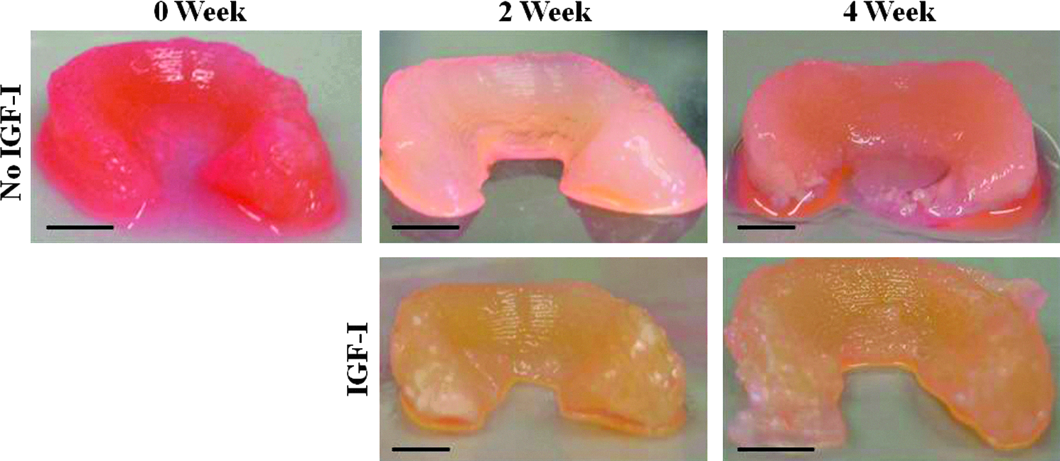
Photographs of alginate menisci after removal from culture (scale bars=5 mm). Color images available online at www.liebertpub.com/tea
Histological analysis
Safranin-O staining demonstrated an increase in GAG localization with time in culture for both IGF-I-treated and untreated groups, with a noticeably increased GAG accumulation in IGF-I-treated 2- and 4-week constructs (Fig. 2). Both Safranin-O and picrosirius red staining revealed the development of a surface layer in samples cultured in IGF-I for 4 weeks. This surface layer was not stained by Safranin-O suggesting no GAG localization. Overall, picrosirius red staining was limited until 4 weeks of culture, at which point untreated constructs had small localized areas of collagen accumulation and IGF-I-treated constructs had strong accumulation of collagen in the surface layer (Fig. 2). When visualized under polarized light, no collagen was visible in untreated 4-week samples, suggesting the small localized areas of collagen accumulation were not organized into collagen fibers. However, the surface layer of the 4-week IGF-I-treated constructs was seen under polarized light and was observed to have a similar organization of collagen to that of the native meniscal surface with fibers aligned parallel to the surface (Fig. 3).

Tissue sections stained with Safranin-O and picrosirius red at 100× under brightfield to determine localization of glycosaminoglycans and collagen, respectively. Arrows point to picrosirius red positive staining. Color images available online at www.liebertpub.com/tea
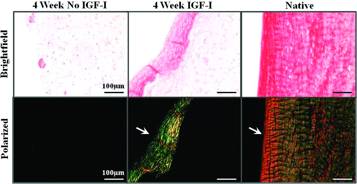
Surface of 4 week insulin-like growth factor-I (IGF-I) untreated and treated alginate menisci and native samples stained with picrosirius red under brightfield and polarized light at 200× demonstrating positive staining and organization of collagen fibers, respectively. Arrows point to organized collagen fibers aligned parallel along the surface. Color images available online at www.liebertpub.com/tea
Immunohistochemical analysis
Immunohistochemical analysis demonstrated slight increases in type I and type II collagen accumulation, concentrated in areas surrounding the cells, in 4-week untreated samples and 2 week treated samples. Constructs cultured in IGF-I for 4 weeks demonstrated a significant increase in collagen accumulation, with strong type I collagen accumulation throughout the construct and surface layer, and type II collagen primarily located in the surface layer (Fig. 4).
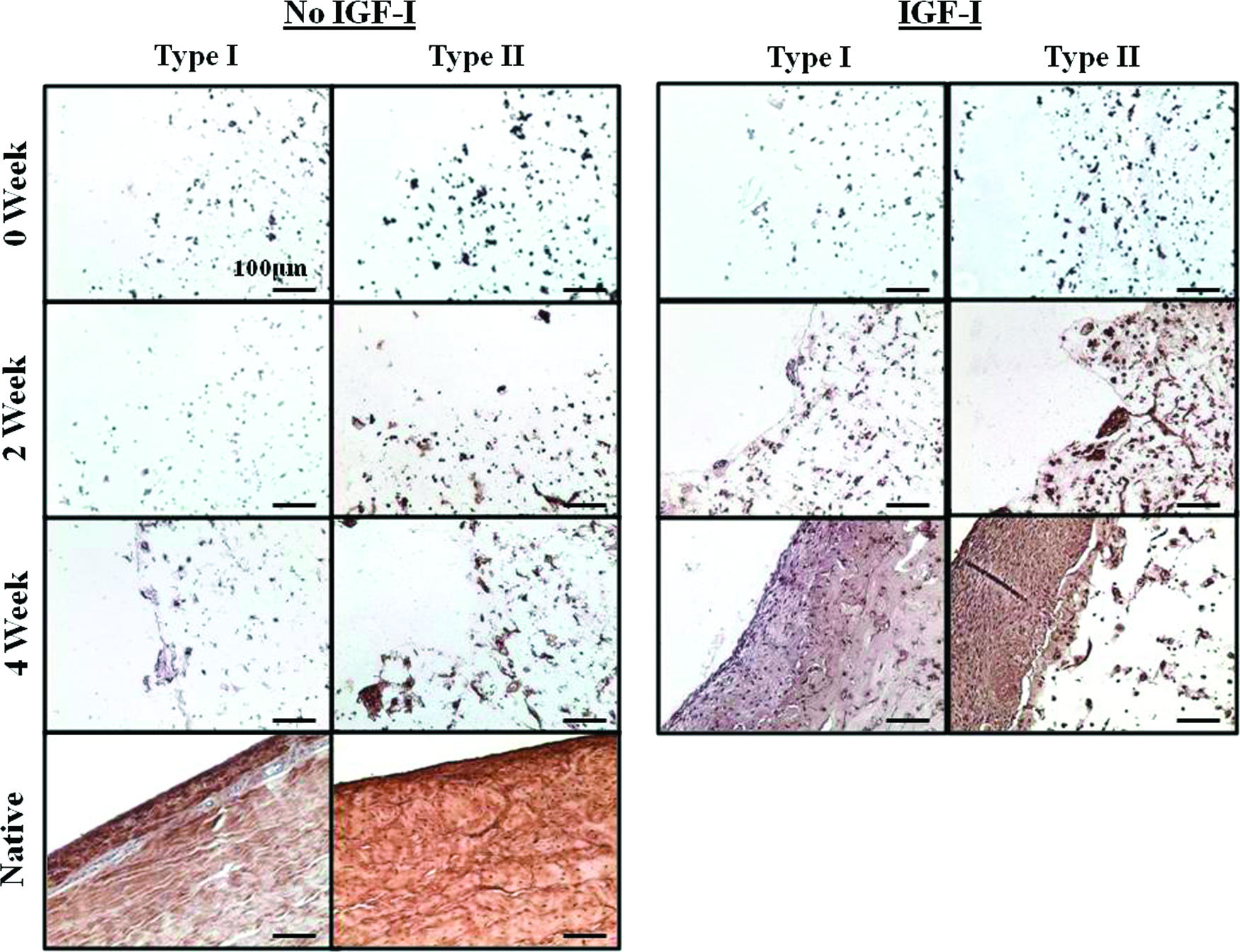
Immunohistochemistry staining for types I and II collagen at 200× in engineered and native meniscal tissues. Color images available online at www.liebertpub.com/tea
Since 4 week IGF-I treated constructs developed a distinct surface layer similar to that of native tissue, further immunohistochemistry analysis for lubricin was conducted because it is localized to the surface of native menisci and is believed to aid in lubrication. Lubricin localized at the surface of 4 week IGF-I treated constructs, similar to that seen in native menisci, and elongated cells aligned parallel to the surface are seen throughout the surface layer (Fig. 5).
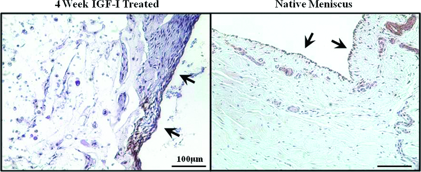
Immunohistochemistry staining for lubricin on the surface of 4 week IGF-I treated alginate and native menisci at 200×. Arrows point to surface of construct where lubricin stained positive. Color images available online at www.liebertpub.com/tea
Biochemical analysis
Both treated and untreated meniscal constructs were found to maintain their DNA content throughout 4 weeks of culture. DNA lost to the media per week decreased after 2 weeks of culture and then remained constant through 4 weeks with IGF-I-treated constructs having significantly more DNA in the media than untreated samples (Fig. 6).
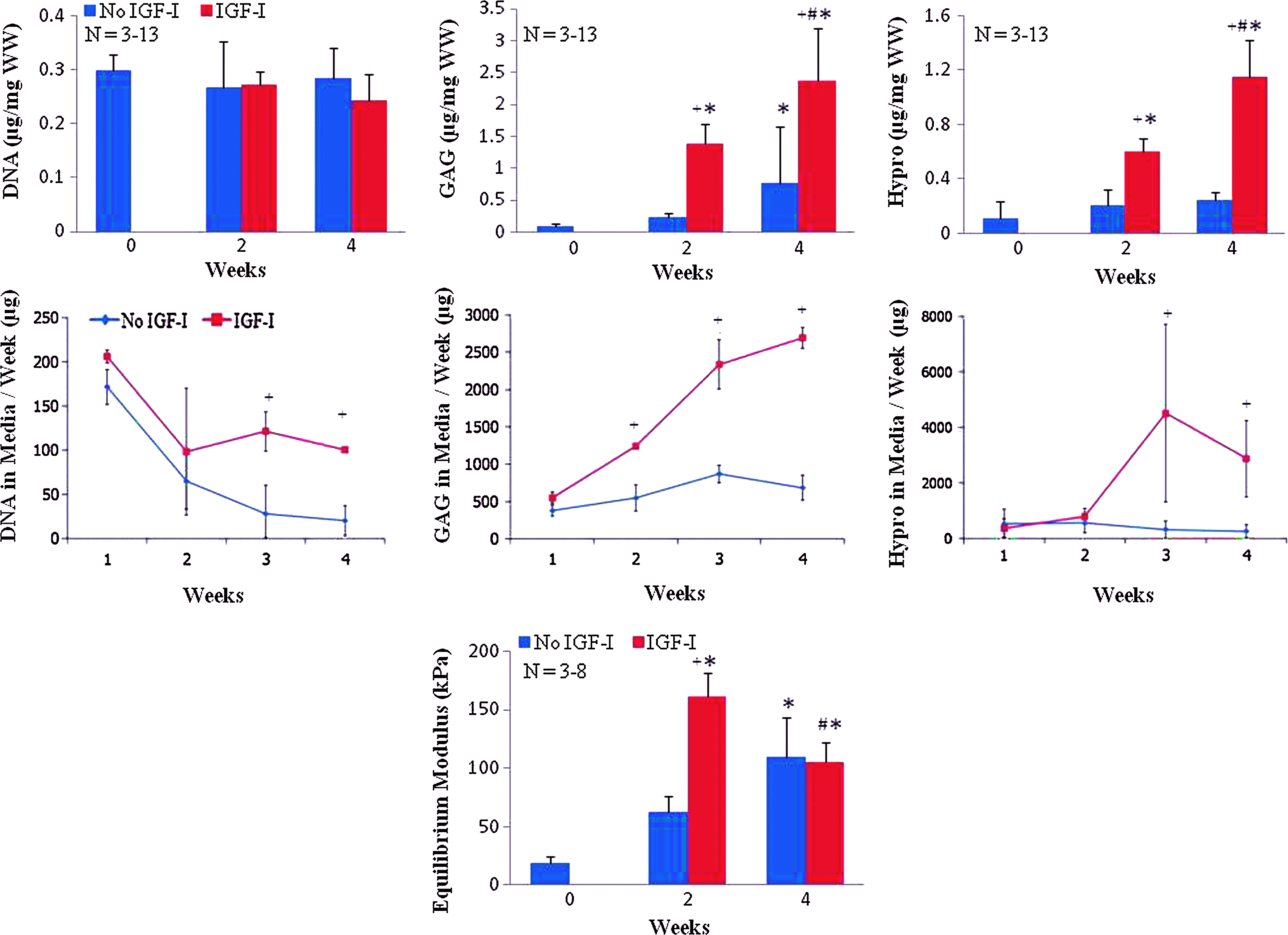
DNA, GAG, and collagen content of alginate meniscal constructs (first row) and total released to the media per week (second row). Equilibrium modulus of alginate meniscal constructs with time in culture (third row). *, difference from 0 week; +, difference from respective group at time point; and #, difference from all other treatments at all other time points. Data presented as mean±standard deviation and p<0.05. Color images available online at www.liebertpub.com/tea
IGF-I treatment significantly increased ECM content in both the meniscal constructs and the media with time in culture. After 2 and 4 weeks of culture, IGF-I-treated constructs had significantly increased GAG accumulation to more than 15- and 26-fold over 0 week constructs at 2 and 4 weeks, respectively. IGF-I-treated constructs also had six and three times higher GAG content compared to 2 and 4 weeks untreated constructs, respectively. After 2 weeks of culture, IGF-I-treated constructs lost significantly more GAG to the media per week than untreated constructs, resulting in an overall significantly greater production of GAG in IGF-I-treated construct than untreated controls (Fig. 6). IGF-I treatment significantly enhanced collagen accumulation at 2 and 4 weeks, with 5- and 10-fold increases over 0 week controls. IGF-I-treated constructs also had a 3- and 5-fold increase over 2 and 4 weeks untreated constructs, respectively. Treated and untreated constructs had very similar collagen loss profiles to the media until 2 weeks. After 2 weeks of culture IGF-I-treated constructs lost significantly more collagen to the media than untreated controls, suggesting greater total production of collagen in IGF-I-treated menisci than untreated controls (Fig. 6).
Mechanical analysis
The equilibrium modulus significantly increased with IGF-I treatment. At 2 weeks of culture, IGF-I-treated constructs had a significantly increased equilibrium modulus over that of 0 week and untreated constructs, with a threefold increase over the untreated control, reaching ∼80%–100% that of human menisci (native tissue=110–200 kPa 40 ). At 4 weeks, treated and untreated constructs had similar equilibrium moduli, which significantly increased from 0 week but decreased from week 2 treated constructs (Fig. 6).
Discussion
In this study, we found that IGF-I treatment enhanced ECM production and mechanical properties of anatomically-shaped tissue-engineered alginate menisci as compared to untreated controls. IGF-I-treated menisci had significantly more GAG and collagen accumulation in the constructs and in the media from 2 weeks on, demonstrating IGF-I greatly increased overall production of biochemical components. Further, IGF-I-treated menisci accumulated 60% of the GAG and 3% of the collagen found in native menisci by 4 weeks of culture 41 and developed an equilibrium compressive modulus that matched native tissue after 2 weeks of culture. 40 The equilibrium modulus did however, decrease after 2 weeks of culture to ∼60%–90% that of native tissue. There were no bursts in release of biochemical components to the media nor a decrease in matrix composition between 2 and 4 weeks for IGF-I-treated constructs, suggesting that this decrease in mechanical properties may be due to degradation of alginate, previously observed in experiments involving culture of anatomically shaped menisci in bioreactor culture. 22
GAG retention in the IGF-I-treated alginate menisci was significantly greater than collagen retention throughout culture. This could be due to the high concentration of IGF-I (100 ng/mL) used in this study. Previous studies have found meniscal explants and fibrochondrocytes in monolayer to produce more GAG than collagen when treated with IGF-I at ≥100 ng/mL.27,42 Additional studies have reported meniscal fibrochondrocytes in monolayer and 3D culture treated with IGF-I at ≤50 ng/mL resulted in only increased collagen incorporation and accumulation, and had no effect on GAG production.24,25,30,32
Further, the accumulation of GAG and collagen in this study could be attributed to a proliferative response. IGF-I has been reported to have a proliferative response at concentrations ≥50 ng/mL in fibrochondrocytes in monolayer25,27 and at 5–10 ng/mL in 3D scaffolds.25,32 Additionally, it has been reported that temporomandibular joint fibrochondrocytes typically have a proliferative response to higher concentrations of growth factors (100 ng/mL), while lower concentrations (10 ng/mL) favor biosynthesis. 43 In this study, we observed no change in DNA concentrations throughout culture for both IGF-I-treated and control constructs; however, IGF-I-treated constructs did have a significant increase in DNA in the media from ∼2 weeks on and the development of a very cell laden surface zone by 4 weeks. Thus, the 100 ng/mL IGF-I treatment used in this study could have had a proliferative effect at the surface of the alginate menisci, but it is unknown whether the increase in DNA in the media is from cells leaving the scaffold or dividing in the media. Additionally, the cell laden surface zone stained negatively for GAGs and very strongly for collagen, suggesting a more fibrochondrogenic response in the surface zone. Typically, proliferative responses to growth factors results in increases in accumulation of both GAG and collagen.
The surface zone of the IGF-I-treated alginate menisci appears similar to the superficial zone of native menisci. The surface zone of the IGF-I-treated menisci at 4 weeks displays organized collagen fibers and elongated cells aligned parallel to the surface very similar to that seen in the superficial zone of native tissue. This organization and alignment of collagen fibers and cells is especially striking since it was developed by IGF-I chemical stimulation, in the absence of any mechanical stimulation. This distinct surface zone also contained types I and II collagen. Type I collagen was located throughout the constructs similar to native tissue, while type II collagen was located primarily in the surface zone. In this system, the action of IGF-I on fibrochondrocytes appears to be highly supportive of fibrochondrogensis, as indicated by stimulation of the production of both proteoglycans and type I collagen. Notably, at 4 weeks, IGF-I stimulation resulted in uniform deposition of proteoglycan and type I collagen throughout the scaffold, similar to what would be expected in native meniscus. 3
Additionally, lubricin, a glycoprotein often localized to the surface of native menisci and believed to aid in lubrication,39,44 was shown to accumulate at the outermost edge of the surface zone. Lubricin is known to decrease the boundary fiction of cartilaginous tissues and thus is believed to be key to joint lubrication and decreased wear of surrounding cartilage. 44 A tissue-engineered meniscus will most likely need a well-organized superficial zone and the ability to localize or produce lubricin at its surface to be functional when implanted in the knee. Previous studies have shown the ability of tissue-engineered meniscal constructs to localize exogenously added lubricin, 39 but the current study is the first to demonstrate the accumulation of endogenously synthesized lubricin in a well-defined surface zone.
The mechanism driving the development of the collagen- and lubricin-rich surface zone is not entirely clear. Previous work 45 suggests that IGF-I does not drive lubricin expression in articular chondrocytes, but there is no comparable data for meniscal fibrochondrocytes. Notably, mensical fibrochondrocytes appear more effective at localizing lubricin in alginate cultures than articular chondrocytes, 39 suggesting that they may be more sensitive to stimulation of lubricin synthesis than articular chondrocytes. Degradation of the alginate scaffold at the surface may facilitate the development of this surface layer, although significant degradation of alginate does not generally occur on this time frame 22 and is not apparent from the appearance of the construct surface (Fig. 1). However, the accumulation of a highly cellular region on the surface in the absence of encapsulating alginate could drive a significantly different cellular response to high concentrations of IGF-I that results in collagen and lubricin accumulation.
IGF-I-treated alginate menisci show great promise as a replacement tissue of the meniscus with a well-defined superficial zone, ∼60% GAGs of native tissue and an equilibrium modulus that matches native menisci by 4 weeks of culture. However, further investigations are needed to improve the collagen content (3% of native tissue) and tensile properties of these engineered menisci. Prolonged culture beyond 4 weeks or decreasing the concentration of IGF-I later in culture could help to further improve collagen content. Additionally, combination of mechanical stimulation and growth factor treatment could serve to be beneficial. We have demonstrated previously that dynamic compression of alginate menisci promotes a fibrochondrogenic response with collagen accumulation and bundle formation over GAG accumulation. 17 Further, it has been suggested that meniscal fibrochondrocytes respond to biochemical and biomechanical stimuli via separate cellular pathways, with dynamic load, and growth factor treatment leading to a synergic improvement of mechanical and biochemical properties.14,42 Thus, combining the response of dynamic compression and IGF-I treatment could serve to further improve the mechanical and biochemical properties of these meniscal scaffolds.
Footnotes
Acknowledgments
The authors would like to thank Sunjoo Park for her contributions to this research. This work was funded by Hunter R. Rawlings III Cornell Presidential Research Scholars, the Sloan Foundation, the Association of Osteosynthesis/Association for the Study of Internal Fixation Foundation, Cornell BME National Science Foundation GK-12 Program: DGE 0841291, and the NSF's Graduate Research Fellowship Program.
Disclosure Statement
The authors have no conflicts of interest or financial disclosures.