




Abstract
Tissue engineering approaches for articular cartilage (AC) repair using collagen type I (Coll)-based hydrogels are limited by their low collagen fibril density (CFD; <0.5 wt%) and their poor capacity to support chondrocyte differentiation. Chitosan (CTS) is a well-characterized polysaccharide that mimics the glycosaminoglycans (GAGs) present in native AC extracellular matrix and exhibits chondroprotective properties. Here dense Coll/CTS hydrogel discs (16 mm diameter, 140–250 μm thickness) with CFD (∼6 wt%) approaching that of AC were developed to investigate the effect of CTS content on the growth and differentiation of three-dimensionally seeded RCJ3.1C5.18 chondroprogenitor cells. Compared to dense Coll alone, cells seeded within Coll/CTS showed increased viability and metabolic activity, as well as a decrease in cell-mediated gel contraction. Immunohistochemistry for collagen type II, in combination with Safranin O staining and GAG quantification, indicated greater chondroprogenitor differentiation within Coll/CTS, compared to cells seeded within Coll alone. The complex interplay between scaffold geometry, microstructure, composition, mechanical properties and cell function was further evaluated by rolling dense planar sheets to prepare cylindrically shaped constructs having clinically relevant diameters (3–5 mm diameter, 9 mm height). The compressive modulus of the cylindrically shaped constructs decreased significantly after 7 days in culture, and remained unchanged up to 21 days for each scaffold composition. Unlike Coll, cells seeded within Coll/CTS showed greater viability along the entire radial extent of the cylindrical rolls and increased GAG production at each time point. While GAG content decreased over time and reduced cell viability was observed within the core region of all cylindrical rolls, the incorporation of CTS diminished both these effects. In summary, these findings provide insight into the challenges involved when scaling up scaffolds designed and optimised in vitro for tissue repair.
Introduction
Hydrogels exhibit a biomolecular structure that is similar to native tissue and, unlike freeze-dried scaffolds, these fully hydrated matrices allow direct cell seeding and homogeneous cell distribution during scaffold preparation and processing. 4 In particular, acid-extracted collagen type I (Coll) hydrogels have been extensively used as scaffolds for cartilage tissue engineering applications5,6 primarily because of the difficulties associated with processing isolated Coll II protein extractions into gel-like scaffolds.7,8 However, Coll hydrogels exhibit poor mechanical properties attributable to their low collagen fibril density (CFD, <0.5 wt%) and undergo a high level of cell-mediated contraction which causes a physical mismatch between the dimensions of the defect and the graft. 9 To overcome both these limitations, plastic compression (PC) of Coll hydrogels has been developed as a method to rapidly increase the CFD by expelling unbound fluid immediately after cell seeding to produce dense hydrogels with increased biomechanical properties and decreased cell-mediated contraction.10–12
Differentiated chondrocytes seeded within Coll scaffolds have been shown to lose their cartilage-like characteristics as they undergo dedifferentiation towards a fibroblastic phenotype.13,14 Therefore, improved scaffold compositions are necessary to ensure the long-term maintenance of the chondrocytic characteristics. Chitosan (CTS) is a polysaccharide composed of randomly distributed N-acetylglucosamine and glucosamine units, which structurally and compositionally resembles the GAGs present within native tissues. The incorporation of CTS within various types of hydrogels has been found to stimulate chondrogenesis by supporting chondrocyte growth and differentiation, as well as the production of cartilage-specific extracellular matrix (ECM) components, such as Coll II and GAGs.15–17 A number of studies have examined the in vivo biological performance of CTS hydrogel implants in both animal and human cartilage defects. No major foreign body reaction has been observed and, in general, these scaffolds have been found to present the formation of hyaline-like cartilage, as well as improve the integration of repaired cartilage within the lesion. 18 However, the effect of CTS on chondrocyte function within dense Coll gels has not been determined.
The present study tests the hypothesis that the incorporation of CTS into dense Coll gels serves as a chondroprotective material that supports chondrocyte differentiation and increases cartilage-like ECM production. Dense Coll/CTS scaffolds with weight ratios approaching those of Coll/GAGs 3 in the native ECM of AC (relative compositions of 2:1 and 1:1 by weight) were prepared and compared with Coll hydrogels. A mesenchymal chondrogenic cell line RCJ3.1C5.18 (RCJ) developed from cells isolated from rat calvaria was used in view of its capacity to undergo a cell differentiation sequence similar to that observed in cartilage in vivo.19,20 Moreover, the RCJ cell line has been widely used for chondrocyte research, which demonstrates its utility as a model cell line for AC tissue engineering.21–23 Seeded RCJ cell metabolic activity, cell-mediated gel contraction, viability and morphology, and cartilage-like aggregate formation within dense Coll/CTS gels, were compared to cells seeded into dense gels made of Coll alone. In addition to support of cell phenotype, the production of constructs of a scale necessary to repair critical-sized clinical defects is a major challenge for cartilage tissue engineering. In this regard, the viability of RCJ cells and GAG content within each specimen was investigated on cylindrically shaped constructs measuring 3 to 5 mm in diameter, depending upon scaffold composition. The study of three-dimensional (3D) AC-ECM mimicking scaffolds, designed according to the structure and properties of native AC, provides a framework for understanding the interactions between chondrocytes and the macromolecular components and morphometric parameters of dense Coll/CTS scaffolds.
Materials and Methods
Cell culture
The nontransformed RCJ mesenchymal clonal chondrogenic cell line isolated originally from foetal rat calvaria was kindly provided by Dr. Jane E. Aubin (University of Toronto, Ontario, Canada). Cells from passages 9 to 20 were cultured in α-minimum essential medium (α-MEM; Gibco-Invitrogen) with ribonucleosides and deoxyribonucleosides containing 1% penicillin/streptomycin (Gibco), 10−7 M dexamethasone (Sigma), 50 μg/mL ascorbic acid (Sigma) and 15% v/v of foetal bovine serum (Hyclone; Thermo Scientific). Cells were incubated at 37°C, in a 5% CO2 humidified incubator, and the medium was changed at 2-day intervals.
Preparation of dense Coll and Coll/CTS gel discs and cylindrical rolls
Sterile, rat-tail tendon-derived type I collagen (2.10 mg/mL of protein in 0.6% acetic acid; First Link Ltd.), and ultrapure CTS powder (79.8% DDA, Ultrasan™; BioSyntech Ltd. [now Piramal Healthcare]) were used as starting materials. Coll/CTS hybrid gels having relative compositions of 2:1 and 1:1 (w/w) were developed as previously described. 12 Briefly, CTS (13.5 and 27 mg for 2:1 and 1:1, respectively) was dissolved in acetic acid (2.2 mL/0.1 M) at 4°C and stirred overnight, followed by the addition of collagen solution (12.8 mL) under gentle stirring. Coll and Coll/CTS self-assembly were achieved by mixing the solution with 10×MEM (Sigma Aldrich) at a ratio of 4:1 and neutralized with 5 M NaOH. For cellular gels, RCJ cells were seeded within the hydrogels at a density of 3×10⁵ cells/mL of Coll, or Coll/CTS solution, immediately prior to gel polymerization.
Neutralized Coll or Coll/CTS solutions were poured in either four-well plates (0.9 mL/well of 4.5 mm height×16 mm diameter) or rectangular moulds (4.5 mL/mould of 18 mm width×40 mm length×6.2 mm height) to produce dense planar discs and cylindrical rolls, respectively. Gelation was achieved by allowing the solutions to set at 37°C in a 5% CO2 incubator for 30 min. Hydrogels were removed from the moulds and dense gels with CFD of 8.13%, 5.26% and 6.38% for Coll, Coll/CTS 2:1 and Coll/CTS 1:1, respectively, were produced by PC as previously reported.10,12 Briefly, highly hydrated gels were placed on a stack of blotting paper, nylon mesh, and metal mesh, and subjected to PC using an unconfined compressive stress of 0.5 kPa for 5 min, to remove the excess casting fluid (Fig. 1). After compression, dense discs (16 mm diameter with approximate thicknesses of 140, 180, and 250 μm for Coll, Coll/CTS 2:1 and 1:1, respectively) were transferred to 12-well plates and cultured under static conditions for 21 days with medium changes at 2-day intervals. To obtain cylindrically shaped constructs (9 mm height×3–5 mm diameter), rectangular sheets (18 mm width×40 mm length×140–250 μm thick) were plied through the middle along the short axis and rolled along their long axis.
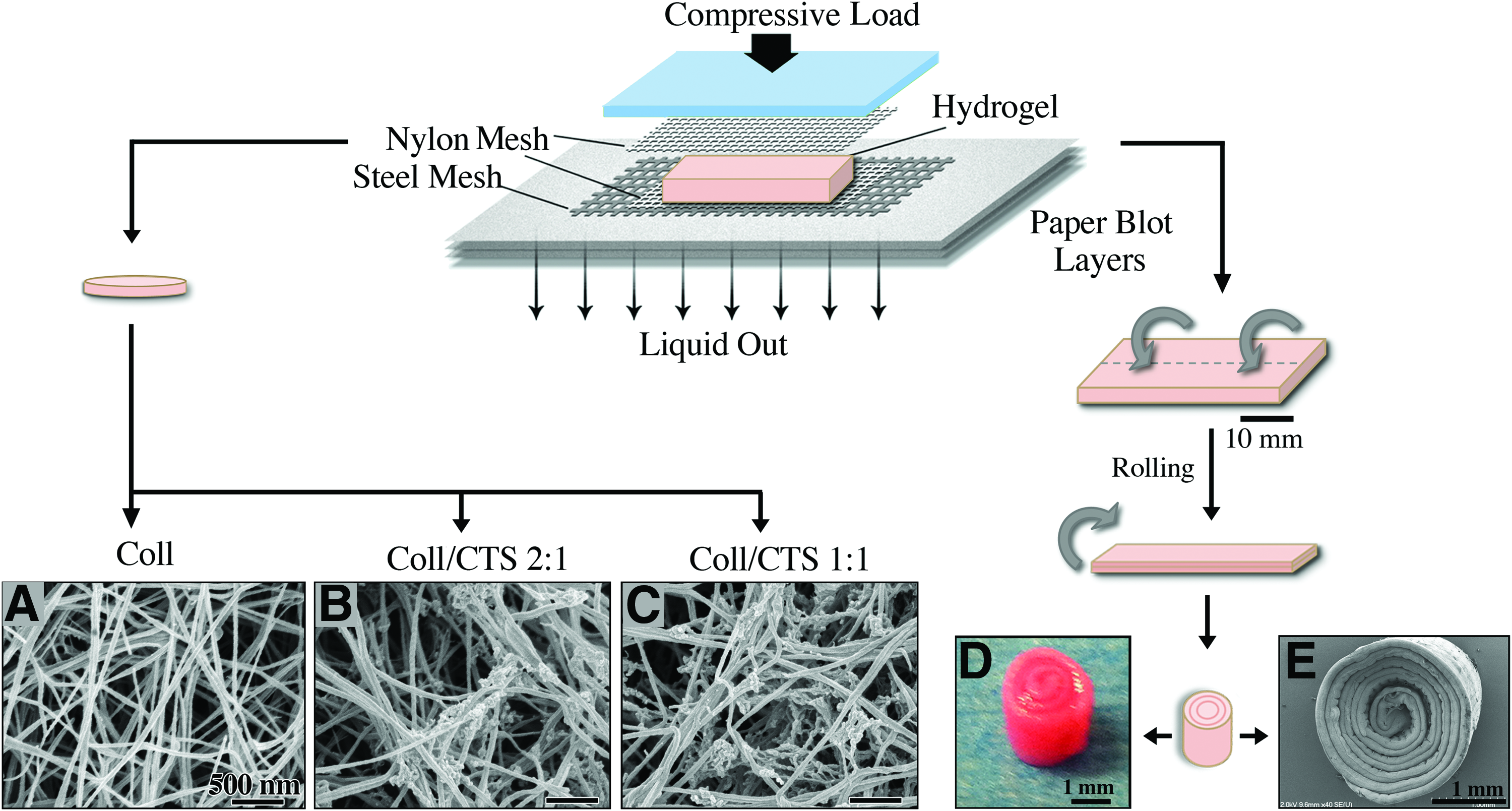
Schematic of production of dense Coll and Coll/CTS hydrogel discs and cylindrical rolls. Dense Coll and Coll/CTS hybrid gels were produced by plastic compression using 0.5 kPa for 5 min.
Scanning electron microscopy
Acellular and cell-seeded gels were morphologically assessed by scanning electron microscopy (SEM, FEG-SEM Model S-4700; Hitachi High Technologies America). Gels were fixed overnight at 4°C in 0.1 M sodium cacodylate buffer containing 4% paraformaldehyde and 2% glutaraldehyde. Fixation was followed by dehydration in a series of increasing ethanol concentrations for 15 min each and critical-point drying (Ladd Research Industries). SEM was carried out on Au/Pd sputter-coated samples (Hummer VI Sputter Coater, Ladd Research Industries).
Cell metabolic activity in dense gel discs
Seeded cell metabolic activity was evaluated up to day 21 in culture using the AlamarBlue™ assay (Molecular Probes™, Invitrogen). Dense gel discs were incubated in complete culture medium containing 10% AlamarBlue for 4 h at 37°C at days 2, 5, 8, 11, 14, 17, and 21. Aliquots (200 μL) of the supernatants (n=3) of each well were pipetted into 96-well plates and the absorbance at 562 and 595 nm was spectrophotometrically measured using a microplate reader (Model EL800; BioInstruments). The background absorbance of the assay medium was accounted for by subtracting the values obtained for acellular gels.
Cell-mediated gel contraction
Cell-mediated contraction of free-floating dense gel discs and cylindrical rolls was determined by measuring the scaffold surface area as a function of time in culture, which was normalized to the initial area at day 0 (n=3). Acellular gels were used as controls. Culture plates were scanned with a Canoscan 8600F (300 dpi resolution) at different time points up to day 21 in culture. Images were analyzed using ImageJ software (1.42q, Rasband W; National Institutes of Health, Bethesda, MD).
Cell viability
Cell viability within discs and cylindrical rolls was performed using the Live/Dead® assay (Invitrogen). Gels seeded with 3×10⁵ cells/mL of Coll or Coll/CTS solution were incubated for 40 min in phosphate buffered saline (PBS; Wisent) containing 1 μM calcein AM and 2 μM ethidium homodimer-1 (EthD-1). Cylindrical rolls were carefully unrolled at each time point prior to staining. Scaffolds were transferred to a glass dish (35 mm in diameter; MatTek) and Live/Dead staining was detected using a confocal laser scanning microscope (CLSM, LSM5 Exciter; Carl Zeiss). Unrolled constructs were partitioned into three subdivisions of ∼13 mm in length (periphery, middle and core) for analysis. Images from each subdivision were obtained through random sampling. Maximum intensity projections of the confocal z-stacks along the entire thickness of the dense gels were obtained using ImageJ software.
Biochemical analyses
Quantification of GAGs and DNA content was performed at days 1, 7, 14 and 21 (n=6). Samples were freeze-dried (BenchTop K freeze dryer; VirTis, SP Industries) and digested with papain (Sigma) for 18 h at 60°C, as previously reported. 24 The supernatant of papain-digested constructs was immediately used for DNA quantification. Unused supernatant volume was stored at −80 °C for GAG quantification. DNA content was determined by detection of Hoechst 33258 (Sigma) staining (FLUOstar OPTIMA; BMG Labtech; 360 nm excitation/460 nm emission) using a calf thymus DNA standard curve. Background fluorescence was accounted for by subtracting the values obtained from extracts of acellular gels.
Total GAG content was measured both within the discs and in the surrounding medium at each time point. Culture medium was collected and concentrated by centrifugation using a Centricon YM-10 filter (Millipore). GAG content was determined by adding dimethyl-methylene blue (DMMB) to all samples followed by taking readings with a microplate reader equipped with two onboard reagent injectors (FLUOstar OPTIMA) at 520 nm. Measured values were compared with that of a GAG standard (chondroitin sulfate from shark cartilage; Sigma) determined for the same wavelength. For the cylindrical rolls, the GAG content within the gels was assessed at days 1, 7, 14, and 21 as explained above.
Histological analyses
Histological analyses were performed at day 21 using Safranin O/Fast Green staining to localize sulfated GAGs within discs. Gels were rinsed with PBS, fixed in 10% neutral buffered formalin overnight at 4°C, and dehydrated in a series of graded ethanol solutions prior to embedding in paraffin. Sections (5 μm) were obtained from middle regions of the samples and mounted on positively charged glass slides, followed by staining with Safranin O/Fast Green.
Immunohistochemical analyses
Paraffin-embedded sections (5 μm) were used for Coll II and aggrecan immunohistochemistry. The sections were deparaffinized with xylene and ethanol, and rehydrated in a graded ethanol solution series. Antigen retrieval was performed by immersing slides in Tris/EDTA at pH 9.0 for 24 h at 60°C. Heat-induced epitope retrieval was followed by digestion in 2% hyaluronidase (Sigma) for 30 min. The sections were permeabilized with 0.25% Triton X-100 (Sigma) for 10 min, placed in 2% bovine serum albumin (blocking buffer; Sigma) for 30 min, and then incubated overnight with rabbit anti-Coll II (1:100; Millipore) and rabbit anti-aggrecan (1:100; Millipore) antibodies. The slides were then incubated with Alexa Fluor® labeled goat anti-rabbit IgG (Invitrogen) for 1 h followed by nucleic acid staining with EthD-1 (1:3000). Finally, samples were coverslipped with mounting medium (Geltol; Thermo Electron Corporation). Z-stacks of fluorescent immunoreactivity towards Coll II and aggrecan of each specimen were acquired using a CLSM. Maximum intensity projections of each scaffold were generated using ImageJ. Negative controls were performed in the absence of primary antibody.
Mechanical analysis
Compressive mechanical tests were carried out on cylindrically shaped constructs using a Bose® ElectroForce® BioDynamic® (Model 5160; Bose Corp.) instrument equipped with a 20 N load cell.12,25 Cell-seeded and acellular cylindrical rolls were assessed at days 1, 7, 14, and 21 in culture. All tests were carried out in displacement control with a cross-head speed of 0.01 mm/s. Measurements were conducted at room temperature while maintaining constant sample hydration using drops of distilled water. The specimens' diameter (2.92±0.23, 3.79±0.13 and 5.21±0.35 mm for collagen, Coll/CTS 2:1 and Coll/CTS 1:1, respectively) and height (3.61±0.21 mm) were measured using a digital calliper and confirmed with a Leitz DMR optical microscope (Leica). Specimens were tested using two parallel nonporous compression platens at up to 80% strain without any evidence of specimen unfolding (n=3–4). The stress was calculated by normalizing the recorded force against the initial resistance area of the specimen, and the strain was calculated by normalizing the displacement against the initial height of the specimen. The compressive modulus values were computed from the slope of the initial linear region (<20% strain) of the stress-strain outputs.12,25
Statistical analysis
All data are presented as mean values±standard deviation of the mean. Statistical significance between groups and between time points was determined using a one-way ANOVA with a Tukey-Kramer's post-hoc multiple comparison of means. The level of statistical significance was set at p=0.05.
Results
Morphological characterization of dense Coll and Coll/CTS gel discs and cylindrical rolls
As-made acellular gels were morphologically characterized by SEM (Fig. 1A–C, E). Micrographs of the internal morphology revealed a porous network of randomly oriented nanofibrils of collagen (Fig. 1A). Incorporation of CTS into Coll, followed by PC, maintained the homogeneous open-pore structure with a high degree of interconnectivity (Fig. 1B, C). Rolling dense sheets along the long axis resulted in cylindrical shaped constructs of 9 mm height and 3 to 5 mm diameter depending upon scaffold composition (Fig. 1D). A low magnification SEM micrograph demonstrates the continuous spiral layers of the rolls (Fig. 1E).
Cell metabolic activity and cell-mediated contraction in dense Coll and Coll/CTS gel discs
Cell metabolic activity measured by AlamarBlue was significantly reduced in Coll gels after 8 days when compared to the hybrid Coll/CTS gels (p<0.05, Fig. 2A). In Coll/CTS 2:1 and 1:1 gels, cell metabolic activity increased up to day 5 and remained unchanged at days 8 and 14, respectively, at which point cell metabolic activity decreased.

Metabolic activity of seeded RCJ cells and cell-mediated Coll and Coll/CTS gel disc contraction up to day 21 in culture.
Cell-mediated contraction of dense discs displayed a sigmoidal relationship between scaffold surface area and time, featuring three contraction phases: an initial lag phase, followed by a linear contraction phase, and then a slow contraction phase (Fig. 2B). Coll/CTS 2:1 and 1:1 gels were significantly less contracted (55 and 50%, respectively) compared to Coll (65%). The extent of contraction between the two Coll/CTS hybrids was not significantly different (p>0.05). No significant changes in scaffold surface area were observed in acellular gel discs with respect to day 0 (p>0.05).
Cell viability and morphology within dense Coll and Coll/CTS gel discs
CLSM of calcein-AM labeled cells was used to monitor cell viability over time. As indicated by green fluorescent cytoplasmic labelling and negligible EthD-1 binding to nucleic acids (red) in maximum intensity projections, all scaffold compositions supported cell viability both immediately after gelation and during subsequent cell culture (Fig. 3). At day 1, all compositions contained uniformly distributed viable cells that displayed a rounded morphology. At day 7, cell density increased throughout the entire thickness, and RCJ cells seeded in hybrid gels formed cartilage-like aggregates with spherical morphology (indicated by white arrows). Aggregate formation progressed up to day 21 with hybrid gels showing a greater tendency to support this.
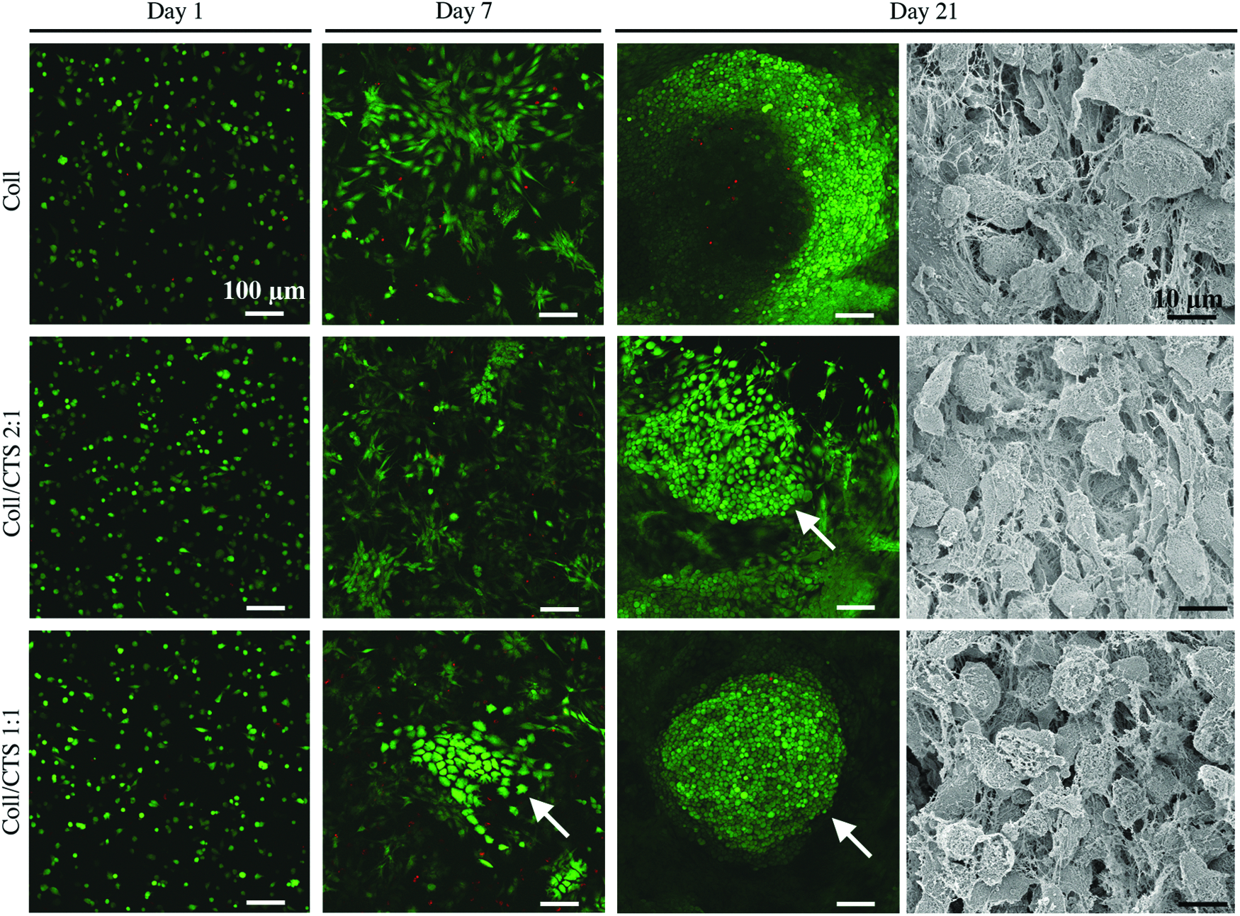
Analysis of RCJ cells viability and morphology within dense Coll, Coll/CTS 2:1 and Coll/CTS 1:1 gel discs. CLSM maximum intensity projection images covering the entire thickness were collected at days 1, 7, and 21. Green fluorescent calcein-AM-labeled cells represent viable cells with no membrane disruption, and red fluorescent EthD-1 positive nuclei indicate cell necrosis and late apoptosis. Aggregates of cells (white arrows) are detected as early as day 7 within Coll/CTS hybrid gels. SEM micrographs obtained at day 21 in culture show the distribution and morphology of seeded RCJ cells within the matrix, with differences in the extent of cell spreading and integration between the different compositions. CLSM, confocal laser scanning microscope; EthD-1, ethidium homodimer-1. Color images available online at www.liebertpub.com/tea
SEM micrographs at day 21 in culture displayed extensive cytoplasmic extensions of seeded RCJ cells integrated within scaffolds. Cells in Coll exhibited a mixture of flat, polygonal cells combined with rounded cells. In comparison, cells with a rounded morphology were more frequently observed in Coll/CTS hybrids. Quantitative analyses of cell spreading (surface area) using ImageJ software (n=10) showed that the average cell surface area within Coll was significantly higher (197±48 μm2) compared to Coll/CTS 2:1 and 1:1 (110±35 and 83±25 μm2, respectively) (p<0.05).
Quantification of GAG synthesis and DNA content within dense Coll and Coll/CTS gel discs
Since CTS binds negatively charged GAGs, 26 the total GAG content was quantified both within the discs and the surrounding media. GAG synthesis significantly increased in all compositions with incubation time up to day 21, except for Coll where it peaked at day 14 (p<0.05, Fig. 4A). The production of GAGs in Coll, Coll/CTS 2:1 and 1:1 constructs increased by a factor of 1.7, 1.8, and 2.4, from days 7 to 14, respectively. Increasing CTS content led to an increase in GAG retention as indicated by a decrease in its content within the surrounding medium (p<0.05). The total amount of secreted GAGs in both Coll/CTS 2:1 and 1:1 hybrid gels was significantly higher compared to Coll beyond day 7 (p<0.05).
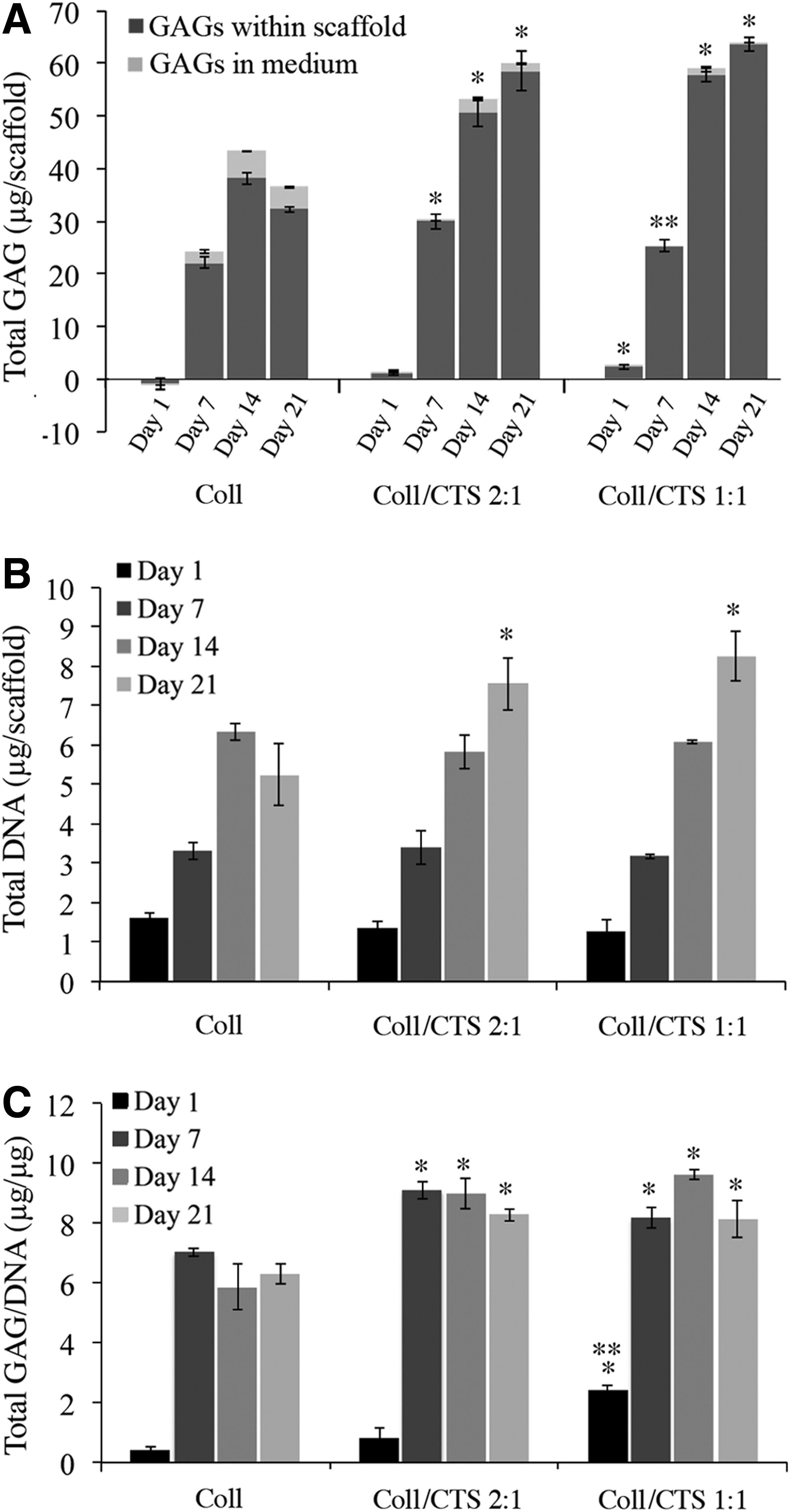
GAG synthesis by differentiated RCJ cells within dense Coll, Coll/CTS 2:1, and Coll/CTS 1:1 gel discs up to day 21 in culture.
DNA content significantly increased over the 21-day culture period, except for Coll where it significantly decreased at day 21 (p<0.05, Fig. 4B). ANOVA indicated no significant differences between hybrids at any given time point (p>0.05). In addition, the total GAG synthesis normalized to DNA content showed that the incorporation of CTS into dense Coll gels significantly enhanced GAG secretion at all times after day 1 (p<0.05, Fig. 4C).
Histological and immunohistochemical analysis of dense Coll and Coll/CTS gel discs
ECM secretion by RCJ cells within the different specimens was assessed through histochemical and immunohistochemical analysis at day 21 (Fig. 5). Sections were histologically stained using Safranin O/Fast Green (Fig. 5A–C) to detect sulfated GAGs. Histological findings in the middle region of the various gel discs showed differences in cartilage-like matrix deposition between all compositions. Coll/CTS 1:1 gels revealed GAG-rich ECM within aggregates, as indicated by intense Safranin O staining, and round chondrocyte-like cells within lacunae characteristic of native cartilage. Staining in the control group (acellular gel) was negative and showed only a light green background counterstaining (data not shown). Differences in the counterstain intensity are attributable to the anionic Fast Green stain having a strong affinity for positively charged CTS. 26 The effect of CTS incorporation into Coll gels was assessed by aggrecan (Fig. 5D–F), and Coll II (Fig. 5G–I) immunostaining. Coll/CTS 1:1 showed stronger positive staining for aggrecan and Coll II compared to Coll/CTS 2:1 and Coll gels.
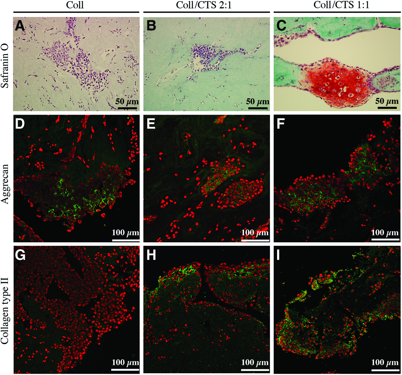
Histological and immunohistochemical analyses of ECM synthesis by differentiated RCJ cells seeded within dense Coll, Coll/CTS 2:1 and Coll/CTS 1:1 gel discs in paraffin embedded sections at day 21 in culture.
Mechanical properties of cell-seeded and acellular gel cylindrical rolls
At the earliest time point (day 1), the incorporation of CTS into Coll significantly increased the compressive modulus of both cellular and acellular rolls (Fig. 6A, B, respectively) from 11.7±2.2 kPa for Coll alone, up to 20.5±1.4 kPa for acellular Coll/CTS 1:1 (p<0.05). At day 7, both cell-seeded and acellular hybrid gels showed a decrease in the compressive modulus, a level that was maintained throughout the 21-day culture period. No significant differences were observed within each group between cellular and acellular cylindrical rolls (p>0.05).
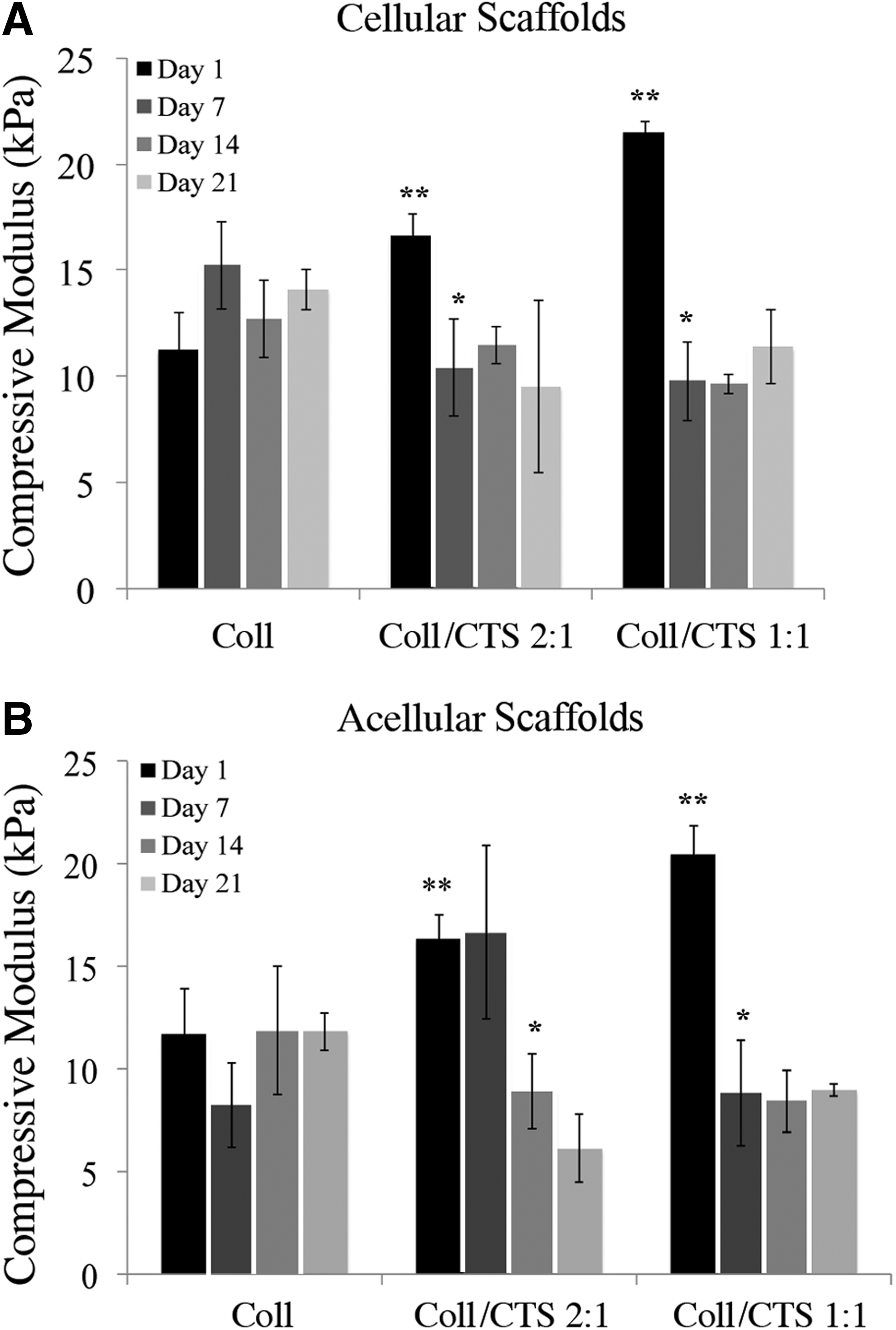
Compressive modulus of
CLSM analysis of cell viability within gel cylindrical rolls
RCJ cell viability in cylindrical rolls was investigated by CLSM detection of Live/Dead staining in unrolled constructs at different time points (Fig. 7A, B). Confocal image z-stacks of the peripheral, middle and core regions of the constructs were acquired at days 1, 7, 14, and 21 from randomly selected regions within three equally spaced, consecutive regions along the construct, and assembled into maximum intensity projection images (Fig. 7C). At day 1, all constructs showed a rounded morphology and a homogenous distribution of viable cells in all regions. At day 7, all constructs exhibited increased cell growth throughout the rolls, as well as initial signs of aggregate formation (red arrows). In addition, the amount and size of aggregates increased with CTS content. At day 14, Coll gels showed a decrease in cell growth, in particular after 13 mm from the periphery. In contrast, hybrid gels showed an increase in cell number up to 26 mm from the periphery, followed by a decrease thereafter.

CLSM analysis of RCJ cells viability within Coll, Coll/CTS 2:1 and Coll/CTS 1:1 cylindrical rolls up to day 21 in culture.
Quantification of GAG synthesis and DNA content within gel cylindrical rolls
Based on the fact that a maximum of 12% of the total GAGs produced by Coll discs was released to the medium and that this amount decreased with the incorporation of CTS and time, GAG content was only quantified within the cylindrical rolls. GAG synthesis (Fig. 8A) and DNA content (Fig. 8B) in all compositions exhibited a similar trend over time. A significant increase in all compositions was observed up to day 14 (p<0.05). GAG and DNA levels at day 21 remained elevated in the hybrid gels compared to Coll where it decreased significantly. GAG content normalized to DNA significantly decreased with time in all compositions (p<0.05); however, the level of GAGs in hybrid gels was higher than in Coll at all measured times, except at day 21, where there was no significant difference between Coll and Coll/CTS 1:1 (Fig. 8C).
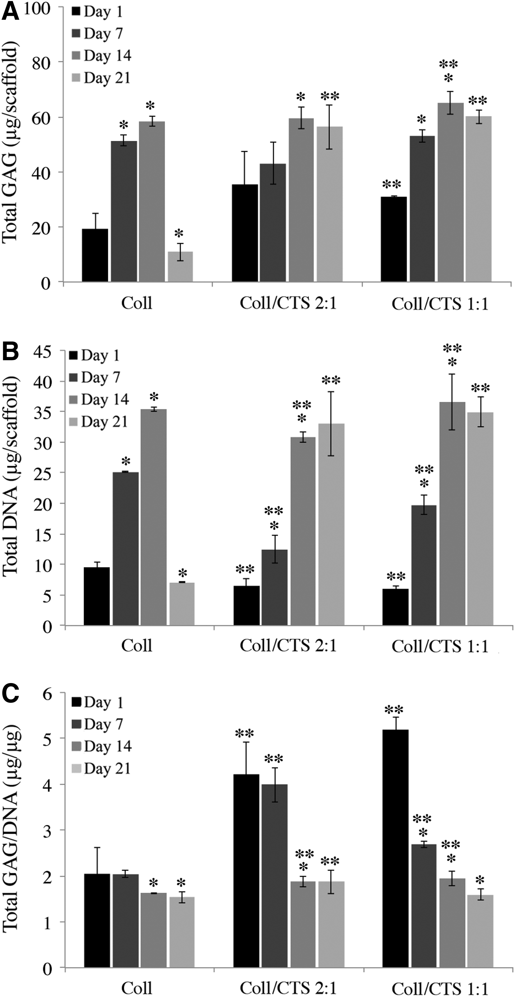
GAG synthesis by differentiated RCJ cells within Coll, Coll/CTS 2:1 and Coll/CTS 1:1 cylindrical rolls up to day 21 in culture.
Discussion
Collagen-based hydrogels are attractive scaffolds for AC repair as they present both microstructural and compositional similarities to native cartilage, and allow for homogenous cell seeding within a highly hydrated environment. 27 However, despite the prevalent use of Coll for tissue engineering purposes, numerous in vitro studies have reported the phenotypic instability of chondrocytes within these scaffolds.13,14 In this study, CTS, a well-known GAG-analog and chondroprotective material, 16 was incorporated into a physiologically relevant, dense Coll nanofibrillar gel, which mimics the major structural component of the extracellular milieu, to determine its effect on RCJ cell function relative to cells grown in dense Coll alone.
RCJ cells seeded into dense hydrogel discs exhibited a higher level of metabolic activity in Coll/CTS compared to Coll from days 8 to 14. The maximum cell metabolic activity of all gels closely corresponded with the onset of cell-mediated disc contraction at day 6, indicating that a minimum threshold cell density was attained to exert sufficient forces required to induce gel contraction. 12 At day 21, the final contraction differed significantly according to CTS content, indicating that RCJ cells in Coll scaffolds may have dedifferentiated into fibroblast-like cells which exert higher contractile forces compared to chondrocytes. 28 Moreover, the increased extent of contraction observed for RCJ cells in Coll gels, compared to that seen in Coll/CTS gels, may be related to a collagen-type effect, which is greater when these cells are in contact with Coll than with Coll II. 29
Quantification of DNA showed that RCJ cells seeded within dense Coll/CTS hydrogels proliferated during the 21-day culture period, in contrast to Coll, which demonstrated a significant reduction at day 21. Differences in the metabolic activity results and DNA quantification may be explained by the fact that the Alamarblue assay is not a direct cell counting technique, as the absorbance signal is affected not only by the number of cells, but also by cell metabolism. In particular, the metabolic activity varies greatly depending on the life-cycle of the cell; thus, the presence of cells in different phases of cell growth results in different oxidative metabolism. 30
RCJ cells seeded within dense Coll/CTS discs were viable and formed aggregates of spherical cells that are characteristic of differentiated chondrocytes, 31 confirming the morphological features observed by SEM. In contrast, cells seeded in Coll alone exhibited a flattened and stellate morphology, with a significantly higher cell surface area that is characteristic of fibroblasts, indicating that cell de-differentiation may have occurred. 32 In contrast, increasing CTS content supported the sustained upregulation of secreted cartilage-specific GAGs, as demonstrated by GAG quantification within the discs and the surrounding medium, Safranin O staining and aggrecan immunoreactivity. The enhanced retention of newly synthesized GAGs within hybrid gels, compared to Coll gels, supports the hypothesis that the biochemical similarities of CTS to GAGs has an important role in modulating chondrocyte function, including chondrocyte differentiation. 33 A similar role for CTS has been reported after incorporation within either hydroxyethyl cellulose hydrogels, freeze-dried Coll/GAGs matrices, and silk fibroin scaffolds.33–36 Consistent with GAG synthesis, Coll II immunoreactivity was also increased by CTS content in this study.
Among the challenges now facing cartilage tissue engineering is the need of the production of constructs of a scale necessary to repair critical-sized defects (>2 mm diameter).1,10,12 In this regard, cylindrically shaped constructs having clinically relevant diameters (3–5 mm diameter) were developed to investigate the complex interplay between scaffold geometry, microstructure, composition, mechanical properties, and cell function. Previous reports have shown that chondrocytes seeded within hydrogel-based scaffolds produce mechanically functional cartilage-like scaffolds owing to the in vitro synthesis of cartilage specific ECM components.37,38 In particular, the proteoglycan network contributes to the compressive stiffness via the repulsive electrostatic interactions amongst its fixed negative charges (GAG).39,40 Thus, in this study, the effect of ECM biosynthesis by RCJ cells on the compressive modulus of dense Coll/CTS cylindrical rolls was assessed as a function of time. The compressive modulus of both cellular and acellular hydrogels at day 1 increased with CTS content. Similar to GAGs in native AC, CTS is thought to associate with the collagen network and increase the swelling pressure due to its high fluid retention capacity, resulting in higher compressive stresses for Coll/CTS when compared to Coll alone.12,40,41After day 1, the modulus of all specimens decreased and remained unchanged for the duration of the study. This decrease may be attributed to extended culture conditions, which may have entrapped free fluid between the spirally wrapped layers that form the cylinder; thus, weakening the scaffold and reducing its compressive modulus. Therefore, the mechanical properties of cellular hydrogels were not significantly modulated by ECM deposition by seeded RCJ cells over the course of the entire culture period.
In an effort to further understand why cellular ECM deposition did not exert a detectable effect on scaffold properties, cell viability was assessed throughout the entire thickness of the cylindrical rolls at each time point using CLSM. Maximum intensity projections of Live/Dead stained cells showed that cell viability within Coll was reduced from the middle regions of the cylinder to the core. However, this phenomenon was diminished by the incorporation of CTS.
Heterogeneous oxygen and nutrient distribution have been reported to lead to nonhomogeneous cell behaviour, with high cell growth and biosynthetic activity at the periphery and an elevated number of necrotic cells at the core regions. 42 Although studies of oxygen diffusion along a 2.3 mm diameter dense Coll roll have shown a sevenfold reduction in the oxygen levels from the periphery to the centre (from 140 to 20 mmHg), only a slight reduction in fibroblasts viability was observed at the core.43,44 Moreover, given that chondrocytes in vivo reside in a hypoxic microenvironment and that prochondrogenic effects of low oxygen tensions have been reported,42,45 reduction in cell viability in the core regions attributable to differences in oxygen diffusion alone are unlikely to explain the above results. In particular, the effect of low oxygen levels in the inner regions of the construct would fail to explain the temporal reduction of normalized GAG levels observed in all specimens. Indeed, it has been reported that GAG synthesis is inversely correlated to oxygen tension, resulting in a pronounced increase in the compressive stiffness of seeded constructs. 46 Nonetheless, in other studies it has been reported that nutrient availability could affect chondrocyte viability within 3D hydrogel constructs, where cell growth is significantly compromised within the core region of the constructs below a certain nutrient threshold.47,48 Based on these reports, nutrient availability within dense cylindrical rolls would represent a more plausible explanation for reductions in both cell viability and GAG synthesis compared to disc scaffolds. It is noteworthy that Coll/CTS cylindrical rolls of ∼5 mm in diameter supported greater cell viability within deeper regions of the cylinder, compared to Coll rolls with diameters that were ∼40% smaller; therefore, supporting the hypothesis that the biochemical similarities of CTS to GAGs play an important role in modulating chondrocyte function. 33
Although GAG/DNA levels decreased over time in all specimens, RCJ cells seeded in cylindrical Coll/CTS rolls synthesized greater GAG/DNA at each time point, as shown for chondrocytes seeded in dense discs. The factors contributing to the attenuated decline in GAG synthesis in constructs containing CTS remain to be determined, but may be attributable to a combination of factors, such as the ability of CTS to support chondrocyte function along with microstructural changes associated with the water retention capacity of CTS, which also contributed to a larger scaffold diameter compared to Coll rolls.12,16 Moreover, given that the Coll cylindrical rolls underwent a 10% contraction at day 14 in culture (Supplementary Fig. S1; Supplementary Data are available online at www.liebertpub.com/tea), an increase in matrix density may have led to a decrease in both scaffold permeability and mass diffusion compared to dense Coll/CTS rolls.49–51 Similarly, static hydrogel culture may have caused heterogeneous distribution of nutrients within the 3D dense cylindrical rolls due to limited nutrient diffusion over long distances. 49 Strategies to enhance the biosynthetic activity of RCJ cells of these hydrogels should therefore, focus on improving nutrient diffusion by using dynamic culturing modes, in analogy to conditions in vivo. 52 In addition, fine-tuning the geometry of such cylindrically shaped constructs may provide better mechanical integrity of the tissue over time, 37 showing potential application as in vitro models for critical-sized AC defects for cartilage tissue engineering.
Conclusions
This study demonstrates that the incorporation of CTS into dense Coll hydrogel discs enhances RCJ cell growth and biosynthesis of cartilage-like ECM. Moreover, analyses of chondrocyte function in larger dense cylindrical Coll/CTS rolls provided new insights into how the morphometric parameters of scaffolds regulate RCJ cell growth and differentiation. Overall, the findings reported herein provide valuable design information for the development of model scaffolds directed towards the clinical repair of critical-sized AC defects.
Footnotes
Acknowledgments
This work was supported by funds from Canadian Natural Sciences and Engineering Research Council, the Canadian Foundation for Innovation, and the McGill University Faculty of Engineering Gerald Hatch Faculty Fellowship (in support of S.N.N.). Florencia Chicatun is also partly supported by McGill Engineering Doctoral Awards, Vadasz and Hatch fellowships and a Bourses Fondation Pierre Arbour scholarship. M.D.M. is a member of the FRQ-S Reseau de Recherche en Santé Buccodentaire et Osseuse and the FRQ-S Groupe de Recherche Axé sur la Structure des Protéines.
Disclosure Statement
Marc D. McKee has received consulting fees and research funding from Biosyntech and consulting fees from Piramal Healthcare (Canada).
References
Supplementary Material
Please find the following supplemental material available below.
For Open Access articles published under a Creative Commons License, all supplemental material carries the same license as the article it is associated with.
For non-Open Access articles published, all supplemental material carries a non-exclusive license, and permission requests for re-use of supplemental material or any part of supplemental material shall be sent directly to the copyright owner as specified in the copyright notice associated with the article.
