




Abstract
Tissue regeneration is a complex process in which numerous chemical and physical signals are coordinated in a specific spatiotemporal pattern. In this study, we tested our hypothesis that cell migration and bone tissue formation can be guided and facilitated by microscale morphological cues presented from a scaffold. We prepared poly(
Introduction
T
Engineering metabolic activities of target cells can be achieved by providing appropriate chemical or physical signals in the context of the cellular microenvironment. As a chemical cue, various growth factors or DNA have been used to enhance cell proliferation or differentiation.8–10 Composite materials (combinations of natural and synthetic polymers or organic and inorganic materials) have also been used for bone formation11,12; scaffolds incorporated with bioactive ceramic such as hydroxyapatite and beta-tricalcium phosphate composites chemically mimicking the microenvironment of natural bone improved bone growth. Meanwhile, many researchers have made efforts to regulate cell behaviors by the fabrication of tissue engineering scaffolds with defined physical cues, including their stiffness, micro/nanoscaled roughness, and topology. For example, myoblasts cultured on patterned film or hydrogels showed structures similar to native muscle tissue with striated architecture in the axial direction and were easily able to form myotubes.13,14 Similarly, patterned surfaces can provide guidance for cell adhesion or migration and have been used for axon elongation of neuron cells or neurite growth. 15
It has been reported that electrospinning is a simple process to fabricate uniaxially aligned fibers, which have been utilized to guide cell adhesion and migration.
16
Dura fibroblasts cultured on the uniaxially aligned fiber rapidly filled void areas in a wound healing model. Cell migration speed of various cells such as fibroblast, Schwann cells, and periodontal ligament cells is faster on the aligned electrospun fibers than on the random fibers, and the faster migration enhanced tissue regeneration in the wound healing model.14,17,18 Although being varied on cell types, the effect of aligned structure on differentiation of progenitors has been studied. For example, human embryonic stem cells were preferentially differentiated into a neuronal lineage on thin aligned nanofibers than that on random fibers.
19
Marrow stromal cells on aligned electrospun poly(
In this study, we hypothesized that oriented electrospun fibers may facilitate in vitro cell migration along the aligned direction and guide in vivo bone regeneration from the surrounding native tissue. To test this hypothesis, we prepared electrospun nanofibers from PLLA that were distributed with either random or aligned morphology. We first examined the effects of fiber orientation on adhesion, proliferation, and migration of mesenchymal stem cells. We then investigated its effect on GBR in vivo using a mouse calvarial critical size defect model. Throughout the experiment, the synergistic effects of chemical modification of polydopamine coated on the surface of electrospun PLLA were also compared.
Materials and Methods
Materials
PLLA (5.7–8.2 dL/g viscosity) was purchased from Boehringer Ingelheim GmbH (Resomer® L 214 S). 1,1,1,3,3,3-hexafluoro-isopropanol (HFIP) was purchased from Wako. Low glucose Dulbecco's modified Eagle's medium (DMEM), phosphate-buffered saline (PBS), trypsin–ethylenediaminetetraacetic acid, and penicillin–streptomycin were purchased from Gibco BRL. Fetal bovine serum (FBS) was purchased from Wisent, Inc. Ascorbic acid was purchased from Amresco Co. Tri-reagent, rhodamine-phalloidin, and Hoechst 22358 were purchased from Invitrogen. Water was distilled and deionized using a Milli-Q System. Unless otherwise specified, all other chemicals and solvents were obtained from Sigma.
Preparation of PLLA electrospun nanofibers with different orientations
We fabricated PLLA nanofibers using an electrospinning technique. PLLA was completely dissolved in HFIP (2 wt% for random, 2.5 wt% for align fibers) and loaded in a 5-mL syringe. The solution was directly electrospun onto the mandrel covered with aluminum foil (16–20 kV, 23G needle, 2 mL/h flow rate, spinning width of 2.5 cm, 25 cm distance between needle and mandrel), which was rotating at different speeds (200 and 2000 rpm for random and aligned fibers, respectively). The prepared electrospun fibers were dried for 1 day at room temperature to remove residual solvent. To improve bioactivity, the electrospun fibers were coated with 3,4-dihydroxyphenethylamine (dopamine) dissolved in a pH 8.5, 10 mM Tris buffer (2 mg/mL) solution for 1 h with shaking, as previously described. 22 Unbound polydopamine was removed by washing several times with distilled water. The codes for prepared samples are presented in Table 1.
Characterization of electrospun fibers
Electrospun fibers were completely dried in vacuum desiccators and coated with platinum sputter. The morphology of the electrospun fibers was observed by field-emission scanning electron microscope (FE-SEM) (JEOL JSM 6330F). To measure the degree of fiber orientation, we took 5 pictures of each fiber and analyzed 30 fibers using a Nikon imaging program (NIS-Elements AR 3.00; Nikon). The reference angle was set by random selection, and the results for measured angle were converted within the range of 0–90°. Finally, the results were divided into nine levels to measure their frequency and presented as percentage to total number of analyzed fibers. After coating with polydopamine, we indirectly measured the coated polydopamine content using a micro-BCA kit (Pierce).
hMSC culture and cell adhesion analysis
Human mesenchymal stem cells (hMSCs) were purchased from Cambrex, Inc. and were expanded with growth media composed of low glucose DMEM, 10% FBS and 1% penicillin/streptomycin under standard culture conditions (37°C, 5% CO2, and 95% humidity). The media was changed every 2–3 day. Before cell adhesion analysis, the electrospun fibers were punched in a circular shape (1.9 cm2), placed in a 24-well culture plate, and sterilized with 70% EtOH under UV light for 2 h. hMSCs were seeded onto the fibers at a density of 2×10⁴ cells/cm2. After 24 h, cells attached on the fibers were washed twice with PBS and fixed with 4% paraformaldehyde solution overnight at 4°C. For fluorescence staining, fixed hMSCs were washed three times with PBS, permeabilized with cytoskeleton buffer for 30 min, incubated with blocking buffer (5% FBS, 0.1% Tween-20, 0.02% sodium azide in PBS) for 1 h at 37°C, and stained with a mixed solution of rhodamine–phalloidine (1:200 in PBS) and Hoechst dye (1:10000) for 30 min. Samples were mounted with VECTASHIELD® Mounting Medium (Vector Laboratories, Inc.) and observed under fluorescence microcopy (TE 2000E; Nikon). Cell adhesion degree was measured in 15 individual cells from 5 pictures to give a total number of 75 cells for the final analysis using the Nikon imaging program. To measure adhesion degree, the long axis of one randomly peaked cell was used as a reference, and the angle of the cells was measured using their long axes. The results were converted within the 0–90° range and divided into nine levels to measure frequency. Final results are presented as percentage of the total number of analyzed cells.
SEM imaging
The cells on fibers were serially fixed with Modified Karnovsky's fixative and 1% osmium tetroxide in 0.05 M sodium cacodylate buffer (pH7.2) for 2 h at 4°C. Fixed cells were washed with distilled water, stained with 0.5% uranyl acetate for 30 min, and serially dehydrated with 30%, 50%, 70%, 80%, 90%, 100% EtOH. For SEM imaging, samples were completely dried using a critical point dryer (CPD 030; BAL-TEC AG) and placed on a mount for platinum coating (sputter coater BAL-TEC/SCD 005; BAL-TEC AG). Cellular morphology was observed using field-emission scanning electronic microscopy (Carl Zeiss).
Cell proliferation analysis
hMSCs cultured on nanofibers were maintained in standard culture conditions for up to 5 days, with media changes every 2 to 3 days. We collected nanofibers after 1, 3, and 5 days of culture and measured DNA content using a Quant-iT™ PicoGreen® dsDNA Assay Kit (Invitrogen). Briefly, cells were washed twice with PBS and treated with RIPA lysis buffer for 1 h at 4°C. The lysates were spun using a centrifuge, and the supernatant was transferred to a 96-well plate. The lysates were reacted with PicoGreen working reagent for 5 min, and fluorescent intensity was measured using a spectrofluorometer (SpectraMax M2e; Molecular Devices) at an excitation wavelength of 480 nm and an emission wavelength of 520 nm.
Cell migration assay
Restricted cell migration analysis was performed to investigate migratory behavior of hMSCs on electrospun fibers with different orientation directions. Fibers were placed on a 100ø culture dish, and a Lab-Tek™ Chamber Slides well (8×8 mm; Nunc, Inc.) was overlaid on the fibers to make a confined area for cell adhesion. The horizontal axis chamber slide was aligned with the horizontal direction of the aligned nanofibers under microscopy to separately measure migration distance in both the parallel and perpendicular directions. Trypsinized hMSCs were seeded within the chamber well and were allowed to adhere for 1 day. After 1 day of culture, we removed the chamber slide well and cultured the hMSCs for another 5 days. Cell migration was observed under Confocal Laser Scanning Microscopy (Carl Zeiss) after staining with rhodamine–phalloidine and Hoechst. Migration image for day 0 was used as the starting point, and we measured the migration distance from three individual images for each group using the Nikon imaging program. Briefly, migration distance (pixel) was separately measured in the parallel and perpendicular directions from a drawn starting point. The distance from the starting point was then converted to μm and divided by time period (h) to calculate the migration speed. The orientation degree of migrating cells was measured using the aforementioned method.
Mouse calvarial critical size defect model
To examine the effect of fiber orientation on bone formation, 6-week-old Institute of Cancer Research mice (Orient Bio Co.) were used as a calvarial critical size defect model. All animal experiments were approved by IACUC in the Hanyang University (HY-IACUC 11-017), and all animals received care following the guidelines for the care and use of laboratory animals. Mice were anesthetized with xylazine (20 mg/kg) and zoletil (60 mg/kg), and the scalp hair was shaved for head skin incision. The surgical region was sterilized with 70% EtOH, and the cranium was exposed by incision from the middle site of the eyes to the nuchal region. Two critical size defects were generated on their skull using a 4-mm diameter surgical trephine bur (Marathon 3 Champion, Foshan Core Deep Medical Apparatus Co., Ltd.). Fibers were punched as a round shape in 5 mm diameter to sufficiently cover the defect site, then sterilized with 70% EtOH for 1 h, and additionally exposed to UV for 30 min before implantation. The left defect remained without fiber implantation as a control, whereas the right defect was covered with the prepared fibers as shown in Table 1 (n=10). After implantation, the implanted site was sutured, and the animals were observed for 2 months.
Radiological and histological analysis
To analyze the bone regenerating effects of fibers, mice were sacrificed using CO2 2 months after implantation. Skull bone with implanted fibers was extracted and immediately put into PBS solution to remove blood. Extracted skull bones were fixed with 10% neutral formalin for 3 days at 4°C. The micro-computed tomography (CT) images were obtained under 80 kV and 124 μA using Skyscan1172 (Bruker-microct). For histological analysis, the skull bones underwent a decalcification process using a decalcification solution (Rapidcal; BBC Chemical). Skull bones were dehydrated and paraffinated by serial dipping in 30%, 50%, 70%, 80%, 90%, 95%, 99%, 100% EtOH, xylene, and paraffin. Samples were embedded with paraffin and sectioned at 6 μm. The sections were deparaffinated and stained with either the hematoxylin and eosin or Masson's Trichrome staining method. Images were obtained by optical microscopy (Nikon 2000).
Electron microscope imaging
For SEM imaging, we chose DR or DA implanted calvarias that were cut in half. DA implanted calvaria was cut in the perpendicular direction along the direction of bone regeneration to confirm fiber direction. Selected calvarias underwent the same procedure for SEM sampling, and images were taken under FE-SEM. For transmission electron microscopy (TEM) imaging, transition was performed with 100% propylene oxide and embedded with Spurr's resin through polymerization at 70°C. Embedded sample were sectioned by ultramicrotome (RMC, MTX; Boeckler Instruments) and stained with 2% uranyl acetate. Samples were observed using TEM (JEOL, JEM1010).
Statistical analysis
Quantitative values are shown as average±standard error of the average, and the data were analyzed using t-test or ANOVA. p<0.05 was considered significant.
Results and Discussion
Preparation and characterization of electrospun fibers
In this study, we hypothesized that both alignment and surface coating of electrospun fibers would affect in vitro adhesion and migration of hMSCs and in vivo bone formation in critical-sized cranial defects created in mice. First, we fabricated random and aligned PLLA fibers with several hundred nanometers in diameter using an electrospinning method and characterized their morphology. SEM images of typical electrospun fibers directly deposited onto the collector are shown in Figure 1a. The diameters of electrospun fibers were ∼1 μm in both structures. To increase the bioactivity, we immersed the electrospun fibers in a dopamine solution, which readily induced the deposition of polymerized dopamine on the surface of the fibers. The successful coating of polydopamine was confirmed by a change in color (yellowish brown) as well as an increase in the roughness of the fibers. After the coating with polydopamine, overall fiber diameter did not change. These results were consistent with our previous study, which showed similar diameter of nanofibers with or without polydopamine coating. 22 The amount of coated polydopamine was similar, 4.9±0.1 and 5.6±0.7 μg/mg for random and aligned fibers, respectively (Fig. 1b). Fiber orientation relative to the reference angle was significantly different between the two groups. The percentage of fibers where the angle closer to 0° indicates more aligned morphology is shown in Figure 1c. Random fibers had a broad distribution of orientation angle, whereas that within 10° was 98.0%±0% and 86.0%±8.8% for aligned fibers with and without polydopamine coating, respectively, suggesting that a highly aligned structure was formed and that process of polydopamine coating had no effect on fiber alignment. To obtain electrospun nanofibers with defined alignment, the rotation speed of the mandrel is important. Previous works showed different morphologies of PLLA electrospun fibers fabricated at 500 and 1200 rpm, suggesting that a lower rotation speed results in a noodle-like arrangement of fibers, whereas a higher speed produces a more defined alignment of fibers. 20 The distribution peak of electrospun PLLA fiber orientation becomes sharper as the speed of the mandrel increases from 250 and 500 to 1000 rpm. 23 Other polymers including poly(caprolactone) (PCL), PCL/silk composite, polydioxanone (PDO), and PDO/silk composite have also been electrospun with aligned orientations collecting at high rotation speeds (8000 rpm). 24 In our experiment, we employed 2000 rpm to obtain highly defined alignment fibers, which resulted in more than 80% of fibers aligned in the reference direction. To fabricate random fibers, 200 rpm was applied. Using a high speed for the mandrel during electrospinning caused an aligned orientation with a decrease in average fiber diameter. 15 Thus, we slightly increased the polymer concentration (2.5 wt%) for aligned fiber fabrication to obtain similar average fiber diameters with random fibers (2 wt%).
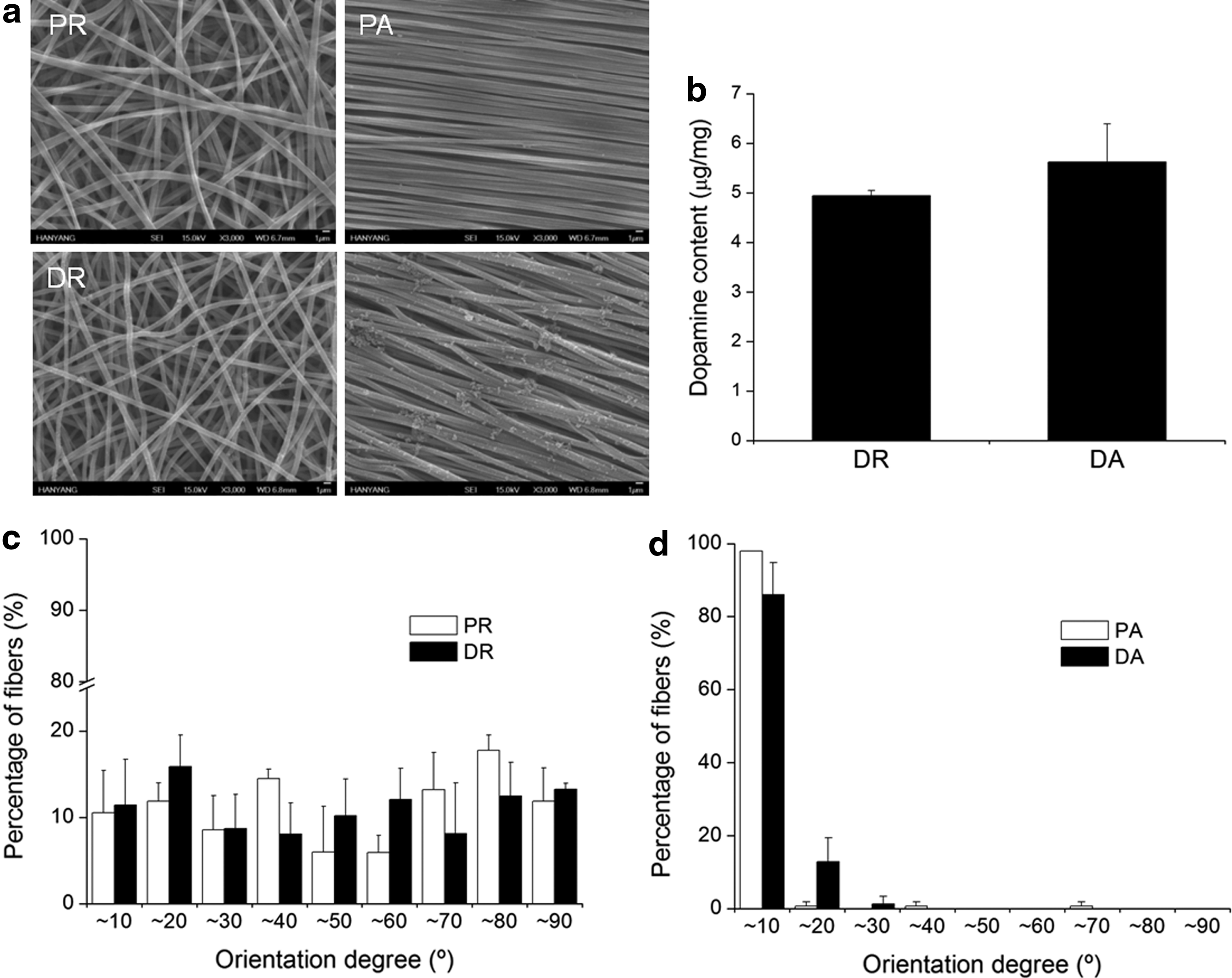
Characteristics of electrospun nanofibers.
Adhesion, proliferation, and migration of hMSCs on fibers
We then visualized the adhesion morphology of hMSCs on the four types of electrospun fibers after 24 h of seeding. Fluorescence staining for f-actin demonstrated that hMSCs organized mature cytoskeletal structures on each fiber (Fig. 2a). The cells on PR adhered with random directions with some cells demonstrating premature spreading, whereas the cells on PA were spread with a spindle shape. Round-shaped hMSCs were not observed on polydopamine-coated random fibers with formation of more mature filopodia, which may be influenced by the polydopamine coating. The direction of growth and migration of hMSCs cultured on aligned fibers with polydopamine coating showed morphologies similar to that observed on PA. We reconfirmed the cell adhesion morphology on the fibers using SEM (Fig. 2b). Consistent with f-actin staining, hMSCs cultured on aligned electrospun fibers were spread and elongated along the direction of the underlying fibers, whereas those on random fibers featured more polygonal shape without a preferential direction. On a polydopamine-coated surface, hMSCs were more widely spread than on electrospun fibers without the coating. To quantify the directionality of cell adhesion, we measured the cell adhesion degree to the reference direction as conducted for the analysis of the electrospun fibers (Fig. 2c). Similar to our results shown in Figure 1, more than 80% of cells were aligned within <10° on aligned electrospun fibers, whereas the orientation angle was widely distributed on random fibers. These results suggest that the directionality of adhered hMSCs reflects the orientation of the underlying substrate. The cell adhesion morphology on the aligned electrospun fibers has been widely investigated with various cell types, including tendon stem cells, human adipose stromal cells (hASCs), cardiomyocytes, and hMSCs.21,25–27 In our previous study, we showed that coating nanofibers with polydopamine for 1 h was sufficient to enhance cell spreading. 22 In the present study, our results showed that the polydopamine coating enhanced cell adhesion compared to that of noncoated PLLA nanofibers.

Adhesion of human mesenchymal stem cells (hMSCs) on electrospun nanofibers.
We measured the DNA content of cells on each fiber during the 5 days of culture to confirm the effect of fiber orientation on cell proliferation (Fig. 3). Overall, we found that fiber alignment had minimal effect on the proliferation of hMSCs. After 1 day of culture, the DNA content on DA was 71.8±7.2 ng/fiber, which was slightly but not significantly higher than that on PR, PA, and DR with values of 58.5±3.6, 57.7±2.5, and 63.7±6.1 ng/fiber, respectively. The trend was consistent with longer culture times of 3 and 5 days. The effect of fiber orientation on cell proliferation is controversial. Fu and Wang reported slightly enhanced proliferation of hASCs on aligned nanofibers compared to random fibers over a 7-day period, 25 whereas it was reported that the proliferation of bone marrow stromal cells was similar on random and aligned electrospun fibers. 20 Moreover, anisotropic patterns with different widths (450, 900 nm) and depths (100, 350, 550 nm) did not affect the proliferation of rat mesenchymal stem cells. 27 Polydopamine coating appears to enhance the hMSC adhesion at day 1 and 3. Regardless of nanofiber alignment, DNA content from hMSCs attached on nanofibers coated with polydopamine was greater than that without polydopamine coating. However, the values were almost comparable at day 5, which may be because hMSCs may have reached confluence level between 3 and 5 days of culture and thereby polydopamine coating was no longer effective in controlling cell proliferation.

Proliferation of hMSCs cultured on electrospun nanofibers after 1, 3, and 5 days (n=5). *p<0.05 versus DNA content of PR on the same culture day point, †p<0.05 versus DNA content of PA on the same culture day point, §p<0.05 versus DNA content of DR on the same culture day point.
We compared the migration distance and the orientation degree of hMSCs cultured on each type of nanofiber structure. A schematic illustration of cell migration analysis is shown in Figure 4a. We restricted the adhesion of hMSCs within the confined square, and adherent cells were subject to migration on the electrospun fibers for over 3 days. A representative image stained for 4′,6-diamidino-2-phenylindole at day 0, which was overlapped to that stained for f-actin of hMSCs cultured for additional 3 days, is shown in Figure 4b. As we expected, cells adhered within the restricted area after the removal of molds (presented with dashed lines, Fig. 5a). After 5 days of migration, hMSCs migrated outside the restricted area (dashed line) and showed polygonal morphology (Fig. 5b). The cells on PR and DR migrated without a specific directionality, whereas the majority of hMSCs on PA and DA migrated in a similar direction (dashed arrow). We separately measured the distances perpendicular and parallel from the starting line to calculate migration speed (Fig. 5c). The migration speeds of hMSCs on random fibers were similar regardless of surface coating with polydopamine; the average migration speed on PR and DR fibers over 5 days was 15.63±3.81 and 12.31±2.1, 16.54±2.5 and 14.38±4.9 μm/h in the perpendicular and parallel directions, respectively, suggesting that the migration of hMSCs was similar on random and aligned fibers. hMSCs on PA and DA migrated approximately two-fold faster on aligned fibers than on random fibers in the parallel direction (34.63±2.9 μm/h and 28.66±5.3 μm/h). However, the migration speed of hMSCs in the perpendicular direction on aligned fibers was significantly decreased compared to that on the random fibers (9.47±3.6 μm/h for PA and 2.74±1.3 μm/h for DA). The migration speed of cells on random and aligned fibers has been compared in other cell types, such as fibroblasts and hASCs.17,25 hASCs migrated two times faster on aligned nanofibers meshes than on the random fibers in the parallel direction, whereas the cell migration speed in the perpendicular direction was less than half of that on the random fibers. These results are consistent with our observations. To confirm the direction of cell migration, we measured the degree of cell orientation after migration against the parallel axis starting point (Fig. 5d). Cells on PR and DR migrated without directionality, whereas more than 80% of cells on PA and DA migrated within 20° of the parallel axis. These results demonstrate that fiber orientation regulates the direction of cell adhesion as well as migration, independent of the presence of polydopamine coating.


Migration analysis of hMSCs on electrospun nanofibers.
Guidance of in vivo bone formation by the structure of electrospun fibers
We investigated the effects of electrospun nanofibers with different orientations on guided in vivo bone regeneration. Bone regeneration in the defect without implantation of fibers was significantly impaired since the critical size defect is limited in spontaneous self-healing (Fig. 6a). Implantation of electrospun fibers without polydopamine coating resulted in slightly increased but not significant bone regeneration on the edge of the defect. PA showed similar results with PR, but the sizes of the bony islands were smaller than in the PR implantation group. The size of new bone from the edge of the site was increased, and bony island size also increased after implantation of DR. Interestingly, the regenerated bone from the edge region seemed to have a specific directionality, and the overall regeneration aspect appeared to be totally different from others. In spontaneous calvaria regeneration, migration of osteoblasts or progenitors from the edge of defects or from the dura membrane to the defect site is important.28,29 Next, we quantified the regenerated bone area (Fig. 6b). Micro-CT imaging demonstrated that implantation of PR and PA showed 3.35%±1.8% and 5.25%±3.7% of regenerated bone area, respectively, suggesting very poor bone regeneration capacity. The regenerated bone area was 10.58%±0.9% after implantation of DR, which was greater than for implantation of PR and PA. The implantation of DA showed the highest regenerated bone area at 28.86%±6.5%, suggesting a synergistic effect of fiber alignment and polydopamine coating on bone regeneration. We confirmed the directionality of regenerated bone after implantation of DA using cross-sectioned 2D micro-CT imaging (Fig. 6c). Sectioning was performed from the upper side of the skull, and the new bone from edge side of calvaria showed directionality at 129.2 μm. The directionality (white arrows) of new bone became clear in the deeper side (181.5, 233.8 and 288.1 μm), which showed aligned new bone fragments from the edge as well as bony islands. To the best of our knowledge, this is the first study to evaluate the effects of fiber alignment and polydopamine coating in enhancing bone regeneration in a calvarial critical size defect model. Our results indicate that the alignment of nanofibers may affect the cell migration direction from the edge region of the defect and may contribute to cell migration into the defect site.
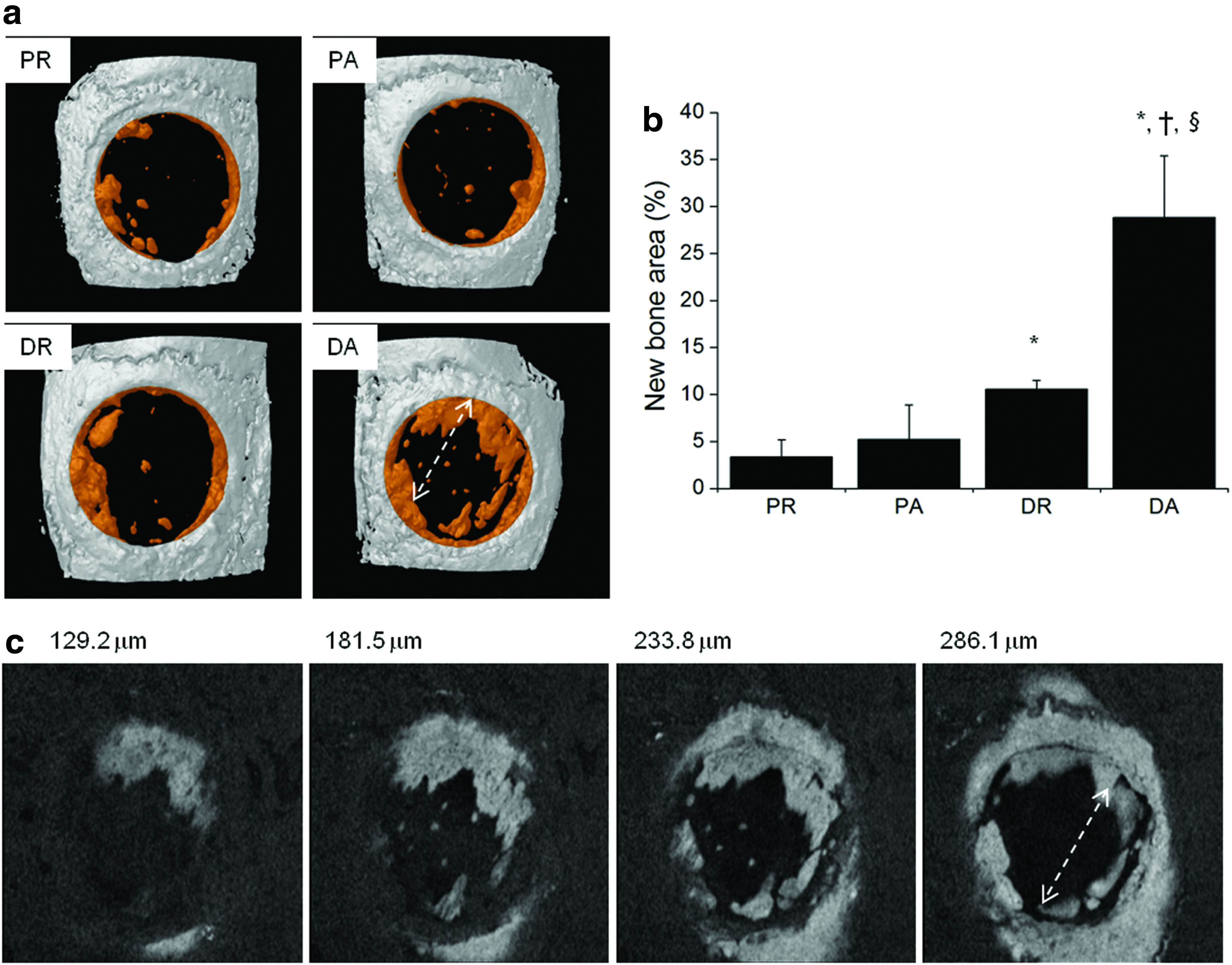
Radiographic analysis of nanofibers after 2 months of implantation into mouse calvarial critical size defect.
Since it was impossible to confirm the direction of fibers after implantation using soft X-ray, micro-CT imaging, or histological analysis, we performed calvaria tissue SEM imaging after cutting in the perpendicular direction against the bone regeneration direction. We found implanted random fibers with complete structure existing under collagen fibers of calvarial tissue (Fig. 7a). Implanted aligned fibers were observed under aligned collagen fibers, with the same orientation (Fig. 7b). We cut the retrieved calvarial tissue perpendicular to the direction of the regenerated bone and found that the fiber alignment direction was the same as the directionality of the regenerated bone. In many studies, implanted materials have been observed by histological analysis. However, we chose tissue SEM imaging to confirm the morphology of implanted nanofibers. Our results support that the enhancement of bone regeneration by DA implantation may occur by the facilitation of cell migration following the directionality of fiber alignment into the defect site. As with SEM sample preparation, we performed tissue TEM to confirm the effects of fiber orientation on collagen fiber distribution and directionality after cutting of tissue perpendicular to the fiber alignment direction. When we implanted random fibers, the cut direction of the collagen bundle was also randomly distributed (Fig. 8a). However, almost all collagen fiber bundles were cut perpendicular to the fiber alignment direction, which indicates that the collagen bundles were arranged in the same direction as the implanted aligned nanofibers (Fig. 8b). Consistent with our results, Baker et al. showed that collagen fibrils oriented around implanted PCL fibers in the parallel direction but not in the perpendicular direction. 30 In the bone regeneration process, the formation and distribution of collagen fibrils are important, but the mechanism of collagen fibril orientation is still not understood. However, the implantation of aligned nanofibers may have induced collagen matrix organization and orientation and then synergistically guided cell migration following their matrix direction.
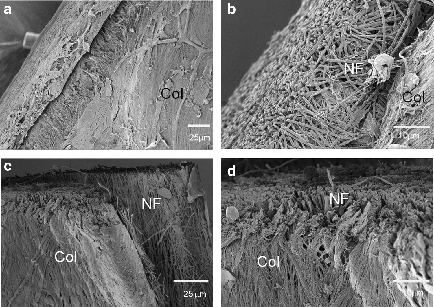
SEM images of calvaria tissues at 2 months postimplantation of nanofibers.
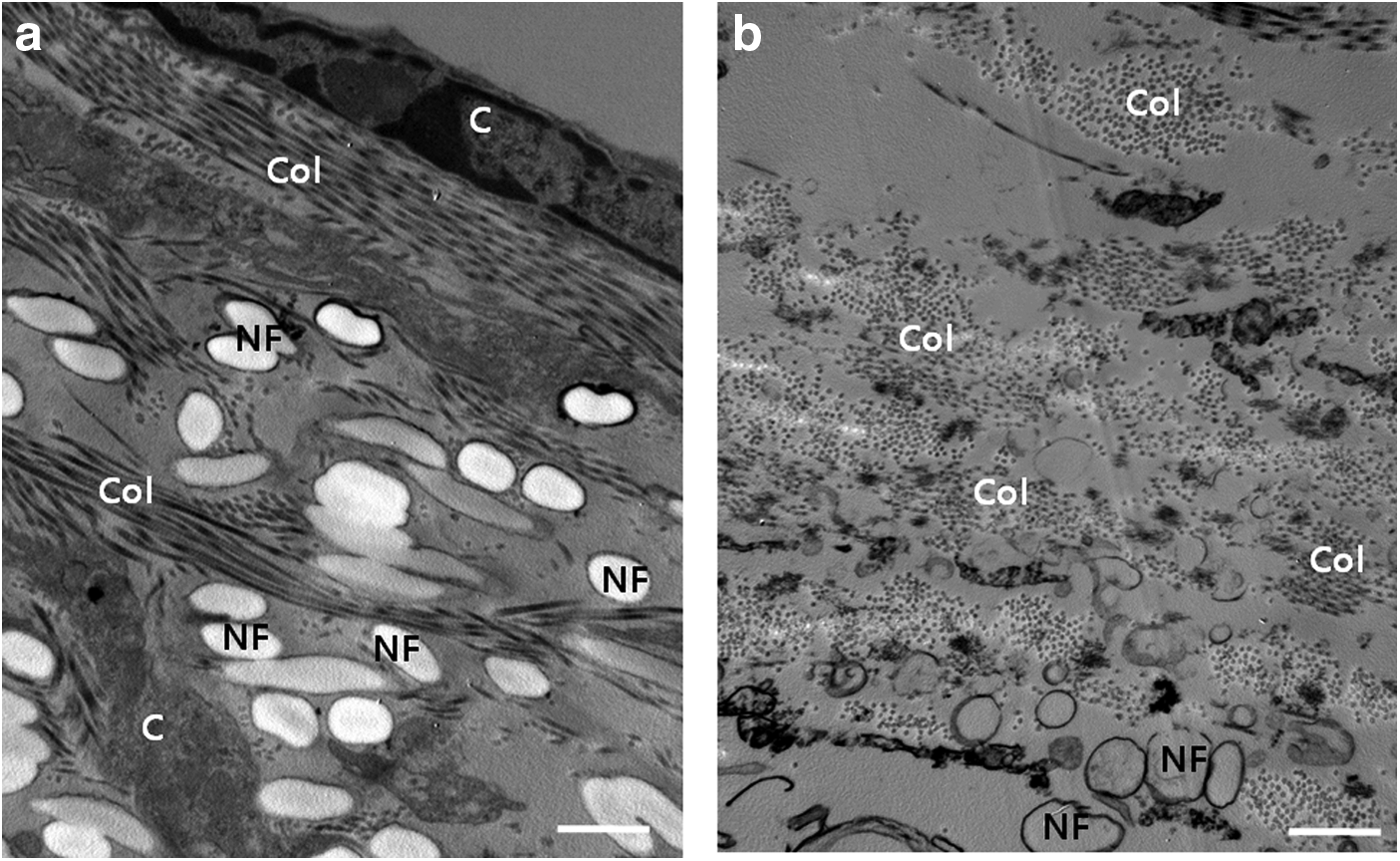
Transmission electron microscopy images of calvaria tissues 2 months postimplantation of nanofibers.
To examine the effect of fibers implantation with different orientations, we performed histological analysis (Fig. 9). We observed that implanted nanofibers were maintained on the defect site with limited fibrous tissue infiltration from all groups. Implantation of PR resulted in the formation of granulated tissues on the fibers but not bone formation. Additionally, the cell linings on the fiber surface were observed with few infiltrating cells. These results indicate that electrospun nanofibers are proper to use as a barrier for cell infiltration. After PA implantation, a premature bone matrix was observed under fibers with no evidence of inflammation. The aligned orientation of fibers may facilitate cell migration as well as extracellular matrix secretion to form the premature bone matrix. In DR implanted bone, small new mature bone was observed at the center of the defect, and DA implantation resulted in increased production of new mature bone formation under implanted nanofibers. Enhanced bone regeneration occurred in DR and DA, which may have resulted from enhanced biocompatibility of materials by polydopamine coating.
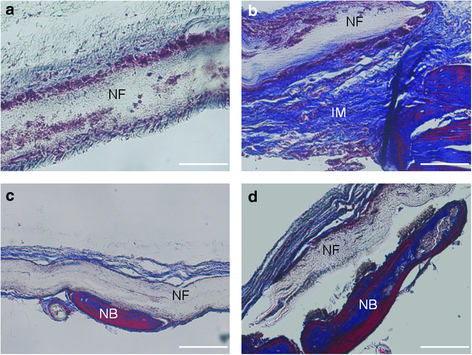
Masson's trichrome staining of calvaria tissues 2 months postimplantation of nanofibers.
The calvarial critical size defect model has been widely used to evaluate the potential of various materials on bone regeneration. Silk fibroin nanofiber membranes were implanted on a rabbit calvarial critical size defect, covering the defect with the fibers, which resulted in ∼45% new bone formation, whereas only 30% new bone formation was observed in the control group. 31 The implantation of an electrospun microfiber bilayer mesh of silicon-doped vaterite/poly(lactic acid) and dense PLA mesh in the same rabbit model showed sufficient bone regeneration 12 weeks after implantation. 6 Fiber implantation was also performed on a rat calvarial model. Highly porous PCL nanofiber implantation resulted in ∼40% bone regeneration, and the effect was significantly enhanced by the incorporation of bone morphogenetic protein-2, which induced an 80% bone regeneration. 32 Although many studies showed in vivo bone regeneration results after fibrous membrane implantation, few studies showed histological staining results. In these studies, new bone formation occurred under the implanted fibers, from the edge of the defect, and from immature collagen matrices. These results are consistent with our results showing new bone formation under implanted fibers with limited cell infiltration, supporting that the electrospun nanofibers with aligned orientation could guide and direct bone regeneration.
Conclusions
In conclusion, the fiber orientation was successfully prepared using an electrospinning technique, and polydopamine was homogeneously coated on the fibers without changing fiber morphology. The cell adhesion direction was consistent with the direction of nanofibers, with or without polydopamine coating, whereas the oriented nanofibers did not affect the proliferation of hMSCs in the 5 days of culture. Importantly, the direction of in vitro cell migration and in vivo bone regeneration was consistent with the direction of the aligned nanofibers as observed by SEM and TEM imaging of tissue. In a mouse calvarial critical size defect model, PA and DA implanted mice showed enhanced bone regeneration compared with the PR and DR implanted groups. Collectively, these results suggest that the structure of aligned fiber can provide spatial guidance for in vitro cell migration as well as in vivo bone regeneration.
Footnotes
Acknowledgments
This work was supported by the research fund of Hanyang University Institute of Aging Society in 2011 (HY-2011-T).
Disclosure Statement
No competing financial interests exist.