
Abstract
Clinical applications of tissue engineering are constrained by the ability of the implanted construct to invoke vascularization in adequate extent and velocity. To overcome the current limitations presented by local delivery of single angiogenic factors, we explored the incorporation of prolyl hydroxylase inhibitors (PHIs) into scaffolds as an alternative vascularization strategy. PHIs are small molecule drugs that can stabilize the alpha subunit of hypoxia-inducible factor-1 (HIF-1), a key transcription factor that regulates a variety of angiogenic mechanisms. In this study, we conjugated the PHI pyridine-2,4-dicarboxylic acid (PDCA) through amide bonds to a gelatin sponge (Gelfoam®). Fibroblasts cultured on PDCA-Gelfoam were able to infiltrate and proliferate in these scaffolds while secreting significantly more vascular endothelial growth factor than cells grown on Gelfoam without PDCA. Reporter cells expressing green fluorescent protein-tagged HIF-1α exhibited dose-dependent stabilization of this angiogenic transcription factor when growing within PDCA-Gelfoam constructs. Subsequently, we implanted PDCA-Gelfoam scaffolds into the perirenal fat tissue of Sprague Dawley rats for 8 days. Immunostaining of explants revealed that the PDCA-Gelfoam scaffolds were amply infiltrated by cells and promoted vascular ingrowth in a dose-dependent manner. Thus, the incorporation of PHIs into scaffolds appears to be a feasible strategy for improving vascularization in regenerative medicine applications.
Introduction
V
Vascular infiltration can also be accelerated by modifications to the scaffold, for example, by incorporating proangiogenic growth factors and cytokines such as vascular endothelial growth factor (VEGF), basic fibroblast growth factor, and hepatocyte growth factor, or plasmids encoding these factors.9,10 However, as these factors are chemically unstable, they have to be incorporated into scaffolds in vastly supraphysiological doses, leading to high costs as well as undesirable side effects.4,11,12 In addition, angiogenesis is a highly complex process that involves multiple crosstalking pathways and factors. In particular, local delivery of single angiogenic factors such as VEGF has been associated with the formation of morphologically abnormal and leaky vessels. 4
Therefore, there remains a great need for vascularization strategies that are simple, cost-effective, reliable, and safe. In the past two decades, a new and potent class of proangiogenic drugs known as prolyl hydroxylase inhibitors (PHIs) has emerged.13,14 These small molecule drugs have a strong capacity for stimulating angiogenesis through hypoxia-inducible factor-1 (HIF-1), by inhibiting HIF prolyl hydroxylases (PHDs). Under normoxia, HIF-PHDs, which require oxygen to function, tag HIF-1α for degradation through the ubiquitin–proteasome pathway. Inhibition of HIF-PHDs leads to the stabilization of HIF-1α, which accumulates and binds with HIF-1β to form HIF-1, a transcription factor that modulates a diverse range of responses to hypoxia. 15 These include the induction of angiogenesis, which HIF-1 achieves by mediating the expression of multiple proangiogenic target genes coding for cytokines (e.g., VEGF, endocrine gland-derived VEGF [EG-VEGF] and transforming growth factor-β3 [TGF-β3]), cytokine receptors (e.g., VEGF receptor 1 [Flt-1]), and other proteins involved in angiogenesis (e.g., matrix metalloproteinases [MMPs], leptin, etc.).16–23 The combination of target proteins which are upregulated by HIF-1 varies depending on the cell type, and this enables HIF-1 to coordinate complex and cell-type-specific responses to hypoxia. 24 Therefore, PHIs can produce a coordinated angiogenic response involving multiple interacting angiogenic factors, in contrast to single-factor approaches.
In a previous study, our group had shown that an established PHI, pyridine-2,4-dicarboxylic acid (PDCA) 25 could stabilize HIF-1α in human fibroblasts and induce ectopic angiogenesis in a zebrafish embryo model. 26 Besides being a potent stimulator of angiogenesis, PDCA is also highly stable, relatively inexpensive, and easily conjugated to scaffolds through its carboxylic acid groups. While the proangiogenic effects of PHIs administered through repeated injections into implanted polyurethane sponges 27 and into bone defects 28 are well-established, to the best of our knowledge, the stable incorporation of PHIs into implantable scaffolds has not been reported yet. We thus set out to explore the incorporation of PDCA into scaffolds as an alternative vascularization strategy, by conjugating it through amide bonds to Gelfoam™, a gelatin sponge that we selected as our test scaffold. Drug release is mediated by the hydrolysis of amide bonds by cellular proteases such as MMPs. We characterized the chemical and material properties of PDCA-Gelfoam scaffolds, and assessed their proangiogenic properties both in vitro and in vivo. Our results show that the incorporation of PHIs into scaffolds is a feasible vascularization strategy.
Materials and Methods
Cell culture
Normal human fetal lung fibroblasts (IMR-90) were purchased from the American Tissue Culture Collection (Rockville, MD). Human osteosarcoma cells (U2OS) expressing green fluorescent protein-tagged HIF-1α (GFP-HIF-1α) were purchased from Thermo Scientific (Lafayette, CO). All media and supplements used for cell culture were purchased from Invitrogen (Carlsbad, CA), unless otherwise stated. All reagents were either purchased sterile or sterilized by filtration through sterile 0.2 μm filter units. The basal medium used for all experiments was high glucose Dulbecco's modified Eagle's medium (HG DMEM, #10569; Invitrogen). For expansion and maintenance, both cell lines were cultured in HG DMEM supplemented with 100 U/mL penicillin, 100 μg/mL streptomycin, and 10% fetal bovine serum (FBS). For assays, cells were seeded and cultured in HG DMEM supplemented with 30 μg/mL
Preparation of PDCA-Gelfoam
All reagents used for drug conjugation were purchased from Sigma-Aldrich (St Louis, MO) unless otherwise stated. The Gelfoam® sterile gelatin sponge (Pfizer, New York, NY) was first cut into smaller pieces with dimensions 2.5×2.5×0.6 cm and placed into six-well polystyrene tissue culture plates (Greiner Bio-One GmbH, Frickenhausen, Germany). The reaction scheme is shown in Figure 1. 1,1′-Carbonyldiimidazole (CDI) and pyridine-2,4-dicarboxylic acid monohydrate were weighed separately in 50-mL polypropylene tubes (Greiner Bio-One GmbH). PDCA and CDI were used at a molar ratio of 1:3, as PDCA was purchased as in monohydrate form. The CDI was first dissolved in anhydrous dimethyl sulfoxide (DMSO) at a concentration of 0.3 g/mL. The resultant CDI solution was then added to the PDCA dropwise with gentle swirling to avoid excessive effervescence, and allowed to react for 1 h at room temperature to allow complete conversion of the carboxylic acid groups in PDCA to acyl imidazole groups (activation). Subsequently, the activated PDCA solution was filtered through sterile Millex-LG 0.2 μm polytetrafluoroethylene (PTFE) syringe filters (Millipore, Bedford, MA), and diluted to appropriate concentrations with anhydrous DMSO. The concentrations of reactants used for each dosage condition are listed in Table 1. The diluted activated PDCA solutions were added gradually to each piece of Gelfoam to a total volume of 4.5 mL per well, and allowed to react for 5 h, during which the acyl imidazole groups in the activated PDCA reacted with the amine groups in Gelfoam to form amide bonds. No-drug controls (“0% w/w”) were similarly incubated with plain anhydrous DMSO. At the end of the reaction, samples were transferred to individual 50-mL polypropylene tubes and washed 10 times with Milli-Q water, with each washing step lasting at least 4 h. The washed samples were transferred to fresh six-well plates, frozen at −80°C, and lyophilized.
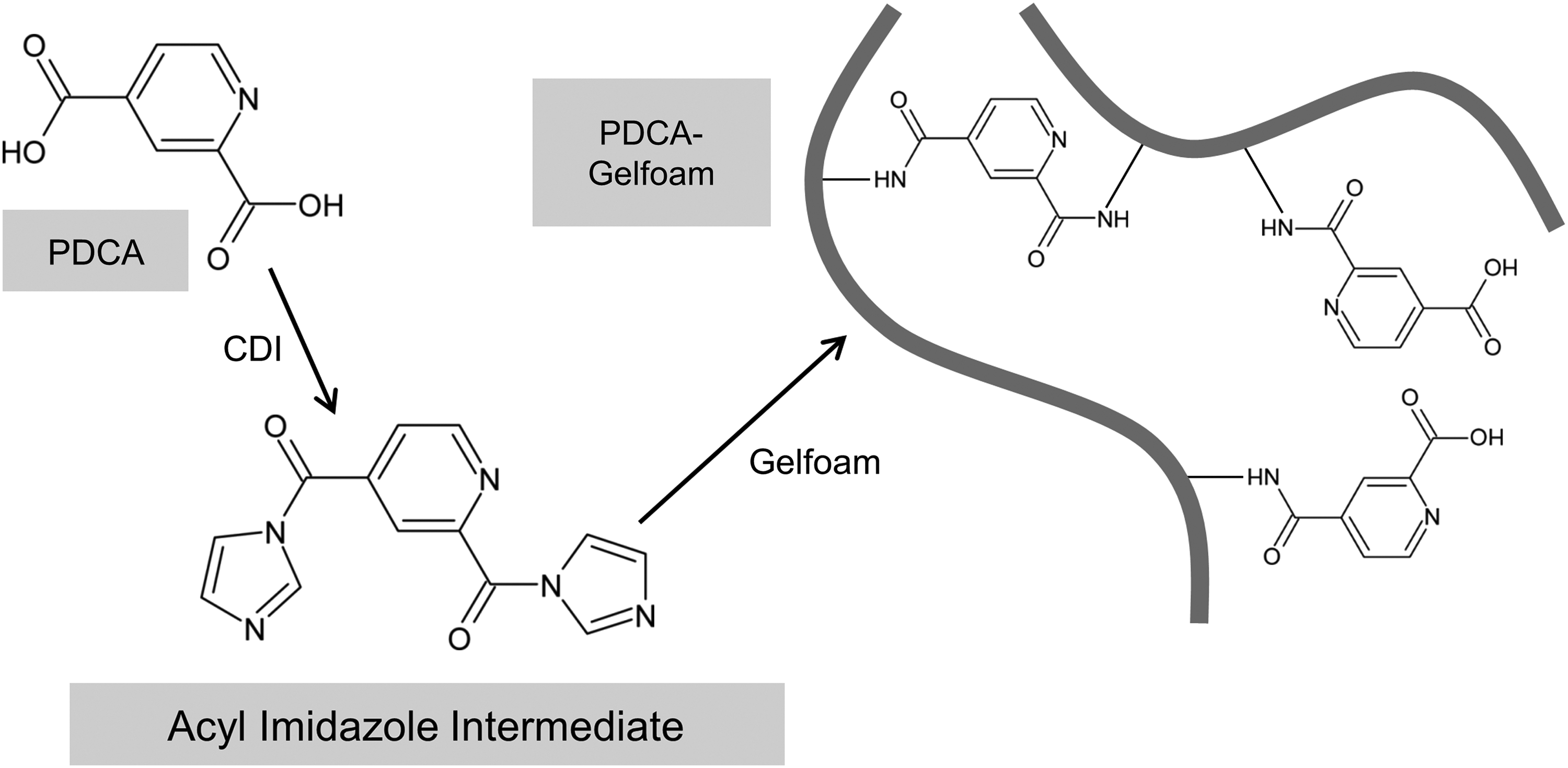
Reaction scheme for the conjugation of pyridine-2,4-dicarboxylic acid (PDCA) to Gelfoam through amide bonds. 1,1′-Carbonyldiimidazole (CDI) was used to facilitate formation of the amide bonds. PDCA's carboxylic groups were first converted by CDI into acyl imidazole groups (activation). Imidazole and carbon dioxide were produced as by-products, with the imidazole remaining in solution and the carbon dioxide escaping as effervescence. When the activated PDCA was added to Gelfoam, the acyl imidazole groups reacted with the amine groups in Gelfoam to form amide bonds. Imidazole occurred again as a by-product, and was subsequently removed by repeated washing of scaffolds.
The concentrations of PDCA and CDI required were determined by extrapolating data from previous experiments conducted to establish the relationship between reactant concentrations and resultant yields.
CDI, 1,1′-carbonyldiimidazole; PDCA, pyridine-2,4-dicarboxylic acid.
Drug loading measurements
The concentration of PDCA in the dissolved PDCA-Gelfoam samples was determined by ultraviolet (UV) spectroscopy, using an Infinite M200 plate reader (Tecan, Research Triangle Park, NC) and UV-transparent 96-well flat bottom microtiter plates (UV-Star; Greiner Bio-One GmbH). PDCA-Gelfoam samples and controls were dissolved at a concentration of 20 mg/mL in 6 N hydrochloric acid by heating at 95°C for 15 min. The dissolved samples were diluted 1:10 with Milli-Q water for spectroscopy, such that the final concentration of hydrochloric acid was 0.6 N. PDCA standards were prepared in 0.6 N hydrochloric acid. UV absorption measurements were made at wavelengths from 230 to 330 nm, at intervals of 5 nm, and UV absorption spectra were obtained by plotting absorbance against wavelength. Absorbance measurements at 290 nm were used for drug loading calculations, as the ratio of the absorbance of PDCA to that of Gelfoam is the greatest at 290 nm. Baseline correction was performed by subtracting the absorbance of the blank plate and the absorbance of 0.6 N hydrochloric acid from the absorbances of all samples. A linear PDCA standard curve that intersects the origin was then plotted. The absorbance of Gelfoam was subtracted from the absorbance of PDCA samples to exclude the absorbance contributions from Gelfoam, and the PDCA concentrations were calculated using the equation of the PDCA standard curve.
Scanning electron microscopy
The morphology and pore structure of the PDCA-Gelfoam scaffolds were assessed by scanning electron microscopy (SEM) using a Zeiss Supra 55 VP scanning electron microscope (Zeiss, Göttingen, Germany) with a variable pressure secondary electron detector (magnification=100×, working distance=6.8–7.5 mm, EHT voltage=10.00 kV).
Preparing reagents for papain digestion
Papain digestion buffer was prepared by dissolving 5 mM
Characterization of protease-based drug release
To characterize the release of conjugated PDCA by proteases, we digested PDCA-Gelfoam samples with papain. PDCA-Gelfoam scaffolds were cut into 9×6×5 mm cuboids, weighed, and placed in individual 15-mL polypropylene tubes. Papain working solution (0.4 μg/mL) was prepared fresh by diluting the 0.5 mg/mL stock serially in papain digestion buffer. The concentration of the papain working solution was selected based on prior optimization, such that digestion could be completed within 8 h. Five milliliters of papain working solution was added to each tube, and the samples were incubated in a 37°C water bath with shaking until the samples had been completely solubilized. At each time point, the timer was paused and the tubes were immersed in ice to slow down proteolysis during sample transfer. Two 100 μL aliquots of solution were then transferred from each tube to UV-Star 96-well plates, after which digestion was resumed by reimmersing the tubes in the water bath. PDCA standards were prepared by dissolving PDCA in the papain digestion buffer. After the last time point, the absorbance values of the standards and the samples were measured at 290 nm using an Infinite M200 plate reader. As the absorbance of Gelfoam at 290 nm is very low compared with that of PDCA, the absorbance values at this wavelength could be used to approximate the concentrations of PDCA, after subtracting the absorbance contributions of papain and buffer components. Since the volumes of solution remaining and removed were known, the cumulative amounts of PDCA released could be calculated by summing the amount remaining in solution and the amounts previously removed. These values were subsequently normalized to sample mass to account for sample mass variations, and plotted as a function of time to obtain the drug release profiles.
Culturing fibroblasts on PDCA-Gelfoam scaffolds
The PDCA-Gelfoam samples were first cut into 8-mm-diameter cylinders using biopsy punches. The cylindrical scaffolds were disinfected by immersion in 70% ethanol and allowed to dry for 30 min, after which the scaffolds were immersed in 8% w/v sodium bicarbonate (Sigma-Aldrich) in the culture medium for 5 min to neutralize the carboxylic acid groups of the conjugated PDCA. The scaffolds were then transferred to Transwell™ polycarbonate cell culture inserts in 24-well plates (#3422; Corning Costar, Corning, NY), rinsed three times with culture medium to remove excess sodium bicarbonate, and conditioned with assay medium overnight. IMR-90 fibroblasts between passages 16 and 18 were seeded at a density of 250,000 cells per scaffold and allowed to attach for 2 h, after which 1 mL of the culture medium was added per well. Culture medium was changed 24 h postseeding and every 2 days thereafter. Culture medium samples harvested during each medium change were either analyzed immediately or stored at −80°C for subsequent analysis.
Cytotoxicity assay
Cytotoxicity of samples were assessed by analyzing aliquots of freshly harvested medium from day 1 postseeding using a Vybrant cytotoxicity assay kit (Invitrogen), which measures the leakage of the cytosolic enzyme glucose 6-phosphate dehydrogenase (G6PD) from compromised cells. Fluorescence measurements were made using a FLUOstar Optima plate reader (BMG Labtech, Durham, NC).
Quantifying cell numbers in scaffolds
Samples that had been cultured for 7 days were rinsed three times with PBS. Excess PBS was gently squeezed out from the scaffolds and removed. The scaffolds were then transferred to individual 1.5-mL polypropylene tubes and digested with 500 μL of 125 μg/mL papain in PBS containing 5 mM
Assessing the distribution of cells within the scaffolds
To facilitate confocal microscopy, the cylindrical PDCA-Gelfoam scaffolds were each cut into two thinner pieces with half the original thickness, such that each piece had a thickness of ∼3 mm, before cell culture. One hundred thousand IMR-90 fibroblasts between passages 16 and 18 were seeded on each half-thickness scaffold and cultured for 7 days, after which the scaffolds were rinsed three times with PBS, fixed in 4% w/v formaldehyde (Thermo Scientific) in PBS for 48 h, and rinsed thrice again to remove the formaldehyde. Fluorescent staining of cell nuclei was performed by immersing the fixed scaffolds in 0.5 μg/mL 4′,6-diamidino-2-phenylindole (DAPI) in PBS for 30 min and rinsing thrice with PBS. After the last rinse, the scaffolds were placed on a glass cover slip and visualized using an Olympus FV500 laser-scanning confocal microscope (Olympus, Tokyo, Japan). Three-dimensional reconstructions showing the positions of the cell nuclei were generated using the ImageJ software (NIH, Bethesda, MD).
HIF-1α reporter assay
To facilitate fluorescence microscopy, the cylindrical PDCA-Gelfoam scaffolds were each cut into four thinner pieces, each with a thickness of ∼1.5 mm, before cell culture. After sterilization by dipping in 70% ethanol, the scaffolds were carefully inserted into a 96-well plate (Greiner Bio-One GmbH) and spread out, such that they lay flat and covered the entire bottom surface of each well. The scaffolds were then allowed to dry for 30 min, after which they were neutralized, rinsed, and conditioned with assay medium overnight. GFP-HIF-1α-expressing U2OS osteosarcoma reporter cells were labeled using a PKH26 fluorescent cell linker kit (Sigma-Aldrich) before seeding, so that the cells could be located even in the absence of GFP-HIF-1α protein. The labeled cells were seeded onto the quarter-thickness scaffold at a density of 100,000 cells per scaffold and cultured for 24 h. GFP-HIF-1α levels were monitored at 4, 8, and 24 h postseeding using an Olympus IX31 fluorescence microscope.
Analysis of VEGF secretion
The quantities of VEGF in aliquots of harvested medium were analyzed by enzyme-linked immunosorbent assay (ELISA) using a DuoSet human VEGF ELISA kit (R&D Systems, Minneapolis, MN). Optical density measurements were made using an Infinite M200 plate reader (Tecan).
Rat perirenal fat implantation model
All animal experiments were reviewed and approved by the Institutional Animal Care and Use Committee of the National University of Singapore (NUS IACUC, protocol number 039/09). Ten to 12 weeks old male Sprague Dawley rats purchased from the NUS Comparative Medicine were used for this study. All PDCA-Gelfoam samples used for animal experiments were prepared under aseptic conditions using sterile materials and equipment, and all reagents used either purchased sterile or sterilized by autoclaving or filtration through sterile 0.2 μm filter units. All surgical procedures were performed as described previously. 30 Before implantation, aseptically prepared PDCA-Gelfoam samples were cut into 1.0×1.0×0.6 cm pieces and disinfected by dipping in sterile-filtered 70% ethanol, as an extra safety measure. The samples were then allowed to dry for 30 min, neutralized by immersion in 8% w/v sodium bicarbonate for 5 min, and rinsed three times with PBS. Anesthesia was induced in the rats and maintained by intraperitoneal injection of ketamine:xylazine (75:10 mg/kg) and inhalational isoflurane (2%). Carprofen (5 mg/kg, subcutaneous) was administered preoperatively as analgesia. A mid-laparotomy was performed, followed by displacement of the gut, mild retraction of the right kidney, and blunt preparation of the retroperitoneal fossa. A pouch was created between the retrorenal fat and the psoas muscle, and a PDCA-Gelfoam sample was inserted into the pouch. This was repeated on the left side, such that two PDCA-Gelfoam samples from the same dosage condition were implanted in each rat. The bowel was subsequently repositioned, and the abdomen closed in two layers of sutures. Carprofen (5 mg/kg, subcutaneous, once daily) and enrofloxacin (25 mg/kg, subcutaneous, twice daily) were administered for 5 days postsurgery. At 8 days postsurgery, the rats were euthanized and the PDCA-Gelfoam samples were explanted for analysis.
Preparation of frozen sections
Explants were fixed in 4% w/v formaldehyde in PBS for 4 h, immersed in 30% w/v sucrose (Sigma-Aldrich) in PBS overnight and frozen-embedded in the Tissue-Tek OCT compound (Sakura Finetek, Torrance, CA). The samples were then cryosectioned at 10 μm thickness.
Morphometric analysis of vascular infiltration
Endothelial cells present in explant sections were visualized by immunohistochemical staining for rat endothelial cell antigen-1 (RECA-1), a rat pan-endothelial cell marker. Mouse monoclonal antibodies against RECA-1 (1:50, HM3012; Hycult Biotechnology, Uden, The Netherlands) were used as the primary antibody, and Alexa Fluor® 594-conjugated goat anti-mouse IgG (1:400, A-11032; Invitrogen) was used as the secondary antibody. Sections were blocked with 3% bovine serum albumin in PBS before immunostaining. Primary antibodies were incubated for 16 h, and secondary antibodies were incubated for 1 h. Cell nuclei were counterstained using 0.5 μg/mL DAPI in PBS. Images were captured using a Zeiss Axio Observer Z1 microscope (Zeiss). Morphometric analysis of immunostained areas and quantification of cell nuclei was performed using the ImageJ software.
Statistical analysis
Data are presented as mean±standard deviation. Statistical analyses were performed by one-way analysis of variance (ANOVA) using the OriginPro software (version 9.1; OriginLab Corporation, Northampton, MA). Multiple comparisons were performed using the Tukey–Kramer post hoc test. Differences were considered statistically significant when p<0.05.
Results
PDCA can be successfully conjugated to Gelfoam
PDCA's UV spectrum displayed two absorption maxima, one below 230 nm (the lower limit of the instrument) and one at 280 nm, corresponding to the carbonyl bonds in the carboxylic acid groups and the pyridine ring, respectively (Fig. 2a).31,32 The UV spectrum of untreated Gelfoam (i.e., in its original state, as purchased from Pfizer) also had an absorption maximum below 230 nm, corresponding to the carbonyl bonds present in the carboxylic acid and amide groups of gelatin, but absorbed relatively weakly in the rest of the wavelength range measured (Fig. 2b). 32 The UV spectra of the PDCA-Gelfoam samples contain spectral characteristics from both PDCA and Gelfoam (Fig. 2c), indicating that the conjugation of PDCA to Gelfoam through amide bonds using CDI had been successful. Absorption at both 230 and 280 nm increased as the concentration of reactants used increased, showing that more PDCA was successfully conjugated to the Gelfoam when higher concentrations of reactants were used.
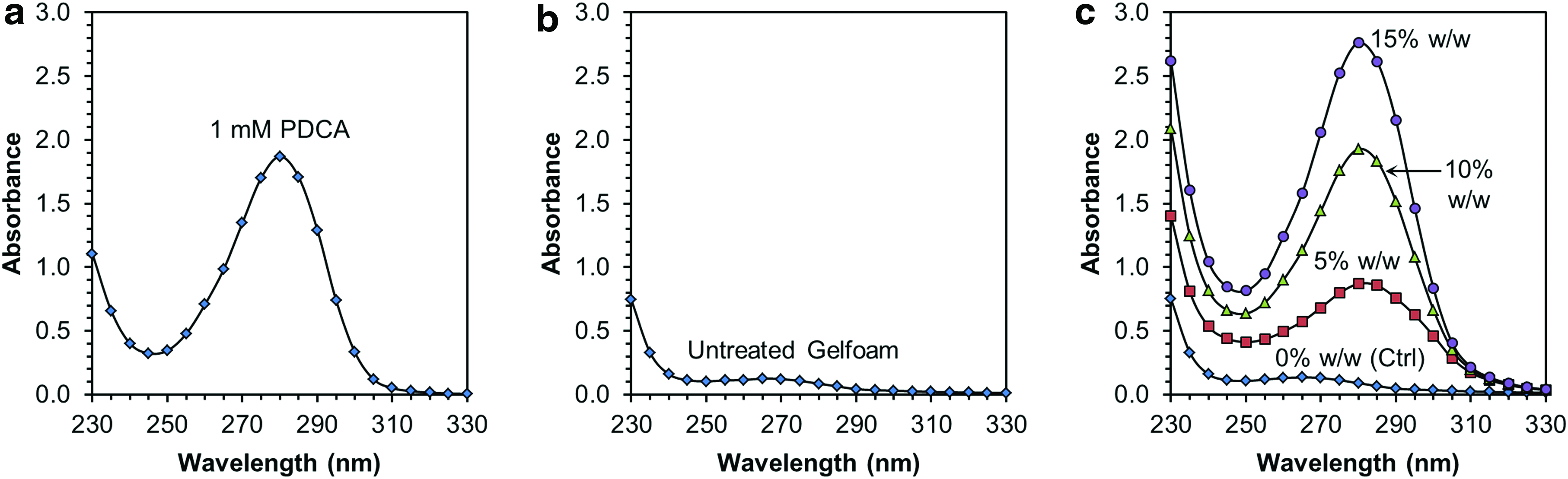
Ultraviolet absorbance spectra of PDCA-Gelfoam.
Gelfoam retains high porosity after conjugation of PDCA
The physical appearance and morphology of untreated original Gelfoam and PDCA-Gelfoam samples did not show major differences (Fig. 3a, b). Compared with the untreated Gelfoam, the PDCA-Gelfoam samples had a rougher texture, but remained highly porous. The morphology of the PDCA-Gelfoam samples appeared similar across all dosages, with and without PDCA, suggesting that the architecture of Gelfoam was not significantly altered by covalent incorporation of PDCA.
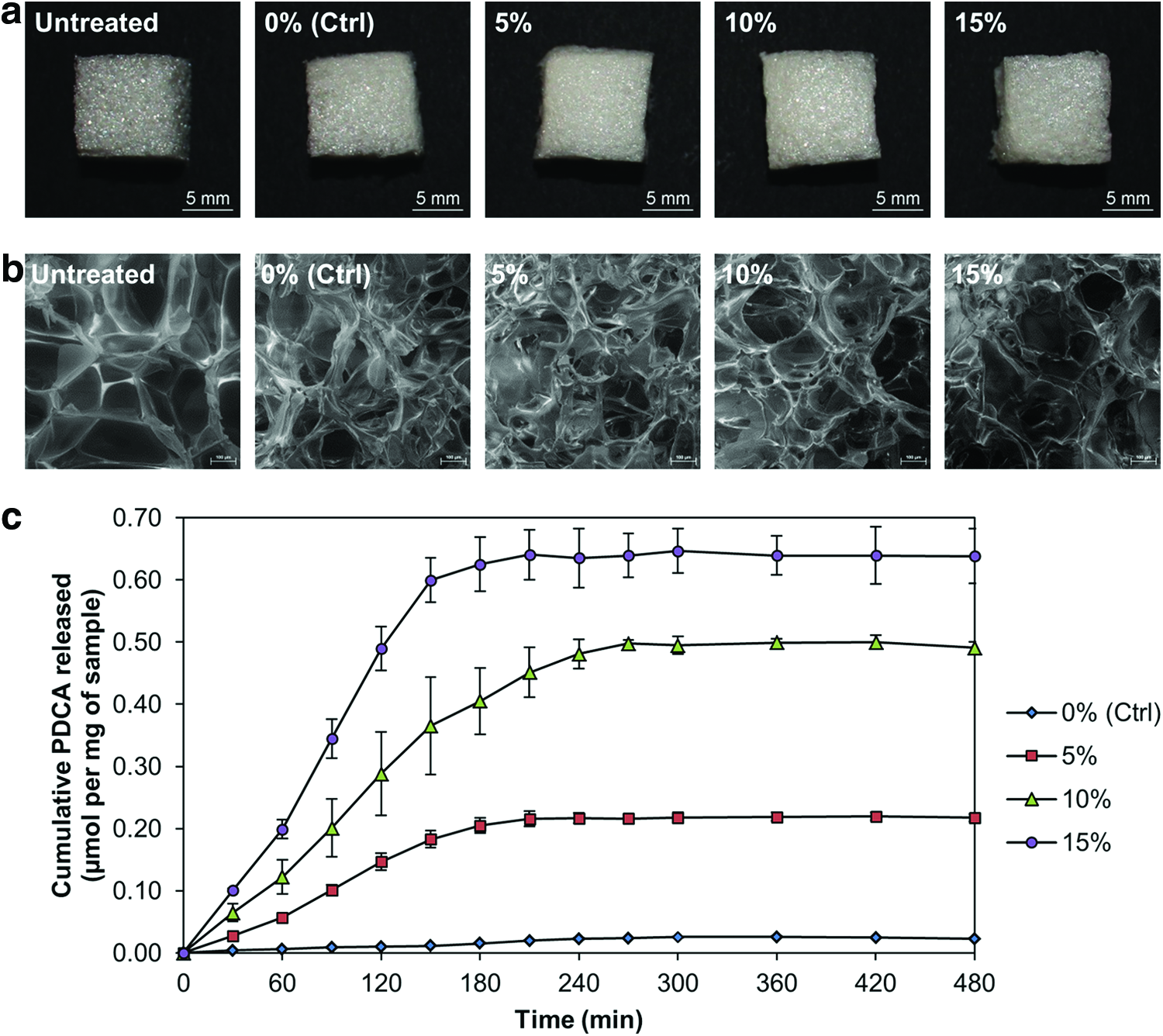
Physical appearance of PDCA-Gelfoam and enzymatic drug release profiles.
Proteolytic release of PDCA occurs at a nearly constant rate
PDCA-Gelfoam samples were digested with 0.4 μg/mL papain as an accelerated simulation of the release of conjugated PDCA by cellular proteases. The resultant cumulative release profiles (Fig. 3c) consisted of two phases: a linear phase, followed by a gradual plateau. The roughly linear slopes of the first phase indicate that most of the conjugated PDCA (∼90%) was released at a sustained and nearly constant rate without an initial burst, consistent with the release characteristics of degradation-based drug delivery systems.
PDCA-Gelfoam shows low cytotoxicity and supports cell proliferation and infiltration
Measurements obtained using the Invitrogen's Vybrant G6PD leakage assay shows that PDCA-Gelfoam has low cytotoxicity (<10%) at all dosages tested (Fig. 4a). Cell numbers measured using the PicoGreen-based DNA measurements showed that by day 7, the cells had proliferated far beyond the numbers seeded (250,000 cells), ascertaining that PDCA-Gelfoam can support cell proliferation (Fig. 4b). Cell numbers at higher dosages (10% and 15% w/w) were, however, significantly lower than in controls (0% w/w), suggesting a dose-dependent antiproliferative effect consistent with prior literature, which showed that PHIs can reduce cell proliferation without affecting cell viability.33,34 The three-dimensional confocal reconstructions of DAPI-stained PDCA-Gelfoam samples showed that cells had infiltrated the scaffolds extensively by day 7 of in vitro culture (Fig. 4c), proving that the conjugated PDCA did not interfere with cell attachment and infiltration at all dosages tested.
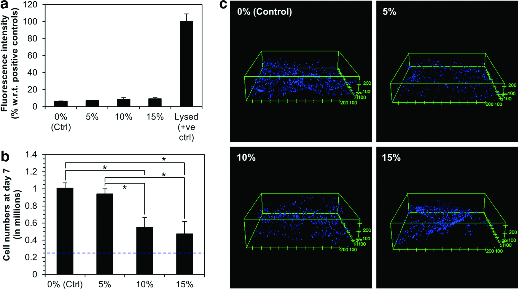
Assessment of PDCA-Gelfoam's effects on the proliferation and viability of IMR-90 fibroblasts.
HIF-1α is stabilized in a dose-dependent manner in cells cultured on PDCA-Gelfoam
Reporter cells transfected with GFP-tagged HIF-1α were cultured on thinly-sliced PDCA-Gelfoam scaffolds and imaged at 24 h postseeding (Fig. 5a). The levels of GFP-HIF-1α were below the limit of detection in the controls (0% w/w), as well as the lowest dosage (5% w/w). At the higher dosages (10% and 15% w/w), GFP-HIF-1α was sufficiently stabilized that their levels were detectable as green fluorescence, with the highest levels observed at 15% w/w. These results demonstrate that PDCA-Gelfoam can release PDCA and stabilize HIF-1α in infiltrating cells.
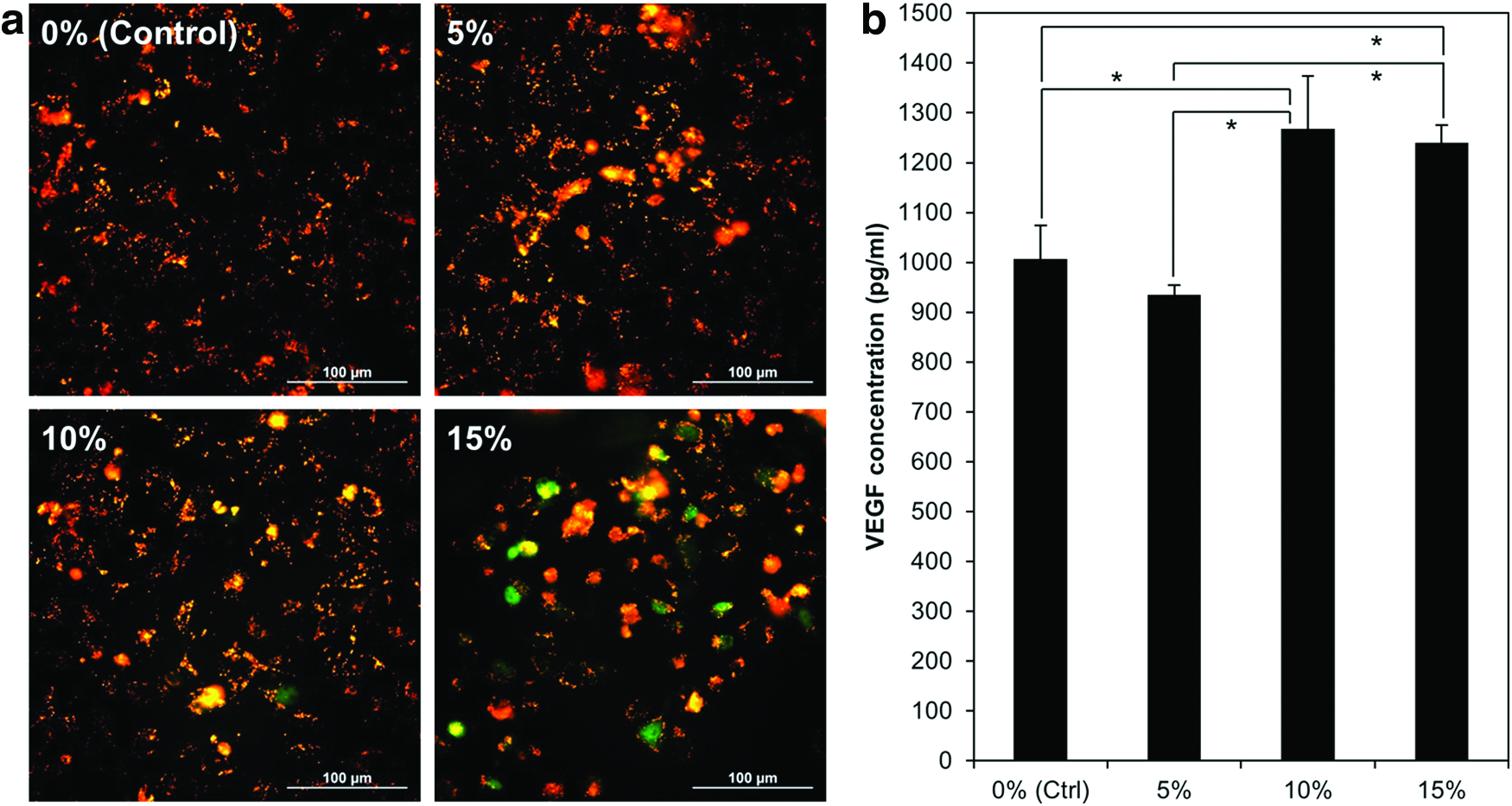
Assessing PDCA-Gelfoam ability to stabilize hypoxia-inducible factor-1α (HIF-1α) and stimulate vascular endothelial growth factor (VEGF) secretion.
PDCA-Gelfoam stimulates VEGF secretion by fibroblasts in vitro
As VEGF is an important proangiogenic cytokine and a direct transcriptional target of HIF-1, we assessed the VEGF secretion as a surrogate marker for PDCA-Gelfoam effects on HIF-1 downstream signaling. Fibroblasts were cultured for 1 day on PDCA-Gelfoam scaffolds, after which the culture medium was harvested and analyzed using a VEGF ELISA kit. Results showed that the higher PDCA dosages (10% and 15% w/w) increased the VEGF secretion by 26% and 23%, respectively (Fig. 5b).
PDCA-Gelfoam stimulates vascular infiltration in vivo
PDCA-Gelfoam samples were implanted into the perirenal fat tissue of Sprague Dawley rats (Fig. 6a), and explanted 8 days later (Fig. 6b). It was observed that the sizes of the explants decreased as the PDCA content in the scaffolds increased. This was due, in part, to differences in the scaffolds' degradation rates, as the conjugated PDCA increased the hydrophilicity of the scaffolds. To assess the PDCA-Gelfoam effects on vascular infiltration, explant cryosections were immunostained for RECA-1, a rat pan endothelial cell marker, and counterstained for cell nuclei using DAPI. We observed that all explants were well-infiltrated by cells, confirming that PDCA-Gelfoam supported good cell attachment and infiltration at all dosages tested (Fig. 7a). The higher dosages (10% and 15% w/w) also had visibly more endothelial cells than the 0% w/w controls (Fig. 7a). To compare the degrees of vascular infiltration across different dosages, the percentages of RECA-1-positive areas in the sections were quantified morphometrically (Fig. 7b). Results showed that the conjugated PDCA increased vascular infiltration in a dose-dependent manner, with the two highest dosages (10% and 15% w/w) having the most pronounced increases in endothelial cell density (7.3- and 8.9-fold, respectively). To rule out the influence of cell density variations, the numbers of nuclei present were counted using ImageJ, and the percentages of RECA-1-positive areas were normalized to the cell numbers and plotted (Fig. 7c). Results showed that the dose-dependent trend of increase in vascular infiltration remained similar after normalization to cell numbers, indicating that the observed increases in vascular infiltration were not due to cell density variations. Taken together, our results have conclusively demonstrated that the conjugated PDCA stimulated vascular infiltration in vivo.

Assessment of PDCA-Gelfoam's effects on vascular infiltration using a rat perirenal fat implantation model.
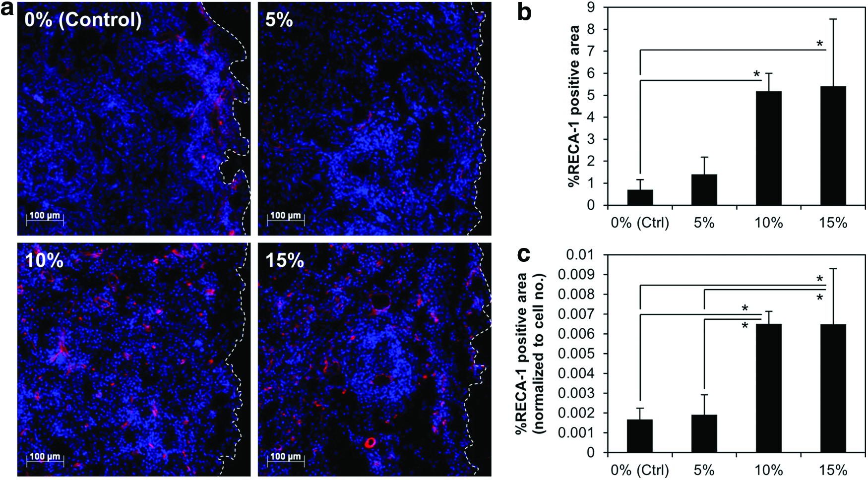
Morphometric analysis of explant cryosections.
Discussion
In this study, we have demonstrated for the first time the feasibility of stimulating vascularization by stably incorporating a PHI into a scaffold for subsequent local release. Our results show that PDCA conjugated to a gelatin scaffold through amide bonds can stabilize HIF-1α in infiltrating cells, stimulate their production of VEGF, and improve vascular infiltration in vivo without negatively impacting cell attachment and viability. While the incorporated PDCA exerted a moderate dose-dependent antiproliferative effect, this effect was not due to cytotoxicity, and cells cultured on PDCA-Gelfoam were still able to multiply and infiltrate the scaffolds. Our findings complement previous studies that showed that repeated injections of PHIs can stimulate localized angiogenesis at the sites of injection.27,28
In a clinical context, repeated injections of drugs into a site of injury cause considerable pain and distress to patients and may increase the risk of infections. The site of implantation may also be located in tissues that are not easily accessible, such as the heart or the spine. Incorporation of drugs into implants for subsequent local delivery is, therefore, preferable and we thus set out to explore the viability of incorporating PHIs into scaffolds as a vascularization strategy. Gelfoam was used as the test scaffold in this proof-of-concept study because it is commercially available, well characterized, and in widespread clinical use. As Gelfoam is composed entirely of gelatin, it is also useful as a representative of protein-containing biomaterials commonly used as tissue engineering scaffolds. PDCA was selected as the PHI for this study because it has been shown to be effective in promoting angiogenesis, and can be easily conjugated to any amine-containing scaffold through its carboxylic acid groups. 26 Aside from being applicable to a wide range of biomaterials, our method also has the additional benefit of not yielding any toxic breakdown by-products upon hydrolysis, as the PDCA is conjugated directly to the Gelfoam scaffold through amide bonds, without the use of cross-linkers.
Although we focused only on the proangiogenic aspects of PHIs in this study, it is worth noting that a variety of PHIs also have an antifibrotic capacity, which is mediated by their ability to cross-inhibit collagen prolyl 4-hydroxylase, an enzyme that is structurally related to HIF prolyl hydroxylases. 25 Inhibition of this enzyme interferes with the posttranslational modification of procollagen, leading to decreased collagen output and thus reduced fibrosis. The antifibrotic function of PHIs may be especially useful in situations where both an increase in angiogenesis and a reduction in fibrosis are desired, for example, in cardiac regeneration following a myocardial infarction. 35
Conclusions
As cells cannot survive without an adequate supply of oxygen and nutrients, it is crucial to the future of the tissue engineering field that the problem of inadequate vascularization be solved. Our findings have demonstrated that the incorporation of PHIs into scaffolds is a feasible strategy for stimulating vascular infiltration in engineered tissues and implanted biomaterials.
Footnotes
Acknowledgments
This work was funded by the National University of Singapore Academic Research Fund, under the Engineering in Medicine Initiative (R-397-000-082-112). The authors would like to thank Ms. Chris Heyjin Park from the Zeiss Microscopy Centre for her invaluable assistance with the SEM imaging in the scope of this work.
Disclosure Statement
The authors hereby declare that no competing financial interests exist.