
Abstract
Objective:
Extracellular matrix (ECM) derived from human amniotic mesenchymal cells (HAMs) has various biological activities. In this study, we developed a novel HAM-derived ECM-coated polylactic-co-glycolic acid (ECM-PLGA) scaffold, examined its property on mesenchymal cells, and investigated its potential as a cell-free scaffold for cartilage repair.
Materials and Methods:
ECM-PLGA scaffolds were developed by inoculating HAM on a PLGA. After decellularization by irradiation, accumulated ECM was examined. Exogenous cell growth and differentiation of rat mesenchymal stem cells (MSCs) on the ECM-PLGA were analyzed in vitro by cell attachment/proliferation assay and reverse transcription–polymerase chain reaction. The cell-free ECM-PLGA scaffolds were implanted into osteochondral defects in the trochlear groove of rat knees. After 4, 12, or 24 weeks, the animals were sacrificed and the harvested tissues were examined histologically.
Results:
The ECM-PLGA contained ECM that mimicked natural amniotic stroma that contains type I collagen, fibronectin, hyaluronic acid, and chondroitin sulfates. The ECM-PLGA showed excellent properties of cell attachment and proliferation. MSCs inoculated on the ECM-PLGA scaffold showed accelerated type II collagen mRNA expression after 3 weeks in culture. The ECM-PLGA implanted into an osteochondral defect in rat knees induced gradual tissue regeneration and resulted in hyaline cartilage repair, which was better than that in the empty control group.
Conclusion:
These in vitro and in vivo experiments show that the cell-free scaffold composed of HAM-derived ECM and PLGA provides a favorable growth environment for MSCs and facilitates the cartilage repair process. The ECM-PLGA may become a “ready-made” biomaterial for cartilage repair therapy.
Introduction
D
Another method for cartilage repair is implantation of a scaffold. Various scaffolds, such as those composed of a collagen matrix, hyaluronan, and polymer-based materials, have been developed, and some have been used in animal experiments and clinical application.7–12 These scaffolds can be implanted as cell-free materials in a one-step procedure for repair and may be useful in a relatively large articular cartilage defect. The cell-free scaffold implantation is often combined with microfracture and is called as “matrix-induced chondrogenesis”. 13 Recent clinical use of such cell-free scaffolds in matrix-induced chondrogenesis is starting to show promising results.13,14 At present, however, there are no comparative clinical studies that show better outcomes than the traditional microfracture without cell-free scaffolds.
Since the repair mediated by a cell-free scaffold depends on the in situ recruitment of endogenous marrow MSCs or other potential cells within the joint, the scaffold used in the matrix-induced chondrogenesis needs to be a biodegradable three-dimensional structure that has biocharacter to allow attachment, growth, and differentiation of the host cells. Therefore, further improvement of the biocharacter of the scaffold should enhance the cartilage repair by matrix-induced chondrogenesis.
We and others have demonstrated that the human amniotic membrane and its stromal extracellular matrix (ECM) have various biological activities such as anti-inflammatory, antiangiogenic, and antifibrotic properties.15–19 Also, the human amniotic membrane provides a favorable environment for cellular attachment and expansion that is a prerequisite for tissue repair.20–24
In this study, we hypothesized that the amniotic ECM composed of a mixture of matrix molecules is an important element of the biological activities of the amniotic membrane and that the amniotic ECM could be used to improve a synthetic scaffold as a cell-free material for cartilage repair. For this purpose, we analyzed amniotic ECM, developed a cell-free scaffold composed of human amniotic mesenchymal cell (HAM)-derived ECM and polylactic-co-glycolic acid (PLGA) (ECM-PLGA), and examined whether the hybrid scaffold could support MSC adhesion and growth. In addition, we implanted the ECM-PLGA to treat a cartilage defect in a rat model.
Materials and Methods
Amniotic membrane and isolation of HAM
The human amniotic membrane was obtained from an uncomplicated cesarean section in the third trimester with informed consent. The study and the use of the amniotic membrane were approved by the Research Ethics Committee of the University of Toyama. A portion of the amniotic membrane was washed and cut into small pieces in phosphate-buffered saline (PBS; Sigma-Aldrich, St. Louis, MO), fixed in 4% paraformaldehyde (PFA)/0.1 M phosphate buffer (pH 7.4) for 1–2 h, embedded in paraffin, and processed for histological analysis.
HAM was isolated from fresh amniotic membranes with sequential trypsin and collagenase digestion as described. 25 For primary culture, the cells were seeded at a density of 2 × 10³ cells/cm2 onto 100-mm culture dishes and cultured in Dulbecco's modified Eagle's medium (DMEM; Sigma, Irvine, United Kingdom) supplemented with 10% fetal bovine serum (FBS; Gibco BRL, Grand Island, NY) and 1% antibiotic solution (100 U/mL penicillin, 0.1 mg/mL streptomycin; Nacalai Tesque, Inc., Kyoto, Japan) at 37°C in 5% CO2.
For stable synthesis of ECM, the isolated HAM was immortalized with retroviral constructs of human papillomavirus type16E6 and E7 (HPV16E6E7) and human telomerase reverse transcriptase gene transduction as described. 26
Generation of the ECM-PLGA scaffold
The PLGA scaffold composed of polylactic and polyglycolic acid in a ratio of 75:25 and 90% porosity was prepared as described previously (GC Corporation, Tokyo, Japan). 27 The PLGA scaffold was cut into a cube shape (2 × 2 × 2 mm), immersed in a cell suspension of immortalized HAM (1 × 10⁷ cells/mL of high-glucose DMEM with 10% FBS and 1% antibiotic solution) and incubated overnight with continuous overturning using the Mild Mixer PR-12 (TAITEC, Saitama, Japan) at 37°C in 5% CO2. The HAM and PLGA composites were carefully transferred into six-well plates and cultured with slow swinging for 14 days. The medium was then changed to chondrogenic medium (Lonza, Walkersville, MD) supplemented with recombinant human bone morphogenetic protein-2 (provided by Pfizer, Cambridge, MA) and cultured for an additional 14 days. At the end of the culture, the HAM and PLGA composites were irradiated with 30 Gy of X-rays by a single 6 min exposure for decellularization. The resulting scaffold, designated as ECM-PLGA, was washed in PBS three times and stored at −80°C until use.
Scanning electron microscopic analysis of the scaffold
The ECM-PLGA scaffold and plain PLGA scaffold were fixed with 4% PFA/0.1 M phosphate buffer (pH 7.4) at 4°C for 1 h, washed with PBS three times and observed using a Miniscope® TM3000 (Hitachi High-Technology Co., Tokyo, Japan) at an accelerating voltage of 10–15 kV.
Histological and immunohistochemical examination of the amniotic membrane and ECM-PLGA scaffolds
Paraffin-embedded amniotic membrane and the ECM-PLGA scaffold were sectioned at 3 μm thicknesses, deparaffinized using Hemo-D (Falma, Tokyo, Japan), hydrated in a series of ethanol, rinsed with distilled water, and stained with basic hematoxylin and eosin or toluidine blue or processed for immunohistochemistry. For antibodies against type I collagen (sc-25974; Santa Cruz Biotechnology, Santa Cruz, CA) and type II collagen (F-57; Daiichi Fine Chemical, Takaoka, Japan), specimens were digested with 20 μg/mL protease K (Sigma-Aldrich) for 15 min at room temperature for antigen retrieval before incubation with antibodies. Microwave treatment was performed before incubation with antibodies against fibronectin (Chemicon International, Temecula, CA). Enzymatic deglycosylation by 1 u/mL chondroitinase ABC (Seikagaku Biobusiness, Tokyo, Japan) was carried out before reactions against hyaluronic acid binding protein (Seikagaku Biobusiness) and anti-chondroitin-6-sulfate antibody (3-B3; Seikagaku Biobusiness), and by 1 u/mL chondroitinase ACII (Seikagaku Biobusiness) before reaction with antichondroitin-4-sulfate antibody (2-B-6; Seikagaku Biobusiness) for 2 h at 37°C. After blocking endogenous peroxidases with peroxidase-blocking solution (Dako, Carpinteria, CA) for 10 min and incubating with BLOCK ACE (Dainippon Pharmaceutical, Osaka, Japan) for more than 30 min, primary antibodies were added to the specimens and incubated overnight at 4°C. Immunoreactivity was detected using biotinylated secondary antibodies followed by the avidin-biotin reaction (Nichirei Bioscience, Tokyo, Japan).
Cell attachment/proliferation assay and cell differentiation assay
MSCs harvested from femoral bone marrow of nude rats (F344/NJcl-rnu/rnu; CLEA Japan, Fujinomiya, Japan) were suspended in DMEM (5 × 10⁵ cells/mL) and inoculated to allow permeation into the ECM-PLGA scaffold, which was placed in a 96-well plate. After overnight culture at 37°C in 5% CO2, the scaffold was transferred into a 48-well plate and cultured for 2 weeks. Methyl thiazolyl tetrazolium assay (MTT cell count kit; Nacalai Tesque, Inc.) was performed to estimate the number of viable cells attached to the ECM-PLGA scaffold at days 1, 7, and 14 (n = 3 each experiment, repeated three times). Optical density of the MTT assay was normalized by the weight variance of the PLGA scaffold at the start of culture, and the average of three independent assays was calculated.
For the differentiation assay of MSCs on the ECM-PLGA scaffold, rat MSCs were purchased [GIBCO Rat (SD) Mesenchymal Stem Cells; Invitrogen, Carlsbad, CA] and cultured for expansion according to the manufacturer's protocol. After passaging five or six times, 3 × 10⁵ cells were suspended in 20 μL DMEM and inoculated onto plain PLGA and ECM-PLGA scaffolds, which were placed in 24-well plates. Cells were allowed to attach and permeate the scaffold for 2 h. One milliliter of chondrogenic medium was then added to each well. The medium was changed twice a week, and the culture was continued for up to 3 weeks. After the culturing periods, the PLGA and ECM-PLGA scaffolds were processed for RNA extraction.
Real-time reverse transcription–polymerase chain reaction
Total RNA was extracted using the SV Total RNA Isolation System (Promega, Madison, WI) according to the manufacturer's protocol. cDNA synthesis was performed using the GeneAmp Gold RNA PCR Reagent Kit (Applied Biosystems, Foster City, CA). Real-time quantitative reverse transcription–polymerase chain reaction (RT-PCR) analyses for chondrogenic genes and other mesenchymal lineage genes were performed using the Mx3000P Real-time QPCR System (Agilent Technologies, Santa Clara, CA) and SYBR® Premix Ex Taq II (Takara Biotechnology, Dalian, China). The PCR cycling parameters were predenaturation at 95°C for 10 s followed by 40 cycles of denaturation at 95°C for 10 s and extension at 60°C for 40 s. The primers used are for Sox9, aggrecan, Col11a2, Col2a1, Col10a1, osteopontin, Pparγ, and Col1a1, and are listed in Supplementary Table S1 (Supplementary Data are available online at www.liebertpub.com/tea). β-Actin was amplified as an endogenous reference gene to normalize the data. Average values of three independent assays were calculated for each sample.
Implantation of ECM-PLGA scaffold into osteochondral defects
Nude rats (F344/NJcl-rnu/rnu; CLEA Japan) were used for implantation of the human ECM-containing PLGA scaffold since xenotransplantation of ECM including native collagen may be possibly immunogenic.28,29 The animal experiment was approved by the Animal Care and Experiment Committee of the University of Toyama. Twenty-six 10-week-old male rats (52 knees) were anesthetized by intraperitoneal injection of 1.5 mg/kg ketamine hydrochloride (Daiichi Sankyo Propharma, Tokyo, Japan) and 3.75 mg/kg xylazine (Nippon Zenyaku Kogyo, Fukushima, Japan). An osteochondral defect (1.8 mm in diameter, 2 mm in depth) was created by thrusting a steel wire into the trochlear groove of bilateral knees through a medial para-patellar approach. The knees were randomly divided into three groups: empty (nonimplanted) control group, PLGA implanted group, and ECM-PLGA implanted group. For the PLGA and ECM-PLGA implanted groups, the scaffolds were cut using a scalpel, placed in the defects, and the incisions were closed in layers. The rats were allowed free activity in their cages. One knee with inaccurate defect creation at surgery and one rat (two knees) that died 3 days after surgery were excluded from subsequent analysis.
Histology of rat knees
The animals were euthanized 4, 12, or 24 weeks after the operation. The knee specimens were resected, fixed in 4% PFA, decalcified in 10% EDTA (Dojindo, Kumamoto, Japan), and embedded in paraffin. Five histology slides of 100 μm apart (obtained from each 20 serial transverse sections of 5 μm thickness) were prepared for each knee and stained with hematoxylin and eosin and toluidine blue. Sections through the center of the defect were then evaluated histologically. Immunohistochemistry was performed as already described using antibodies specific for type I collagen (LB-1101, LSL; Cosmo Bio Co., Ltd., Tokyo, Japan) and type II collagen (F-57; Daiichi Fine Chemical, Takaoka, Japan). Enzymatic pretreatments were performed with 2.5% hyaluronidase (Sigma-Aldrich) for type I collagen and 20 μg/mL protease K for type II collagen.
The degree of cartilage repair was evaluated by two coauthors (T.K. and S.S.) in a blinded manner based on a previously described histological scale, 30 which is a modification of the scale described by Wakitani et al. 31 and Pineda et al. 32 The mean scores of the two observers were adopted for the result.
Statistical analysis
Statistical analysis was carried out using the software Statcel 3 (OMS Ltd., Saitama, Japan). Data distributions were analyzed using the chi-square goodness-of-fit test. For parametric data, equalities of variances in the two groups were compared with the F-test. Statistical differences in data with homogeneous variance were calculated using Student's t-test. Otherwise, Welch's t-test was used for the calculation. For nonparametric data, statistical differences were calculated using Mann–Whitney's U-test. For histological evaluation scores in rat osteochondral defects model, interobserver reliabilities were evaluated using Spearman's rank correlation coefficient. All data are presented as the means ± standard deviation. Values of p < 0.05 were considered as significant differences.
Results
Histological features of the ECM of the amnion
Histological and immunohistochemical staining showed that amniotic stromal tissue contained diffuse ECM components such as type I collagen, fibronectin, hyaluronic acid, chondroitin-4-sulfate, and chondroitin-6-sulfate (Fig. 1). No staining was seen with toluidine blue, and no immunoreactivity for type II collagen was observed.
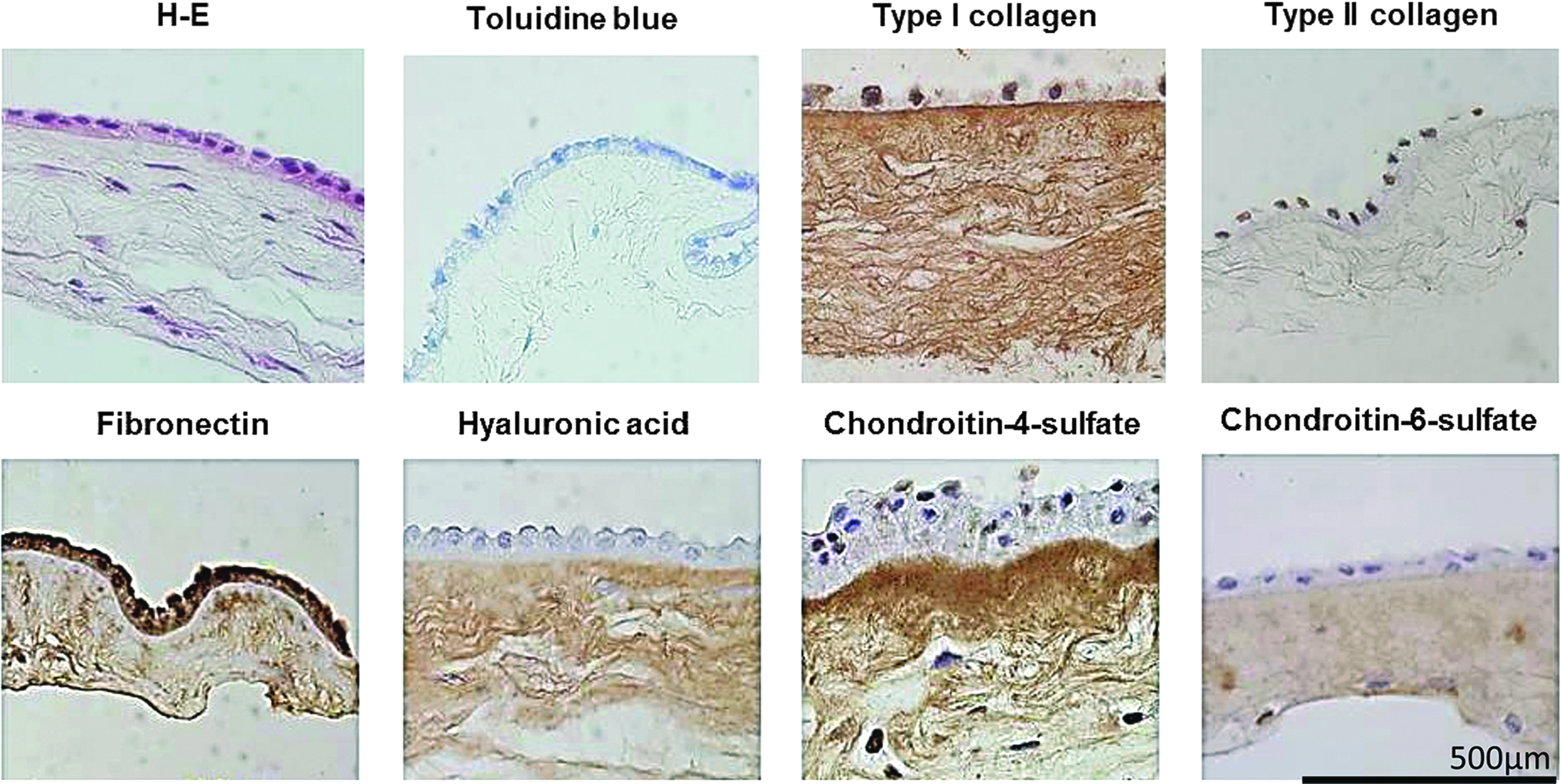
Native ECM of the amniotic membrane. Amniotic stromal tissue contained diffuse type I collagen, fibronectin, hyaluronic acid, chondroitin-4-sulfate, and chondroitin-6-sulfate. No metachromatic staining with toluidine blue or immunoreactivity with antitype II collagen was seen. Scale bar = 500 μm. ECM, extracellular matrix. Color images available online at www.liebertpub.com/tea
Morphology of PLGA and ECM-PLGA scaffolds
The stereomicroscopic appearance of PLGA and ECM-PLGA scaffolds included extensive ECM accumulation on PLGA after 14 days of culture. Additional culture until day 28 promoted more abundant ECM deposition (Fig. 2, upper panel). Scanning electron microscopy showed a porous three-dimensional structure of the plain PLGA scaffold. Moderate ECM deposition was seen on the pore surface at day 14, and further abundant ECM deposition surrounding the PLGA scaffold was seen at day 28, mostly coating the surface porous structure (Fig. 2, middle panel). After irradiation, preservation at −80°C, and thawing before use, the ECM-PLGA displayed presence of micropores of 1–20 μm in the deposited ECM (Fig. 2, lower panel).
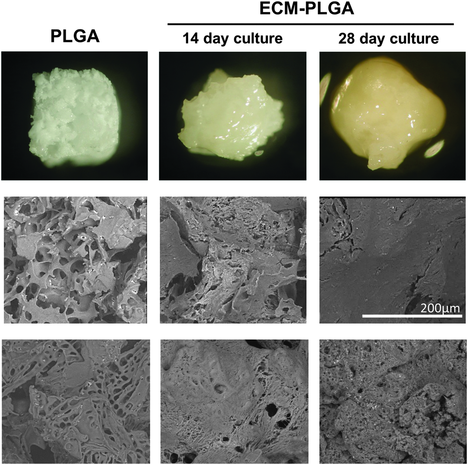
Morphology of PLGA and ECM-PLGA scaffolds. Stereomicroscopic appearance of PLGA and ECM-PLGA scaffolds (upper panel). Scanning electron microscopic images of the PLGA and ECM-PLGA scaffolds (middle panel, preirradiation; lower panel, after irradiation, freezing, and thawing before use). Scale bar = 200 μm. PLGA, polylactic-co-glycolic acid. Color images available online at www.liebertpub.com/tea
Histological analysis of the ECM-PLGA scaffold showed accumulation of HAM-derived ECM within the PLGA scaffold as well as on its surface. Toluidine blue staining showed little metachromasia in the ECM-PLGA scaffold. Immunohistochemical staining revealed that the ECM-PLGA scaffold was positive for type I collagen, fibronectin, hyaluronic acid, and chondroitin-4-sulfate (Fig. 3). A low level staining for chondroitin-6-sulfate was also present. These results indicated that the deposited ECM molecules in the ECM-PLGA scaffold mimic the ECM present in the native amniotic membrane.

Histological and immunohistochemical examination of the ECM-PLGA scaffold. Accumulation of HAM-derived ECM was apparent around and within the PLGA scaffold. Toluidine blue staining showed little metachromasia. Immunohistochemical stainings for type I collagen, fibronectin, hyaluronic acid, chondroitin-4-sulfate, and chondroitin-6-sulfate were positive. Scale bar = 500 μm. HAM, human amniotic mesenchymal cell. Color images available online at www.liebertpub.com/tea
ECM-PLGA is a suitable scaffold for MSC attachment and proliferation
A MTT assay performed at day 1 indicated that a significantly higher number of MSCs attached to the ECM-PLGA scaffold than to the plain PLGA scaffold (Fig. 4a). During culture of the MSCs with the scaffolds for 7 and 14 days, a higher MTT value was detected in the ECM-PLGA scaffold than in the plain PLGA scaffold, indicating better cell attachment and subsequent proliferation with HAM-derived ECM coating of the PLGA scaffold (Fig. 4b).
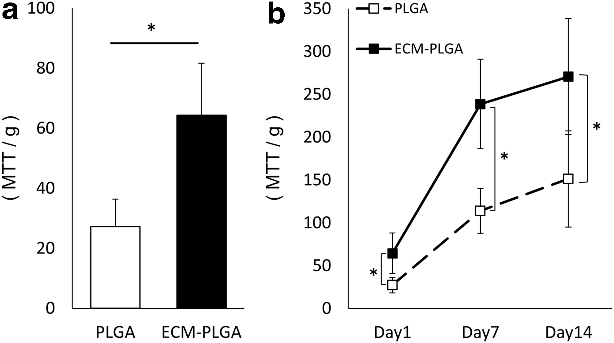
MSC attachment and proliferation on the PLGA and the ECM-PLGA scaffolds.
ECM-PLGA scaffolds and MSC differentiation
To examine the potential of MSC differentiation in vitro in the cell-free scaffold, exogenous MSCs were inoculated and cultured on the ECM-PLGA scaffold in chondrogenic medium. During the 3-week MSCs culture, the mRNA expression of Sox9, Col11a2, Col2a1, and Col10a1 gradually increased. Col2a1 expression was significantly higher in MSCs cultured on the ECM-PLGA scaffold than on the plain PLGA scaffold (Fig. 5). Expression of aggrecan, however, remained at relatively low level regardless of scaffold types. Expression of osteopontin, Pparγ, and Col1a1 remained at low level and no apparent difference was observed between the ECM-PLGA scaffold and the plain PLGA scaffold. These results may suggest that the MSCs showed differentiation toward chondrogenesis to a certain level in vitro and the ECM-PLGA scaffold may be a better scaffold than the plain PLGA scaffold to be used for cartilage repair.
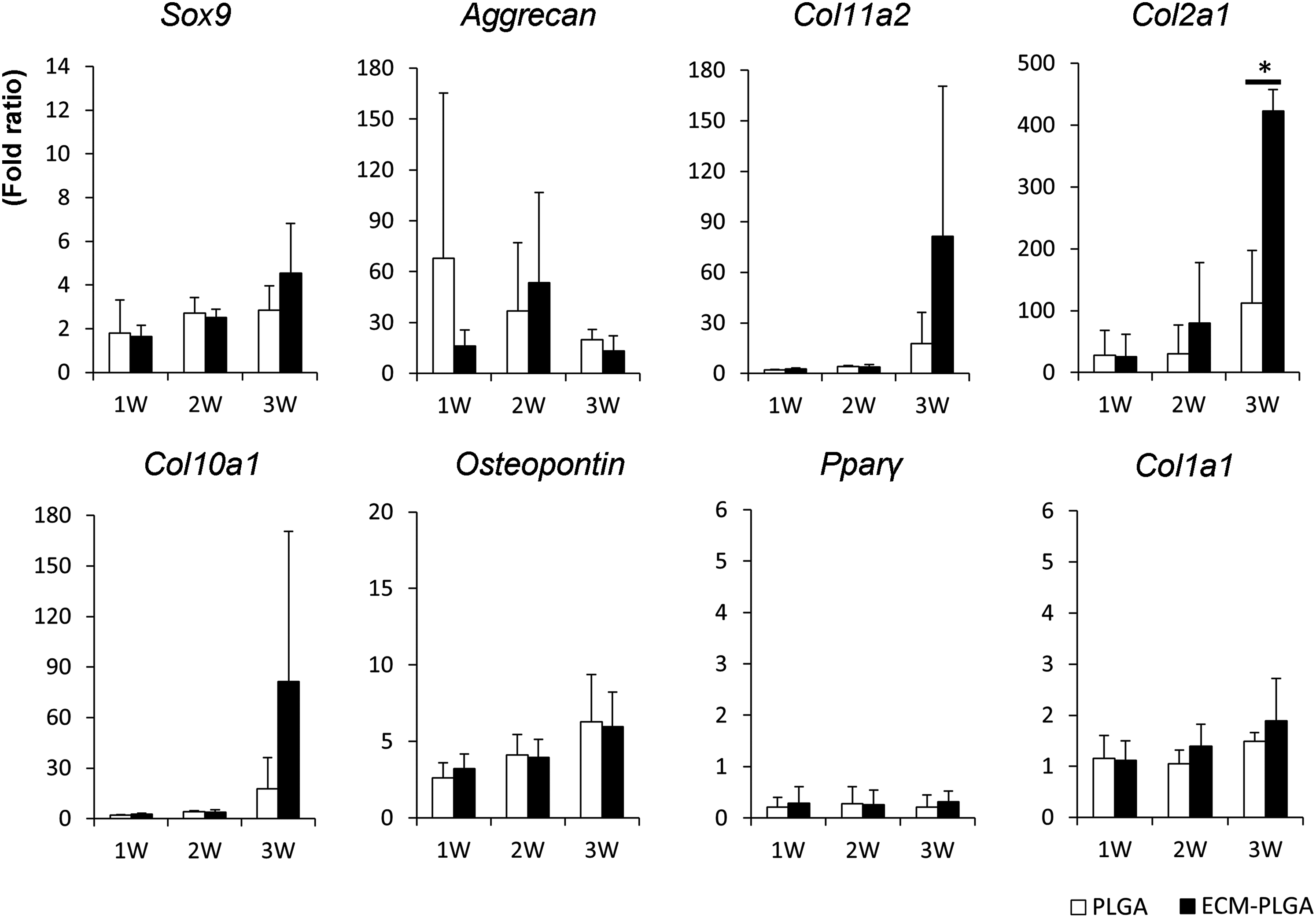
Quantitative RT-PCR analysis of MSC differentiation on PLGA and ECM-PLGA scaffolds. Several chondrogenic genes increased during 3 weeks in chondrogenic culture. Nonchondrogenic genes remained relatively low. The mRNA expression of type II collagen was significantly higher in the ECM-PLGA scaffold than in the plain PLGA scaffold at 3 weeks (*p < 0.01). RT-PCR, reverse transcription–polymerase chain reaction.
Repair of osteochondral defects with implantation of the ECM-PLGA scaffold
Representative images from each group are shown in Figure 6a and Supplementary Figure S1. In the empty (nonimplanted) control group, the defect was filled with a newly formed fibrous tissue and metachromatic tissue in the deeper part of the defect at 4 weeks. The deeper zone was then replaced with regenerated subchondral bone by 12 weeks. The upper part of the defect infrequently showed faint metachromatic staining at this stage and became fibrous tissue by 24 weeks. In the plain PLGA implanted group, the defect was filled with a mixture of PLGA scaffold and regenerated tissue at 4 weeks. Subchondral restoration seemed to occur somewhat slowly by 12 weeks with some remaining scaffold and the upper part of the defect showed slight metachromatic staining. At 24 weeks, however, the fibrous surface layer became predominant with reconstituted subchondral structure. In the ECM-PLGA implanted group, the defect was filled with the scaffold and regenerated tissue at 4 weeks. The subchondral reconstitution again seemed to be gradual with some remaining scaffold, but an area of apparent metachromatic staining was observed within the surface zone at 12 weeks. At 24 weeks, mature hyaline cartilaginous repair of the surface zone and subchondral reconstitution was achieved and maintained (Fig. 6a).
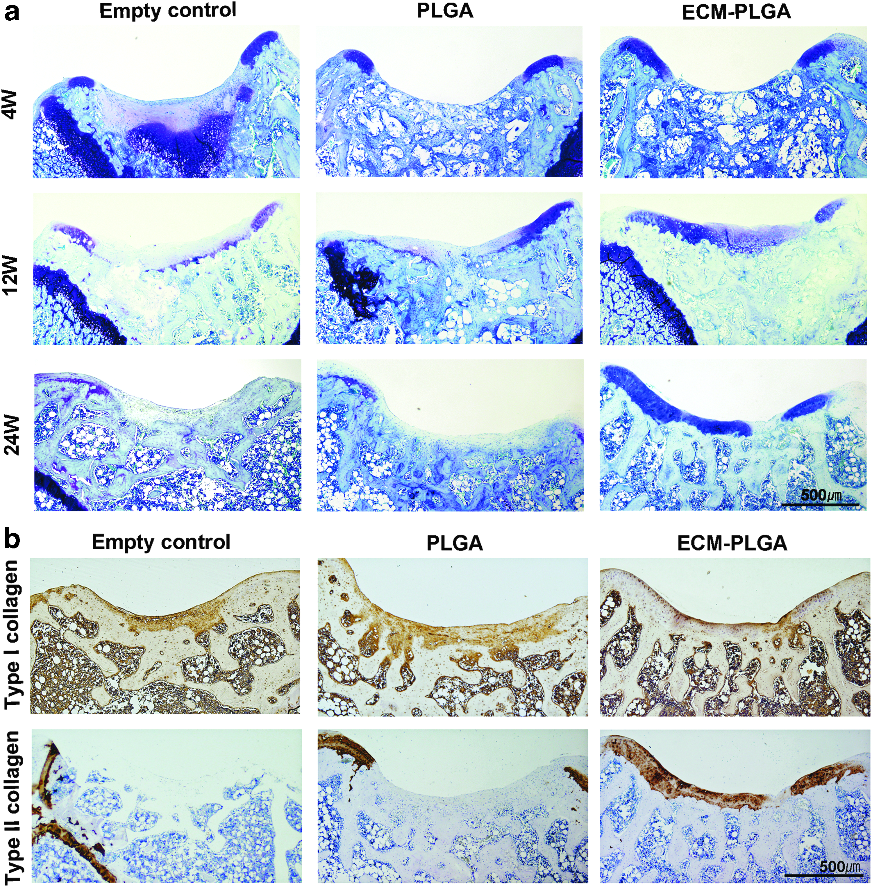
Representative histological sections of osteochondral tissue formation.
Immunohistochemistry indicated that regenerated tissue after ECM-PLGA implantation was stained intensely for type II collagen, again supporting the hyaline cartilage nature of the surface zone at 24 weeks (Fig. 6b). On the contrary, staining for type I collagen was predominant in the surface layer of empty control and PLGA-implanted groups.
The total scores of the three groups varied with no significant difference in the total scores among the groups at 4 and 12 weeks (Fig. 7). The score of subcategories suggested that the PLGA group showed inferior defect filling and subchondral reconstitution at 4 weeks (Supplementary Figure S1 and Supplementary Table S2), whereas the ECM-PLGA group showed better surface regularity. From 12 to 24 weeks, the total score of the empty control group tended to worsen (i.e., increase score) and that of the PLGA group was maintained. In contrast, the ECM-PLGA group showed gradual improvement of the score and the total score was significantly better than that of the empty control group at 24 weeks. The inter-observer reliability value calculated for histological scores by Spearman's rank coefficient was 0.92 (Z value 6.37, 95% confidence interval >1.96).
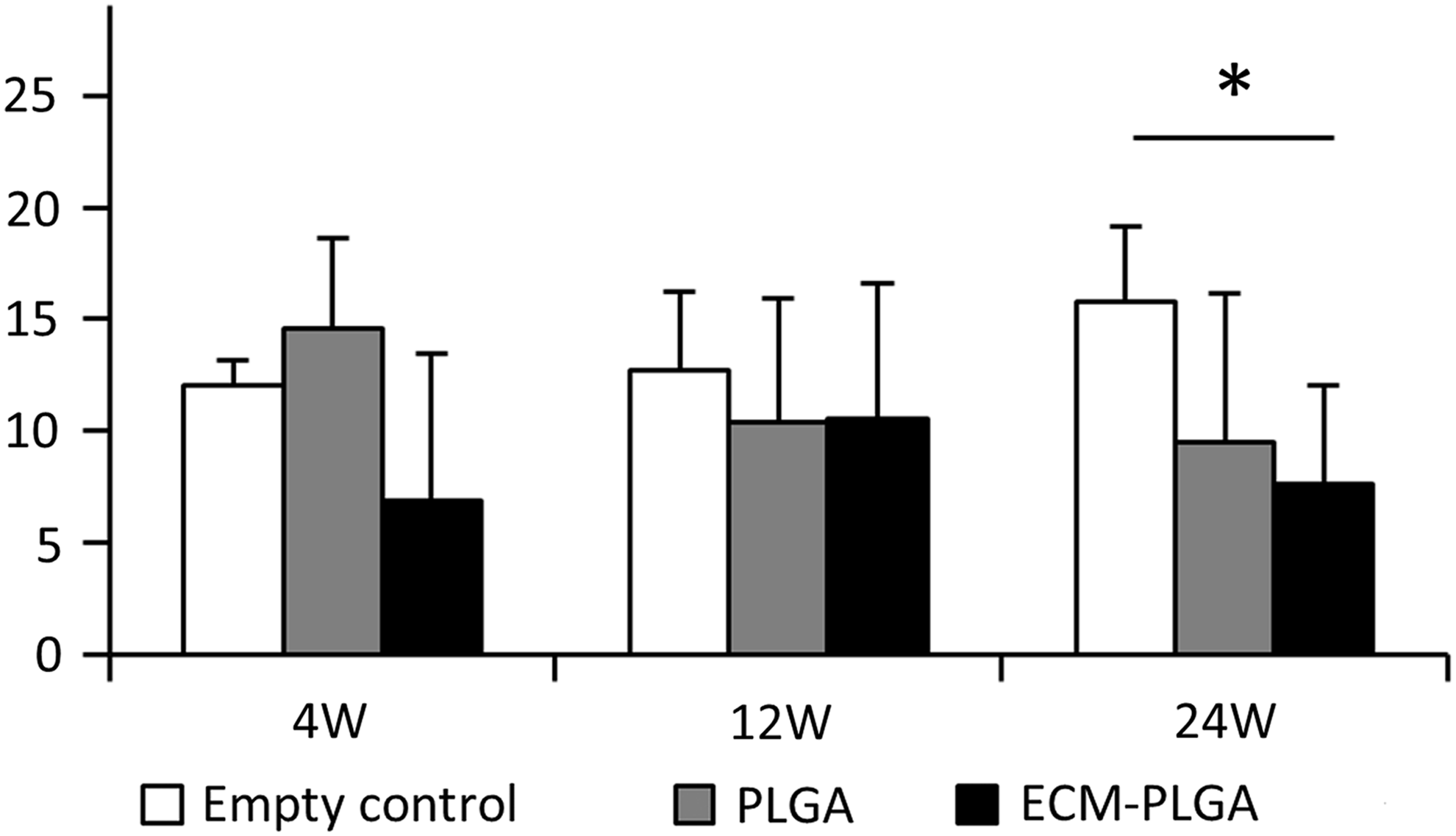
Histological grading scale (total score) for cartilage repair. Data are shown as average scores and error bars representing standard deviation (*p < 0.05).
Discussion
This study demonstrated that a cell-free scaffold composed of HAM-derived ECM and PLGA provided a suitable in vitro environment for the attachment and growth of MSCs. In addition, after implantation into an osteochondral defect in the rat knee, the cell-free ECM-PLGA scaffold seemed to support the differentiation of in situ endogenous cells and promoted regeneration of hyaline-like cartilage.
Various attempts have been made to repair articular cartilage using cell-free scaffold implantation. Among these, implantation of a synthetic polyglycolic acid scaffold soaked in hyaluronan and serum was reported to repair full-thickness cartilage defects in sheep. 9 Similarly, a polyglycolic acid and hyaluronan scaffold was used to cover human chondral defects, and when combined with microfracturing or drilling, the technique was reported to improve the quality of repaired cartilage.33,34 PLGA is another well-known synthetic biodegradable polymer, and a three-dimensional porous scaffold composed of PLGA in combination with microfracture promotes regeneration of hyaline-like cartilage. 35 These observations suggest that implantation of cell-free synthetic scaffolds may be useful for cartilage repair. In addition, the results of implantation of cell-free scaffolds may not be inferior to those with cell transplantation. 12
When implanted without cells, a prerequisite for a synthetic scaffold is the ability to facilitate recruitment and ingrowth of endogenous cells in situ. Plain PLGA scaffolds, for example, are biologically inert and have a hydrophobic surface and low potential for cell adhesion and tissue regeneration. To solve this limitation, we used HAM-derived ECM to modulate the biological property of the PLGA scaffold and developed a hybrid scaffold of ECM and PLGA that aimed to provide a better environment for endogenous MSCs to migrate, adhere, and proliferate.
The HAM-derived ECM used in this study is similar or identical to that of amniotic membrane composed of types I, III, and VI collagens, fibronectin, hyaluronic acid, chondroitin-4-sulfate, chondroitin-6-sulfate, and other molecules.36,37 Therefore, the ECM of the current ECM-PLGA scaffold is a mixture of various matrix macromolecules. Among the ECM components, type I collagen plays a structural role and provides a suitable environment for cell proliferation, which is needed for tissue repair. 11 Type I collagen can also be used to modify synthetic biodegradable polymers. 38 Fibronectin, another major component of the ECM, plays not only a primary role in cell–matrix adhesion but also mediates various cellular functions such as cell proliferation and differentiation. 39 Hyaluronic acid and glycosaminoglycans also support differentiation and chondrogenesis.7,10 Thus, the role of ECM molecules is far greater than simply providing physical support for the cells. Because each ECM component plays different roles in cell adhesion, growth, and regulation of physiological signals, attributing the observed effect of the ECM-PLGA scaffold in this study to a single component of the HAM-derived ECM is difficult. However, a suitable environment for the MSCs is likely one that closely mimics the natural mesenchymal environment of multiple ECM components. We hypothesize that a mixture of ECM components with a composition that mimics that of the amniotic ECM provided a superior environment for the cells to mediate regeneration during the repair of osteochondral defects.
In this study, we demonstrated that implantation of ECM-PLGA scaffolds into osteochondral defects promoted regeneration of hyaline-like cartilage. To our knowledge, no study has demonstrated cartilage repair by implanting a human ECM-modulated synthetic scaffold. However, we noted that the repair occurred gradually by 24 weeks and the scaffold degradation was relatively slow during this period. Undegraded scaffold was still observed in the subchondral region at 4 and 12 weeks. It is possible that the slow biodegradability of the PLGA may have retarded new tissue regeneration that occurs concurrently with scaffold degradation. If so, modulation of the PLGA properties and biodegradability may enhance the cartilage repair. We also speculate that slow degradation of the PLGA/ECM-PLGA scaffolds and subchondral reconstitution, as observed in this study, may rather favorably affect the regenerated surface cartilage to allow remodeling toward a more mature hyaline-like structure. Further investigation of the time course of biodegradation and remodeling of cartilage and subchondral bone is warranted, together with long-term follow-up.
Some biomimetic cell-free scaffolds synthesized from natural matrices are also currently in use to treat cartilage defects. A three-dimensional chitosan-based scaffold has been reported to improve the clinical symptoms and magnetic resonance imaging (MRI) of the repaired tissue in comparison with microfracture at 1 year after operation. 40 Kon et al. reported an improvement of clinical and MRI scores after osteochondral defect treatment with nanocomposite trilayered scaffold composed of type I collagen and hydroxyapatite at 5 year follow-up. 41 These results further support the usefulness of various cell-free scaffolds in clinical use. Nevertheless, the MRI images also showed that the repaired tissue was not a mature hyaline cartilage in both studies, suggesting the difficulty of the host cells to fully grow and differentiate into chondrogenic cells in these scaffolds. We believe that further development or modification of the biological property of the scaffolds as attempted in this study should help to overcome this difficulty toward hyaline repair.
There are some limitations in this study. First, the number of animals for the in vivo experiment was small and the superiority of the ECM-PLGA scaffold during early stage of cartilage repair was not demonstrated. Future long-term as well as short-term studies with larger number of animals will be necessary. Second, the current in vivo results were derived from a rat model. We used nude rats because xenoimplantation of human ECM-containing materials may be immunogenic. At the next stage, the potential of the ECM-PLGA scaffold should be confirmed in large and immunologically close animal models. Third, the ECM used in this study was from one HAM cell-line. Therefore, ECM from other HAM cell-lines may show slightly different biological properties.
Conclusion
We developed a HAM-derived ECM-coated PLGA scaffold with suitable properties for attachment, proliferation, and differentiation of MSCs. The implantation of a cell-free ECM-PLGA scaffold into osteochondral defects promoted ingrowth of endogenous cells and resulted in good cartilage repair. Because no autologous cells are needed, ECM-PLGA scaffolds may be “ready-made” biomaterials for use in one-step cartilage repair therapy.
Footnotes
Acknowledgments
We thank Etsuko Furuichi for sectioning the paraffin-embedded cartilage tissue. This work was supported by JSPS KAKENHI Grant Number 24390350.
Disclosure Statement
The authors T.N., T.Y., M.O., C.S., M.N., and T.K. have Japanese unexamined patent application publication No. 2013-248221.
References
Supplementary Material
Please find the following supplemental material available below.
For Open Access articles published under a Creative Commons License, all supplemental material carries the same license as the article it is associated with.
For non-Open Access articles published, all supplemental material carries a non-exclusive license, and permission requests for re-use of supplemental material or any part of supplemental material shall be sent directly to the copyright owner as specified in the copyright notice associated with the article.