
Abstract
Aqueous tear-deficient dry eye disease is a multifactorial chronic disorder, in which the lacrimal gland fails to produce enough tears to maintain a healthy ocular surface. Some severe cases may develop corneal damage and significant vision loss. Treatment primarily involves palliation using ocular surface lubricants, but can only provide temporary relief. Construction of a bioengineered lacrimal gland having functional secretory epithelial cells is a potentially promising option for providing long-term relief to severe dry eye patients. Using sphere-forming culture techniques, we cultured adult rabbit lacrimal gland progenitor cells and prepared a lacrimal gland scaffold by decellularization. When progenitor cells were seeded onto the decellularized scaffold, they formed duct- and acinar-like structures in the three-dimensional culture system. Lacrimal gland epithelial cells showed good cell viability, cell differentiation, and secretory function in decellularized lacrimal gland matrix, as indicated by morphology, immunostaining, and β-hexosaminidase secretion assay. This study demonstrated the potential suitability of utilizing tissue-specific progenitor cells and a tissue-derived bioscaffold for lacrimal gland restoration.
Introduction
T
Aqueous tear-deficient dry eye is a multifactorial chronic disorder, in which the lacrimal gland fails to produce enough tears to maintain a healthy ocular surface.1,6 Current treatment modalities primarily involve palliation using ocular surface lubricants or pharmacological stimulation of tear secretion. However, these approaches only provide temporary relief of the symptoms and do nothing to treat the degenerated lacrimal gland. Once the lacrimal gland is atrophied or injured, the condition often becomes irreversible and recovery of function is rare. In a few cases, lacrimal gland tissues regenerate and their functions may be restored; however, in severe cases, especially in those with permanent damage to the lacrimal gland, there is a need to replace the gland and restore its functionality using appropriate cell therapy. 7 Allogeneic lacrimal gland transplants may help restore tear film production and function, but because of donor shortages and lack of immune suppression to prevent transplant rejection, there is a need to explore alternative solutions. 8
Construction of a bioengineered lacrimal gland possessing functional secretory epithelial cells is a potentially promising option to investigate lacrimal gland biological behavior and provide long-term relief to severe dry eye patients. It is therefore important to find suitable cell sources as well as develop and evaluate a functionally competent in vitro three-dimensional (3D) cell culture system. Biological scaffold materials derived from the extracellular matrix (ECM) of intact mammalian tissues have been successfully used in a variety of tissue engineering/regenerative medicine applications, both in preclinical animal studies and clinical situations. The ECM is composed of a complex mixture of functional and structural molecules that affect a variety of cell activities, including cell migration, proliferation, and differentiation. 9 Decellularization provides a unique opportunity for engineering 3D scaffolds that structurally recapitulate the complex architecture of the original tissue/organs. The cells seeded on such biological scaffolds could potentially produce a functional tissue with a direct connection to the patient's vasculature, forgoing many of the problems associated with tissue and organ transplantation.
In the current study, a method to decellularize rabbit lacrimal gland was developed and adult rabbit lacrimal gland epithelial cells, including a subpopulation of lacrimal gland progenitor cells, were introduced into sphere-forming culture. The lacrimal gland cells were thereafter seeded onto collagen gel and decellularized scaffolds to investigate cell growth and differentiation, and to evaluate the potential functionality of these reseeded cells.
Materials and Methods
Tissue preparation and cell cultures
Female adult New Zealand White (NZW) rabbits (Robinson Service, Inc., Mocksville, NC) weighing 2–3 kg were used in accordance with the guidelines in the ARVO Statement for the Use of Animals in Ophthalmic and Vision Research and with the approval of the Animal Care and Use Committee at Johns Hopkins University. The rabbits were first anesthetized with xylazine and ketamine and then euthanized with Euthasol® (Virbac Corp., Ft. Worth, TX). After removal, the lacrimal glands were dissociated by mincing and cocktail enzyme digestion, as described elsewhere 10 with the following modifications.
Briefly, the glands were cut into small pieces, washed, and enzymatically digested by collagenase (350 U/mL, 17018-029; Invitrogen, Thermo Fisher Scientific, Waltham, MA), hyaluronidase (300 U/mL, LS02592; Worthington Biochemical Corp., Lakewood, NJ), and DNAse (40,000 U/mL, 260913; EMD Millipore, Billerica, MA) at 37°C for 30 min with vigorous shaking. The cell digest was then centrifuged and washed twice, before filtering through a 70 μm mesh (BD Biosciences, Franklin Lakes, NJ). The retained cell suspension was centrifuged through a Ficoll (Sigma-Aldrich, St. Louis, MO) gradient, and the obtained cell subpopulation was washed, centrifuged twice, and resuspended in the culture medium.
Digested cells were plated onto low attachment dishes (Sigma-Aldrich) and cultured in Hepato-STIM Culture Medium (BD Biosciences) supplemented with epidermal growth factor (EGF, 5 ng/mL),
Tissue decellularization
To avoid sacrificing live animals for scaffold preparation, NZW rabbit lacrimal glands, purchased from Pel-Freez Biologicals (Rogers, AR), were decellularized based on our laboratory's protocols. Briefly, NZW rabbit lacrimal glands excised from orbits were treated with 1% sodium dodecyl sulfate (SDS) or 1% Triton X-100 for 36 h followed by DNase 1 (2000 U/mL) treatment for 2 h, then stored in phosphate-buffered saline (PBS) with 1% PS antibiotics. All incubation steps were performed at 4°C under continuous agitation. The tissues were thoroughly washed with PBS between each step. Histological analysis was performed to evaluate the efficiency of decellularization and to determine the ECM components that remained after processing. 4′,6-Diamino-2-phenylindole (DAPI) staining was performed to confirm the removal of cell nuclei. Scanning electron microscopy (SEM) was performed to determine the remaining collagen fiber status.
Scanning electron microscopy
Samples were prepared for SEM by fixation in 3.0% formaldehyde/1.5% glutaraldehyde in 0.1 M sodium cacodylate buffer with 2.5% sucrose for 1 h. Samples were then postfixed with 1% osmium tetroxide for 30 min before dehydration with graded ethanol solutions. Samples were dried using carbon dioxide critical point drying, followed by sputter coating with platinum, and images were taken with an FEI Quanta 200 scanning electron microscope (Hillsboro, OR).
Three-dimensional culture with lacrimal gland sphere cells
Rabbit lacrimal gland cells formed spheres in passage 0 (P0) from day 2, and then the spheres were dissociated by TrypLE Express treatment on day 7. To test the differentiation potential of the sphere-derived cells, 2 × 10⁶ of the dissociated cells in passage 1 (P1) were embedded in type I collagen gel (Cosmo Bio, Tokyo, Japan) according to the manufacturer's instructions and maintained in serum-free Hepato-STIM with the CT medium for 4 weeks. The dissociated sphere cells (2 × 10⁶) in P1 were seeded onto the decellularized lacrimal gland scaffold (1 cm diameter) that was precoated with Matrigel for 3D culture as well. The lacrimal constructs were maintained in the Peter's complete medium (PCM) or Hepato-STIM medium for up to 30 days. PCM was prepared by mixing Ham's F-12 (Thermo Fisher Scientific, Agawam, MA) and DMEM with 1.5 g/L glucose (Thermo Fisher Scientific) at a 1:1 ratio and adding the other reagents to the following final concentrations: penicillin 100 U/mL, streptomycin 0.1 mg/mL, linoleic acid 0.3 μM, n-butyric acid 2 mM, transferrin 5 μg/mL, insulin 5 μg/mL, sodium selenite 30 nM, hydrocortisone 5 nM, laminin 4 μg/mL, carbachol 0.1 μM/mL, and
Histology and immunohistochemistry
Samples of isolated adult rabbit lacrimal glands, the decellularized lacrimal gland tissues, and cultured recellularized constructs (n = 3 for each time point for each group) were embedded in the optimal cutting temperature (OCT; Sakura Finetek USA, Torrance, CA) compound and frozen in liquid nitrogen. Frozen sections were stained with hematoxylin–eosin (H&E) for histologic examination. The lacrimal gland sphere cells were cultured in low attachment dishes and then embedded in OCT (∼150 spheres per sample prepared). The frozen sections were fixed with ice-cold acetone for immunofluorescence (IF) staining. After background staining was blocked with 10% normal donkey serum, the cells were treated with the following monoclonal primary antibodies: anti-pan-cytokeratin (PCK) antibody (Sigma-Aldrich), anti-α-smooth muscle actin (α-SMA) antibody (Abcam, Cambridge, MA), anti-c-kit antibody (Santa Cruz Biotechnology, Santa Cruz, CA), anti-nestin antibody (Santa Cruz), anti-ABCG2 antibody (Santa Cruz), anti-ΔNp63 antibody (Santa Cruz), anti-cytokeratin 4 antibody (K4; Sigma-Aldrich), anti-cytokeratin 14 antibody (K14; Santa Cruz), anti-aquaporin-5 antibody (AQP5; Abcam), and anti-lactoferrin antibody (Abcam). The cells were then treated with the fluorescent dyes Alexa Fluor® (AF) 488- or AF568-conjugated secondary antibodies (Life Technologies, Thermo Fisher Scientific). Cell nuclei were counterstained with DAPI (1 μg/mL; Sigma-Aldrich). IF images were taken with a Zeiss LSM 510 Confocal Microscope (Carl Zeiss, Jena, Germany). Positive-staining spheres were manually counted from a minimum of four fields of view (20× objective) per slide from three slides per group.
Live and dead assay
The live/dead assays were performed at days 2, 7, 14, and 30 to assess cell viability using a LIVE/DEAD Cell Viability Kit (Life Technologies, Thermo Fisher Scientific), a two-color fluorescent assay based on differential permeability of live and dead cells, according to the manufacturer's protocol. Briefly, the lacrimal gland constructs were soaked in the medium with live/dead reagents, calcein AM (green fluorescent), and ethidium homodimer-1 (red fluorescent) stain for 40 min, and then the specimens were washed thrice in PBS for 1 min. The lacrimal gland constructs in the PCM or Hepato-STIM media were examined in parallel with a Nikon Eclipse TE200 fluorescence microscope (Nikon, Inc., Melville, NY) immediately thereafter.
β-Hexosaminidase secretion assay
The β-hexosaminidase secretion assay was performed to test the functionality of the acinar cells in the lacrimal gland constructs at days 2, 7, 10, 14, 17, and 30. Stimulation of the cultured cells by carbachol results in a significant increase of β-hexosaminidase release over baseline, which suggests that healthy acinar cells have carbachol-dependent β-hexosaminidase secretion in vitro.
11
At least three wells were set per condition. Before stimulation, culture samples were carefully rinsed with DMEM three times, filled with 500 μL DMEM, and then incubated at 37°C with 5% CO2 for 2 h. After removing a basal sample, carbachol was added to a final concentration of 100 μM in stimulation wells that were then incubated for 60 min. After incubation, the supernatants were collected and centrifuged for 10 min and stored frozen at −80°C until use. For measurement of β-hexosaminidase activity, 4-methylumbelliferyl N-acetyl-β-
RNA isolation and real-time polymerase chain reaction
RNA was isolated from freshly harvested lacrimal gland cells, as well as from lacrimal tissue constructs on days 2, 7, and 14 using the RNeasy Plus Mini Kit (Qiagen) and reverse transcribed to cDNA using the iScript™ cDNA Synthesis Kit (Bio-Rad, Hercules, CA). Real-time polymerase chain reaction (RT-PCR) was performed with a StepOne RT-PCR detection system (Applied Biosystems, Thermo Fisher Scientific) using a Power SYBR® Green PCR Master Mix (Life Technologies, Thermo Fisher Scientific), according to the manufacturer's instructions, with designed primers (HEXB, 5′-CAACAAAGTTTGGGGAGCAT-3′, and 5′-CCATGGCATCCAGAGTTCTT-3′). The amplification program included an initial denaturation step at 95°C for 10 min, followed by denaturation at 95°C for 10 s, and annealing and extension at 60°C for 30 s, for 40 cycles. SYBR Green fluorescence was measured after each extension step, and the specificity of amplification was evaluated by the melting curve analysis. The results of the relative quantitative RT-PCR were analyzed by the comparative Ct method and normalized to β-actin as an internal control.
All experiments were performed with at least three independent biological replicates.
Results
Stepwise decellularized lacrimal gland maintained the scaffold structure
After treatment with 1% SDS or 1% Triton X-100, the decellularized lacrimal glands became pale and semitranslucent, yet the lobular and ductal structure remained clear (Fig. 1A). H&E staining demonstrated complete removal of cell nuclei with preserved ECM. In addition, DAPI-positive staining was absent in SDS-treated samples and there was very little DAPI-positive staining in Triton-treated samples (Fig. 1B). SEM showed that the collagen fiber bundles were well maintained in decellularized samples (both SDS- or Triton X-100-treated) compared to normal lacrimal gland tissues (Fig. 2).

Effect of decellularization reagent on rabbit lacrimal gland. The gross appearance and H&E staining
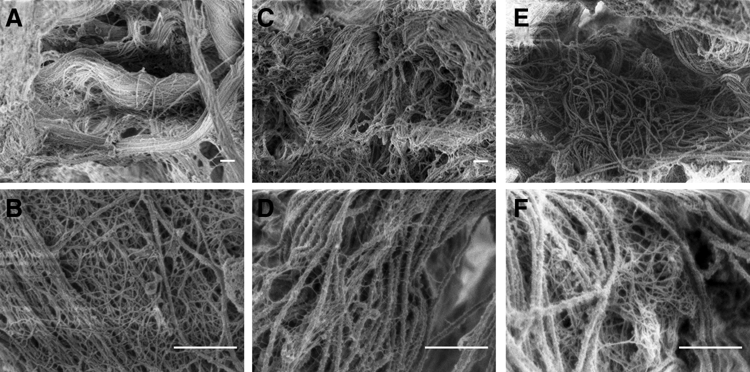
Scanning electron microscopy images of processed lacrimal gland tissue. The collagen fiber status in the decellularized scaffolds
Sphere cultures of adult rabbit lacrimal gland epithelial cells
Spheres appeared in both DF12 and Hepato-STIM media on day 2. Figure 3A–D shows time-dependent changes in the density and diameter of lacrimal gland cell spheres from day 3 to 7. Moreover, after subculture on day 7, secondary spheres were found on day 10 (Fig. 3E, F). While grown from the same seeding density of single cells, the higher density of spheres on day 3 and larger diameter of the primary and secondary spheres in DF12 medium could suggest higher cell proliferative capacity in this medium.

Cultured lacrimal sphere cells in serum-free media at P0 and P1. There was a time-dependent increase of lacrimal gland spheres at P0 in DMEM/F12 media
The day 3 and 7 sphere cells were prepared for frozen slides. Characterization of these spheres was performed by immunostaining. Figure 4 shows representative positive staining. To test the lineage of the sphere cells, detection of PCK (for epithelial cells), α-SMA (for myoepithelium), and K4 (for ductal epithelium) was performed, as well as detection of ocular surface stem/progenitor cell markers K14, nestin, ABCG2, and ΔNp63. Staining results were similar in spheres grown in DF12 (Fig. 4) or Hepato-STIM (data not shown) media. K4, a ductal epithelial marker, demonstrated positive staining in the duct of normal lacrimal gland (Fig. 4N) and was also detected in around 70% of examined day 3 spheres (Fig. 4B). Nestin-positive staining pattern (Fig. 4O) in the normal lacrimal gland was similar to K4 staining, and nestin-positive spheres were found on days 3 (∼80%) and 7 (∼50%) (Fig. 4E, J). K14, a marker for the progenitor basal layer of the ocular surface and skin, has been found in the duct systems of mammary and salivary glands.13–15 K14-positive staining was found in some duct locations in normal lacrimal gland (Fig. 4M) and was also detected in day 3 spheres (∼20%). Most spheres were also PCK (Fig. 4F, K, over 90%) and α-SMA (Fig. 4G, L, ∼80%) positive on days 3 and 7. ABCG2-positive (Fig. 4C, I) and ΔNp63-positive (Fig. 4A, H) staining was also found on days 3 and 7 in ∼50% of spheres.
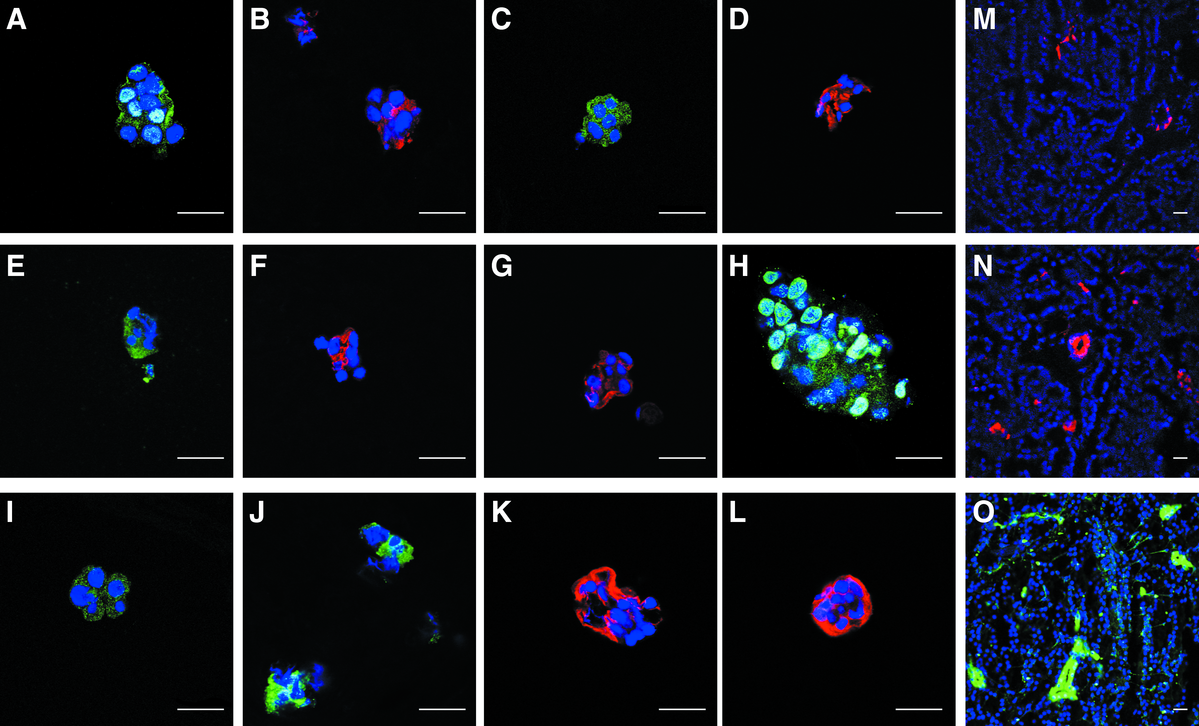
Characterization of rabbit lacrimal sphere cells. Most spheres on day 3
Three-dimensional culture of rabbit lacrimal gland sphere cells
To test the differentiation potential of the sphere-derived cells in 3D culture, subcultured and dispersed sphere cells in P1 were placed in collagen I on day 7. Collagen I gel was used as the ECM for lacrimal gland germs and salispheres, which both successfully formed 3D gland structures. It is also suitable for continuous direct observation of morphology due to its transparency. The sphere-derived cells survived in 3D type I collagen and gradually transformed into duct- (Fig. 5A–C) and acinar-like (Fig. 5D) structures from day 3 to 4 weeks.
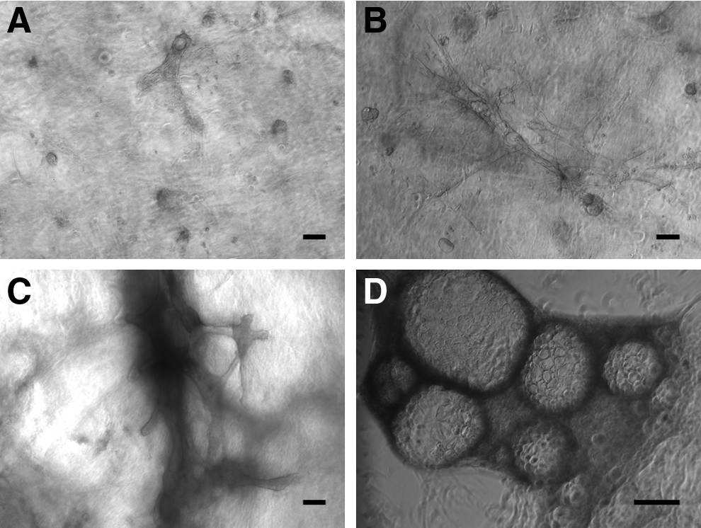
Cultured rabbit sphere cells gradually formed duct- and acinar-like structures in collagen gel. After 3 days, formation of branching structures was evident
When dispersed P1 sphere cells were cultured on the decellularized scaffold, the majority of the isolated lacrimal cells survived for at least 7 days (Fig. 6A–C) in both the PCM and Hepato-STIM groups; however, cell viability decreased dramatically at day 14 (Fig. 6D–F) in the PCM group. Cell viability in the Hepato-STIM media was well maintained through the first month of culture. The morphology of the live cells also demonstrated tubular- and acinar cell-like structures. Compared to the collagen gel culture, which already had apparent ductal structures beginning on day 5 (Fig. 5B), the cells cultured on the decellularized scaffold demonstrated round or elongated cell clusters on day 7 (Fig. 6B, C), and then gradually formed tubular structures on day 14 (Fig. 6E, F). Scattered round cyst structures representing acinar cell-like morphologies were demonstrated both in collagen gel and decellularized matrix after 4 weeks of culture. Cell viability was similar in the SDS-treated scaffold (Fig. 6) and the Triton-treated scaffold (data not shown).
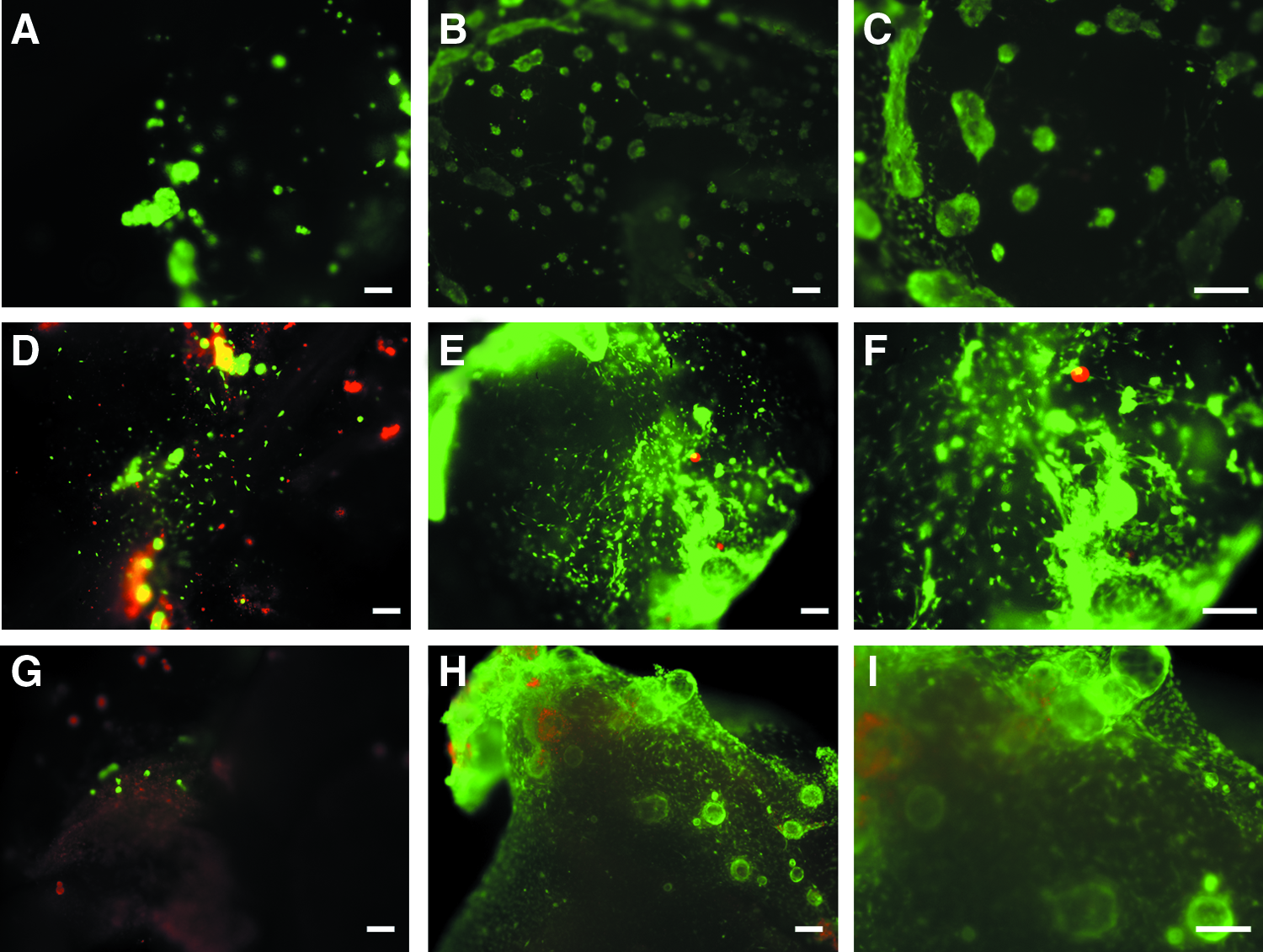
Representative photos of live/dead assay performed in lacrimal gland constructs. The Hepato-STIM group
Functional lacrimal gland cells survived on the decellularized scaffold
The mature lacrimal gland consists of three types of cells: duct cells, acinar cells, and myoepithelial cells. Since positive staining of PCK and c-kit is found in human lacrimal gland acinar and ductal cells, 6 and rabbit lacrimal gland has the most similarity to humans among the common models to study lacrimal gland function, 16 we used PCK and c-kit to identify the differentiated rabbit lacrimal gland epithelial cells and α-SMA to identify myoepithelial cells in the constructs. IF staining of the lacrimal gland constructs demonstrated the presence of the epithelial marker PCK-positive cells (Fig. 7A, B) and c-kit-positive cells (Fig. 7C–F) with tubular morphology or budding morphology. Double staining also demonstrated the presence of α-SMA-positive myoepithelial cells surrounding the c-kit-positive epithelial cells (Fig. 7C, D). AQP5, a membrane water channel protein that has an essential role in fluid secretion and is expressed on the ductal and apical membranes of acini, was present in the apical side of duct-like structures and epithelial cyst sites of the bioengineered constructs (Fig. 7A, B). The expression of lactoferrin, a protein secreted by the lacrimal gland, was found in most of the reseeded cells present in the bioengineered constructs (Fig. 7G, H). Increased β-hexosaminidase activity in the supernatant following stimulation with 100 μM carbachol (Fig. 8A) also suggested that there are differentiation and survival of lacrimal secretory epithelial cells in the lacrimal gland constructs for more than 2 weeks. There was no significant carbachol-stimulated β-hexosaminidase activity in day 30 samples (data not shown). While previous literature showed a secretory response to carbachol stimulation of acinar cells that decreased over the culture period, 17 the differentiated secretory cells in lacrimal gland constructs also lost the ability to respond to carbachol in our 1-month culture. Moreover, RT-PCR showed gradually increasing mRNA levels of the β-hexosaminidase (HEXB) gene during culture, which is consistent with the β-hexosaminidase assay results (Fig. 8B).
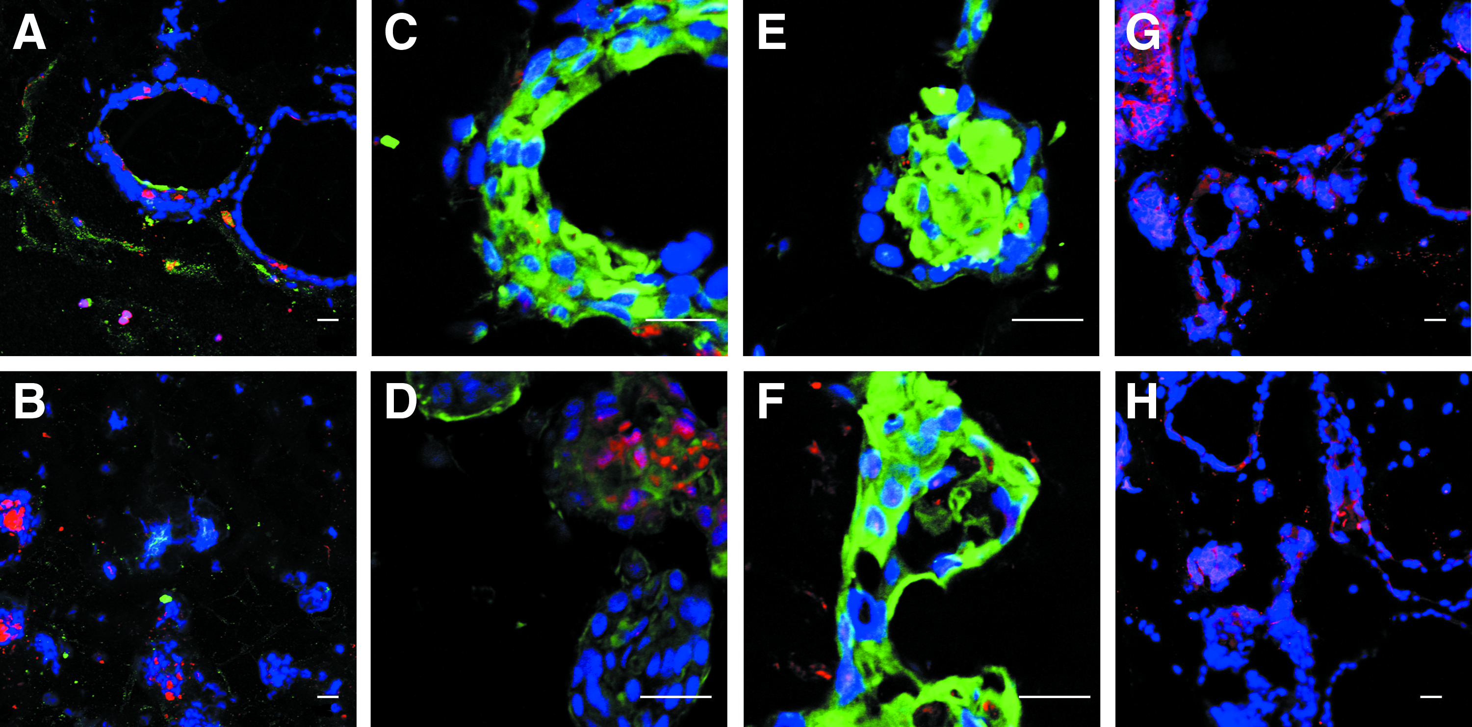
Immunofluorescence staining of secretory epithelial cells and myoepithelial cells in lacrimal gland constructs. Immunostaining of lacrimal constructs showed PCK-

The β-hexosaminidase assessment. β-Hexosaminidase activity significantly increased (asterisks indicate statistical significance, p < 0.05) in the supernatant of lacrimal construct cultures following stimulation with 100 μM carbachol on days 7, 10, 14, and 17
Discussion
Restoration of lacrimal gland function potentially could provide a curative therapy for dry eye disease, and transplantation of an artificial lacrimal gland is a potentially promising approach for severe dry eye patients. Recently, one study demonstrated that orthotopic transplantation of bioengineered lacrimal gland germs can restore the physiological functions of the lacrimal gland in vivo. 18 However, utilization of embryonic tissue-derived cells presents a problem for later translational exploration. Thus, it is necessary to identify somatic and other tissue-derived stem/progenitor cell populations for the regeneration and bioengineering of lacrimal gland.
Stem cells in adult tissues have been extensively studied because of their potentially wide-ranging clinical use. Several studies on exocrine glands, such as the pancreas, salivary, and mammary glands, have shown that stem/progenitor cells exist in these tissues and are involved in their regeneration.19–22 However, there are few reports about stem cells in the lacrimal gland.23–25 It is increasingly suspected that the lacrimal gland also has its own tissue-specific stem/progenitor cells that might generate three differentiated cell phenotypes: acinar cells, myoepithelium, and ductal epithelial cells.
Previous studies have demonstrated that, when salivary spheres generated from newborn mice were transplanted into irradiated salivary glands, secretory function increased. 14 Furthermore, single mammary stem cell transplantation has been shown to generate cells with both luminal and myoepithelial lineages, thus generating functional lobuloalveolar units during pregnancy. 19 Since the lacrimal gland could be repaired after experimental injury, 26 and due to its similarity to the salivary gland, we can hypothesize that lacrimal gland stem/progenitor cells could exist and could survive in similar culture conditions.
In this study, we report on a sphere-forming method for culturing adult rabbit lacrimal gland epithelial cells, which potentially contain lacrimal gland progenitor cells, in media containing CT. Previous reports on adult human/mouse limbal epithelial cell and mouse lacrimal gland cell cultures demonstrated that the addition of CT to the culture medium successfully selected for epithelial cells. 27 We adapted the addition of CT to the culture medium and found far fewer mesenchymal cells in contrast to culturing without CT (data not shown). We also used a sphere culture condition (DF12) similar to the one previously described for salivary glands (documented to support the growth of salivary stem cells derived from the duct cells), 28 which achieved more numerous and larger spheres compared to another serum-free medium. Lacrimal sphere cells demonstrated positive staining with progenitor cell markers in floating culture and also showed the potential to differentiate into both ductal cells and acini when cultured in collagen gel or decellularized scaffold. Lacrimal sphere-derived cells cultured in the collagen gel formed duct-like structures earlier than acini-like structures and formed more complex duct-like structures than in decellularized matrix. The 3D architecture of these sphere-derived cells, displayed during culture, indicated differentiation into various cell types and resembled the branching morphogenesis that occurs during organogenesis. The results support the hypothesis that lacrimal gland epithelial cells have unique progenitor cells and that, similar to salivary glands, the origin of the lacrimal gland progenitor cells may be in the duct cells of lacrimal glands. In addition, the survival of lacrimal secretory cells in a 3D biological scaffold was demonstrated by β-hexosaminidase assessments and the live/dead assay, plus morphologic investigation and IF staining. Therefore, the current study indicated that lacrimal gland progenitor cells exist and can be initially cultured in decellularized lacrimal gland tissue to develop secretory function. The promising results of our study warrant further investigation into the biological characteristics and behavior of lacrimal gland progenitor cells.
The cell lineage of a tissue at a specific developmental stage provides important information for characterizing tissue-specific stem/progenitor cells. During development of the lacrimal gland, a budding of the gland originates from conjunctival epithelium, which branches and differentiates into mature tissue. This suggests that the ocular surface epithelial cells and the lacrimal gland cells may have similar specific stem/progenitor cell markers. Several articles have suggested K14, ΔNp63, ABCG2, and nestin to be ocular surface stem/progenitor cell markers.29–31 In the current study, spheres formed early in the culture stage contained cells positive for these stem/progenitor markers, which suggest the existence of lacrimal gland progenitor cells in our current culture method and serum-free culture system. However, further studies are needed on the cells' multipotent potential and the ability to efficiently differentiate into three cellular phenotypes in vivo. The purification of lacrimal stem/progenitor cells by specific cell surface markers needs further exploration as well. After purification and proliferation, a greater amount of lacrimal gland progenitor cells in the ex vivo culture system will aid in bioengineering lacrimal glands.
Several models of cultured lacrimal gland cells have been established to better understand their physiology and pathophysiology.32–35 Primary cultures of rabbit lacrimal gland could proliferate on plastic, but exhibited morphological differentiation only slightly resembling what is seen in vivo.36,37 When cultured with ECM components, such as laminin and Matrigel, isolated rabbit lacrimal gland acinar cells remained more histotypic. 38 The current study compared different culture media and reported that the primary culture of rabbit lacrimal gland cells could not only proliferate in the defined media without serum (better in DF12 medium) in spheres but also transform further into the duct- and acinar-like morphologies in 3D cultures (in the Hepato-STIM medium).
The aqueous tear layer is produced by the lacrimal gland and contains water and various tear proteins. To identify the functional secretory epithelial cells in the cultures, IF staining of lactoferrin, AQP5, and β-hexosaminidase assessment was performed. Lactoferrin is the main tear protein produced by the lacrimal gland and has an essential role in tear function, including tear stability, moisturization, wound healing, and antibiotic effects. Our results demonstrated positive staining of lactoferrin in the 1-month lacrimal gland constructs, indicating differentiation into lacrimal secretory acini of the reseeded cells. The cells had correct cell polarity as demonstrated by the expression of AQP5 in the apical surface of duct-like structures in the lacrimal construct. Moreover, the protein assay and RT-PCR results suggested the presence of β-hexosaminidase-secreting lacrimal epithelial cells in the 3D constructs.
Decellularized tissues and organs have been successfully used in a variety of tissue engineering/regenerative medicine applications. Other than using defined ECM as collagen gel, decellularization also provides a unique opportunity for engineering 3D scaffolds that structurally recapitulate the complex architecture of the original tissue/organs. Decellularization methods vary as widely as the tissues and organs of interest used. The present study compared two methods of decellularization and showed satisfactory efficacy of lacrimal gland cell removal, which may result because of the gland's relatively loose structure. SEM showed an intact preserved collagen fiber structure after decellularization procedures. The different pattern of differentiation of lacrimal gland progenitor cells on the collagen gel and decellularized matrix in a serum-free environment also illustrated the role of the ECM in affecting cell differentiation. However, the effect of decellularization treatments on the remaining ECM scaffold, which is likely to influence recellularization and host response to the biomaterial, requires further investigation. Despite this, the efficacy of lacrimal gland cell removal was acceptable with both methods employed in our study and both supported reconstitution of acinar-like and duct-like structures.
In conclusion, adult rabbit lacrimal gland epithelial cells can be successfully isolated and subcultured in serum-free media with CT. Isolated lacrimal gland cells can be successfully cultured in 3D collagen gels and decellularized lacrimal gland tissue, forming duct and acinar cell-like morphologies. The matrix can successfully maintain cell viability and secretory function for 4 weeks. To achieve the goal of establishing a clinically applicable construct, further studies are necessary to optimize the decellularization process to minimize disturbance to the ECM, to improve the reseeding process by using a bioreactor, and to identify approaches to improve the duration of cell viability and function. Moreover, further studies are required to confirm the existence of tissue-specific stem cells in the adult lacrimal gland of different species (including human samples) and to evaluate their regenerative potential in animal models and future clinical application.
Footnotes
Acknowledgments
The authors thank an unrestricted grant from the Research to Prevent Blindness and the Maryland Stem Cell Research Fund for partial financial support.
Disclosure Statement
No competing financial interests exist.