
Abstract
Link N (DHLSDNYTLDHDRAIH) is a peptide that occurs naturally in the intervertebral discs (IVDs) and cartilage as a result of proteolytic cleavage of Link protein. Several studies have identified Link N as a growth factor capable of stimulating matrix synthesis in these tissues. We have recently discovered that annulus fibrosus cells can release an enzyme (possibly cathepsin K) that can further cleave Link N resulting in an eight amino acid peptide, we called short Link N (sLink N). Separately, we recently developed and validated an organ culture model that has the vertebrae attached (vIVDs; IVD with intact vertebrae). The aims of this study were (i) to examine if sLink N has the potential to repair early degenerate discs and (ii) to determine if this new model can be used to test potential drugs for disc repair. To determine if sLink N was able to stimulate repair of the degenerate disc, vIVDs with trypsin-induced degeneration (DG) were used. After 4 weeks of culture, the proteoglycan content measured as glycosaminoglycans was stimulated by sLink N in the degenerated discs, and the staining of proteoglycan was observed throughout the tissue irrespective of its proximity to the cells. The quantity of extractable type II collagen and aggrecan was also increased when the degenerate discs were treated with sLink N. Taken together, the results suggest that sLink N can increase key disc matrix molecules, namely type II collagen and aggrecan. Thus sLink N is an attractive peptide for tissue engineering and regeneration of the disc due to its anabolic effects. Finally, we show the feasibility of using the long-term whole organ culture system with adjacent intact vertebrae for studying the DG and regeneration of the IVD.
Introduction
D
Human IVDs are avascular structures that receive their nutrients predominantly from the vascular openings of the vertebral bone by diffusion through the endplates.3,4 Aggrecan is a high molecular weight molecule and principle proteoglycan in the disc. 2 In the AF the collagen fibrillar network provides the ability to resist tensile forces while aggrecan of the NP is important for resisting compressive loads. 5 To resist compression generated by weight and bending, the NP swells due to the high water retention of proteoglycans.
Continued catabolism of the disc matrix, accompanied by impaired synthesis of aggrecan and collagen, leads to disc degeneration (DG). 2 Some of the risk factors impacting IVD DG are related to abnormal mechanical loading, increased age, loss of nutrients,6–9 biochemical changes,10–14 and genetic factors.15–18 Moreover, as we age there is loss of notochordal cells, senescence of mesenchymal cells, decline in blood vessels of the vertebrae, and mineral formation in the vertebrae, factors that are all detrimental to disc homeostasis.1,3,19–21 As the glycosaminoglycan (GAG) content in the NP diminishes, the matrix transforms from a gelatinous to a fibrotic structure.22,23 However, it is not clear why some people who have degenerative discs experience back pain while others do not.
Certainly, progressive disc damage due to aggrecan loss, aberrant loading, or trauma may lead to innervating nociceptors and initiate back pain as a protective mechanism. 24 At present, a dire need exists for a more effective treatment for IVD DG as surgical intervention, 25 which can result in DG of neighboring discs. 26 Therefore, IVD biological repair would be a preferable nonsurgical alternative.
The adult IVD possesses limited ability of self-repair. 26 However, choosing which method to use to repair the disc nonsurgically may depend on the severity of DG. Thus tissue engineering approaches such as the use of growth factors, cells, and biomaterials are also applicable in this study. 1
Link N is a bioactive 16 amino acid peptide released by proteolysis from its parent protein, link protein. Many studies using disc cells or a rabbit model of disc DG have shown that it has proteoglycan and collagen anabolic effects.27–33 In a recent publication, we found that cells from the AF can release an enzyme that can cleave Link N generating a one-eight bioactive amino acid peptide termed short Link N (sLink N). 28 Separately, we developed a novel organ culture model which possesses vertebrae and can be kept in culture alive for more than 6 months. 34 The aim of this study was to evaluate the effect of sLink N on matrix restoration in this model.
Materials and Methods
Synthesis of sLink N
Short Link-N DHLSDNYT (Link N 1–8) with a mass of 964 Da was synthesized by CanPeptide (Pointe Claire, Canada).
Disc culture with vertebrae
We obtained tails from 22- to 28-month-old steers. These were from a nearby abattoir and not more than 4 h after the slaughter as described previously. 34 After cleaning the tails with an antiseptic called Dovidine Solution (10% Povidone-Iodine) from Laboratoire Atlas, Inc. (Montreal, Canada), we removed the skin, pedicles, muscles, and ligaments. Parallel cuts through the vertebrae at about 1 cm from the endplates (IVD with intact vertebrae [vIVDs]) were conducted using an IsoMet®1000 precision sectioning saw from Buehler (Lake Bluff, IL). 34 After isolation, the discs were rinsed in 1× phosphate-buffered saline (PBS; Cat# 311-010-CL; Wisent, Inc., Montreal, Canada). One thousand units per microliter penicillin, 1000 μg/mL streptomycin, and 0.25 μg/mL fungizone (GIBCO; Burlington, Canada) were also added. Sterile 60 mL LeakBuster™ Specimen Containers from Starplex Scientific, Inc. (Etobicoke, Canada) were used to place vIVDs and then incubated in 30 mL PrimeGrowth™ Isolation Medium (Cat# 319-511-EL; Wisent, Inc.) for 1 h on an orbital rocker at 37°C. This was done to improve the diffusion of nutrients. Following incubation, vIVDs were washed thrice for 2 min with PrimeGrowth Neutralization Medium (Cat# 319-512-CL; Wisent, Inc.). One week of preconditioning was followed in sterile 80 mL specimen containers (Starplex Scientific), which had 30 mL PrimeGrowth Culture Medium (Cat# 319-510-CL; Wisent, Inc.), at 37°C with 5% CO2 (Fig. 1). After preconditioning, 50 μg trypsin (Sigma-Aldrich, St. Louis, MO) dissolved in 40 μL PBS was injected into the center of the disc using a 28G1/2 needle.27,32 Trypsin was prepared in PBS, and the pH was adjusted to 7.4. The temperature of the enzymatic reaction was 37°C to ensure optimal enzymatic activity. At 1 week after enzymatic degradation, six discs, which were treated with trypsin, were injected with 100 μg sLink N bringing the total volume to 100 μL PBS (DG + sLN) and six with 100 μL PBS alone (DG). Six cultured discs, which had not been injected and those that had not undergone DG, served as controls. Finally, 0.2 μm filter vented 60 mL LeakBuster Specimen Containers filled with 30 mL PrimeGrowth Culture Medium (Cat# 319-510-CL; Wisent, Inc.) were used to culture the discs for 4 weeks. Media were changed every 3 days.

Schematic of the experimental setup. Following preparation with PrimeGrowth™ disc isolation media, vIVDs were preconditioned in culture for 1 week. Fifty micrograms of trypsin was dissolved in 40 μL PBS and injected into the center of the disc using a 28G needle. The discs were cultured for 1 week before injection with sLink N (100 μg) or PBS as the degenerative control. Control vIVDs were prepared without any injection of trypsin. The vIVDs were maintained in culture using PrimeGrowth Culture Medium for 4 weeks following treatment. IVD, intervertebral disc; vIVDs, IVD with intact vertebrae; PBS, phosphate-buffered saline.
Live/dead assay
After 4 weeks, the vIVDs were dissected to separate the NP and AF regions. The LIVE/DEAD® Assay Kit on a 4-mm biopsy punch was used to determine cell viability using the manufacturer's instructions (Live/Dead Viability/Cytotoxicity Kit, Cat# L3224; Thermo Fisher Scientific, Waltham, MA), and a confocal microscope was used to visualize the live and dead cells.
Histology
At the end of the experiments, 5 μm sections from the vIVDs were fixed in Accustain (Sigma-Aldrich) and then embedded in paraffin. Sections were rehydrated using alcohol and water before staining with Safranin O, and Permount was used to mount them (Thermo Fisher Scientific).
Proteoglycan GAG analysis
The 1,9-dimethylmethylene blue (DMMB) dye-binding assay was used to quantitate sulfated proteoglycans extracted from NP and AF samples.35,36
Western blotting of collagen and proteoglycan
The procedures used to analyze type II collagen and aggrecan by Western blotting have been described previously 27 using specific antibodies for type II collagen Abcam (Toronto, Canada) and aggrecan G1 domain. 37 Visualization of the bound antibody was done by chemiluminescence (GE Healthcare, Baie d'Urfe, Canada), while a Bio-Rad VersaDoc was used to analyze the images (Bio-Rad, Mississauga, Canada).
Statistical analysis
For statistical purposes, a one-way analysis of variance and Tukey's post hoc test were used to test for the effects of sLink N. The results of the mean ± standard deviation of six separate experiments are presented at a significance level of p < 0.05.
Results
We recently demonstrated that sLink N treatment of disc cells is sufficient to induce the synthesis of one of the major load-bearing matrix molecules, proteoglycan in disc cells in vitro. 28 It is, however, not known if this peptide can contribute to disc homeostasis by balancing proteoglycan loss with formation in the whole intact disc. To address this, bovine vIVDs that had undergone proteoglycan degradation with trypsin were treated with sLink N (Fig. 1). The discs were cultured for 4 weeks after treatment, and proteoglycan distribution was analyzed using safranin O staining (Fig. 2A). Staining of proteoglycan in the control discs that had undergone DG was decreased. In contrast, treatment with sLink N (DG + sLN) led to an increase in proteoglycan staining similar to that of discs that had not been treated with trypsin (Control).
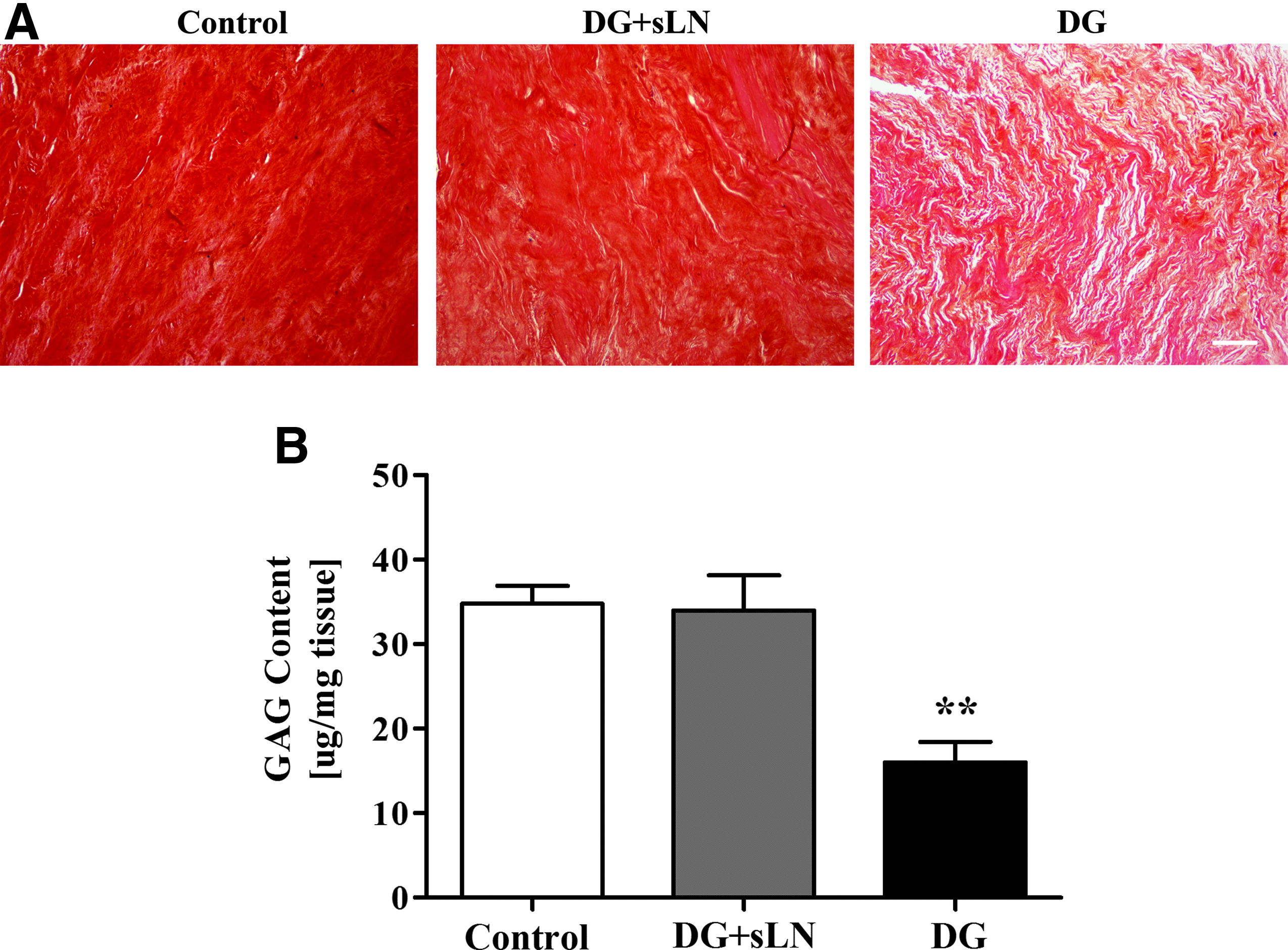
Proteoglycan intensity and content in vIVDs.
Since, safranin O staining intensity may not be a direct proportional indicator of disc proteoglycan content, we further used a DMMB assay to determine GAG content (Fig. 2B). There was a 50% decrease in proteoglycan content in discs that were treated with trypsin compared to the control. In contrast, treatment with sLink N led to a significant enhancement of the proteoglycan content compared to discs treated with trypsin alone and was similar to that in control discs. These results suggest that restoration of proteoglycan content in degenerated discs using sLink N as a bioactive peptide is possible and that the vIVD organ culture model used for the first time in this study is an approach in which to study disc repair.
Next, aggrecan structure was evaluated by measuring protein levels in proteoglycan extracts using Western blotting. (Fig. 3). Aggrecan was significantly reduced in degenerated discs. In contrast, aggrecan was significantly elevated by sLink N injection to a level higher than that in the control discs. Taken together, our data confirm our previous results regarding the fact that the size of aggrecan stimulated by Link N is identical to that in normal discs. 27
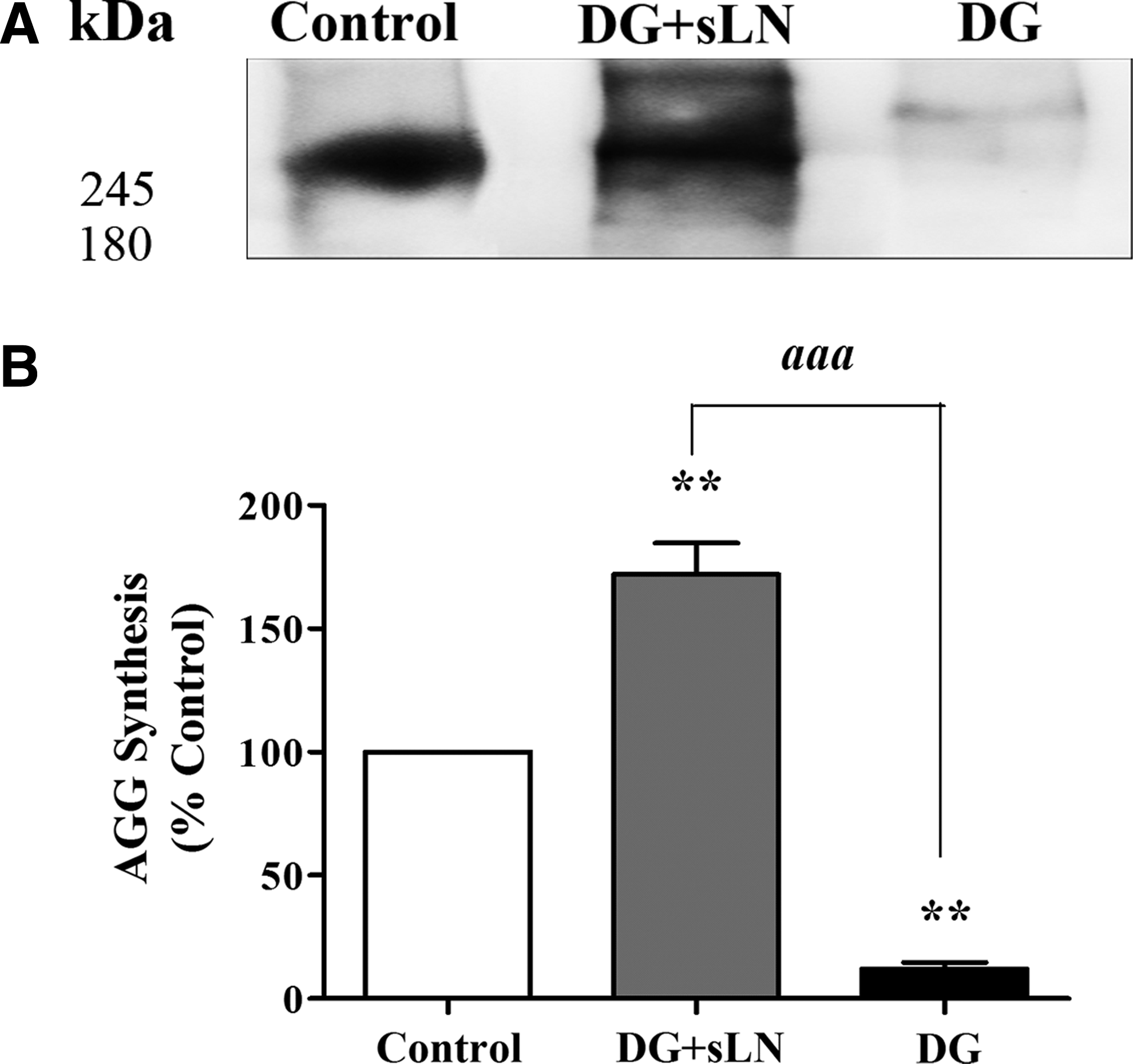
Western blotting of aggrecan core protein in the NP of vIVDs.
Apart from aggrecan, collagen is another important molecule the IVD depends on for function. The type II collagen fibrillar network strengthens the gelatinous NP of the IVD. To address this, we analyzed the levels of newly synthesized type II collagen (Fig. 4). It is notable that type II collagen in DG discs was significantly decreased compared to those in control discs. Our findings also show that injecting sLink N led to an increase in type II collagen to levels that were slightly higher than that detected in control discs. From a tissue engineering perspective, this is significant as the ability of sLink N to stimulate both aggrecan and collagen fibrils is vital for repair.
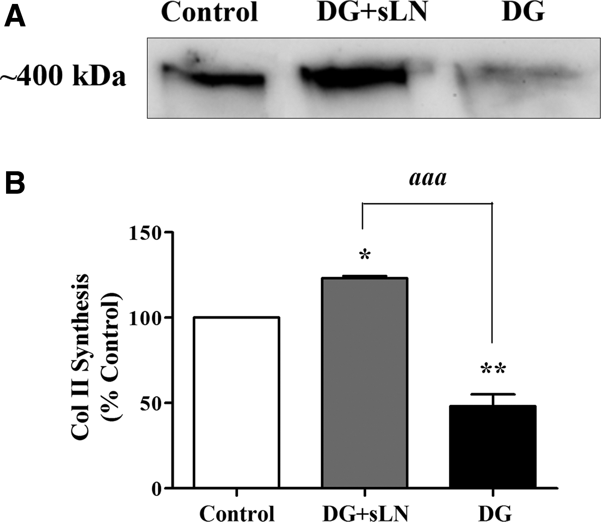
Analysis of newly synthesized type II collagen in vIVDs.
Cell viability was assessed at the end of the experiment to verify that injection of 100 μg of sLink-N (1 mg/mL) and 4 weeks in culture were not detrimental to IVD cells. NP cell viability was maintained at >95% irrespective of the condition (Fig. 5A, B). This suggests that sLink N is not toxic to disc cells. Although we chose a higher concentration of sLink N compared to that we determined as optimal in cells entrapped in alginate beads, 28 we assumed that the majority of the sLink would bind to the disc matrix and, thus, be unavailable to the cells. Indeed, results from some pilot studies in which we injected vIVDs with different concentrations of sLink N showed that only 100 μg generated the best response, importantly that this dosage of slink N (or any other that we have tested) was not toxic. Furthermore, we found that similar to what was observed with Link N, the stimulation of matrix synthesis by sLink N also occurs in the absence of any cell division (Fig. 5C).
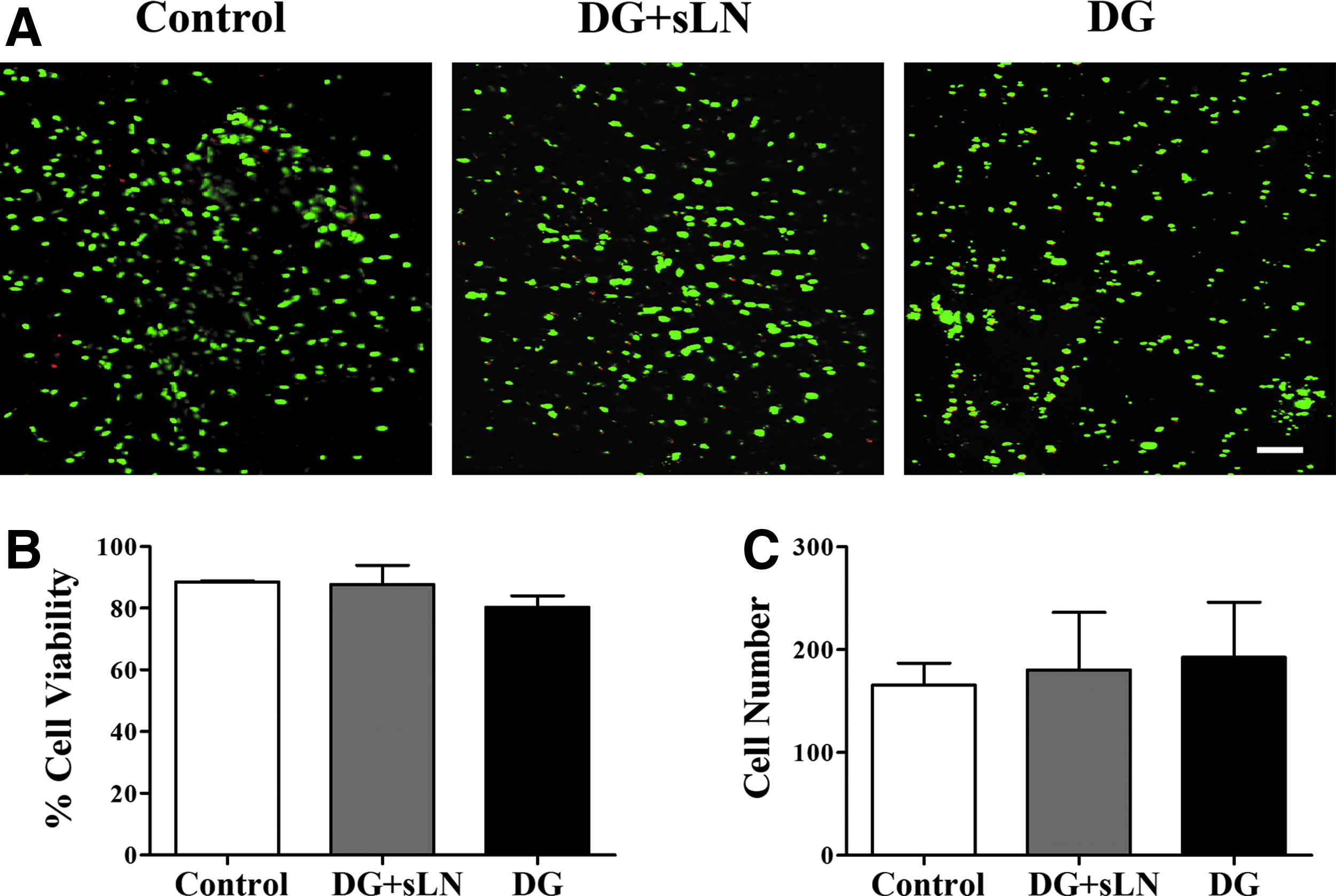
Cell proliferation and viability in vIVDs.
Discussion
We have shown for the first time that sLink N can restore IVD proteoglycan content in a novel organ culture model. 34 The level of increased proteoglycan content in the degenerate disc after injection of sLink N was similar to that observed with Link N supplementation, 27 which suggests that although AF cells can cleave Link N, 28 the peptide generated is bioactive. Treatment with sLink N can also enhance the production of collagen, which is another important molecule in the disc. Furthermore, we show that the culture of intact motion segments, for long periods of time developed recently by our group 34 can be used for testing sLink N and possibly other therapeutics in a well-controlled environment.
Recent work has shown that Link N can stimulate the production of proteoglycan in IVDs from human spines 29 and increase the height of the rabbit degenerate disc after injection. 32 Link N is also capable of stimulating disc repair29–33,38,39 and restoring proteoglycan content. 27 Our laboratory recently pioneered a technique to culture bovine IVD with vertebrae for long term and showed the synthesis of proteoglycans and collagens. We have now for the first time demonstrated the use of this culture model to advance our understanding of sLink N's bioactive effects. Although no load was applied in these studies, the inclusion of bone with a flat surface makes it possible to include complex loading to this model.
In the current study, the model we used is similar to an early DG and suggests that the administration of sLink N has the potential to restore matrix in a mildly degenerated disc and perhaps in more advanced DG as long as the nutrition is not severely compromised. Certainly, this may require adding scaffolds and/or cells and since there is no benign site, stem cells are the more likely candidate. Trypsin and papain have previously been used to degenerate discs for biological repair, but trypsin may be preferable as it is cost effective over other enzymes such as chymopapain or collagenase. 40
Changes in the structure, composition, cellularity, nutrition, and levels of oxygen can result in IVD DG. However, despite numerous efforts, there are no FDA-approved structure-modifying drugs that slow, halt, or reverse the progression of disc DG. Thus, strategies aimed at ameliorating disc DG represent a highly significant opportunity to contain healthcare costs while enhancing the quality of life for many people. We recently reported that sLink N can function in the presence of an inflammatory mediator, such as interleukin-1β, produced by degenerative disc tissue. 28 However, there is little evidence to indicate that these changes have any effect on the stability and structural integrity of sLink N. In terms of mechanisms, Link N exerts its anabolic effects primarily through the Smad1/5/8 signaling pathway. 39 The signals produced by Link N binding to BMP type II receptors on IVD cells may govern disc homeostasis and the prevention of disc DG. We previously showed that the stimulation of matrix synthesis in the disc by Link N occurs with no cell division.32,33 Similar results were obtained in this study with sLink N.
This is the first time this model, with bony vertebrae, has been used for stimulation with any therapeutic agent. These findings will permit us to use the model as a novel preclinical screening of potential therapeutic drugs. Certainly, the potential for an increase in disc repair by modulating BMP signaling in this model will outline the possibility of similar strategies with an eye towards clinical trials.
Conclusion
Following the initial work that identified sLink N as a bioactive peptide and the development of a novel motion segment culture system, we have utilized this model for the first time to show that sLink N can stimulate disc repair by increasing proteoglycan and collagen synthesis.
Footnotes
Acknowledgments
The authors thank Intervertech, Inc. and Wisent Bio Products, Inc. for production and supply of PrimeGrowth media. This work was funded by the Canadian Institute of Health Research (CIHR).
Disclosure Statement
A percentage of the sales of PrimeGrowth™ media will be used for research funding in Dr. Mwale's laboratory. Dr. J.A. and Dr. F.M. hold shares in Trepso Therapeutics.