
Abstract
Polyacrylamide (PAAM) and chitosan were fabricated by inverted colloidal crystal (ICC) method for scaffolds comprising regular pores. The hybrid PAAM-chitosan ICC scaffolds were grafted with poly(lactide-co-glycolide) (PLGA) nanoparticles (NPs) for a rougher pore surface and grafted with transactivator of transcription von Hippel-Lindau (TATVHL) peptide for a better differentiation of induced pluripotent stem (iPS) cells toward neural lineage. By scanning electron microscopy, we found that iPS cells cultured in PAAM-chitosan ICC scaffolds with PLGA NPs at 1.0 mg/mL and TATVHL peptide at 15 μg/mL elongated the axonal length to 15 μm. A combination of PLGA NPs and TATVHL peptide favored the adhesion of iPS cells, reduced the embryonic phenotype after cultivation, and guided the production of βIII tubulin-positive cells in PAAM-chitosan ICC scaffolds. In addition to the differentiation toward neurite-like cells, an increase in the content of TATVHL peptide in PAAM-chitosan ICC scaffolds inhibited the differentiation of iPS cells toward astrocytes. ICC scaffolds composed of PAAM, chitosan, PLGA NPs, and TATVHL peptide can be an efficacious matrix to differentiate iPS cells toward neurons and retard the glial formation for nerve regeneration.
Introduction
E
For a uniform cell distribution in the porous microenvironment, inverted colloidal crystal (ICC) scaffolds were evolved to prevent the disordered and discrete voids. 14 For example, ICC scaffolds with spherical cavity of 97 μm were employed to cultivate Hep-G2 cells and HS-5 cells and demonstrated a satisfactory cell migration and proliferation for 5 weeks. 15 In addition, an improved efficiency in the nutrient transport in ICC scaffolds was confirmed by Brownian dynamics and Monte Carlo simulations. 16 In a study on hepatocytic bile capillaries, spheroids with a structure similar to liver tissue were formed in ICC scaffolds prepared with glass beads of 170 mm. 17 In addition to the size and shape of porogen, the pore configuration of ICC scaffolds was closely related to the solvent used in the self-assembly for colloidal template. It has been found that arrayed monodisperse polystyrene microspheres in 99.8% ethanol were much regular than those in acetone and ethylene glycol. 18
In regard to the pore topography, surface roughness can be another crucial factor responsible for material property and is generally categorized into macroroughness with size from 100 μm to millimeters, microroughness with size from 100 nm to 100 μm, and nanoroughness with size smaller than 100 nm. The surface roughness including bulge, etched channel, and groove might also affect the cell adhesion, proliferation, differentiation, and genetic expression. A roughened titanium surface, for instance, could intensely increase the activity of alkaline phosphatase and the secretion of transforming growth factor-β1 by cloned MG63 osteosarcoma cells.19,20 Moreover, a creation of surface humps and pits could be one of the solutions to guide axonal outgrowth for neural regeneration in biomaterials.21–25
Transactivator of transcription (TAT) peptide, an immediate transactivation protein from human immunodeficiency virus-1, had the capability to permeate cell membrane and carry cross-linked heterologous molecules to cells.26–28 In addition, von Hippel-Lindau (VHL) peptide, containing residues 63–154 of stranded β-sandwich and 155–204 of helical α-domain, was involved in an inherited syndrome about various carcinogenicities.29,30 Intriguingly, VHL protein could bind to the site of elongin BC of differentiating neural progenitor cells. 31 In a study on neural repair for Parkinson's disease, TATVHL peptide was drawn to accelerate endocytosis into skin-derived precursors and intensify the expression of neuronal markers. 32 In a treatment for the spinal cord injury, TATVHL peptide also showed an entire entry to neural stem cells, triggered a differentiation toward neuron-like cells, and facilitated a neurofilament expression. 33 In a study on differentiation of multipotent stem cells isolated from the epidermis of elderly patients with a mean age of 69.1 ± 9.5 years, the percentage of neurite-like cells induced with TATVHL peptide was 78.3% ± 12.5% whereas that induced with TAT peptide was only 18.6% ± 7.3%. 34
The aim of this study was to develop hybrid polyacrylamide (PAAM)-chitosan ICC scaffolds to produce neurons from induced pluripotent stem (iPS) cells. In addition to the pore structure, PAAM-chitosan ICC scaffolds were grafted with poly(lactide-co-glycolide) (PLGA) nanoparticles (NPs) to create rougher surfaces and with TATVHL peptide to induce the differentiation of iPS cells toward neurite-like cells. During neural repair, the regenerative capacity of nerve tissue is feeble and when encountering an obstruction of glial scar, the extension of neurofilament is even worse. Hence, an effective differentiation of iPS cells with an inhibition to astroglial formation is important for neural regeneration. We also examined the efficiency of PAAM-chitosan ICC scaffolds with PLGA humps in the adhesion and neuronal differentiation of iPS cells.
Materials and Methods
Preparation of PAAM-chitosan ICC scaffolds
Polystyrene microspheres (Thermo Fisher Scientific, Fremont, CA) with diameter of 161 ± 3 μm were dried at 40°C for 24 h. The dehumidified monodisperse polystyrene microspheres of 20 mg were immersed in 99.8% ethanol (Riedel-de Haen, Seelze, Germany) of 0.3 mL in a cylindrical container with diameter of 0.5 cm and height of 3 cm, vibrated ultrasonically for 10 min, swung in a bath-reciprocal shaker at 100 rpm for 1 h to array the self-assembly particles, dehydrated at 40°C for 24 h, and sintered at 120°C for 4 h to form a cross-linked colloidal template. PAAM (Sigma-Aldrich, St. Louis, MO) of 1 g was dissolved in ultrapure water (Barnstead, Dubuque, IA) of 40 mL at 300 rpm for 12 h and chitosan (85% deacetylation, Sigma-Aldrich) of 1 g were mixed in 40 mL of ultrapure water containing 1% (v/v) acetic acid (Showa, Tokyo, Japan) at 300 rpm for 12 h. PAAM and chitosan gels were centrifuged at 2500g for 10 min, vibrated ultrasonically for 10 min, stirred in weight ratio of 0:1, 1:2, 1:1, and 2:1, reacted with 2% (w/w) genipin (Challenge Bioproducts, Taichung, Taiwan) for 5 min, added to a centrifugal tube, and vibrated ultrasonically for 5 min. The mixed gel and cross-linked colloidal template were centrifuged at 2625g for 10 min for gel penetration to the interstices among microspheres. The wet gel in the colloidal template was cross-linked for 24 h and dehydrated in an oven at 40°C for 24 h. The gel penetration and dehydration steps were repeated eight times. The composite of biomaterials and colloidal template was immersed in tetrahydrofuran (THF; J.T. Baker, Phillipsburg, NJ) for 12 h and swayed at 100 rpm for 10 min to produce regular ICC pores. After dissolving the template, the scaffolds was washed with acetone (Mallinckrodt, Hazelwood, MO) for 1 h, immersed in 95% ethanol (Taiwan Sugar, Tainan, Taiwan) for 5 h, rinsed with ultrapure water for 5 h, dehydrated at 40°C for 24 h, and trimmed with a cryostat microtome (Slee, Mainz, Germany) to cuboids with dimension of 3 × 3 × 1 mm. In addition, PAAM-chitosan freeform scaffolds were prepared with the corresponding gel. PAAM and chitosan gels were added to a mold with dimension of 23 × 23 × 16 mm, vibrated ultrasonically for 5 min, placed at 25°C for 24 h, prefrozen in an ultralow temperature freezer (Panasonic Healthcare, Gunma, Japan) at −80°C for 24 h, lyophilized using a freeze dryer (Eyela, Tokyo, Japan) at 2–4 torr for 24 h, and trimmed to cubes with dimension of 3.5 × 3.5 × 3.5 mm.
Grafting with PLGA NPs and TATVHL peptide
PLGA NPs were prepared by an emulsion–diffusion method. An organic phase of 8 mL containing PLGA (85/15; Sigma-Aldrich) of 16 mg in acetone (Mallinckrodt Baker, Hazelwood, MO) were emulsified with an aqueous phase of 0.1 mL containing polysorbate (Fisher Scientific, Fair Lawn, NJ) of 2 mg in ultrapure water at 700 rpm for 2 h and then at 1150 rpm for 15 min. Ultrapure water of 20 mL was added to the homogenized fluid to precipitate out PLGA NPs. The suspension was continuously stirred at 200 rpm and 35°C for 2 h to evaporate acetone and filtered with a filtration paper. The filtrate was ultracentrifuged at 2465g for 20 min, suspended in ultrapure water with 1% (w/v)
Porosity and swelling ratio of PAAM-chitosan ICC scaffolds
The porosity of PAAM-chitosan ICC scaffolds (P (%)) was studied by immersing scaffolds in ethanol and calculated by
where Ww, Wd, and W0 are the wet weight of PAAM-chitosan ICC scaffold with ethanol, dry weight of PAAM-chitosan ICC scaffold, and weight of PAAM-chitosan ICC scaffold in ethanol, respectively. In addition, the swelling ratio (RS (%)) was defined as
where W1 is the wet weight of PAAM-chitosan ICC scaffold with ultrapure water.
Scanning electron microscope image of PAAM-chitosan ICC scaffolds with PLGA NPs and constructs with differentiating iPS cells
The morphology of PAAM-chitosan ICC scaffolds with PLGA NPs and constructs was investigated using a field emission scanning electron microscope (SEM; JSM-6330 TF, Jeol, Tokyo, Japan). PAAM-chitosan ICC scaffolds were sliced, washed with DPBS, dehydrated at 25°C for 24 h, vacuum-dried, and sputter-coated with gold at 4 mA and 1 kV for 7 min. In addition, the constructs with differentiating iPS cells were sliced, washed with DPBS, fixed with 2.5% glutaraldehyde (Riedel-de Haen) for 4 h, dehydrated with ethanol from 70% to 99.8% for 7 h, vacuum-dried, and sputter-coated with gold at 4 mA and 1 kV for 7 min.
XPS spectra of PAAM-chitosan ICC scaffolds with PLGA NPs and TATVHL peptide
The X-ray photoelectron (XPS) spectra of PAAM-chitosan ICC scaffolds with PLGA NPs and TATVHL peptide were obtained using an XPS spectroscope (Kratos, Kanagawa, Japan). Samples of PAAM-chitosan ICC scaffolds with PLGA NPs and TATVHL peptide were lyophilized using the freeze dryer at 2–4 torr for 6 h, placed on a sliced cover slide of 5 × 5 mm, vacuum-dried at a vacuum grade of 2 × 10⁻⁷ Pa and 300 W for 15 min.
Adhesion of iPS cells in PAAM-chitosan ICC scaffolds with PLGA NPs and TATVHL peptide
The method for expanding iPS cells (System Biosciences, Mountain View, CA) in a humidified incubator (NuAire, Plymouth, MN) was described in the previous study. 35 PAAM-chitosan ICC scaffolds with PLGA NPs and TATVHL peptide were rinsed with 70% ethanol and dried. The multiplied iPS cells of 8–12th passage were injected into the scaffolds at a density of 3.6 × 10⁵ cells/construct, placed in a 24-well microtiter plate (Falcon, Franklin Lakes, NJ), and cultivated with ESGRO complete PLUS clonal grade medium (Millipore, Billerica, MA) containing 1% penicillin-streptomycin-glutamate solution (Gibco, Carlsbad, CA) in the CO2 incubator at 37°C for 8 h. After seeding, the construct was immersed in the medium of 0.2 mL for releasing of iPS cells. The quantity of free cells was evaluated by a hemocytometer (Neubauer, Marienfeld, Germany) under a phase-contrast biological microscope (Motic, Richmond, BC, Canada). The adhesion efficiency of iPS cells in PAAM-chitosan ICC scaffolds with PLGA NPs and TATVHL peptide was defined as [(3.6 × 10⁵ – number of free iPS cells)/(3.6 × 105)] × 100%.
Immunochemical staining of differentiating iPS cells cultured in PAAM-chitosan ICC scaffolds with PLGA NPs and TATVHL peptide
The constructs composed of PAAM-chitosan ICC scaffolds with PLGA NPs and TATVHL peptide and seeded iPS cells at a density of 3.6 × 10⁵ cells/construct were cultured with ESGRO complete PLUS clonal grade medium in the CO2 incubator for 7 days. The medium was replaced every 2 days. The cultured constructs containing differentiating iPS cells were fixed with 10% (v/v) formaldehyde solution (Sigma-Aldrich) for 4 h, dehydrated with ethanol from 70% to 99.8% for 7 h, immersed in o-xylene (Fluka, Buchs, Switzerland) for 2 h, imbedded in paraffin (Sigma-Aldrich) for 2 h, and sliced using the cryostat microtome to samples with thickness of 4 μm. The samples were heated at 56°C for 1 h, immersed in o-xylene for 10 min, treated with ethanol from 99.8% to 70% for 5 min, rinsed with ultrapure water for 10 min, boiled in citric acid buffer (J.T. Baker) for 5 min, washed with DPBS for 10 min, immersed in 0.5% (v/v) Triton X-100 (Acros Organics, Geel, Belgium) for 10 min, washed with DPBS for 10 min, blockaded with serum blocking solution (Zymed Laboratories, South San Francisco, CA) of 30 μL for 20 min, and reacted with chicken anti-200 kD neurofilament heavy chain polypeptide (NF-H; Abcam, Cambridge, MA) in a dilution of 1:1000, with rabbit anti-sex determining region Y (SRY)-box 2 (SOX-2; Abcam) in a dilution of 1:1000, and with mouse anti-astrocyte marker glial fibrillary acidic protein (GFAP; Abcam) in a dilution of 1:100 at 4°C overnight. After having washed with DPBS, the samples were stained with chicken anti-immunoglobulin (Ig) Y (heavy and light; H&L) carrying fluorescein isothiocyanate (FITC) label (Abcam) in a dilution of 1:200, with rabbit anti-IgG carrying rhodamine label (Sigma-Aldrich) in a dilution of 1:400, and with mouse anti-IgG carrying Texas red (Abcam) in a dilution of 1:1000 at 25°C for 1 h in darkness, washed with DPBS for 10 min, counterstained with 0.5% (w/v) 4′,6-diamidino-2-phenylindole (DAPI; Sigma-Aldrich) in 0.5% (v/v) Triton X-100 at 25°C for 3 min in darkness, dehumidified for 12 h, and treated with ProLong gold antifade reagent (Biocompare, South San Francisco, CA). Immunofluorescent images were visualized using a confocal laser scanning microscope (LSM 510, Zeiss, Oberkochen, Germany) with a filtered argon laser. The excitation wavelengths of red (rhodamine and Texas red), green (FITC), and blue (DAPI) were 555, 488, and 405 nm, respectively. The emission wavelengths of red, green, and blue were 578, 528, and 460 nm, respectively.
Flow cytometry of differentiating iPS cells
After induction for 7 days, the constructs composed of PAAM-chitosan ICC scaffolds with PLGA NPs and TATVHL peptide and differentiating iPS cells were washed with DPBS for 10 min, centrifuged at 200g for 5 min, detached with TrypLE select (Gibco) of 0.5 mL with mild squeezing for 3 min, and flushed with ESGRO complete PLUS clonal grade medium of 200 μL for 10 times. The flushed suspension was collected and centrifuged at 200g for 5 min. Bottom pellets were treated with 0.5% (v/v) Triton X-100 in DPBS for 10 min, washed with DPBS for 10 min, centrifuged at 200g for 5 min, blockaded with serum blocking solution for 1 h, and identified with Milli-Mark anti-stage-specific embryonic surface antigen (SSEA)-1 cloning MC-480 phycoerythrin (PE) label (Millipore) in a dilution of 1:100, with rabbit monoclonal [EP1331Y] anti-βIII tubulin (Abcam) in a dilution of 1:100, and with anti-GFAP at 4°C for 1 h in darkness. The differentiating iPS cells were stained with goat polyclonal secondary antibody to mouse IgG H&L carrying FITC label (Abcam) in a dilution of 1:100 and with mouse anti-IgG carrying Texas red label in a dilution of 1:100 at 4°C for 1 h in darkness. The stained differentiating iPS cells were washed with DPBS for 10 min, centrifuged at 200g for 5 min, resuspended in DPBS of 0.5 mL, classified using a flow cytometer (Becton Dickinson, Franklin Lakes, NJ), and calculated with CELL Quest software (BD Pharmingen, Franklin Lakes, NJ).
Results and Discussion
Porosity and swelling ratio of PAAM-chitosan scaffolds
Table 1 shows the porosity and swelling ratio of PAAM-chitosan ICC and freeform scaffolds. As indicated in this table, an increase in the weight percentage of PAAM slightly enhanced the porosity of PAAM-chitosan freeform scaffolds. This was because the viscosity of chitosan was high in 2.5% (w/v) gel. Thus, a higher content of chitosan resulted in a denser solid structure and a lower porosity of PAAM-chitosan freeform scaffolds. However, as revealed in Table 1, a variation in the weight percentage of PAAM had no significantly influence on the porosity of PAAM-chitosan ICC scaffolds. In addition, the porosity of freeform scaffolds was higher than that of ICC scaffolds. This was because the pores in PAAM-chitosan ICC scaffolds resulted from the empty space after dissolution of polystyrene microspheres with THF and the pores of freeform scaffolds were from the sublimation of ice crystals.
CPLGA = 1.0 mg/mL; n = 3.
Concerning regular pore pattern in PAAM-chitosan ICC scaffolds, the packing of polystyrene microspheres was related to the solvent used to arrange the self-assembly. In ideal hexagonal closest packing, 1 polystyrene microsphere contacted other 12 identical particles, leading to about 74% volume occupation. Nonetheless, the porosity of PAAM-chitosan ICC scaffolds was slightly higher than 74%. This was because the high viscosity of chitosan was not easy to completely fill the voids among polystyrene microspheres during centrifugation. It is worth to note that porosity is one of the most essential properties of biomedical scaffolds. For example, high porosity of biomaterials could accelerate the transfer of nutrients and favor the release of metabolites out of the matrix.13,36 It has been observed that porosity of biopolymers could be higher than 90% by lyophilization. 37 Although porosity of PAAM-chitosan freeform scaffolds was higher than that of PAAM-chitosan ICC scaffolds, the interconnected pores in the latter benefited the mass transfer efficiency throughout the scaffolds. 38
As exhibited in Table 1, an increase in the weight percentage of PAAM slightly decreased the swelling ratio of PAAM-chitosan scaffolds. This was because the hydrogen bonding between water and –NH2/–OH groups in chitosan could be stronger than that between water and –NH2 groups in PAAM. Thus, –NH2 and –OH in chitosan could seize more water than –NH2 in PAAM. As displayed in Table 1, the swelling ratio of PAAM-chitosan freeform scaffolds was higher than that of PAAM-chitosan ICC scaffolds. This was mainly because the former had a higher vacancy for the uptake of water than the latter.
Surface atom on PAAM-chitosan ICC scaffolds grafted with PLGA NPs and TATVHL peptide
Table 2 shows the results of the XPS spectra for atomic ratio of PAAM-chitosan ICC scaffolds grafted with TATVHL peptide. As indicated in this table, an increase in the concentration of TATVHL peptide enhanced the quantity of surface sulfur at 165 eV. This resulted from the fact that the cross-linker bis(3-sulfo-N-hydroxysuccinimide ester) to graft TATVHL peptide contained sulfur. In addition, an increase in the concentration of TATVHL peptide enhanced the quantity of surface nitrogen. This was because the atomic ratio of nitrogen to all atoms in TATVHL peptide was higher than that in PAAM-chitosan ICC scaffolds with 50% (w/w) PAAM. Considering the different organic solvents used during scaffolds preparation, the toxic residues after processing were detected by FTIR (data not shown). No absorption of residual THF was found in the current matrix. 39 In our previous studies, solid-state 13 C NMR spectroscopy also could not observe the characteristic peaks of acetone in polymeric scaffolds. 40
rPAAM = 50%.
Pore structure and PLGA NPs in PAAM-chitosan ICC scaffolds
Figure 1 shows the internal structure and grafted PLGA NPs on the pore surface in PAAM-chitosan ICC scaffolds. As indicated in Figure 1a–d, the pore size was about 160 nm, which well agreed with the size of polystyrene microspheres in this study. The diameter of interconnected holes between two pores was 40–50 nm, originating from the cross-linking of polystyrene at 120°C. As revealed in Figure 1a, the slight irregular pore shape manifested a partially incomplete infiltration of viscous chitosan gel to the interstices among microspheres. In addition, Figure 1a–d was consistent with the data shown in Table 1. Since a high content of chitosan in PAAM-chitosan ICC scaffolds obstructed the gel infiltration and promoted the water absorption, PAAM-chitosan ICC scaffolds with 50% (w/w) PAAM could be selected for the culture of iPS cells. As exhibited in Figure 1e–g, the size of grafted PLGA NPs on the surface was 200–300 nm. An increase in the concentration of PLGA NPs also increased the quantity of grafted PLGA NPs on the pore surface in PAAM-chitosan ICC scaffolds.
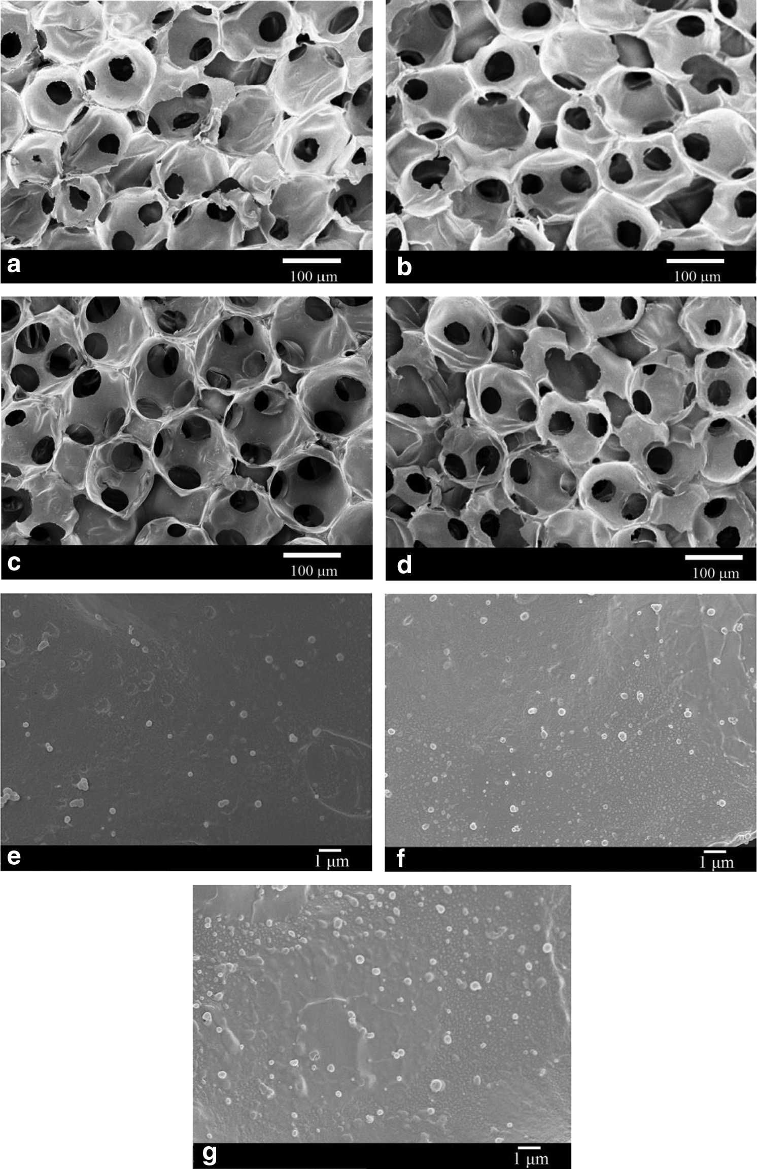
SEM micrograph of PAAM-chitosan ICC scaffolds.
Adhesion of iPS cells in PAAM-chitosan ICC scaffolds grafted with PLGA NPs and TATVHL peptide
Table 3 shows the effect of the concentration of PLGA NPs and TATVHL peptide on the adhesion efficiency of iPS cells in PAAM-chitosan ICC scaffolds. As indicated in this table, the concentration of PLGA NPs had a slight effect on the adhesion efficiency of iPS cells. It has been observed that an increase in the surface roughness favored the adhesion of calvarial and endothelial cells in biomaterials.41,42 However, surfaces with a high roughness could aggregate neural cells and hinder the neurite extension. 43 Based on Table 3, PAAM-chitosan ICC scaffolds grafted with PLGA NPs at 1.0 mg/mL could be proper for the adhesion of iPS cells.
n = 3.
As revealed in Table 3, an increase in the concentration of TATVHL peptide promoted the adhesion efficiency of iPS cells. However, this promotion was not obvious when the concentration of TATVHL peptide increased from 10 to 15 μg/mL. Two reasons could explain this behavior. First, at the concentration of TATVHL peptide of 15 μg/mL, the enhancement in the quantity of cross-linked amino groups became partly inactive to attract iPS cells to PAAM-chitosan ICC scaffolds. Second, for the grafting reaction on the surface of PAAM-chitosan ICC scaffolds, the best concentration of TATVHL peptide was close to 10 μg/mL. Although both PLGA NPs and TATVHL peptide reacted with amino groups on the pore surface, TATVHL peptide of 15 μg/mL yielded the highest adhesion efficiency of iPS cells and could have a high applicability for neuronal differentiation in PAAM-chitosan ICC scaffolds.
Morphology of differentiating iPS cells in PAAM-chitosan ICC scaffolds grafted with PLGA NPs and TATVHL peptide
Figure 2 shows the morphology of cultured construct of PAAM-chitosan ICC scaffolds grafted with PLGA NPs and TATVHL peptide and differentiating iPS cells. As indicated in this figure, porous materials with partially degraded ICC structure were observed. In addition, Figure 2a–d evidenced the clusters of differentiating iPS cells without neuronal trait in PAAM-chitosan ICC scaffolds grafted with PLGA NPs of 0.5 mg/mL. Figure 2e and f manifested that an induction with TATVHL peptide favored the differentiation of iPS cells with extended cell peripheries. Moreover, Figure 2g and h suggested that surface PLGA NPs of 1 mg/mL could benefit neuronal differentiation of iPS cells in PAAM-chitosan ICC scaffolds after an induction with TATVHL peptide of 10 μg/mL.

SEM morphology of differentiating iPS cells cultured in PAAM-chitosan ICC scaffolds grafted with PLGA NPs and TATVHL peptide for 7 days. rPAAM = 50%.
When compared with Figure 2a–j exhibited more mature neuron-like cells with axonal outgrowth on the pore surface grafted with TATVHL peptide of 15 μg/mL The neuronal differentiation in Figure 2i with neurite length about 15 nm was more obvious than that in Figure 2j, suggesting that PLGA NPs of 1.0 mg/mL provided an optimal surface roughness in neurofilament regeneration.
It has been observed that the growth of neurons on rough membrane surface could be better than that on smooth one. 43 The porosity of the current scaffold appeared not sufficiently spacious for the colonization of iPSCs because of the characteristics of ICC topography at micrometric level. However, the regular scaffold morphology with interconnected holes favored the nutrition diffusion and cell migration inside the scaffold, although an organized nanostructure was important for cell adhesion. Moreover, it is well known that cells seeded into a three-dimensional scaffold require active nutrition transport to growth. 44 In our previous studies on cartilage tissue engineering, the dynamic cultivation in spinner system was a prerequisite in nutrition supply and metabolite removal for long-term chondrogenesis.45,46
Immunofluorescence of differentiating iPS cells in PAAM-chitosan ICC scaffolds grafted with PLGA NPs and TATVHL peptide
Figure 3A shows the immunostaining images of expressed SOX-2, GFAP, and NF-H by differentiating iPS cells after cultivation in PAAM-chitosan ICC scaffolds grafted with PLGA NPs and TATVHL peptide for 3 days. As indicated in Figure 3A (a–d, h–k), an increase in the concentration of TATVHL peptide increased the expression of NF-H and decreased the quantity of GFAP in PAAM-chitosan ICC scaffolds. Three conclusions could be drawn. First, TATVHL peptide on the surface of PAAM-chitosan ICC scaffolds stimulated the neuronal differentiation of iPS cells. Second, TATVHL peptide in PAAM-chitosan ICC scaffolds reduced the quantity of phenotype of iPS cells after the neuronal induction. Third, TATVHL peptide in PAAM-chitosan ICC scaffolds could decelerate the production of astroglial cells. It has been observed that VHL peptide could inhibit the signal transducer and activator of transcription-3 factor for astrocyte differentiation. 47

As indicated in Figure 3A (e–g, l–n), the expression of NF-H in Figure 3A (e, f) was higher than that in Figure 3A (g) and the expression of NF-H in Figure 3A (l, m) was higher than that in Figure 3A (n). In addition, the quantity of SOX-2 in Figure 3A (e–g) and GFAP in Figure 3A (l–n) was low. This suggested that surface PLGA NPs stabilized the maturation of neuron-like cells differentiating from the firm adhesive iPS cells in PAAM-chitosan ICC scaffolds.
Neuronal differentiation of iPS cells in PAAM-chitosan ICC scaffolds grafted with PLGA NPs and TATVHL peptide
Figure 3B shows the cell sorting diagram of differentiating iPS cells cultivated in PAAM-chitosan ICC scaffolds grafted with PLGA NPs and TATVHL peptide. The corresponding data are listed in Table 4 and 5. As indicated in Figure 3B (a) and Table 4, the percentage of iPS cells identified with anti-SSEA-1 were very high after cultivation in PAAM-chitosan ICC scaffolds. This manifested a high portion of iPS cells remaining in embryonic phenotype. In addition, the percentage of differentiating iPS cells with βIII tubulin in PAAM-chitosan ICC scaffolds was low, demonstrating a weak neuronal differentiation.
Differentiating iPS cells are stained against SSEA-1 and βIII tubulin. rPAAM = 50%.
Differentiating iPS cells are stained against GFAP and βIII tubulin. rPAAM = 50%.
GFAP, glial fibrillary acidic protein.
As revealed in Figure 3B (b–d) and Table 4, an increase in the concentration of TATVHL peptide enhanced the percentage of βIII tubulin-positive cells. However, when the concentration of TATVHL peptide increased from 10 to 15 μg/mL, the enhancement (2.49%) was not significant. As exhibited in Figure 3B (d, e) and Table 4, when the concentration of PLGA NPs increased from 0 to 5 mg/mL, the percentage of βIII tubulin-positive cells slightly reduced (1.67% decrease), and the percentage of cells in the second quadrant (cells with SSEA-1 only) reduced from 2.05% to 0.66%.
As displayed in Figure 3B (d–g) and Table 4, the maximal percentage of βIII tubulin-positive cells differentiating from iPS cells occurred at PLGA NPs of 10 mg/mL. The rationale behind this differentiation behavior was explained as follows. First, the rough pore surface with grafted PLGA NPs benefited the adhesion of iPS cells for neuronal differentiation (adhesion data shown in Table 3). Second, a high concentration of PLGA NPs reduced the surface amino groups for TATVHL peptide cross-linking and decreased the capacity of PAAM-chitosan ICC scaffolds to guide differentiation (surface nitrogen-to-carbon ratio shown in Table 2).
As indicated in Figure 3B (h–k) and Table 5, an increased in concentration of TATVHL peptide reduced the percentage of GFAP-positive cells in PAAM-chitosan ICC scaffolds. This suggested that TATVHL peptide not only enhanced the production of neuron-like cells but also hindered astroglial differentiation in PAAM-chitosan ICC scaffolds. As revealed in Figure 3B (l–n) and Table 5, the minimal percentage of astrocyte-like cells differentiating from iPS cells occurred at PLGA NPs of 10 mg/mL. Thus, PLGA NPs of 10 mg/mL could produce sufficient roughness on pore surface of PAAM-chitosan ICC scaffolds and could be an efficient condition for nerve tissue engineering.
Conclusions
Fifty percent (w/w) PAAM in the polymeric matrix can be appropriate to fabricate PAAM-chitosan ICC scaffolds with interconnected pore topography and PLGA NPs can generate the roughness of pore surface. An incorporation of PLGA NPs and TATVHL peptide in PAAM-chitosan ICC scaffolds could effectively promote the adhesion of iPS cells. The most effective concentrations of PLGA NPs and TATVHL peptide for the adhesion of iPS cells were 1.0 mg/mL and 15 μg/mL, respectively. Based on immunostaining and flow cytometry, surface TATVHL peptide benefited the differentiation of iPS cells toward neurons and the concentration of TATVHL peptide above 10 μg/mL was effectual to retard the production of astroglia in PAAM-chitosan ICC scaffolds.
Footnotes
Acknowledgment
This work is supported by the Ministry of Science and Technology of the Republic of China.
Disclosure Statement
No competing financial interests exist.