
Abstract
Our objective was to test the efficacy of collagen–hyaluronan scaffolds reinforced with poly(
Introduction
T
Meniscal tears compromise the tissue's mechanical properties and function, causing pain, discomfort, and instability. One of the most commonly performed orthopedic surgeries is meniscectomy, 7 or the removal of damaged tissue to prevent locking and catching. While these procedures typically provide short-term symptom relief, the amount of long-term articular cartilage damage is closely correlated to the amount of tissue removed.8,9 This can be attributed to increases in peak contact stresses between the femoral condyle and tibial plateau. 10 Meniscal allograft transplantation is one potential replacement, but concerns include long-term graft failure, size matching, graft preservation, and the risk of disease transmission. 11 Tissue engineering provides a potential approach to create a mechanically functional, long-term meniscal replacement.
Previously, our group successfully tested a novel resorbable scaffold for total meniscus reconstruction in an ovine model. It consisted of a collagen–hyaluronan sponge reinforced with poly(desaminotyrosyl-tyrosine dodecyl ester dodecanoate) [p(DTD DD)] fibers. 12 The previous trials with p(DTD DD) fibers demonstrated that following 16, 32, and 52 weeks in vivo, the total meniscus replacement scaffold maintained functional mechanical properties, experienced tissue ingrowth and remodeling into neomeniscal tissue, and prevented the progression of osteoarthritis.13,14
Results using p(DTD DD) fibers were excellent, but from a product development standpoint, clinical translation might be simplified by utilizing fibers made from a more commonly used degradable polymer. Thus, the purpose of this study was to evaluate the efficacy of a total meniscus replacement scaffold using our same design but with poly(
There were two phases of this study. First, we measured the tensile strength and in vitro strength retention of individual fibers, and the strength of whole scaffolds, and determined that PLLA fiber-reinforced scaffolds had appropriate mechanical properties for this application. Then, for the total meniscus implantation study, we tested the hypotheses that the scaffolds would (i) remain intact and maintain their shape; (ii) experience improvement in compressive properties over time; (iii) support continuing neomeniscus tissue deposition and organization; and (iv) prevent the progression of articular cartilage damage.
Materials and Methods
The initial strength and strength retention after degradation in physiologic saline for various time periods were evaluated by mechanically testing individual PLLA fibers. PLLA fiber-reinforced scaffolds were fabricated and characterized by an ultimate tensile test. For the in vivo study, one scaffold was implanted into a sheep for 8 weeks to determine the feasibility of using the PLLA scaffold in this model. Then, PLLA scaffolds were implanted into eight additional sheep for up to 32 weeks. Explants were evaluated mechanically with confined compression creep testing, histologically for cellular and extracellular matrix (ECM) organization, and biochemically for collagen and sulfated glycosaminoglycan (S-GAG) content. Articular surfaces were also analyzed macroscopically and histologically.
Fiber and scaffold production
PURASORB PL38 (IV = 3.8 dL/g) was obtained from Purac Biomaterials (Gorinchem, Netherlands) and melt-extruded and hot-drawn into 100-μm- diameter fibers at the New Jersey Center for Biomaterials (Department of Chemistry, Rutgers University, Piscataway, NJ). Scaffolds were produced as previously described 13 (Fig. 1). Fiber was quasi-woven around a semilunar set of pins to create an ovine medial meniscus-sized network of fibers with extended tails, with 831 fibers per cross section and ∼150 m of total fiber. 14 The medial meniscus was chosen as a template due to its higher susceptibility to injury than the lateral meniscus. 31 Collagen–hyaluronan dispersion (20, 0.25 g/L) was injected into and around the network of fibers, frozen via ethanol-dry ice bath, and lyophilized (−50°C, 0.05 mbar). Cross-linking was performed in 1-ethyl-3-(dimethyl aminopropyl) carbodiimide (EDC, 10 mM) and N-hydroxysuccinimide (NHS, 5 mM) for 6 h (50 mL/scaffold). Scaffolds were then washed in deionized (DI) H2O (3 × 10 min), sodium phosphate solution (Na2HPO4, 3 h), and DI H2O (4 × 6 h). Scaffolds were lyophilized again, packaged, and sterilized by gamma irradiation at 25 kGy (Sterigenics, Rockaway, NJ).
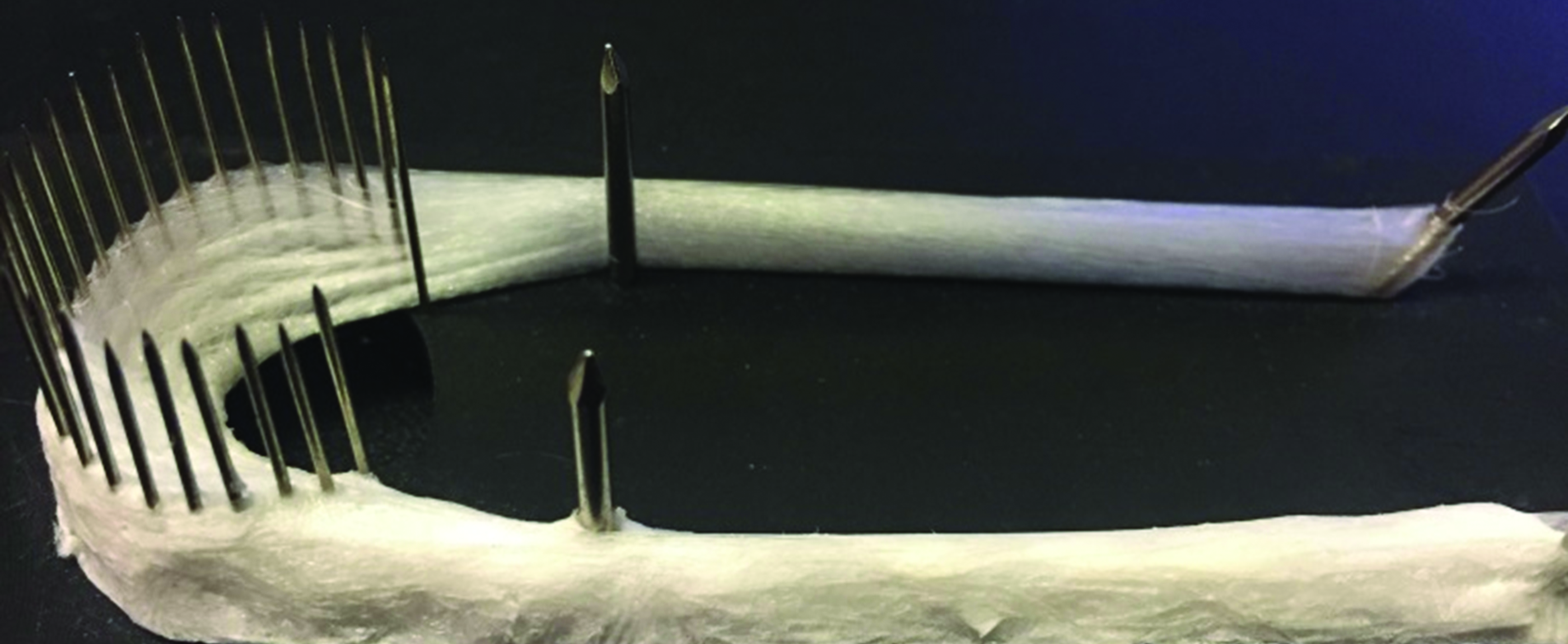
PLLA fiber (top) woven around a circumferential set of pins. Fibers were impregnated with type I bovine collagen sponge (bottom). The extended tails are for surgical fixation. PLLA, poly(
Strength retention of fibers
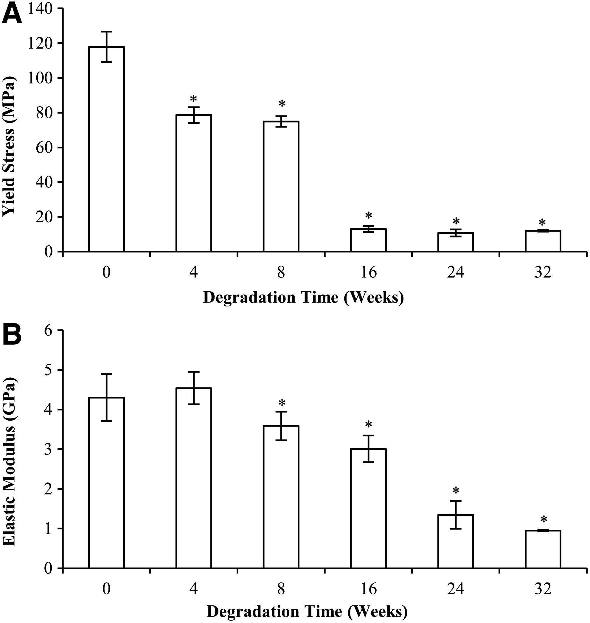
PLLA fibers were cut into 100 mm segments and split into six groups (n = 6 per group). Fibers were placed in phosphate-buffered saline (PBS, pH 7.4) at 37°C for 0, 4, 8, 16, 24, and 32 weeks. Fibers were superficially dried and 25 mm on each end were taped for gripping, leaving a 50 mm gauge length. Taped ends were loaded into pneumatic grips and tested in tension until failure at 30 mm/min (Instron #5542, Norwood, MA). 32 Elastic modulus was obtained by finding the steepest portion of the linear region of the stress–strain curve, and a 0.2% offset strain line was used to obtain the yield stress.
Scaffold tensile properties
Three PLLA fiber-reinforced collagen–hyaluronan scaffolds were tested in uniaxial tension. Samples were hydrated for at least 30 min before testing and loaded into electro-cooling cyro-clamps (Bose Electroforce Systems, Eden Prairie, MN) with the anterior and posterior regions of the scaffold within the clamps. The body region was left between the grips with a gauge length of 10 mm. Samples were tested at 10 mm/min until failure, and the ultimate tensile load and tensile stiffness were obtained.
Total meniscus replacement model
Total medial meniscectomies were performed on nine skeletally mature (2–3 years, 50–70 kg) Dorset Finn cross sheep, and PLLA scaffolds were implanted into the right hind leg of all sheep as previously described. 13 Tibial bone tunnels were drilled at the anterior and posterior horns, and scaffold tails were fixed with interference screws (Smith & Nephew, Andover, MA). Animals were allowed unrestricted movement 3 h postoperatively and received antibiotic, anti-inflammatory, and pain-killing medication as necessary. Animals were sacrificed at specific predetermined time points (n = 1 at 8 weeks, 2 at 16 weeks, n = 3 at 24 and 32 weeks). Explants were analyzed grossly for size, shape, location, and integrity, and, after the 8-week time point, subjected to compression creep, histological, and biochemical analyses. The femoral condyles and tibial plateaus were also retrieved and observed macroscopically (International Cartilage Repair Society [ICRS] mapping scheme) and histologically for cartilage damage. Contralateral knees were recovered for comparison to native tissue.
Confined compression creep testing
Cylindrical plugs (4 mm diameter by 4 mm height) were taken from the anterior, body, and posterior regions of each explant and from each of three native menisci using a biopsy punch, giving nine samples in the native, 24-week, and 32-week groups and n = 6 in the 16-week group. Plugs were hydrated for at least 60 min and inserted into a 4-mm-diameter cylindrical chamber with unidirectional fluid flow filter. Samples were subjected to 1 N for 3600 s (Instron #5542). Mow's biphasic theory 33 and least-squares fit were used to obtain aggregate modulus and permeability.
Explant histology
Radial cross-sectional tissue samples were taken from the anterior, body, and posterior region of each explant and a native meniscus. Samples were fixed in Carson's buffered formalin and paraffin embedded. Sections were cut (8 μm thick) and stained with hematoxylin and eosin (H&E; AML Labs, Baltimore, MD). Qualitative observations were made with regard to cell concentration and morphology, ECM deposition and organization, vasculature, and inflammatory response.
Biochemical analyses
For quantitative biochemical analyses, compression creep plugs (native meniscus, 16-, 24-, and 32-week explants) were hydrated and weighed wet, and were then lyophilized and weighed dry. The dry samples were digested in papain solution (100 μL solution/1 mg tissue; 125 μg/mL papain, 5 mM
For hydroxyproline quantification, 100 μL of each digested sample was hydrolyzed in 100 μL of 12 M HCl for 24 h at 120°C. Hydrolyzed samples were diluted in 800 μL of 6 M HCl. Two 10 μL aliquots of each diluted sample were transferred to a 96-well plate and subject to evaporation under a vacuum hood. Each well (including 2 blank wells and 7 × 2 standard wells) was incubated in 100 μL chloramine T solution (20 min, room temperature). Solutions were dyed with 100 μL dimethylaminobenzaldehyde (DMAB; 30 min, 60°C). Absorbance was measured at 530 nm.
For S-GAG quantification, two 20 μL aliquots of papain-digested tissue were diluted in 80 μL PBS and stained with 100 μL of 1,9-dimethylmethylene blue (DMB). Absorbance was measured at 525 nm. Hydroxyproline and S-GAG concentrations by wet weight were calculated using a regression analysis on ranges of hydroxyproline and chondroitin-6-sulfate standards. Hydroxyproline concentration was used to calculate collagen content with a scale factor of 7.46. 34 Collagen and S-GAG content as a percentage of that in native tissue was calculated. Water content was also calculated.
Cartilage histology
One medial-to-lateral slice (∼5 mm) was taken from the most damaged region of the femoral condyle and tibial plateau and fixed in Carson's buffered formalin. Samples were embedded in paraffin and decalcified with an acid-EDTA solution (Decal Stat; StatLab, McKinney, TX), and 8-μm-thick sections were stained with Safranin-O/Fast Green (AML Labs). One section per slice was graded using the Mankin scoring system on the most damaged region of each section (scale of 0–14: 0 represents no damage; 14 is maximal damage). 33 Mankin scoring was also performed on three randomly chosen contralateral knees. Scoring was performed on regions of cartilage not affected by any joint abnormalities.
Statistical analysis
In the fiber strength retention assay, average yield stress and elastic modulus for each time point were compared to the control group (t = 0 weeks) with a one-tail, type 2 t-test. Two-tail, type 2 t-tests were used to make comparisons between PLLA scaffolds and native menisci in the ultimate tensile test. One-way ANOVA with a post hoc Tukey's honest significant difference was used for the compression creep and biochemical assays. Mankin scores were analyzed with a Kruskal–Wallis test with a post hoc Dunn's nonparametric pairwise comparison. Any p-values less than 0.05 were considered statistically significant differences. Statistical comparisons were performed using Stata/MP 14 statistical software.
Results
Fiber and scaffold production
Fibers had an average diameter slightly above 100 μm (114.6 ± 7.7 μm). PLLA-reinforced scaffolds were fabricated with dimensions and morphology similar to native ovine medial menisci.
Strength retention of fibers
The PLLA t = 0 fibers had a yield stress of 117.9 ± 8.7 MPa and elastic modulus of 4.30 ± 0.59 GPa. The mechanical properties of the PLLA fiber through the 32-week degradation assay are shown in Figure 2. Yield stress decreased significantly after 4 weeks and between 8 and 16 weeks, there was an 82.6% reduction. Elastic modulus of fibers decreased ∼30% by 16 weeks.
Scaffold tensile properties
The ultimate tensile load of PLLA-reinforced scaffolds (590.8 ± 45.6 N) was comparable to that of the native ovine medial meniscus (572.6 ± 210.9 N). The PLLA scaffolds (340.5 ± 27.3 N/mm) were 137.3% stiffer than native menisci (143.5 ± 41.6 N/mm). Native menisci values were obtained from our previous study. 14
Total meniscus replacement model
All animals were standing ∼2 h postoperatively and showed no differences in weight bearing between legs by 8 weeks. In the 8-week pilot study, the explant had meniscus-like morphology and appeared fibrous and vascularized (Fig. 3, left). No narrowing in the body region or indications of rupture were noted. Slight superficial abrasions were present on the femoral condyle (Fig. 3, right), and no osteophytes or bone erosion was observed.
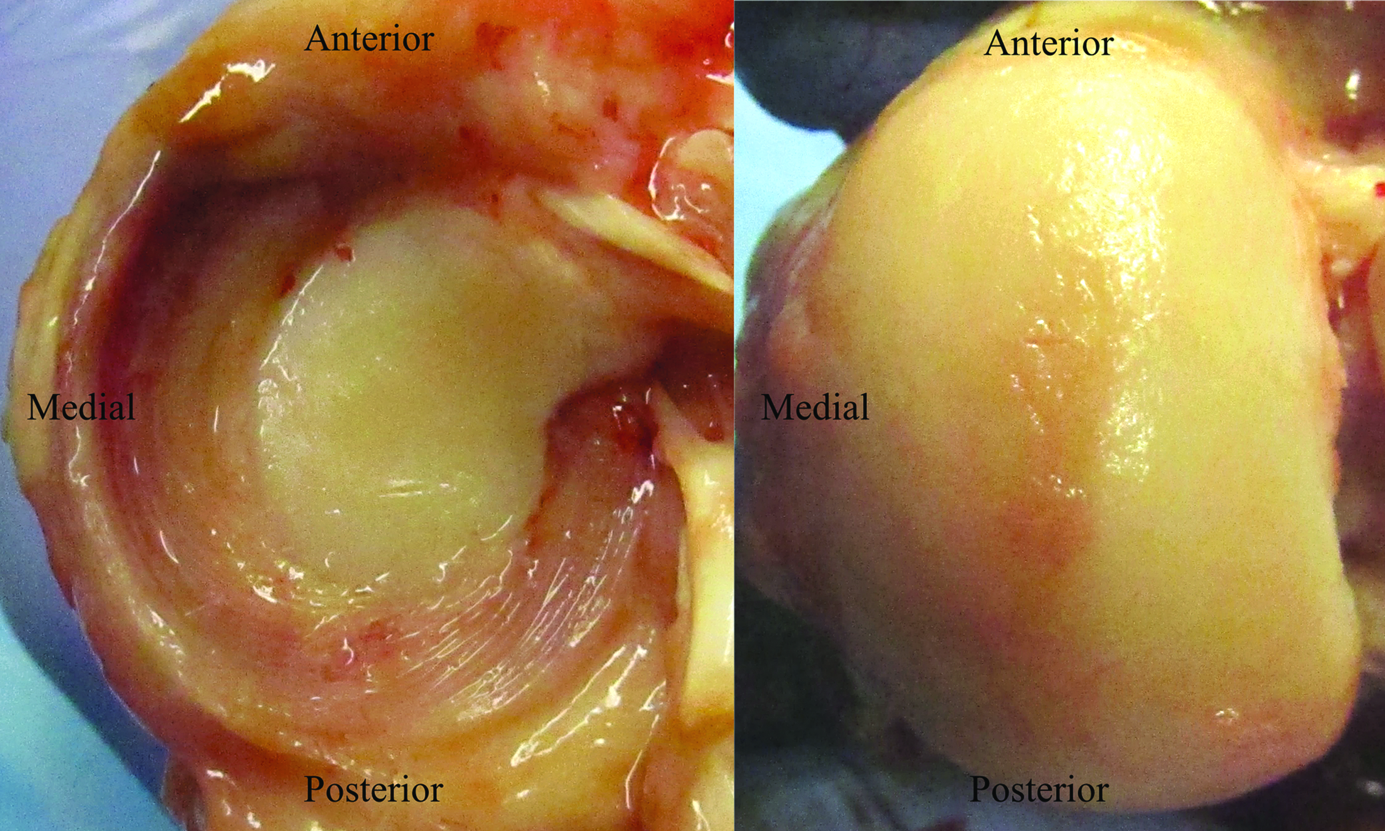
Macroscopic images from an 8-week pilot study. (Left) Explant on tibial plateau and (right) femoral condyle with slight superficial abrasions and no joint abnormalities. Color images available online at www.liebertpub.com/tea
At later time points, one explant at 24 weeks and two explants at 32 weeks experienced rupture in the body region. All other explants were thin and narrow in the body region, leaving a large area of the tibial plateau cartilage exposed. Sample explants from each time point are compared to the native meniscus in Figure 4 (top row). The anterior region of the explants appeared thicker and wider than the body and posterior regions.
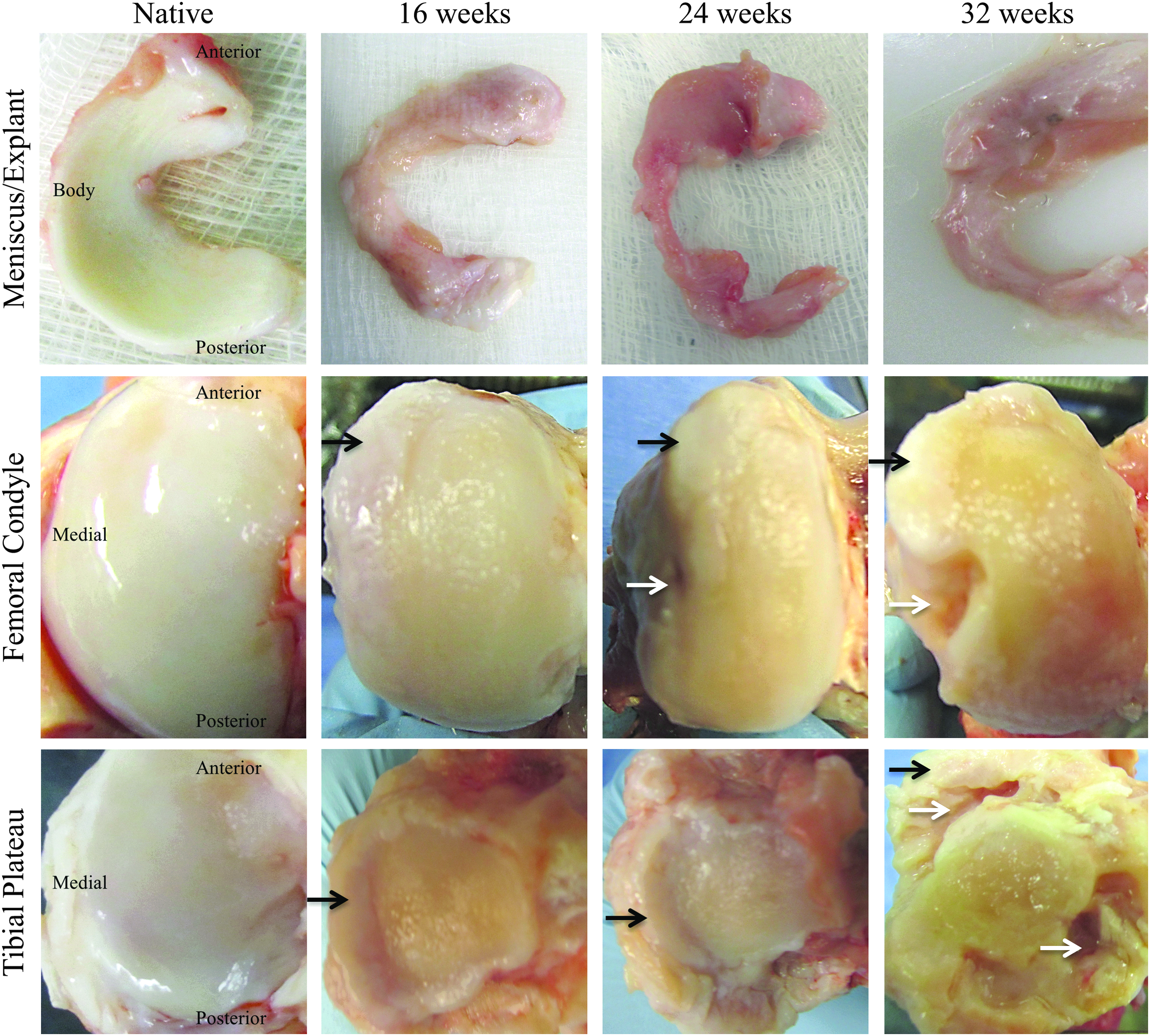
Native meniscus and articular surfaces compared to explants, femoral condyles, and tibial plateaus at 16, 24, and 32 weeks. Osteophytes are highlighted by black arrows; bone erosion is highlighted by white arrows. Color images available online at www.liebertpub.com/tea
In all eight animals, osteophytes were observed on the medial aspect of the medial femoral condyles and beneath the explants on the medial tibial plateaus, both regions in direct contact with the meniscus implants. These osteophytes were smooth and very hard, and their size and severity increased from 16 to 24 to 32 weeks. Bone erosion was noted in five of eight femoral condyles, in the region corresponding to the body of the explant. Additional erosion was seen under the anterior and posterior horns of the explant on the tibial plateau, with one extreme case at 32 weeks (Fig. 4, bottom right).
For the areas of cartilage not affected by osteophytes or erosion, slight superficial abrasions on both the femoral condyle and tibial plateau were observed, primarily at the point of contact between the two surfaces in the standing position of the knee. This corresponded to the central–central region, as defined by the ICRS, and cartilage scoring was performed on these regions. Abrasions at 32 weeks covered a greater portion of the articular surfaces than at earlier time points, spreading into the anterior and posterior regions.
Confined compression creep testing
The compressive properties are shown in Figure 5. One native compressive creep sample was omitted due to improper fit of the least-squares equation. For aggregate modulus, the 16-week explants (0.34 ± 0.16 MPa) had a value more than double that of unimplanted scaffolds (∼0.15 MPa 13 ), but significantly less than native tissue (0.81 ± 0.41 MPa). The aggregate modulus decreased by 5.49% from 16 to 24 weeks, and by another 17.80% from 24 to 32 weeks. With regard to permeability, no statistically significant differences were calculated, but all three explant groups had permeabilities approximately twice that of native tissue. A slight increase in average permeability was seen between 16 and 32 weeks.
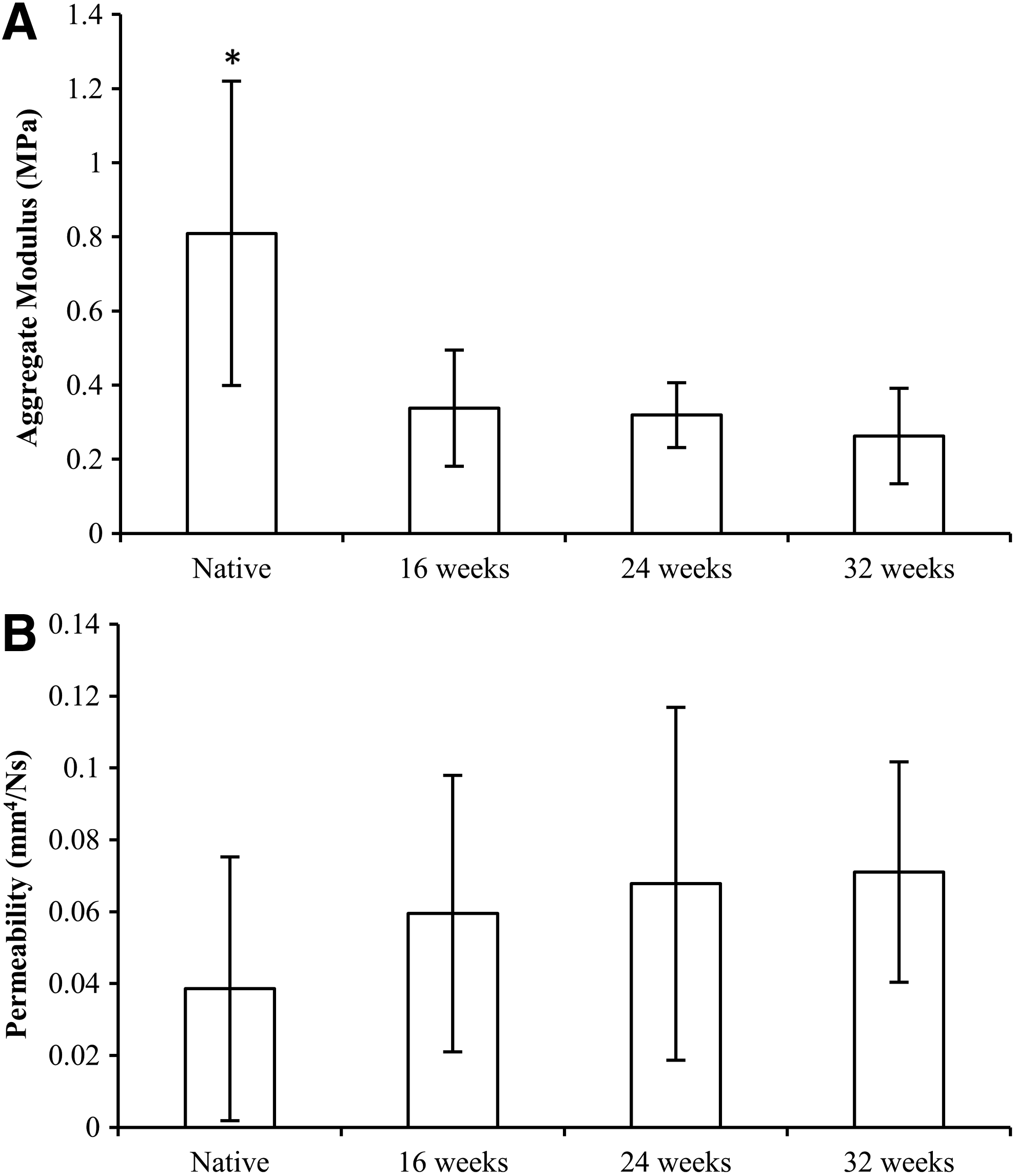
Confined compression creep results.
Explant histology
Anterior, body, and posterior cross sections stained with H&E at 16, 24, and 32 weeks are shown in Figure 6. Sections from the body region of explants were much smaller than those from the anterior and body regions. Polymer fibers were observed at all three time points, indicated by circular void spaces. The anterior and posterior regions at all three time points contained more remaining polymer fiber than the body region. Cellular infiltration was seen through the interior of the explant, with no discernible differences in cell density between the inner and outer margins, but more cells were observed in the anterior and posterior regions than in the body region. Cells appeared elongated near polymer fibers, likely corresponding to fibroblasts. Native meniscus tissue contained very few cells that were well dispersed. New collagen deposition and organization were detected in explants at 16 weeks, indicated by a pink-purple color with some aligned collagen fibers. However, at 24 and 32 weeks, ECM staining did not appear as strong as at 16 weeks. For all three time points, the body region contained more areas of aligned ECM, indicative of organization. Blood vessels were observed in low quantities (one to five per section) in all three regions at all three time points, but were most common in the anterior region. No encapsulation or rejection response was observed, and a few macrophages per section were observed, usually close to the remaining polymer fiber.
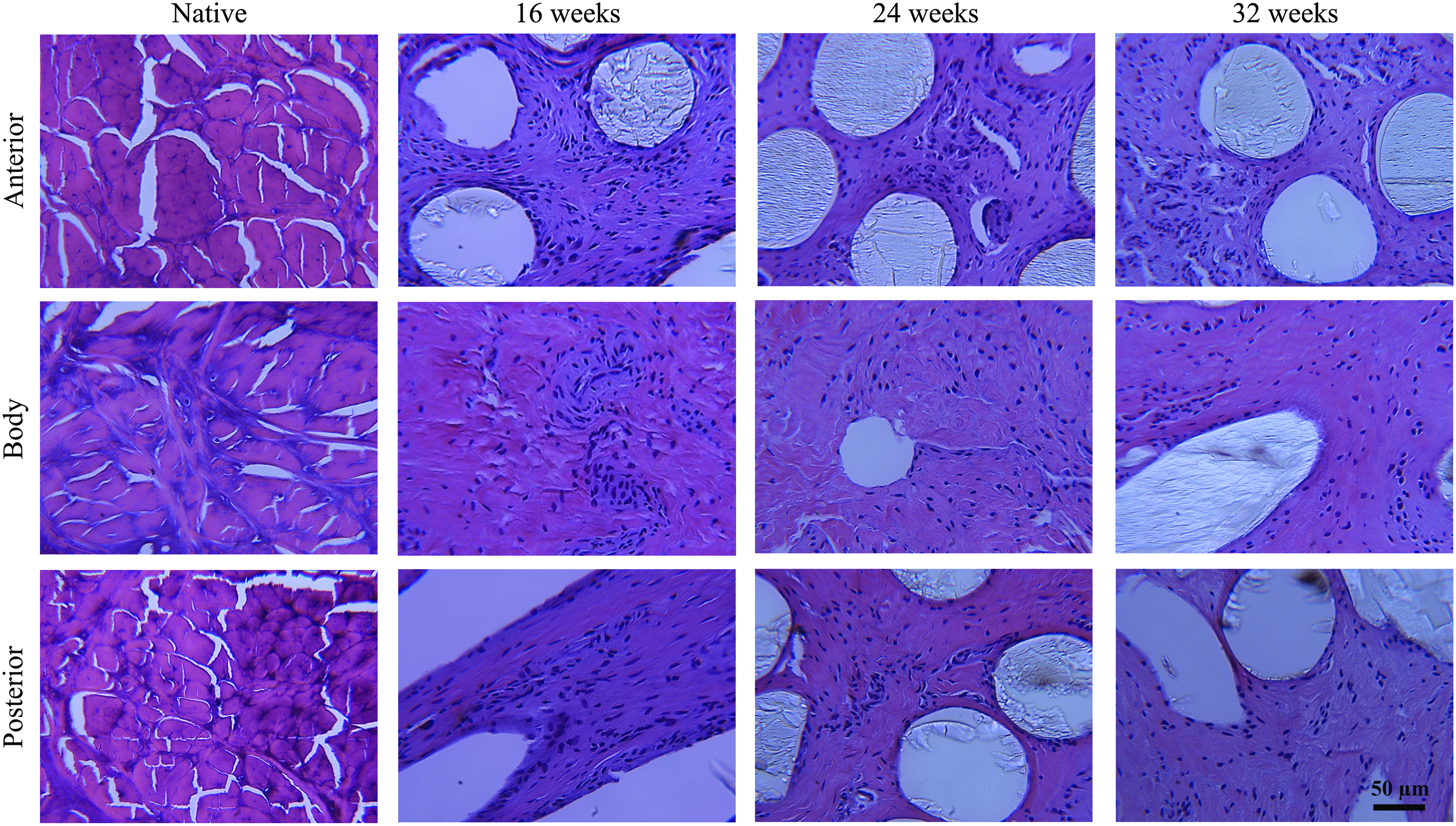
H&E-stained images of PLLA fiber-reinforced meniscus explants at 16, 24, and 32 weeks compared to the native ovine meniscus (magnification 200 × ) in the anterior, body, and posterior regions. Circular void spaces indicate PLLA fiber, which was largely removed during histological processing. H&E, hematoxylin and eosin. Color images available online at www.liebertpub.com/tea
Biochemical analyses
Water content did not statistically differ between native (67.78% ± 3.42%), 16-week (75.97% ± 6.99%), 24-week (71.44% ± 7.72%), and 32-week samples (69.44% ± 5.66%). Native tissue had significantly greater collagen and S-GAG content than all explant groups. Explant groups did not statistically vary between time points for either biochemical analysis, but a slight progression was seen from 16 to 24 to 32 weeks. By 32 weeks, collagen and S-GAG content was 26.28% and 64.39% of native tissue, respectively. Collagen and S-GAG content of explant groups as a percentage of native tissue is shown in Figure 7.

COL and S-GAG content of explant groups at 16, 24, and 32 weeks as a percentage of content in native tissue. Mean ± standard deviation. COL, collagen; S-GAG, sulfated glycosaminoglycan.
Cartilage analysis
Histology of the areas of articular surfaces unaffected by osteophytes or erosion verified superficial damage; the largest clefts were no more than 40% deep at 32 weeks. These clefts were primarily in the central region of cartilage; the medial and lateral regions experienced relatively little damage. Some hypocellularity and cell cloning were noted, as was a loss in proteoglycan stain. All tidemarks stayed intact. Tibial plateau Mankin scores were significantly different from native cartilage at 24 weeks (p = 0.0093) and 32 weeks (p = 0.0066). Mankin scores increased with time, without statistical significance, for both the tibial and femoral surfaces. Mankin scores at 32 weeks (femoral condyle: 4.33 ± 1.76, tibial plateau: 6.00 ± 1.32) were nearly twice the values at 16 weeks (femoral condyle: 2.75 ± 1.06, tibial plateau: 2.25 ± 0.35) (femoral condyle: p = 0.1865, tibial plateau: p = 0.0829). Mankin scores with sample histological images can be seen in Figure 8.
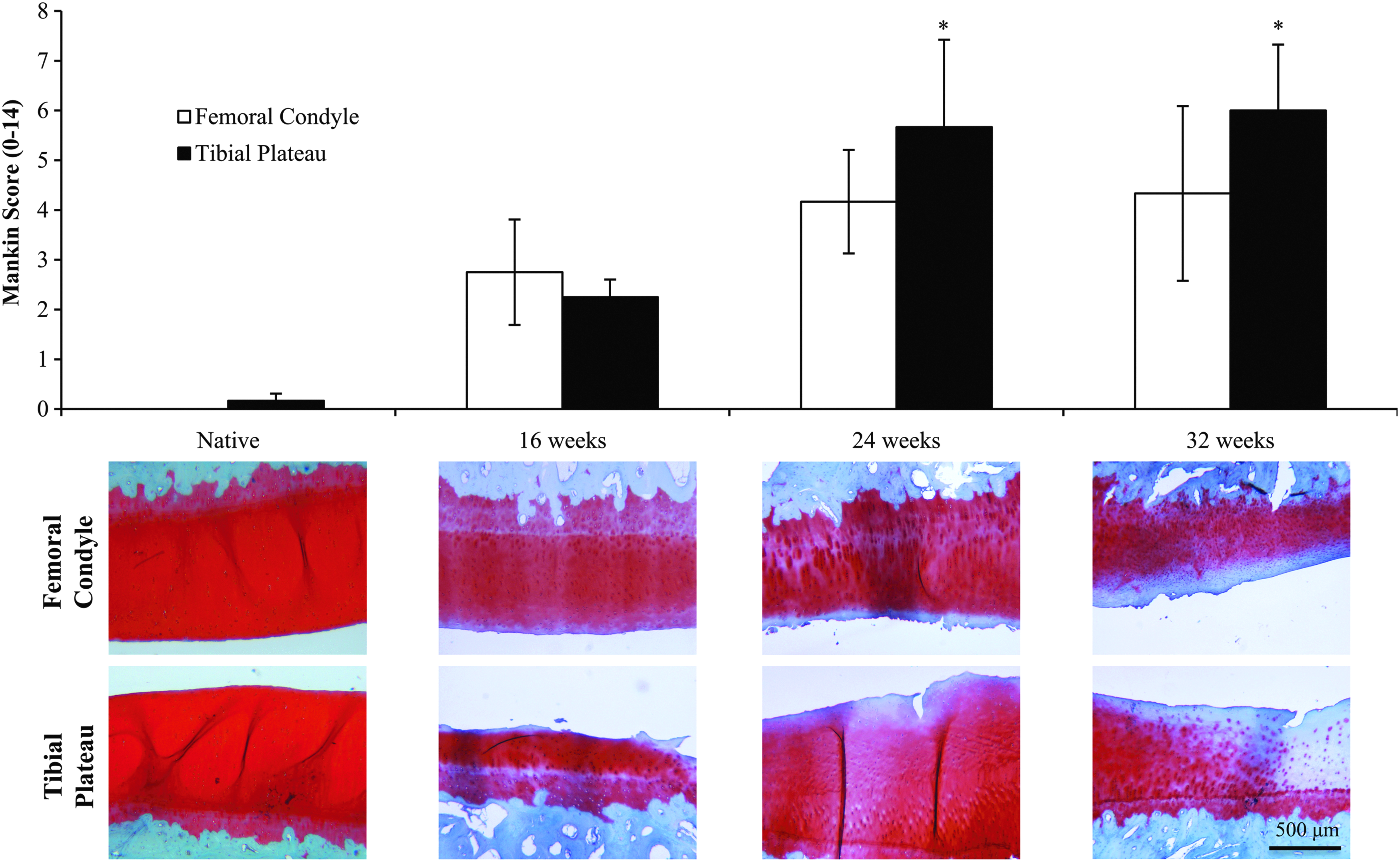
Mankin scores for femoral condyle and tibial plateau of native and experimental surfaces at 16, 24, and 32 weeks. Scores range from 0 (no damage) to 14 (full damage). Images below the graph are representative histology images (Safranin O/Fast Green) for each time point. *Denotes significant difference from native cartilage. Mean ± standard deviation. Color images available online at www.liebertpub.com/tea
Discussion
Previously, our group demonstrated efficacy of a collagen–hyaluronan sponge reinforced with p(DTD DD) fibers for total meniscus reconstruction through 52 weeks in an ovine model.13,14 Neomeniscal tissue formed within the scaffold, functional biomechanical properties were maintained, and articular cartilage was protected from damage. In contrast, results of the present study, using the same scaffold design but with PLLA reinforcing fibers, were negative with unanticipated damage to the knee joint.
PLLA was selected as a potentially useful polymer for this application due its use in other FDA-approved medical devices and musculoskeletal tissue engineering applications. It has a relatively high modulus (3.6–5.4 GPa 35 ) compared to other biodegradable fibers [p(DTD DD): 2.7 GPa, 32 PCL fibers 3.7 GPa 36 ] and has a similar modulus to native type I collagen fibrils (4.20–7.69 GPa 37 ). Yuan et al. previously established the mechanical properties of melt-extruded and hot-drawn PLLA fibers (110–165 μm diameter) with a few different starting molecular weights, noting a yield strength of 112–148 MPa and elastic modulus of 3.6–5.4 GPa. 35 The PLLA fibers from the present study exhibited similar mechanical properties (yield stress: 118 ± 9 MPa, elastic modulus: 4.30 ± 0.59 GPa), suggesting proper fabrication of these fibers. Furthermore, ultimate tensile testing of our scaffold showed comparable, if not greater, strength and stiffness than native meniscal tissue.
In the in vitro strength retention study, there was a marked change in PLLA fiber yield stress between 8 and 16 weeks, leaving the fibers more susceptible to rupture under load. This large decrease in yield strength may have been due to autocatalytic degradation; once the PLLA fibers begin degrading, the free carboxylic acid end-groups may self-catalyze the hydrolysis of the remaining PLLA. 38 For example, Huang et al. 39 showed partial degradation of PLLA in a composite bone scaffold at 12 weeks, allowing hydrolysis to occur at a faster rate. By 18 weeks, their scaffolds had completely collapsed, similar to the major decrease in tensile properties between 8 and 16 weeks in the present study.
Assuming similar degradation kinetics in vivo, this leaves a small window of time for sufficient tissue ingrowth and remodeling to occur, in order for the tissue, not the fibers, to bear mechanical loads.
The PLLA scaffold in this study remained intact through 8 weeks, exhibiting morphology consistent with the native meniscus. However, at 16 weeks, significant implant narrowing had occurred, and by 32 weeks, rupture occurred in two out of three animals. Therefore, the hypothesis that implants would remain intact and maintain shape was rejected. These implant ruptures and narrowing were consistent with in vitro findings, where a significant loss in mechanical integrity occurred between 8 and 16 weeks in the strength retention study. Autocatalysis may have accelerated degradation of the PLLA fiber, due to the inability of the knee joint to clear the lactic acid degradation products around the 16-week time point. In areas of the body with limited vascularity, like the intrasynovial environment of the knee, this buildup of lactic acid can be especially problematic.
A buildup of lactic acid and/or the rapid loss of scaffold mechanical integrity due to PLLA degradation are probable causes for the joint abnormalities observed in this study. The articular surfaces in direct contact with the PLLA scaffold displayed osteophyte growth and bone erosion that progressed with time. There have been previous reports of PLLA-based scaffolds and/or implants causing bone erosion, osteolysis, and synovitis. The applications explored in those studies included wrist suture anchors, 40 shoulder tacks, 41 and rotator cuff implants. 42 Lactic acid can attack neighboring tissue, in this case, the articular surfaces. With the body region of the scaffold containing little tissue, the fiber that degraded from this region could have caused erosion on the femoral condyle. In addition, the higher concentration of fibers within the scaffold tails and at the horn sites could have caused the erosion seen next to the bone tunnel attachment sites.
We hypothesized that the compressive properties would progress toward native values with time; however, the opposite occurred, causing the rejection of this hypothesis. Following explant rupture or narrowing, the cells recruited to the scaffold may not have received sufficient mechanical stimulus, meaning they would not deposit and organize tissue. This reasoning helps to explain the compressive creep results, where a twofold increase in aggregate modulus from time-0 scaffolds to 16-week explants indicates that tissue deposition occurred while fibers remained intact. However, the narrowing and ruptures following 16 weeks did not allow for this neomeniscal tissue growth to continue, causing the modulus for the PLLA explants to decrease. The lack of dense ECM development in the explants would also allow fluids to move more freely, explaining the relatively high permeability of the explants compared to the native meniscus.
The histological evaluation of the explants was consistent with the poor biomechanical results. The tissue achieved some deposition and organization by 16 weeks, but did not become further organized following this time point. While the body region contained some areas of aligned collagen fibers even at 32 weeks, the cross sections were thin and narrow. Therefore, the hypothesis that PLLA implants would support neomeniscus tissue deposition and organization was rejected. In addition to not being properly mechanically loaded, a buildup of lactic acid within the explant could have also prevented tissue deposition and organization. There are several studies that have shown that a buildup of lactic acid in the intervertebral disc (IVD) can cause tissue degeneration. While a buildup of acid in the IVD is caused by inadequate nutrient supply as opposed to degrading fiber, it affects the ability of IVD cells to produce and maintain ECM, leaving the tissue susceptible to rupture.43–45 Due to the similarities between the IVD and meniscus, the lactic acid from the hydrolysis of PLLA fibers may have prevented, and even reversed, the neomeniscal tissue deposition and organization after 16 weeks. The potential effect of lactic acid on neomeniscal tissue formation was also evident in the biochemical analyses. While collagen and GAG content slightly increased between time points, their values were still well below those of native tissue.
In addition to the cartilage destroyed by osteophytes or erosion, the intact regions of cartilage also experienced progressive damage. The Mankin scores at 32 weeks in this study (femoral condyle: 4.33 ± 1.76, tibial plateau: 6.00 ± 1.32) resembled those found by Hannink et al. (femoral condyle: 6.0, tibial plateau: 6.5), and the joints in that study were osteoarthritic by 24 months. 46 Assuming a similar trend with the PLLA scaffold from this study is justified based on the increasing scores. It is also important to note that the cartilage from the abnormal regions would have Mankin scores close to or at the maximum of 14, and that up to 40% of these surfaces could be classified with such a score. Therefore, the progression of cartilage damage and joint abnormalities from 16 to 24 to 32 weeks indicates that these joints would almost certainly have progressed to osteoarthritis, contrary to the hypothesis that PLLA implants would prevent the progression of articular cartilage damage. In fact, the overall joint damage observed in this study was worse than in meniscectomized knees in our previous study, further suggesting that the abnormalities were due to the choice of material. 13
To our knowledge, this is the first report documenting poor outcomes when PLLA-based fibers were used as the major component of a total meniscus implant. However, these results are consistent with several reports of poor tissue compatibility in response to degrading PLLA-based devices in other musculoskeletal applications.40–42 Negative outcomes have also been observed in large animal studies using other synthetic polymers, but these were due to device design failure rather than a response to the material. Chiari et al. 47 tested a porous PCL-hyaluronan composite scaffold in an ovine model for 6 weeks, and the scaffold was augmented with circumferential PLLA fibers. However, only four fibers were used, to achieve suture fixation. The porous composite displayed excellent tissue in-growth, but implant compression and extrusion were observed and attributed to mechanical issues and improper surgical fixation.
There were some limitations in this study. Tensile testing was not performed on explanted PLLA scaffolds, because with three complete ruptures and limited tensile integrity in the intact samples, compression creep testing was the only possible mechanical characterization. An additional limitation of this study is that the animals were allowed to stand freely several hours after the procedure, a practice that we used in previous studies but is not representative of postoperative care in humans.
The implant failures and cartilage damage indicate that PLLA fiber as fabricated in this study cannot be used as reinforcement in the total meniscus reconstruction device. Furthermore, the formation of significant osteophytes and bone erosions less than 1 year postoperatively suggests that not only is the PLLA implant lacking long-term load transmitting qualities but is also negatively impacting the joint, most likely by a buildup of acidic by-products during degradation.
In contrast, p(DTD DD) does not release acidic by-products during degradation. A p(DTD DD) fiber-reinforced total meniscus replacement scaffold was previously shown to remain intact, maintain functional mechanics, support deposition of organized tissue, and protect the articular surfaces through 52 weeks.13,14 In those studies, compressive modulus and tissue deposition were shown to increase toward native properties with time. In addition, collagen and S-GAG content was 65% and 110% of native tissue, respectively, with the p(DTD DD) scaffolds, roughly twice the values found for the PLLA scaffolds. Results of these separate studies strongly suggest that scaffolds reinforced with p(DTD DD) fibers are more efficacious than scaffolds reinforced with PLLA fibers. Thus, the next steps in development of the total meniscus replacement device will be to test the p(DTD DD) fiber-reinforced scaffold to longer time points in the sheep model, and complete additional studies using p(DTD DD) scaffolds designed and sized for clinical use.
Footnotes
Acknowledgments
This study was funded, in part, by a research contract from Novopedics, Inc. The authors thank the Orthopaedic Research Laboratory, the vivarium staff, and especially Barbara Perry, for their assistance with the surgeries and postoperative care. They also thank Sanjeeva Murthy, PhD, for his assistance in extruding and drawing the polymer fibers.
Disclosure Statement
One or more of the authors have declared the following potential conflict of interest relating to NovoPedics, Inc, a startup company developing the meniscus implant for potential future commercialization. J.M.P. is a consultant for NovoPedics, Inc. J.K. is on the scientific advisory board of NovoPedics, Inc. C.J.G. is on the board of directors of, is interim president of, and owns stock in NovoPedics, Inc. M.G.D. is interim secretary and treasurer of and owns stock in NovoPedics, Inc. Neither receives any salary from the company. C.J.G. and M.G.D. are inventors on two United States patents, and A.R.M. is an inventor on one United States patent for the meniscus implant design described in the article.