
Abstract
Nanohydroxyapatite (nanoHA) is a well-established synthetic bone substitute with excellent osteoconduction and osteointegration. However, brittleness coupled with slow degradation curtails its load-bearing and bone regeneration potential, respectively. To address these limitations, nanoHA composite matrix reinforced with electrospun fibrous yarns was fabricated and tested in vitro and in vivo. Different weight percentages (5, 10, 15 wt%) and varying lengths (short and continuous) of poly(
Introduction
B
Native bone, at the nanoscale, comprised mineral apatite, which is reinforced by collagen fibrils, and this interaction is known to significantly dictate its strength and toughness.
5
Based on this perception, to mimic the bone architecture, researchers have developed fiber-reinforced CPC matrices, mainly utilizing melt-spun fibers. These include both nonresorbable (polyamide 6.6, aramide, carbon fibers) and resorbable fibers (polylactides, poly (
As an alternative to melt-spun fibers, electrospun fibers with high surface area and nanoscale dimensions that mimic the natural extracellular matrix emerged.
12
Electrospun fibrous matrices have shown to enhance the adhesion and osteogenic differentiation of mesenchymal stem cells (MSCs).
13
However, studies on electrospun fiber-reinforced CPC matrices are sparse. While one group used poly (
In this study, we report the reinforcement of silica-coated nanoHA composite matrix using electrospun continuous yarns to generate a biomimetic scaffold having porosity, mechanical stability, biodegradability, and osteoconductivity. Nair et al. have demonstrated that silica coating of HA imparts enhanced osteogenic differentiation, bone formation, and biodegradation when compared to pure HA.18,19 Based on this, we utilized amorphous silica-coated nanoHA—gelatin matrix, which was reinforced with PLLA fibrous yarns (short and continuous yarns at 5, 10, and 15 wt%). A detailed in vitro analysis of the viability, proliferation, and osteogenic differentiation of adipose-derived mesenchymal stem cells (ADMSCs) was carried out. Finally, the ability of this composite fibrous scaffold to support bone regeneration in a critical-sized segmental defect in rat femur was analyzed in comparison to commercial HA graft. This is the first report demonstrating the efficacy of electrospun continuous yarn reinforced bioceramic composite to regenerate bone in a critical-sized defect in vivo.
Materials and Methods
Fabrication and characterization of electrospun yarns
PLLA (14% w/v) (Mw = 100 kDa; Good Fellow, United Kingdom) was dissolved in chloroform and acetone in a 3:1 volume ratio. PLLA yarns were fabricated using a novel electrospinning technique as reported by our own group. 20 Briefly, the electrospinning setup consists of dual spinnerets, an open-ended collector comprising plurality of tines connected at one end forming an open structure. Polymeric solution was loaded into a syringe maintained at +12 kV and injected at a flow rate of 3.5 mL/h. The fluffy deposit formed at the center of the grounded collector was drawn to form continuous yarns. The surface morphology of the fabricated yarns was characterized by scanning electron microscopy and the tensile strength by materials testing machine (Tinius Olsen L-series, H5KL).
Development of composite fibrous scaffolds
HA nanoparticles were synthesized by an aqueous precipitation method. Diammonium hydrogen phosphate (0.3 M; Merck) solution was added dropwise into calcium chloride (0.5 M; Merck) solution at 90°C with constant stirring. Simultaneously, ammonium hydroxide solution (Merck) was added to the solution mixture to maintain pH of 10–12. The solution mixture was centrifuged, washed with deionized water, dried, machine powdered, and coated with silica as reported earlier. 21 This powder was then dispersed in an aqueous solution of gelatin (10% wt/v Type-A; HiMedia) at a weight ratio of 65:35. Electrospun PLLA yarns were then randomly dispersed in the composite slurry at two different lengths (short yarns of 3 mm length and continuous yarns) and at three different weight percentages (5%, 10%, and 15%). The slurry was extruded into suitable molds, freeze-dried, crosslinked with 0.5% glutaraldehyde (Sigma Aldrich) solution, washed with distilled water for five to six times, and lyophilized. The scaffolds developed in the study were grouped as follows: (1) CS—Composite scaffold, (2) CSF5%—Composite scaffold with 5 weight% fibers, (3) CSF10%—Composite scaffold with 10 weight% fibers, and (4) CSF15%—Composite scaffold with 15 weight% fibers. Figure 1 depicts the overall process of fabricating the composite scaffold.
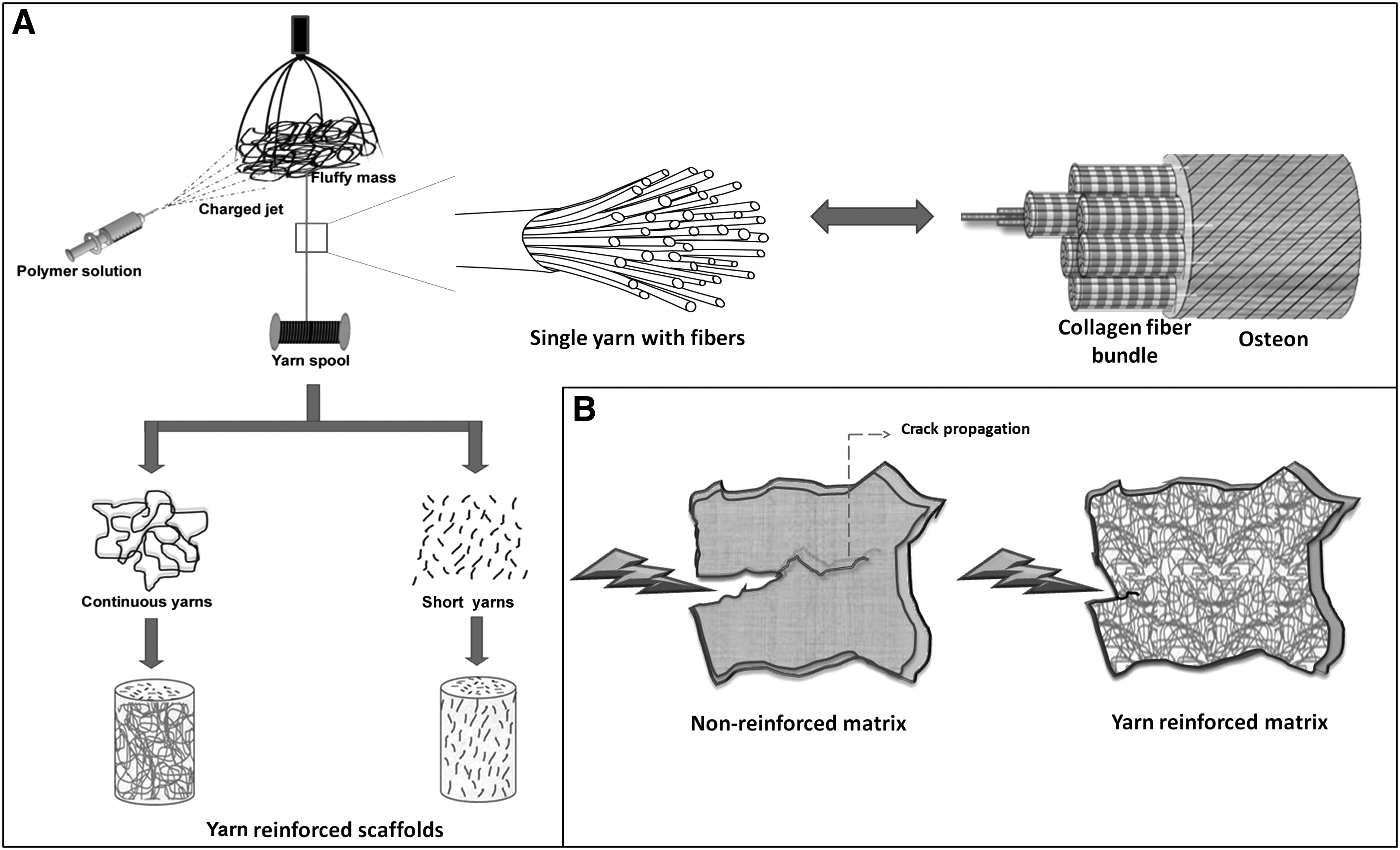
Schematic representation of
Physicochemical characterization of composite fibrous scaffolds
Morphological analysis
The morphology and pore size of the composite scaffolds were analyzed by scanning electron microscope (SEM; JEOL, JSM-6490LA, Tokyo, Japan). The scaffolds were cut to expose its internal architecture, gold sputtered (JEOL, JFC-1600), and imaged by SEM.
Mechanical testing
The compressive strength of scaffolds (n = 6) (14 × 7 mm) was evaluated by subjecting it to 80% compression using 5 kN load cell at a crosshead speed of 5 mm/min in a universal testing machine. The failure stress was obtained by dividing the maximum load registered during the test by the resistant section area. The three-point flexural test (n = 8) (scaffold size of 20 mm length × 4mm thickness × 3 mm width) was used to fracture the specimens in a universal testing machine (Instron 3365, United Kingdom) with a span of 20 mm at a crosshead speed of 1 mm min−1, and the corresponding load–displacement curve was recorded by computer. Flexural strength was calculated using the equation,
Porosity evaluation
The three-dimensional (3D) morphometry and pore size of the scaffolds were scanned and imaged using micro-CT (Micro-CT 40; Scanco Medical AG, Switzerland). The scanning was carried out with a slice thickness/slice increment = 16 mm, X-ray beam energy = 70 kV, and X-ray intensity = 114 mA. The porosity was also quantified (n = 3) using the mercury intrusion technique (Micrometrics, AutoPore IV). A few discs of known weight (0.5 g) were loaded in the penetrometer cell of the equipment, evacuated, and filled with mercury (Hg) under pressure (0.52 psi). The volume of mercury that intruded was measured as a function of pressure. The total porosity and percentage of open porosity were evaluated from the total volume of the Hg intruded.
In vitro cytocompatibility studies
MSCs were isolated from adipose tissue of adult Wistar rats after approval from the Institutional Animal Ethics Committee. The cells were cultured in α-minimal essential medium with 10% fetal bovine serum (FBS) at 37°C with 5% CO2 atmosphere. The scaffolds (4 mm diameter × 3 mm height) were sterilized by ethylene oxide for 12 h at 37°C. The cells (5 × 10⁴ cells in passage number 2–4) were seeded onto scaffolds for 1 h for an initial attachment followed by an addition of basal medium. After 24 h, the medium was supplemented with 10 mM β-glycerophosphate, 10−8 M dexamethasone, and 0.05 mg/mL L-ascorbic acid (Sigma Chemical Co., India) to differentiate ADMSCs into osteogenic lineage 22 and subjected to medium change twice a week. Commercially available porous HA blocks (BioGraft, India) with pore size in the range of 50–400 μm were used as controls. All in vitro and in vivo experiments were carried out with CSF10 in comparison to CS except live/dead assay and SEM for which CSF15 was also included.
Cell adhesion and viability
After 24 h of osteogenic induction, the cells on scaffolds were washed with PBS, stained with calcein AM (4 mM in DMSO) and ethidium bromide (2 mM in 1.4 DMSO/H2O) for 5 min, and imaged by confocal microscope (Leica TCS SP5 II) under green (480 nm/520 nm) and red (605 nm/635 nm) filters. For cell adhesion studies, the same scaffolds were washed with PBS, fixed with 1% glutaraldehyde in Sorenson phosphate buffer, and dehydrated with increasing concentrations of ethanol (50%, 70%, and 80% for 10 min twice, and 90%, 100% for 15 min each). The samples were then air-dried, sputter-coated with gold, and viewed under SEM (JEOL, JSM-6490LA).
A quantitative analysis of cell viability on the scaffolds after 1, 7, 14, 21, and 28 days of culture was obtained by the measurement of cytosolic LDH activity using the Cytotox96 kit (Promega, Madison, WI). For this, the cells on the scaffolds (n = 6) were lysed with 1% Triton X-100 for 50 min with sonication for 10 min. An aliquot of each cell lysate (50 μL) was mixed with LDH substrate at room temperature and the enzymatic reaction was stopped after 30 min. The absorbance was then read at 492 nm (Biotek Powerklave XS). The percentage cell viability was calculated with respect to the viability of cells on porous BioGraft HA blocks (control).
Cell proliferation
Cell proliferation after 1, 7, 14, 21, and 28 days of cultivation (n = 6) was determined using the PicoGreen® dsDNA Quantitation kit (Invitrogen) according to the manufacturer's instructions. For this, the cells were lysed as described above and the lysate (50 μL) was mixed with PicoGreen in Tris-EDTA buffer (200 mM Tris-HCl, 20 mM EDTA, pH 7.5). The intensity of fluorescence was measured with a multifunction microplate reader (DTX 880 multimode detector; Beckman Coulter) at excitation and emission wavelengths of 485 and 535 nm, respectively. Relative fluorescence units were correlated with cell number using a calibration line constructed with increasing concentrations of cells. The percentage cell viability was calculated with respect to the viability of cells on porous BioGraft HA blocks (control).
Osteogenic differentiation
Alkaline phosphatase activity
Alkaline phosphatase (ALP) activity of osteogenic-induced rat ADMSCs after 1, 7, 14, and 21 days of culture was determined based on the hydrolysis of p-nitrophenyl phosphate (Sigma) to p-nitrophenol (n = 6). For this, the cell cultured materials were lysed with 1% Triton-X-100. The lysate was added to 50 mL ALP reaction buffer (pH 9.8; Sigma), incubated at 37°C for 30 min, followed by the addition of 3N sodium hydroxide to stop the enzymatic reaction, and the absorbance was read at 405 nm (Biotek Powerwave XS). A calibration line was plotted using different concentrations of ALP enzyme.
Osteocalcin release
The concentration of osteocalcin (ng/mL) in the medium in which cells were grown on scaffolds (n = 3) was determined using the ELISA technique (QnDSystems™-Rat Osteocalcin ELISA Kit). For this, the medium was collected on days 7, 14, 21, and 28 and the level of osteocalcin was analyzed as per the manufacturer's instructions. The absorbance was recorded at 405 nm against a reference filter of 630 nm. A calibration curve was plotted and the osteocalcin concentration was determined with respect to calibration curve.
In vivo studies
Animal surgical procedure
Six- to seven month old Wistar rats (n = 6), weighing 300–350 g, were used for the study. Method and procedures adopted for the management, surgery, and care of the animals were approved by the Institutional Animal Ethics Committee of Amrita Institute of Medical Sciences and Research Centre, Kochi, India. The animals were maintained under anesthesia via intramuscular injection of ketamine hydrochloride at 75 mg/kg body weight and xylazine hydrochloride at 5 mg/kg body weight. Rats were placed in a right lateral recumbent position, and a long transverse skin incision of ∼2.5 cm running the full length of femur was created, exposing muscles and fascia. After removing the periosteum, a 5 mm segmental defect was created in the mid-diaphysis using a dental burr and the defect was stabilized with stainless steel plates and screws. The scaffolds (CS, CSF10, and BioGraft HA) were placed at the defect site, after which the defect was closed using vicryl sutures of size 4-0. Sham (defect without scaffolds) was used as the control. It was noted that composite matrix not without fibers was losing its integrity on contact with blood during the procedure and it was only through very careful handling that we could get a minimum number of samples for implantation (at least two to three scaffolds per animal). However, the yarn reinforced scaffolds were intact throughout the study (Supplementary Fig. S1; Supplementary Data are available online at www.liebertpub.com/tea). Meloxicam (5 mg/kg body weight) analgesic and ceftriaxone antibiotic (20 mg/kg body weight) were administered subcutaneously and intramuscularly, respectively, after the surgery for 5 days. The animals were euthanized after 2 and 4 months of implantation through an overdose of CO2 gas in a euthanasia chamber. Femoral tissue was harvested and fixed in 10% neutral buffered formalin. Radiographs of the defect site were taken immediately after surgical implantation (day 0) and at the end of 2 and 4 months using an in vivo imaging system (KODAK in-vivo Imaging Systems; Carestream).
Histological and histomorphometric analysis
For histological analysis, the postimplanted samples were dehydrated in graded ascending series of alcohol, infiltrated, and embedded in polymethyl methacrylate (Merck). Longitudinal sections (100–120 μm) were made from each block using a high-speed precision saw (Isomet 5000 Precision Saw; Buehler) and polished to a thickness of 50–70 μm. The sections were stained with Stevenal's blue and van Gieson's picrofuchsin and viewed under a light microscope (10X; Olympus BX51). Histomorphometric analysis was done using calibrated stereomicrographs (Leica MZ 10 R, Germany) and image analysis software ImageJ. The percentage area of the newly formed bone (orange-red by van Gieson's picrofuchsin staining) and the material that remained (bluish black) at the defect site were quantified by marking the total area of the defect (5 mm) as 100%.
For hematoxylin and eosin (H&E) staining, formalin fixed tissues were decalcified using 5% nitric acid, dehydrated in increasing concentrations of ethanol, and embedded in paraffin. Serial sections were stained with H&E and imaged at 10X magnification using a light microscope (Olympus BX51).
Morphological and elemental analysis
To know the degradation nature of scaffolds, the tissues after 4 months of implantation were fixed in 3% glutaraldehyde in Sorenson phosphate buffer at 4°C overnight, washed, and viewed under SEM (JEOL JSM-6490 L). The elements present in the samples, especially Ca, P, Si, and O, were plotted by energy dispersive X-ray spectroscopy (EDS) (JEOL JSM-6490 L). Line mapping was done in three different fields of two samples to know the distribution of Si throughout the defect.
Statistics
All experimental trials were performed using six separate samples. Statistical analysis was carried out by one-way analysis of variance. All results are expressed as mean of all values ± standard deviation, and p-values less than 0.05 were considered significant for all analyses.
Results
Physicochemical characterization
Characterization of electrospun yarns
As a first step toward the development of the composite fibrous scaffold, polymeric yarns were obtained through a novel electrospinning technique. Figure 2A depicts the continuous PLLA yarn spool having yarns of typical diameter 250–400 μm (Fig. 2B) and constituted of individual fibers of diameter 200–600 nm (Fig. 2C). It is clearly evident from the SEM images that the fibers within the yarns are well aligned, with a compactly twisted architecture. The twisting process imparted good tensile strength (27.6 ± 4.42 MPa) to these micron-sized polymeric yarns.

Characterization of electrospun yarns and composite fibrous scaffold.
Scaffold morphology
The electrospun yarns were incorporated into gelatinous matrix containing silica-coated nanoHA at different weight percentages (5, 10, 15) and lengths (short and continuous). Figure 2D–I shows the scanning electron micrographs of CS and CSF at low and high magnifications, revealing their porous geometry with pore sizes ranging from 50 to 350 μm. No significant differences in the pore size were noted between the scaffolds reinforced with short and continuous yarns, although the distribution of fibrous yarns in the matrix was markedly different. In case of scaffolds with continuous yarns, bundles of yarns were uniformly and randomly arranged (Fig. 2H, I), while in the other group, the yarns were loosened and individual fibers were randomly dispersed in the matrix (Fig. 2F, G).
Mechanical strength
An evaluation of the mechanical strength of the scaffolds with short and continuous yarns was conducted. Generally, the compressive strength of yarn reinforced scaffolds was noticeably higher compared to the scaffold devoid of yarns. However, the yield strength was not significantly altered for scaffolds reinforced with shorter yarns (Fig. 3A). On the contrary, scaffolds reinforced with continuous yarns exhibited significantly improved yield strength, with values identical to their ultimate compressive strength (Fig. 3B). Figure 3C and D depicts the variations in stress–strain behavior for CSF (with short and continuous yarns) in comparison to CS, clearly displaying differences in their break pattern. While CS underwent failure at lower strain values, short yarn reinforced scaffolds followed a quasibrittle pattern of material failure at all weight percentages of fiber loading. In contrast, continuous yarn reinforced scaffolds exhibited a more ductile pattern of failure, especially at 10 and 15 wt% of fiber loading. These scaffolds could withstand more stress in comparison to short yarn reinforced ones and did not undergo failure and maintained integrity with increasing load.
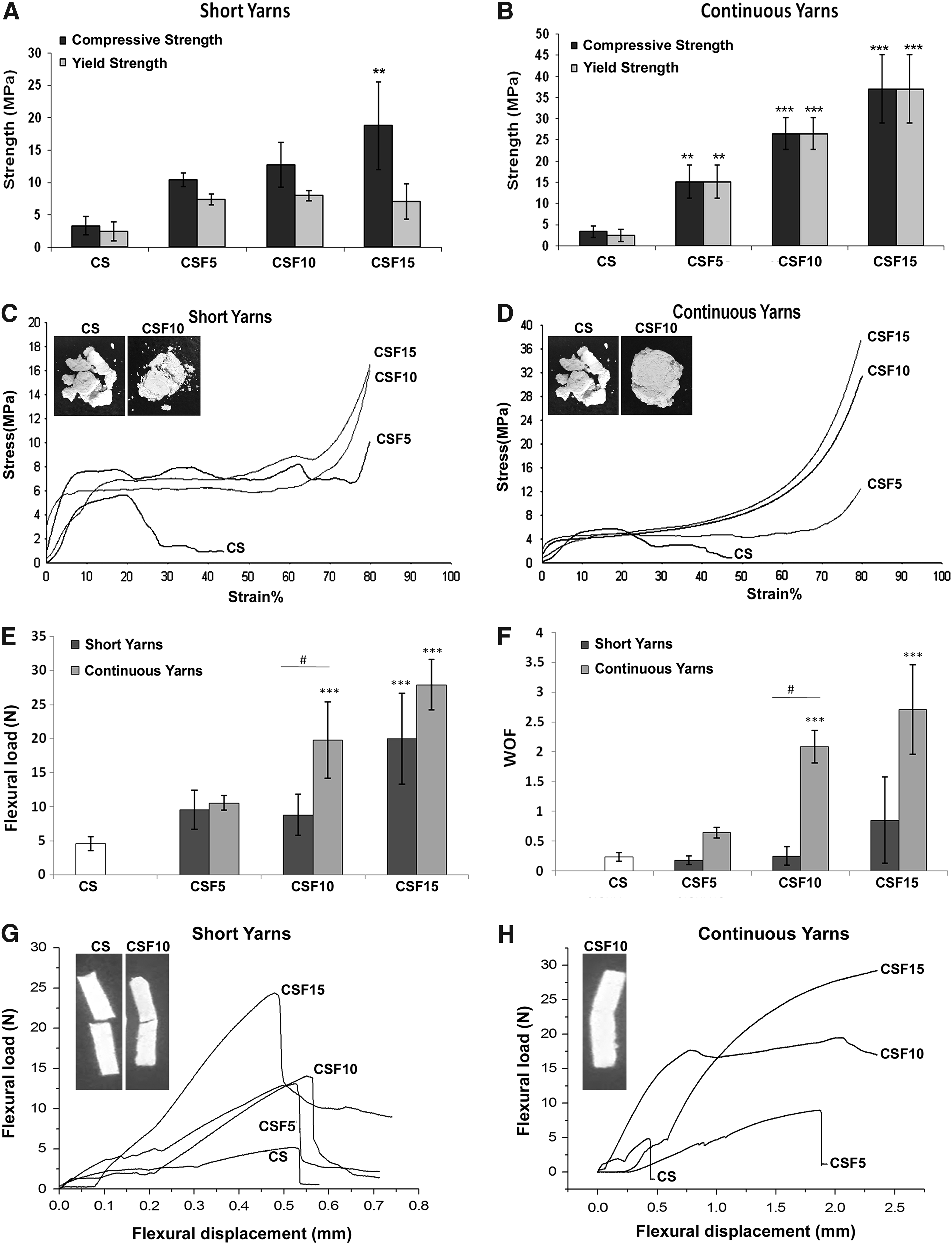
Evaluation of mechanical properties of the composite scaffolds reinforced with short and continuous yarns at different weight percentages.
Flexural testing of the scaffolds was evaluated by a three-point bending test. Both flexural load and work of fracture (Fig. 3E, F) were considerably augmented with continuous yarn incorporation in the matrix, especially for 10 and 15 wt% yarns. For short yarn reinforced scaffolds, although the flexural load increased significantly for 15 wt%, the WOF values showed no improvement with yarn reinforcement. The load–displacement curves obtained from three-point bending tests are illustrated in Figure 3G and H. As clearly evident, composite matrix was brittle and failed at lower strain values. Similar was the case with short yarn reinforced scaffolds, at all proportions of fiber loading. In contrast, scaffolds incorporating 10 and 15 wt% continuous yarns (CSF10 and CSF15) (Fig. 3H) demonstrated resistance to cracking even for displacements as large as ∼2 mm and endured higher loads. Owing to the poor mechanical characteristics, short yarn reinforced scaffolds and continuous yarn reinforced scaffolds with 5 wt% were omitted from additional analysis.
Porosity
Porosity of the scaffolds was verified by mercury porosimetry and micro-CT (Fig. 4). As evident from the representative micro-CT images, composite matrix with and without yarns were porous in nature (Fig. 4A, B). Mercury porosimetry studies evinced that the material has an open network of pores and most of the pores were in the size range of 50–350 μm (Fig. 4C). The percentage porosity calculated for CS, CSF10, and CSF15 was 60.0% ± 6.53%, 58.83% ± 7.35%, and 62.14% ± 5.10%, respectively.
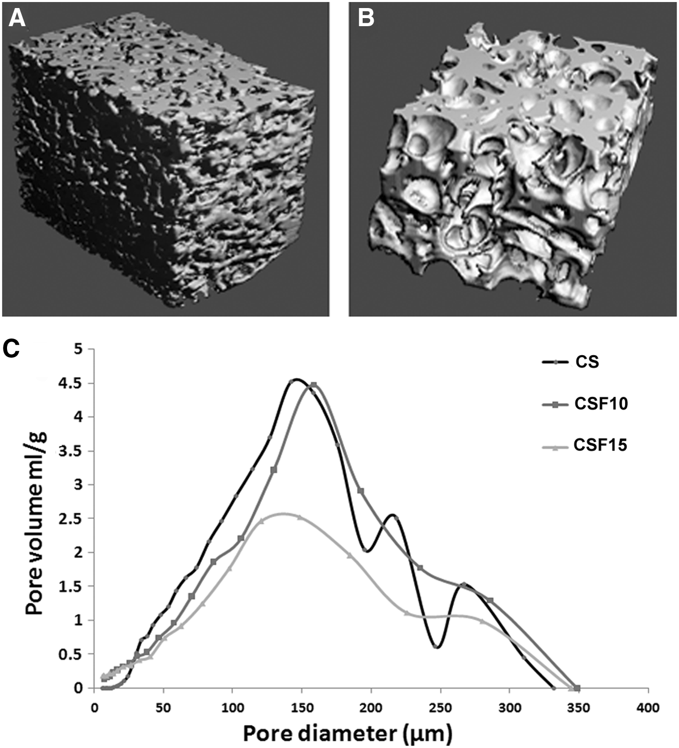
Porosity evaluation of composite fibrous scaffolds; Micro-CT images of
Cytocompatibility studies
The viability and adhesion of ADMSCs on CS, CSF10, CSF15, and HA were evaluated by scanning electron microscopy and confocal microscopy after 24 h of osteogenic induction. Live cells appear to have adhered and spread on the surface of all the group of scaffolds, without occluding the pores (Fig. 5A, B). Nevertheless, the number of viable cells was relatively less on CSF15 than CSF10. Therefore, additional in vitro and in vivo studies were continued only with CSF10 scaffolds and their properties were compared to the scaffold devoid of fibers and commercially available BioGraft HA blocks.
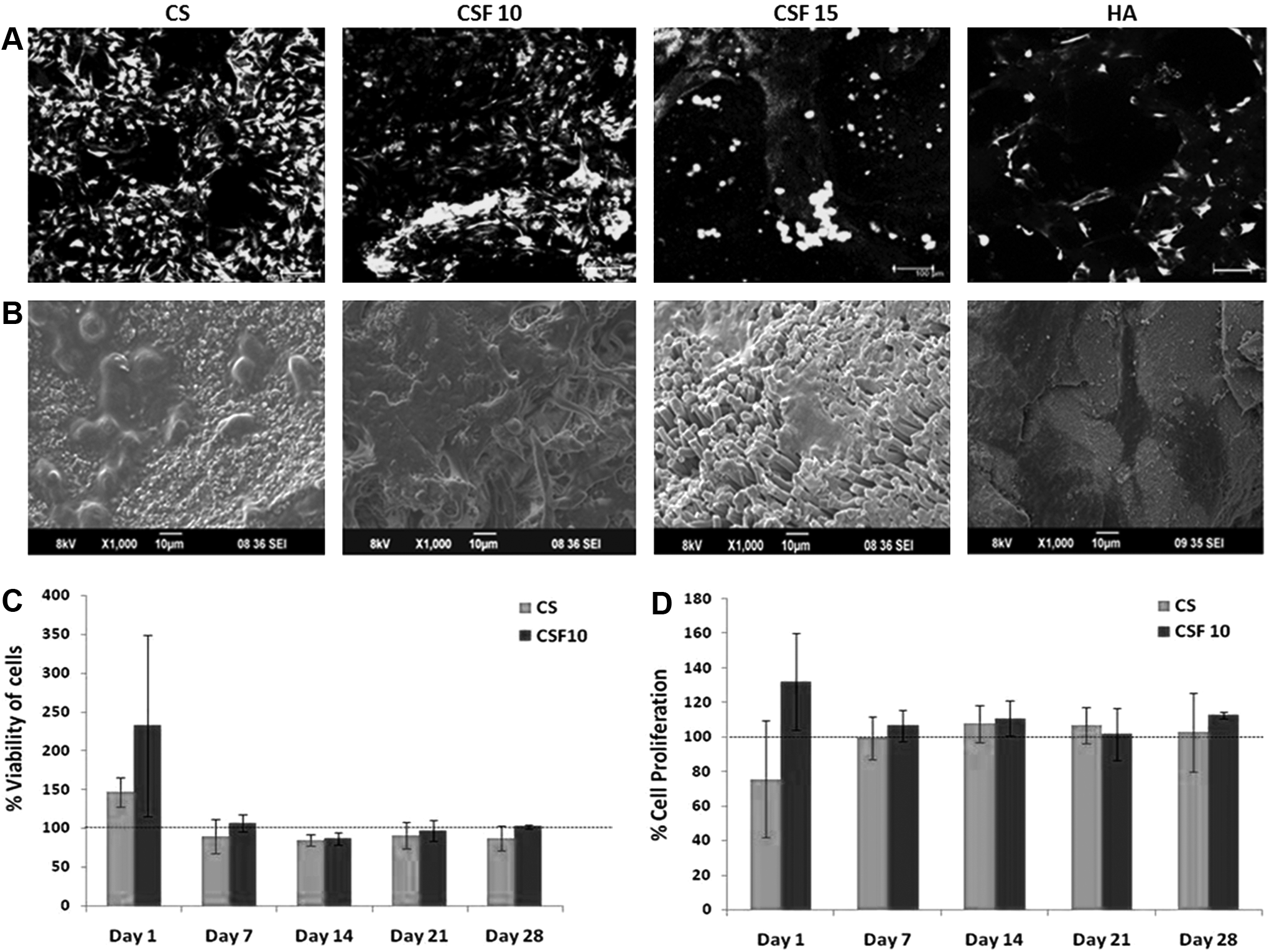
Response of osteogenic-induced rat ADMSCs on composite scaffolds.
The percentage cell viability on the scaffolds was quantified by LDH assay for 4 weeks with respect to the viability of cells on HA blocks (Fig. 5C). The cells were viable on all the groups of scaffolds on all days of culture and there was no significant difference between the groups as well as at different time points of culture. These data were again validated with PicoGreen assay and illustrated a similar trend (Fig. 5D).
To study the extent of osteogenic differentiation, the cells on the scaffolds were evaluated for ALP activity (Fig. 6A) and osteocalcin release (Fig. 6B). The ALP activity was significantly higher on CS and CSF10 scaffolds when compared to BioGraft HA on day 7 and 14. The release of osteocalcin by the cells cultured on all the groups of scaffolds was considerably higher from day 14 to 28, with CSF10 scaffold showing better release. Thus, ALP activity maximized at day 7, while osteocalcin release was the highest on all scaffolds by day 28.

Osteogenic differentiation of rat ADMSCs on the scaffolds.
In vivo studies
The animals after implantation of scaffolds did not display any evidence of inflammation or other postoperative complications. All the groups of scaffolds showed good integration with the host bone at the defect site, which was evident from the gross images (Fig. 7A). It can be noted that the scaffolds remained intact at the surgical site at 2 months in all the groups, while the residual material present at the site was remarkably less for CS and CSF10 groups at 4 months.
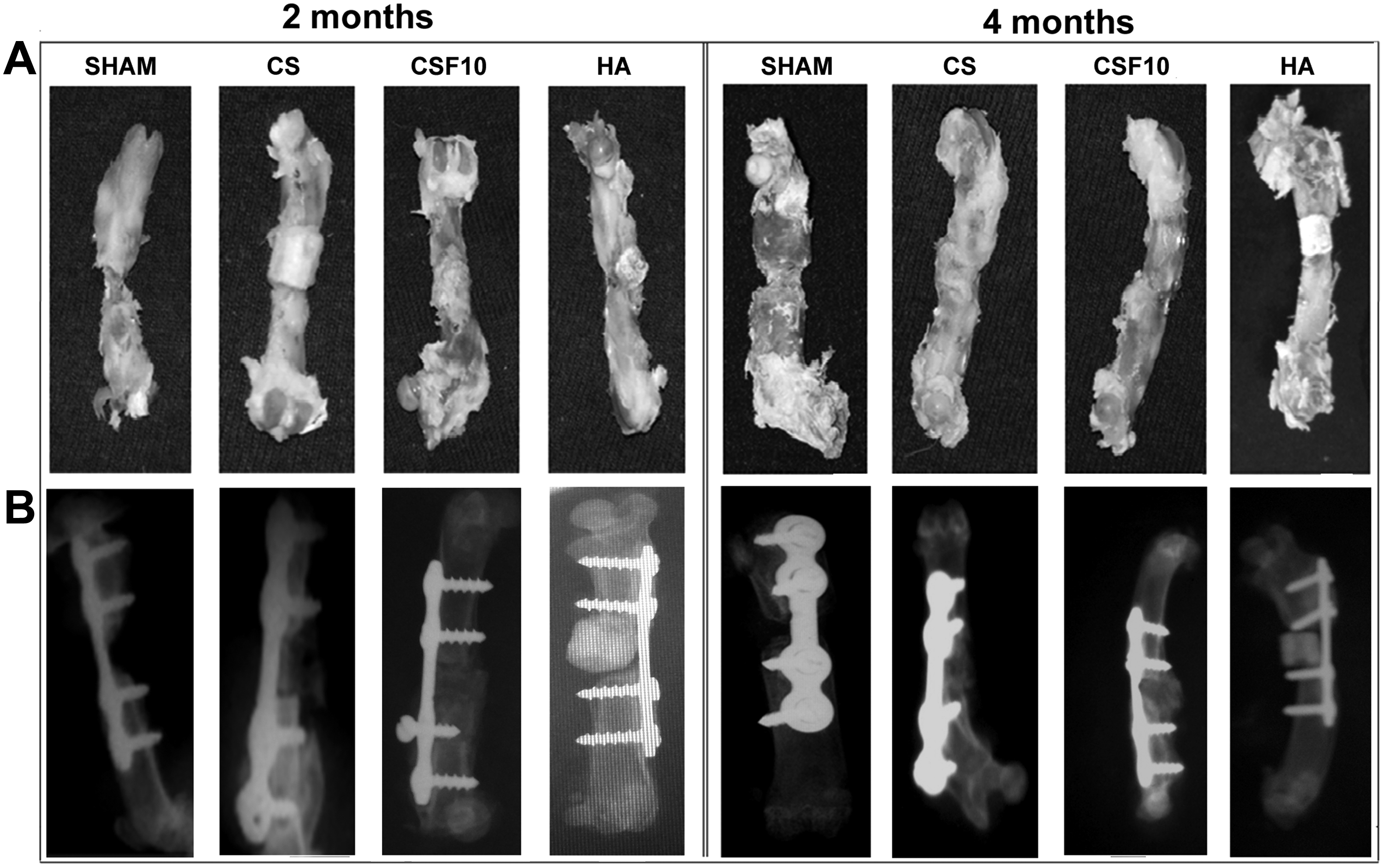
Evaluation of postimplanted samples.
The radiographs of the implanted site taken on the day of implantation did not explicitly reveal the presence of the material in CS and CSF10 groups (Supplementary Fig. S1) and that may be due to the translucent nature imparted by the gelatin content in the matrix. At 2 and 4 months, the radiopacity of the defect site implanted with CS and CSF was augmented periodically, which can be related to new bone formation. In contrast, in BioGraft HA groups, the material could be clearly visualized throughout the defect at 2 and 4 months. In the sham group, there was no union at the defect site even after 4 months (Fig. 7B).
Histological examination of PMMA-embedded tissues demonstrated the union of host bone with the new bone regenerated at the defect site for all the groups, with no evidence of fibrous tissue formation around the implant (Fig. 8). Stained sections depicted cellular infiltration, material remnants, and newly formed bone. It was evident that the peripheral region of the defect showed enhanced new bone formation as well as material degradation than the mid region. In general, there was no observable difference between the groups in the peripheral region of the defect, except sham. In peripheral areas, immature woven bone lined with osteoblast cells together with few material remnants was evident at 2 months, while an organized bone was seen at 4 months. Nevertheless, in the midregion of the defect at 2 months, the material was not completely degraded, but existed as small islands embedded among the infiltrated osteoblast cells. Interestingly at 4 months, organized new bone formation was noticed only in the CS and CSF10 group, whereas in the BioGraft HA group, immature bone and material islands lined with osteoblast cells were seen. In the sham group, there was no bone formation. The new bone formation at the defect site was further confirmed with decalcified H&E-stained tissue sections (Supplementary Fig. S2). The percentage of new bone and the material that remained at the defect site was quantitated at 4 months through ImageJ software (Fig. 8B). The area of new bone formed at the defect site implanted with CS, CSF10, and HA was 30.28% ± 0.31%, 43.69% ± 5.9%, and 7.20% ± 0.34%. The new bone formation and material degradation were significantly higher in the CS and CSF10 groups when compared to BioGraft HA. However, there was no difference between the CS and CSF10 groups.
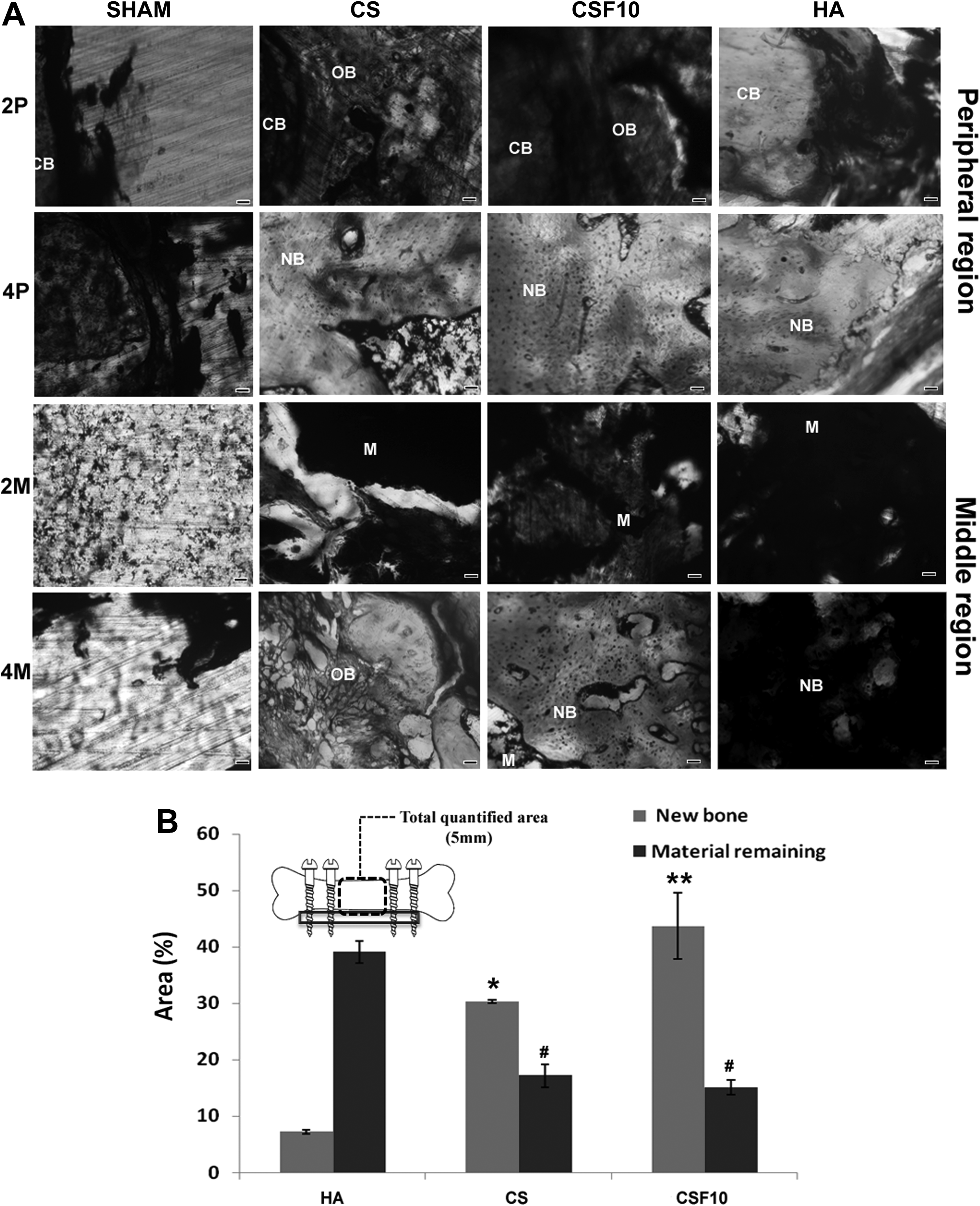
Histological and histomorphometric examination of PMMA-embedded bone tissue sections at 2 and 4 months.
The degradation nature of the composite fibrous scaffold was analyzed by SEM-EDS at 4 months. The fiber bundles in each yarn were loosened to form individual fibers, which were broken into smaller segments in many areas, as verified by SEM. The smaller fiber segments were embedded well in the new bone regenerated at the defect site (Fig. 9A, B). In addition, the elemental distribution in different areas was investigated under SEM-EDS. There was uniform distribution of Ca, P, and O throughout the tissue. However the mass% of Si was significantly reduced at the defect site at 4 months (1.27% ± 0.63%) when compared to that on day 0 (10.5% ± 1.2%), representing the biodegradation of the scaffold (Fig. 9C, D).

SEM-EDS of the defect implanted with composite scaffolds at 4 months. Top panel shows fiber degradation at low
Discussion
Bone mineral is made up of nanoHA in the form of needles or rods, having an average length and width of 50 and 25 nm, respectively. 23 Loiselle et al. demonstrated that HA nanotopographies (50–60 nm) enhance osteogenic differentiation and new bone formation with mechanical properties equivalent to live autografts. 24 In addition, doping or coating of silica onto HA has a major impact on bone remodeling. Si has shown to promote the biosynthesis of collagen by upregulating the expression of prolyl 4-hydroxylase, a key enzyme involved in collagen synthesis. 25 Silica can also act as a nucleating apatite for bone bonding and thereby promotes bone formation, while undergoing degradation in vivo. 26 Herein, rod-like nanoHA of 200 nm length and 15–20 nm diameter was developed, which was coated with a thin layer of amorphous silica and dispersed in gelatinous matrix reinforced with electrospun yarns.
Generally, the electrospinning process generates two-dimensional (2D) nonwoven sheets with aligned or random fibers using flat targets/collectors. However, the inclusion of 2D sheets into the bioceramic matrix is practically difficult. Furthermore, the porosity and pore size distribution of 2D electrospun fibrous sheets are less, restricting the migration of cells toward the inside of matrix, thereby hindering the suitability for 3D applications. 27 To overcome these limitations, electrospun PLLA yarns were used for nanoHA matrix reinforcement in this study. The yarns were developed using a novel open collector with peripheral tines, whose rotation permitted rapid yarning with controlled yarn architecture. 20
Factors such as fiber length, orientation, and volume/weight fraction play an important role in determining the mechanical properties of the composite scaffold. Gorst et al. investigated the effect of random orientation of fibers, in which random patterning preserved the modulus at increased strength, whereas regular fiber orientation led to decreased modulus. 28 Invoking the positive results, herein, we have oriented fibrous yarns in a random manner in the gelatinous matrix. Apart from fiber orientation, its length and weight/volume percentages in the matrix are an imperative factor. Earlier reports on fiber reinforcement either used short fibers randomly distributed in the matrix or continuous fibers that were nearly as long as the specimen. 12 Although increased fiber content and length is known to induce efficient ductilization in CPC matrices, its mixing and injectibility were compromised. 17 In our study, continuous yarn reinforced scaffolds not only enhanced compressive strength and ductility of the material but also dispersed well in gelatinous matrix even at higher weight percentages (up to 15 wt%). In the composite matrix reinforced with short yarns, yarns were seen as loosened individual fibers and not as intact bundles (verified by SEM micrographs). Thus, the inefficiency of short fibers to improve the mechanical properties of the scaffold may be attributed to the lower strength of loosened fibers.
Adequate pore size (100–300 μm) is an important prerequisite for bony scaffolds to allow proper vascularization, cell infiltration, and regeneration. 29 Lack of macropores is a major concern for CPC-fiber composites. Certain approaches adopted to increase the porosity considerably affected the strength and stiffness of the CPC matrix. Addition of 40 wt% mannitol to CPC powder reinforced with aramid fibers (6% volume) created well-formed macropores, having a total porosity of 70.8%, but with reduced composite strength (1.4 ± 0.4 MPa), which was much lower than the strength of bare CPC without mannitol (15.0 ± 1.8 MPa). 30 When polyglactin/PLGA resorbable sutures were utilized with a purpose of imparting reinforcement and eventual creation of macroporous channels of ∼300 μm size, a significant decrease in the material strength and work of fracture was seen in 2–4 weeks. 7 In another study, the incorporation of biodegradable electrospun fiber bundles was shown to increase porosity, but the flexural strength and elastic modulus were decreased considerably. The authors pointed that this variation is attributed to the porous structure of the fiber bundles, which weakened its strength. 15 In our study, the yarns fabricated through a novel electrospinning process were mechanically robust (tensile strength of 27.6 ± 4.42 MPa). The technique of freeze-drying adopted to develop the gelatinous matrix with those yarns (especially with 10 and 15 wt%) yielded highly interconnected porous scaffolds without compromising their mechanical strength. However, owing to the decreased cell adhesion and viability of high weight percentages of PLLA (CSF15) imparted by its hydrophobicity, 31 our biological studies were confined to CSF10 in comparison to CS alone and commercially available BioGraft HA.
Quantitative osteogenic differentiation studies on CS and CSF10 scaffolds demonstrated enhanced cellular activity when compared to BioGraft HA. The markers selected for verifying osteogenic differentiation included ALP activity and osteocalcin secretion. ALP, a cell membrane enzyme, is known to be related with early osteoblast differentiation, while osteocalcin is a marker of late osteoblast differentiation and matrix mineralization. 32 Timely cellular expression of ALP (maximized on day 7) and osteocalcin (highest on day 28) characterizes the maturation and mineralization of identified cells on CS, CSF10, and BioGraft HA, with the highest expression shown on the composite scaffolds.
The composite fibrous scaffolds, devoid of cells or bioactive molecules, were implanted in a rat femoral critical-sized segmental defect to understand the true and inherent ability of the scaffold to regenerate bone. Periosteum, which contains multipotent MSCs and osteoprogenitor cells that contribute to normal bone growth, healing, and regeneration, 33 was removed during surgery. This made bone tissue regeneration at the femoral defect created in this study more challenging. The surgical site was not able to heal spontaneously in the sham group, confirming that the defect was critically sized. 34
Ideally, the scaffold should have mechanical properties similar to the implantation site and, from a practical perception, it must be strong enough to allow surgical handling during implantation. 35 The matrix without yarns (CS) and HA blocks were losing their integrity during the implantation procedure (at least two to three blocks were used per animal) (Supplementary Fig. S1). In contrast, CSF10 remained intact owing to the toughness imparted by the fibrous yarns in the matrix. The fibrous scaffold could also be shaped for precise fit into the defect site. Once implanted, all the groups of scaffolds were stabilized with stainless steel plate and screws to withstand the force passing through load-bearing sites. Accordingly, the biomechanical properties of the fibrous scaffold during the bone remodeling process were not studied.
When materials such as HA-βTCP and coral were implanted in the segmental defect in a goat model, bone union mainly appeared at the peripheral region, while the midportion of these defects showed fibrous tissue deposition.36,37 Interestingly, in our study, the defect implanted with CS and CSF10 showed osteoblast infiltration and new bone formation throughout the defect, without the intervention of any fibrous tissue, and these scaffolds performed better than BioGraft HA. Nevertheless, there were no significant differences in bone formation between CS and CSF10. This indicates that the biomimetic chemical composition and nanotopography of the composite scaffold played a pivotal role in enhancing osteogenic differentiation and bone regeneration. Akin to bone regeneration, material degradation is an important aspect in bone tissue engineering. Herein, biodegradation was observed for both CS and CSF10 scaffolds when compared to BioGraft HA, as evident from the radiographic and histologic data. The superior resorption of the composite matrix may be triggered by the fast degradation nature of a natural polymer such as gelatin. 38 Moreover, coating of HA nanoparticles with amorphous silica may have improved biodegradation, mediated through osteoclast cells.18,39 The fibrous PLLA yarns used for reinforcement also had started to degrade, which was verified through SEM data.
Data presented here reveal that although fiber reinforcement is beneficial for enhancing mechanical strength, the chemical composition of the biomaterial matrix would dictate the ultimate biological response of a material. Incidentally, matrix reinforcement using biomimetic fibers (instead of pure PLLA fibers) could provide a better biological effect in vitro and in vivo without compromising the mechanical strength of the scaffold and those studies are in progress.
Conclusion
In this study, we demonstrate a novel biomimetic nanocomposite matrix reinforced with fibrous PLLA yarns that provided the requisite features of biocompatibility, porosity, and mechanical strength, coupled with biodegradation and bone regeneration. Continuity of the fibrous yarns in the matrix imparted augmented toughness and compressive strength, retaining its interconnected porous architecture. This fibrous matrix could also offer a favorable milieu for osteoid deposition and new bone formation in a critical-sized segmental defect in rat model. Thus, the developed biomaterial is a promising bone substitute for healing critical-sized bone defects, currently a major challenge in orthopedics.
Footnotes
Acknowledgments
The authors acknowledge the funding received from the Department of Science & Technology, Government of India, through a Thematic Unit of Excellence (SR/NM/NS-07/2011), and are grateful to Amrita Vishwavidyapeetham for all infrastructural support. Mr. Sajin P Ravi, Mr. Sharath S, Mr. Dennis Mathew, and Dr. Anusha Ashokan are duly acknowledged for their technical help; Dr. A.K.K. Unni, Dr. P. Reshmi, P. Dr. Renjith, and Mr. Amit G. Krishnan for animal surgical experiments.
Disclosure Statement
No competing financial interests exist.
References
Supplementary Material
Please find the following supplemental material available below.
For Open Access articles published under a Creative Commons License, all supplemental material carries the same license as the article it is associated with.
For non-Open Access articles published, all supplemental material carries a non-exclusive license, and permission requests for re-use of supplemental material or any part of supplemental material shall be sent directly to the copyright owner as specified in the copyright notice associated with the article.