
Abstract
Current treatment options for cartilage injuries are limited. The goals of this study are to create a biodegradable polymer scaffold with the capabilities of sustaining chondrocyte growth and proliferation, enable cell-to-cell communication and tissue regeneration through large pores, and assess the biological augmentation of the scaffold capabilities using platelet lysate (PL). We synthesized biodegradable polycaprolactone fumarate (PCLF) scaffolds to allow cell–cell communication through large interconnected pores. Molds were printed using a three-dimensional printer and scaffolds synthesized through UV crosslinking. Culture medium included alpha modified Eagle's media with either 10% fetal bovine serum (FBS) or 5% PL, a mixture of platelet release products, after being seeded onto scaffolds through a dynamic bioreactor. Assays included cellular proliferation (MTS), toxicity and viability (live/dead immunostaining), differentiation (glycosaminoglycan [GAG], alkaline phosphatase [ALP], and total collagen), and immunostaining for chondrogenic markers collagen II and Sox 9 (with collagen I as a negative control). The large interconnected pores (500 and 750 μm) enable cell-to-cell communication and cellular infiltration into the scaffolds, as the cells remained viable and proliferated for 2 weeks. Chondrocytes cultured in PL showed increased rates of proliferation when compared with FBS. The chondrogenic markers GAG and total collagen contents increased over 2 weeks at each time point, whereas the osteogenic marker ALP did not significantly change. Immunostaining at 2 and 4 weeks for the expression of chondrogenic markers Collagen II and Sox 9 was increased when compared with control human fibroblasts. These results show that the PCLF polymer scaffold enables chondrocytes to attach, proliferate, and retain their chondrogenic phenotypes, demonstrating potential in chondrocyte engineering and cartilage regeneration.
Introduction
C
There is a critical need for tissue engineering to generate a scaffold to serve as a matrix for new cartilage formation. This scaffold would ideally provide the framework for new chondral regeneration, 7 while being compatible with surrounding cells and tissue, and be able to be loaded with growth factors to augment the regenerative process. One option includes the use of three-dimensional (3D) printing technology and the biologically friendly and degradable polycaprolactone fumarate (PCLF) to synthesize scaffolds.8–18 Prior research has demonstrated that mesenchymal stem cells derived from adipose tissue will attach, proliferate, and differentiate with increased collagen expression in the presence of ligament and tenogenic growth factors. 19
Our goal is to bridge the polymer and biological strategies to create a chondral scaffold. To achieve this, we aimed to create (1) a biodegradable polymer scaffold with the capabilities of sustaining chondrocyte growth and proliferation, (2) contain large pores that would theoretically enable cell-to-cell communication and cellular infiltration, and (3) assess the strategies to augment cellular viability, proliferation, and differentiation, such as platelet lysate (PL).
Materials and Methods
Synthesis of PCLF
PCLF was synthesized as previously described.8–10,20 Scaffolds were designed to mimic a small chondral defect (5 mm ×5 mm), with pores/channels (500 × 500 μm or 750 × 750 μm) that facilitate cellular infiltration, nutrient acquisition, and eventually replacement by native cartilage. Scaffolds with large interconnected pores were designed using the 3D printing technology previously described. 20 To ensure consistency between batches of PCLF, the molecular weight was determined as previously described using gel permeation chromatography. 11
Platelet lysate
The in vitro assays analyzed the alpha modified Eagle's media (aMEM) with 10% fetal bovine serum (FBS) and 5% PL medium, as previously described. 20 PL was produced with concentrations of vascular endothelial growth factor, platelet derived growth factor, fibroblast growth factor, and transforming growth factor-beta previously described by Crespo-Diaz et al., 21 in brief PL represents the lysed product of 500 distinct patient's platelets, divided into groups of 8–12.21,22
Rabbit chondrocyte isolation
Under the compliance of the institution's IACUC protocol, we isolated and cultured rabbit auricular chondrocytes, which have been shown to produce a collagen pattern identical to hyaline chondrocytes while having higher proliferative potential in vitro (Kuhne, 2010 #237; Madsen, 1984 #238). In brief, rabbit cartilage was isolated, minced, and soaked in 0.075% collagenase type II for 1.5 h in a 37°C incubator. After centrifugation for 10 min, a cell strainer with pores around 70 μm was utilized (BD Biosciences, San Jose, CA). Rabbit anterior cruciate ligament (ACL) fibroblasts were isolated and expanded in the same manner in compliance with the IACUC.
Dynamic rotational bioreactor cell seeding and attachment
Utilizing solution containing 1 million chondrocytes/mL, scaffold seeding of the cells were performed by one of three approaches: (1) static group: directly pipetting 1 mL of medium containing the cells onto the scaffolds over a period of 5 min per scaffold; (2) suspension group: same as static group, but once finished, immersing the scaffolds in medium with the cells for 12 h; (3) dynamic group: a rotational bioreactor (Synthecon, Houston, TX) was used to seed 12 different scaffolds per chamber filled with medium, rotating at 18 rpm to remove the influence of gravity for 18 h. Quantification was performed using both the MTS cell proliferation assay (Promega Corporation, Madison, WI) and Trypan blue-stained cell counting for viable cells.
Proliferation assays
After seeding the cells on the scaffolds using the dynamic rotational bioreactor, culturing in static culture plates with aMEM, cellular proliferation was quantified using both MTS assays and cell counting, as previously described.19,23
Cell viability
After seeding the cells on the PCLF scaffolds using the dynamic bioreactor, individual scaffolds were positioned in a single well of a 24-well plate. The medium, based on PL or FBS, was changed every other day. The viability of the cells and their apoptosis rates were assessed using a Live/Dead Kit (Molecular Probes, Eugene, OR) and quantified using Image J (National Institute of Health, Bethesda, MD) as previously described. 19
Glycosaminoglycan assays
The glycosaminoglycan (GAG) assays were performed to assess cartilage differentiation and phenotype, with 10 scaffolds at each time point. At end points of 1, 7, and 14 days, the scaffolds were placed in a solution of 50 μg per mL of proteinase K in 100 mM K2HPO4, pH 8.0, at 60°C for 16 h. A heated bath of 90°C was used to deactivate proteinase K. The GAG assay was then performed with Blyscan Glycosaminoglycan Assay Kit (Biocolor, Newtonabbey, United Kingdom). In brief, 100 μL of sample was combined with 1 mL of Blyscan Dye Reagent and standards and vortexed for 30 min, followed by 10 min of centrifugation. Excess solution was disposed of. One milliliter of dissociation reagent was added to the pellet and dissolved. Samples were added in duplicate to a 96-well plate and the absorbance was measured with a spectrophotometer at a wavelength of 656 nm within 2 hours.
Total collagen assay
The total collagen quantity on each scaffold was assessed using the Sircol Collagen Dye Binding Assay Kit (Biocolor, Ltd., Newtonnabby, Ireland). 24 In brief, the cells were seeded and statically cultured on the scaffolds for 1, 7, 14, and 28 days, with 10 scaffolds per group. The cells were cultured in either PL or FBS-based medium. At the end of each time point, the medium was aspirated and the scaffolds underwent the kit's protocol, which stains specifically for the helical collagen, without staining any immature, unwound, or degraded helices. After addition of the releasing agent, the absorbance was measured at 540 nm, with a standard curve obtained using the collagen standards.
PicoGreen cell proliferation standardization
The GAG assay and the total collagen assay were standardized using the total DNA content on each PCLF scaffold through the Picogreen Cell Proliferation Kit (Molecular Probes).
Immunofluorescence staining
Immunofluorescence staining was utilized to examine the live expression of collagen I, collagen II, and Sox 9, whereas the amount of Cy3 expression was quantified using Image J software (National Institute of Health) as previously described.19,24 In brief, the cells were cultured on the PCLF scaffolds for 2 and 4 weeks in either PL or FBS-based medium. Rabbit ACL fibroblasts were used as a control to ensure the chondrocytes did not dedifferentiate to progenitor cells or fibroblasts. At each time point, the scaffolds were fixed with 4% paraformaldehyde (Wako, Richmond, VA), then underwent the live immunostaining as previously described. 25 The concentrations of the mouse primary antibodies were Collagen I at a concentration of 1:250 (Sigma, St. Louis, MO), Collagen II at a concentration of 1:250, and Sox 9 at a concentration of 1:200. The secondary antibody goat antimouse Cy3 (Jackson ImmunoResearch, Westgrove, PA) was added to each scaffold at a concentration of 1:500. Each scaffold was counterstained using 4′,6-diamidino-2-phenylindole (Sigma) at a 1:500 concentration.
Statistical analysis
Comparisons between outcomes were performed with either a two-tailed Student's t-test. For the immunostaining and Pico Green assays, Image J quantification was used from 10 representative images. Statistical significance was set at p < 0.05.
Results
PCLF morphology
The molecular weight of the PCLF scaffolds was similar to previously described weights, with a mean of 11,250 g/mol. 11 This remained consistent throughout each batch prepared for the experiments. The scaffold dimensions can be modified to match the size of the defect. For this study, the scaffold dimensions were 4 mm × 6 mm × 3 mm.
Figure 1 shows the porosity secondary to the large pores and channels traversing the scaffolds. When comparing the theoretical and actual pore size after processing and sterilization measured on the electron microscopy, the pore sizes decrease by a mean of 14% while the scaffold diameters surrounding the channels swells by a mean of 12% the initial size. Despite the swelling of the walls, the 500-μm scaffolds maintain pores of 420 μm, whereas the 750-μm scaffolds sustain pores of 659 μm.
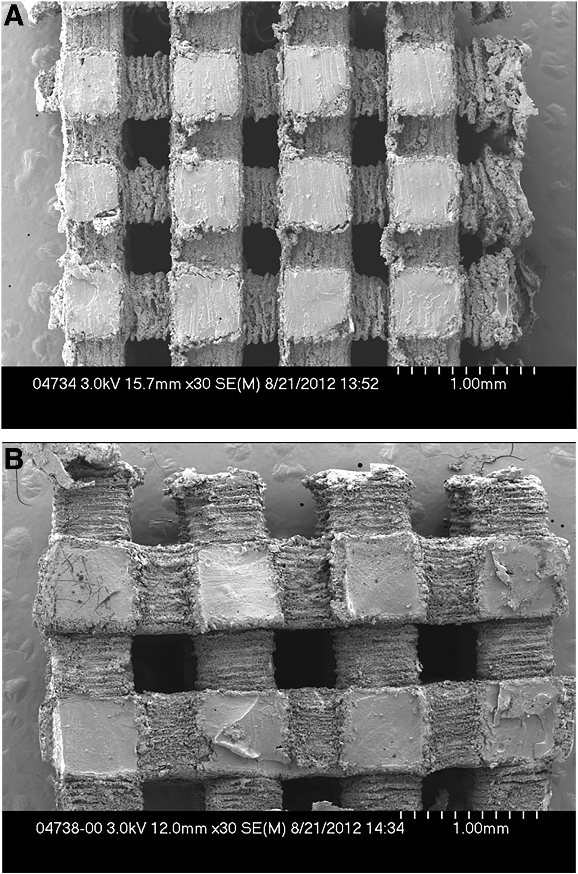
MicroCT analysis. Gross and MicroCT images of the 500 μm pore
Chondrocyte attachment, viability, and distribution
Figure 2A displays selected photographs of the PCLF scaffolds at the time points 1, 7, and 14 days for scaffolds with 500 and 750-μm pore sizes at days 1, 7, and 14, where the calcein AM stains the viable cells (green) and ethidium bromide for the cells undergoing apoptosis (red). As the chondrocytes proliferated throughout the scaffolds, there was no difference between the two morphologies of PCLF scaffolds. The chondrocytes covered the scaffolds and infiltrated throughout the pores by the seventh day compared with that by the first day (p < 0.01), covering all aspects of the scaffold into the channels and pores by the 14th day (p < 0.01) (Fig. 2B).
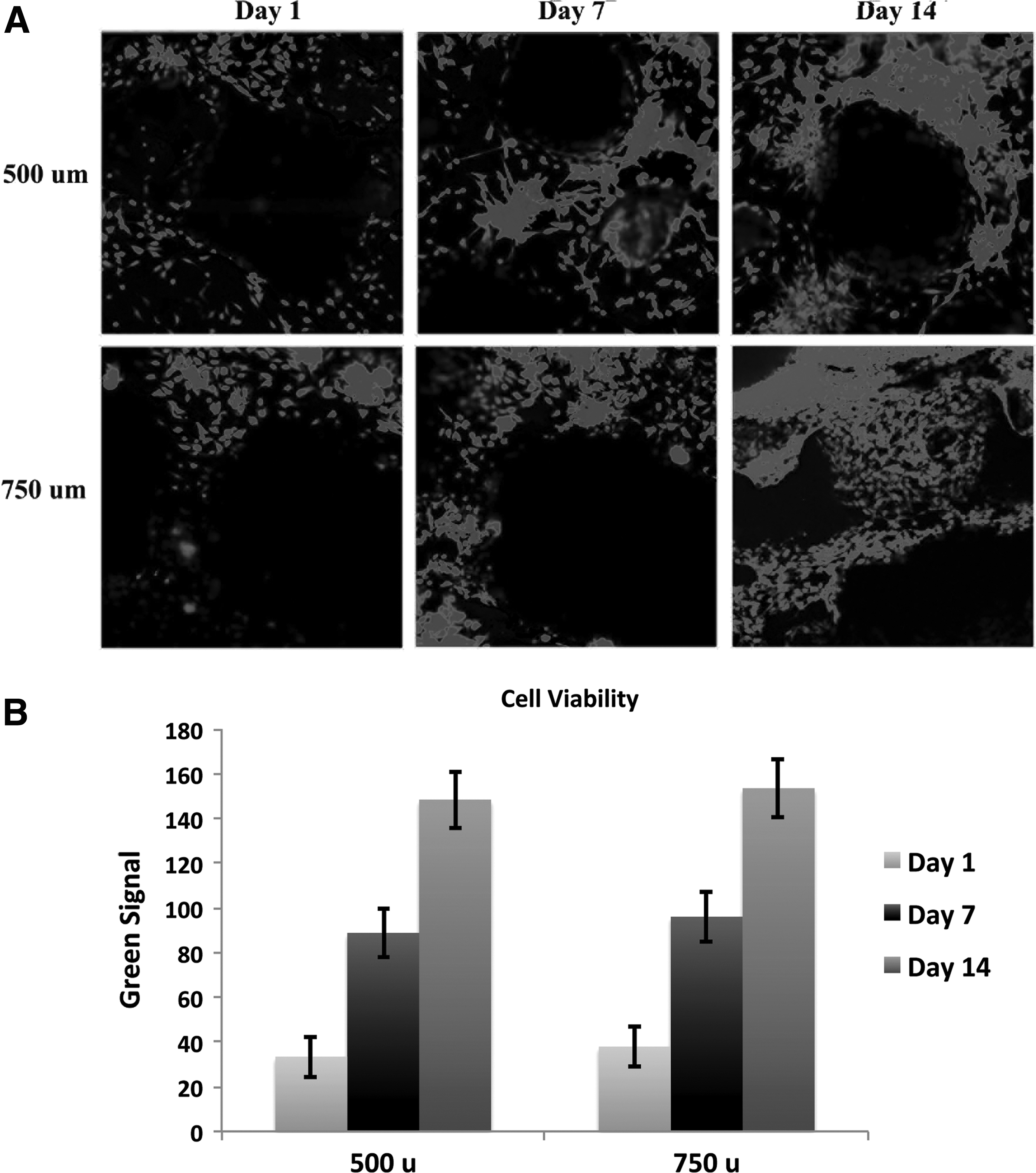
Chondrocyte viability assay.
Cellular attachment and proliferation
The presence of PL in the culture medium significantly increased the rate of chondrocytes proliferation compared with that in the FBS medium (p < 0.05), but did not impact cellular attachment (Fig. 3). There were no significant differences in proliferation or cellular attachment between the two pore sizes of PCLF scaffolds. Interestingly, the assay involving chondrocyte counting showed an increase in number of chondrocytes at first, followed by a sharp decrease, as the cells and their matrix appeared to become resistant to trypsin by the seventh day (Fig. 3).
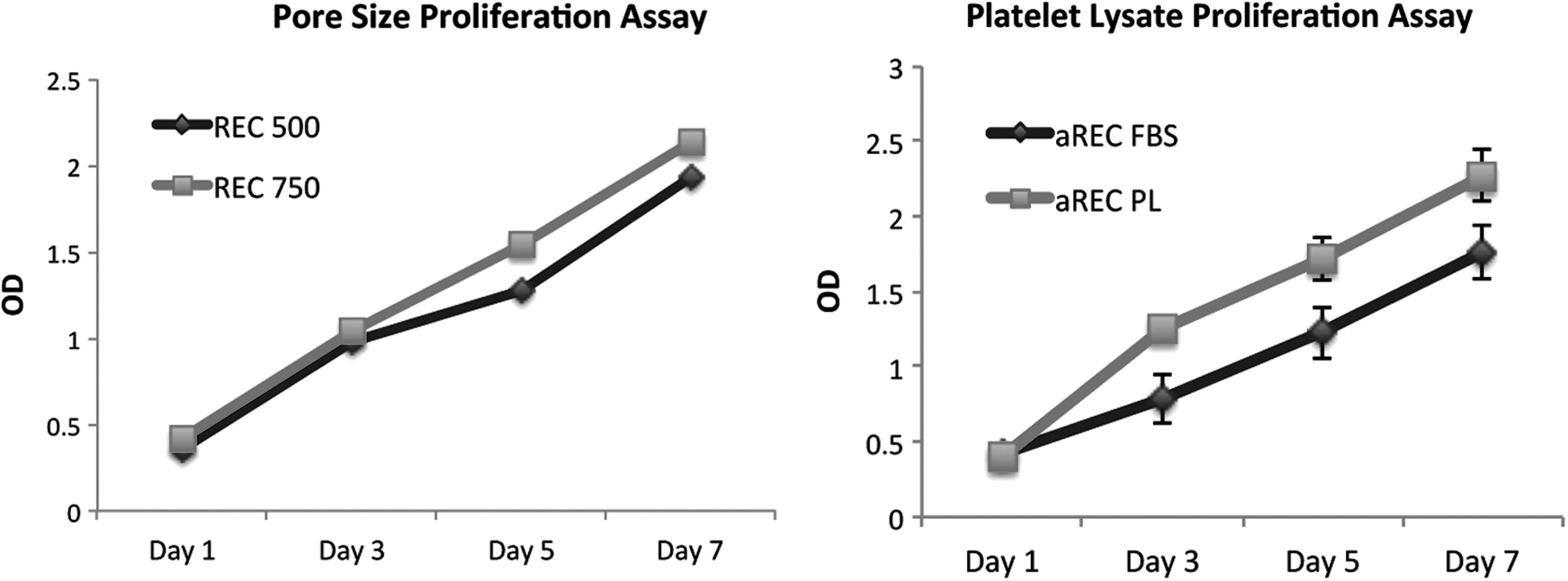
MTS cell proliferation assay. Chondrocytes were seeded onto scaffolds with pore sizes of 500 and 750 μm, without any difference in their proliferation rates. Cells were cultured in the presence of FBS or PL, with an increased proliferation rate in the PL group. Error bars represent standard error. FBS, fetal bovine serum; PL, platelet lysate.
Chondrocyte GAG expression
One important aspect of cartilage biology is its propensity to dedifferentiate into progenitor cells. This was assessed using the GAG assay to quantify this marker of chondrogenic differentiation. The GAG present on the PCLF scaffolds containing chondrocytes with pore sizes of 750 μm was compared with cells cultured on flat sheets of PCLF and flat tissue culture plates in both FBS and PL. However, over the 14-day time period, there was a decrease in the overall GAG content expressed by the cells cultured on flat sheets of PCLF or flat tissue culture plates. Alternatively, there was a slightly increased amount of GAG expressed by the chondrocytes cultured on the PCLF scaffolds (Fig. 4). The difference in GAG expression on the scaffolds was significantly higher at days 7 and 14 than the expression from the cells on flat surfaces (p < 0.01).

Chondrogenic differentiation and total collagen assays. The chondrocyte differentiation marker GAG and total collagen levels were assessed at different time points. These assays demonstrated that the chondrocytes did not dedifferentiate on the 3D scaffolds, when compared with the flat tissue culture plates or sheets of polycaprolactone fumarate. Error bars represent standard error. GAG, glycosaminoglycan; 3D, three-dimensional.
Chondrocyte total collagen expression
The total amount of collagen expressed in the extracellular matrix (ECM) on the scaffolds by the chondrocytes steadily amplified over 14 days of cell culture (Fig. 4). The expression spiked at 7 days, while increasing gradually from days 7 to 14. Furthermore, the cells cultured on the scaffolds had higher expression of collagen than the cells cultured on the flat sheets of PCLF or the tissue culture plates.
Chondrogenic marker immunostaining
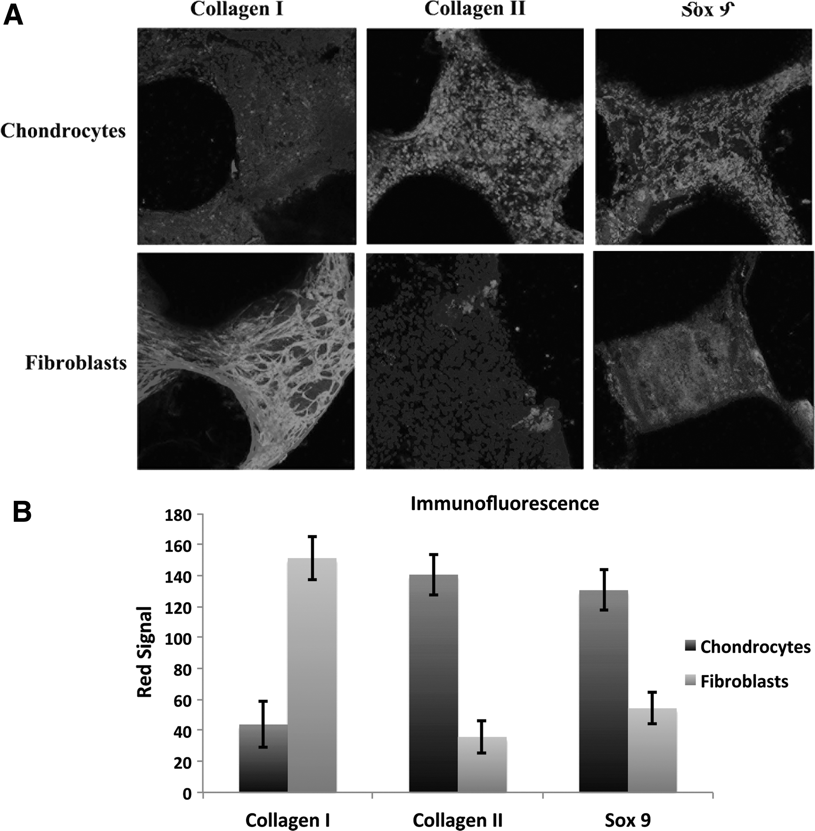
Multiple markers representing different aspects of chondrogenic differentiation were analyzed, including the ECM marker Collagen II and the chondrogenic transcription factor Sox 9. These were compared with a negative control for fibrous and osteogenic ECM formation Collagen I.
At 2 weeks, Collagen II and Sox 9 expression were significantly upregulated in the chondrocytes compared with that in the fibroblasts (p < 0.01) (Fig. 5A). After 2 weeks of chondrocyte cultures on the scaffolds, the ECM protein Collagen II and transcription factor Sox 9 were expressed throughout every surface and into every channel of the scaffolds. Alternatively, the negative control Collagen I was expressed by fibroblasts in much higher proportions than the chondrocytes (p < 0.01) (Fig. 5B).
Chondrogenic differentiation live immunostaining assay.
Discussion
Cartilage injuries are challenging problems, in part, because their poor healing potential is likely associated with its intrasynovial location, limited blood supply, and limited availability of regenerative or pluripotent cartilage regeneration cells.3–5 Furthermore, when cartilage regeneration therapies are attempted, such as the microfracture surgery, biomechanically inferior fibrocartilage is formed that cannot withstand the mechanical loads. 3 There is a critical need for both biological regeneration and polymer scaffolds to aid in cartilage repair and regeneration.1,2,26 Tissue engineering strategies typically focus on decellularized allografts, natural polymers, or synthetic polymer scaffolds. 7 Many recent strategies have attempted to combine one of these three strategies with biological regeneration therapies, including progenitor cells and growth factors.1,2,26 The potential of this strategy deals with combining the structural stability of the scaffolds with the regeneration potential of the cells and proteins.
The purpose of this investigation was to create a scaffold that possessed multiple properties conducive to augmenting its structural stability with biological therapy: (1) biologically friendly material to create a scaffold with a surface that promotes chondrocyte adherence and ongrowth; (2) large interconnected channels traversing the body of the scaffold to promote transportation of nutrients, as well as chondrocyte and eventual tissue infiltration; and (3) a material that creates a local milieu compatible for maintenance of chondrogenesis and eventual replacement of the scaffold with a chondrogenic matrix. We created a novel synthetic polymer scaffold utilizing 3D printing technology with a biologically friendly environment that facilitates attachment and sustained viability of mature chondrocytes that release a chondrogenic-like matrix throughout the surfaces of the PCLF scaffolds.
We chose the synthetic biodegradable PCLF because of its biocompatibility, cross-linking capability, and easily modifiable material properties. PCL and PCLF have both been used in many prior studies for a variety of applications, with excellent cytocompatibility and biocompatibility.8–18 In addition, although this material was originally synthesized to act as an adjunct in bone scaffolds, given its flexibility and compressibility, it became apparent that this had potential in cartilage engineering. It also has been able to cross-link with PLGA microspheres containing essential growth factors. The mechanical properties (i.e., strength and stiffness) of this polymer are easily modified by changing the molecular weight, degree of cross-linking, and polymer branching characteristics.11,16,18 In addition, sterilization of this biodegradable material with FDA-approved ethylene oxide or autoclave does not influence its mechanical properties or cytocompatibility. 11 In our study, we developed a novel methodology for synthesizing PCLF using a CAD software to design a specific geometric design to be printed on a 3D printer. Using the software and 3D printing technology, we synthesized a PCLF scaffold with sizeable channels, connected to each other throughout the body of the scaffolds, with the size designed to replace a small cartilage defect.
One important factor that influences cyto-compatibility of scaffolds is the surface roughness and size of the pores/channels. Smooth surfaces make cellular attachment more difficult than rougher surfaces, whereas increasing size of pores enhances the ability of cells and tissue to infiltrate the scaffolds' inner core when implanted in vivo. 10 Furthermore, the large channels that were visible by the naked eye, traversing the scaffolds in multiple different axes, likely played a role in the viability of the cells over the 14-day cultures, infiltrating deep into the channels. In fact, the scaffolds maintained a higher overall porosity than initially designed on the 3D software (Table 1). Another important consideration is biocompatibility, as many previous types of scaffolds have been demonstrated to release toxic byproducts or prevent cell attachment because of their hydrophobic nature and acidic microenvironment.27–32 Alternatively, the excellent cytocompatibility of PCLF created a very friendly microenvironment, both on the surface and deep in the body of the scaffolds, as demonstrated by the chondrocytes propensity to proliferate, infiltrate, remain viable, and produce various chondrogenic markers in vitro culture (Supplementary Fig. S1; Supplementary Data are available online at www.liebertpub.com/tea).9,10,16,18
Scaffolds must also sustain viability and enable proliferation to facilitate eventual tissue infiltration throughout the scaffolds. Strategies to augment cellular attachment and induce proliferation often involve utilizing natural proteins and growth factors. To augment cell attachment and tissue infiltration into silk and synthetic polymer scaffolds, surface attachment molecules, such as RGD peptides, fibronectin, or hyaluronic acid, or collagen hydrogels, have been combined with the scaffolds.7,32–37 We did not find this to be necessary, as the chondrocytes were able to attach to the rough surfaces of the scaffold, initially with the help of the dynamic bioreactor, and maintained this attachment for multiple weeks.
In cartilage engineering, there has been debate about the role of a 3D environment to mimic the native cartilaginous environment.3,5,7,26 We found that a 3D porous scaffold facilitated chondrocytes to retain their phenotypes through 2 weeks of cell culture. In contrast, when cultured in a two-dimensional (2D) cell culture plate or on a flat sheet of PCLF, the cells decreased their expression of the chondrogenic marker GAG. This could be because of the large pores and resultant ability for the cells to communicate with cell-to-cell contact through multiple axes (x, y, and z), as opposed to only one axis of contact seen in 2D cell culture. Furthermore, the chondrocytes were demonstrated to not only remain viable but also retain their chondrogenic phenotype after 2 and 4 weeks in culture, when compared with the negative control fibroblasts. The chondrocytes expressed high levels of the hyaline cartilage ECM marker collagen II and the chondrogenic transcription factor Sox 9.
As seen in other studies, PL did not significantly impact the differentiation status of the chondrocytes, but did impact their proliferation rates. 21 However, its use in chondrogenic tissue regeneration remains unclear, with a need for future studies attempting to better elicit its role in cartilage differentiation.
In conclusion, through the utilization of a novel 3D printing technology, we designed a cytocompatible PCLF scaffold with large pores/channels interconnected throughout the body in three different axes. Chondrocyte attachment, viability, and proliferation were facilitated by the scaffolds' large pores, surface roughness, and friendly microenvironment, augmented by the presence of PL. The chondrocytes retained their phenotypes and expressed chondrogenic ECM and transcription factors over a long static cell culture. We hope to utilize the potential of PCLF and PL clinical translation through examining copolymers for PCLF and additional strategies of chondrogenic differentiation induction, with PCLF scaffolds serving as the foundation to facilitate cartilage tissue regeneration.
Footnotes
Acknowledgment
This study was performed at Mayo Clinic, Orthopedic Department, Rochester, MN. The authors thank the Center of Regenerative Medicine at Mayo Clinic for funding this project.
Disclosure Statement
Dr. M.J.Y. holds a patent on PCLF. The remaining authors have no other relevant financial disclosures.
References
Supplementary Material
Please find the following supplemental material available below.
For Open Access articles published under a Creative Commons License, all supplemental material carries the same license as the article it is associated with.
For non-Open Access articles published, all supplemental material carries a non-exclusive license, and permission requests for re-use of supplemental material or any part of supplemental material shall be sent directly to the copyright owner as specified in the copyright notice associated with the article.