
Abstract
Although bone morphogenetic protein-2 (BMP-2) has been frequently used to stimulate bone formation, it has several side effects to be addressed, including the difficulty in optimization of clinically relevant doses and unwanted induction of cancerous signaling processes. In this study, an osteogenic peptide (OP) derived from BMP-2 was investigated as a substitute for BMP-2. In vitro studies showed that OP was able to enhance the osteogenic differentiation and mineralization of human mesenchymal stem cells (hMSCs). The peptides were then conjugated onto biocompatible poly-ι-lactide electrospun nanofibers through polydopamine chemistry. Surface chemical analysis proved that more than 80% of the peptides were stably retained on the nanofiber surface after 8 h of polydopamine coating during at least 28 days, and the amount of peptides that was retained increased depending on the polydopamine coating time. For instance, about 65% of the peptides were retained on nanofibers after 4 h of polydopamine coating. Also, a relatively small dose of peptides could effectively induce bone formation in in vivo critical-sized defects on the calvarial bones of mice. More than 50.4% ± 16.9% of newly formed bone was filled within the defect after treatment with only 10.5 ± 0.6 μg of peptides. Moreover, these groups had similar elastic moduli and contact hardnesses with host bone. Taken together, our results suggest that polydopamine-mediated OP immobilized on nanofibers can modulate the retention of relatively short lengths of peptides, which might make this an effective therapeutic remedy to guide bone regeneration using a relatively small amount of peptides.
Introduction
B
The incorporation of osteoinductive peptides derived from potent bone-inductive growth factors such as bone morphogenetic proteins (BMPs) onto commonly used membrane materials has been investigated to facilitate bone formation. 9 For instance, one study demonstrated that an osteogenic growth peptide (OGP) derived from BMP-7 possessed a high osteogenic differentiation capacity, which can induce the differentiation of mesenchymal stem cells (MSCs) into osteogenic cells. 10 Moreover, another study confirmed vasculogenic bone formation through the immobilization of BMP-2-derived peptide onto nanoparticles. 11 These BMP-derived peptides can reduce protein form-related side effects and are able to maintain their three-dimensional (3D) structures under physiological conditions because of their simple structural conformations. In addition, higher amounts of peptides could be available to be delivered in vivo compared to that of the BMPs. 11 However, the removal of the synergy domains of BMP targeting various signaling pathways often reduces the osteoinductive property, which requires relatively large amounts of peptides for a comparable level of bone formation, and therefore, the retention of the peptide from the membrane is critical for the effective bone formation with a compact lamella structure and a high mechanical strength.12–14
Several approaches for delivering biomolecules have been developed, including modifying the surface of fibers with aminolysis, heparin, and coelectrospinning.15–17 However, peptides composed of short amino acid sequences, and few functional groups are difficult to retain within the materials, and this often results in limitations on long-term delivery. Generally, after 28 days of incubation under physiological conditions, the peptides incorporated on the membrane were fully detached, while 40–50% of the proteins were retained.11,18,19 Therefore, a series of immobilization techniques have been developed to increase the retention of the peptides, such as the use of polyethylene oxide linkers, 10 fibronectin, 18 and the chemical conjugation of materials with peptides.20,21 Polydopamine is another widely used linker molecule that can be coated onto various substrates, enabling covalent reactions with biomolecules through the catecholamine group present in its structure.22,23 Our previously reported study demonstrated that immobilizing BMP-2 onto polydopamine-coated nanofibers enhanced bone formation.24,25
In this study, an osteogenic peptide (OP) derived from BMP-2 with 13 amino acid sequences was immobilized onto PLLA nanofibers through previously reported polydopamine chemistry. However, we were more focused on how the surface retention of the peptide can be improved by the controlled coating of polydopamine on the surface of the nanofibers. Given that, the specific objectives of this study were as follows: (1) to characterize the osteoinductive properties of the peptide, (2) to investigate the effect of polydopamine coating time on the retention of OP on the PLLA nanofibers, and (3) to evaluate the in vivo regenerative capacity of surface-modified electrospun nanofibers using a mouse calvarial critical-sized defect model over 2 months.
Materials and Methods
Materials
PLLA with 5.7–8.5 dL/g inherent viscosity was purchased from Evonik, Resomer® L214S and, 1,1,1,3,3,3-hexafluoro-2-propanol (HFIP) was from Wako. Tris-HCl was from Alfa Aesar and dopamine hydrochloride was purchased from Sigma-Aldrich. Phosphate-buffered saline (PBS), fetal bovine serum (FBS), trypsin- ethylenediaminetetraacetic acid, and penicillin–streptomycin were from Wisent. Dulbecco's modified Eagle's medium (DMEM) with low glucose was from Gibco BRL. Distilled water was produced by an Elix advantage system (Millipore). A Micro BCA™ Protein assay Kit was purchased from Thermo Scientific. OP (DWIVAGSGDWIVA) was purchased from AnyGen Co., Ltd..
Culturing of human MSCs and characterization of the effects of OP on them
Human MSCs (hMSCs) were purchased from Lonza and subcultured on tissue culture plates in a growth medium (low glucose DMEM with 10% FBS, and 1% PS) at 37°C, 95% air, and 5% CO2 in a humidified environment with media changed every 2–3 days. For the study, hMSCs were enzymatically lifted from the culture plate using 0.125% trypsin-EDTA. First, the effect of OP dose on the viability of hMSCs was examined. The hMSCs were seeded onto 96-well tissue culture plates at a density of 5000 cells/well and cultured in media containing 0, 10, 50, or 100 μg/mL of OP for up to 7 days (1, 3, 5, and 7 days). A 3-(4,5-dimethylthiazol-2-yl)-2,5-diphenyl-2H-tetrazolium bromide (MTT) assay was performed according to the manufacturer's guidelines. In brief, hMSCs were treated with 10 μL of MTT solution (5 mg/mL in ion-free PBS) and 125 μL of DMEM for 2 h. After incubation, the formazan salt was diluted with 100 μL of dimethyl sulfoxide for 10 min, and the absorbance at a wavelength of 550 nm was measured using a spectrometer (SpectraMAX M2e; Molecular Devices).
To investigate the effects of OP on the osteogenic differentiation of hMSCs, hMSCs were cultured for 10 days in growth media with or without 50 μg/mL of OP. RNA was extracted by scraping each sample and using Ambion® Trizol Reagent (Life Technologies), and the concentrations of RNA were estimated by measuring absorbance at 260 nm using a nanospectrometer (Nanodrop 2000; Thermo Scientific). The cDNA was synthesized from 1 μg of RNA using a Maxime RT PreMix kit from Intron Biotechnology and was processed in a Bio-Rad Thermocycler (Bio-Rad Laboratories). Before RT-PCR was performed, 2 μL of cDNA solution, which was diluted with Ambion diethyl pyrocarbonate (DEPC)-treated water (Life Technologies), was mixed with 10 μL of SYBR® Premix Ex Taq™ (2×) Tli RNaseH Plus from Takara, 0.4 μL of 10 pmol primer pairs, 0.4 μL of ROX reference dye (50×), and 6.8 μL of DEPC-treated water. The sample solutions were then sent to an RT-PCR StepOnePlus™ instrument (Life Technologies) and the amplification reaction was performed with denaturation at 95°C for 10 min, followed by annealing at 95°C for 15 s and extension at 60°C for 1 min for 40 cycles. Finally, the melting curve stage was performed from 60.0°C to 95.0°C in increments of 0.5°C per 5 s. All reactions were conducted in triplicate. The sequences of the primers utilized were as follows: glyceraldehyde 3-phosphate dehydrogenase (GAPDH) (Fw: 5′-GACCTGACC TGCCGTCTAGA-3′, Rv: 5′-CCTGCTTCACCACCTTC TTG-3′), alkaline phosphatase (ALP) (Fw: 5′-CCCCGTGG CAACTCTATCTT-3′, Rv: 5′-GATGGCAGTGAAGGGCT TCT-3′), collagen1a (Col1a) (Fw: 5′-GGACACAATGGAT TGCAAGG-3′, Rv: 5′-TAACCACTGCTCCACTCTGG-3′), osteocalcin (OCN) (Fw: 5′-TGTGAGCTCAATCCGGA CTG-3′, Rv: 5′-GCCGATAGGCCTCCTGAAA-3′), and Runx2 (Fw: 5′-TCTGCTGAGCTCCGGAATC-3′, Rv: 5′-CCACTCCGGCCCACAA-3′). The expression levels of the osteogenic genes were analyzed by the comparative Ct method and are presented as the fold change relative to Ct value of the human housekeeping gene GAPDH. Each gene expression level was normalized by GAPDH and was then normalized again by the gene expression level of the group cultured without OP.
Finally, after 14 days of culture in the osteogenic medium (low-glucose DMEM with 10% FBS, 1% PS, 50 μg/mL ascorbic acid, 0.01 M glycerol-2-phosphate, and 10−7 M dexamethasone), the amount of calcium deposited by the hMSCs was measured. Samples were dissolved in 0.6 N HCl and incubated at 37°C for 24 h. After the samples were spun down, the amounts of calcium in the supernatants were quantified using a QuantiChrom™ Calcium Assay kit (Bioassay Systems) according to the manufacturer's procedure. Finally, the optical densities of the samples were estimated by measuring their absorbances at 570–650 nm with a spectrometer.
Preparation of electrospun fibers, immobilization of peptides, and surface characterization
PLLA was dissolved in HFIP with stirring for at least 2 days. The polymer solution was ejected at 2 mL/h through a 23 G blunt-end needle using a syringe pump onto an aluminum foil-covered rotating collector (13 kV, 200 rpm) separated from the needle by 30 cm. Electrospun fiber sheets were dried at room temperature for 1 day. The fiber sheet was first hydrated by using 70% ethanol and distilled water before coating, and sequentially immersed in dopamine hydrochloride solution (2 mg/mL, 10 mM Tris-HCL buffer, pH 8.5) for 4 or 8 h on an orbital shaker for homogenous coating of polydopamine on the fiber. After the coating, the samples were thoroughly rinsed by stirring in distilled water for 1 day to remove unreacted residues. The polydopamine-coated fibers were then immersed in OP solution (50 μg/mL, 10 mM Tris-HCL buffer, pH 8.5) and incubated overnight at 37°C for the immobilization. After the reaction, surface atomic compositions of scaffolds were analyzed with X-ray photoelectron spectroscopy (XPS) (Theta Probe base system; Thermo Fisher Scientific). To ensure the morphological change after the immobilization of OP, the samples were coated with platinum using ion sputtering and examined using field emission scanning electron microscopy (SEM) (observed at an acceleration of 5 kV).
Quantitative analysis of OP immobilization
The amount of polydopamine present after 4 and 8 h of coating was examined using the Micro BCA Protein Assay Kit. Samples were punched as circles matching to the size of wells in a 24-well culture plate and treated with BCA solution for 2 h at 37°C, and then, the optical densities of the samples were measured at 562 nm using a spectrometer. The efficiency of immobilization of OP on the polydopamine-coated nanofibers was calculated indirectly, in which the initial concentration of OP in the working solution was measured, which was then subtracted by the amount of OP remaining within the supernatant at each assay point. We also confirmed the nonspecific adsorption of OP onto the reaction chamber, which was also considered for the measurement of OP density on the surface of nanofiber. Three types of samples were characterized: (1) OP physically adsorbed onto PLLA fibers and PLLA fibers on which OP was immobilized following (2) 4 h or (3) 8 h of coating with polydopamine. After the reactions, the supernatants were collected and their optical densities were then measured. The OP binding efficiency was then back calculated based on the value from the stock OP solution. To confirm the stability of OP on the fibers, the aforementioned samples were incubated at a pH of 7.4 and temperature of 37°C, and samples of supernatant (1 mL each) were collected at 4, 12, 24, and 48 h and 4, 7, 10, 14, 20, and 28 days. After each sample collection, the medium was replenished with 1 mL of fresh PBS. The optical densities of the collected supernatants were estimated by measuring the absorbance at 562 nm.
In vivo bone formation in a mouse calvarial defect model
The effect of in vivo guided bone regeneration was examined using a mouse calvarial defect model (6-week-old female mice; Narabiotech, Seoul, Korea). Animal studies were approved by Institutional Animal Care and Use Committee (IACUC) of Hanyang University (HY-IACUC-2014-0183) and performed under IACUC guidelines. We cared for all mice for 1 week after purchasing them. The mice were anesthetized with Zoletil (60 mg/kg) and xylazine (20 mg/kg) and the hair on head skin was shaved to prepare the surgery site. On each mouse, a skin incision was performed after sterilization with 70% ethanol. After incision, two critical-sized defects, which were circular in shape with 4 mm diameters, were created using a 4-mm diameter surgical trephine bur. Sterilized nanofibers were implanted within the defects on the left sides, and the defects on the right sides received no treatment as negative controls. The sample groups were defect only, PLLA fibers (PLLA), PLLA fibers coated with polydopamine (PD-PLLA), PLLA fibers coated with 50 μg/mL of OP without polydopamine-coated PLLA fibers (OP-PLLA), and OP immobilized on PLLA fibers following 8 h of polydopamine coating (OP-8PD-PLLA). Finally, the site of surgery was sutured and treated with povidone-iodine to prevent infections. All mice were sacrificed by CO2 suffocation after 8 weeks, and then, the calvarial bones were collected and fixed with 10% formalin solution for further analysis.
Analysis of in vivo study
Microcomputed tomography (micro-CT) was performed to analyze the new bone formation. Extracted skull bones were fixed with 10% neutral formalin and stored at 4°C for at least 3 days. The samples were exposed to X-rays under fixed condition (80 kV, 124 μA) in the micro-CT machine (Skyscan1172; Bunker microCT). The 3D images from the micro-CT scanning were analyzed with Adobe Photoshop CS6 (Adobe Systems) to calculate the bone area, and the newly formed bone volume (BV) dataset was obtained by CT Analyser (Bunker microCT). For our analysis, BV/total volume (TV) value was calculated. TV was not a defect volume, rather it was optional values that equally fit on every group.
The samples were decalcified with RapidCal™ solution (BBC Biochemical) for 2 weeks and the solution was replaced every 2 days. Before staining, the samples were dehydrated by immersion in graded concentrations of ethanol (70%, 90%, and four times at 100%), xylene, and paraffin. Finally, dehydrated samples were embedded in paraffin wax and hardened into a paraffin block for sectioning. The blocks were sliced into thicknesses of 6 μm using a microtome. The sections were subjected to a deparaffinization step and hydration step, and were then stained with Harris's hematoxylin and eosin solution, which stained the nuclei and cytoplasm of the tissues, and Goldner's trichrome staining solution, which stained the mineralized collagen a light green color. After staining, the samples were dehydrated and mounted with mounting solution from Richard-Allan Scientific. Stained samples were observed with an optical microscope.
The elastic modulus and contact hardness of the regenerated bone tissue were analyzed by the nanoindentation method. Calvarial specimens were fixed and embedded with acrylic resin (Ortho-Jet; Lang Dental), and sectioned into 2–mm-thick slices with a low-speed diamond saw (Isomet Buehler). The center of the specimens was longitudinally cut. The surface of the sections was polished using silicon carbide abrasive paper and aluminum oxide paste, glued onto stainless steel holders, and mounted on a nanoindenter (Nano-XP; MTS). All indentations were conducted at a depth of up to 500 nm with loading and unloading displacement rates of 10 nm/s. The indentation force–displacement curves were then used to obtain the contact hardness by dividing the peak indentation force by the projected area at the end of loading, and the elastic modulus was calculated using the unloading slope following the established nanoindentation methodology based on contact mechanics.26,27 The distance between indent locations was at least 30 μm to avoid any interruptions from the adjacent indents. A total of 52 nanoindentation sites were analyzed, including 28 in the regenerated bone tissues and 24 in the host bone tissues.
Statistical analysis
All of the quantitative values were calculated from more than triplicate samples (n ≥ 3) and expressed as the mean ± standard deviation of the average. The statistical significance was carried out using Student's t-test or analysis of variance with Tukey's HSD procedure. Values of p less than 0.05 were considered significant.
Results
Although synthetic polymers have been frequently used in bone tissue engineering, the inability to provide appropriate osteogenic signals often limits their extended use. Herein, the electrospun PLLA nanofibers were coated with adhesive polydopamine, and OP was subsequently immobilized upon them. The objectives of this study were as follows: (1) to investigate the effects of polydopamine coating time on the immobilization efficiency of OP, and (2) to examine the effects of the nanofibers with immobilized OP on in vivo bone formation.
Characterization of the effect of OP on hMSCs
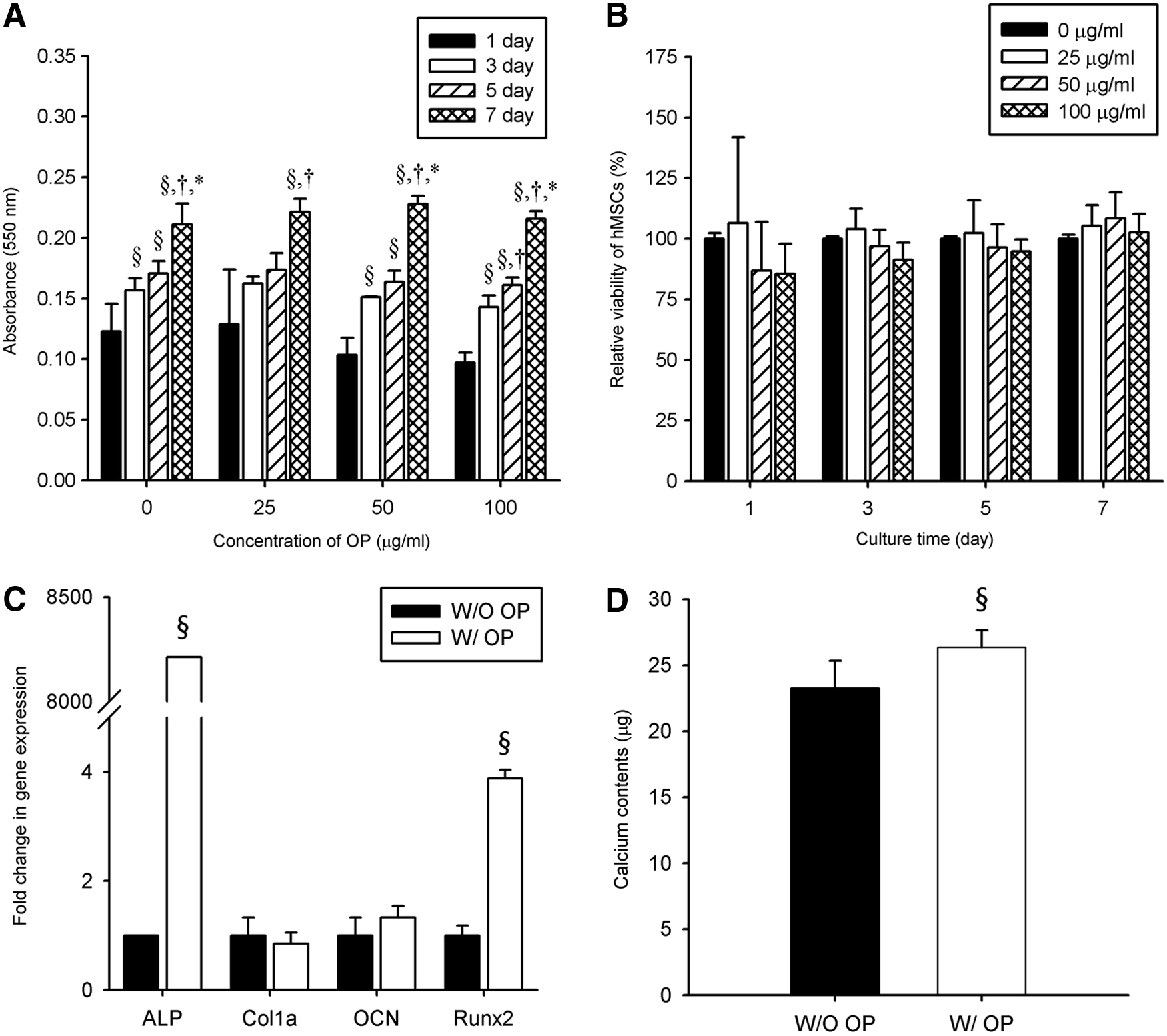
An MTT assay demonstrated that the peptide had no effect on the proliferation of hMSCs at OP concentration of up to 100 μg/mL (Fig. 1A, B). All groups showed continuous proliferation over 7 days. For example, the optical density from the group treated with 50 μg/mL OP was 0.103 ± 0.014 (1 day), and this significantly increased to 0.228 ± 0.006 at 7 days. In addition, the relative viability of hMSCs presented no statistically significant difference among the groups over 7 days in the presence of peptide at any tested concentrations. The hMSCs cultured under the abovementioned conditions exhibited a well-stretched and spindle-shaped morphology, and were tightly attached to the substrate. The effects of OP on the osteogenic differentiation of hMSCs were then investigated by measuring the osteogenic gene expression and calcium deposition following the culturing of hMSCs in the presence of 50 μg/mL OP for 10 days. As shown in Figure 1C, the expression of the ALP and Runx2 genes were significantly greater in the OP-treated group. For example, the gene expression levels of ALP were 1.00 ± 0.00 versus 8211.53 ± 0.07 and those for Runx2 expression were 1.00 ± 0.18 versus 3.88 ± 0.15 in the group without OP group versus the OP-treated group, respectively. As shown in Figure 1D, an analysis of calcium contents revealed that hMSCs cultured in the presence of 50 μg/mL OP showed a significant increase in calcium (26.3 ± 1.30 μg) compared to the control group (23.2 ± 2.10 μg). Collectively, the treatment with OP induced the in vitro osteogenic differentiation and mineralization of hMSCs.
Characterization of nanofibers immobilized with OP
Figure 2A is a diagrammatic scheme of the OP immobilization method. SEM images showed that unmodified PLLA nanofibers exhibited a smooth and clear fibrous morphology and also had proper pore-size membrane (Fig. 2B). In case of 8 h polydopamine coated PLLA (8PD-PLLA), no major difference was observed compared with untreated PLLA (Fig. 2C). However, when 50 μg/mL of OP was immobilized onto the 8PD-PLLA nanofibers, the surfaces of the nanofibers became rough and small aggregates were locally distributed throughout the fibers (Fig. 2D). XPS analysis shown in Fig. 3, carbon (C 1 s) (288 eV) and oxygen (O 1 s) (533 eV) were similarly detected on all PLLA nanofiber surfaces; however, nitrogen (N 1 s) peak (399 eV) was found only in the 8PD-PLLA and OP-8PD-PLLA samples. Likewise, high-resolution carbon spectrum analysis revealed that C-N (286.0 eV) peaks were only observed in 8PD-PLLA and OP-PD-PLLA samples. We also calculated the N/C atomic ratios of the specimens, and PLLA showed a negligible amount of nitrogen (<0.5%), while it was significantly increased in OP-8PD-PLLA (10.55%) and 8PD-PLLA (9.02%), indicating the successful coating of PLLA nanofibers with polydopamine and peptide.
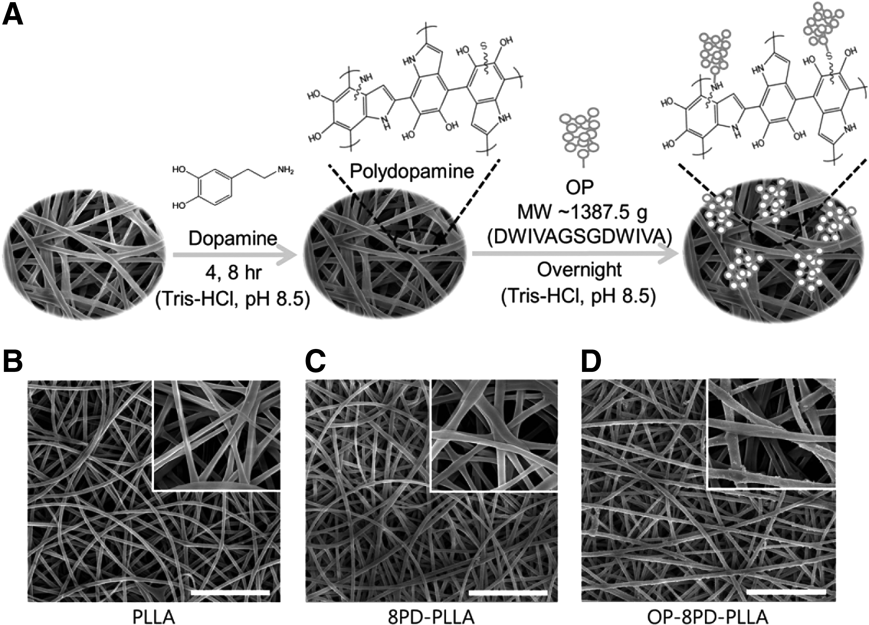
Fabrication of surface-modified nanofibers.
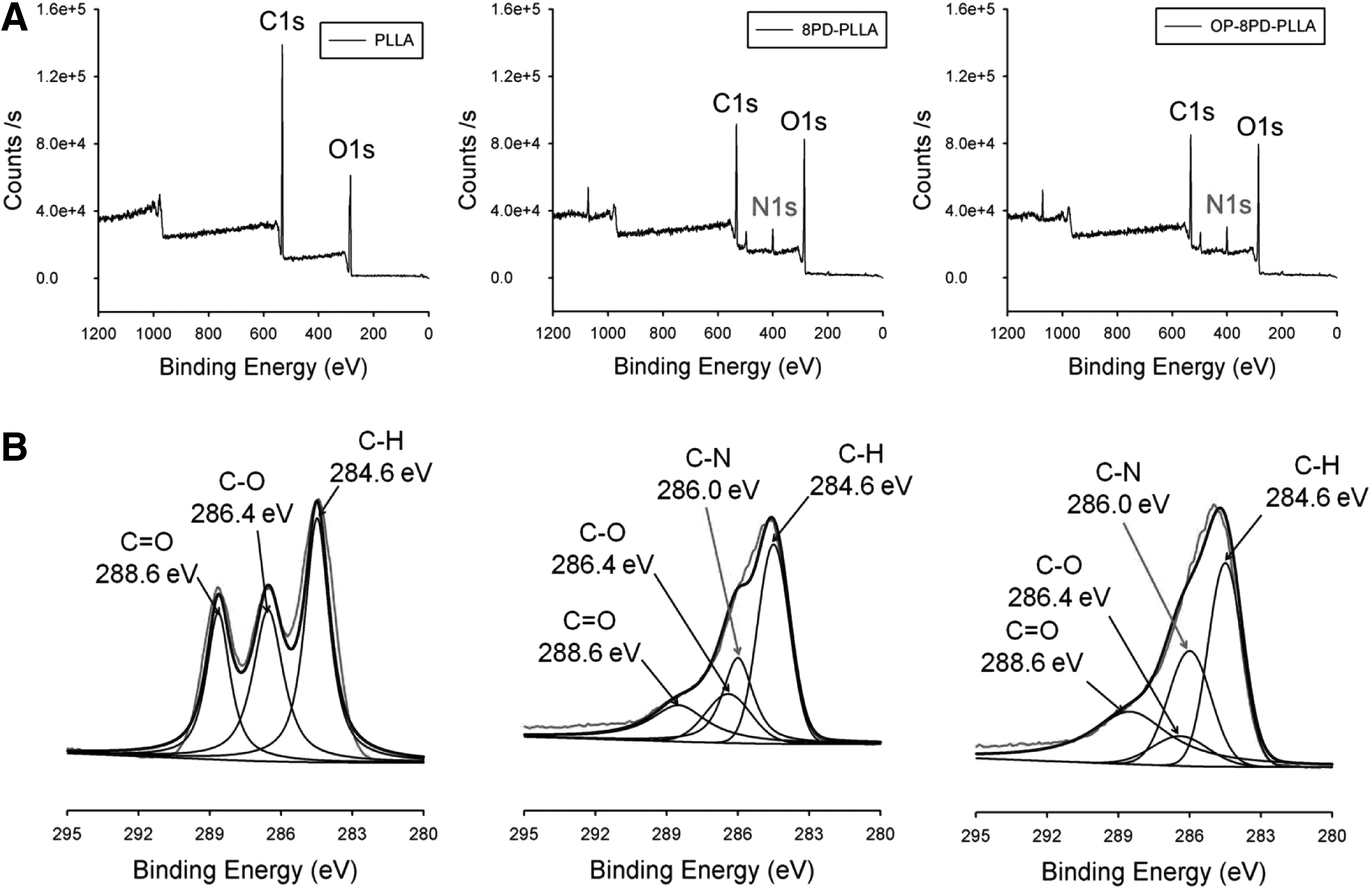
Surface chemical composition of the different nanofibers.
Quantitative analysis of OP immobilization
The amount of polydopamine coated on the nanofibers was dependent on the coating time, where the amount of amine groups was quantified as 101.96 ± 11.37 μg/cm2 after 4 h of coating, and this was significantly increased to 207.91 ± 7.49 μg/cm2 after 8 h of coating (Fig. 4A). We also confirmed nonspecific adsorption of OP onto the reaction chamber, which showed that the physically adsorbed amount of peptide on the culture plate was negligible (data not shown). Consistent with PD coated amounts, the binding efficiency of OP onto a polydopamine-coated surface was accordingly affected by the available amount of polydopamine on the surface. As shown in Figure 4B, the binding efficiency for OP-4PD-PLLA was 62.1% ± 4.73%, and this was significantly increased to 84.5% ± 4.84% for OP-8PD-PLLA. OP was also physically adsorbed onto the PLLA nanofibers without polydopamine modification, although the quantity was significantly lower than that available on polydopamine-coated nanofibers (43.88% ± 2.41%). The analysis of the amounts of peptides retained over 28 days demonstrated that 80.3% ± 0.219% of OP relative to the initially immobilized peptide remained, whereas other samples showed a greater initial burst, followed by the retention of 60.9% ± 0.736% of OP for OP-4PD-PLLA and only 36.3% ± 1.71% of peptide for OP-PLLA (Figs. 4C, D).
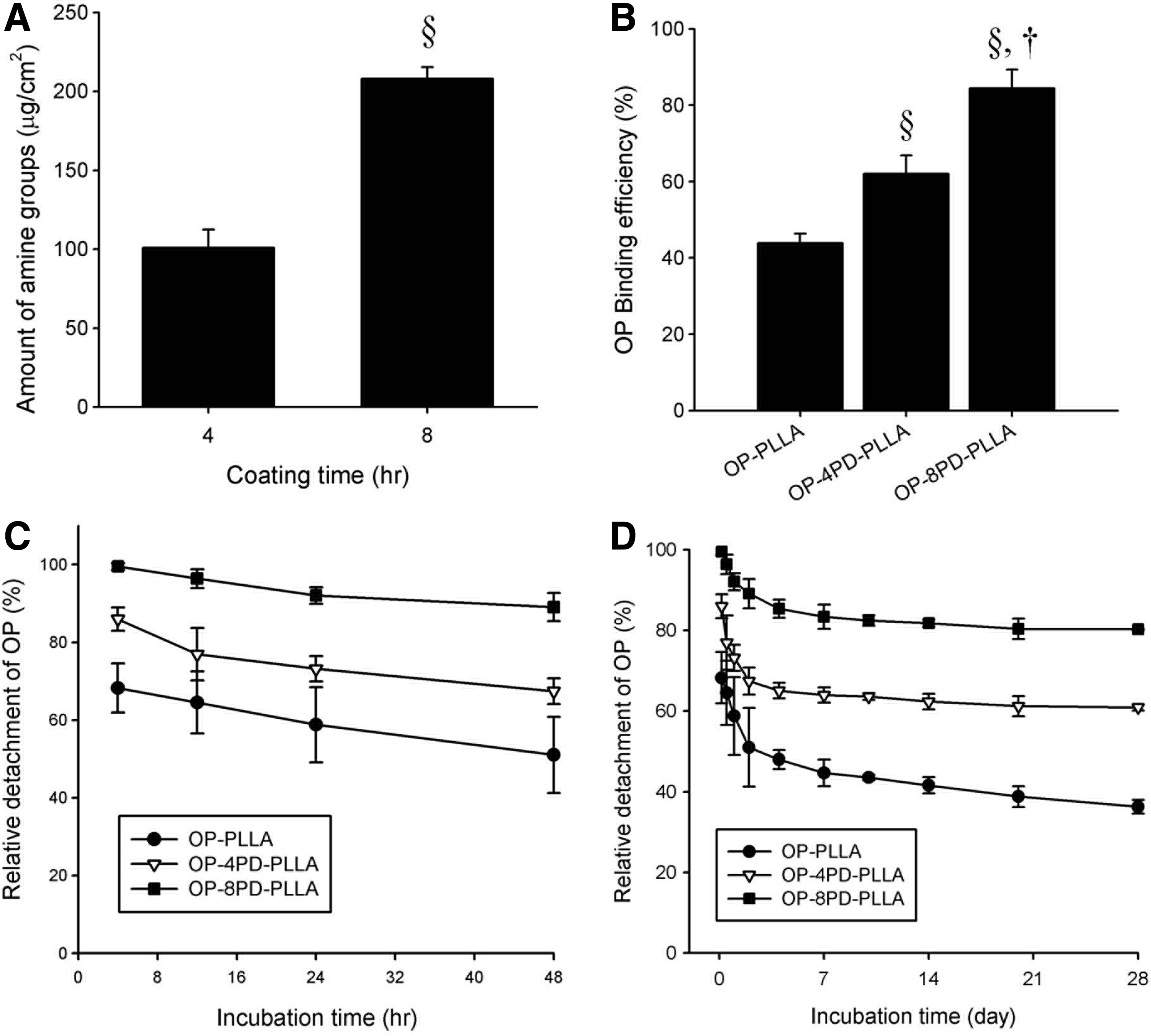
Quantification of the OP retained on the electrospun fibers.
Enhanced in vivo bone formation and mechanical strength
We then implanted the fibers into mouse calvarial critical-sized defect models and analyzed the in vivo bone regeneration after 2 months. As shown in Figure 5, the quantified results of micro-CT images showed the greatest bone formation from the group implanted with OP-8PD-PLLA (50.4% ± 16.9%) compared with the other groups, 8PD-PLLA (17.7% ± 13.1%), OP-PLLA (12.2% ± 2.54%), PLLA (15.1% ± 13.9%), and defect only (8.14% ± 4.07%). In addition, the regenerated BV displayed a similar tendency, in which none of the groups was significantly different from the defect-only group, except for the OP-8PD-PLLA group (1.91% ± 0.640%) (Fig. 5B). Furthermore, the elastic modulus and contact hardness of the newly formed bone tissues were compared with those from their corresponding host bone tissues (Fig. 5C). The elastic moduli of the defect (3.84 ± 0.783 GPa), PLLA (8.26 ± 5.84 GPa), and OP-PLLA (6.56 ± 5.69 GPa) groups were measured significantly lower than those of the host bone tissue (13.5 ± 4.07 GPa), while the OP-8PD-PLLA group (14.7 ± 4.11 GPa) had a comparable value with that from the host bone (p value = 0.26). Consistently, the contact hardness from the OP-8PD-PLLA group had a value similar to that from the host bone tissue (0.41 ± 0.15 GPa vs. 0.35 ± 0.15 GPa) (p value = 0.14).
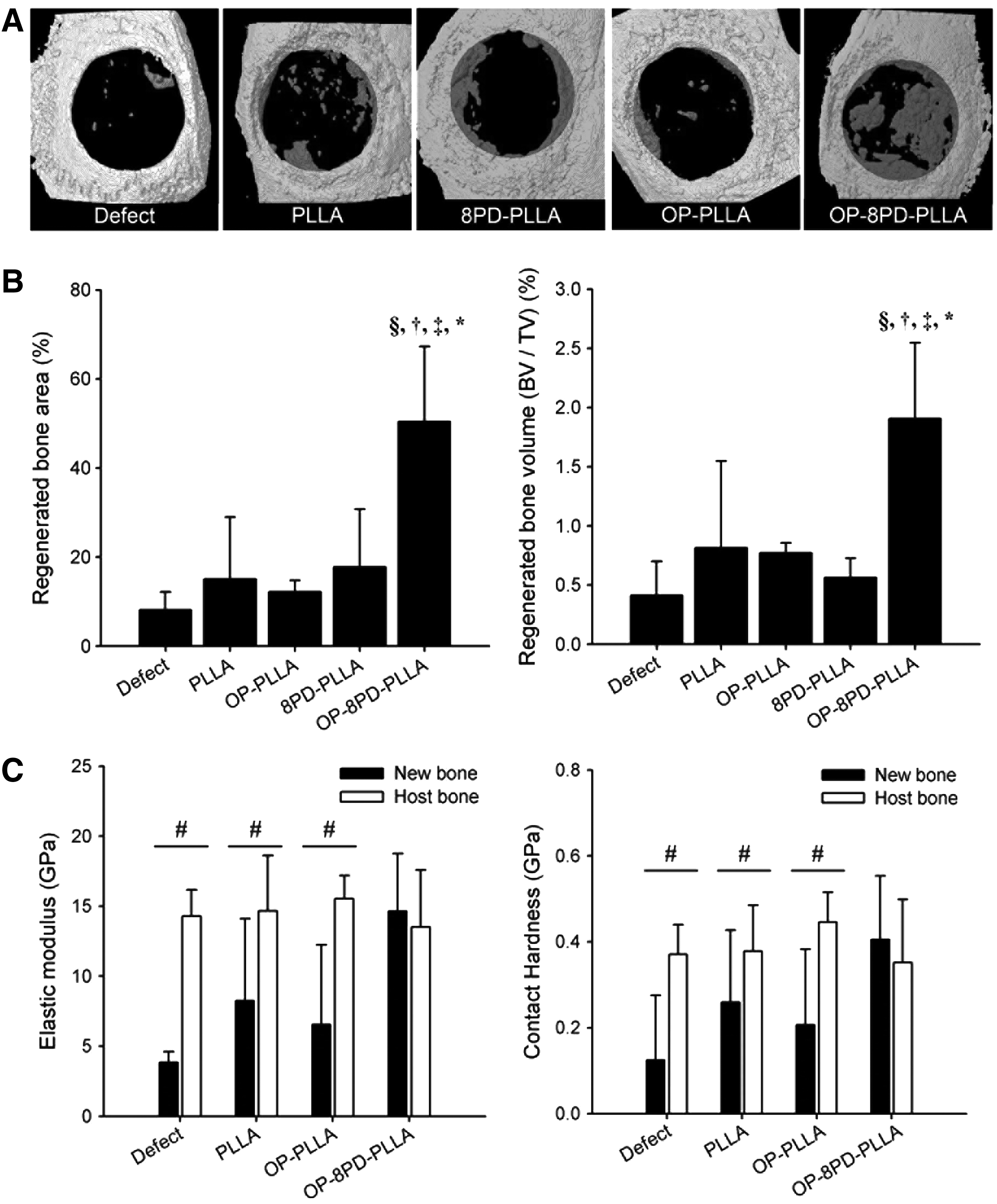
Radiographic analyses of mouse calvarial defects implanted with nanofibers at 2 months after surgery.
Histological analysis demonstrated that the groups implanted with nanofibers presented thicker and highly organized collagen fibrils throughout the defect areas, as shown in Figure 6A and G. The PLLA-implanted group showed thick tissue layers; however, they appear to be composed of fibrous connective tissues, indicating the formation of nonwoven collagen tissues (Fig. 6B, H). Similarly, the mice implanted with OP-PLLA and 8PD-PLLA nanofibers also had connective tissue formation in the middle of the defect sites, while regenerated compact bone structures were detected only on the edges of the defect areas (Fig. 6C, I). However, the OP-8PD-PLLA implantation group showed positive staining for mature newly formed compact bone throughout the whole defect area, and the regenerated tissues were tightly integrated with the host bone tissue (Fig. 6D, E, J, K).
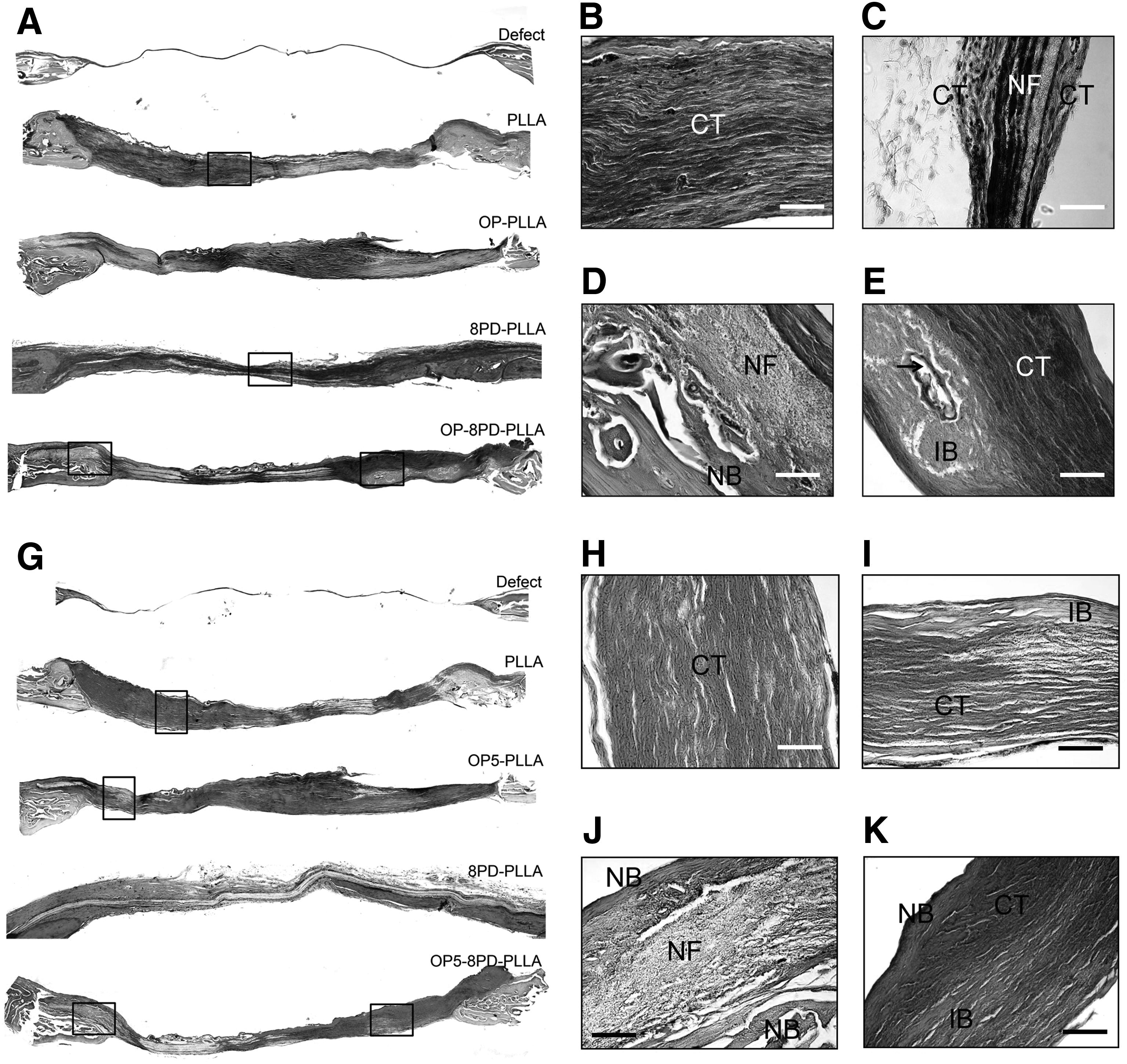
Goldner's trichrome staining and hematoxylin and eosin staining of sectioned mouse calvarial defect model after 2 months of implantation.
Discussion
To utilize the OP as osteogenic moieties on a nanofiber membrane prepared from a synthetic polymer PLLA, its effect on the viability of cells and its bone regeneration capacity should be studied. 28 Our results showed that no cytotoxicity was observed at up to 100 μg/mL of OP. In addition, the expression levels of the ALP and Runx2 genes were significantly increased in the presence of OP (Fig. 1C). ALP is one of the major representative markers during the osteogenic differentiation of hMSCs. 29 In addition, a constantly upregulated level of Runx2 gene expression is essential to induce the ALP activity. 30 Thus, the increased expressions of ALP and Runx2 genes indicate that OP is a potent signaling factor to induce the differentiation of hMSCs to an osteogenic lineage. Calcium assay demonstrated that OP in the culture medium was also able to induce mineralization (Fig. 1D). The control group also showed calcium mineralization because hMSCs are able to differentiate in the osteogenic culture medium without additional biomolecules. 30 However, OP containing the osteogenic medium showed more statistically significant mineralization than that from the osteogenic medium only. Likewise, several recent studies have also demonstrated the in vitro osteogenic efficacy of peptides derived from BMP-2 for controlling cell behavior. For instance, a BMP-2 mimicking peptide (CGKIPKASSVPTELSAISTLYL–OH), which was conjugated with a hydrogel successfully directed the in vitro osteogenic differentiation of hMSCs at a concentration of 50 μM of the peptide, 31 and 100 μM of BMP-2 peptide (KIPKASSVPTELSAISTLYLGGK) delivered by nanoparticles also induced the osteogenic differentiation of hMSCs. 32 Compared with these in vitro studies, a relatively lower concentration of OP (36 μM) appeared to be needed to exhibit a similar osteogenic differentiation efficiency, although pairwise comparisons should be warranted.
Electrospun nanofibers have been utilized as a matrix for delivery of proteins or peptides, including BMP-2 for bone regeneration.33–35 For example, Paletta and his colleagues incorporated lyophilized BMP-2 into PLLA solution by emulsification before electrospinning for sustained delivery. Although the results demonstrated that the BMP-2 containing group significantly enhanced bone formation during 3 months of implantation, the used amount was relatively large (∼200 ng/animal) and potential side effect of BMP-2 diffused out from the implanted site was not fully studied. In contrast, out study may be advantageous such that, synthetic peptide derived from BMP-2 may reduce the potential off target effect of BMP-2, and immobilization of the peptide on nanofibers will lead to more stable retention on the target, minimizing any extra stabilization steps, including emulsification. 34 It has been reported that the thiol and amine groups in proteins and peptides can bind to the catechol/quinone groups of the polydopamine structure by a Schiff`s base reaction or Michael addition. 23 For example, a previous study from our group demonstrated that BMP-2 was immobilized onto polydopamine-coated PLLA nanofibers and over 90% of the immobilized BMP-2 was retained for 28 days. 24 Also, one study demonstrated that a cell adhesive peptide (CGGRGD) on a decellularized vein matrix increased its immobilization efficiency up to 70% by using polydopamine (compared to less than 30% immobilization without polydopamine). 36 Similarly, in this study, the primary amine group of OP (DWIVAGSGDWIVA) can bind to the catechol/quinone groups of the polydopamine structure through the same reaction and be retained on the surface of the polydopamine-coated substrate. However, the complex underlying the mechanism of reaction between biomolecules (such as proteins and/or peptides) and polydopamine has not yet been fully elucidated. To prove the polydopamine-mediated peptide conjugation, surface characteristic analyses were implemented. Rash-like dots were detected on the OP-8PD-PLLA group and these particles were homogeneously disseminated throughout the nanofibers (Fig. 2D). This observation is consistent with our previous study, in which surface roughness was changed after the immobilization of bone forming peptide-1 derived from BMP-7 onto PLGA nanofibers. 37 Moreover, XPS analysis also demonstrated that polydopamine was properly coated onto the fibers in a time-dependent manner (Fig. 3). Our previous results also proved that polydopamine modification enhanced the C-N peak compared to unmodified nanofibers and the amount of coated polydopamine was increased depending on the coating time.38,39
For peptide quantification, we found that it is hard to directly detect the immobilized amount of peptides since the sequence is too short and primary amine residues in the peptide, which are frequently targeted as a ligand of detection antibodies, are bound with polydopamine. However, many studies, including our work, indirectly quantified the surface density of biomolecules in supernatant through protein assay or enzyme-linked immunosorbent assay (ELISA).11,40,41 It should be noted that peptide immobilization could be controlled by the amount of polydopamine available on the surface. We found that over 80% of the immobilized OP was stably retained on the surface of the OP-8PD-PLLA nanofiber group (Fig. 4C, D). One recent study demonstrated that BMP-2 was more stably immobilized on the surface of poly(lactide fumarate) nanoparticles than was a BMP-2 derived peptide (LYLTSIASLETPVSSAKPIK), since protein generally exhibits more binding sites and functional groups toward biomaterial surfaces. It is difficult for short peptides to maintain an attachment to substrate, and therefore most of them were detached within 30 days. 11 Moreover, over 60% of the OGP immobilized onto a titanium substrate with the aid of fibronectin was detached within 400 h. 18 However, it was demonstrated in this study that, over 80% of the OP immobilized by polydopamine was retained on the surfaces for 28 days, indicating that polydopamine-mediated conjugation is effective to capture the peptide. Furthermore, mechanically attached peptides were detached within 48 h, while chemically conjugated peptides were tightly attached to surfaces of nanofibers.42,43 As shown in Figure 4A, the amount of polymerized dopamine coated onto the PLLA scaffold surfaces was increased depending on the coating time, which could be attributed to a greater amount of peptides immobilized onto OP-8PD-PLLA than onto OP-4PD-PLLA. It could be explained that 35% of OP was retained on unmodified PLLA by hydrophobic interaction between hydrophobic biomolecules and the substrate material.44–46 Many hydrophobic groups within the OP (DWIVAGSGDWIVA) could be attracted to PLLA nanofibers by hydrophobic interactions even when the fibers were under a polydopamine coating.
The in vivo bone formation efficacy of the OP-incorporated nanofibers was then investigated by the implantation of the nanofibers into calvarial critical-sized defect mouse models (Fig. 5A, B). Considering the in vitro binding efficiency of OP and polydopamine-coated PLLA (84.47%), ∼10.56 μg of the OP was attached to the 8PD-PLLA fibers. This amount is indeed significantly lower than that in previous studies that usually delivered several hundred micrograms to several milligrams of peptides.47–49 Directly delivered and/or loosely conjugated peptides easily disappeared in in vivo environments into the surrounding tissues, and this phenomenon would be a major reason why higher amounts of peptides were administered. 50 Herein, 8PD-PLLA tightly seized the peptides, and thus, the stably conjugated peptides significantly enhanced the bone formation. Bone is one of the organs with high viscoelastic strength, and therefore, the mechanical properties of newly regenerated bone should be analyzed to elucidate its supporting capacity. Elastic modulus and contact hardness values could be promising indicators of the structural assembly and mineralization within the bone formation. 51 Nanoindentation studies demonstrated that the mechanical properties at the tissue level of the regenerated bone in the OP-8PD-PLLA group were similar to the values of native host bone (Fig. 5C). These results suggest that the characteristics and functions of the newly regenerated bone tissues induced by OP immobilized on nanofibers by polydopamine are comparable to those of host bone. Furthermore, our histological outcomes demonstrated that the OP-8PD-PLLA group showed many lacunae spaces along with a well-organized lamella structure of new bone (Fig. 6) and the formation of bone marrow (black arrows) space with blood vessels (Fig. 6D, E) in the OP-8PD-PLLA group. In particular, the most representative lamella-structured new bone was located at the edges of the defect area in the OP-8PD-PLLA group compared to the other groups, suggesting that bone regeneration was triggered from edges of the defect area. Based on these results, it is concluded that OP-immobilized nanofibers at the defect site facilitated bone maturation with constantly increasing mechanical properties similar to those of the host bone.
Conclusions
In this study, we developed and characterized biodegradable nanofibers on which OP was immobilized using polydopamine applied for different coating time for bone regeneration. Surface chemical characterization demonstrated that the polydopamine coating can be regulated by coating time, and furthermore, the immobilization efficiency and retention of OP on the surface of nanofibers were then correspondingly regulated. Quantification of the peptide on the nanofiber showed that 80% of the OP on the OP-8PD-PLLA nanofibers was retained even after 1 month of incubation under in vitro physiological conditions. Significantly enhanced bone formation was observed in the PLLA fibers on which OP was immobilized using polydopamine (OP-8PD-PLLA group), compared to the other groups. In addition, this group displayed mechanically well-organized bone regeneration with mechanical properties very similar to those of the host bone. Taken together, our results suggest that polydopamine-mediated OP immobilization on nanofibers can modulate immobilization and retention of relatively short length of peptides, which might be an effective therapeutic remedy to guide bone regeneration using a relatively small amount.
Footnotes
Acknowledgment
This work was supported by a National Research Foundation of Korea (NRF) grant funded by the Korean government (MEST) (NRF-2016R1A2B3009936) and Technology Innovation Program, 10050526, Development of disposable diaper based on biomass-oriented biodegradable super absorbent polymers funded by the ministry of Trade, Industry & Energy (MI, Korea). We also acknowledge Dr. Yonghoon Jeong, Ohio State University for his advice in characterization of mechanical properties of regenerated bone.
Disclosure Statement
No competing financial interests exist.