
Abstract
Background:
Decellularization of xenogenous cardiovascular structures is a promising approach to create scaffolds for tissue engineering. Unfortunately, handling and pliability of the unfixed tissue is challenging. N-(3-dimethylaminopropyl)-N9-ethylcarbodiimide (EDC) is an alternative cross-linking agent to glutaraldehyde (GA). Applied in native tissue, it provides biocompatibility and shows no potential for calcification. In addition, EDC can be used to link growth factors (GFs) to tissue scaffolds after decellularization. EDC cross-linking could thereby help to improve decellularized tissue without the toxicity of GA.
Material and Methods:
Porcine aortic wall tissue specimens (TS) were decellularized, treated with EDC, and coated with fibroblast growth factor (FGF) or vascular endothelial growth factor (VEGF). Afterward, TS were subcutaneously implanted in 36 Lewis rats along with one decellularized TS without EDC treatment. After 2, 4, and 6 weeks TS were explanted from 12 rats, respectively. Vital cells were evaluated by RNA quantification, general cellular infiltration by hematoxylin and eosin staining (H&E), macrophage infiltration by CD68 staining, calcification by Von-Kossa staining, and tissue degradation by measurement of TS thickness.
Results:
Quantification of vital cells showed reduced reseeding of EDC-treated TS compared to noncross-linked TS after 2 (p < 0.05) and 4 weeks (p < 0.05), while after 6 weeks only EDC+VEGF showed fewer viable cells (p < 0.01). Histological evaluation confirmed a reduced infiltration of EDC-treated TS. Macrophage infiltration decreased in all groups from 2 to 6 weeks, with the smallest population in EDC+VEGF-treated TS (p > 0.05). In EDC+FGF-treated TS, macrophages were reduced after 2 weeks compared to noncross-linked TS (p < 0.05), while after 4 and 6 weeks no significant difference was found (p > 0.05). Von-Kossa staining revealed no calcification in any of the specimens. Thickness of noncross-linked and EDC+FGF-treated TS was not different at the respective times of explantation, but decreased in both groups toward 6 weeks.
Conclusion:
EDC cross-linking combined with GF coating of decellularized aortic wall tissue showed encouraging results. The treatment did not impair the advantages of decellularized tissue such as long-term recellularization, absence of calcification, and tissue integrity. Based on the low macrophage infiltration and minimal tissue degradation, treatment with EDC and VEGF could be useful after decellularization. However, further research is necessary to verify these findings in models, including mechanical stress.
Background
A
Established chemicals for tissue fixation, like glutaraldehyde (GA), are cytotoxic and lead to calcification.6–10 A new cross-linking method using N-(3-dimethylaminopropyl)-N9-ethylcarbodiimide (EDC) in combination with N-hydroxysuccinimide (NHS) shows several advantages compared to GA. 11 These cross-linking agents facilitate amide bond formation between carboxylic and amino groups of xenogenous matrices without becoming part of the linkage themselves or causing cytotoxic reactions.12–15 EDC treatment of decellularized tissue improves mechanical strength, offers biocompatibility, and shows no potential for calcification.16,17 In addition, this method has demonstrated a reduced inflammatory response in animal models. 17
A further advantage of EDC cross-linking is the possibility to covalently bind heparin to the decellularized matrix.18,19 Heparin functions as an inhibitor of blood coagulation and can therefore be used to improve blood compatibility. The heparinized extracellular matrix can then be coated with growth factors (GFs) such as fibroblast growth factor (FGF) and vascular endothelial growth factor (VEGF).20–24 Both GFs possess a heparin binding site.25,26 This in vitro coating could be useful to accelerate and adjust the in vivo recellularization. Both, FGF and VEGF, are known to modulate the migration and proliferation of endothelial cells, smooth muscle cells, and fibroblasts. In addition, they are angiogenic factors and play an important role in the promotion and regulation of vascularization.26–28 Zhou et al. have already shown promising results with a heparin-coated and VEGF modified decellularized vascular graft. 20 Binding of FGF to heparinized collagenous matrices led to an improved endothelialization. 22
EDC cross-linking combined with GF coating has the potential to improve decellularized tissue by stabilizing the tissue, reducing inflammation, and allowing recellularization. Therefore, we subcutaneously implanted decellularized and EDC-crosslinked aortic wall tissue specimens (TS) coated with FGF or VEGF in rats and determined the effect on general recellularization, macrophage response, and tissue degradation in comparison to noncross-linked decellularized tissue.
Materials and Methods
All experiments were performed in accordance with the Principles of Laboratory Animal Care prepared by the National Society of Medical Research and the Guide for the Care and Use of Laboratory Animals prepared by the Institute of Laboratory Animal Resource and published by the National Institute of Health (NIH Publ. 85-23, Rev 1985). This study has been approved by the local Ethics Committee.
Preparation of TS
Porcine aortic wall tissue was obtained from healthy pigs aged 6 to 9 months and was treated according to an established decellularization protocol for pulmonary heart valves. 1 In brief, extracted tissue was decellularized using 1% deoxycholic acid (Sigma-Aldrich Productions, Steinheim am Albuch Germany) for 16 h. To remove cell fragments and detergent residues, the tissue was extensively washed with a saline solution and sterilized with ethanol. On account of the aortic wall tissue thickness, this process was repeated thrice, whereafter the tissue was cut into specimens (TS) of 1 cm2. Cell removal was confirmed with RNA quantification and hematoxylin and eosin (H&E) staining. Afterward, cross-linking using EDC (Carl Roth Productions, Karlsruhe Germany) and NHS (Sigma-Aldrich Productions, Steinheim am Albuch, Germany) and covalent heparin (AppliChem, Darmstadt Germany) immobilization were performed as described previously with slight modifications. 20 In summary, for the reagent solution 1 g heparin, 2 g EDC, and 1.2 g NHS were solubilized in 500 mL 2-morpholinoethanesulfonic acid buffer (0.05 M, pH 5.6). After incubation in the preheated reagent solution for 4 h at 37°C the specimens were rinsed in 0.1 M Na2HPO4 for 2 h. Then, TS were rinsed in 4 M NaCl for 24 h and twice in distilled water for 24 h.
Cross-linked and heparinized TS were coated with 5 ng/mL FGF (Biochrom, Berlin, Germany) or 5 ng/mL VEGF (Biochrom, Berlin, Germany) for 24 h at 37°C. All incubation and washing steps were performed under gentle shaking.
Implantation
Decellularized EDC cross-linked TS coated with FGF or VEGF along with one unfixed decellularized TS were implanted in 36 Lewis rats (Charles River, Sulzfeld, Germany) aged 32 to 39 days using the subdermal model according to the technique of Mako. 29 After induction of anesthesia with Ursotamin and Rompun, the backs of the rats were shaved and disinfected. Subdermal pockets were created using scissors. After implantation of respective TS, the pockets were closed with intracutaneous sutures (Monocryl 4-0, Ethicon, Somerville, MA).
The positions of the TS varied in every experimental animal to avoid bias. After 2, 4, and 6 weeks, TS were explanted from 12 rats, respectively. Gross examination was performed, and TS were prepared for further analysis.
Evaluation of explanted TS
TS were bisected, and one half was used for quantification of RNA to reveal the relative amount of viable cells (RNeasy Mini Kit, Qiagen, Hilden, Germany). The other half of the bisected TS was preserved in formalin and embedded in paraffin. Longitudinal sections were made from the middle of the TS. H&E staining was performed on all specimens to allow general evaluation of cellular infiltration. Staining for macrophages (CD68, Acris Antibodies, Herford, Germany) was performed to evaluate the respective cellular infiltration. Histological examination was performed using a Leica DM1000 microscope (Leica, Solms, Germany), and representative pictures were taken using a Leica DSC 290 (Leica, Solms, Germany). Cellular infiltration was evaluated on the basis of the entire longitudinal sections from the middle of the TS according to the following criteria: None (0), partial and superficial (1), continuous and superficial (2), moderate infiltration (3), and high infiltration (4). To evaluate tissue degradation, tissue thickness was measured using the 700-119-20 LCD Digital Thickness Gage (Mitutoyo, Neuss, Germany). Van Gieson staining was performed to evaluate collagens histologically. TS were Von-Kossa stained to evaluate calcification.
Statistical analysis
All data were analyzed with GraphPad Prism 5 (GraphPad, Inc., CA). Descriptive statistics is reported as the mean ± standard deviation. Normal distribution was tested by the Shapiro–Wilk test. The non-normally distributed data were analyzed using the Mann–Whitney test. Statistical significance was set at a p-value of p < 0.05 (*significant), p < 0.01 (**very significant), and p < 0.001 (***highly significant). Significance was shown between coated TS and noncross-linked control at the respective time points (*) and between different time points in each group ( ). Graphs were created with GraphPad Prism 5 as well.
Results
Decellularization of aortic wall tissue reduced the RNA content by 99% (n = 3), from 112 ± 20 μg/g in native tissue to 1 ± 0.2 μg/g. H&E staining showed the complete removal of cell nuclei (Fig. 1). After confirmation of acellularity, TS were cross-linked with EDC, coated with FGF or VEGF, and subcutaneously implanted in rats for 2, 4, and 6 weeks.
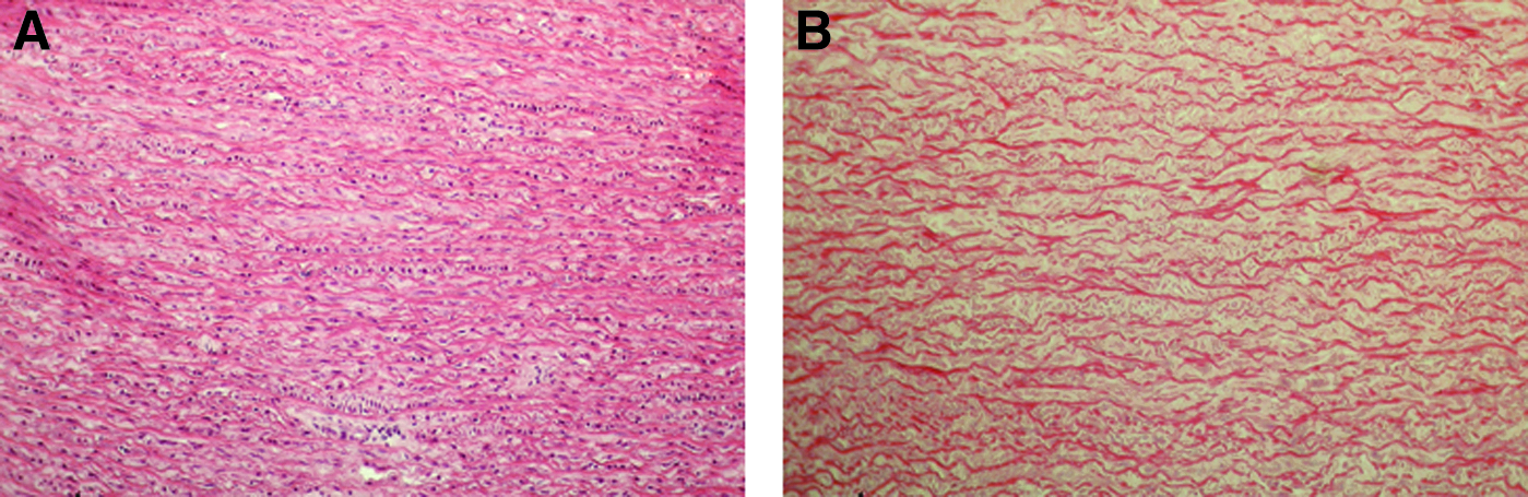
H&E staining of
During the study interval, all animals survived without complications. After explantation, gross examination exposed no visible inflammatory reaction in and surrounding any of the TS.
RNA quantification of the explanted TS revealed a comparable process of recellularization in all groups with an increase from week 2 to 4 and a regression after 6 weeks (Fig. 2). However, regarding their RNA contents, EDC cross-linked TS differed from the decellularized TS at the respective time points. EDC cross-linkage in combination with FGF coating (EDC+FGF) and VEGF coating (EDC+VEGF) resulted in a significant lower RNA content after 2 and 4 weeks (Fig. 2). After 6 weeks, only the EDC+VEGF-treated TS showed significantly lower RNA quantities.
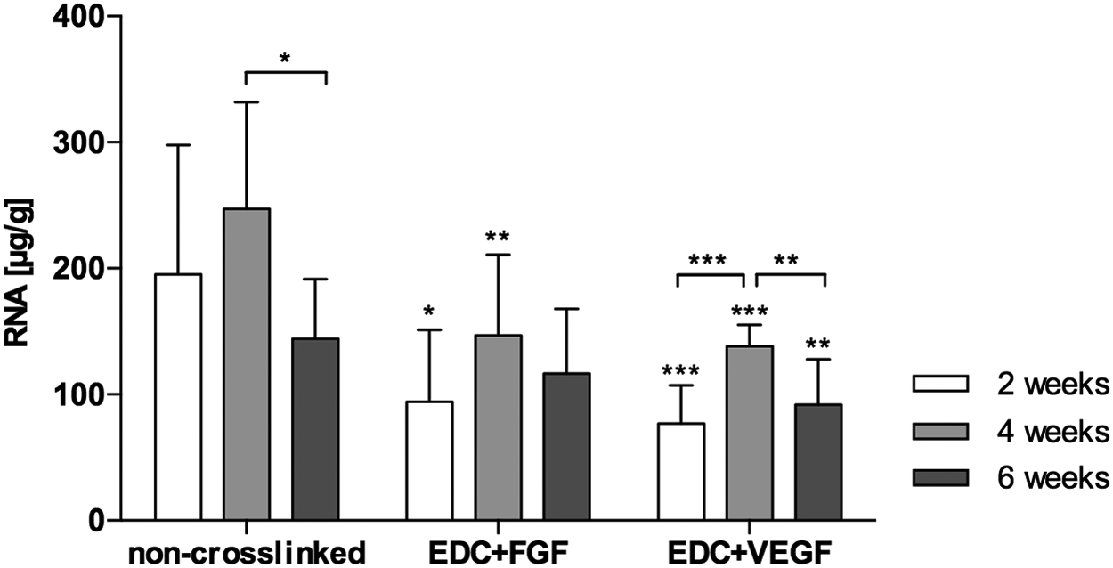
RNA quantification of differently treated TS after subcutaneous implantation. Decellularized TS without EDC treatment (noncross-linked); decellularized TS treated with EDC and FGF (EDC+FGF); and decellularized TS treated with EDC and VEGF (EDC+VEGF) were explanted after 2, 4, and 6 weeks. *p < 0.05; **p < 0.01; ***p < 0.001. EDC, N-(3-dimethylaminopropyl)-N9-ethylcarbodiimide; FGF, fibroblast growth factor; TS, tissue specimens; VEGF, vascular endothelial growth factor.
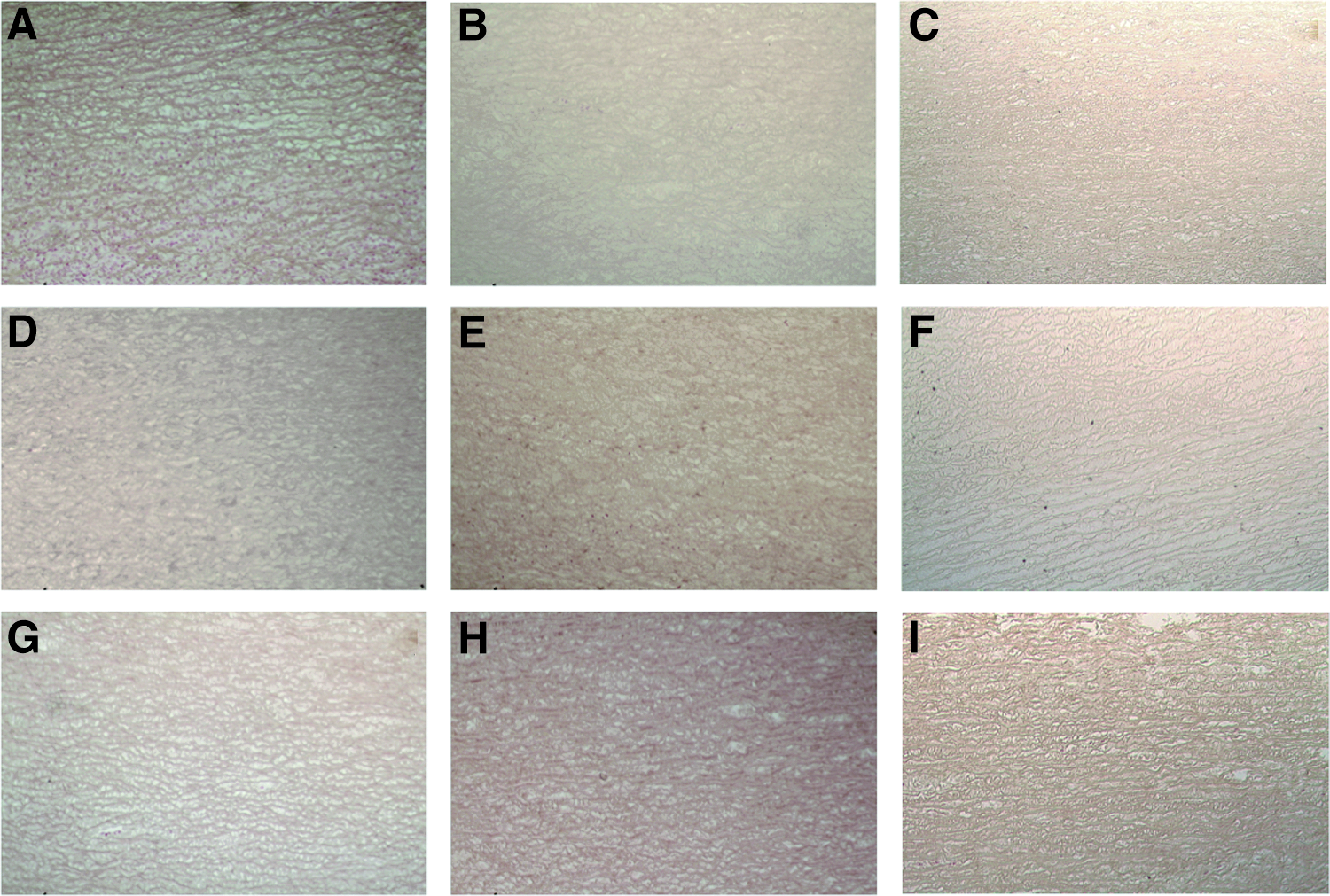
Histological sections of the TS stained with H&E showed an intact collagenous scaffold in all explants (Fig. 3). Surrounding all specimens a fibrotic layer was seen, and the recellularization process was observed as a slow infiltration from the outside to the inside of all TS. Mostly, cellular infiltration was observed from the trimmed edges of the TS rather than from adventitia and intima.

Representative H&E stained sections of explanted TS after subcutaneous implantation.
Based on the H&E staining, recellularization was analyzed by a semiquantitative method, which took into consideration the amount of cells at the surface of the TS and inside the TS, as well as the depth of infiltration; it was graded from 0 (no recellularization) to 4 (high recellularization). The evaluation revealed a significantly lower recellularization of EDC-treated TS (Fig. 4). While decellularized TS showed a moderate infiltration, EDC-treated TS were solely recellularized in the outer part of the explant. This recellularization did not increase over time and stayed equal up to 6 weeks.
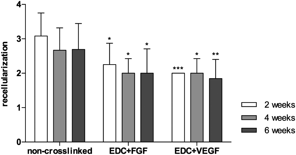
Analysis of the general recellularization of H&E stained TS after subcutaneous implantation. Recellularization of decellularized TS without EDC-treatment (noncross-linked); decellularized TS treated with EDC and FGF (EDC+FGF); and decellularized TS treated with EDC and VEGF (EDC+VEGF) were analyzed after 2, 4, and 6 weeks and graded as follows: 0, none; 1, partial and superficial; 2, continuous and superficial; 3, moderate infiltration; 4, high infiltration. *p < 0.05; **p < 0.01; ***p < 0.001.
For the evaluation of macrophage response to the implanted TS, CD68-positive cells were analyzed. The population of CD68-positive cells decreased from 2 to 6 weeks in all groups (Figs. 5 and 6). Significant differences between the groups were found after 2 weeks, with a significantly lower recellularization of EDC treated explants. Similar to H&E staining, TS without EDC treatment showed moderate infiltration. In EDC treated TS, on the contrary, CD68-positive cells were mostly present in the outer area of the TS. The group treated with EDC and VEGF showed the smallest population of specifically stained cells after 2 and 6 weeks.
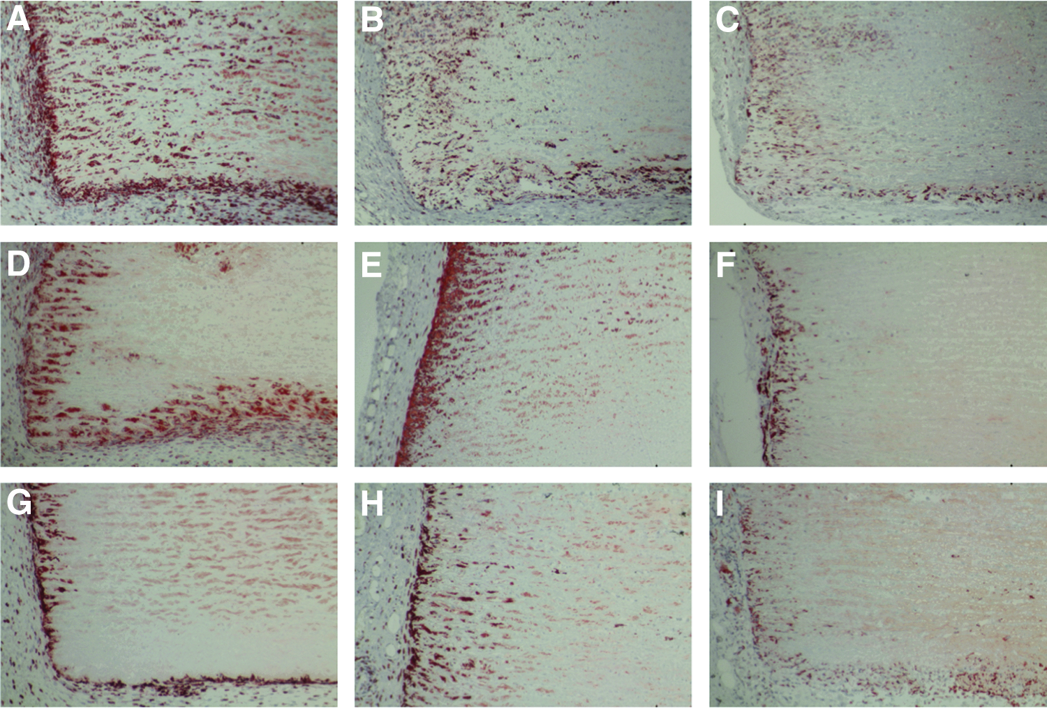
Representative sections of explanted TS stained for CD68-positive cells after subcutaneous implantation.

Immunohistological staining of CD68-positive cells in TS after subcutaneous implantation. The staining of CD68-positive cells was used to evaluate the macrophage population in decellularized TS without EDC treatment (noncross-linked); decellularized TS treated with EDC and FGF (EDC+FGF); and decellularized TS treated with EDC and VEGF (EDC+VEGF) after 2, 4, and 6 weeks. Macrophage recellularization of TS was graded from 0 (no recellularization) to 4 (high recellularization). *p < 0.05; **p < 0.01; ***p < 0.001.
Evaluation of TS thickness revealed a significant reduction from 2 to 6 weeks in the groups treated with EDC+FGF and without EDC treatment. However, no significant difference between these two groups was observable (Fig. 7). In comparison, the EDC+VEGF group merely showed a slight reduction of tissue thickness after 6 weeks. Moreover, EDC+VEGF treated TS were significantly thicker than TS of the other groups. Regardless of the tissue thickness, Van Gieson staining revealed visually no degradation of collagen fibers over time (Fig. 8).
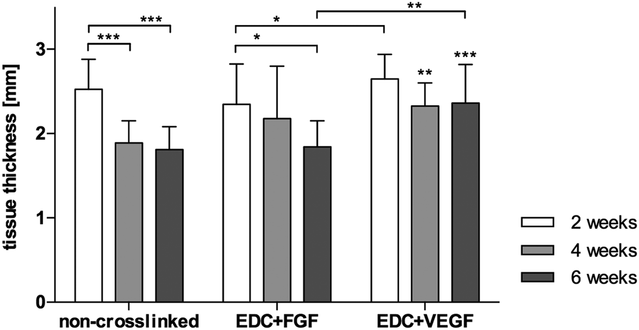
Thickness of TS 2, 4, and 6 weeks after subcutaneous implantation. After explantation tissue thickness of decellularized TS without EDC-treatment (noncross-linked, n = 12); decellularized TS treated with EDC and FGF (EDC+FGF, n = 12); and decellularized TS treated with EDC and VEGF (EDC+VEGF, n = 12) was measured to evaluate tissue degradation. *p < 0.05; **p < 0.01; ***p < 0.001.

Representative Van Gieson stained sections of explanted TS after subcutaneous implantation.
In addition, Von-Kossa staining revealed no sign of calcification in any of the TS at all time points (Fig. 9).
Representative Von-Kossa stained sections of explanted TS after subcutaneous implantation.
Discussion
Decellularization of xenogenous tissue is a possible approach to create suitable scaffolds for tissue engineering. As previously shown, deoxycholic acid can be used for complete cell removal, while preserving the extracellular matrix and allowing repopulation in vivo.1–5 Nevertheless, handling and pliability of decellularized tissue can be challenging. Since repopulation of acellular tissue is important for its long-term function and durability after implantation, treatments to stabilize the tissue and facilitate handling should not influence the recellularization negatively.
The commonly used fixation agent GA is cytotoxic, inhibits recellularization and leads to calcification.6–10 As an alternative fixation method after decellularization, EDC can stabilize the tissue without the drawbacks of GA. 11 EDC in combination with NHS facilitates amide bond formation between carboxylic groups from aspartic and glutamic acid residues and ɛ-amino groups from (hydroxy-) lysine residues without adding further linking groups.12–15,18 During cross-link formation, the generated urea derivative is released and can be easily rinsed away.15,16 Therefore, EDC cross-linked matrices elicit very low cytotoxicity, which was already shown in vitro and in animal models.16,17,21 Additional GF coating with FGF or VEGF showed promising results such as enhanced endothelialization in vitro and good biocompatibility.20–22,24 Thus, decellularized cardiac tissue could benefit from this treatment in its later in vivo development.
In this study, we investigated the effect of EDC cross-linkage and GF coating of decellularized aortic wall tissue on recellularization, macrophage response, and tissue degradation in the subcutaneous rat model. Treatment with EDC in combination with VEGF or FGF led to reduced recellularization throughout the different times of TS explantation. Uncoated TS showed strong cellular infiltration during the whole study period, while EDC-treated TS contained fewer cells which were mostly present near the tissue surface. Regardless of the reduced recellularization, repopulation still occurred in the fixed TS, confirming the reported noncytotoxic properties of EDC-treated matrices.
Furthermore, decellularized TS without additional coating demonstrated strong macrophage infiltration after 2 weeks. As an estimation of tissue degradation, measurement of TS thickness 4 weeks after implantation revealed a decrease in tissue thickness. This observation can be due to macrophage induced enzymatic degradation in the first study interval. After 4 and 6 weeks, macrophage infiltration decreased and no further tissue degradation was observed. Compared to uncoated tissue, macrophage infiltration in EDC+FGF treated TS was slightly lower after 2 weeks, but showed no difference after 4 and 6 weeks. In line with these findings, both groups showed a similar trend of tissue degradation, which progressed toward 6 weeks without noticeable differences at the different time points. EDC+VEGF-coated TS, on the contrary, demonstrated the lowest macrophage infiltration after 2 and 6 weeks and no tissue degradation. These results indicate that VEGF seems to be more effective than FGF, as tissue degradation was more extensive in FGF-coated TS. Despite the loss of tissue thickness, collagen fibers stayed intact, which indicates that mechanical stability might not be impaired. The EDC cross-linking method may have affected cellular migration by forming a protective barrier, which impeded cellular and leukocyte migration into the tissue. This might explain the reduced macrophage infiltration of coated TS, but the dissimilar results of the GF coated TS make this very unlikely since a significant influence of the GFs was observed.
Other research groups, such as Xu et al., reported reduced immunogenicity of decellularized tissue by EDC cross-linking. They compared fresh and decellularized porcine pulmonary arteries in vivo with samples that were decellularized and treated with EDC. The EDC treated tissue showed the lowest level of rejection, which may prevent or minimize rejection-induced tissue degradation.16,30 The advantage of cross-linking decellularized tissue, on the one hand, is the enhanced mechanical stability and handling. On the other hand, this method masks remaining cellular debris and antigens in case of incomplete decellularization, thus preventing tissue rejection and degradation.
Decellularized TS without EDC treatment and those with EDC+FGF treatment showed a similar course of macrophage infiltration and degradation, therefore indicating the absence of cellular remnants in the implanted decellularized TS.
This means that, aside from mechanical stability, the EDC+FGF coating has no beneficial effects on biocompatibility and immunogenicity of decellularized aortic wall tissue. On the contrary, the EDC+VEGF coating seemed to retain macrophage infiltration throughout 6 weeks, thus preventing degradation of the implanted TS. However, it should be considered that tissue degradation is not necessarily a sign of rejection, since decellularized implants are generally degraded over time and replaced by the body's own extracellular matrix.31–34 To elucidate these processes in the explanted TS, macrophage characterization will be necessary, since these cells are classified as either type M1 or M2 that either promote an inflammatory host response or facilitate tissue repair and regeneration.35,36
A major factor in the degradation of heart valve prostheses and tissue failure is calcification. 37 Untreated and EDC-fixed tissue showed no sign of calcification in this study. On the contrary, GA, which is the established fixation chemical for biological heart valve prostheses, leads to calcification. Recent studies support the anticalcification effect of the EDC-based cross-linking method compared to GA-fixed tissues. 17 Thus, this is a strong argument for the biocompatibility of the EDC treatment, which could thereby result in longer valve durability.
In conclusion, this study demonstrated that fixation of decellularized cardiovascular structures with N-(3-dimethylaminopropyl)-N9-ethylcarbodiimide could be a promising approach to generate biocompatible and durable cardiovascular prostheses. However, it must be taken into account that the subcutaneous position of the TS influenced the repopulation and development of the TS. 38 Furthermore, mechanical stress and the physiological conditions in the blood stream were not properly included in this study setup. Therefore, further studies are necessary to clarify these promising results in a physiological environment.
Footnotes
Acknowledgments
The excellent technical assistance of Mrs. Katrin Krüger is gratefully acknowledged. This work was supported by the BMWi (Bundesministerium für Wirtschaft), project KF 3002401FR2.
Disclosure Statement
No competing financial interests exist.