
Abstract
Recent studies suggested that notochordal cells (NCs) and NC-conditioned medium (NCCM) can stimulate cell viability and matrix production of nucleus pulposus cells (NPCs). However, the potential of notochordal cell-rich nucleus pulposus (NRNP) incorporating the native environment of the intervertebral disc (IVD) has not been evaluated. The objective of this study was to develop an optimal NRNP model and test whether it can allow a significant level of NPC activation in vitro. Rabbit NRNP explants were divided into three groups according to different digestion time: digestion NRNP of 8 h, partial digestion NRNP of 2 h, and natural NRNP. Cell viability and NC phenotype were compared between these groups after 14 days of incubation. The products of the selected partial digestion NRNP group were then cocultured with human degenerated NPCs for 14 days. NPC viability, cell proliferation and senescence, the production of glycosaminoglycan (GAG) found in extracellular matrix, and NP matrix production by NPCs were assessed. The results showed that coculturing with partial digestion NRNP significantly improved the cell proliferation, cell senescence, and disc matrix gene expression of NPCs compared with those in the monoculture group. In addition, GAG/DNA ratio in the coculture group increased significantly, while the level of collagen II protein remained unchanged. In this study, we demonstrated that partial digestion NRNP may show a promising potential for NPC regeneration in IVD tissue engineering.
Introduction
L
Nucleus pulposus plays an important role in the absorption of the axial compressive forces, facilitating load transmission and allowing the multiaxial flexibility of spine. 5 Therefore, maintaining NP homeostasis under physiological conditions is crucial.6–8 As the disk degeneration advances, the number and cellular functions of nucleus pulposus cells (NPCs), responsible for the production of the extracellular matrix (ECM), decrease, which eventually leads to the development of symptomatic DDD.9,10 Therefore, the techniques for increasing the number of NPCs and restoration of the intervertebral disc (IVD) to a healthy state should be urgently investigated.
Although stem cells have been extensively tested for this purpose,11–13 the adverse microenvironmental conditions, such as acidic pH, hypoxia, hyperosmolarity, and limited nutrition in the IVDs, hinder the survival of exogenous cells. 14 Therefore, a possible strategy would be the restoration of the original NP function by the repopulation of the degenerating discs with healthy NPCs.
Recently, notochordal cells (NCs), large cells with numerous vacuoles found in the immature NRNP tissue in young individuals, showed promising effects in IVD regeneration.15,16 Under physiological conditions, human NCs disappear around the age of 10, while the degeneration of IVD is initiated shortly after this. 17 Furthermore, notochordal cell-conditioned medium (NCCM) was demonstrated to have a positive stimulatory effect on NPCs.18,19 Therefore, it can be assumed that NCs may stimulate the activation of NPCs by secreting soluble mediators,20,21 representing a potential source of therapeutic factors.22–26 To date, several studies demonstrated that NCs or NCCM can promote the proliferation of the NPCs, 27 inhibit NPC death, 23 and even modulate the expression of inflammatory mediators in DDD.28–30 However, it is very difficult to induce the proliferation of NCs in vitro, 31 and these cells are sensitive to nutrient deprivation in monolayer culture. 32
Notochordal cell-rich nucleus pulposus (NRNP) explant culture, recently reported, may be suitable for coculture with NPCs due to its intact native environment and the appearance of cell clusters. 15 Since NCs can promote NPC proliferation and matrix production,33–35 maintaining NCs in their native environment may be beneficial for the secretion of nutrient factors. In addition, this explant culture system was previously reported to maintain the viability and biological function of cell in vitro.20,36,37
The first aim of our study was to identify the optimal conditions for maintaining NRNP tissue viability using three digestion types: digestion NRNP of 8 h, partial digestion NRNP of 2 h, and natural NRNP. The second aim was to investigate the effects of coculturing the optimal selected NRNP model and NPCs on NPC morphology, cell proliferation, cell senescence, and extracellular gene/protein expression.
Materials and Methods
Isolation and culturing of rabbit NRNP tissue and human NPCs
This study was conducted in compliance with recommendations in the Guide for the Care and Use of Laboratory Animals written by the National Institutes of Health. The study protocol was reviewed and approved by the Institutional Animal Care and Use Committee of Navy General Hospital (No.2015-0824). Eighteen three-month-old male New Zealand white rabbits (1500–2000 g) were used in this study. 36
Briefly, following the application of intravenous general anesthesia, the rabbits were sacrificed and the lumbar NRNP tissue was harvested from each disc and washed with Dulbecco's modified Eagle's medium (DMEM) containing 100 U/mL penicillin–streptomycin. Subsequently, the NP tissue samples were divided into three groups according to different time of incubation with 0.025% collagenase II (Sigma-Aldrich): digestion NRNP (incubation for 8 h), partial digestion NRNP (incubation for 2 h), and natural NRNP (no digestion). Afterward, all three groups, containing samples in triplicates were placed into 24-well transwell inserts and incubated in the culture medium at 37°C in 5% CO2 (Fig. 1). Every second day, the medium was removed and replaced.

Flow diagram, showing the selection of the best NRNP culture type. Rabbit NP tissue was harvested and divided into three groups according to different time of digestion with 0.025% collagenase II: 0 h
For the isolation of human NPCs, nucleus pulposus tissues were obtained from five patients who underwent posterior discectomy surgery of lumbar degenerative disease (Table 1). Human NPCs were harvested and cultured as previously described.
4
Briefly, after the patient NP tissues were cut and filtered using a 100-μm nylon mesh, they were digested with 0.025% collagenase type II solution (Sigma-Aldrich) in a serum-free medium overnight at 37°C in 5% CO2 atmosphere. Following the centrifugation at 1000 g for 5 min, the isolated cells were cultured at a density of 1 × 10³ cells/cm2 in the mixture of DMEM and Ham's F12 medium (DMEM/F12; Hyclone) containing 10% fetal bovine serum (FBS; Gibco), 2 mM
After 3 days, the medium was removed and replaced with the complete culture medium, and then replaced every 3 days. The cells were harvested with trypsin/ethylenediaminetetraacetic acid (EDTA) (0.05%/0.02%; Gibco) and passaged at 1:3 ratio at confluence. Finally, NPCs at passage 3 were used for the following experiments.
Coculture assay
A noncontact NRNP and NPC coculture system was maintained in 24-well plates. Six 24-well culture plates were seeded with 1 × 10⁴ NPCs/well. Cells seeded in three of these plates were cocultured with NRNP by inserting the NRNP transwell inserts. Other NPC-seeded plates served as the controls (Fig. 2). The cells were incubated at 37°C, 21% O2, and 5% CO2. The culture medium was replaced every second day.
NCs in the three different NRNP explants in vitro.
NRNP viability assay
To assess the NRNP tissue viability after 14 days of culturing, NRNP tissues were dyed with fluorogenic ester calcein-AM (CAM; Dojindo) to detect live cells and with propidium iodide (PI; Sigma-Aldrich) to detect dead cells. The tissues were incubated with 2 mM CAM and 4.5 mM PI for 30 min at 37°C in the dark and gently rinsed with phosphate-buffered saline (PBS) thrice. A fluorescence microscope (CFM-300; Nikon) was used for image acquisition.
Cell proliferation and cell counting
Cell proliferation of NPCs was evaluated using cell counting kit-8 reagents (CCK-8; Dojindo). The WST-8 in the CCK-8 reagent can be reduced by the dehydrogenase of viable cells to formazan dye, which is yellow and of high water solubility. First, the standard curve of CCK-8 was decided as bellow: the cell suspension was counted by cell counting plate and then inoculated into a 96-well plate according to the 1/2 ratio, followed by medium diluted solution to 5 cell concentration gradient, with each concentration repeating five times. After 3 h of inoculation, the CCK-8 reagent was added and the OD value was tested after 0.5-, 1-, 2-, and 4-h incubation, respectively.
Based on the cell number as the X axis and OD value as Y axis, the standard curve was determined and the best linear correlation was chosen for further evaluation. In the study, after removing the culture medium and NRNP, 10 μl CCK-8 solution was added in each 100 μL fresh medium and incubated at room temperature. Finally, the samples were placed in 96-well plates for the final measurements. The absorbance at 450 nm was measured using a microplate reader (Elx800; BioTek). The experiments were repeated thrice for each sample and the results were averaged for further analysis. In addition, 4′,6′-diamidino-2-phenylindole (DAPI) staining was used to visualize cell nuclei. Briefly, NPCs were washed with PBS and fixed with 4% paraformaldehyde for 15 min. Then, the cells were incubated with DAPI solution (1:1000; Invitrogen) at room temperature in dark for an additional 15 min. The stained cells were photographed using a fluorescence microscope (CFM-300; Nikon). The cell numbers were counted thrice using a cell counter and were averaged for the analysis.
Senescence-associated β-galactosidase (SA-β-gal) staining
After 2 weeks of incubation, NPCs were analyzed by using the Senescence β-Galactosidase Staining Kit (Beyotime Institute of Biotechnology). Briefly, cells were washed with PBS, fixed in the SA-β-gal fixative solution for 15 min at room temperature, rinsed thrice with PBS, and then incubated in the SA-β-gal working solution (Reagents A, B, C, and X-Gal) overnight at 37°C in the atmosphere without CO2. Quantification was performed by counting the number of SA-β-gal-positive cells and the total number of cells in five randomly selected 0.41 mm by 0.33 mm areas from each of six wells for every sample.
Gene expression
On day 0 and 14, total RNA was extracted using Trizol, as previously described. 3 Afterward, cDNA molecules were obtained using a reverse transcription reaction. One microliter of RNA was mixed with 2 μL of 5× PrimeScript RT MasterMix and 10 μL of RNase Free dH2O was added. The solutions were incubated for 15 min at 37°C and then at 85°C for 5 s, and finally stored at −80°C for qPCR. The housekeeping gene glyceraldehyde-3-phosphate dehydrogenase (GAPDH) was used as the internal control.
The mRNA levels of ECM-related genes (collagen Iα1, collagen IIα1, SOX9, and aggrecan [ACAN]), NC phenotype (brachyury, KRT18, aggrecan, and collagen IIα1), and cellular senescence-related genes (p16, p21, and p53) related to replicative and stress-induced senescence pathways were analyzed. 38 The primers used in this study are shown in Table 2 (Sangon Biotech Corporation). Following one cycle at 95°C for 20 s, the samples were amplified 40 times at 95°C for 5 s and at 60°C for 20 s. The cycle threshold (Ct) value was obtained for each sample and the values of triplicate samples were averaged. The 2−ΔΔCt values were used to evaluate the relative expression levels of these genes.
Collagen II staining and GAG/DNA biochemical evaluation
After 2 weeks of culturing, the NPCs were fixed in 4% paraformaldehyde for 15 min at room temperature. The cells were treated with 0.5% Triton X-100 for 1 min and incubated for 30 min with the blocking solution (1% BSA, 0.4% Triton X-100, and 4% normal serum in PBS). Afterward, the cells were incubated with rabbit polyclonal anti-collagen II antibody (1:100; Abcam) for 12 h at 4°C, and with 488-conjugated goat anti-rabbit IgG secondary antibodies (1:200; Abcam) for 1 h at room temperature. Following this, cell nuclei were stained with DAPI solution (1:1000; Invitrogen) for 5 min at room temperature. The samples were examined and photographed using a fluorescence microscope (FV-1000; Olympus).
GAG content was determined using dimethylmethylene blue (DMMB) assay.
39
Briefly, the NPCs were placed in 1 mg/mL papain in 100 mM sodium phosphate (pH 6.5), 5 mM
Statistical analysis
The data are presented as mean ± standard deviation (SD). The obtained mean values were compared using analysis of variance with SPSS13.0 software (SPSS). The Student-Newman-Keuls test (homogeneity of variance) or the Tamhane's test (heterogeneity of variance) was performed for comparisons between the groups. p values <0.05 were considered statistically significant.
Results
NRNP preparation
For natural NRNP sample, large, vacuolated NCs were observed mainly organized in clusters. When digested with collagenase II for 2 h, several NC clusters with many single NCs around were observed in partial digestion NRNP samples. After 8 h of digestion in NRNP samples, most of NCs with large vacuoles were present as single dispersed cells, with only a few NC clusters (Fig. 1).
NC viability and phenotype in NRNP tissues
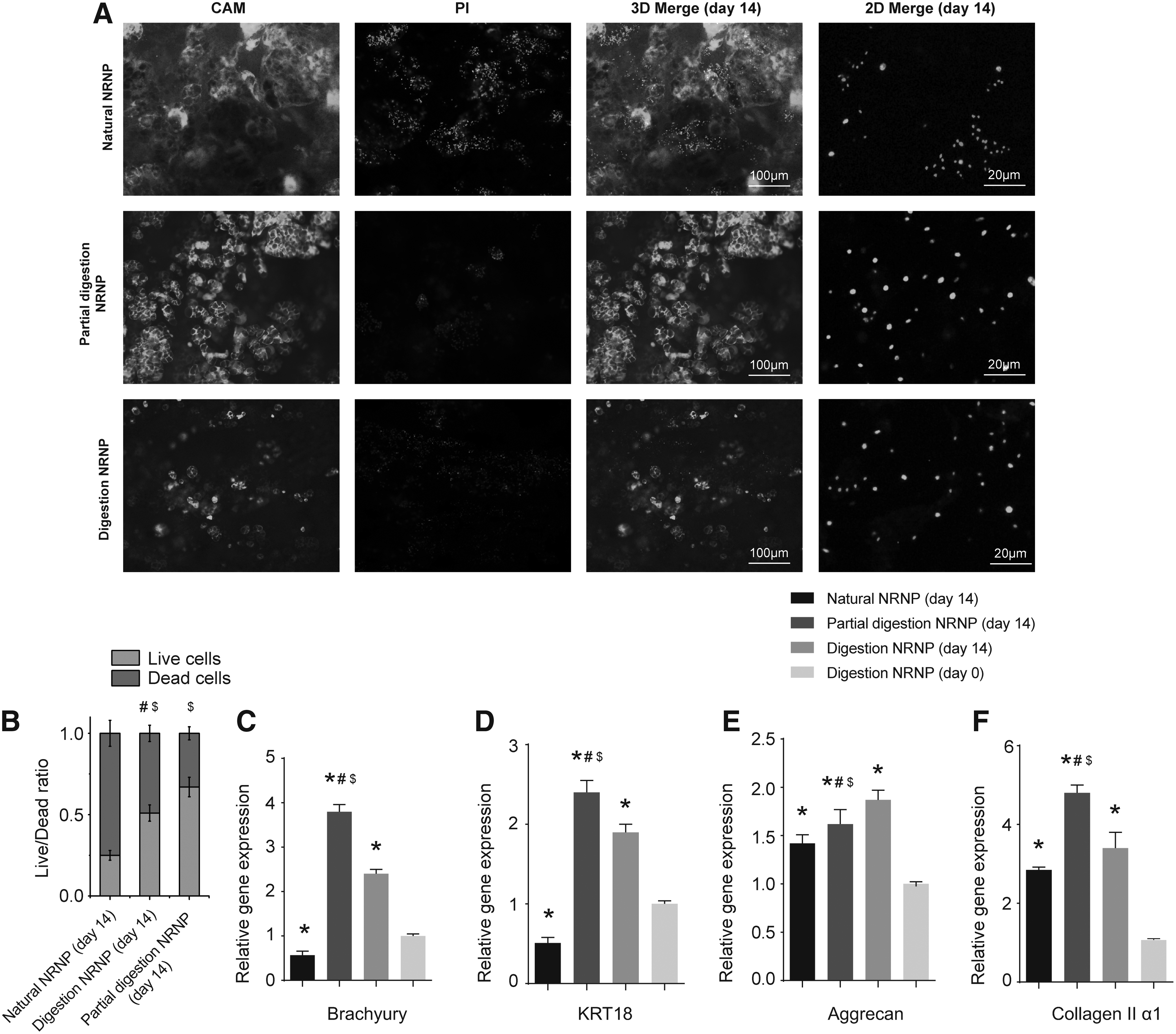
NC viability in NRNP tissues was analyzed, and green staining indicated viability, while red-stained cells were considered dead. The percentages of live and dead cells were compared among the three groups (natural NRNP, partial digestion NRNP, and digestion NRNP). After 14 days of culturing, the percentage of viable cells in natural NRNP samples decreased to (25 ± 4)%, while more viable cells were observed in the digestion NRNP group with (51 ± 5)% of viable cells and partial digestion NRNP group with nearly (67 ± 7)% viable cells (Fig. 2A, B).
In addition, the expression of NC markers, brachyury, KRT18, aggrecan, and collagen IIα1, compared with the levels of these genes at day 0 in digestion NRNP samples, were significantly higher in partial digestion NRNP and digestion NRNP after 14 days of incubation, but considerably lower in natural NRNP samples (Fig. 2C–F). Although digestion NRNP sample showed the highest gene expression in aggrecan, the expression of brachyury, KRT18 and collagen were all shown to be much higher in partial digestion NRNP samples than in nature NRNP and digestion NRNP samples after 14 days of culture (Fig. 2C–F).
NPC number and proliferation
Cell numbers and proliferative potential of NPCs were determined in each group. Although NPCs in both the coculture group and control group were shown to proliferate, a significantly higher number of NPCs were detected in the coculture group than that in the control group on day 6, 8, 10, 12, and 14 (all p < 0.05; Fig. 3A, B). Furthermore, the CCK-8 assays demonstrated a 39% increase in cell numbers on day 7 and 52% increase on day 14 in the coculture samples, in comparison with those in the controls (both p < 0.05; Fig. 3C).

NPC proliferation in the coculture and control groups. Representative images of DAPI-stained NPCs in the coculture and control groups
Cellular senescence analysis
SA-β-gal staining was used to detect cell senescence in the samples. 40 A reduced number of SA-β-gal-positive NPCs were observed in partial digestion NRNP-NPC coculture group in comparison with that in the control group on day 7 and 14 (both p < 0.05; Fig. 4A, B). Furthermore, the quantitative analysis revealed that the expression of p21 and p53 was significantly lower in the coculture samples than in the control group (both p < 0.05), while no differences in the levels were found for p16 expression (Fig. 4C).
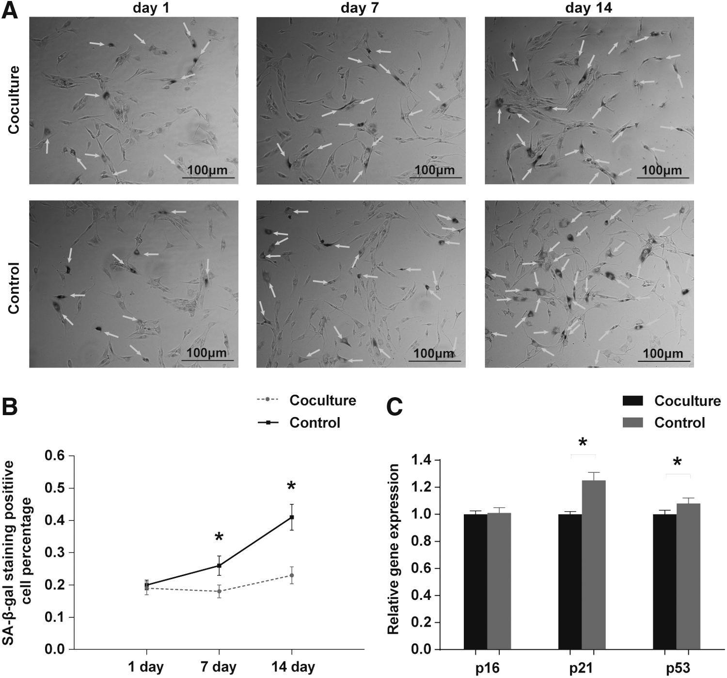
NPC senescence after 14 days of culture in the coculture and control groups.
ECM-related gene expression and GAG/DNA ratio
To examine the effect of partial digestion NRNP-NPC coculture on the ECM production of NPCs, the expression levels of ACAN, SOX9, collagen Iα1, and collagen IIα1 were assessed. On day 7 and 14, mRNA levels of ACAN, SOX9, and collagen IIα1 were shown to be significantly increased, while the levels of collagen Iα1 decreased on day 14, compared with those in the control groups (Fig. 5A–D). However, no significant difference in collagen II protein expression was observed between the two groups (Fig. 5E). Furthermore, we examined the GAG/DNA ratio of NPCs and it was shown to increase more rapidly with time, as observed on day 9, 12, and 15 in the coculture group comparing with the control group (p < 0.05; Fig. 5F).

ECM-related gene expression in the coculture and control groups. The gene expression level of ACAN, SOX9, and collagen IIα1 was significantly higher in coculture group compared with the control group in both 7- and 14-day examination, while the expression of collagen Iα1 was significantly lower in coculture group compared with the control group in 14 days
Discussion
The advantage of biological therapy of DDD is that this therapy represents the fundamental treatment of the original pain source rather than symptoms alone. An increasing number of studies suggested that NCs or NCCM may act as stimulators, controlling NPC viability, cell proliferation, and the expression of matrix proteins.15,18,19,23,27,34 However, the native tissue environment that NCs adhere to is lost during cell isolation and passaging.15,36
Therefore, in this study, we initially developed three types of NRNP explant culture systems and selected partial digestion NRNP for further analyses based on viability cells and NC gene expression. We demonstrated that partial digestion NRNP may potentially stimulate the anabolism of degenerative NPCs by enhancing cell proliferation, decreasing cellular senescence, and promoting GAG production, together with the gene expression of ECM proteins, such as collagen I, collagen II, SOX9, and ACAN. Collectively, our results demonstrate a promising potential of partial digestion NRNP for the development of novel strategies for DDD treatment.
Previous studies demonstrated that NCs play important roles in maintaining the health of IVD by behaving as trigger cells, which direct the functioning of other IVD cells.22–24,41 However, the monolayer culture was shown to be unfavorable for the maintenance of NC phenotype in vitro.27,36,42 Therefore, many researchers consider NCCM a better choice for the coculture system.18,35,43–45 Instead of using NCs isolated using a sequential pronase/collagenase digestion,8,9 in the recent studies, NCCM is generally produced using NRNP tissue.15,18,46 This allows NCs to remain clustered, which can serve to preserve NC phenotype better in vitro.18,35,47 Nevertheless, NCCM may require modifications as well due to the indirect coculturing, and nutritional components may be lost in the production and storage processes.
In addition, harvesting sufficient numbers of NCs from human degenerative IVDs still represents an obstacle due to physiological removal of NCs at an early age and numerous difficulties related to their amplification and passaging. Fortunately, the factors secreted by the NCs isolated from different species can also have regenerative effects on human degenerative NPCs, demonstrating a cross-species effect. 48 Therefore, in this study, we used rabbit-derived NRNP for the regeneration of degenerative human NPCs in vitro.
When culturing the NP tissue in a standard culture medium, it is difficult to avoid its swelling due to the difference in osmotic pressure between the tissue and the medium.20,36,37 Feng et al. 36 cultured rabbit NP explants for 14 days in free swelling conditions and the gelatinous NP tissue gradually swelled and could not be maintained further. Le Maitre et al. 20 maintained NP explants inside a plastic ring for 21 days, but even though these samples macroscopically preserved matrix integrity, the possibility that the samples would swell if not fully constrained remained.20,37 Another group attempted to improve physiological oxygen and glucose levels to prevent or reverse the swelling process, but no beneficial effects were observed. 37
Since the swelling is mainly due to the ECM pressure, the removal of its components may reduce the swelling, and therefore, the selection of an optimal NRNP explant that can maintain the native environment and stable NC behavior is of great importance. In this study, we initially generated three NRNP types: digestion NRNP, partial digestion NRNP, and natural NRNP. Our results demonstrated that the digestion of NRNP for 2 h retains good cell viability levels. In addition, the expression levels of NC phenotype-related genes were higher in partial digestion NRNP than in the other two models. Therefore, we selected partial digestion NRNP for further experiments assessing the stimulation of degenerative NPCs. Maintaining the NC phenotype in an NRNP explant is difficult for long periods22,36 and therefore, we choose 14 days of incubation since NC phenotype does not change during this time.
As the disc degenerates, the number and phenotypic characteristics of NPCs, responsible for the production of NP matrix components, such as proteoglycans and collagens, change accordingly.15,49 Therefore, regenerative therapies rely on the stimulation of cell proliferation, as well as matrix production. 50 In accordance with previous studies demonstrating the positive effects of NCCM and NCs,15,18,19 our data found that NPC proliferation and matrix production are stimulated in vitro, and an increase in the number and proliferative potential of NPCs in partial digestion NRNP-NPC coculture was observed. These data are of great importance for potential treatments due to a decreasing number of cells observed in DDD.
Partial digestion NRNP-NPC coculturing led to an increased production of matrix-related proteins as well, demonstrating the effects similar to those of NCs and NCCM. Although no statistical differences in collagen II protein expression were found between two groups, the expression of collagen IIα1 and ACAN was shown to be induced and the expression of collagen Iα1 gene was shown to be decreased in the partial digestion NRNP-NPC coculture group. This discrepancy between collagen II mRNA and protein expression may be ascribed to the short coculture time or altered cellular functions of degenerative NPCs. In addition, the observed reduction in collagen Iα1 gene expression on day 14 may indicate a decrease in the process of fibrosis. Furthermore, the expression levels of SOX9, 18 associated with a healthy NP phenotype, were shown to be higher in the coculture than in the control group. This indicates that partial digestion NRNP might have the potential to stimulate degenerative NPCs to a healthier state. Furthermore, after washing the NPCs with PBS several times to avoid the effects of GAG from partial digestion NRNP, 18 the DMMB analyses showed considerable amounts of GAG in the NPCs of coculture group, suggesting that the coculture medium may promote GAG synthesis. Taken together, our results indicate that partial digestion NRNP explant may have regenerative effects on degenerative NPCs in vitro.
Disc cell senescence, leading to a decreased number of functional cells, was considered to have a detrimental role in DDD.51,52 SA-β-gal is a biomarker used for the identification of senescent cells. 53 In our study, a significantly lower percentage of SA-β-gal-positive cells was observed in the coculture group than in the control group, indicating a protective role of partial digestion NRNP on the senescence of NPCs.
This result was further confirmed at the gene level. The p53-p21-pRb and p16-pRb pathways were two important cell senescence pathways in IVD degeneration controlled by internal and extrinsic factors, respectively.54–56 In our study, compared with the cells in the control group, NPCs of coculture group expressed significantly lower amounts of p53 and p21, further verifying the inhibitory effect of partial digestion NRNP on cell senescence, most likely by affecting the replicative p53-p21-pRb pathway. However, the p16 gene expression reflecting the extrinsic factors in coculture group was not significantly changed. This may be explained by the culture medium in both groups that made it hard to distinguish any slight change through the extrinsic pathway. Considering that NPCs play a crucial role in the maintenance of normal NP tissue homeostasis, the accumulation of senescent NPCs may reduce the rate of repair and regenerative potential of NP tissues, eventually leading to the development of DDD.
Although the obtained results demonstrated a promising potential of partial digestion NRNP use in disc repair therapy, this study had some limitations as well. Even though we maintained NCs in a native condition, the complex characteristics of the discogenic environment, such as the acidic pH and hyperosmolarity, were difficult to mimic perfectly in vitro. 14 In addition, NCs were reported to be highly sensitive to mechanical loading and changes in the nutritional environment,16,19 which makes the application of NCs in tissue engineering rather difficult. Furthermore, due to the addition of partial digestion NRNP to the NPC cells, the coculture system required additional nutrients, and therefore, we replaced the culture medium every other day.
After all, only the initial steps of determining how partial digestion NRNP may contribute to the development of future strategies for cell based DDD therapy were performed in this study. Based on the previous studies, connective tissue growth factor (CTGF or CCN2),19,21,57 Noggin and Chordin,25,58 released by NCs were demonstrated to hold potential in IVD regeneration. However, since the production of partial digestion NRNP for IVD regeneration would require large amounts of NP tissue, the factors influencing the regeneration of NPCs in partial digestion NRNP should be identified in future studies.
Conclusion
In this study, we developed a novel partial digestion NRNP-NPC coculture system, allowing the maintenance of high cell viability and NC phenotype. Coculture with partial digestion NRNP can enhance NPC proliferation rate, decrease NPC senescence, and promote the production of matrix-related proteins. Our results may help further identification of trophic factors secreted by NCs as a promising strategy for the regeneration of DDD.
Footnotes
Acknowledgments
Disclosure Statement
No competing financial interests exist.