
Abstract
Large skeletal muscle defects that result in volumetric muscle loss (VML) result in the destruction of the basal lamina, which removes key signaling molecules such as hepatocyte growth factor (HGF) from the wound site, eliminating the endogenous capacity of these injuries to regenerate. We recently showed that HGF-loaded fibrin microthreads increased the force production in muscle tissues after 60 days in a mouse VML model. In this study, we created an in vitro, three-dimensional (3D) microscale outgrowth assay system designed to mimic cell recruitment in vivo, and investigated the effect of HGF-loaded, cross-linked fibrin microthreads on myoblast recruitment to predict the results observed in vivo. This outgrowth assay discretely separated the cellular and molecular functions (migration, proliferation, and chemotaxis) that direct outgrowth from the wound margin, creating a powerful platform to model cell recruitment in axially aligned tissues, such as skeletal muscle. The degree of cross-linking was controlled by pH and microthreads cross-linked using physiologically neutral pH (EDCn) facilitated the release of active HGF; increasing the two-dimensional migration and 3D outgrowth of myoblasts twofold. While HGF adsorbed to uncross-linked microthreads, it did not enhance myoblast migration, possibly due to the low concentrations that were adsorbed. Regardless of the amount of HGF adsorbed on the microthreads, myoblast proliferation increased significantly on stiffer, cross-linked microthreads. Together, the results of these studies show that HGF loaded onto EDCn microthreads supported enhanced myoblast migration and recruitment and suggest that our novel outgrowth assay system is a robust in vitro screening tool that predicts the performance of fibrin microthreads in vivo.
Introduction
T
To assess SC recruitment, a variety of in vitro chemotactic assays have been described to rapidly measure SC migration. Many chemotactic assays such as migration across membranes in Boyden chambers, 11 or over defined two-dimensional (2D) substrates 12 do not accurately mimic the complex three-dimensional (3D) environment present in wound healing. To improve 2D migration studies, sophisticated cell tracking algorithms have been developed to follow cell movement in 3D gel matrices.13,14 While the analysis of migration through gels remains a significant improvement to 2D migration assays, these models still do not mimic the highly axially organized structure present in fibrillar tissues such as skeletal muscle, tendon, or ligament.
Our laboratory developed a novel 3D in vitro model system designed to analyze the role of surface cues on cellular outgrowth along biopolymer microthread materials.15,16 Cells were seeded into type I collagen gels that mimicked the provisional matrix at the wound margins. These collagen gels were cast around 3D biopolymer microthreads, and the rate of cell outgrowth on the microthreads was measured. 15 This method of cell outgrowth, which is a combination of migration and proliferation, occurs during skeletal muscle regeneration, when trauma triggers the longitudinal recruitment of SCs along myofibers to the injury site.9,17
The extracellular matrix of the basal lamina of skeletal muscle is composed of a combination of extracellular proteins including type IV collagen and laminin-2. 18 Additionally, fibrin is rapidly deposited as a provisional matrix following injury in skeletal muscle as a provisional matrix to support wound healing, suggesting that a fibrin gel matrix may more closely mimic the provisional matrix at the wound margins than type I collagen. 19 Further, there is evidence to suggest that early recruitment of SCs in muscle injuries occurs as a result of cell–matrix interactions, such as the release of chemotactic agents from the matrix or from integrin signaling.20,21 As such, we aimed to develop a model system that would serve as a first approximation to predict SC recruitment for skeletal muscle regeneration following skeletal muscle injuries based on these cell–matrix interactions.
In this study, we describe an in vitro assay to rapidly assess myoblast outgrowth onto HGF-loaded fibrin microthreads, as a predictor of in vivo recruitment of SCs to the injury site. Since HGF is secreted into the wound site for 3 days after injury, our goal was to develop a strategy to mimic this activity profile with HGF-loaded fibrin microthreads. We hypothesized that the passive adsorption of HGF to the surface of fibrin microthreads would facilitate a rapid delivery mechanism for the growth factor and that HGF released from fibrin microthreads would enhance the rate of myoblast outgrowth. Recently, we showed that fibrin microthreads loaded with HGF increased the number of differentiated myoblasts in the injury site 14 days after implantation and the force production in muscle tissues 60 days after implantation in a mouse model of a VML defect. 22
Here, we demonstrate that HGF was adsorbed to carbodiimide cross-linked fibrin microthreads in an active form that supported 2D myoblast proliferation and migration, and cellular outgrowth in a 3D model system. The release of adsorbed HGF from cross-linked microthreads as indicated by measuring the response of myoblasts to treated microthreads in these studies also supports our in vivo data, as there were significantly more myogenin-positive myoblasts in the injury site 14 days after injury.22,23 The results of these data demonstrate that we have developed an in vitro platform to model myoblast recruitment into VML injury sites, and supports findings observed in our in vivo model of VML injury, despite being a simplified model. This novel in vitro cell recruitment assay can be utilized as a platform to determine how various cytokines direct cell infiltration at injury sites. Further, we anticipate that this assay system can be used to predict long-term in vivo remodeling of complex injuries.
Materials and Methods
Fibrin microthread preparation
Microthread extrusion
Fibrin microthreads were co-extruded from solutions of fibrinogen and thrombin using techniques described previously.24,25 Briefly, fibrinogen from bovine plasma (F8630; Sigma, St. Louis, MO) was dissolved in HEPES (N-[2-Hydroxyethyl]piperazine-N′-[2-ethanesulfonic acid]) buffered saline (HBS, 20 mM HEPES, 0.9% NaCl; pH 7.4) at 70 mg/mL and stored at −20°C until use. Thrombin from bovine plasma (T4648; Sigma) was dissolved in HBS at 40 U/mL and stored at −20°C until use.
To fabricate microthreads, fibrinogen and thrombin solutions were thawed and warmed to room temperature, and thrombin was mixed with a 40 mM CaCl2 (Sigma) solution to form a working solution of 6 U/mL. Fibrinogen and thrombin/CaCl2 solutions were loaded into 1 mL syringes, which were inserted into a blending applicator tip (SA-3670; Micromedics, Inc., St. Paul, MN). The solutions were combined in the blending applicator and extruded through polyethylene tubing (BD, Sparks, MD) with an inner diameter of 0.86 mm into a bath of 10 mM HEPES (pH 7.4) in a Teflon-coated pan at a rate of 0.225 mL/min using a dual syringe pump. After 10 min, 25.4 cm, amorphous fibrin microthreads were removed from the buffer solution and stretched to form three 19-cm microthreads and air dried under the tension of their own weight. Dry microthreads were placed in aluminum foil and stored in a desiccator until use.
Fibrin microthread cross-linking
Fibrin microthreads were cross-linked using techniques described previously. 25 Briefly, microthreads were hydrated in an acidic buffer of 50 mM 2-(N-morpholino)ethanesulfonic acid (MES; Sigma, pH 5.2) or a neutral buffer of 100 mM monosodium phosphate (NaH2PO4; Sigma, pH 7.4) for 30 min and then cross-linked in either acidic (MES buffer, EDCa) or neutral (NaH2PO4 buffer, EDCn) buffer containing 16 mM N-hydroxysuccinimide (NHS; Sigma) and 28 mM 1-ethyl-3-(3-dimethylaminopropyl)carbodiimide (EDC; Sigma) for 2 h at room temperature. After cross-linking, the buffered EDC/NHS solution was aspirated and the microthreads were rinsed thrice in deionized (DI) water, air dried, and stored in a desiccator until use.
Adsorption of HGF to microthreads
HGF was adsorbed to fibrin microthreads using techniques described previously. 22 Briefly, three similarly treated microthreads (uncross-linked (UNX), EDCn, or EDCa), each 1.8 cm in length, were attached to polydimethylsiloxane rings (PDMS, 1.9 cm inner diameter; Dow Corning, Midland, MI), sterilized with 70% ethanol for 90 min, rinsed in DI water thrice, and air dried in a laminar flow hood overnight. Sterile microthread-PDMS constructs were hydrated in Dulbecco's phosphate-buffered saline (DPBS) and the PDMS surfaces were blocked with 0.25% (w/v) bovine serum albumin (BSA; Sigma) in DPBS for 1 h. These solutions were aspirated and replaced with 1 mL of varied concentrations of HGF (0, 5, 10, 20, 40, or 100 ng/mL of HGF; Peprotech, Rocky Hill, NJ) in DPBS and incubated at room temperature for 2 h. The microthreads were rinsed five times in DPBS and immediately used for experiments.
To assess the feasibility of HGF adsorption, we loaded microthreads with 100 ng/mL fluorescein isothiocyanate (FITC)-labeled BSA (Sigma) for 2 h, rinsed the microthreads five times in DPBS, and imaged the microthreads with a Zeiss inverted microscope (Zeiss, Thornwood, NY) using a matched filter cube (green, Ex/em 495/515 nm). The relative fluorescence intensity (RFI) of BSA-loaded microthreads was calculated comparing the RFI along the microthreads against background RFI. Each condition was run in triplicate and repeated twice.
Cell culture
Immortalized mouse myoblasts (C2C12; ATCC, Manassas, VA) were cultured in a 1:1 (v/v) ratio of high glucose Dulbecco's modified Eagle medium (DMEM; Gibco BRL, Gaithersburg, MD) and Ham's F12 (Gibco) supplemented with 4 mM
Quantifying activity of HGF from microthreads
To quantify the activity of adsorbed HGF as a function of time, HGF-loaded microthreads (0, 40, or 100 ng/mL HGF) were attached to PDMS rings (three microthreads per ring) by inserting them into slits cut into the PDMS rings, and they were incubated in 1 mL of serum-free medium (SFM; 1:1 v/v high glucose DMEM/F-12) for a period of 96 h. After 1, 2, and 4 days, two 0.5 mL aliquots of conditioned-SFM (C-SFM) were removed and incubated on C2C12 myoblasts to determine the ability of the C-SFM to induce myoblast proliferation. Therefore, the medium is completely replaced each day, and each time point corresponds to a subsequent 24 h conditioning of the medium by the microthreads. Myoblasts were seeded in 24-well plates at a density of 10,000 cells/well and incubated in SFM for 4 h before incubation with C-SFM to maintain a uniform cell population for each experiment.
As negative and positive controls, myoblasts were cultured in SFM containing 0 and 5 ng/mL of soluble HGF, respectively. Each experimental condition was run in triplicate. After an incubation time of 4 h in C-SFM, cells were fixed with ice-cold methanol, permeabilized with 0.1% Triton X-100 (Sigma), immunostained with a primary antibody against Ki67 (1:400, rabbit polyclonal, D3B5; Cell Signaling Technologies, Danvers, MA) and an Alexafluor 568 secondary (10 μg/mL, goat anti-rabbit; Life Technologies, Carlsbad, CA), and then counterstained with Hoechst 33342 (1.7 μg/mL; Molecular Probes). To determine the percentage of proliferating cells, five images were taken of each well with a Zeiss inverted microscope and the number of Ki67-positive nuclei were normalized to the total number of nuclei counted in each image.
Two-dimensional motogenic response from HGF-loaded microthreads
To measure the activity of HGF released from fibrin microthreads, we developed a purpose-built, dual-well cell migration assay system. This PDMS-based device is 5 mm thick, with two 1.9 cm diameter chambers and a 1 cm long, 3 mm wide, cell migration channel between the two chambers, which is initially sealed with a PDMS barrier (Fig. 1A). PDMS constructs were autoclave sterilized and secured to the bottom of 60 mm Petri dishes (BD Falcon, Franklin Lakes, NJ) with sterile vacuum grease (Dow Corning). Three HGF-loaded (0, 40, or 100 ng/mL HGF) microthreads were placed in the first chamber to serve as an HGF reservoir with 1 mL of medium, and the second chamber was seeded with 80,000 myoblasts in 1 mL of medium.

2D assay developed to quantify the migration of myoblasts toward HGF-loaded microthreads.
As negative and positive controls, the first chamber was loaded with 0 and 100 ng/mL of soluble HGF, respectively. After 4 h, the medium in the cell-seeding chamber was replaced with fresh medium to remove any nonadherent cells from the well, and then the barrier separating the two chambers was removed (Fig. 1B), allowing the medium in the two chambers to mix and come to an initial equilibrium. The cell front was imaged every 6 h for 24 h on a Leica inverted microscope (Leica, Wetzlar, Germany) coupled with Leica imaging software (Fig. 1C). Each experimental condition was run in duplicate. The position of the cells was determined at each time based on their distance from their starting location using ImageJ (NIH), and a linear regression curve was fit for each sample using Excel (Redmond, WA). The slope of the curve represents the migration rate (μm/h) (Fig. 1D), and these migration rates were averaged for each treatment group for statistical analyses.
Three-dimensional motogenic outgrowth from HGF-loaded microthreads
To measure cell outgrowth in a 3D model, we developed an outgrowth assay based upon an assay previously described by our laboratory. 15 Raised rectangular platforms of Thermanox® tissue culture plastic (3 × 13 mm) (Nalge Nunc, Rochester, NY) were elevated 2–3 mm above the surface of six-well tissue culture dishes using molded PDMS plugs (2 mm diameter), and secured with medical-grade silicone adhesive (Factor II, Lakeside, AZ) (Fig. 2A). The outgrowth assays were sterilized with 70% ethanol for at least 2 h, rinsed in DI water thrice, and then air dried in a laminar flow hood.
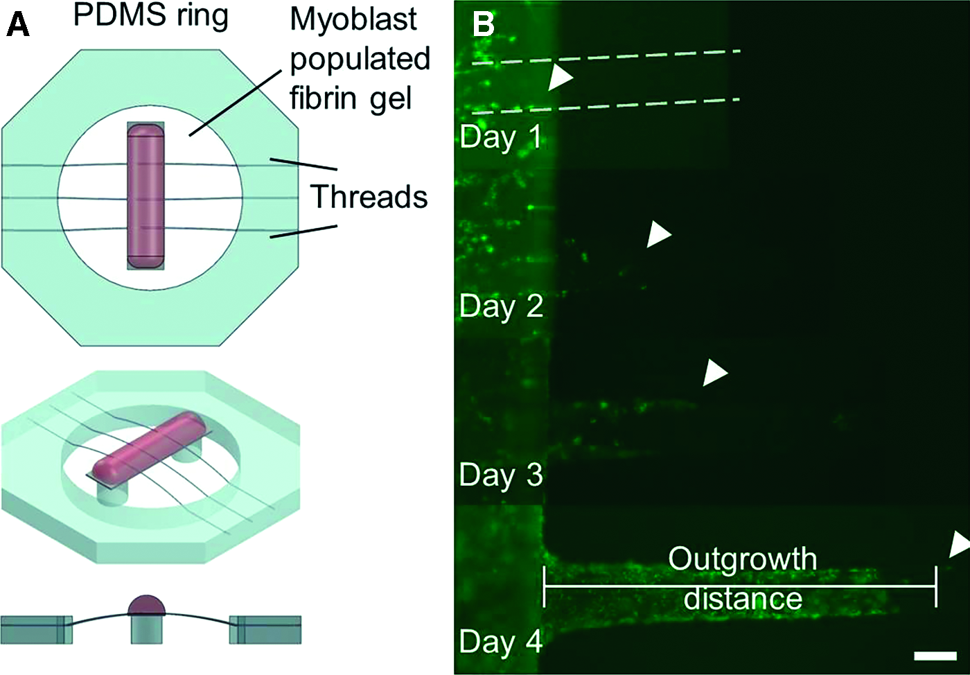
Schematic diagram of the in vitro myoblast outgrowth assay. Images show
After 0, 5, 10, 20, or 40 ng/mL of HGF was adsorbed to fibrin microthreads, the PDMS rings were affixed to the outgrowth assay with sterile vacuum grease so that the microthreads were in contact with the central region of the raised platforms (Fig. 2A). Each platform system had six microthread/platform interfaces and each condition was done in duplicate.
To create cell outgrowth assays, 40 μL of myoblast-populated fibrin gels were placed on each platform. These fibrin gels were produced by mixing a 1 mL aliquot of sterile fibrinogen (5.22 mg/mL), thrombin (2.35 U/mL), calcium chloride (31.25 mM), and cell solution (909,000 cells/mL) in a 8:1:1:2 ratio to produce a fibrin gel with a final fibrinogen concentration of 3.5 mg/mL and a final cell concentration of 150,000 cells/mL. Before mixing into the fibrin gel solution, myoblasts were loaded with DiO (Life Technologies) for 20 min according to the manufacturer's instructions. Myoblast-populated fibrin gels were incubated at 37°C for 1 h to facilitate gel formation, after which the wells were flooded with 4 mL of medium to submerge the entire gel.
To analyze cell outgrowth on the fibrin microthreads, the microthread–coverslip interfaces were imaged daily to measure outgrowth, and the position of the leading cell was determined based on its distance from the edge of the coverslip (Fig. 2B). To measure the effect of HGF on myoblast outgrowth, the outgrowth rate was calculated as the slope of the linear regression curve over a period of 2 days. After 2 days, the cells were reloaded with DiO by adding 1 mL of medium supplemented with 5 μL of DiO solution. Microthreads were incubated in DiO-supplemented medium for 20 min, and then 3 mL of medium was removed from the wells and replaced with 2 mL of fresh medium. The wells were gently agitated to mix the medium, and then an additional 2 mL was removed from each well and replaced with 2 mL of fresh medium.
After 4 days of culture, outgrowth assays were fixed in 4% paraformaldehyde and two microthreads per treatment group were permeabilized with 0.1% Triton X-100 (Sigma), immunostained with a primary antibody against Ki67 (1:400, D3B5; Cell Signaling Technologies, Danvers, MA) and an Alexafluor 568 secondary (10 μg/mL; Life Technologies, Carlsbad, CA), then counterstained with Hoechst 33342 (1.7 μg/mL; Molecular Probes). A single image from each microthread, focusing on the leading cell and the following confluent layer of cells, was imaged with an inverted Zeiss microscope, and the percentage of Ki67-positive nuclei were normalized to the total number of nuclei present the image.
Statistical analyses
Statistical analyses were performed using a one-way analysis of variance with p < 0.05 indicating significant differences between groups using SigmaPlot 11.0 software (Systat Software, Inc., San Jose, CA). For post hoc analyses, a Holm-Sidak pairwise multiple comparison test was performed to determine significance between experimental groups using an overall significance level of p < 0.05. Where indicated, a Student's t-test was performed with p < 0.05 indicating significant differences between sample groups. Data are reported as mean ± standard errors, where the sample size indicates the number of experimental replicates performed.
Results
Rapid and sustained activity of HGF from microthreads stimulates myoblast proliferation
To characterize the mechanism by which passively adsorbed HGF is released from fibrin microthreads, we developed several assays to study HGF activity. Based on its similarity to the molecular weight of HGF, FITC-labeled BSA was used as a model molecule to confirm protein adsorption on UNX and cross-linked microthreads. For UNX and EDCn microthreads, no fluorescent signal was detected with 0 ng/mL BSA (Fig. 3A, B). When FITC-labeled BSA was adsorbed to UNX and EDCn microthreads, there was an increase in detectable fluorescence signal, with qualitatively more BSA adsorption observed on EDCn microthreads than on UNX microthreads (Fig. 3D, E). Interestingly, there was no notable evidence of BSA adsorption onto EDCa microthreads, relative to control samples (Fig. 3C, F). There was a significantly higher RFI on UNX and EDCn microthreads when loaded with FITC-labeled BSA, and the RFI of BSA-loaded EDCn microthreads was significantly higher than BSA-loaded UNX or EDCa microthreads (Fig. 3). Together, these findings suggest that cross-linking the fibrin microthreads in a neutral pH environment affects the ability of proteins to bind to the surface of the microthreads.
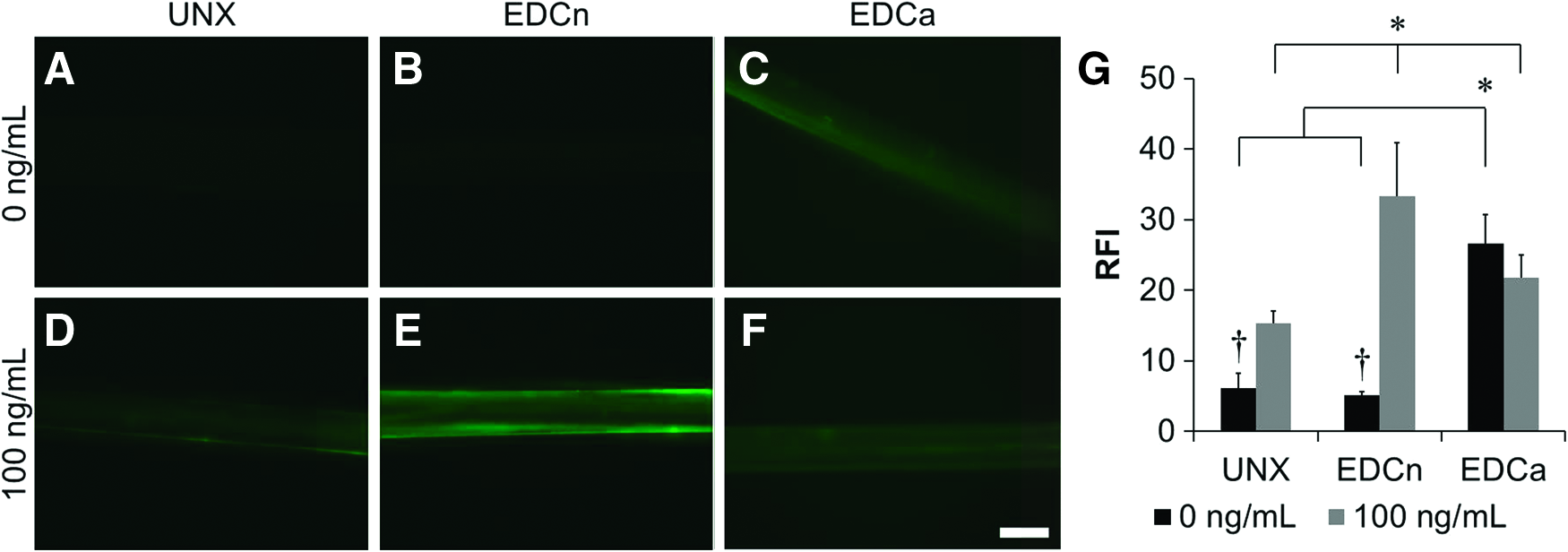
Representative images of microthreads loaded with FITC-labeled BSA. UNX
To quantitatively evaluate the temporal activity of HGF eluted from microthreads, myoblasts were incubated in SFM conditioned with HGF-adsorbed microthreads (C-SFM) and cells were assayed for increases in Ki67 expression. We previously reported that the percent of Ki67 positive myoblasts significantly increased when cells were cultured in C-SFM from 40 and 100 ng/mL HGF coated EDCn microthreads (56.0% and 54.3%, respectively) and UNX microthreads (51.6% and 48.2%, respectively) for 1 day, HGF coated EDCn microthread C-SFM from both 40 and 100 ng/mL at 2-day incubations (53.7% and 52.9%, respectively) also stimulated myoblast proliferation, and that neither 40 or 100 ng/mL HGF coated EDCn (40.3% and 38.0%, respectively) or UNX (40.6% and 42.4%, respectively) microthread C-SFM stimulated myoblast proliferation at day 4 incubations. 22
In all cases including this study, ∼40% of myoblasts were proliferating in the negative controls and ∼55% of myoblasts were proliferating in the positive controls. Here, EDCa microthread C-SFM stimulated myoblast proliferation regardless of the concentration of HGF after 1 day (Fig. 4) with respect to negative controls (Fig. 4B). This response is comparable to the response observed when cells were cultured in the presence of 5 ng/mL of soluble HGF (Fig. 4C), which is in the range of HGF concentrations found in the literature to maximally stimulate proliferation in myoblast populations.22,26 There were significantly lower percentages of Ki67+ myoblasts when incubated in EDCa microthread C-SFM from 0 and 40 ng/mL HGF at 2 (Fig. 4E) and 4 days (Fig. 4F) than with C-SFM collected from 1 day (Fig. 4D).
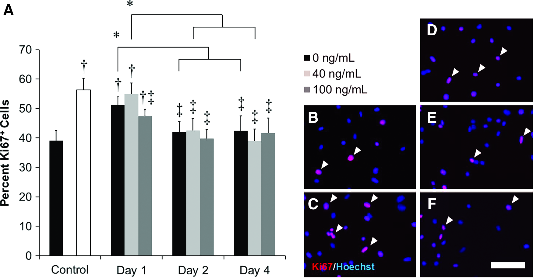
Percentage of C2C12 Ki67+ myoblasts stimulated from EDCa microthread C-SFM.
HGF-adsorbed fibrin microthreads increases 2D myoblast migration
To confirm that HGF-adsorbed microthreads stimulated myoblast migration, a dual-well PDMS device was fabricated where one reservoir had three HGF-loaded microthreads and the second chamber was seeded with C2C12 myoblasts (Fig. 1A). The PDMS barrier separating the two channels was removed after 4 h (Fig. 1B) to measure myoblast migration toward HGF-adsorbed microthreads in a 2D system. Migration was observed for 24 h. There was a twofold increase in the rate of myoblast migration stimulated by HGF-adsorbed EDCn microthreads; independent of the amount of HGF adsorbed. This suggests that incubating microthreads with 40 ng/mL of HGF either saturates the surface of the microthreads, or that 40 ng/mL of HGF generates a similar cell response to that of HGF-adsorbed microthreads incubated with 100 ng/mL of HGF (Fig. 5).
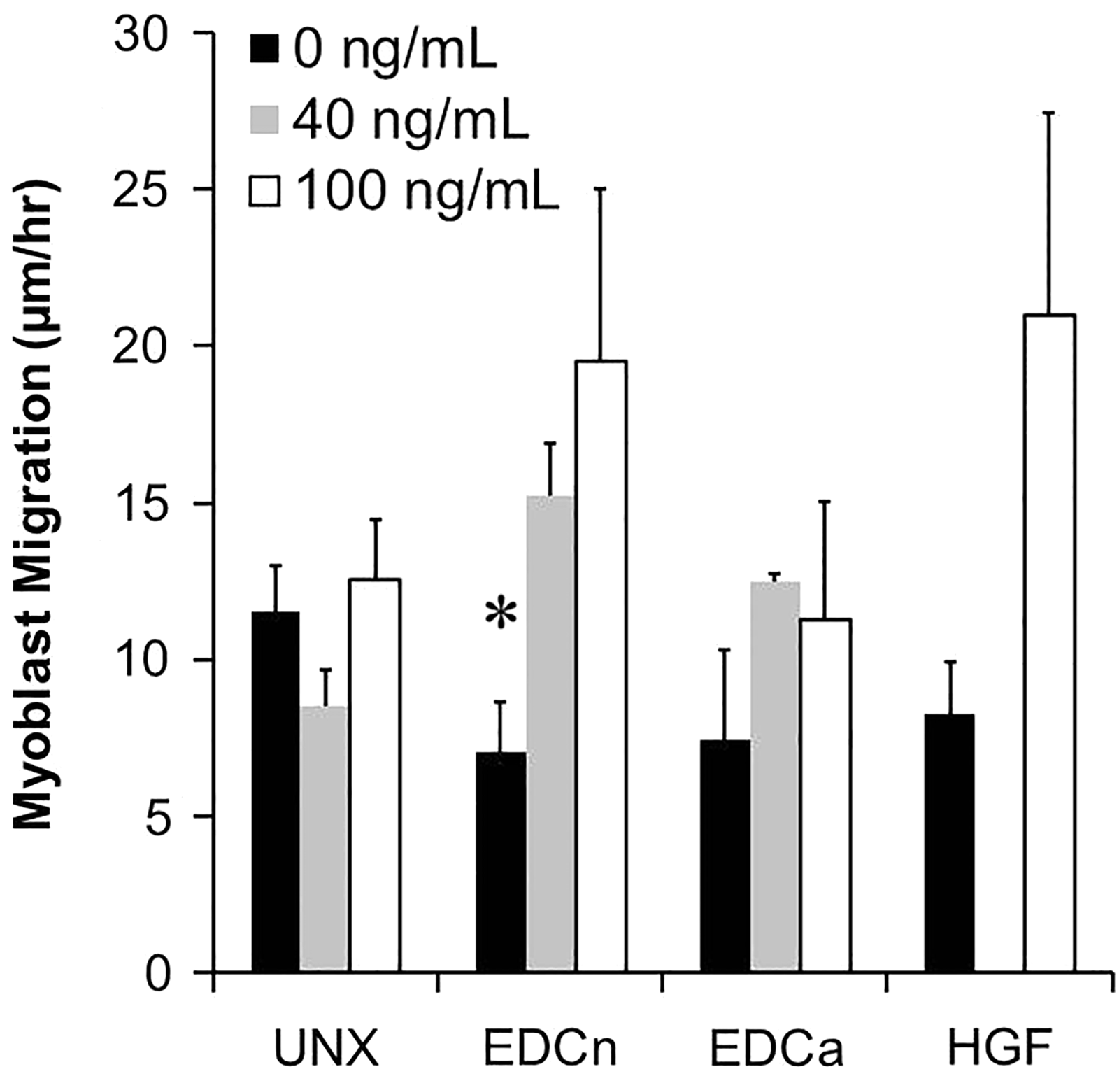
Plot showing myoblast migration toward HGF-loaded microthreads in a 2D model system. Migration toward HGF-loaded EDCn microthreads increased twofold relative to unloaded EDCn microthreads. There was no significant increase in cell migration toward UNX or EDCa microthreads with increasing amounts of adsorbed HGF. Soluble HGF at 0 ng/mL (HGF, black bar) or 100 ng/mL (HGF, white bar) was added to some microthread chambers as negative and positive controls, respectively. *Indicate significant differences between migration rates of unloaded microthreads and HGF-loaded microthreads of similar cross-linking type as determined by one-way ANOVA with Holm-Sidak post hoc analysis (p < 0.05, n ≥ 2).
The migration rates stimulated by both HGF loading concentrations (40 and 100 ng/mL) on EDCn microthreads were significantly higher than negative control reservoirs and they were comparable to experimental conditions that contained 100 ng/mL of soluble HGF; which was found to maximally stimulate migration of SCs along muscle fibers. 27 Further, the response from each of these loading concentrations were similar to each other, suggesting a plateau in the activity of HGF-adsorbed EDCn microthreads at or below a loading concentration of 40 ng/mL. Despite measuring an increase in expression of Ki67 from myoblasts incubated in HGF-loaded UNX microthread C-SFM, we did not observe a significant increase in the motogenic response from UNX microthread C-SFM. Similarly, there was no significant increase in the motogenic response toward EDCa microthreads regardless of the amount of HGF adsorbed.
HGF-adsorbed EDCn fibrin microthreads enhances myoblast outgrowth
To analyze the effects of HGF-loading on cell outgrowth along fibrin microthreads, we developed a novel in vitro 3D assay system to quantify the rate of cell outgrowth. Because there were no significant differences in 2D cell migration between medium that was conditioned with microthreads that were loaded with 40 or 100 ng/mL of HGF, we assumed that we had reached an adsorption saturation limit in our system and adsorbed 0, 5, 10, 20, or 40 ng/mL of HGF to the surface of microthreads to evaluate the effect of HGF on the 3D outgrowth of myoblasts from a cell-seeded fibrin gel. The HGF-loaded microthreads were positioned over raised Thermanox coverslips (Fig. 2A), and C2C12 myoblasts were preloaded with the membrane dye tracker DiO to enable daily visualization of cellular outgrowth on the scaffolds (Fig. 2B).
The myoblast outgrowth rate, a measure of total cell infiltration, was significantly higher by twofold on EDCn microthreads adsorbed with 40 ng/mL of HGF compared to unloaded EDCn controls (Fig. 6). Interestingly, there was no HGF-mediated increase in myoblast outgrowth on EDCa or UNX microthreads. The total distance myoblasts traveled, a measure of cell migration and recruitment, along EDCn microthreads after 4 days also significantly increased when 10, 20, or 40 ng/mL of HGF was loaded onto the microthreads; compared to 0 or 5 ng/mL of HGF (Fig. 6B). The total distance myoblasts traveled along UNX microthreads also significantly increased from 0 to 20 ng/mL of HGF was loaded onto the microthreads. These data suggest that preadsorption of 20 or 40 ng/mL of HGF to the surface of EDCn microthreads will significantly increase the outgrowth rate and distance these myoblasts will travel within 4 days.
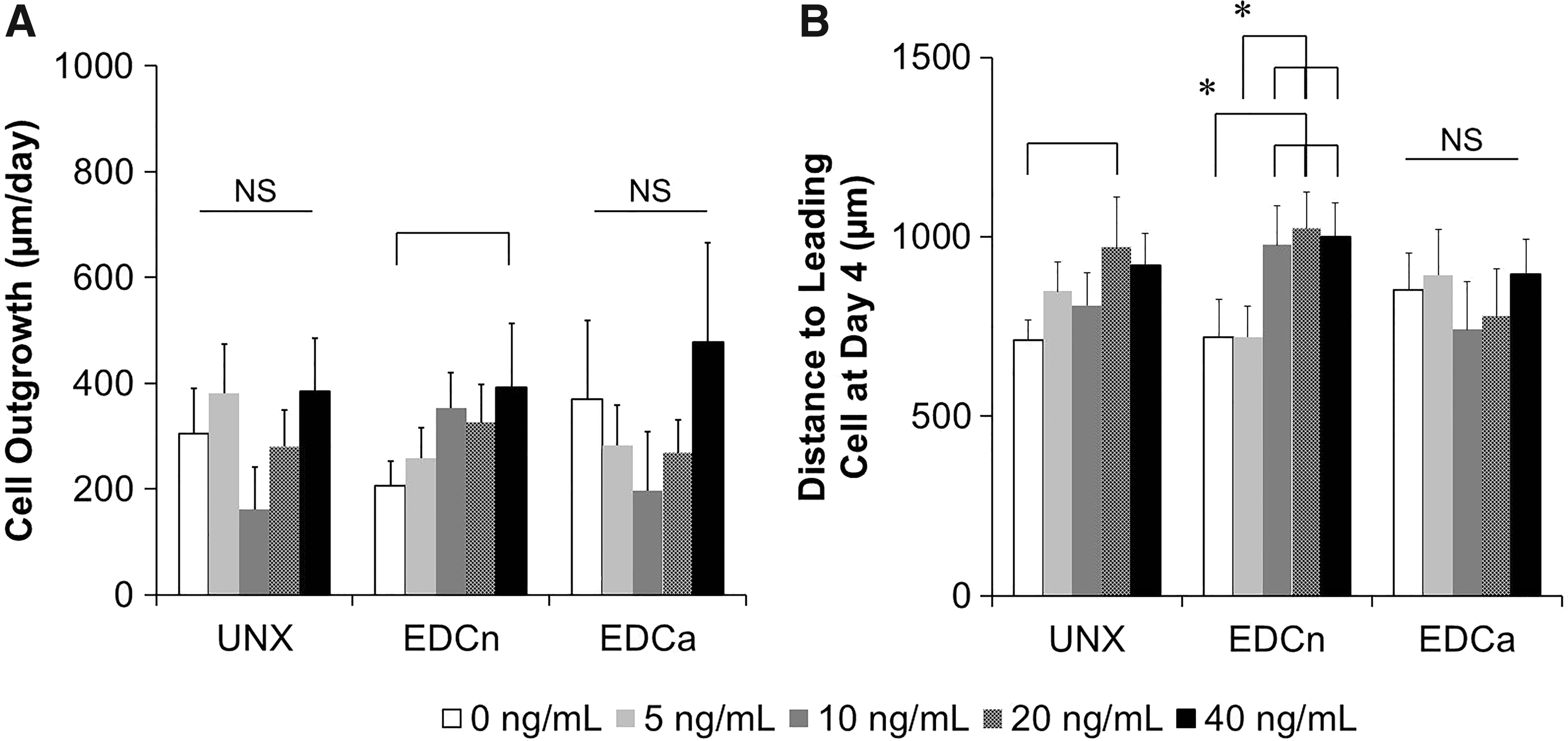
Cell outgrowth of C2C12 myoblasts on HGF-loaded fibrin microthreads.
Fibrin microthreads enhance myoblast proliferation
Cell outgrowth is a phenomenon that includes both the cellular functions of migration and proliferation. To uncouple these mechanisms and to characterize the relative role of cell proliferation in cellular outgrowth, we quantified the number of Ki67 positive cells present on the microthreads at day 4. For most conditions, the cells were observed to be a confluent layer with several cells ahead of the migration front (Fig. 7A–F). Interestingly, Ki67 positive cells were evenly distributed among the leading cells and within the layer of cells behind the migration front. There was a significant increase in the percentage of proliferating cells on the EDCn and EDCa microthreads compared to UNX microthreads (Fig. 7H).
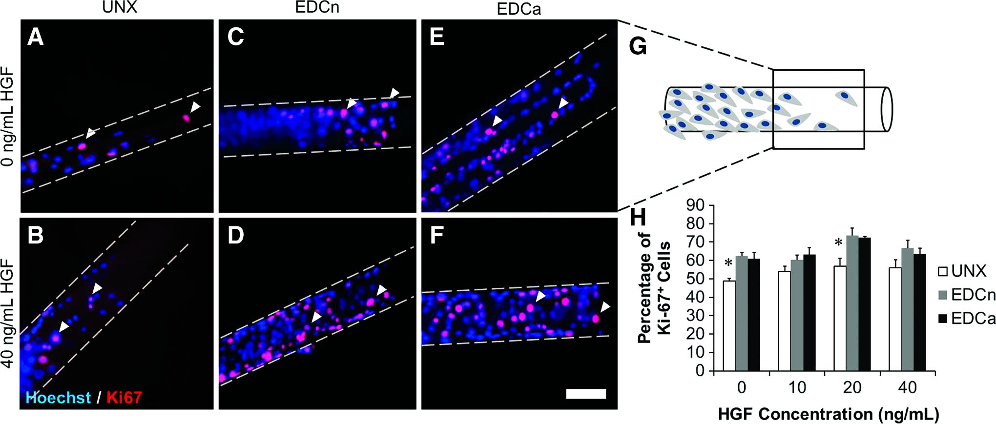
Ki67 staining of myoblasts on fibrin microthreads from cellular outgrowth assays at day 4.
Discussion
Treatment of VML injuries remains a significant healthcare concern due to the inability of skeletal muscle to functionally regenerate these critically sized injuries. One reason that VML defects are difficult to regenerate via the endogenous skeletal muscle regenerative pathway is the loss of the native ECM from these injury sites, eliminating essential biophysical and biochemical cues that typically initiate tissue regeneration. One of the first signaling molecules that is released from damaged skeletal muscle is HGF, which functions to both activate and recruit SCs to the wound site.28,29 In this study, we developed a 3D in vitro model system to predict myoblast recruitment to the wound site and we demonstrated that HGF-adsorbed EDCn microthreads facilitate enhanced myoblast outgrowth along EDCn microthreads. 22 Conversely, while EDCa microthreads supported myoblast migration, their inability to bind and release growth factors such as HGF makes them unsuitable for the delivery of factors for skeletal muscle regeneration. The findings from these studies suggest that HGF-loaded EDCn cross-linked microthreads will enhance endogenous skeletal muscle regeneration due to this increased ability to recruit myoblasts to the wound site.
To support the findings that cell outgrowth was enhanced on HGF-loaded EDCn microthreads, results from our FITC-labeled BSA study indicated that a significant increase in fluorescence was observed on UNX EDCn microthreads. In contrast, there were no significant changes in RFI for EDCa microthreads. While the RFI increased on UNX microthreads, the BSA-FITC-loaded UNX microthreads had a significantly lower RFI than EDCn microthreads. Carbodiimide cross-linked surfaces have an increased amount of amide bonds due to the coupling of free amino and carboxyl groups, and we confirmed this in a separate study where we observed a significant increase in the relative amount of amide bonds between EDCa microthreads and UNX microthreads via Fourier transform infrared spectroscopy (FTIR) analysis. 30
We hypothesize that EDCa microthreads are composed of additional aspartate and glutamate amino acid residues because of the hydrolysis of the amine containing residues asparagine and glutamine, respectively, which may further decrease the surface charge of EDCa microthreads with respect to EDCn microthreads. 25 FTIR analyses also showed that surfaces of EDCa microthreads appear to undergo alkylation with ethylene oxide sterilization, which significantly reduced the ability of cells to attach to these microthreads, and it suggests that the surface chemistry of EDCa microthreads may be significantly altered by microthread cross-linking techniques. 30 Together, these data suggest that EDCa microthreads exhibit limited capacity to adsorb bioactive molecules such as BSA-FITC or HGF. While these materials may have passive material properties that support cell infiltration, their inability to adsorb significant quantities of growth factors, which strategically direct SC recruitment to VML injuries, remains a limitation in their utility in this application.
It is interesting to note that while HGF adsorbed to EDCn microthreads stimulated proliferation and migration, HGF adsorbed to UNX microthreads only stimulated proliferation. HGF is a pleiotropic growth factor that stimulates a variety of cell functions including proliferation, 31 migration,32,33 and survival. 34 Regardless of which downstream pathway is activated, HGF signaling is initiated by its interaction with the tyrosine kinase receptor, c-Met.35,36 Studies assessing the motogenic effects of HGF have found its effect to be maximized with concentrations at or below 100 ng/mL, and in general, higher concentrations elicit increased motility,27,37,38 while concentrations between 2 and 10 ng/mL have been found to stimulate myoblast proliferation.4,26,39
Based on the observations that HGF is a globular (spherical) protein with a molecular weight of 100 kDa, it is estimated that the minimum hydrodynamic radius of the molecule is 3.06 nm and the minimum surface area is 59 nm2. 40 Assuming that microthreads are ∼130 μm in diameter, 25 the surface area of each microthread is 4.08 mm2/cm of microthread. From these calculations, we estimate the theoretical adsorption limit of HGF to microthreads to be 11.5 ng of HGF per 1.0 cm length of microthread. Each microthread in the assay was 1.8 cm in length, yielding an adsorption limit of 20.7 ng of HGF per microthread. Given that HGF is being adsorbed to three microthreads at a time, the maximum amount of HGF that could be adsorbed would be 62.1 ng.
Because we did not observe any significant differences in myoblast proliferation or migration between adsorbing EDCn microthreads with a loading concentration of 40 or 100 ng/mL, we assume that we did not reach this theoretical maximum. However, the minimum concentration of HGF that has been reported to induce a chemotactic response on myoblasts is 10 ng/mL. 32 These observations, coupled with our data, suggest that the HGF-loaded EDCn microthreads are releasing at least 10 ng/mL of HGF per day to enhance 2D and 3D migration. Finally, we used FITC-conjugated BSA as a molecular weight analog of HGF and we observed the binding efficiency of the adsorbed protein on two different microthread scaffolds. We observed an increase in adsorption of FITC-labeled BSA on EDCn microthreads, further supporting the hypothesis that EDCn microthreads are facilitating the adsorption of more HGF than UNX microthreads.
It is interesting to note that the results of this in vitro model system mimicked the results observed in our recent in vivo work using HGF-loaded EDCn microthreads in a mouse VML model. 22 EDCn microthreads supported similar amounts of cell outgrowth as UNX microthreads, demonstrating that this cross-linking method did not adversely affect cell mobility. Further, EDCn fibrin microthreads, loaded with 40 ng/mL of HGF promoted a twofold increase in the rate of myoblast outgrowth. Similarly, the total distance that the myoblasts migrated along the microthreads followed a comparable trend: HGF-loaded EDCn microthreads increased the migration distance of the leading cell along the surface of the scaffold compared to control EDCn microthreads. For a scaffold to successfully regenerate skeletal muscle in VML defects, the material must recruit myoblasts to the wound site and persist for 2–4 weeks, long enough for the entire wound to regenerate. 41
We observed a significant increase in myogenin positive myoblasts 14 days after injury in a mouse model of VML, 22 supporting the validity of this model to accurately predict increases in myoblast infiltration into the injury site. It is worth noting that the similarities between the in vitro model and in vivo study are presented in a facile, first-order system. This in vitro model does not account for contributions to myoblast recruitment and migration from the immune system or from vascularization or innervation into the wound site, all of which certainly have an effect on this dynamic regenerative environment. These results highlight the importance of early cell-cytokine signaling events in skeletal muscle regeneration and suggest that these signaling events are critical for the early recruitment of myoblasts and for positive remodeling outcomes in VML injuries. Furthermore, the modular nature of this in vitro model system will enable the systemic analyses of these synergistic factors in future studies, by incorporating additional cell types or conditioned medium to the model system.
While we did not observe evidence of HGF adsorption to EDCa microthreads, it is interesting to note that C-SFM conditioned for 1 day by EDCa microthreads stimulated an increase in myoblast proliferation, independent of the HGF loading process. This phenomenon was only observed when medium was conditioned with EDCa microthreads during the first day, suggesting that the acidic cross-linking conditions may be generating mitogenic factors within the fibrin microthreads. We previously noted that prolonged incubation in acidic buffer degraded fibrin microthreads. 25 Together, these results suggest that incubation of fibrin microthreads in acidic buffer may be creating fibrin degradation products, which have been shown to direct cell functions such as proliferation and migration in a variety of model systems.42–45 It is important to note that while fibrin degradation products stimulate cell functions such as proliferation and migration, these effects are independent of growth factor supplementation. 45 We hypothesize that these degradation products likely formed during microthread rehydration, while microthreads were incubating in acidic buffer before cross-linking, and released within a 24-h period to stimulate myoblast proliferation. Future studies will analyze C-SFM from EDCa microthreads to identify the presence of fibrinolytic mitogens, specifically focusing on fibrin degradation products. 43
Throughout the cellular outgrowth study, we consistently observed a group of cells migrating ahead of what appeared to be a confluent cell front on the surfaces of microthreads. This suggests that the migration of several cells along the microthread substrate precedes the proliferation of these cells to achieve a confluent layer. This phenomenon was observed each day that the microthreads were imaged (Fig. 2B for a representative data set), suggesting that this is a continuous process, where several motile cells migrate ahead of a more confluent layer of cells that results from cell proliferation. A similar phenomenon has been observed for endothelial cell outgrowth, and this cellular migration effect was attenuated when the system was treated with mitomycin C, suggesting that both proliferation and migration are necessary for outgrowth. 37
Interestingly, when the percentage of Ki67 positive cells was quantified along microthreads at 4 days of culture, a substrate-dependent increase in proliferation was observed. Substrate stiffness has been reported to increase myoblast proliferation in other 2D studies, 46 and we have previously reported that EDCn and EDCa microthreads are significantly stiffer than UNX microthreads (11.66, 22.42, and 3.38 MPa, respectively). 25 Together, these observations suggest that HGF-adsorbed microthreads enhance the migration of motile cells along EDCn microthreads and substrate-mediated cell proliferation is concurrently promoting the formation of a confluent layer of cells behind the leading edge of the migrating cells. Future studies will look at later time points to assess the ability of these microthreads to support myoblast differentiation in myofibers and analyze the effects of incorporating different cell types such as monocytes or endothelial cells to encourage vascularization.
Conclusions
In this study, we (i) developed an in vitro model system to estimate cell recruitment in complex 3D environments, and (ii) loaded a dose-dependent increase in the concentration of HGF to the surface of cross-linked fibrin microthreads to stimulate an increase in myoblast outgrowth. Further, the results from myoblast recruitment along HGF-loaded EDCn microthreads within this in vitro system closely align with results observed in our recent study where HGF-loaded EDCn microthreads were found to significantly increase the amount of myoblasts within the injury site of a mouse VML defect 14 days after injury, demonstrating that our system can accurately model in vivo cellular responses. The synergistic contributions of cell migration and proliferation, and soluble factors, can be uncoupled and systematically analyzed within this outgrowth assay, demonstrating the utility of this tool to serve as a powerful platform to model cell recruitment in axially aligned tissues, such as skeletal muscle.
Our immunofluorescence analyses suggest that the surface of EDCn microthreads facilitate the greatest concentration of HGF loading. We also showed that these substrates are capable of stimulating myoblast proliferation in addition to 2D and 3D migration. These effects plateaued when 40 ng/mL of HGF were adsorbed to the surfaces of the scaffolds. UNX microthreads supported some HGF adsorption that promoted myoblast proliferation, but these scaffolds did not significantly enhance myoblast migration. We hypothesize that changes in the surface chemistry between UNX and EDCn microthreads are responsible for this difference in the growth factor loading capacities of the microthreads. Regardless of the adsorption of HGF, the proliferation of myoblasts 4 days after cells migrated along the microthreads was increased on cross-linked microthreads. Taken together, these results suggest that our novel outgrowth assay is a robust, in vitro screening tool that can be utilized to determine how different cytokines affect cell infiltration, and can be used to predict the performance of biomaterials remodeling complex injuries in vivo.
Footnotes
Acknowledgments
This research was funded in part by US Army (W81XWH-11-1-0631, G.D.P. and R.L.P.), NIH R01-HL115282 (G.D.P.), NIH F31-DE023281 (J.M.G.), and NIH F32-DE026058 (J.M.G.). The authors wish to thank Michelle Zayas for creating the CAD designs and Laura Pumphrey and Brianna Sheldon for their assistance in fabricating the migration and outgrowth assays.
Disclosure Statement
G.D.P. discloses that he is a cofounder and has an equity interest in Vitathreads L.L.C., a company that has licensed intellectual property associated with fibrin microthreads.