




Abstract
Collagen has been used extensively in tissue engineering applications. However, the source of collagen has been primarily bovine and porcine. In view of the potential risk of zoonotic diseases and religious constraints associated with bovine and porcine collagen, fish collagen was examined as an alternative. The aim of this study is to use tilapia fish collagen to develop a cross-linked electrospun membrane to be used as a barrier membrane in guided bone regeneration. As there is limited data available on the cytotoxicity and immunogenicity of cross-linked tilapia collagen, in vitro and in vivo tests were performed to evaluate this in comparison to the commercially available Bio-Gide® membrane. In this study, collagen was extracted and purified from tilapia skin and electrospun into a nanofibrous membrane. The resultant membrane was cross-linked to obtain a cross-linked electrospun tilapia collagen (CETC) membrane, which was characterized by scanning electron microscopy (SEM), differential scanning calorimetry (DSC), degradation studies, cytotoxicity studies, and cell proliferation studies. The membranes were also implanted subcutaneously in rats and the host immune responses were examined. The DSC data showed that cross-linking increased the denaturation temperature of tilapia collagen to 58.3°C ± 1.4°C. The in vitro tests showed that CETC exhibited no cytotoxicity toward murine fibroblast L929 cells, and culture of murine preosteoblast MC3T3-E1 cells demonstrated better proliferation on CETC as compared to Bio-Gide. When implanted in rats, CETC caused a higher production of interleukin IL-6 at early time points as compared to Bio-Gide, but there was no long-term inflammatory responses after the acute inflammation phase. This finding was supported with histology data, which clearly illustrated that CETC has a decreased inflammatory response comparable to the benchmark control group. In all, this study demonstrated the viability for the use of CETC as a tissue engineering scaffold and provides an insight on the in vivo immune responses toward xenogenic collagen scaffolds.
Introduction
C
Traditionally, commercial type I collagen has been extracted from mammalian sources, especially from bovine and porcine skin. However, there has been a growing concern about the risk of transmissible diseases from mammalian tissues to human recipients, such as bovine spongiform encephalopathy.3–5 In addition, bovine and porcine collagen products may pose a problem for patients with religious constraints. 6 In view of the limitations of mammalian collagen, fish collagen has recently become an attractive alternative in biomedical applications. Beside the low risk of disease transmission and less religious limitations, fish collagen is also highly abundant in fish skin, scales, and bones, which are often discarded as waste by the fish-processing industry.1,7 Hence, exploiting this under-utilized resource would generate value out of unwanted fish parts, and lead to a reduction of waste.
Among the various fish species, Nile tilapia (Oreochromis niloticas), a tropical freshwater fish, is considered to be the most promising source of collagen, due to its high popularity in fish farms around the world. Dubbed by fishery experts as the “aquatic chicken,” tilapia is farmed in >100 countries due to its fast growth, high protein content, high resistance to disease, and good adaptability in a wide range of breeding conditions. 8 More importantly, tilapia type I collagen has the highest denaturation temperature (Td) among various reported fish collagens, which allows the processing procedures to be less temperature sensitive. 9
For tilapia collagen to be employed as tissue engineering scaffolds, it needs to be converted from its water soluble state into a solid porous structure to support cell attachment and nutrient diffusion. Electrospinning is increasingly employed in the production of collagen scaffolds because the nanofibrous architecture and biochemical properties of the electrospun membranes can be easily adjusted by varying the solution composition and spinning parameters.4,5,10 The nanofiber membrane can be stabilized by physical or chemical cross-linking treatment to preserve its structure and mechanical integrity, but there is little data available on the cytotoxicity and immunogenicity of cross-linked tilapia collagen.
Guided bone regeneration (GBR) is a dental surgical procedure to restore bone tissue in an alveolar defect, with the help of a barrier membrane to prevent the entry of epithelial cells into the defect and promote osteoinduction within the defect. 11 In this study, we aimed to fabricate a cross-linked electrospun tilapia collagen (CETC) membrane and compare its biological responses with Bio-Gide®, a commercial porcine collagen membrane, to determine the suitability of CETC for GBR. The in vivo immune response studies were conducted in rats with a cage implant model and a subcutaneous implantation model. We hypothesize that the CETC membrane will have similar biological responses as Bio-Gide and will be a suitable material for GBR.
Materials and Methods
Chemicals reagents
Phosphate-buffered saline (PBS), Dulbecco's modified Eagle's medium (DMEM), minimum essential medium alpha (MEM-α), fetal bovine serum (FBS), penicillin-streptomycin, and PicoGreen assay kit were obtained from Life Technologies. All other chemicals and reagents were obtained from Sigma-Aldrich, unless otherwise stated.
Isolation of tilapia collagen
Collagen was isolated from tilapia skin following the procedures described by Zhang et al. 4 and Zhou et al. 5 with slight modifications. Fresh tilapia skin was stirred in 0.1 M NaOH solution for 6 h to remove noncollagenous proteins, 10% 1-butanol, and 20% isopropanol in water for 24 h to remove fats and fat-soluble pigments, and 0.5 M acetic acid for 48 h to solubilize the collagen. The crude collagen extract was separated from the skin residue by centrifugation, and 5 M NaCl solution was added dropwise to the supernatant to a final concentration of 1 M to precipitate the collagen. After centrifugation (18,000 g) for 1 h, the precipitate was redissolved in 0.5 M acetic acid. The solution was filtered, dialyzed in DI (deionized) water overnight, and lyophilized to obtain collagen sponges for subsequent use. The whole process was performed at room temperature (23°C ± 1°C).
Fabrication of CETC membrane
The collagen sponge was dissolved in hexafluoroisopropanol with a weight:volume ratio of 1:10. The solution was placed into a 3 mL syringe with a needle (inner diameter 0.25 mm), and inserted vertically into a syringe pump (KDS-100-CE; KDS Scientific), with the needle tip 10 cm above the aluminium foil collector. A voltage of 10 kV was applied between the needle and the collector, and the pump flow rate set at 0.7 mL/h. After 4 h of collection, the membrane was then cross-linked with glutaraldehyde vapor for 2 h and vacuum-dried overnight. The whole process was performed at room temperature (23°C ± 1°C).
Characterization of CETC membrane
The membrane morphology was analyzed by scanning electron microscopy (SEM). The collagen membrane was dehydrated with increasing concentrations of ethanol and dried overnight in vacuum. The samples were splutter-coated using a platinum ion coater (JFC-1600; JEOL, Japan) and analyzed with the SEM (JSM-6700F; JEOL).
The thermal stability was evaluated by differential scanning calorimetry (DSC). The samples was dissolved in 0.05 M acetic acid solution and placed into a sample pan. The denaturation temperature and denaturation enthalpy of the samples were determined with the DSC machine (Diamond DSC; Perkin Elmer) with a heating rate of 10°C/min.
The mechanical strength of the membranes was determined by uniaxial tensile testing. Prewetted membranes were tapped to remove the excess water, cut into 30 × 5 mm strips, and secured onto the tensile tester (Instron 5543; Instron) with clamps in a 100 N load cell. The dimensions of the sample were measured with a micrometer and recorded in the tensile testing software. The load cell was calibrated and the sample was pulled with an extension rate of 10 mm/min until the sample broke. The maximum tensile stress and Young's modulus, derived from the tensile testing software, were recorded and compiled.
The degradation profile of the CETC membrane was performed according to the procedure described by Zhang et al.
4
with some modifications. Each 1 × 1 cm sample was freeze-dried, accurately weighed, and placed in 2 mL PBS under aseptic conditions. The tubes were sealed and incubated at 37°C for 7, 14, 21, and 28 days. After each time point, samples was taken out and rinsed in DI water, freeze-dried, and weighed. The weight loss of each sample was calculated by the following equation.
where W0 represents the initial weight (g) of the dried sample before incubation and Wt represents the weight of the degraded sample after the respective time points.
Cell culture for in vitro studies
Murine-derived preosteoblast cell line (MC3T3-E1) (ATCC®) and murine fibroblasts (L929) (ATCC) were used in this study. MC3T3-E1 cells were cultured in MEM-α and L929 cells in DMEM+GlutaMAX supplemented with 10% FBS. Cells were incubated at 37°C and passaged every 2–3 days. All samples for the in vitro studies were sterilized with 70% ethanol for 2 h and washed with respective medium before use in cell culture.
Cytotoxicity studies
Indirect cytotoxicity studies were performed in accordance to ISO 10993-5 protocols. Samples were sterilized as above and individually incubated in DMEM at 37°C for 24 h to obtain an extract of the test sample. Simultaneously, L929 cells were seeded into 24-well plates (Corning) at a density of 2 × 10⁴ cells/well and grown overnight. The culture media were then replaced by the sample extracts and after 24 h incubation, cells were visualized by light microscopy and cell viability determined using Alamar blue assay (Invitrogen). Polyurethane film containing 0.1% zinc diethyldithiocarbamate (ZDEC) (Food and Drug Safety Center, Hatano Research Institute, Japan) and high-density polyethylene (Food and Drug Safety Center, Hatano Research Institute, Japan) were used as positive (toxic) and negative (nontoxic) control respectively.
Cell adhesion, viability, and proliferation assay
Samples were cut into circular disc of 10 mm diameter, sterilized as previously described, and placed into a 24-well plate (Corning). MC3T3-E1 were seeded onto the samples at a density of 2 × 10⁴ cells/well. After 1, 3, and 7 days, cell adhesion was evaluated by observing the cell morphology using SEM, and cell metabolic activity was tested using Alamar blue assay. DNA of the attached cells was extracted using extraction kit (Favorgen, Taiwan) and quantified with PicoGreen assay (Life Technologies).
In vivo experiments in rats
All animal experiments were performed in the Animal Research Facility (ARF), Nanyang Technological University (NTU) under the rules and regulation of NTU's Institutional Care and Use Committee (NTU-IACUC).
Cage implant system
The implantation of cages and the subsequent analysis of extracted exudate were performed according to the procedures described by Schutte et al. with slight modification. 12 Surgical-grade stainless steel mesh was cut and formed into cylindrical cages (2 × Ø 1 cm). The cages were autoclaved and divided into three groups; empty, Bio-Gide, and CETC. Membranes of 1 × 1 cm were inserted aseptically into each cage and one cage from each group was implanted subcutaneously into the back of a male adult Sprague-Dawley rat (n = 3) under anesthesia with isofluorane. On days 1, 2, 7, and 14 after implantation, the rats were anesthetized and the exudate fluid in each cage drawn. These were centrifuged (300 g) for 10 min at 4°C, and the supernatants frozen at −80°C for later analysis.
Exudate analyses
After the last time point (14 days after implantation), the exudate samples were assayed for cytokines with a Milliplex® Map Rat Cytokine/Chemokine assay kit (Merck Millipore, Germany). Cytokines interleukin-1β (IL-1β), IL-2, IL-4, IL-6, IL-10, IL-13, monocyte chemoattractant protein 1 (MCP-1), tumor necrosis factor alpha (TNF-α), and vascular endothelial growth factor (VEGF) were measured according to the manufacturer's protocol. The sample concentrations (pg/mL) were determined from mean fluorescence intensities with respect to the standard curves using the Cubic Spline curve fitting method to analyze the data.
Subcutaneous implantation
Two sets of samples—Bio-Gide and CETC membrane—were implanted subcutaneously through a 2 cm skin incision into the back of a male adult Sprague-Dawley rat (n = 3 per time point) under anesthesia with isofluorane. The samples (1 × 2 cm) were implanted in the subcutaneous space on the back of the animals. On days 7 and 14 after implantation, the rats were sacrificed and the tissue around each sample was harvested en block. Paraffin-embedded sections were sliced along the longitudinal axis into 5 μm thick sections with a microtome (RM2255; Leica, Germany), stained with hematoxylin and eosin, and observed with light microscopy (BX51; Olympus, Japan). The inflammatory response around the implant was quantified by counting the number of cell layers surrounding the implant.
Statistical analysis
Quantitative results are expressed as mean ± standard deviation and differences between mean values were evaluated using a two-tailed Student's t-test. A p-value of <0.05 was considered to be statistically significant.
Results
Fabrication and physical characterization of CETC
Collagen was successfully extracted from tilapia skin, purified, lyophilized, and electrospun into a collagen nanofiber membrane. The morphology of the electrospun tilapia collagen membrane was visualized by SEM (Fig. 1A), which showed that the collagen nanofibers were smooth and the structure was porous. However, after cross-linking in glutaraldehyde vapor for 2 h, the collagen nanofibers merged and the structure became less porous, as seen in the SEM image of CETC (Fig. 1B).

Physical characterization of tilapia collagen scaffolds
DSC results showed that the extracted tilapia collagen has a denaturation temperature of 36.9°C ± 0.7°C (Fig. 1C). After cross-linking in glutaraldehyde vapor, the denaturation temperature increased to 58.3°C ± 1.4°C. In comparison, the denaturation temperature of Bio-Gide, which is derived from porcine collagen, was 51.0°C ± 1.3°C.
Our CETC had a dry thickness of 0.1–0.2 mm after 4 h of electrospinning and 2 h of cross-linking. When hydrated, CETC swells to a thickness of 0.4–0.6 mm, which is similar to the thickness of Bio-Gide.
Uniaxial tensile testing was done on Bio-Gide, electrospun tilapia collagen and CETC (Table 1). Bio-Gide has a maximum tensile stress of 1.54 ± 0.16 MPa and a Young's modulus of 8.27 ± 1.38 MPa. CETC has a maximum tensile stress of 0.44 ± 0.05 MPa and a Young's modulus of 1.30 ± 0.11 MPa.
All membranes were prewetted and excess water removed before testing. 1 MPa (megapascal) = 1 N/mm2.
CETC, cross-linked electrospun tilapia collagen.
The degradation rates of Bio-Gide and CETC were evaluated and compared (Fig. 1D). After 2 weeks in PBS at 37°C, Bio-Gide samples retained 80% ± 1% of their weight while CETC samples retained only 57% ± 8% of their weight. After 4 weeks, the weight of Bio-Gide maintained at 80% ± 1%, while the weight of CETC further dropped to 31% ± 7%.
In vitro cytotoxicity testing of samples
The morphology of L929 cells after incubation with the sample extract was evaluated qualitatively by SEM (Fig. 2A–F) and the metabolic activity was evaluated by Alamar blue assay (Fig. 2G) to determine the sample cytotoxicity. The blank control refers to cells incubated with just the culture medium (no test extract added). In the cells treated with the positive control, the cells had mostly become rounded and cell confluency was low. In other samples, the cells were elongated and cell confluency was high. There were no visible differences in morphology for cells incubated with Bio-Gide, electrospun tilapia collagen, and CETC. The results obtained from the Alamar blue assay (Fig. 2G) is in agreement with the observed images. It was observed that the positive control had a significant drop in cell viability as compared to the blank control. For Bio-Gide, electrospun tilapia collagen, and CETC, there were no significant differences in cytotoxicity (p > 0.1).

Indirect cytotoxicity studies using murine fibroblast L929. Representative phase contrast images of L929 cells showing their general morphology after 24 h incubation with the extract from
Attachment and proliferation of MC3T3-E1 cells
The ability of MC3T3-E1 cells to adhere onto the surface of the CETC and proliferate was examined qualitatively by SEM (Fig. 3A, B) and quantitatively by Alamar blue assay (Fig. 3C) and PicoGreen assay (Fig. 3D). SEM images on Day 3 showed that the cells adhered well on CETC, and by Day 7, the cells had formed a continuous cell sheet on CETC. The proliferation of the cells was also confirmed with the Alamar blue assay and PicoGreen assay, which showed increasing growth with time. When compared against Bio-Gide, there was a greater number of metabolic-active cells adhering on the CETC at both time points.
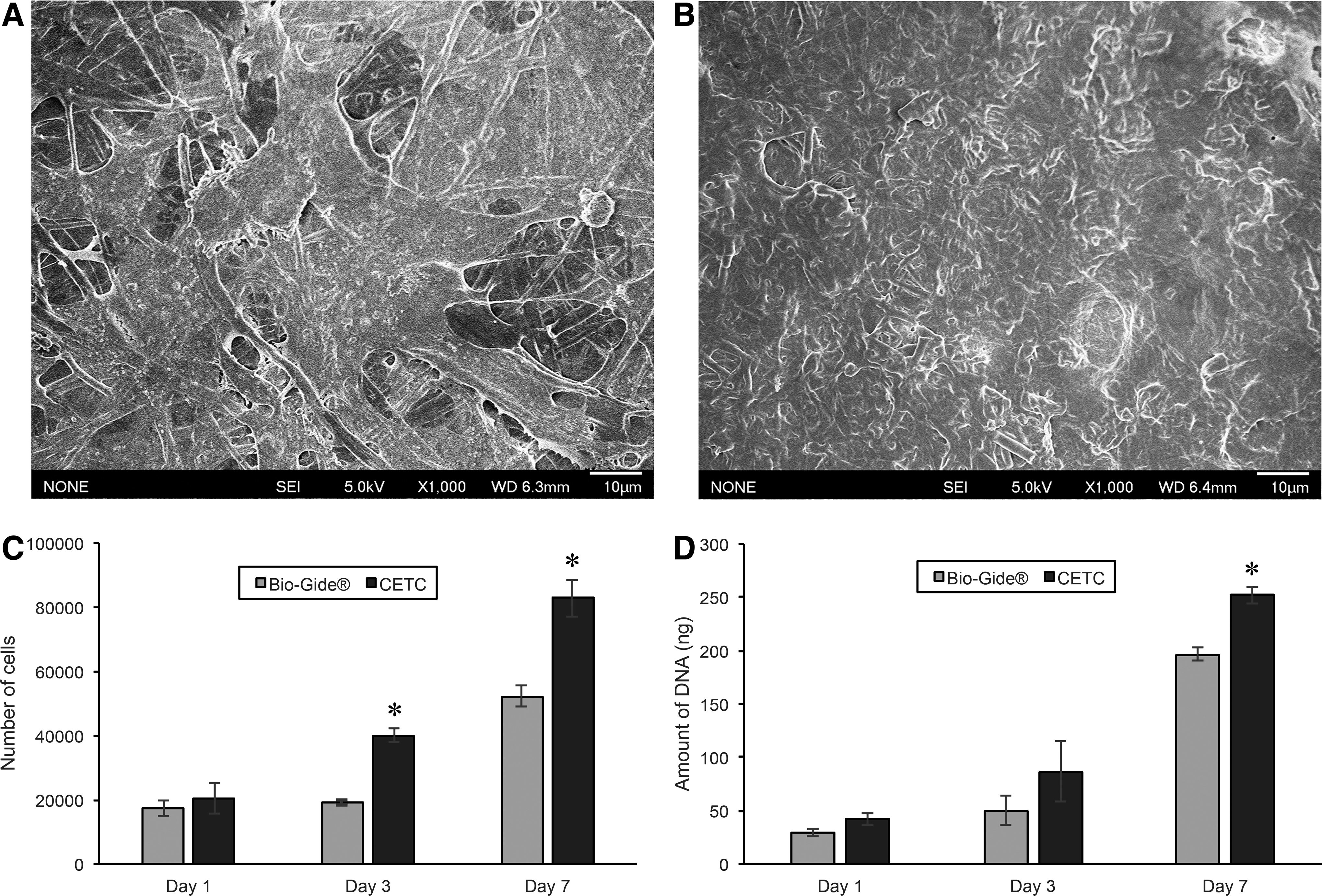
Adhesion and proliferation of murine ostegenic cells MC3T3 on Bio-Gide and CETC.
Cytokines levels in implanted cages
Exudate samples were extracted from the empty and sample-containing stainless steel mesh cages and the quantities of nine cytokines—IL-1β, IL-2, IL-4, IL-6, IL-10, IL-13, MCP-1, TNF-α, and VEGF—were measured with a multiplex magnetic bead array system (Fig. 4). A high production of proinflammatory cytokines was observed at the early time points and the production decreased with time, as seen for IL-1β, IL-6, IL-10, and MCP-1. Notably on Day 1, Bio-Gide and CETC induced a significantly higher production of IL-1β than the empty cage, and CETC induced a significantly higher production of IL-6 and a slightly higher production of IL-10 than the empty cage and Bio-Gide. Such a difference diminished after 7 days. Comparable production of cytokines at all time points with little or no differences between the empty cage, Bio-Gide, and CETC was observed for IL-2 and TNF-α. The prohealing cytokines IL-4, IL-13, and VEGF were observed in low production at the early time points and the production increased with time. For IL-4 and IL-13, cytokine production was only noticeable on Day 14. For VEGF, cytokine production increased with time and peaked on Day 7, before dropping to very low levels on Day 14.
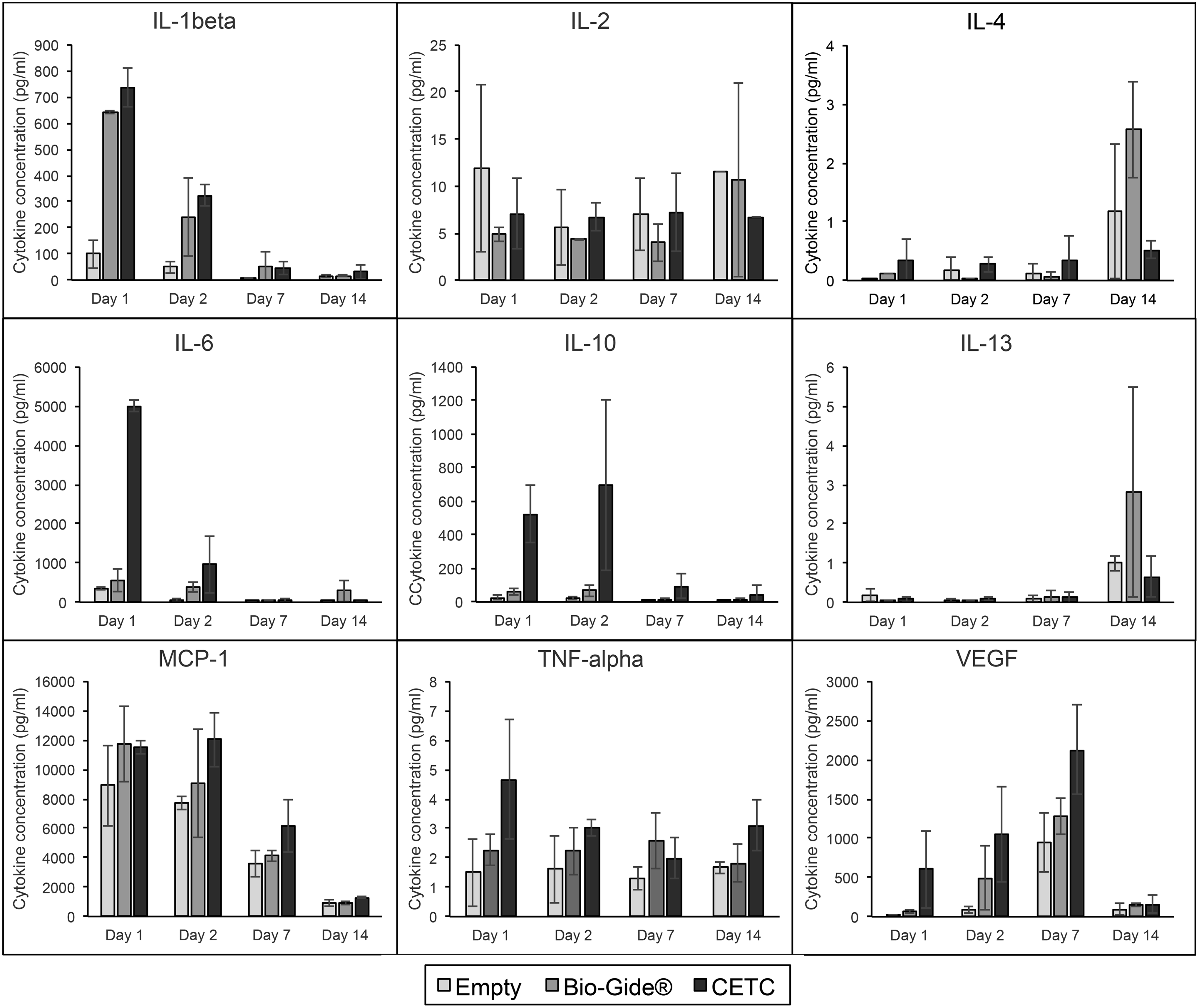
Cytokine levels in rat exudates on 1, 2, 7, and 14 days after subcutaneous implantation of empty cages, cages with Bio-Gide, and cages with CETC. IL, interleukin; MCP-1, monocyte chemoattractant protein 1; TNF-α, tumor necrosis factor alpha; VEGF, vascular endothelial growth factor.
Histology of host tissue response
The host tissue response was evaluated at 1 and 2 weeks after Bio-Gide and CETC were subcutaneously implanted into the back of male adult Sprague-Dawley rats. The histological sections were stained with hematoxylin and eosin and shown in Figure 5. After 1 week of implantation, Bio-Gide samples demonstrated thin localized fibrous tissue encapsulation. No obvious granulomatous reactive tissue or inflammatory cells seen in either of the groups while electrospun collagen samples did not show any fibrous tissue encapsulation. After 2 weeks, the encapsulation was more distinct and thicker in the Bio-Gide samples while that of the electrospun collagen still had a lack of an encapsulating layer of cells. The approximate cell thickness of the encapsulation is represented in Figure 6.
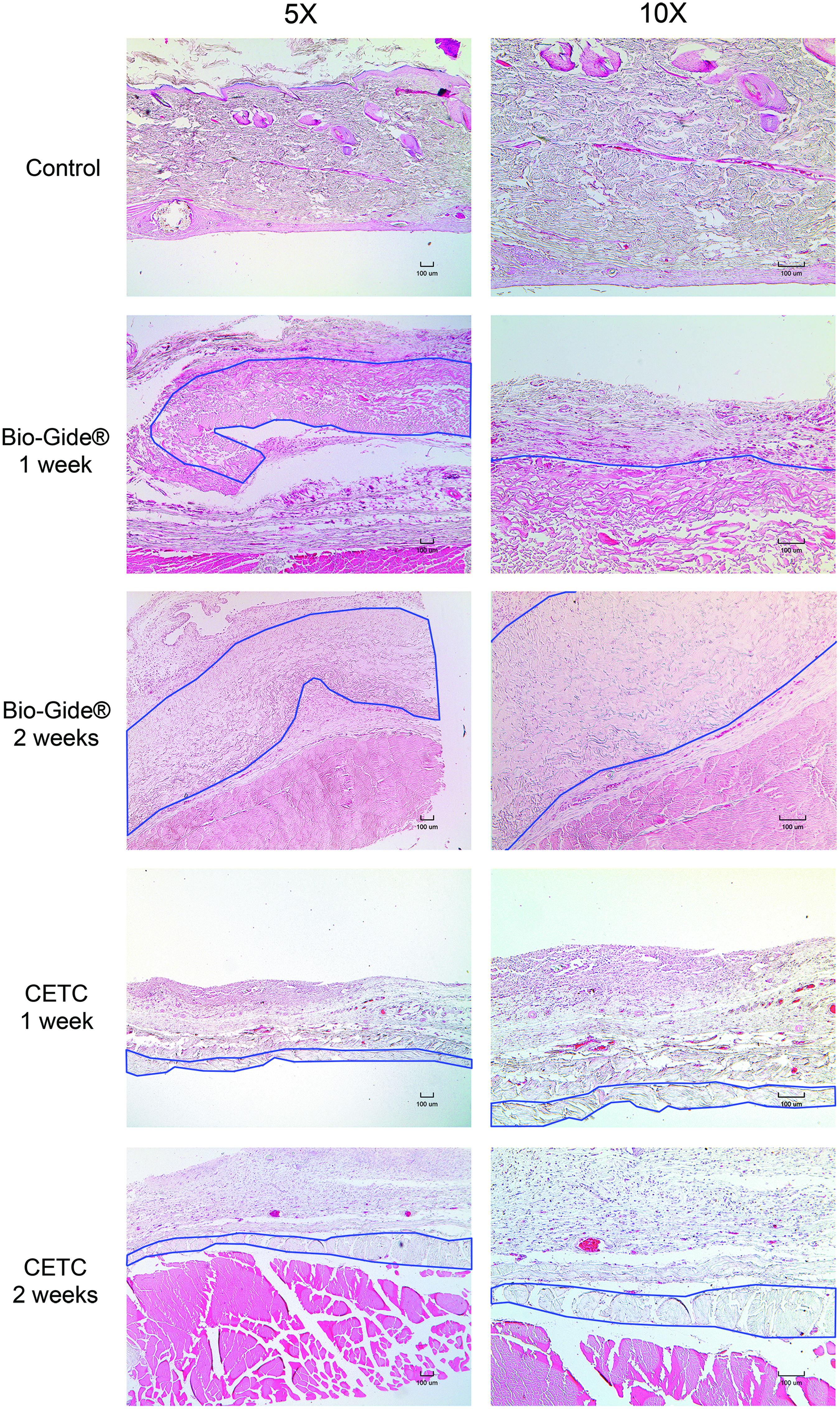
Histology sections of the subcutaneous implants stained with H&E. The implanted samples are identified as Bio-Gide and CETC. The implants are highlighted with a blue line to indicate their location. Sections are representative of the samples. Scale bars are 100 μm. H&E, hematoxylin and eosin. Color images available online at www.liebertpub.com/tea

Layers of cell encapsulation around the implant after 1 and 2 weeks in vivo. Each point on the graph represents the maximum number of cell layers in one rat.
Discussion
Fish collagen is gaining interest as an alternative to mammalian collagen as it alleviates concerns of disease transmissions and religious constraints associated with mammalian products 6 and its extraction from fish waste can generate additional revenue for the fishing industry and reduce wastage.1,7 However, most fish collagens are limited by low Td of <30°C unlike mammalian collagens with Td >40°C, so they would easily denature in the presence of heat during processing or in the site of implantation. 9 To overcome this limitation, tilapia collagen was chosen due to its high Td among all fish collagens. In this study, our tilapia collagen has a Td of 36.9°C, which is close to the value reported by Ikoma et al. (35°C). 13
To mimic the structure of ECM, tilapia collagen was electrospun to form a nanofiber membrane. The electrospun membrane was cross-linked in glutaraldehyde vapour to make the product stable in human body temperature (37°C) and to make up for the loss of mechanical strength during the acid extraction process.14,15 However, the cross-linking changed the morphology of the nanofiber structure, as the nanofibers contracted and merged with one another, making the material less porous. The cross-linking also caused the membrane to shrink physically and modified its mechanical properties (Table 1). Despite the cross-linking, the CETC fabricated has lower mechanical strength as compared to the commercial Bio-Gide. Our future work will focus on improving the mechanical properties such that it is comparable to other commercial collagen membranes. This can be achieved by increasing the thickness of the CETC,16,17 modifying the cross-linking conditions such as reagent concentration and duration,11,18 using cross-linking agents, which will vary the degree of collagen cross-linking, 14 or using a combination of chemical, physical, and/or enzymatic cross-linking methods. 18 Alternatively, the CETC can be combined with a mechanically stronger layer to produce a bi-layer membrane, to mimic the bi-layer structure of Bio-Gide. In this bi-layer structure, the CETC would act as the rough layer to support bone cell adhesion and osteoinduction, while the mechanically stronger layer would provide structural support and protect the CETC layer from epithelial cells and oral bacteria.11,19,20
Ever since tilapia collagen was identified as a potential biomaterial, tilapia collagen was proven to be biologically safe 21 and it was shown to have good biocompatibility with a variety of cells such as rat odontoblast-like cell line MDPC-23 22 and baby hamster kidney fibroblast BHK-21. 23 However, the results from these studies may not represent the biological properties of CETC, as the cross-linking process may have modified the physical and chemical properties of the collagen. In a previous study by Zhou et al., CETC nanofibers were reported to promote adhesion, proliferation, and differentiation of human keratinocytes, and accelerate skin wound healing in rats. 5 Immune response studies were also performed by Zhou et al., but the studies were done on noncross-linked tilapia collagen sponge. In this study, we focused on the biological properties of CETC, in particular the cytotoxicity, cell proliferation using murine osteogenic cells MC3T3-E1, in vivo cytokine expression, and in vivo inflammatory responses, to evaluate the suitability of CETC in GBR.
Results from the cytotoxicity studies showed that Bio-Gide, electrospun tilapia collagen, and CETC are nontoxic, as indicated by the high cell confluency and the elongated shapes of the cells incubated with these materials. From the quantitative data, the metabolic activities of the cells incubated with these three materials were higher than that of the nontoxic negative control and the blank control, confirming that these materials are nontoxic. The results also proved that the cross-linking process did not contribute any cytotoxicity despite the use of glutaraldehyde, a toxic chemical. We believed most of the unreacted glutaraldehyde was removed during the overnight vacuum drying.
As the CETC membrane is intended for use as a barrier membrane in GBR, it has to be biocompatible with osteogenic cells to promote osteoinduction and bone regeneration. The osteogenic biocompatibility of CETC was evaluated with murine preosteoblast cells MC3T3-E1, and results showed that CETC exhibited excellent compatibility toward MC3T3-E1 cells. SEM images showed the spreading of MC3T3-E1 cells on the CETC surface, while the Alamar blue and PicoGreen assays showed an increase in cell proliferation with time. The assays also showed that on Day 7, cell proliferation is significantly higher on CETC than on Bio-Gide, suggesting that CETC can support growth of MC3T3-E1 cells better than Bio-Gide.
In this study, we have used cage and subcutaneous implants to examine the in vivo immune response of the membrane and this experimental design is supported by the work performed by other groups that have used similar models.24–26 To determine the in vivo immune response of Bio-Gide and CETC, nine cytokines were selected and quantified using a multiplex magnetic bead array system. Among the tested cytokines, IL-2, IL-6, and TNF-α, promote inflammation.27–29 Proinflammatory/prowound healing cytokines, which include IL-1β and MCP-1, can activate both inflammatory cells and wound healing cells while anti-inflammatory/antiwound healing cytokine IL-10 does the opposite by suppressing both cells.30–32 Anti-inflammatory/prowound healing cytokines, which include IL-4 and IL-13, inhibit inflammation and promote wound healing. 33 Another important cytokine VEGF promotes the formation of blood vessels to support growth of new tissue. 34
From the results, some general trends were found among all the groups. First, all implantation groups induced a strong expression of MCP-1 and a mild expression of IL-1β, IL-2, IL-6, and TNF-α after 1 day postimplantation, indicating that the stainless steel cages alone caused some degree of inflammation. The high production of MCP-1 after 1 and 2 days postimplantation and the decrease of MCP-1 after 7 and 14 days indicated the accumulation of monocytes in the acute inflammation phase and the clearance of monocytes in the wound healing phase respectively. 30 Pronounced production of IL-1β in the collagen containing groups was found in the first 2 days of implantation, which was not noticeable after 7 days. Second, the expression of IL-4 and IL-13 for all the groups only became evident after 14 days postimplantation indicating the implantation sites progressed from the acute inflammation phase into the wound healing phase. Third, the concentration of VEGF for all samples increased with time and reached a peak after 7 days postimplantation, before dropping back to very low levels. This suggested that the implantation site was undergoing vascularization for the first 7 days, and once a network of blood vessels was formed to supply oxygen, nutrients, and soluble factors into the cages, VEGF was no longer secreted at high levels.
To evaluate the suitability of CETC as a barrier membrane for GBR, the cytokine responses of CETC and Bio-Gide were compared. Since tilapia collagen and porcine collagen are reported to have similar properties, 9 we expected CETC to have similar cytokine responses as Bio-Gide, which is derived from porcine collagen. Indeed it was found that CETC and Bio-Gide had comparable cytokine profiles in terms of proinflammatory cytokines IL-1β, IL-2, MCP-1, and TNF-α. From the in vitro degradation test, CETC lost 43% of its weight in 2 weeks in PBS, so it was expected that CETC would degrade even faster in vivo due to the presence of proteases. For IL-6, CETC had a higher cytokine response at earlier time points, but later, CETC had comparable responses as Bio-Gide and the empty cage. One possible explanation for the high response at the earlier time points is that CETC was degrading rapidly in vivo and the tilapia collagen fragments triggered the high expression of IL-6. As the tilapia collagen fragments continued to be broken down and get resorbed, the cytokine response at the later time points became similar to that of the empty cage. The low concentrations of proinflammatory cytokines at late time points showed that the metabolites of tilapia collagen are safely resorbed by the body and do not cause chronic inflammation.
This study further investigated the host tissue response to the samples subcutaneously. Host reactions following biomaterial implantation may include fibrous capsule formation, foreign body reaction, injury, blood–material interactions, provisional matrix formation and inflammation to name a few.35,36 From the histology data, it is evident that the CETC membrane elicits a lower tissue response from the host as indicated by the lack/thin biomaterial encapsulation as compared to that of Bio-Gide. In general, biomaterials that have cross-linking may in some cases result in the nonincorporation and graft failure, also the preclusion of immune cell penetration, making such grafts unable to participate in normal remodeling.37,38 However, despite chemical cross-linking of the collagen membrane, no increase in cellular encapsulation was observed after 2 weeks. Typically, a foreign body response occurs under normal physiological conditions and is based on nonspecific protein adsorption, immune, and inflammatory cells and occurs to protect the body from the foreign objects. 39 Importantly, in this inflamed environment, the immune cells contribute to the development of a dense layer of fibrotic connective tissue, which is detrimental to the implants' function, safety, and biocompatibility36,39,40 The lack of encapsulation suggests that the host does not mount a typical foreign body response to CETC and this observation is in agreement with the results from the cytokine assay.
Conclusion
In this study, we have fabricated a CETC membrane, and evaluated its cytotoxicity, biocompatibility, in vivo cytokine responses, and in vivo inflammatory responses to determine its suitability as a barrier membrane for GBR. CETC was shown to be nontoxic and have a higher denaturation temperature and better biocompatibility than Bio-Gide. However, CETC degraded faster in PBS and may have caused a higher production of IL-6 when implanted. The results from the cytokine assay and histology images suggested that CETC had no long-term inflammatory responses but it was resorbed more quickly than Bio-Gide. For CETC to be suitable as a barrier membrane for GBR, it would be desirable to increase its thickness and/or mechanical stability to extend its in vivo lifetime.
Footnotes
Acknowledgments
The authors would like to thank Dr. Mun-Keat Chong from Merck Pte. Ltd. for his help with the Milliplex® Map Rat Cytokine/Chemokine assay, and Ms. Nur Aishah Binte Ali from National Dental Centre Singapore for her help with the histology. This work was funded by the Nanyang Technological University research grant (M4081390) and Nanyang Technological University–National Dental Centre Singapore joint grant (M4081405).
Disclosure Statement
No competing financial interests exist.