
Abstract
Intra-articular ligamentous injuries are typically unrepairable and have limited outcomes after graft reconstruction. A combination of porous polycaprolactone fumarate (PCLF) scaffolds with polyethylene terephthalate (PET) sutures was developed with the goal of regenerating intra-articular ligaments. Scaffolds were fabricated by injecting PCLF over three-dimensional-printed molds containing two strands of PET suture down its central pore followed by cross-linking. Scaffolds were seeded with human mesenchymal stem cells (MSCs) from adipose tissue. To demonstrate cell attachment and proliferation in culture, we performed live/dead staining and cell proliferation assays. These experiments showed that MSCs remain viable and continue to proliferate on the scaffolds in culture for at least 2 weeks. Bare scaffolds were then used to reconstruct the rabbit anterior-cruciate ligament (ACL), while control rabbits underwent semitendinosus autograft reconstruction. The specimens underwent micro-computed tomography (CT) imaging, histological examination, and biomechanical testing at 8 weeks. The ultimate pull-out strength of the PCLF-PET scaffolds and tendon autografts was initially 72 ± 30 N and to 45 ± 10 N, respectively (p < 0.06). On inspection after 8 weeks in vivo, the intra-articular portion of the PCLF-PET scaffolds was fragmented while the tendon autografts remained intact. Cross-sectional areas of bone tunnels in the PCLF-PET scaffolds (11.3 ± 1 mm2) were enlarged compared to tendon autografts (3.8 ± 0.5 mm2) (p < 0.004) as measured by micro-CT. These studies show that PET-reinforced PCLF scaffolds are capable of initial ACL reconstruction and supports stem cell growth. The intra-articular portion of the scaffold may need to be re-engineered to support their use in ligament regeneration.
Introduction
I
The ACL has been the focus of a number of ligament tissue engineering studies utilizing scaffolds, multipotent stem cells, and growth factors. Biomaterial scaffolds including collagen, silk, and polymers like poly(glycolic acid), poly-L-lactide, polycaprolactone (PCL), and poly(lactic-co-glycolic acid) have all been investigated but are hampered by inherent weakness and immunogenicity. 19 Our group has previously developed a versatile cross-linkable macromer, polycaprolactone fumarate (PCLF), which has been used in multiple tissue engineering applications, including bone, chondrocyte, and nerve regeneration.20–23 Its ability to self-cross-link when exposed to ultraviolet (UV) light allows fabrication into complex three-dimensional (3D) porous scaffolds using 3D-printed molds. This study evaluated the use of PCLF in ligament regeneration.
Preliminary in vitro studies have demonstrated that multipotent mesenchymal stem cells (MSCs), which are derived from the stromal vascular fraction of adipose tissue, will attach to these 3D scaffolds and proliferate. Our study uses human MSCs that are expanded in human platelet lysate and have a characteristic cell surface profile indicating they represent pericyte-like immature mesenchymal stromal cells. 24 In their undifferentiated state, these adipose-derived MSCs express many different extracellular matrix proteins and are capable of tri-lineage potential.25,26 When these MSCs are seeded on PCLF scaffolds and exposed to the ligamentous growth factors (e.g., fibroblastic growth factor-2; [FGF-2]), they actively express collagen I (COL1A1), collagen III (COL3A1), and tenascin-C (TNC) similar to ACL derived fibroblasts. 27 Like other biodegradable polymer scaffolds under study for potential use in ACL tissue engineering, PCLF has a low tensile strength, which is not optimal for ACL reconstruction. In preliminary tests performed in our lab ultimate tensile strength of porous PCLF scaffolds was only 10 N, which is far less than the 130 N of an intact rabbit semitendinosus tendon, which is commonly used for rabbit ACL reconstruction models. Our goal was to enhance or reinforce these PCLF scaffolds to make them suitable for ACL reconstruction in a rabbit model.
To enhance the mechanical properties of the PCLF scaffolds we investigated synthetic ACL grafts. Synthetic grafts were initially developed in the 1970s secondary to the shortcomings of using tendon grafts. These synthetic grafts avoided the disadvantages of tendon grafts as they were strong, easily available, resulted in decreased surgical times, smaller bone tunnels, and more aggressive rehabilitation times.28,29 Synthetic grafts have been made from carbon fibers, Gore-Tex, polypropylene, and polyesters such as polyethylene terephthalate (PET). Despite the benefits of synthetic grafts, long-term outcomes are limited by failure to integrate with host tissue, thus leading to wear, effusions, and ultimate graft failure.29–31 PET grafts designed for biointegration with the host tissues are still used commercially for ACL reconstruction with reasonable success in short-term studies, however, with longer follow-up incomplete biointegration results in graft rupture.31–37
Multiple investigations have attempted to increase the tissue ingrowth of the PET grafts by coating them with polystyrene sodium sulfonate, hydroxypropylcellulose, hydroxyapatite, or hyaluronic acid.28,38–40 These surface coatings have shown promise by increasing PET biointegration in animal models, which should theoretically decrease the long-term wear rates of the PET graft.
Considering both the potential of PET and PCLF, we propose designing, fabricating, and testing a novel combination of cellular compatible porous PCLF scaffolds with the initial stability and strength of PET suture to reconstruct the ACL in a in vivo rabbit model.
Methods
Scaffold production
PCLF was synthesized using a method our group described previously. 20 All chemicals were supplied by Aldrich (Milwaukee, WI) unless specifically mentioned. In brief, PCLF was synthesized by polycondensation of PCL with fumaryl chloride with a methylene chloride and triethylamine catalyst. Scaffolds were fabricated by injecting liquid-state PCLF over 3D molds designed with computer-aided design (CAD) software (Solidworks, Waltham MA) and printed using a 3D printer (Solidscape, Wilton NH) (Fig. 1). Before injection of liquid PCLF into the mold, two strands of PET braided suture (Ethibond-0, Ethicon, Som NJ) were threaded through the center of the mold allowing for a complete interface with the polymer. These molds were placed into lubricated cylindrical glass tubes with an inner diameter of 2.5 mL with a rubber stopper at one end.
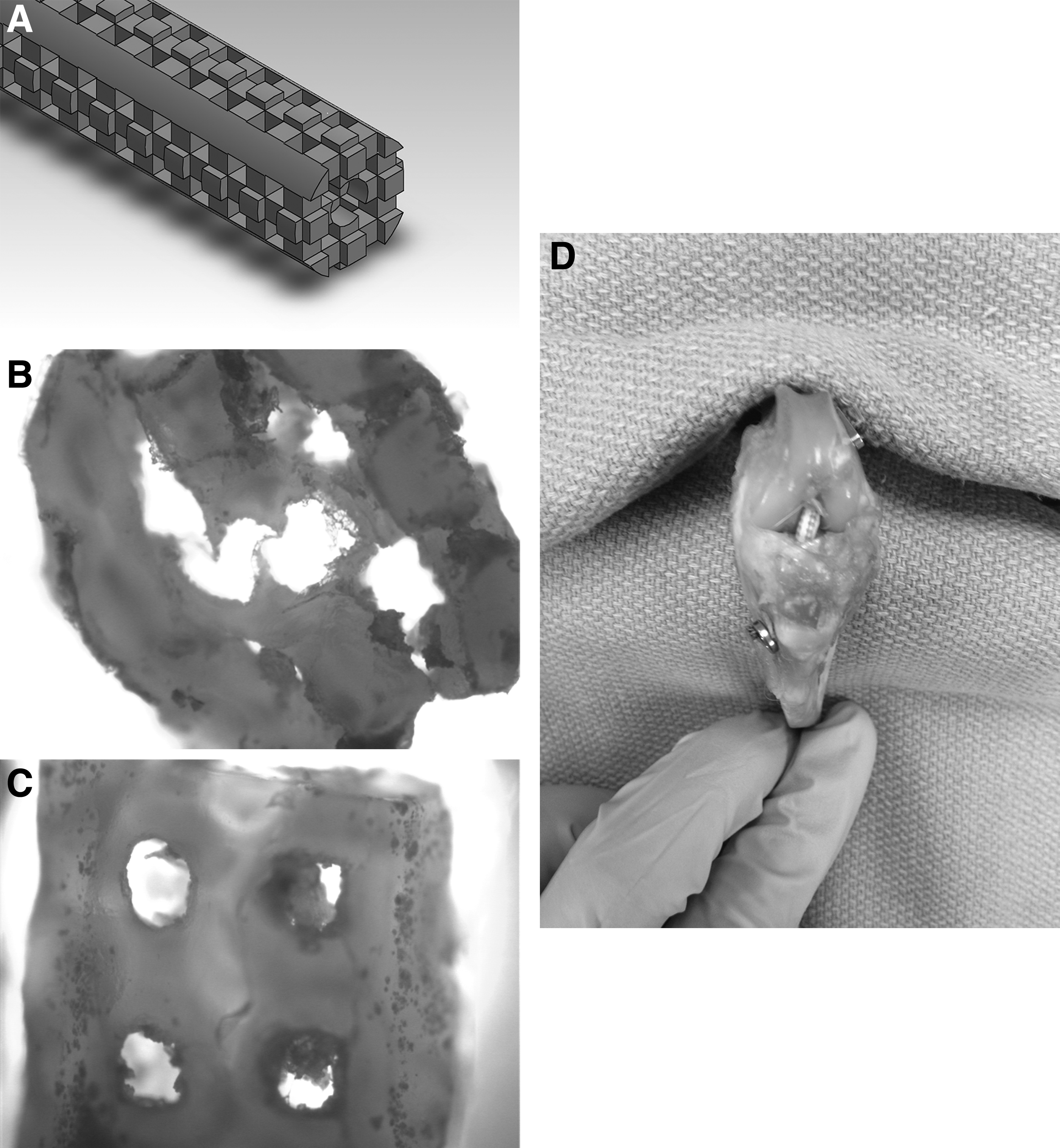
After injection of the PCLF the other end of the glass tube was capped. Scaffolds were cured in an UV oven for 2 h then removed from the glass tubes. Excess polymer was sharply shaved from four sides of the scaffold to expose the mold within. The mold was then dissolved in a mixture of acetone and methanol, which has been used previously without significantly affecting PCLF properties. 22 The final scaffold dimensions were similar to that of the rabbit semitendinosus tendon graft, being tubular in shape with a diameter of 2.5 mm and 3 cm in length with square pores (500 × 500 μm) (Fig. 2).
PCLF-PET scaffolds alongside a rabbit semitendinosus tendon grafts. The final scaffold diameter was 2.5 mm and 3 cm in length with square pores (500 × 500 μm).
Cell viability and proliferation testing
Previously Wagner et al. 27 showed that MSCs attached and proliferated on larger square PCLF scaffolds. We first wanted to determine cell viability, proliferation, and ideal cell seeding duration on the redesigned circular PCLF scaffolds embedded with PET suture. MSCs were obtained as previously described through an institutional review board approved process. 41 Briefly, human adipose tissue was obtained from general surgical operations and mechanically minced, incubated in 0.075% collagenase type I (Worthington Biochemical) for 90 min at 37°C, centrifuged at 500 g for 10 min and passed through a 70 μm cell strainer (BD Biosciences).
One centimeter sections of the scaffolds were first disinfected with ethanol washes of, respectively, 100%, 90%, and 70% for 30 min each followed by sterile phosphate-buffered saline. Human MSCs were concentrated at a level of 1 million cells per milliliter (mL) of media. Scaffolds were placed inside a dynamic rotational bioreactor (Synthecon, Houston TX) containing 50 mL of media, which consisted of a preparation of advanced minimum essential medium (Life Technologies, Carlsbad CA), 1% glutamax, 2 Units/mL heparin, and 5% platelet lysate (PLTMax; Mill Creek Lifescience LLC, Rochester, MN).
The gravitational component of culturing was negated by application of a constant rotational force through the dynamic rotation bioreactor applied at 16 Hz. In a prior study scaffolds were seeded for 18 h in this reactor and resulted in uniform seeding of all scaffold surfaces. 22 The goal of this study was to evaluate the differences in cell seeding for 24 and 72 hours to determine whether this would result in increased cell concentration and activity. The scaffolds were then removed and plated in static 24-well cell culture plates. Media was changed every third day. Six specimens were removed from culture at day 1, 7, and 14. Three specimens were used for a cell viability assay (LIVE/DEAD, Life Technologies, Carslbad CA) and three specimens were used for a (3-[4,5-dimethylthiazol-2-yl]-2,5-diphenyltetrazolium bromide) tetrazolium (MTS) cell proliferation assay (Cell titer 95 One solution, Promega, Madison WI).
Ex vivo ACL reconstruction
ACL reconstructions in cadaveric rabbit knees were done with PCLF-PET scaffolds and semitendinosus autografts (n = 4 in each group). A cannulated 2.7 mm drill was used to create the femoral and tibial bone tunnels after achieving proper positioning with a 0.062 mm k-wire. For the tibial tunnel, the drill was placed anterograde through the ACL foot print exiting in the middle of the anteromedial face of the proximal tibia, anterior to the medial collateral ligament and medial to the tibial tubercle. The femoral bone tunnel was drilled retrograde from the femoral origin of the ACL, in the 10 o'clock direction in the coronal plane, exiting anterolateral on the distal femur.
For the autograft reconstruction 3-0 Ethibond was used in a Krakow stitch fashion to secure both ends of the tendon. A suture passer was used to deliver the grafts and scaffolds through the tunnels. The ACL was tensioned maximally in 30 degrees of flexion. Both grafts and scaffolds were fixed to the end of the bone tunnels with stainless steel buttons (Arthrex, Naples FL). The cadaveric limbs were obtained from rabbits sacrificed in a separate Institutional Animal Care and Use Committee (IACUC) approved study, which did not affect the ligaments or bone quality.
In vivo ACL reconstruction
IACUC approval was obtained before any animal testing. Female New Zealand White rabbits, weighing 3.5–4.5 kg, underwent unilateral ACL reconstruction with either a PCLF-PET scaffold or a semitendinosus autograft through a lateral parapatellar approach as described previously under general anesthesia. Six rabbits were included in each group. The PCLF-PET scaffold was incubated in media containing 5% platelet lysate under sterile conditions for 24 h before implantation. This media bath was to act as a control for future studies that would seed scaffolds with MSCs before implantation. This media was shown increased MSC proliferation and collagen expression on PCLF porous scaffolds. 22 The semitendinosus autograft was harvested from the ipsilateral side at the time of surgery. Animals were monitored postoperatively for any signs of undue stress or infection. All rabbits recovered well and regained mobility. No restrictions on weight bearing or activity were placed. There were no deaths in either group. All 12 animals were sacrificed at the 8 week time point.
Biomechanical testing
Biomechanical testing was performed the same day and the limbs were harvested for both the ex vivo and in vivo reconstructions. The legs were sectioned through the mid-femur and mid-tibia and potted in cement. All muscle and ligamentous attachments except the ACL reconstruction were carefully removed before load to failure testing was performed.
An additional group of ex vivo PCLF-PET reconstructed specimens underwent fatigue testing before load to failure testing to determine the effects of time and wear on the specimens. In this group, the specimens were placed in a warm normal saline bath for 1 week before testing to demonstrate in vivo conditions. A custom-made motorized device (specifically designed for repetitive knee flexion in a rabbit model) was employed to put the knees through 5000 cycles of motion before testing. 42 Every 1000 cycles the specimens were visually assessed for macroscopic damage. After fatigue testing was completed, load to failure was tested in the same manner as the nonfatigue time zero group.
A universal testing machine (MTS Systems, Eden Prairie MN) was used for load to failure testing at a rate of 20 mm per/min. The knees were tested in 30 degrees of knee flexion to ensure that uniaxial force would be aligned with the orientation of the ACL. 43 The mechanical data gathered from the autograft and native ACL groups was compared to those in the literature to validate our testing method and reconstruction technique.43,44 After failure the specimens were sectioned and inspected.
Micro-computed tomography
Two specimens from each group were spared from biomechanical testing, placed in formalin, and underwent micro-computed tomography (micro-CT). The cross-sectional area of the femoral bone tunnel was calculated from the micro-CT by measuring the radius of the bone tunnel on two different perpendicular lines through the longest axis of the tunnel. The cross-sectional area of the femoral bone tunnel was measured on 10 different continuous axial CT slices starting at the intra-articular bone tunnel opening.
Histology
After performing micro-CT the two intact specimens from each group were subjected to decalcification, paraffin embedding, sectioning, and Gomori trichrome staining for further evaluation of bony integration of the scaffolds.
Statistics
Nonparametric statistical tests were used for non-normally distributed data as determined by the Shapiro–Wilk Wtest. The Wilcoxon Signed-Rank test evaluated differences within groups. The Wilcoxon Rank Sum test evaluated unpaired continuous variables between groups. All analyses were carried out using JMP statistical software package (Version 8, SAS Institute Inc., Cary, NC). A power analysis based on the data of a prior study that tested semitendinosus suture-button ACL reconstructions at 8 weeks suggested that four specimens in each arm would obtain a power of 80% to detect a mean difference of 50% in load to failure.45,46 A p-value <0.05 was considered statistically significant.
Results
Cell proliferation and viability
To assess the overall viability of MSCs seeded on PCLF scaffolds, we assessed mitochondrial activity, which represents a proxy for cell proliferation, using the MTS assay. Activity was measured in scaffold cultures that were seeded for either 24 or 72 h and were measured at 1, 7, and 14 days after seeding. The results show that MSCs seeded for 24 h had higher metabolic rates compared to cells seeded for 72 h, and the metabolic rate continued to increase in the 24 h group while decreasing in the 72 h at day 14 (Fig. 3). Differences in MTS cell proliferation activity reached significance at day 7(p < 0.008) and day 14 (p < 0.005).

The MTS proliferation assay showed increased cellular activity in samples seeded in the rotating bioreactor for 24 h compared to 72 h, reaching significance at day 7 (p < 0.008) and 14 (p < 0.005). Error bars represent standard error.
The same cultures were subjected to the LIVE/DEAD cell viability assay, which labels cells with fluorescent probes that selectively discriminate between live (green) and dead (red) cells. The results corroborate findings of the MTS cell proliferation assay, showing that there was a greater number of viable cells at day 1 in those scaffolds seeded for 24 h compared to cells seeded for 72 h. At day 7 and 14 the amount of viable cells had increased, reaching confluence in both groups (Fig. 4).

LIVE-DEAD staining at 2.5× magnification of PCLF-PET scaffold samples seeded for 24 h versus 72 h at day 1, 7, and 14 of static culture demonstrating greater cellular viability at day 1in the 24 h group. Both groups reached confluence and retained cellular viability at 14 days. Green color represents viable cells.
Biomechanical testing
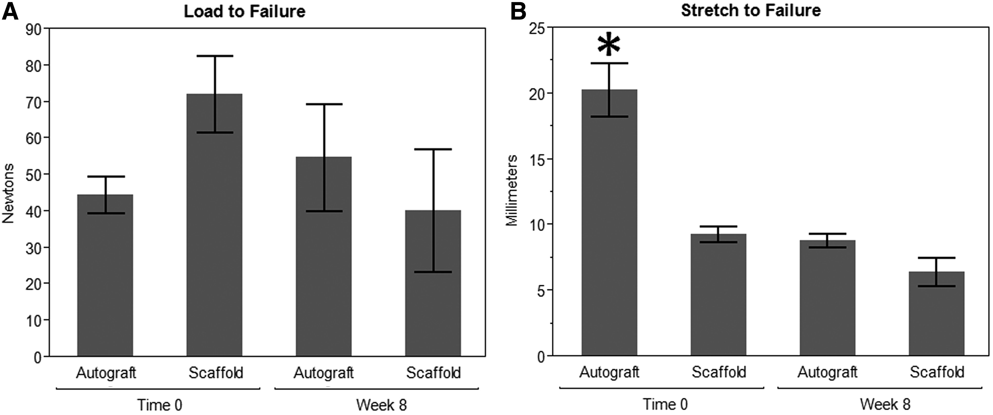
Ex vivo biomechanical testing of intact native rabbit ACLs demonstrated a mean load to failure of 353 ± 33 N (mean ± standard deviation [SD]) and stretch at failure of 3 ± 1 mm, values consistent with a prior study. 44 At time zero, the mean load to failure of the PCLF-PET scaffold reconstruction and the autograft group was 72 ± 30 and 45 ± 10 N, respectively (p < 0.06), while stretch at failure at time zero was 9 ± 1 and 20 ± 4 mm, respectively (p < 0.03) (Fig. 5). The PCLF-PET scaffold reconstructions submitted to fatigue flexion testing demonstrated intact scaffolds with no evidence of breakdown. Fatigue tested and nonfatigue tested scaffolds had a load to failure of 49 ± 20 and 72 ± 25 N (p < 0.4), respectively, and a stretch at failure of 10 ± 2 and 9 ± 1 mm (p < 0.7), respectively.
Load to failure
After 8 weeks in vivo the load to failure in the autograft had increased from to 45 ± 10 to 55 ± 30 N (p < 0.8). The autograft reconstruction stiffness also increased with the stretch at failure significantly decreasing from 20 ± 4 to 9 ± 1 mm (p < 0.03). The mean load to failure of the PCLF-PET scaffold had decreased from 72 ± 30 to 40 ± 34 N and stretch to failure had increased from 6 ± 2 to 9 ± 1 mm. No significant differences in load to failure (p < 0.6) or stretch at failure (p < 0.1) were found between the PCLF-PET scaffolds and the autografts at the 8 week time point.
Macroscopic analysis at week 8
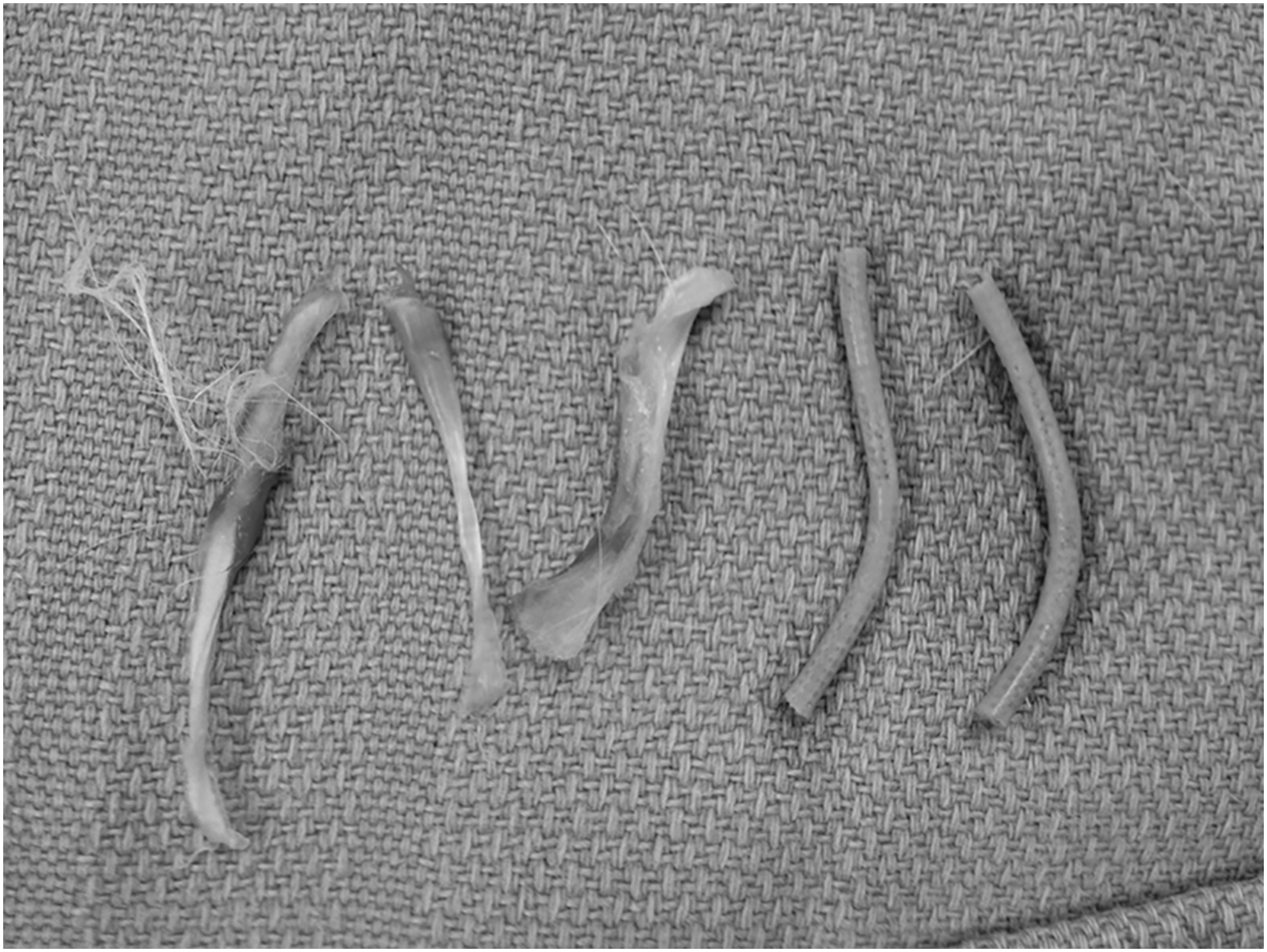
After 8 weeks in vivo rabbits were sacrificed and anatomically examined for graft integrity. In the autograft group, all six grafts remained intact with visible hypertrophy. In contrast, PCLF-PET constructs showed extensive intra-articular scaffold destruction in all specimens. The portion of the scaffold in the bone tunnels remained intact. Polymer debris was evident throughout the joint and the bone tunnels appeared to increase in size (Fig. 6). One strand of the PET suture had ruptured in all specimens, while one strand remained intact.
Micro-CT
The average cross-sectional area (±SD) of the bone tunnels at 8 weeks increased in the PCLF-PET scaffold specimen compared to the autograft specimen (11.3 ± 1 and 3.8 ± 0.5 mm2, respectively; p < 0.004). The bone tunnels in both groups were made using a drill with a cross-sectional area of 5.7 mm2, suggesting that bone tunnels in the autograft group grew in around the graft, while the tunnel in the PCLE-PET group expanded.
Histology
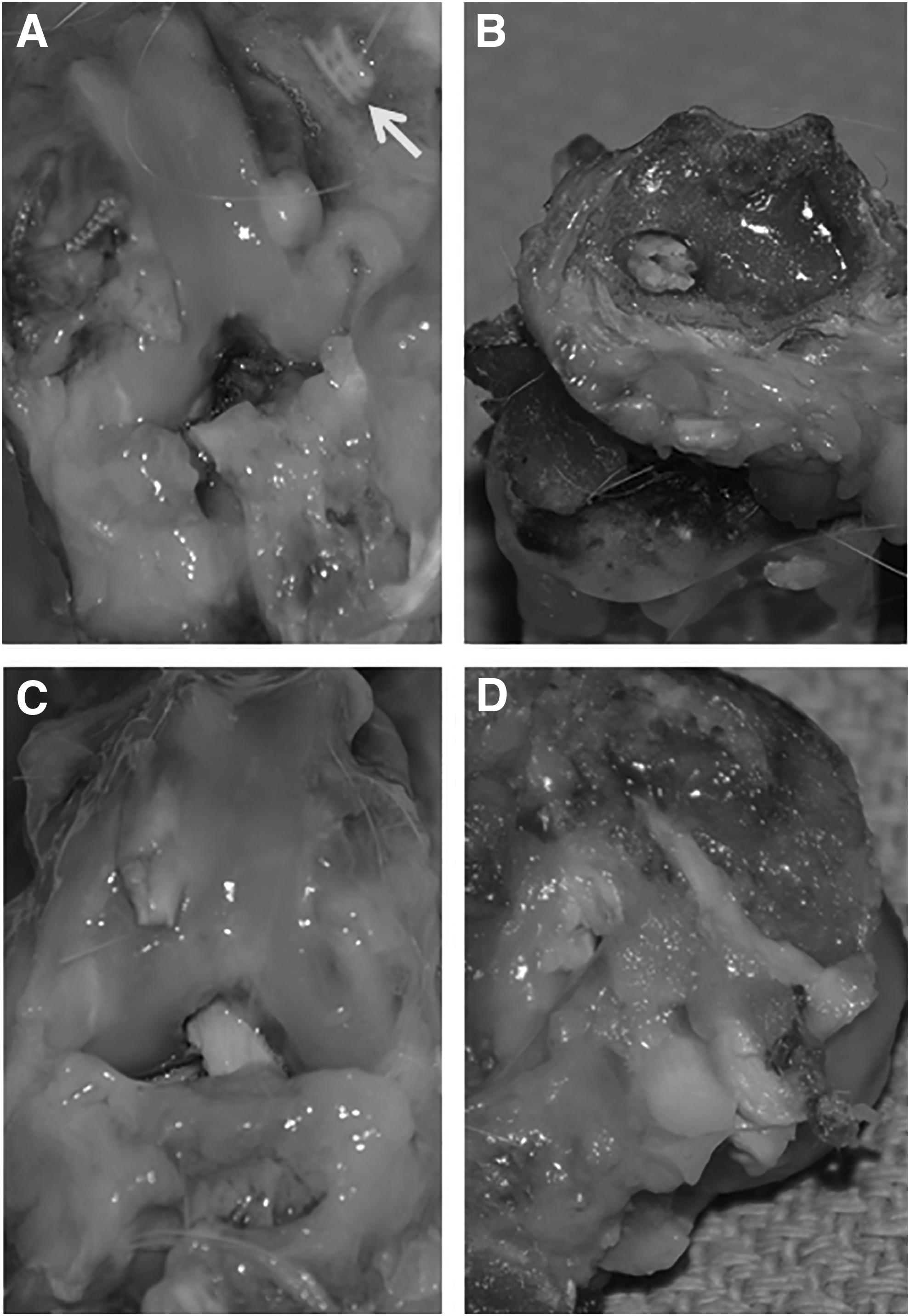
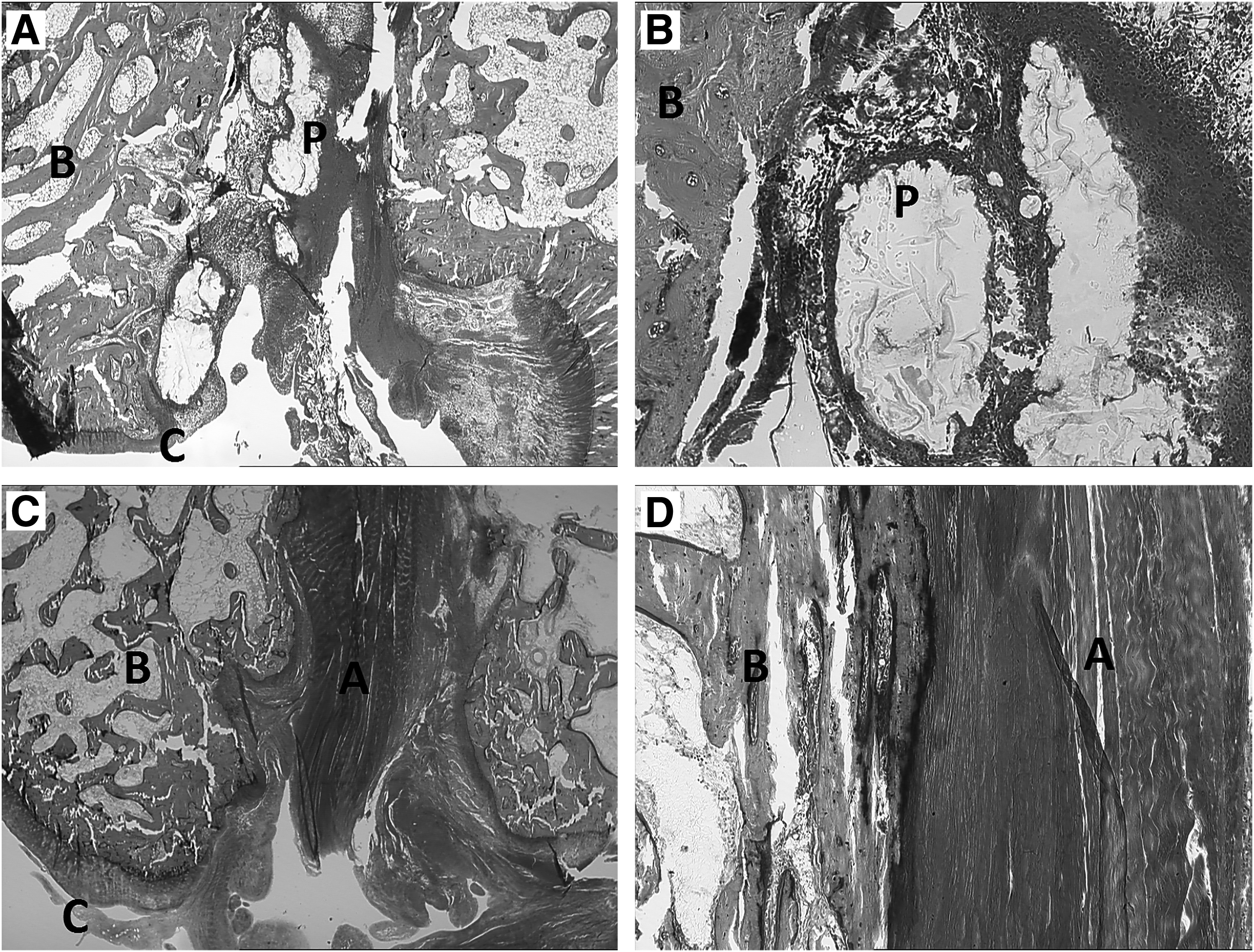
Gomori trichrome staining of femoral specimens demonstrated minimal collagenous ingrowth in the bone tunnels containing PCLF-PET scaffolds with diffuse areas of polymer debris surrounded by inflammatory cells, while the autograft reconstructions demonstrated intact autograft tendon with viable cells, close approximation with the bone tunnels throughout (Fig. 7).
2×
Discussion
PCLF is a polycaprolactone-based macromere that contains a fumarate cross-linker. This biopolymer has excellent cellular compatibility and degradation rates for tissue-engineering purposes, and it can be fabricated into complex porous scaffolds. Previously our group has applied these 3D scaffolds successfully toward guided bone and nerve regeneration.20,23
We were encouraged by our initial in vitro studies demonstrating the attachment, proliferation, and viability of MSCs on the scaffold for up to 2 weeks with cells expressing ligamentous extracellular proteins (collagen I, III, and tenascin-C) after culture in platelet lysate and FGF2 factor. 27 One limitation of porous PCLF scaffolds for ligamentous regeneration is its limited ultimate tensile strength. We, therefore, set out to combine the biointegrative properties of PCLF with the excellent tensile strength of PET suture to develop a tissue-engineered intra-articular ligament scaffold.
We chose PET because it has had relative clinical success for ACL reconstruction. The Ligament Augmentation and Reconstruction System (LARS) artificial ligament (Surgical Implants and Devices, Arc-sur-Tille, France) is a PET graft that is commercially available and has similar outcomes compared to tendon autografts in short and medium-term clinical studies.31–37 The LARS ligament differed from earlier synthetic grafts because it is designed for tissue ingrowth. 47 Biointegration of the graft provides stronger osseous fixation and protects from grafts wear both in the bone tunnels and the intra-articular space. 31 Notwithstanding the benefits of its design, long-term outcomes of LARS ligament reconstruction have been disappointing with reports of inevitable wear and iatrogenic arthritis.29,30 In an ovine model, ingrowth of the construct was nonuniform and did not extend through the intra-articular space. The authors of the study recommended against the grafts use secondary to its eventual wear and subsequent weakening. 30
Since incomplete biointegration has been linked to LARS ligament wear and rupture, several investigations have looked into increasing the tissue ingrowth of the PET scaffold by coating it with substances like polystyrene sodium sulfonate, hydroxypropylcellulose, hydroxyapatite, and hyaluronic acid.28,38–40 These surface coatings have shown promise by increasing PET biointegration in animal models, which should theoretically decrease the long-term wear rates of the PET graft.
By combining the biointegrative properties of PCLF scaffolds with the immediate fixation strength of PET suture we sought to create a graft for reconstruction and regeneration of the ACL. After initial reconstruction the PCLF-PET scaffolds had superior load to failure and stiffness compared to the autograft reconstruction. Before animal experimentation specimens underwent fatigue testing, which involved 5000 knee cycles after spending a week in a warm normal saline bath. Fatigue testing did not demonstrate any appreciable wear of decrease the mechanical properties of the PCLF-PET graft. We did, however, notice degradation of the graft when placed within an intra-articular environment in a live animal model after 8 weeks.
A possible improvement may be to develop an electrospun graft that can tolerate the tension and torsion-like forces it is subjected to. Although PCLF is initially quite resilient and flexible after fabrication, we have observed that over time it becomes brittle and rigid. This has not been described in prior literature on PCLF. Consistent with this finding, PCLF was not damaged by fatigue testing that took place immediately after fabrication. Increased brittleness with aging may be due to indirect UV exposure in the laboratory setting and continued spontaneous cross-linking of fumarate bonds in the PCLF overtime. This physical property may explain why in vivo, the scaffold degenerated within the articulating part of the joint, but remained intact within the static environment of the bone tunnels. The intra-articular environment is bathed in synovial fluid and is avascular, which limits soft tissue healing, contributing to the poor outcomes of primary ACL repair. These same factors likely affected the graft durability, preventing the cellular ingrowth necessary for scaffold integration.
Beyond biomaterial properties, the overall design of the PCLF-PET construct itself may have played a role in its failure. Although the PCLF was cross-linked around the two PET suture strands the stress of the knee motion may have led to micro-motion of the suture creating a “cheese-cutter” effect, destroying the PCLF from the inside out. The specimens that underwent flexion fatigue testing did not show any evidence of this, however, these PCLF scaffolds were <10 days out from the original fabrication so there original flexibility remained intact.
We found that scaffolds seeded for 24 h in the dynamic bioreactor resulted in an increased cellular activity compared to those seeded for 72 h. Cellular activity may have been lower in the 72 h group due to depletion of the nutrients in the broth. Another explanation could be that these cells seeded for 72 h were 48 h older than the cells cultured for 24 h; so the differences in the MTS assay could be secondary to the cells in the 72 h group being at a later point in the lifecycle.
Despite the ex vivo cellular compatibility, histology of the failed PCLF reconstructions demonstrated diffuse polymer debris within the bone tunnel with dense inflammatory cells compared to the autograft reconstructions, which were intact and had close adherence with the walls of the bone tunnel. It is not evident whether the inflammatory reaction was the result of gross movement of the scaffold within the bone tunnel or reaction to the polymer debris. CT scans correlated these finding by demonstrating that the bone tunnels in the PCLF reconstructions had widened while the bone tunnels with autograft had narrowed. This was visualized on the gross specimens and with the tendon grafts being integrated and fixed to the bone tunnels while the remaining PCLF scaffold was loose inside the bone tunnels.
Based on our data it is evident that the ideal scaffold for reconstruction of intra-articular ligaments must remain flexible and durable enough to survive multiple joint forces (e.g., tension and rotation), while permitting robust biointegration to improve graft success. Recently, Petrigliano et al. 48 reconstructed the ACLs in rats with an electrospun PCL fiber scaffold. After 12 weeks in vivo the grafts remained intact, with increased stiffness, and histological evidence of collagen infiltration of both the bone tunnel and the intra-articular regions. The initial success of this fiber scaffold, along with others including collagen and silk, in small animal models provides an exciting future direction for intra-articular ligament regeneration.48–50
Conclusion
We noted degradation of the PCLF-PET scaffold when placed within an intra-articular environment. This may be related to the structure of the scaffold and ongoing studies are being conducted to enhance its ability to tolerate torsional loads as seen within the knee joint. The ideal scaffold for reconstruction of intra-articular ligaments must remain flexible and durable enough to survive multiple joint forces while permitting robust biointegration to improve graft survival.
Footnotes
Acknowledgments
This study was funded by intramural grants and philanthropic support. We thank Steven Chase for scaffold design and polymer synthesis, as well as Yan Su for assistance with cell culture.
Disclosure Statement
PCLF was developed and patented by Dr. Yaszemski while a member of the department of Orthopaedic Surgery and the Tissue Engineering and Biomaterials Laboratory. No other relevant financial conflicts to report.