
Abstract
Dental pulp tissue engineering is possible after insertion of pulpal stem cells combined with a scaffold into empty root canals. Commonly used biomaterials are collagen or poly(lactic) acid, which are either difficult to modify or to insert into such a narrow space. New hydrogel scaffolds with bioactive, specifically tailored functions could optimize the conditions for this approach. Different synthetic and natural hydrogels were tested for their suitability to engineer dental pulp. Two functionalized modifications of polyethylene glycol were developed in this study and compared to a self-assembling peptide, as well as to collagen and fibrin. Cell viability of dental pulp stem cells in test materials was assessed over two weeks. Cells in selected test materials laden with dentin-derived growth factors were inserted into human tooth roots and implanted subcutaneously into immunocompromised mice. In vitro cell culture exhibited distinct differences between scaffold types, where viability was significantly higher in natural compared to synthetic materials. In vivo experiments showed considerable differences regarding scaffold degradation, soft tissue formation, vascularization, and odontoblast-like cell differentiation. Fibrin appeared most suitable to enable generation of a pulp-like tissue and differentiation of cells into odontoblasts at the cell–dentin interface. In conclusion, natural materials, especially fibrin, proved to be superior compared to synthetic scaffolds regarding cell viability and dental pulp-like tissue formation.
Introduction
C
To date, revitalization, a regenerative endodontic procedure, is a valid alternative to the conventional treatment of apexification.1,2 During a regenerative procedure, bleeding into the root canal is induced; the blood clot is covered with a bioactive calcium silicate cement and a coronal seal. After provocation of bleeding, stem cells from the apical papilla are flushed into the canal, 3 the blood clot reorganizes and serves as a guide rail for cellular attachment and migration. 4 This treatment can result in the resolution of periapical inflammation and a completion of root development caused by formation of a vascularized, immunocompetent new tissue inside the root canal, which is capable of mineralization. A study reported a survival rate of 100% for revitalization after a follow-up period of 18 months, which was lower for mineral trioxide aggregate (MTA) apexification with 95%. 5 A multitude of clinical case reports and case series demonstrates the potential of such biology-based regenerative treatment approaches.6,7 However, animal studies, as well as singular histology reports after regenerative endodontic procedures in patients, show that formation of pulp tissue with its original architecture and function cannot be taken for granted.8–10 Apposition of cementum or ingrowth of bone tissue may take place, which can be interpreted as completion of root formation after radiographic assessment.
Although patient-based, as well as clinician-based, outcomes after regenerative endodontic procedures seem satisfactory, 11 the goal of functional regeneration of the dentin-pulp complex might not be achieved regularly with this approach. Questions remain regarding potential reinfection, obliteration of the root canal by overproduction of mineralized tissue, replacement resorption, or even ankylosis, which is a serious complication in young patients with incomplete skeletal growth.
To better control the parameters of regenerative endodontic treatment procedures, application of the tissue engineering principle might be a promising approach. The combination of scaffolds, growth factors, and stem cells is well applicable to the clinical situation of endodontic treatment. Advancements in the development of scaffold materials have opened new possibilities. Early studies on dental pulp tissue engineering with tooth slices or segments used polylactic or polyglycolic acid.12–14 Subsequently, various other scaffold materials, including natural polymers such as alginate,15,16 collagen,17,18 and fibrin19,20 or synthetic polymers such as polylactic-co-glycolic acid, 14 polyethylene glycol (PEG), or self-assembling peptide (SAP),21–23 have been explored. While these materials are applicable for various tissue engineering approaches, little work has been done to select and modify a material to create a custom-made bioactive scaffold that is optimized specifically for dental pulp tissue engineering. Desirable properties of such a material include injectability, control over the mechanical properties, mimicry of the extracellular matrix of dental pulp, cell-mediated biodegradability, and bioactivity by specific motifs or binding and slow release of growth factors. While natural materials better mimic the cells' natural environment, synthetic materials offer high control over the material properties such as chemistry, charge, and stiffness and allow for customization, for example, biodegradability by specific enzymes. A previously developed SAP with defined stiffness and mode of gelation was modified with a cell adhesion motif and a matrix metalloproteinase-2 (MMP-2)-cleavable site to enable cell-mediated degradation by dental pulp cells. 24 Evaluation of this material in vivo after implantation of tooth roots filled with scaffold material, growth factors, and cells in the backs of immunocompromised mice showed promising results and the formation of a vascularized pulp-like tissue. 21 Thus, we hypothesized that synthetic materials might be better candidates for dental pulp tissue engineering compared to natural polymers.
In the larger scheme of identifying an optimized biomaterial for this approach, the aim of this study was twofold: (1) to develop a customized and bioactive polyethylene glycol derivative for dental pulp tissue engineering and (2) to test and directly compare the developed material with different natural materials, as well as the previously described synthetic scaffold 21 for compatibility with dental pulp cells in vitro and their suitability to enable the formation of pulp-like tissue in vivo.
Materials and Methods
Cell culture
Primary dental pulp cell cultures were established from extracted third molar tooth germs after approval by an appropriate institutional review board as described before 25 and cultured in MEMα supplemented with 10% fetal bovine serum, 100 U/mL Penicillin, and 100 μg/mL Streptomycin. Reagents were purchased from Gibco (Invitrogen Corporation, Carlsbad, CA). Primary human dental pulp cells at passage 3 were used for cell viability testing in combination with different scaffold materials. Primary cells at passage 2 were selected by magnetic activated cell sorting (MiniMACS™ Separator; Miltenyi Biotec GmbH, Bergisch Gladbach, Germany) using the mesenchymal stem cell marker STRO-1 (primary antibody: purified anti-human STRO-1 mouse IgM, BioLegend, San Diego, CA; secondary antibody: anti-mouse IgM Micro Beads, Miltenyi Biotec GmbH, Bergisch Gladbach, Germany). To validate successful sorting and enrichment of STRO-1-positive cells, immunohistochemistry was conducted, and differentiation after culture with inductive cell culture media (StemPro® Adipogenesis, Osteogenesis and Chondrogenesis Differentiation Kit; Invitrogen Corporation) was confirmed by alizarin red after osteogenic induction, staining of proteoglycans after chondrogenic induction, and by visualization of lipid droplets by oil-red staining after adipogenic induction as shown previously. 26 Sorted cells at passage 5 were used for the in vivo experiments.
Preparation of scaffold materials
Two classes of synthetic and natural hydrogel materials were tested for cytocompatibility with primary human dental pulp cells. Synthetic materials were modifications of PEG and two different types of SAP, as natural materials, fibrin, and collagen type I were included.
Polyethylene glycol
Three different PEG-based materials were tested; two of them were developed in the course of this study. Design criteria included generation of an injectable material suitable for use in a dental office, a defined curing mode (chemical reaction or light-induced polymerization, time for polymerization ≤5 min), and addition of bioactivity by incorporation of a cell adhesion motif and an enzyme-cleavable site for pulp cell-mediated degradation. The chemical structures of the functionalized polymers are depicted in Figure 1.
(1) For a chemically curing hydrogel (PEGchem), four-armed PEG (10 kDa molecular weight, 4armPEG10k) was functionalized with amino (NH2) or succinimidyl (SC) groups to synthesize 4armPEG10k-NH2 and 4armPEG10k-SC as previously described. 27 Hydrogel formation occurred after dissolution of both components in phosphate buffered saline (PBS) containing cells. Subsequent covalent bonds were formed between amino and SC groups. The final polymer concentration was 10% by weight.
(2) To develop a light-curing hydrogel (PEGlight), PEG5k-acrylamide (5 kDa molecular weight) was synthesized from PEG5k-NH2 as described in the Supplementary Data (Supplementary Method; Supplementary Data are available online at www.liebertpub.com/tea). After synthesis, PEG5k-acrylamide was dissolved at a concentration of 10% by weight in PBS containing the initiators eosin Y (0.01 μmol/mL), triethanolamine (10 μmol/mL), and N-vinylpyrrolidone (5 mg/mL), and cells. Light-induced polymerization occurred by use of a dental curing unit (Bluephase® C8; Ivoclar Vivadent AG, Schaan, Liechtenstein) for 5 min with 800 mW/cm2.
(3) For biomimetic hydrogels (PEGbio), PEG5k-acrylamides were functionalized with a cell adhesion motif (Arg-Gly-Asp-Cys; PEG5k-RGDC-acrylamide) and a MMP-2-sensitive enzyme-cleavable site (Gly-Arg-Leu-Ser-Ala; PEG5k-GRLSA-acrylamide) using Fmoc-chemistry (Supplementary Method). To induce polymerization, the components were dissolved in PBS with cells at 10% by weight and mixed in equal amounts; initiators for polymerization were added, and the material was light cured as described above.
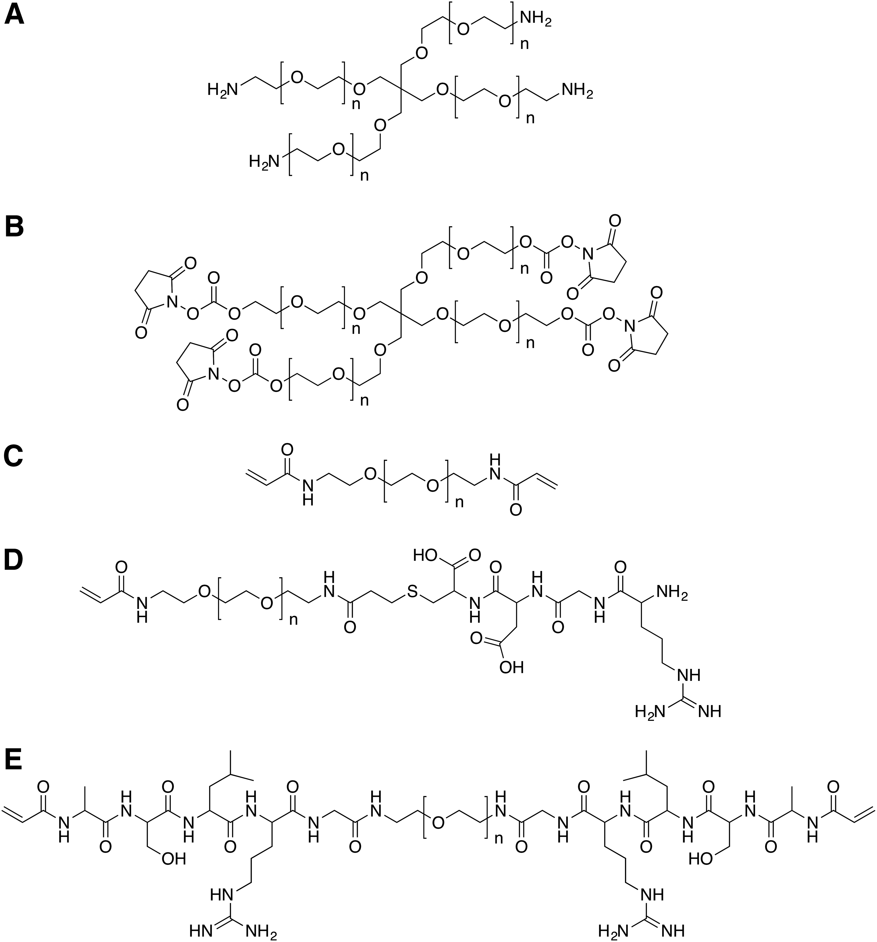
Chemical structure of tested PEG derivatives.
Self-assembling peptide
The previously described MMP-2-sensitive, cell adhesive 21 amino-acid peptide sequence K(SL)3RG(SL)3KGRGDS was synthesized and purified to 97% (21st Century Biochemicals, Marlborough, MA). 24 The lyophilized product (SAPbio; custom-made) was dissolved at 30 mg/mL in deionized water with 298 mM-sucrose, and the pH was adjusted to 7.4. Addition of a cell suspension in PBS induced gelation, where the charge of lysine-containing peptides was screened by the negatively charged phosphate. The final peptide concentration was 15 mg/mL (1.5% by weight). Furthermore, a commercially available SAP (SAPcom, PuraMatrix™ Peptide Hydrogel; BD Biosciences, Franklin Lakes, NJ), a 16 amino acid peptide with a RADA repeat that forms stable β-sheet structures in water, was tested. 28
Fibrin
Fibrinogen and thrombin from bovine plasma were purchased from Sigma-Aldrich (St. Louis, MO). Fibrinogen was dissolved in PBS, and thrombin was dissolved in CaCl2. Final concentrations were adjusted to 10 mg/mL fibrinogen in PBS and 75 U/mL thrombin in 20 mM CaCl2. Fibrin gels with cells were generated by resuspending a cell pellet in 50 μL fibrinogen and addition of an equal volume of thrombin for gelation.
Collagen
Collagen solution (Rat Tail Collagen, Type 1; BD Biosciences) was prepared at a concentration of 3 mg/mL according to the manufacturer's instructions. Cells in PBS were resuspended in collagen solution; adjustment to a neutral pH and incubation at 37°C and 5% CO2 for 15 min allowed for gelation.
Cell seeding
Cytotoxicity of the initiating compounds eosin Y (0.01 μmol/mL), triethanolamine (10 μmol/mL), and N-vinylpyrrolidone (5 mg/mL) were tested with monolayer primary pulp cells at passage 3 before experimentation in three-dimensional cultures. Cell viability was assessed in methylthiazolyldiphenyl-tetrazolium bromide (MTT) assays after 24 and 48 h for each component alone, as well as the combination of all three at dilutions of 1, 1:2, 1:5, and 1:10.
To test materials in three-dimensional cultures, 100 μL of liquid biomaterial containing 1 × 10⁵ cells were inserted in wells of 96-well plates. Cells were suspended in PBS and added to one of the components before mixing in cell culture plates as specified above, and gelation was induced according to the type of material. For light-induced polymerization, the light duct was placed directly in contact with the well plate to ensure minimum distance between light source and test material. Samples were seeded in triplicates. After gelation, 200 μL of cell culture media were added on top, and gels were incubated for 14 days at 37°C and 5% CO2. Culture media were changed thrice a week.
Cell viability testing and live/dead staining
To test cell viability of three-dimensional cell cultures in different hydrogel materials, a modified MTT assay was performed as described previously. 26 The optimum time period for elution of converted MTT dye after incubation was assessed in pilot experiments. Briefly, cells in hydrogels were incubated with 0.5 mg/mL MTT (Thiazolyl Blue Tetrazolium Bromide; Sigma-Aldrich) at 37°C and 5% CO2 for 1 h. Subsequently, culture media were removed from the gels and replaced by dimethyl sulfoxide (DMSO) solution. Cell culture plates were placed on a shaker at 220 rpm for 3 h to allow for a release of the dye from the hydrogels into solution. Absorbance was measured in a new 96-well plate spectrophotometrically at 570 nm on day 1, 3, 5, 7, 10, and 14 (Infinite® 200; Tecan, Männedorf, Switzerland). Two independent experiments were performed for PEGchem, PEGbio, and SAPcom (n = 6) and three for PEGlight, SAPbio, Fibrin, and Collagen (n = 9) to calculate median values and the 25–75% percentiles.
To visualize live and dead cells and confirm the results from MTT assays, live/dead staining in three-dimensional cultures was performed exemplarily for cells in collagen and functionalized variants of PEG5k-acrylamide (PEGbio) after 3 and 5 days in culture (Live/Dead Viability/Cytotoxicity Kit; Molecular Probes, Eugene, OR). The three-dimensional structure was sterically reconstructed based on single confocal laser scanning microscopy images without further quantification (LSM 510 META; Carl Zeiss Microscopy, Jena, Germany).
Root canal model in vivo
Out of the seven materials tested in vitro, the following four were chosen for an in vivo model for dental pulp regeneration: fibrin, chemically curing PEG (PEGchem), light-curing functionalized PEG (PEGbio), and functionalized SAP (SAPbio).
Caries-free, extracted human molars were collected by local oral surgeons after written consent of the patients and stored in 0.5% chloramine (Chloramine T trihydrate; Sigma-Aldrich) at 4°C to avoid bacterial growth. Ninety-six dentin cylinders with a height of 5 mm, an inner diameter of 1 mm, and an outer diameter of 3 mm were prepared from tooth roots using a low speed handpiece and a cylindrical diamond bur under constant irrigation with water for continuous cooling. The root canal was enlarged to prepare the shape of a hollow tube and remove remaining tissue attached to the canal walls.
Ethylenediaminetetraacetic acid (EDTA)-soluble dentin matrix proteins (eDMP) were collected from ground human teeth and concentrated after a filtration process as described previously. 29 After isolation, the amount of TGF-β1 in this protein mixture was quantified using ELISA (Quantikine® ELISA Kit; R&D Systems, Inc., Minneapolis, MN). In test groups, biomaterials were prepared with eDMP in PBS, where the concentration of TGF-β1 was adjusted to 500 pg/mL. In control groups, PBS alone was used. Furthermore, dentin cylinders from tooth roots for test groups were immersed in 268 mM EDTA for 10 min before cell seeding, followed by a brief rinse in PBS. Test and control groups were established as shown in Table 1.
eDMP, EDTA-soluble dentin matrix proteins; EDTA, ethylenediaminetetraacetic acid; PEG, polyethylene glycol; SAP, self-assembling peptide; SC, succinimidyl.
Cells were seeded into the respective biomaterials, and the cell-material mixture was injected into 12 dentin cylinders per group. 5 × 10⁵ cells were seeded per cylinder in a volume of ∼30 μL of liquid scaffold. Gelation took place inside dentin cylinders, either through light-induced polymerization for 5 min (PEGbio), chemical reaction (PEGchem), self-assembly (SAPbio), or polymerization of fibrin from fibrinogen and thrombin (Fibrin) as described above.
For the surgical procedure, immunodeficient mice (8- to 10-week-old females; NMRI nu/nu, University of Regensburg, Regensburg, Germany) were used as subcutaneous implant recipients according to the specifications of an approved small-animal protocol (Ethics number: 14-101-0358; University of Regensburg, Regensburg, Germany). Twelve implants were placed per treatment group, four implants per animal. Operations were performed under anesthesia achieved by intraperitoneal injection of 0.35 to 0.4 mL of a mixture of ketamine/xylazine at 50/5 mg/kg. Two longitudinal incisions were made on the lateral dorsum of each animal. Four subcutaneous pockets were created by blunt dissection; one dentin cylinder was placed per pocket and, thus, four implants per animal. Incisions were closed with surgical staples.
Animals were sacrificed 4 weeks after implantation. The implants were retrieved and fixed in 4% buffered paraformaldehyde solution for 3 days at 4°C. After several washes in PBS, implants were transferred to Tris-EDTA buffer for demineralization (TE buffer with 300 mM Tris and 268 mM EDTA at pH 7, Trizma® base, Sigma-Aldrich; EDTA disodium salt 2-hydrate, AppliChem GmbH, Darmstadt, Germany). Cylinders were decalcified at room temperature on a shaker at 60 rpm for 28 days, and the solution was changed daily. After demineralization, the implants were processed through ethanol series for dehydration, embedded in paraffin, sectioned at 6 μm thickness, and mounted to glass slides (Fisherbrand™ Superfrost™ Plus Microscope Slides; Fisher Scientific International, Inc., Pittsburgh, PA). Histologic findings were classified as (1) pulp-like tissue formation, which was indicated by the extension of cellular processes into the dentinal tubules by the cell layer adjacent to dentin, (2) loose connective tissue, (3) scattered cells, or (4) empty.
Histology and immunohistochemistry
Sections were deparaffinized in xylene and rehydrated through ethanol series. For histology, the tissues were stained with hematoxylin and eosin and with Masson's trichrome to visualize collagen formation (Trichrome Stain [Masson] Kit; Sigma-Aldrich). For dentin sialoprotein (Dsp), anti-human Dsp antibody (DSP Antibody [H-300]; Santa Cruz Biotechnology, Inc., Dallas, TX) was diluted (Dako Antibody Diluent; Dako Denmark A/S, Glostrup, Denmark) and applied at a concentration of 4 μg/mL. Color development was performed with a Peroxidase-based System Kit (EnVision®+; Dako Denmark A/S, Glostrup, Denmark), and sections were counterstained with hematoxylin. For controls, one section per group was treated with PBS instead of primary antibody, and an isotype control was performed with nonimmune IgG (Rabbit IgG, monoclonal [EPR25A]—Isotype Control; Abcam, Cambridge, United Kingdom). Further immunohistochemistry was performed to distinguish mouse and human cells. A dilution of 1:250 of a rabbit monoclonal antibody to human lamin A + C (Anti-Lamin A + C antibody [EPR4100]—Nuclear Envelope Marker; Abcam) was applied for 2 h, followed by application of anti-rabbit secondary antibody (N-Histofine® Simple Stain Max PO; Nichirei Biosciences, Inc., Tokyo, Japan), and human cells were visualized after color development as described above.
Scanning electron microscopy
In addition, histological specimens were analyzed under a scanning electron microscope. Deparaffinized sections were platinum coated in a vacuum chamber and mounted onto aluminum stubs using self-adhesive carbon disks (Leit-Tabs; PROVAC GmbH, Sprendlingen, Germany). The tissue-dentin interface of prepared samples was examined on a FEI Quanta 400 environmental scanning electron microscope with a field emitter (FEI Europe B.V., Eindhoven, The Netherlands) and operated at high-vacuum scanning electron microscopy imaging mode.
Statistical analysis
Cell viability data were treated nonparametrically, and Mann–Whitney U-test was used for pairwise statistical analysis and comparison of test groups at each day (α = 0.05) (GraphPad Prism 6; GraphPad Software, La Jolla, CA). Detailed results are provided in the Supplementary Data (Supplementary Table S1).
Results
Cell viability testing and live/dead staining
Pilot experiments on cytotoxicity testing of initiator systems for light-induced polymerization in PEG hydrogels showed unaltered cell viability after addition of eosin Y and triethanolamine in either concentration. N-Vinylpyrrolidone reduced cell viability after 48 h by 40%; dilutions ≥1:2 did not affect cell viability. Combination of all three initiators showed the same reduction of cell viability as N-vinylpyrrolidone alone.
Cell viability in three-dimensional hydrogels was generally higher in natural than in synthetic biomaterials (Fig. 2). Highest viability was observed in fibrin, which showed a typical progression of cell proliferation with a lag phase between day 3 and 7 and a plateau phase after day 7. The profile for collagen was similar, but cell viability was lower compared to fibrin. In synthetic PEG materials, cell viability was lowest in nonfunctionalized PEG5k-acrylamide (PEGlight). Modification with a cell adhesion motif and an enzyme-cleavable site (PEGbio) increased viability during the initial phase of the culture period; however, a continuous decrease was observed during the 14 days of in vitro culture. Chemically curing PEG (PEGchem) showed a nearly identical graph. Both types of SAP (SAPcom and SAPbio) allowed for higher cell viability compared to PEGbio. Viability in SAP hydrogels was still significantly lower compared to both natural materials collagen and fibrin.
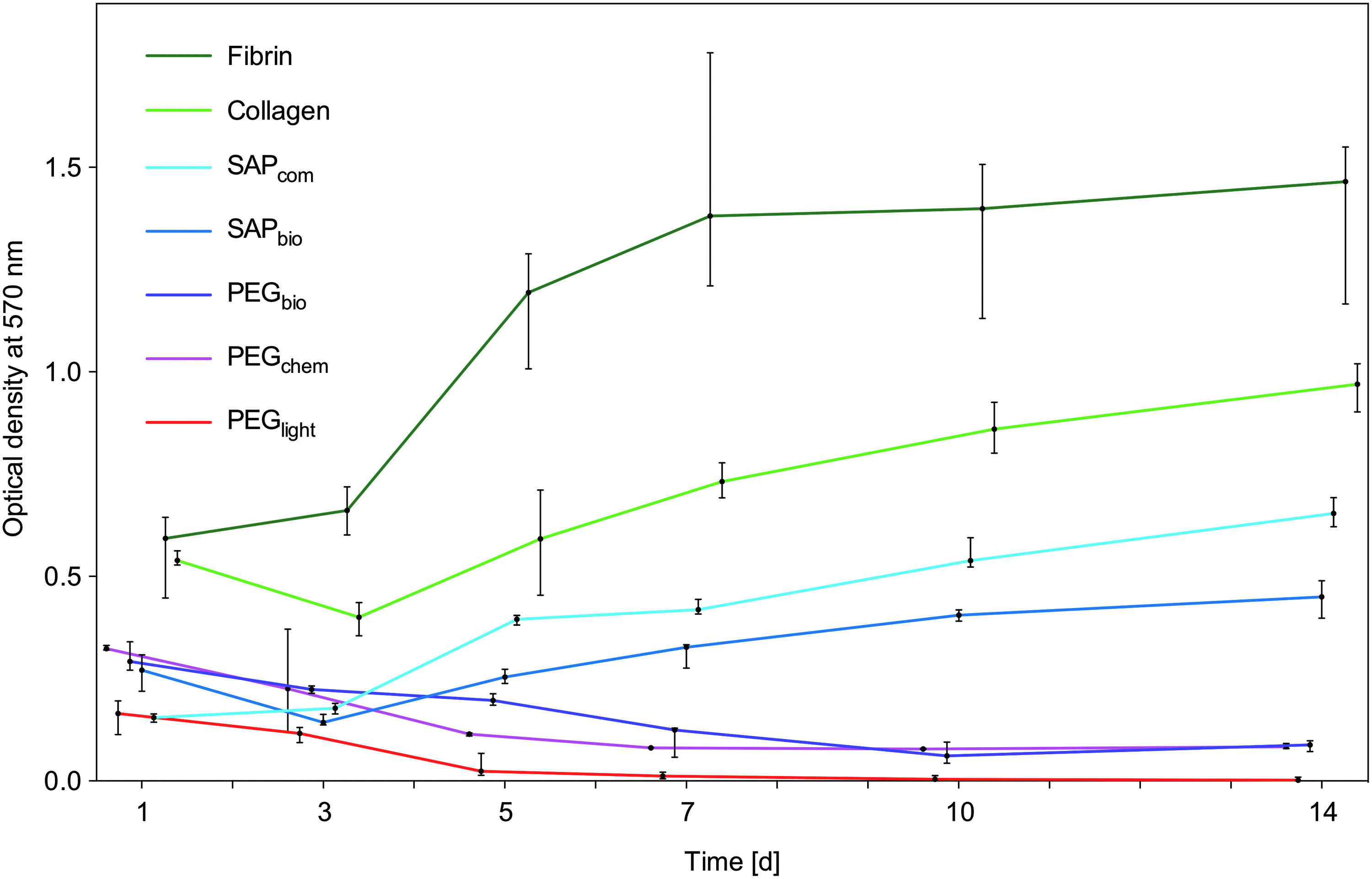
Cell viability is considerably higher in natural compared to synthetic materials. Light-curing PEG5k-acrylamide (PEGlight) shows lowest cell viability, which is higher in biomimetic PEGbio and chemically curing PEGchem initially, but drops to a similarly low level after 10 days of culture. SAP hydrogels (SAPbio and SAPcom) allow for intermediate cell viability, which is higher compared to PEG materials, but lower compared to natural materials fibrin and collagen. Depicted are medians with 25–75% percentiles, computed from experiments that were performed at least twice with triplicate samples. SAP, self-assembling peptide.
Live/dead stain confirmed the observations from cell viability assays. Exemplarily, cells in PEGlight, PEGbio, and Collagen at day 5 are shown in Figure 3. No live cells were observed in PEGlight (Fig. 3A), less than half of the cells were vital in PEGbio (Fig. 3B), and cell bodies appeared round and secluded. In collagen, the majority of cells remained vital and formed cell-to-cell contacts resulting in the formation of a three-dimensional network (Fig. 3C).
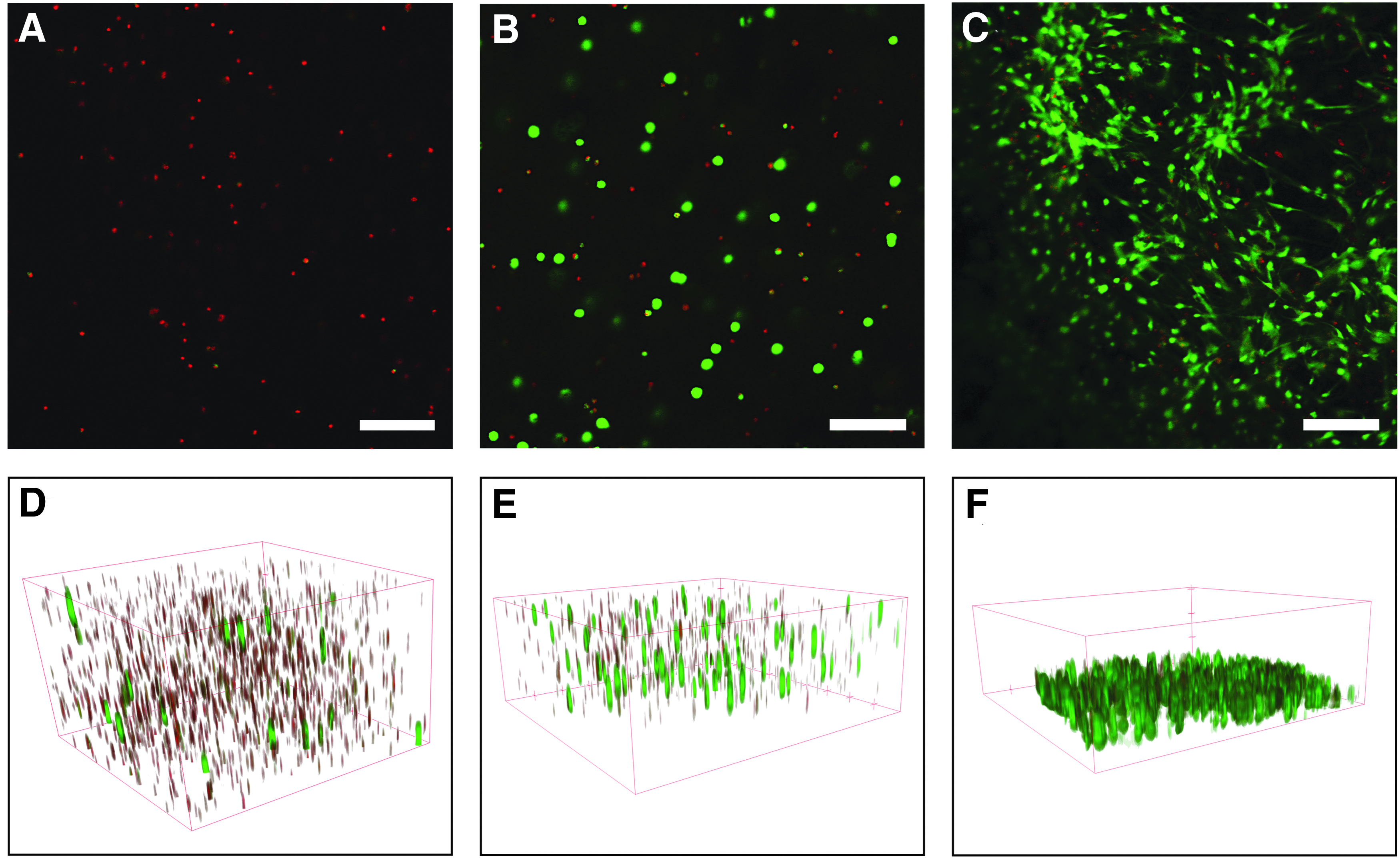
Live/dead staining of cells in PEGlight
Root canal model in vivo
In vivo experiments showed considerable differences in terms of tissue formation both between tested materials, as well as between test and control groups. Histological analysis of the explanted specimens revealed a distinctly higher cell activity regarding proliferation, matrix degradation, and formation of connective tissue in test groups, where scaffolds were laden with dentin matrix proteins. Among the tested scaffolds, both PEG materials showed scattered cell bodies or smaller islands of connective tissue. Fibrin enabled tissue formation in all cylinders; 10 out of 12 cylinders revealed pulp-like tissue with differentiated cells at the cell–dentin interface. SAP scaffolds generated both loose connective tissue, as well as pulp-like tissue. Images from histology and immunohistochemistry are depicted in Figure 4; the results from histologic analyses are summarized in Figure 5. Dentin cylinders from tooth roots with functionalized PEG-acrylamide gels showed cells embedded in a cavernous matrix, where they remained spheroidal and the material had been degraded merely around the cell bodies (Fig. 4A, B). In the respective control group without dentin matrix proteins, the scaffold material appeared disintegrated as a loose network, with only few spheroidal cell bodies (Fig. 4C). Chemically curing PEG allowed for partial formation of a loose connective tissue in test groups (Fig. 4D), whereas control groups presented mainly remnants of decomposed material without cells (Fig. 4E) or empty constructs. For fibrin scaffolds, most constructs in test groups were filled with pulp-like tissue (Fig. 4F, G). Deposition of a collagenous matrix against the existing dentin wall of the tooth root was evident after Masson's trichrome stain in three out of 12 cylinders (Fig. 4H). At the cell–dentin interface, cellular processes extended into the dentinal tubules (Fig. 4I). Where cells had formed an intimate association with the dentin wall, immunohistochemistry revealed expression of dentin sialoprotein in the adjacent cell layer (Fig. 4J, K). For fibrin, the control group showed empty cylinders in half of the constructs; the other half showed a fibrous tissue with remnants of scaffold material, which had not been fully degraded in the center (Fig. 4L). For SAP scaffolds, pulp-like tissue formation was observed in one-third of the cylinders (Fig. 4M, N) with cellular processes in dentinal tubules at the contact area of cells with the dentin wall (Fig. 4O). In the control group, most cylinders were empty or contained remnants of the scaffold material only; two cylinders showed fibrous tissue (Fig. 4P). In all cylinders that showed tissue formation, vascularization was present. Cells inside the dentin cylinders stained positive for the human nuclear envelope marker, confirming human origin, whereas blood vessels were derived from the donor. Capsules of connective tissue surrounding each cylinder did not stain and were thus identified as mouse tissue. Analysis of the tissue distribution in test and control groups for the different materials revealed higher amounts of tissue formation in test than control groups and demonstrated that fibrin enabled pulp-like tissue formation in most of the implanted constructs.

Histologic analysis of constructs after subcutaneous implantation. In the PEGbio test group, cells remained scattered and did not form networks and the scaffold material was degraded only around the cell bodies
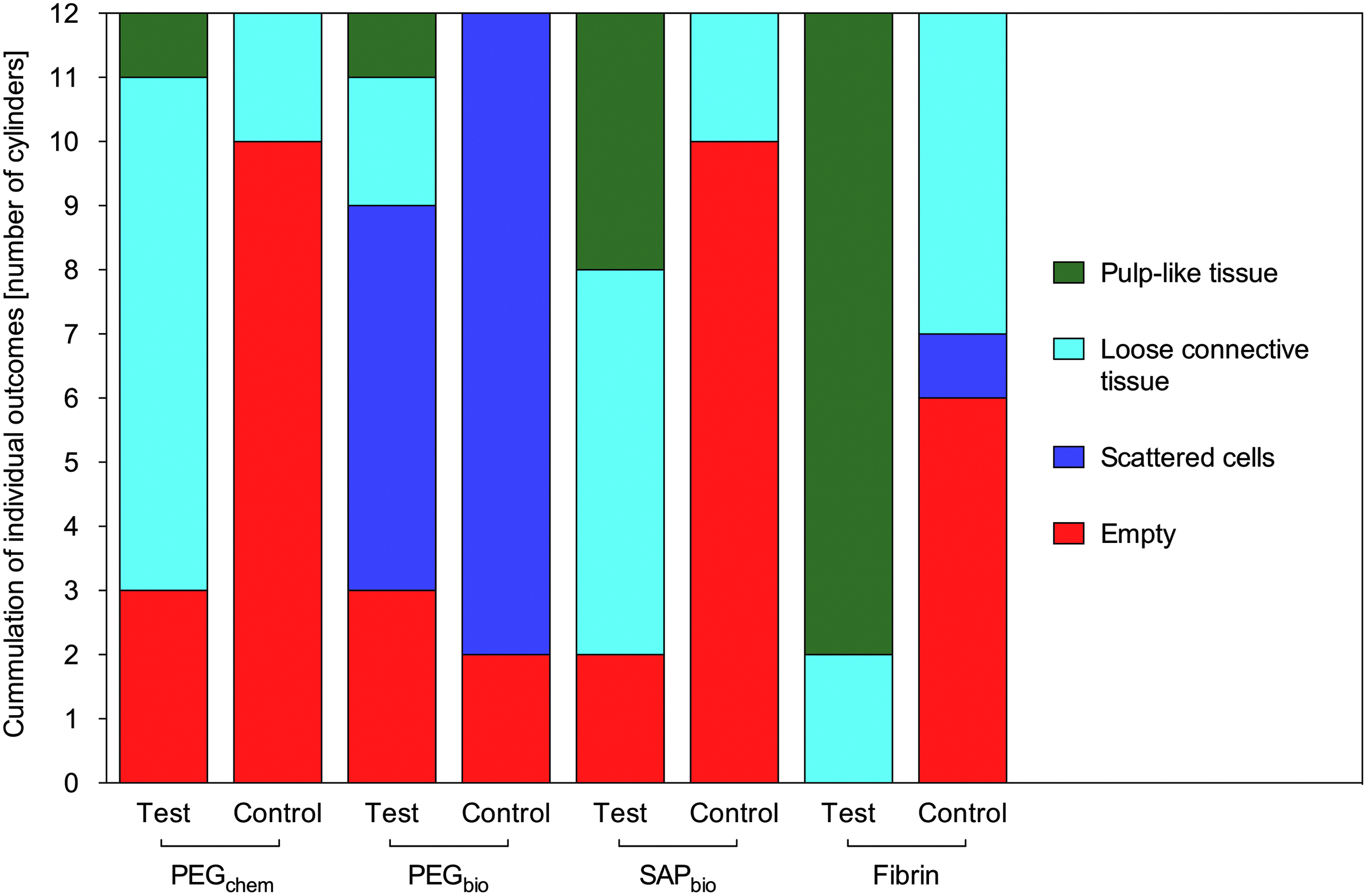
Summary of histological analysis of test groups (with extracted dentin matrix proteins) and control groups (without dentin matrix proteins). Results were classified into the following four groups: pulp-like tissue, loose connective tissue, scattered cells, and empty (root canal).
Further morphological insight was gained by scanning electron microscopic examination of the interface between soft and hard tissue (Fig. 6). Images confirm the extension of cellular processes into the dentinal tubules as observed in histologic sections.
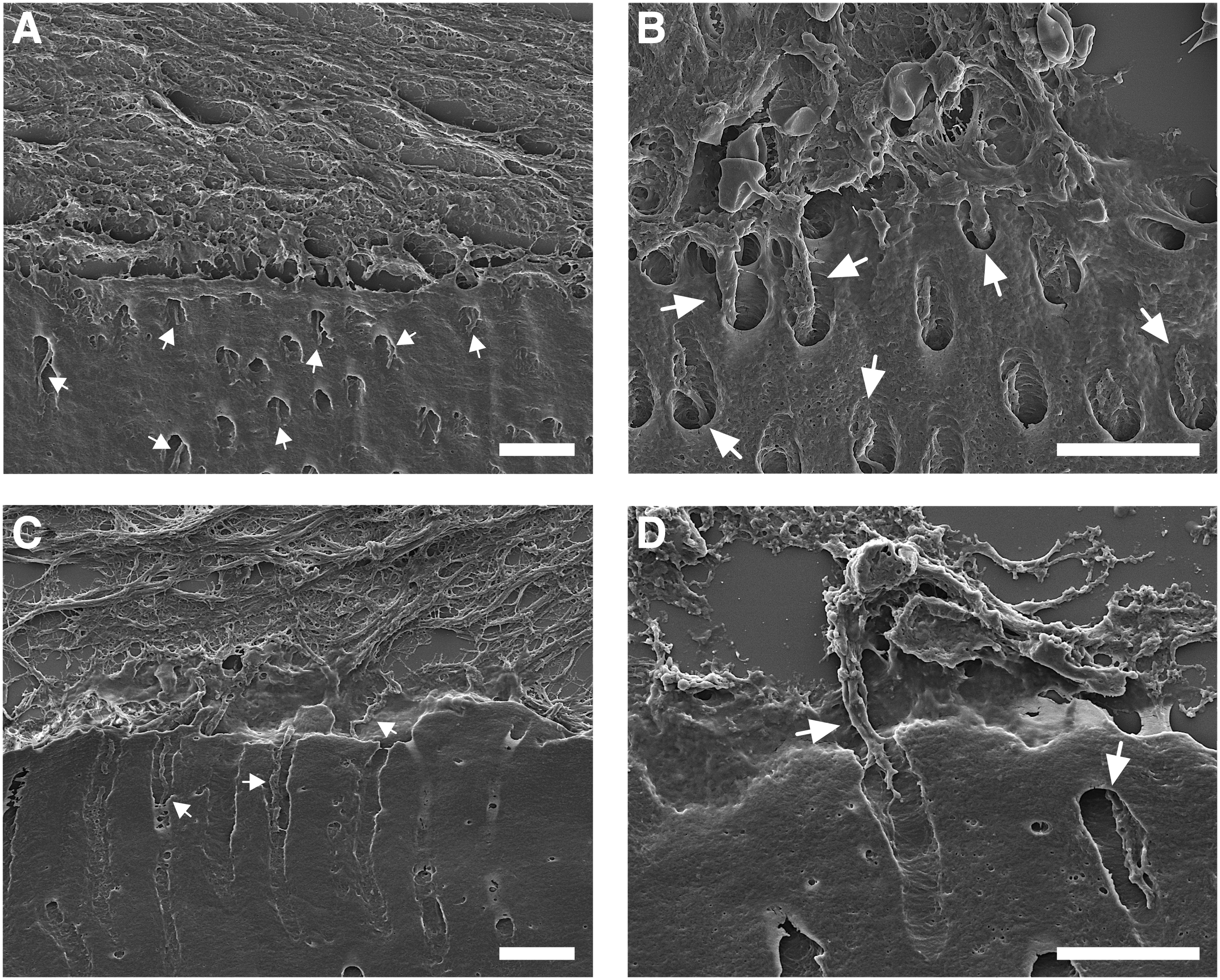
Scanning electron microscopy imaging of the interface between dentin and soft tissue in tooth roots. A layer of cells lined the dentin surface and extended processes into dentinal tubules (arrows) with fibrin
Discussion
This study reports for the first time on a direct comparison of different hydrogel scaffold materials for dental pulp tissue engineering and parallel examination both in vitro and in vivo.
The first consideration in the selection process was to compare natural and synthetic materials. Collagen and fibrin were chosen as natural materials, new and functionalized PEG materials with bioactive motifs (cell adhesion motif and enzyme-cleavable site) were developed in this study, and a previously generated functionalized SAP was included. Both natural materials have been used for dental pulp tissue engineering previously and seemed to be feasible candidates.17,20 Our own work on SAP hydrogels has demonstrated that customized synthetic materials might be applicable for this approach. PEG-based materials were chosen due to their characteristics of a hydrogel with high control over the material properties, the applicability for tissue engineering, and the possibility to define the mode of curing, as well as to include bioactive motifs. Cell culture experiments with different types of hydrogels showed higher cell viability in natural than in synthetic materials. This is in line with own previous data on fibrin and SAP.23,24 Fibrin seemed to be the most favorable material, which appears feasible as its primary purpose is to serve as a guide rail for cells in wound healing and regeneration. Viability in collagen was characterized by a deflection at day 3, which might be due to contraction of the collagen matrix inducing a temporary contact inhibition around this time point. For synthetic hydrogels, cell viability in light-curing PEG was lowest. Functionalization of the material increased cytocompatibility only during the initial phase of the culture period, which confirms previous results on SAP. 24 The light-curing PEG generated in this study could easily be polymerized in a dental office by use of a polymerization lamp for dental composites, thus it was initially considered a feasible candidate material. The initiating compounds for polymerization exert cytotoxic effects, which are concentration dependent. 30 Although cytotoxicity of acrylamide groups and initiating compounds for polymerization should be abrogated as soon as the material is polymerized, PEG-acrylamide proved to be unfavorable for cell survival with decreasing cell viability during the culture period and, thus, unsuitable as a scaffold for pulp tissue engineering. Chemically curing PEG, which was initially used as a reference material, was tolerated better by the cells than PEG-acrylamide and might thus have been a better candidate for further functionalization. Furthermore, the stiffness of the material might play a role. Chemically curing PEG hydrogels are softer than light-curing PEG, which offer a higher conversion rate. Both types of SAP turned out to be more cytocompatible than PEG, but less than both natural materials. Certainly, the basic chemical composition will affect cytocompatibility. Positive charges of lysine in customized SAP might exert a negative effect, but functionalization a positive effect. The commercially available product, which lacks both, allows for similar cell viability compared to the tested functionalized SAP. To corroborate cell viability assessment in three-dimensional cell cultures, live/dead staining was performed exemplarily, which confirmed observations from MTT assays.
This study furthermore enabled us to directly correlate results from in vitro and in vivo experiments. Four materials were selected for animal work based on the following considerations: Fibrin was chosen based on its excellent cytocompatibility and handling characteristics. Collagen was excluded, as it is contracted considerably by the cells. Contraction of the material after insertion into a root canal might lead to detachment of the cells from the dentin walls, result in voids, and thus compromise new tissue formation. Chemically curing PEG was tested along with light-curing functionalized PEG, as the aim of the study was to design and test customized PEGs for dental pulp tissue engineering. Out of the two SAPs, the customized and functionalized appeared to be the more promising candidate due to the possibility for cell-mediated degradation, despite similar results for cytocompatibility of SAPbio and commercially available SAPcom. Furthermore, previous experience with the functionalized SAP offered a good reference to test other materials against. 21
Data obtained from the animal work closely reflected the findings from cell culture experiments, where low cell viabilities in scaffold materials were linked to the disability of pulp-like tissue formation in the respective materials and vice versa. Functionalized PEGbio allowed only for sparse cell survival and matrix degradation around cell bodies; chemically curing PEGchem gave rise to small islands of connective tissue. Constructs with SAP displayed connective or pulp-like tissue. Fibrin, which offered highest cytocompatibility, enabled pulp-like tissue formation with extension of cellular processes into dentinal tubules in the majority of the constructs. Thus, the originally stated hypothesis that synthetic materials, which are well defined and can be created based on the specific requirements for regeneration of the target tissue, had to be rejected, at least for the materials designed and tested in this study.
Yet another relevant finding is the fact that endogenous growth factors from dentin, which were exposed on the dentin surface of the dentin cylinders after EDTA conditioning, and additionally incorporated into the scaffold before gelation, rendered significant differences in cell activity, matrix degradation, cell proliferation, and differentiation between test and control groups. It is evident that dentin matrix harbors a plethora of bioactive proteins,31–33 which play roles in immunomodulation and the stimulation of chemotaxis, cell proliferation, angiogenesis, and differentiation.34–38 Our previous work demonstrates that amounts of dentin matrix proteins that can be extracted from the root canal surface of a tooth during endodontic treatment might be sufficient to evoke a desirable cellular behavior. 29 The use of endogenous dentin-derived growth factors might be an elegant way to avoid the controversies afflicted with recombinant growth factors for tissue engineering approaches and, thus, appears to be a promising alternative. Mixing dentin matrix proteins with the biomaterials without further mechanisms of binding will result in a burst release and most likely in rather quick degradation of the bioactive proteins. Binding and slow release of growth factors by incorporation of binding sites, for example, by use of heparin, have been reported previously.21,39 However, of all test materials, fibrin possesses an endogenous capability of growth factor binding and slow release. 40 This effect might have contributed to the favorable results for pulp-like tissue formation for this material in combination with dentin-derived proteins.
Conclusions
In conclusion, the natural materials tested in this study allowed for significantly higher viability of dental pulp stem cells compared to synthetic materials, which was reflected by in vivo data, where pulp-like tissue formation was observed in most of the constructs for fibrin, but not the synthetic materials. Fibrin appeared to be the most promising scaffold material for dental pulp tissue engineering among the tested hydrogel scaffolds. This result is contradictory to the stated hypothesis that synthetic materials might be better candidates for this approach, as they offer high control over the material properties and can be functionalized with specific bioactive motifs. Advantages of fibrin, however, include easy handling and low cost, commercially available fibrin-derivatives that are approved for clinical applications, high cytocompatibility, and facilitation of pulp-like tissue formation. Thus, fibrin should be explored further in future clinical studies on dental pulp tissue engineering and regeneration.
Footnotes
Acknowledgments
The authors thank Dr. Thilo Spruss for his valuable assistance with the animal experiments, as well as Helga Ebensberger and Gerlinde Ferstl for their support on scanning electron microscopy. This work was supported by University Hospital Regensburg and DMG.
Disclosure Statement
No competing financial interests exist.
References
Supplementary Material
Please find the following supplemental material available below.
For Open Access articles published under a Creative Commons License, all supplemental material carries the same license as the article it is associated with.
For non-Open Access articles published, all supplemental material carries a non-exclusive license, and permission requests for re-use of supplemental material or any part of supplemental material shall be sent directly to the copyright owner as specified in the copyright notice associated with the article.