
Abstract
Chemokine stromal cell-derived factor-1 (SDF-1) is a powerful chemoattractant for the localization of CXCR4-positive bone marrow mesenchymal stem cells (BMSCs) into the bone marrow. We studied the effects of SDF-1 on the cartilage defect repair by recruiting BMSCs and promoting its chondrogenic differentiation in vitro and in vivo. Chemotaxis analysis with Transwell plate showed that SDF-1 could recruit BMSCs through SDF-1/CXCR4 axis. Real-time polymerase chain reaction, enzyme-linked immunosorbent assays, and Western blot results suggested that the levels of type II collagen and GAG were increased after incubating BMSCs with SDF-1 compared with the without SDF-1 group. More positive BrdU-labeled BMSCs were detected at the cartilage defect region in the SDF-1 + poly [lactide-co-glycolide] (PLGA) scaffold group (SP) in which those animals showed a smooth and transparent cartilage tissue with a strong staining of toluidine blue and type II collagen compared with the no-SDF-1 groups. ICRS score suggested that the repair effect in the SDF-1 + PLGA-treated animals was improved compared with PLGA scaffold group alone at 4 and 8 weeks after surgery; the repair effect from the SDF + PLGA-treated animals was significantly improved compared with the PLGA alone at 12 weeks after surgery. Our in vitro and in vivo results indicated the following: (1) SDF-1 could recruit the BMSCs into cartilage defect area. (2) SDF-1 induces BMSCs expressing type II collagen and GAG, which may accelerate the BMSCs transforming into chondrocytes under the cartilage microenvironment in vivo. (3) PLGA scaffold attached with SDF-1 remarkably promoted the cartilage defect repairing. The defected cartilage was filled with transparent cartilage 12 weeks after the surgery, which shared a similar structure with the adjacent normal cartilage. Taken together, this research provides a new strategy for cartilage defect repairing.
Introduction
T
Stromal cell-derived factor-1 (SDF-1) is the well-known chemoattractant protein that is secreted by bone marrow matrix cells as well as other relative matrix cells. 4 SDF-1 reacts with its receptor CXCR4 and regulates the stem cell homing by stimulating numbers of downstream signal pathways. SDF-1 and its receptor CXCR4 play important roles in the activation of the hematopoietic stem cell 5 and regulation of BMSC migration. 6 In addition, the collected evidence showed that BMSCs expressing CXCR4 had the powerful chemotaxis to SDF-1, and SDF-1 could increase the expression of CXCR4 itself and promote the migration of BMSCs. 6 However, its effects on BMSC differentiation into chondrocyte and cartilage repairing are seldom reported.
The material of the scaffold is another important issue in tissue engineering. 7 Artificial poly [lactide-co-glycolide] (PLGA) is the Food and Drug Administration-approved biodegradable material. Research showed that PLGA could promote cartilage repair for excellent biocompatibility. 8 Moreover, PLGA was beneficial for the regeneration of cartilage by inhibiting the infiltration of macrophages and decreasing the inflammatory factors. 9 We assumed that the PLGA with SDF-1 can repair cartilage defect by recruiting BMSCs and promoting BMSC chondrogenic differentiation under endogenous cartilage microenvironment in vivo.
In this study, BMSCs were identified by flow cytometry from adult rabbit bone marrow and in vitro chemotactic effect of the SDF-1 to BMSCs was detected using transwell plates. To test the chondrogenic differentiation effect of SDF-1 on BMSCs, Type II collagen and GAGs expression were analyzed using real-time polymerase chain reaction (RT-PCR), enzyme-linked immunosorbent assays (ELISA), and Western blotting. The chemotactic effect of SDF-1 on BMSCs in vivo was confirmed by counting the number of BrdU-labeled BMSCs using immunohistochemical staining. The ICRS histological scoring and immunohistochemistry were used to analyze the effects of SDF-1/PLGA scaffold repairing cartilage compared with without SDF-1 groups.
Materials and Methods
Isolation and identification of BMSCs
Three-month male New Zealand Rabbits (provided from animal center in Shanxi Medical University) were sacrificed. The bone marrow cells were isolated and cultured as previously described. 10 The third generation of BMSCs was incubated for 10 h at 4°C with CD14, CD44, CD45 (BD Pharmingen), and CD90 (AbCam) antibody, respectively, and in the negative control group, BMSCs were incubated with IgG1 antibodies. Secondary antibodies labeled with fluorescein isothiocyanate (FITC) were added and incubated for 1 h at 4°C. Labeled cells were analyzed using flow cytometry.
In vitro chemotaxis analysis with transwell plate
Cells were cultured in a 24-well transwell plate (Corning Inc) with 8.0 μm pore polycarbonate membrane inserted to separate the wells into upper and lower compartments and the cell density was adjusted to 1 × 10⁶/mL with L-DMEM media out of Bovine Serum (Gibco). Cells were divided into the following groups: (1) control group (0 ng/mL SDF-1 in both upper and lower compartments); (2) 50 ng/mL SDF-1 group (upper compartment: 0 ng/mL SDF-1, lower compartment: 50 ng/mL SDF-1); (3) 10 μg/mL AMD group (upper compartment: SDF-1/CXCR4-specific blocker-AMD3100, lower compartment: 0 ng/mL SDF-1); (4) SDF-1 + AMD group (upper compartment: 10 μg/mL AMD3100, lower compartment: 50 ng/mL SDF-1). One hundred microliters BMSC suspension was added in upper compartments and L-DMEM was added in lower compartments. The transwell plate was kept at 37°C in 5% CO2 for 24 h, the polycarbonate membrane was put out, and fixed with 4% paraformaldehyde for 15 min and DAPI (32670, Sigma) staining for 30 min, observing and counting the BMSCs under the fluorescence microscope (Olympus, Japan).
The mRNA level detection using RT-PCR
The third generation of BMSCs was randomly divided into four groups: (1) control group: only incubating with L-DMEM media; (2) 50 ng/mL SDF-1 group; (3) 10 μg/mL AMD3100 group; and (4) SDF-1 + AMD group. The levels of the Col-II, Agg, and MMP-13 expression were determined using RT-PCR after 72 h of incubation. The total RNA was extracted from the BMSCs and total RNA (0.5 μg) was reverse transcribed using the PrimeScript™ RT Kit (K1642, Fermentas). RT-PCR amplification was performed with the SYBR® Premix Ex Taq™ Kit (K0241, Fermentas). mRNA levels were normalized to GAPDH. The relevant expression level of the mRNA would be reflected by the Ct value calculated by 2−ΔΔct method, in which ΔΔCt = ΔE−ΔC, ΔE = Ctexp−CtG, and ΔC = Ctctl−CtG. The sequences of the primers were as follows: Col-2 forward 5′-ACACTGCCAACGTCCAGATG-3′ and reverse 5′-GTGATGTTCTGGG AGCCCTC-3′ (D83228); AGG forward 5′-TCTACCGCTGTGAGGTGATGC-3′ and reverse 5′-TTCACCACGACCTCCAAGG-3′ (L38480); MMP-13 forward 5′-ACACC GGATCTGCCAAGAGA-3′ and reverse 5′-CTGG AGAACGTGATTGGAGTCA-3′ (001082037); and GAPDH forward 5′-GGTGAAGGTCGGAGTGAACG-3′ and reverse 5′-AGTTAAAAGCAGCCCTGGTGA-3′ (L23961).
The level of Agg, Col-II and MMP-13 detected by ELISA in cell supernatant
On the first, third, and sixth day after the incubation of BMSCs with SDF-1, the cell supernatant was collected. ELISA Kits Agg (E10H2109, R&D), Col-II (E10H2107, R&D), and MMP-13 (E10H2108, R&D) were employed to test the metabolic and inflammatory concentration.
The Col-II protein assay by Western blot in vitro
Total protein was extracted from BMSCs 48 h after incubation with SDF-1, sample proteins were separated and transferred onto polyvinylidene fluoride (PVDF) membranes as previously described. 11 The membranes were incubated with a primary antibody overnight at 4°C, followed by a secondary antibody for 2 h at 4°C (Primary antibody: rat anti-rabbit Col-2, Sigma; HRP IgG Goat anti-rat as the secondary antibody, Sigma). The film signals were quantified with Alpha View SA software. Each blot was normalized to its corresponding internal control-β-actin value.
In vivo experiment to verify SDF-1-recruiting BMSCs in cartilage defect region
The biocompatibility of the BMSCs and PLGA scaffolds was observed with scanning electron microscope (JEOL100-C, OLYMPUS, Japan) after coculture for 7 days. To observe the effect of SDF-1 recruiting BMSCs, the third generation of BMSCs was coincubated with 40 μmol/L BrdU for 72 h. The BrdU-labeled BMSCs were counted and adjusted as 1 × 10⁷ cells/mL. Three-month male New Zealand Rabbits were anesthetized by injection of 3% pentobarbital (Sigma). As previously described, 12 a 3-mm parapatellar incision was made to expose the knee joint, the femoral trochlea was revealed, and a 4-mm diameter, 3-mm tall cylindrical osteocartilage piece was drilled on the trochlea with a corneal trephine: the PLGA scaffold with phosphate-buffered saline (PBS) (control group, n = 3), the PLGA scaffold with 50 ng/mL SDF-1 (SDF-1 group, n = 3), the PLGA scaffold with 10 μg/mL AMD3100 (AMD3100 group, n = 3), and the PLGA scaffold with 50 ng/mL SDF-1 + 10 μg/mL AMD3100 (SDF-1 + AMD3100 group, n = 3) were attached into the defected region, respectively. 4 × 10⁵ U penicillin was injected for three consecutive days. One milliliter BrdU-labeled BMSCs (1 × 10⁷/mL) were injected into the ear vein 2 h after surgery. The knee joints were collected at 4 weeks postoperatively and the vertical sections were assessed by two blinded observers using an Olympus BX51 microscope at 400 × magnification. The percentage of positively stained chondrocytes was calculated by counting the number of BrdU positively and negatively stained cells in five random areas of each slide.
Establishment of the model of cartilage defect and repairing with PLGA scaffold attached with SDF-1
The osteocartilage defect model was created as mentioned above. Sixty adult male New Zealand Rabbits (3.0 ± 0.2 kg) were randomly divided into four groups: (1) Group D (defect control group), without planting any materials and directly closed the articular cavity; (2) Group P (PLGA scaffold alone), PLGA sponge scaffolds, the diameter and thickness of which were 4.0 mm and 1.5 mm, respectively, were overlapped implanted into the defected region; (3) Group SP (50 ng/mL SDF-1 + PLGA scaffold); (4) and Group SPA (50 ng/mL SDF-1 + PLGA scaffold +10 μg/mL AMD3100). After the surgery, animals were allowed to freely move and the penicillin was injected for three consecutive days.
ICRS histological scoring
Femurs were withdrawn at 4, 8, and 12 weeks after surgery. The tissues were fixed, decalcified, and embedded; the vertical trochlea was sectioned and HE staining, Safranin O staining, and Toluidine blue staining were performed. The quality of cartilage repair was assessed using the ICRS histological scoring system. 13
Col-II immunohistochemistry detection
As previously described, 11 the sections were incubated with anti-rabbit Col-II polyclone antibody (Cat. No. cp18 Calbiochem, Japan) at 4°C overnight. For the negative controls, the sections were incubated with 0.01 M PBS. On the second day, the sections were incubated with a goat anti-IgG secondary antibody (Boster, Inc.), followed by standardized development in diaminobenzidine (DAB). Photography was performed with Olympus BX51.
Statistics
Data were collected and processed by SPSS18.0 software. Nonparametric Kruskal–Wallis rank sum test was used for statistical analysis in overall scoring. Data were presented as M ± QR rung, and considered statistical significance at p < 0.008. The rest of the experiment was processed by Tukey's one-way analysis of variance, in which mean ± SD was considered the statistical description, and the statistical significance was at p < 0.05.
Results
The identification of BMSCs
The flow cytometry results indicated that CD44- and CD90-positive cells were 99.17% and 93.37%, respectively, while CD45 and CD14 were negative on the third generation BMSCs (Fig. 1). These results suggested that the majority of these cells are BMSCs after three-generation subculture.
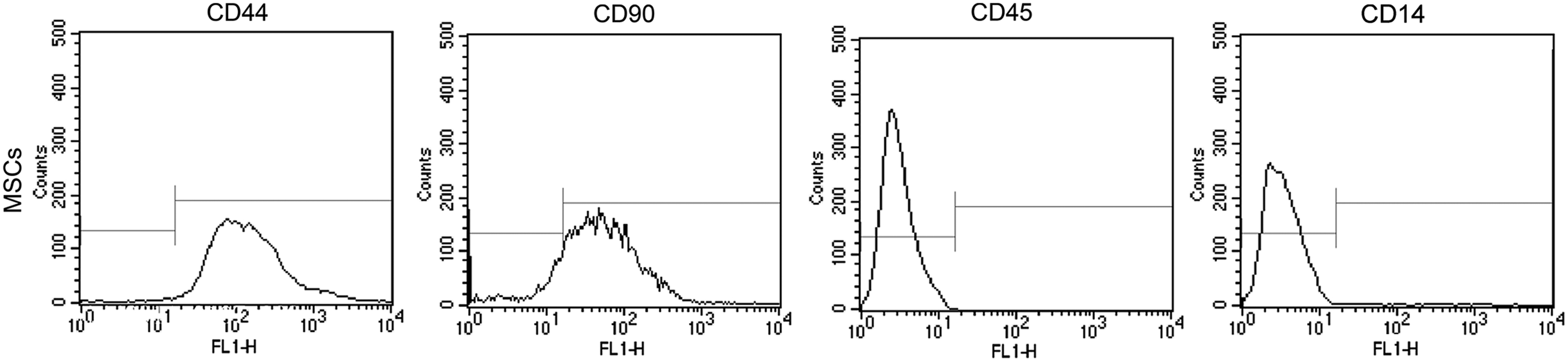
The identification of third generation BMSCs using flow cytometry. As shown in the figure, BMSCs were positive for the cell surface markers CD44 and CD90, and negative for CD45 and CD14. BMSCs, bone marrow mesenchymal stem cells.
SDF-1 recruits BMSCs through SDF-1/CXCR4 axis
As shown in Figure 2A and B, the number of cells transported in the 50 ng/mL SDF-1 group was significantly higher than the number in the control group (p < 0.01). By adding the 10 μg/mL AMD3100, the number of cells transported was significantly decreased compared with the SDF-1 group (p < 0.01), there was no significant difference between the 10 μg/mL AMD-alone group and control group (p > 0.05). Our experiment results confirmed that the receptor CRCX4 was wildly expressed on BMSC surface (Fig. 2C)
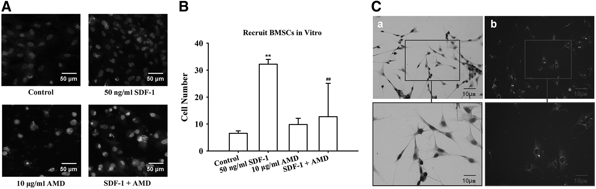
SDF-1 could recruit BMSCs through SDF-1/CXCR4 axis.
SDF-1 could stimulate the BMSCs to secrete the cartilage matrix and promote BMSC synthesis of Col-II and Agg
To explore the effect of SDF-1 on the differentiation of BMSCs into chondrocytes, we employed RT-PCR to analyze the relative mRNA expression of the cartilage marker Agg and Col-II. As is shown in Table 1, the mRNA level of Agg and Col-II in the SDF-1 group was prominently increased (p < 0.01) compared with the control group, while the mRNA levels of these two markers in the SDF-1 + AMD group were both lower than that in the SDF-1 group (p < 0.01); the mRNA level of MMP-13 was not significantly different among all groups.
p < 0.01, control group.
p < 0.05 versus SDF-1 (50 ng/mL) group.
p < 0.05, control group.
p < 0.01 versus SDF-1 (50 ng/mL) group.
SDF, stromal cell-derived factor.
As shown in Table 2, the MMP-13 protein level did not present remarkable changes among all groups (p > 0.05) after 1, 3, and 6 days of coculture. The Tables 3 and 4 showed that there was no difference in the Agg and Col-II secretion among groups (p > 0.05) after 1 day of coculture. However, after 3 and 6 days of coculture of SDF-1 and BMSCs, the level of Agg and Col-II in the SDF-1 group was significantly increased compared with the control group (p < 0.01), the secretion level of Agg and Col-II in the SDF-1 + AMD group was significantly lower than the SDF-1 group (p < 0.01), and there was no significant statistical difference in the AMD, SDF-1 + AMD, and control group (p > 0.05).
p < 0.01, Control group.
p < 0.01 versus 50 ng/mL SDF-1 group.
p < 0.01, Control group.
p < 0.01 versus 50 ng/mL SDF-1 group.
Western blot assay suggested that the higher level of Col-II protein was found in the SDF-1-treated group (1.22 ± 0.03) compared to the control group (0.98 ± 0.02, p < 0.01). While blocking the SDF-1/CXCR4 pathway by AMD3100, the protein levels of Col-II were decreased both in the ADM (1.11 ± 0.03) and SDF-1 + AMD group (1.02 ± 0.01) (p < 0.01, Fig. 3).
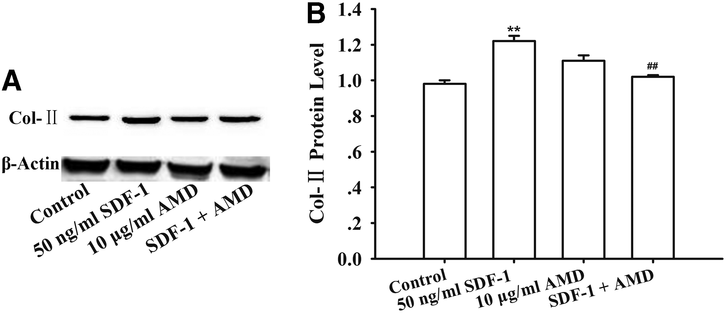
SDF-1 promoted the BMSC synthesis of Col-II.
SDF-1 recruits BMSCs into the cartilage defect region in vivo
The percentage of BrdU-positive cells was 93.73% ± 4.01% and the brown color Brdu-positive signal was located in the nucleus 72 h after the third generation BMSCs were cocultured with BrdU in vitro (Fig. 4A). Furthermore, we found that BMSCs were intensively attached on the surface of the PLGA scaffold after coculture of BMSCs with PLGA scaffold in vitro by scanning electron microscope (SEM), which suggested that PLGA has good histocompatibility (Fig. 4B). IHC staining in vivo results showed that the percentage of migration cells (BMSCs labeled with BrdU) was 35.25% ± 3.96% in 50 ng/mL SDF-1-treated group, which was remarkably increased compared with the control group (10.17% ± 4.2%, p < 0.01), while the percentage of the migration cells in the SDF-1 + AMD group (19.02% ± 6.00%) was less than that in the 50 ng/mL SDF-1 group (p < 0.01, Fig. 5A, B).

The staining of BrdU-labeled negative BMSCs and detection of BMSC histocompatibility.
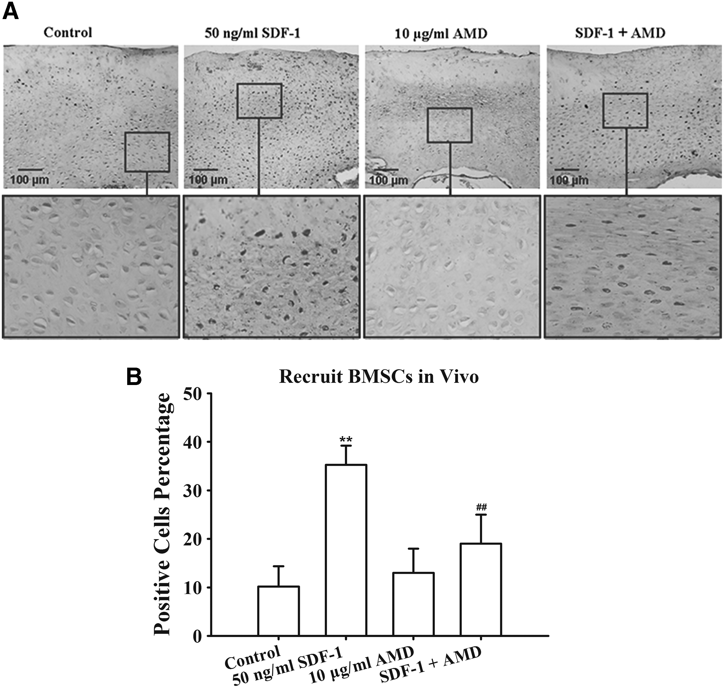
SDF-1 recruits BMSCs into the cartilage defect region in vivo after 8 weeks of the surgery.
SDF-1 promoted the cartilage repair in vivo
As shown in Table 5, the ICRS score suggested that PLGA scaffold attached with SDF-1 had a higher score compared with group P and SPA at the same time point (p < 0.008), no significant difference was observed between the group SPA and P after 4 and 8 weeks of surgery (p > 0.008), but at 12 weeks after the surgery, the ICRS score of the group SP was higher than that in group P and SPA. At the same time point, the score of group SP was significantly higher than that in the control group (p < 0.008). As shown in Figure 6, the cartilage defect was completely refilled with the normal cartilage similar to the adjacent normal cartilage in group SP; for group D, the cartilage defect was supplemented with white fibrous tissue; after 12 weeks of surgery, there was a clear boundary around cartilage tissue, the group P and SPA were similar to group D, the defected region was refilled with the translucent tissue, the surface of cartilage tissue was rough, and the boundary was obvious. The above results indicated that the cartilage defect in the animals treated with PLGA/SDF-1 started to repair with cartilage tissue from 4 weeks after surgery compared with other groups. The repairing effect was continuing to improve with time.
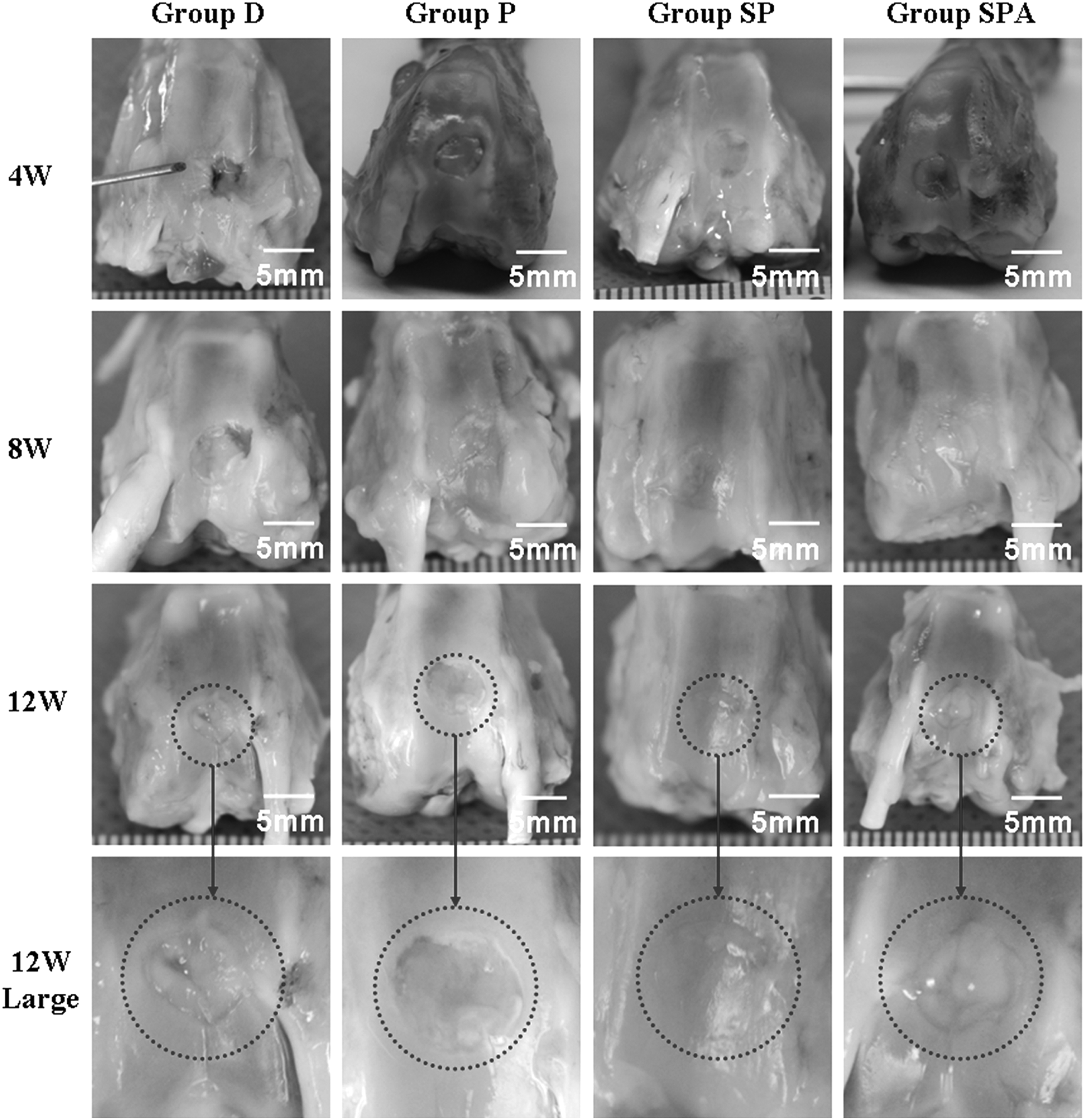
The gross observation at 4 W, 8 W, and 12 W after the operation. The surface of the repaired cartilage in the SP group was smooth and the boundary between the supplementary tissue and the surrounding tissue was almost undetectable, and the color of the repair tissue was similar with the adjacent normal tissue compared with the other groups.
p < 0.008, versus SDF-1 + PLGA group.
p < 0.008, versus Defect Control group.
The histological detection of Col-II
In group D, a few fibroblast-like cells were apparent along the defect borders and a limited degree of chondrocyte proliferation was detected within vicinal native tissue; the toluidine blue staining and Col-II staining were negative after 4–12 weeks of surgery. In group P and SPA, the defected region was repaired with the transparent fibrous combined cartilage-like tissue, in which there were few scattered chondrocytes and fibroblasts. The tissue was negative or slightly positive for both toluidine blue staining and Col-II staining after 4 weeks of surgery, while after 8 and 12 weeks of surgery, a fibrous cartilage-like tissue with smooth surface was formed in the defected region; both the toluidine blue staining and Col-II staining were positive. In contrast, the repaired cartilage from the animals treated with PLGA/SDF-1 showed that the repair tissue demonstrated the presence of chondrocyte-like cells and hyaline cartilage-like structure and matrix, both toluidine blue staining and Col-II staining were positive, and the specimens were free of ectopic calcification and vascularization (Fig. 7).
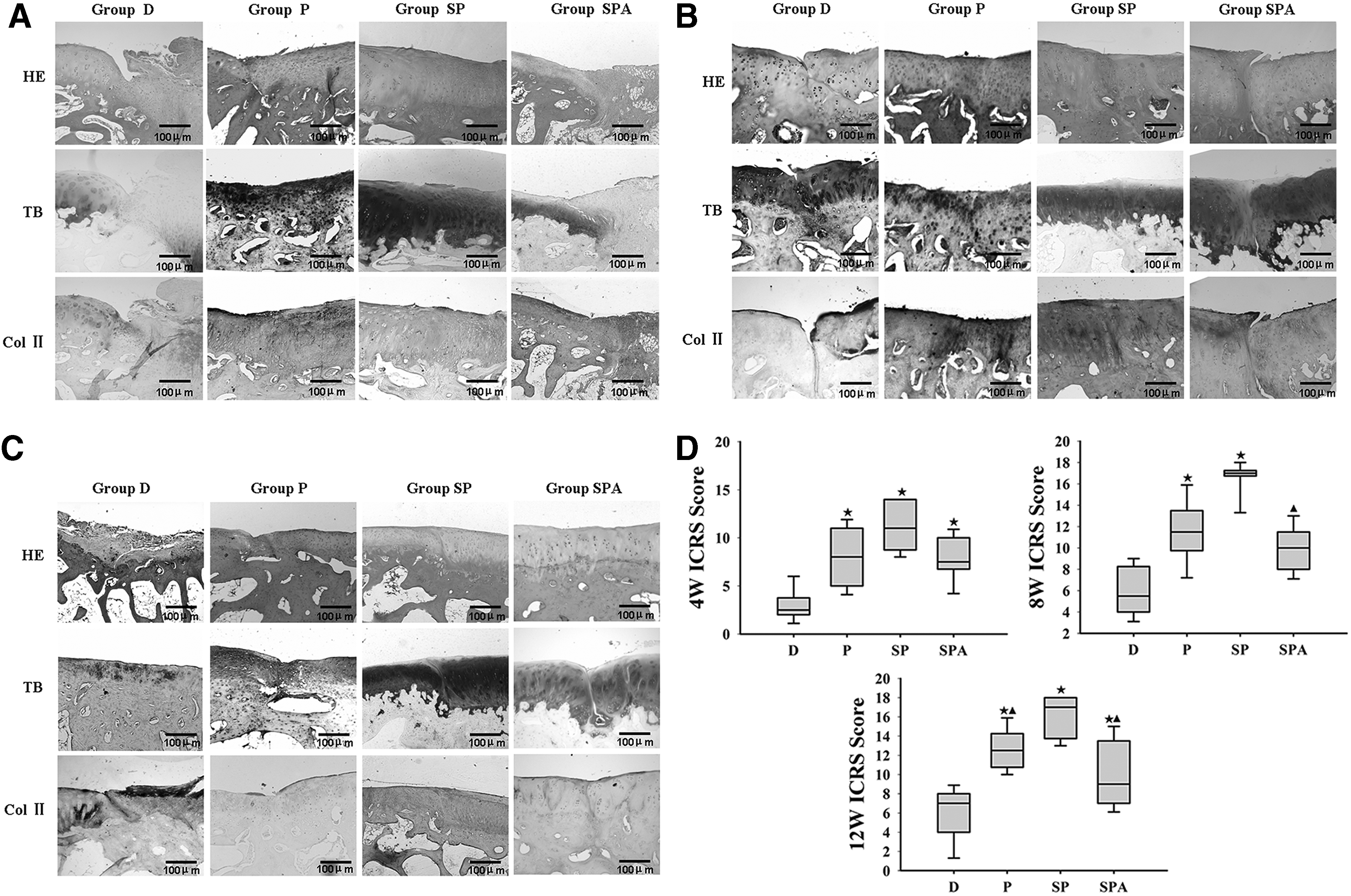
Different group hematoxylin-eosin, toluidine blue, and Col-II immunohistochemical staining in 4 W
Discussion
The cartilage defect can disrupt the stability of articular metabolism and induce the pain and dysfunction of the articular cartilage tissue, which ultimately results in osteoarthritis. 14 The cartilage tissue has very limited self-regenerative ability and the traditional treatment of the cartilage defect has some disadvantages, 15 which make tissue engineering a new pathway for the cartilage defect treatment.
It was reported that SDF-1 could activate the migration of stem cells or progenitor cells for tissue defect repairing.16,17 SDF-1 is a highly conservative molecule that is synthesized and released by bone marrow cells. It plays a critical role in the formation of the bone marrow hemopoiesis microenvironment. 18 Our in vitro results suggested that SDF-1 could promote the chemotaxis of the BMSCs, which could be blocked by the CXCR4 antagonist AMD3100. This further verified that the SDF-1/CXCR4 axis plays a key role in the migration of stem cells. In this study, we further showed that 50 ng/mL SDF-1 could promote BMSCs to secrete more Col II and aggrecan, and there was no reaction of inflammation.
The tissue engineering could be the most promising pathway for cartilage defect. 19 One of the necessary condition for this technique is to provide sufficient, normal function seed cells. 1 BMSCs are a kind of mesenchymal stem cells (MSC) with pluripotent differentiation and self-renew abilities, which exist in bone marrow. They have a certain level of plasticity, which can differentiate into different cells under certain conditions, such as bone cells, chondrocytes, adipocytes, muscle cells, and neurons. Moreover, autologous BMSCs have better histocompatibility than transplanted chondrocytes. Taken together, the above features suggest that BMSCs are ideal seed cells for cartilage tissue engineering.
SDF-1 is a powerful chemoattractant for the localization of BMSCs with CXCR4 expression. 10 However, it has not been reported whether BMSCs can reach the defected region through SDF-1/CXCR4 signaling. In this experiment, the BrdU-labeled BMSCs were injected through ear vein; the results found that SDF-1 could recruit more BrdU-labeled BMSCs into the cartilage defect region and the specific antagonist of CXCR4 decreased the positively recruited cells. This result indicates that SDF-1/CXCR4 signaling could recruit more BMSCs for cartilage repairing. This finding is consistent with previous report that the SDF-1/CXCR4 signaling pathway associates with the migration and homing of the CXCR4-positive hematopoietic stem cells/progenitor cells. 5
Currently, there are several common treatments for the application of tissue engineering7,22: first, the chondrocytes were transplanted into cartilage defect region; second, noncellular scaffold transplant was used to repair the defect cartilage alone in which the scaffold can recruit endogenous bone marrow or blood stem cells for repairing cartilage tissue; and third, the transplanted cells incubated with the scaffold were used to repair the defect cartilage. However, the first two methods could cause the loss of transplanted cell or cellular heterization. 23 Our experimental results in vivo and in vitro indicated that SDF-1-attached PLGA scaffold provided a better repairing effect compared with the PLGA alone, which suggested the SDF-1 may recruit BMSCs and promotes these cell differentiates into chondrocytes under cartilage microenvironment in vivo. However, the exact mechanism remains unexplored.
The previous research has suggested that microenvironment in the articular cartilage could promote BMSC differentiation into chondrocyte. 12 In this experiment, we employed PLGA scaffold, which is the better vehicle of the cell in tissue engineering, it can provide the continuous adherent and growth environment of the cells. Our SEM data indicated that PLGA provided larger three-dimensional space for cells, which allows cells to move and migrate in three-dimensional space, overcoming cell-mediated inhibition in monolayer culture in vitro. The scaffold is benefit for the communication of information, nutrition, and metabolism among cells; meanwhile, the cells and secreted extracellular matrix can be confined to the three-dimensional structure of the scaffold, which is conducive to the structural modification and integration of the new tissue.
Conclusion
PLGA scaffold attached with SDF-1 could enhance the repair of defected cartilage by recruiting BMSCs into the cartilage-defected area and promoting Col-II and GAG production. This study provided the experimental and theoretical evidence using SDF-1 as a new tissue engineering approach for the cartilage defect repair.
Footnotes
Acknowledgment
This project was supported by the National Natural Science Foundation of China (31200728 and 81572098).
Disclosure Statement
No competing financial interests exist.