




Abstract
Three-dimensional (3D) bioprinting technology is a promising new technology in the field of bioartificial organ generation with regard to overcoming the limitations of organ supply. The cell source for bioprinting is very important. Here, we generated 3D hepatic scaffold with mouse-induced hepatocyte-like cells (miHeps), and investigated whether their function was improved after transplantation in vivo. To generate miHeps, mouse embryonic fibroblasts (MEFs) were transformed with pMX retroviruses individually expressing hepatic transcription factors Hnf4a and Foxa3. After 8–10 days, MEFs formed rapidly growing hepatocyte-like colonies. For 3D bioprinting, miHeps were mixed with a 3% alginate hydrogel and extruded by nozzle pressure. After 7 days, they were transplanted into the omentum of Jo2-treated NOD Scid gamma (NSG) mice as a liver damage model. Real-time polymerase chain reaction and immunofluorescence analyses were conducted to evaluate hepatic function. The 3D bioprinted hepatic scaffold (25 × 25 mm) expressed Albumin, and ASGR1 and HNF4a expression gradually increased for 28 days in vitro. When transplanted in vivo, the cells in the hepatic scaffold grew more and exhibited higher Albumin expression than in vitro scaffold. Therefore, combining 3D bioprinting with direct conversion technology appears to be an effective option for liver therapy.
Introduction
C
Hepatocytes derived from induced pluripotent stem cells (iPSCs) or embryonic stem cells (ESCs) cannot be maintained because they do not multiply. 14 Several researchers have suggested methods of long-term culture of hepatocytes, such as combining them with mesenchymal stem cells (MSCs), 15 use of collagen sandwiches, 16 and the addition of special media components. 17 Although “sandwich culture,” an in vitro approach of mimicking three-dimensional (3D) culture, has been proposed as a solution to long-term culture and maintenance of hepatocyte function, it has some limitations because it cannot be used to construct an actual 3D model 18 ; 3D structures are particularly important for generating functional hepatocytes in an in vivo-like state.
3D bioprinting is one of the most promising 3D culture methods using stem cells, which overcomes many limitations of earlier cell transplantation methods. 19 Nevertheless, bioprinting faces some of the same challenges that confront all tissue engineering and regenerative medicine technologies. 20 We describe the use of mouse-induced hepatocytes (miHeps) in conjunction with 3D bioprinting as a technique that could be used in cell therapies and drug discovery to eventually facilitate transplantation of bioartificial organs.21,22
Materials and Methods
Generation of miHeps
The production of miHeps has been described previously. 23 In brief, to produce miHeps, 5 × 10⁴ mouse embryonic fibroblasts (MEFs) were transduced with pMX retroviruses expressing hepatic transcription factors Hnf4a and Foxa3 (a gift from Prof. Dong Wook Han of Konkuk University). After 48 hrs, the cells were further cultured in miHep medium on a Type I collagen-coated dish to induce lineage transition toward miHeps. The cells were cultured in a 100 mm diameter dish (Corning) with DMEM/F-12 (11965; Gibco, CA) supplemented with 10% fetal bovine serum (Gibco), 10 mM nicotinamide (Sigma-Aldrich, MO), 0.1 μM dexamethasone (Sigma-Aldrich), 1% insulin-transferrin-selenium (ITS) (Gibco), 1% penicillin/streptomycin (Gibco), 20 ng/mL hepatocyte growth factor (HGF) (Peprotech, NJ), and 20 ng/mL epidermal growth factor (EGF) (Peprotech) at 37°C in a CO2 incubator. The medium was changed daily.
Production of mCherry lentivirus
Viruses were produced as previously described. 22 mCherry was packaged by cotransfection with psPAX2 lentiviral packaging plasmid and pCMV-VSV-G plasmid in human embryonic kidney 293T cells. Culture supernatants were harvested after 48 and 72 hrs and stored at −80°C. Lentiviral transduction of mCherry was carried out in culture medium supplemented with 8 μg/mL polybrene (Sigma-Aldrich).
3D bioprinting
3D-printed strands of packaged cells were produced with a 3D bioprinting system made by the Korea Institute of Machinery and Materials. The system can control the pressure and velocity needed to fabricate the 3D hydrogel, which was plotted through the pneumatic dispenser. The material dispensed consisted of 3% alginate to form the hydrogel scaffold and a calcium chloride (CaCl2) solution to solidify the hydrogel. Before 3D printing, the miHep cells were suspended in 3% alginate solution. The 3D-printed scaffold measured 25 by 25 mm. For cell printing, constant air pressure was applied to the dispenser, and the cell-encapsulating alginate was squeezed out onto a 100 mm diameter culture dish. After printing, the 3D alginate hydrogel was soaked in 1% CaCl2 solution to strengthen crosslinking, washed with phosphate-buffered saline (PBS), and placed in miHep culture medium.
RNA isolation and quantitative real-time polymerase chain reaction
Total RNAs were isolated using Trizol Reagent (Gibco). Two-microgram RNA samples were reverse transcribed with a Transcriptor First Strand cDNA Synthesis Kit (Roche, PA), and real-time polymerase chain reaction (PCR) was performed using 10 μL of quantitative polymerase chain reaction (qPCR) PreMix (Dyne bio, Korea), 1 μL of cDNA, and oligonucleotide primers on a CFX Connect Real-Time PCR Detection system (Bio-rad, CA). Reactions were analyzed in triplicate for each gene; the primers used for mouse Albumin, ASGPR1, AFP, HNF4a, and 18s rRNA are listed in Supplementary Table S1 (Supplementary Data are available online at www.liebertpub.com/tea). PCR cycles consisted of 40 cycles of 95°C for 20 s and 60°C for 40 s. Melting curves and melting peak data were obtained to characterize the PCR products.
Hematoxylin and eosin staining and immunohistochemistry
Four-micrometer sections were deparaffinized by washing three times with xylene for 5 min each time. The printed miHeps were rehydrated in a series of 100%, 90%, and 70% ethanol for 5 min each, and finally rinsed in tap water. The sections were stained with hematoxylin (Sigma) for 5 min and counterstained with eosin for 1 min after washing with running water for 5 min. Finally, they were washed in running water and dehydrated in 95% ethanol for 5 min. The 3D-printed mCherry-miHep alginate scaffold was fixed in 4% buffered formalin and embedded in paraffin. Four-micrometer sections were cut, mounted on silane-coated glass slides, deparaffinized, and rehydrated in graded alcohols. After washing, endogenous peroxidase was blocked with 3% H2O2 in methanol for 15 min at room temperature. Antigen was retrieved by autoclaving the slides in pH 6.0 citric acid buffer.
For immunohistochemistry, the slides were incubated in a moist chamber with rabbit anti-mCherry antibody (1:200; Abcam, Cambridge, United Kingdom), goat antimouse serum Albumin antibody (1:200; Abcam), mouse antihuman cytokeratin 18 (1:200; DAKO, CA), cytokeratin 19 (1:100; Santa Cruz Biotechnology, TX), or Ki67 (1:200; Leica, NCL, United Kingdom) overnight at 4°C. The next day, the cells were washed twice with staining solution in PBS supplemented with 1% fetal bovine serum for 5 min, then incubated with secondary antibody, goat antimouse IgG Alexa Fluor 488 (1:250; Molecular probes, OR), goat antirabbit IgG TRITC (1:250; Molecular Probes), or donkey antigoat IgG Alexa Fluor 488 (1:250; Molecular Probes) for 1 hr 30 min at room temperature. Nuclei were counterstained with Hoechst 33342 (1:10000; Molecular Probes) in PBS. After incubation with secondary antibodies, the slides were washed in running tap water, dehydrated, cleared, and mounted.
Experimental animal model
NOD.Cg-Prkdcscid Il2rgtm1Wjl/SzJ mice were purchased from the Jackson Laboratory and housed under specific pathogen-free conditions in accordance with the Principles of Laboratory Animal Care and the Guide for the Use of Laboratory Animals of Samsung Biomedical Research Institute (20170116003). Twenty-four hours after intraperitoneal injection of Jo2 antibody/PBS (BD Pharmingen, CA) at 0.1–0.2 mg/kg body weight, a 3D alginate hydrogel containing miHeps (∼0.5 × 0.5 cm) was implanted into the peritoneal cavity of mice. After implantation, the mice received 100 mg/L ciprofloxacin (CJ Pharma, Korea) in their drinking water to prevent infection. At termination, mouse tissue and the 3D alginate hydrogel scaffold were immediately fixed in 10% formalin.
Statistical analysis
Quantitative data are presented as mean ± standard deviation with inferential statistics (p-values). Statistical significance was evaluated using two-tailed t-tests with significance set at *p < 0.05, **p < 0.01, and ***p < 0.001.
Results
Induction of hepatic lineage cells from MEFs by direct conversion methods
To generate the miHeps, MEFs were prepared from an E16.5 embryo. The fibroblasts were virally transduced with reprogramming pMX vectors encoding two transcription factors, Foxa3 and HNF4a (Fig. 1A). After 10–12 days, hepatocyte-like cells appeared. To evaluate the hepatic characteristics of these miHeps, hepatic marker gene expression levels were measured by quantitative real-time polymerase chain reaction (qRT-PCR). Expression levels of Albumin, AFP, ASGR1, and HNF4a in miHeps were significantly higher than in MEFs (Fig. 1B). Moreover, miHeps were positive for hepatic proteins, Albumin, E-cadherin, ASGR1, HNF4a, CYP1a2, and CK18 on immunofluorescence staining as compared with MEFs (Fig. 1C). Also, Albumin secretion and urea synthesis were increased on miHeps compared with MEFs (Supplementary Fig. S1). These results suggest that direct conversion techniques are effective for generating hepatocyte-like cells from fibroblasts.
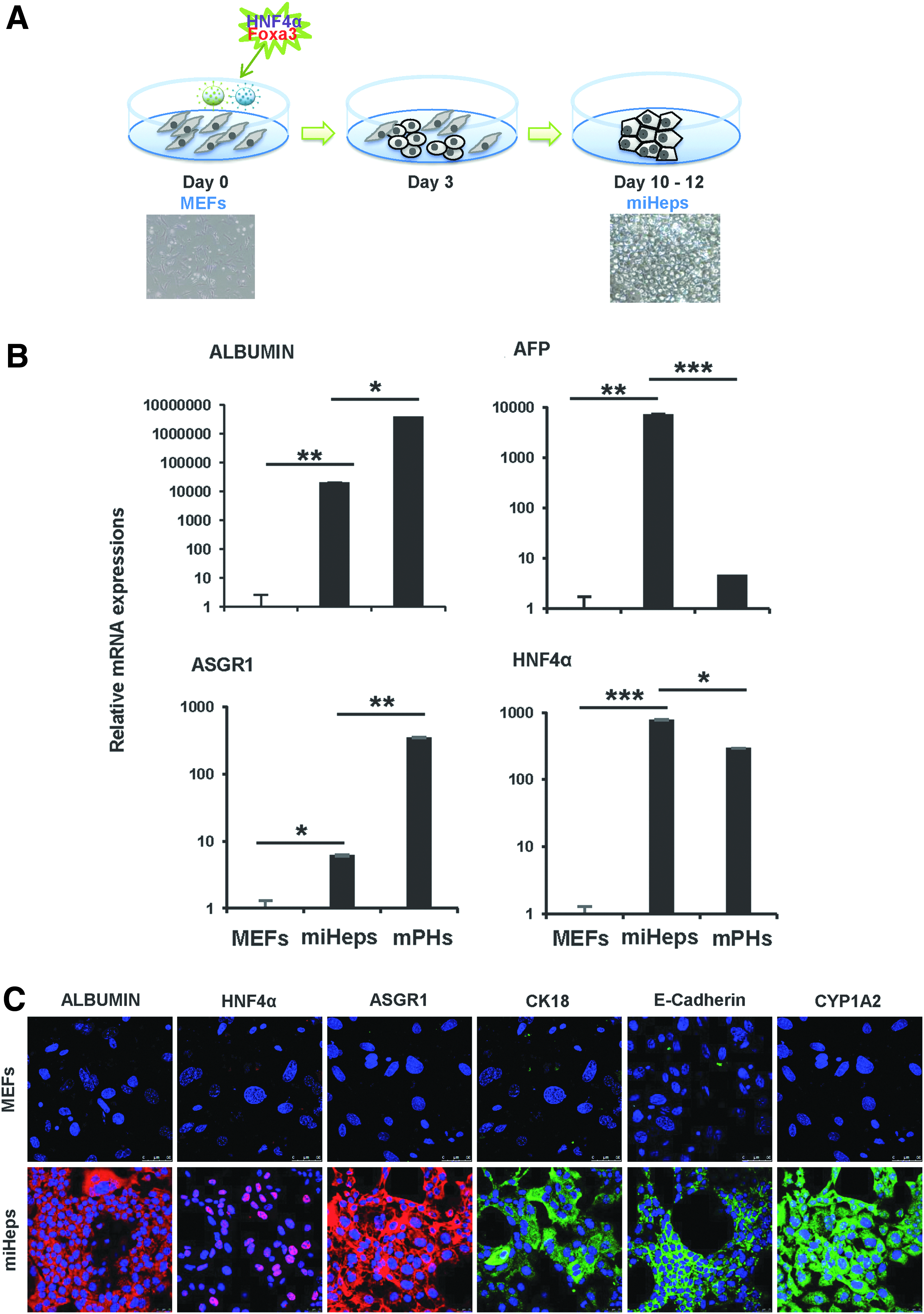
Generation and characterization of miHeps.
3D bioprinting of miHeps with alginate, and improvement of hepatic function
Before 3D printing, we labeled miHeps with mCherry lentivirus to trace miHeps proliferation and migration in 3D scaffold (Fig. 2A). To produce 3D hepatic scaffold, mCherry-miHeps and alginate mixtures were ejected from a 3D printer (Fig. 2B). Under fluorescence microscopy, we confirmed mCherry-miHeps distribution on day 1 (Fig. 2B). They were cultured in vitro with miHeps medium for 28 days to form miHeps colonies. To evaluate gene expression in the scaffold, we performed qRT-PCR at several time points. Expression of Albumin gradually increased, for up to 28 days, but ASGR1 and HNF4a increased only on day 28 and diminished on day 14 after printing (Fig. 2C). AFP, which is not expressed in normal hepatocytes, gradually decreased. Overall, these hepatic function data suggest that directly converted miHeps mature during incubation upon 3D-printed scaffold.

Generation of 3D hepatic structures using a 3D bioprinter.
Proliferation of functional miHeps in 3D-printed scaffold
We investigated whether 3D-printed hepatic scaffold facilitates proliferation of miHeps. As we expected, the mass of mCherry expressing miHeps increased gradually day by day (Fig. 3A–E), and proliferation of the cells was confirmed by fluorescence microscopy (Fig. 3F–J). miHeps appeared to be highly compact by days 14 and 28 after printing, as revealed by hematoxylin and eosin (H&E) staining (Fig. 3K–O). Surprisingly, the cells strongly expressed Albumin and CK18, demonstrating that rapidly proliferating miHep cells retained typical hepatic characteristics in the scaffold (Fig. 3P–Y). In addition, no cell death was apparent in these structures (Supplementary Fig. S2). These data demonstrate that the 3D-printed framework facilitates miHep cell proliferation without loss of hepatic function.

Proliferation of functional miHeps in 3D hepatic scaffolds in vitro.
Transplantation of a 3D-printed scaffold with miHeps into mice
To evaluate miHeps proliferation and hepatic function in vivo, 3D hepatic scaffold was transplanted into omentum of Jo2-treated NOD Scid gamma (NSG) mice. 3D-printed hepatic scaffold was stabilized for 7 days in vitro and transplanted (Fig. 4A). Twenty-eight days after transplantation, miHep scaffold was found in the omentum, and some pieces were observed to be also surrounding the liver (Fig. 4B). We harvested the scaffold to check the status of the miHeps and confirmed hepatic marker gene expression. As shown in Figure 3, in vivo, proliferating miHeps in the scaffold could be detected after 28 days (Fig. 4C). Surprisingly, the miHeps grew more rapidly and expressed higher levels of Albumin in vivo than in vitro (Fig. 4C, D). Also, we performed immunocytochemistry to evaluate whether miHeps differentiated into cholangiocytes in vivo. Interestingly, some miHeps were stained with CK19 antibody, and they were colocalized with mCherry expression around the bile duct on day 28 (Supplementary Fig. S3). These proliferating cells were detected (Supplementary Fig. S4). These results confirm that miHeps proliferate and differentiate faster when transplanted on 3D scaffold than in vitro.
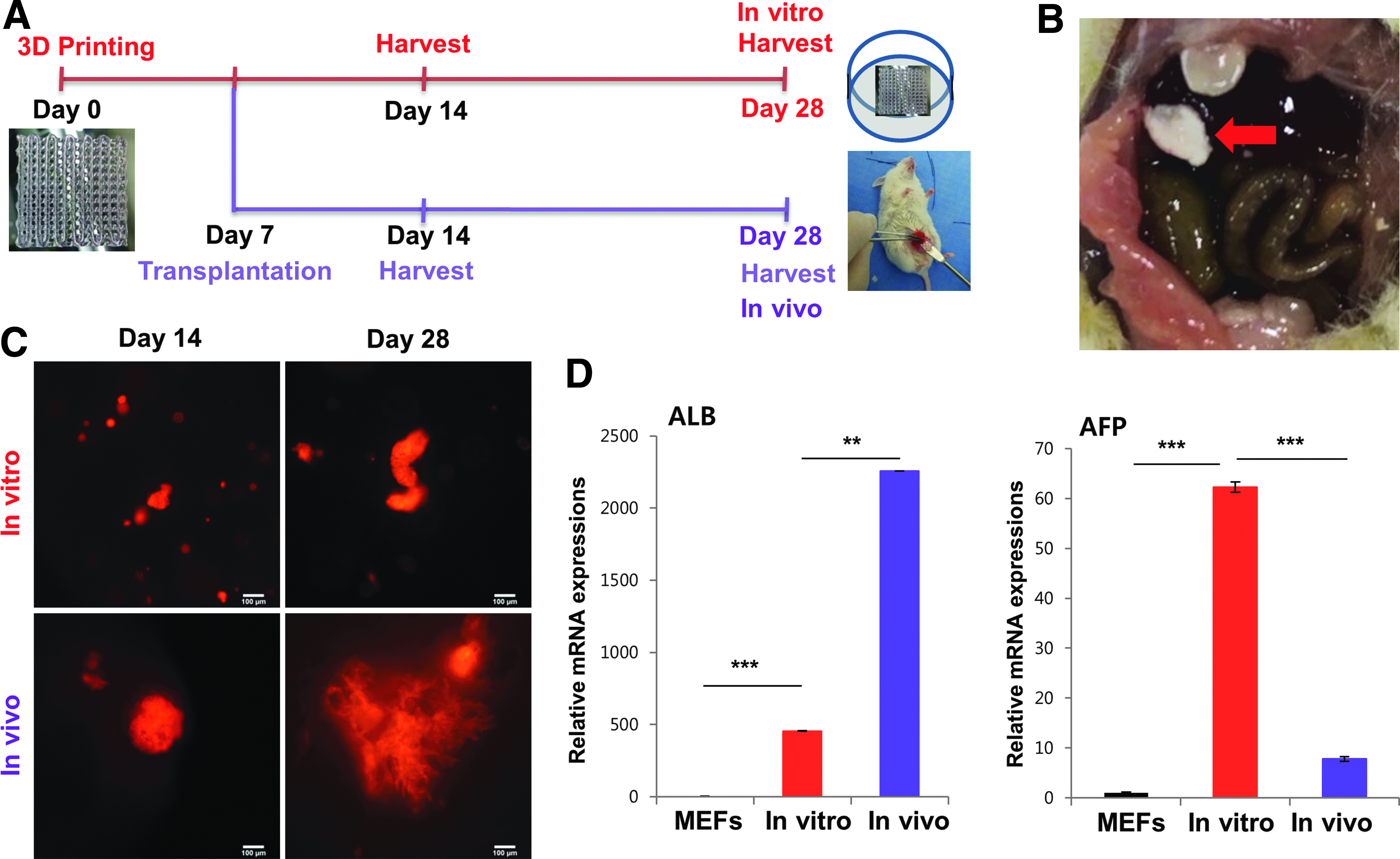
3D hepatic scaffolds transplanted into mouse omenta.
We further examined the hepatic function of 3D hepatic scaffold around the liver. We were able to detect mCherry fluorescence around mouse liver tissue on days 14 and 28 (Fig. 5A–D). Albumin (Fig. 5E, G) and CK18 (Fig. 5F, H) were also expressed, and expression of both proteins colocalized with mCherry expression (Fig. 5 M–P, enlarged panel in Q–T). These observations indicate that bioprinted 3D hepatic scaffold can engraft around liver tissue, and may offer an alternative to liver transplantation.
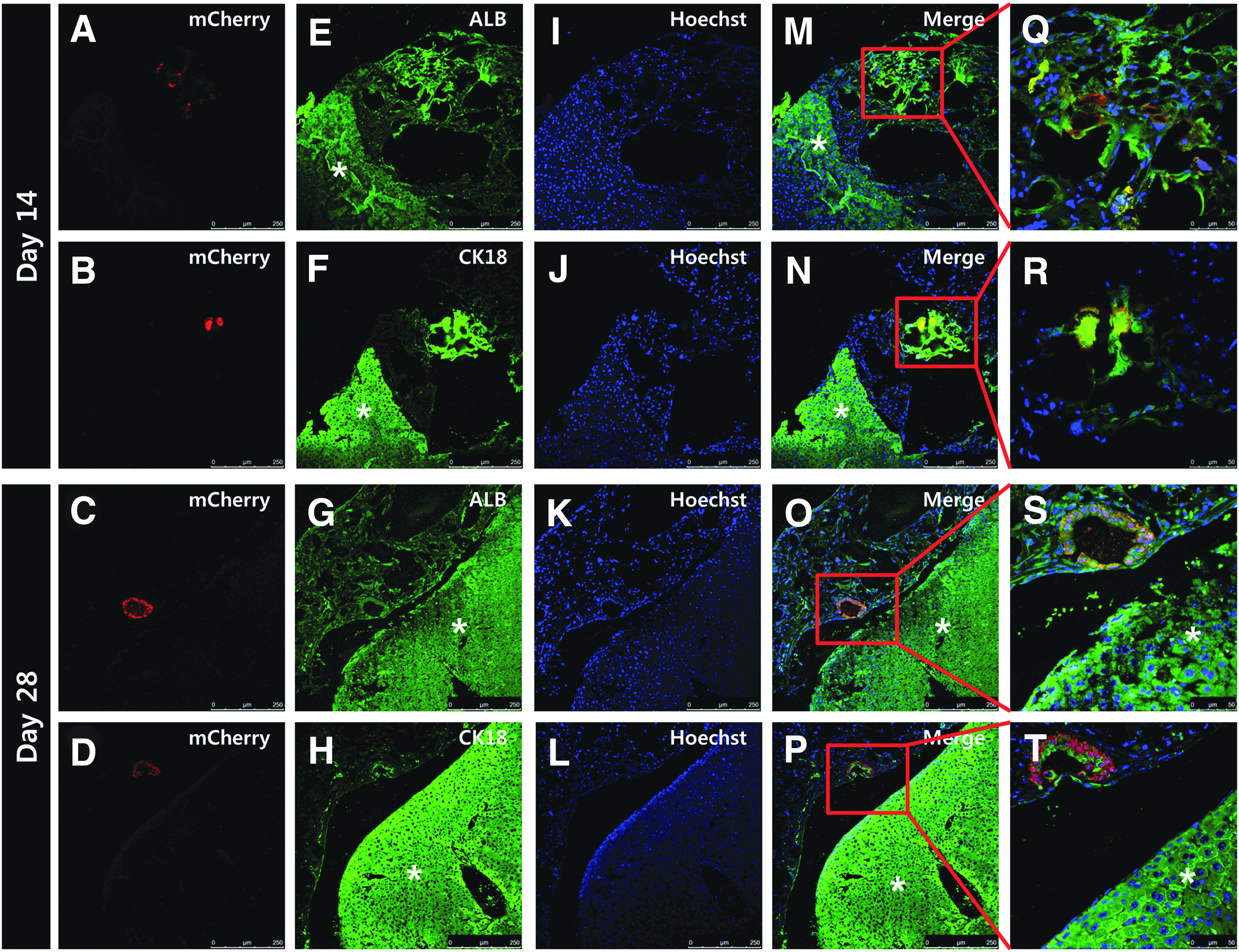
Histological analysis of 3D hepatic scaffolds with mouse liver.
Discussion
Liver transplantation is a vital procedure for patients with end-stage chronic liver disease.24,25 However, the number of potential liver transplantation recipients are increasing faster than the number of potential donors. 21 To overcome this donor shortage, many researchers have attempted cell therapies using stem cells, 26 and are now focusing on generating 3D scaffold made up of stem cells within a matrix. 27 In this study, we used a direct conversion technique to generate hepatocyte-like cells as an alternative cell source for cell therapy, together with 3D printing to form 3D scaffold of liver organoids. Although hepatocyte-like cells can be derived from human pluripotent stem cells,3–6 there are disadvantages to this process. The major issues are that their differentiation into functional mature hepatocytes is challenging to achieve, and sometimes these cells can form tumors in vivo. 27 In contrast, directly converted hepatocyte-like cells do not need to follow liver development mechanism during differentiation, and they do not form teratomas after transplantation. 27 Therefore, we transduced HNF4α and Foxa3 into MEFs, and by 3 or 4 days after infection, the fibroblasts had transformed into polygonal epithelial cells referred to as miHeps. 28 This process activates the mesenchymal–epithelial transition and hepatocyte markers. 29 Lim et al. have reported that treatment with A83-01, which is a transforming growth factor beta receptor inhibitor, boosts miHeps generation with just one gene. miHeps of generation was first reported with small molecules. 29 For clinical trials of directly converted hepatocyte-like cells to be approved, they must be generated without exogenous genes or induced pluripotent stem cells. 30 Therefore, we will attempt miHeps generation without exogenous genes such as mRNA or other small molecules in the future.
The geometry of the extracellular matrix that was formed using collagen sandwiches can maintain hepatocytes long term and to expand their function. 18 Therefore, we hypothesized that miHeps would also maintain their hepatic function and proliferation on 3D scaffold or 3D structure. Although collagen is theoretically the optimal extracellular matrix material, we used alginate hydrogels as bioink. This is because collagen requires certain temperatures and pH levels to harden, 31 and 3D bioprinters are incapable of handling collagen as an extracellular matrix material. Therefore, further development of bioinks and techniques for seeding cells under the desired conditions is needed.
In summary, we have shown that miHeps can be used as a liver cell source in 3D hepatic scaffold and that they increase in number and function when seeded upon scaffold and incubated. Thus, the transplantation of 3D-printed miHeps suggest that 3D bioprinting techniques will improve cell therapy techniques and survival rates. Although challenges related to 3D bioprinters, bioink, and cell sources remain, bioprinting is poised to become one of the most promising methods of creating bioartificial organs.
Footnotes
Acknowledgment
This work was carried out with the support of the “Cooperative Research Program for Agriculture Science and Technology Development (Project No. PJ01100202),” Rural Development Administration, Republic of Korea.
Disclosure Statement
No competing financial interests exist.
References
Supplementary Material
Please find the following supplemental material available below.
For Open Access articles published under a Creative Commons License, all supplemental material carries the same license as the article it is associated with.
For non-Open Access articles published, all supplemental material carries a non-exclusive license, and permission requests for re-use of supplemental material or any part of supplemental material shall be sent directly to the copyright owner as specified in the copyright notice associated with the article.
