
Abstract
Fat tissue, due to its high concentration of stem cells, has a role in aesthetic medicine and reconstructive surgery. However, poor survival of the transplanted cells still limits the usefulness of this material in regenerative medicine. Several studies indicated that platelet-rich plasma (PRP) may improve adipose tissue viability due to its growth factor content. This study aimed at investigating the effects of PRP and hyperacute serum (HAS) on the adipogenic lineage in vitro. PRP was prepared by using two centrifugation steps in the presence of anticoagulants, and HAS was isolated from activated platelet-rich fibrin within 10 min of blood drawing to prevent the propagation of inflammatory cascades. Metabolic activity and proliferation rate of human bone marrow-derived mesenchymal stem cells (hMSCs) cultivated in media supplemented with three types of serum additives (fetal calf serum [FCS], human PRP, or HAS) was determined by using a tetrazolium assay. Adipogenesis was evaluated in standard and pro-adipogenic media and tested by oil red staining, triglyceride content, and expression of specific genes. Adipogenic regulators in the sera were measured by multiplex ELISA assays. We observed that proliferation of hMSCs was supported by both FCS and HAS in a time-dependent manner, but surprisingly, PRP had a much weaker effect (change in proliferation rate after 5 days relative to metabolic activity on day 0—FCS: 5.4-fold change, HAS: 5.8-fold change, serum free 1.9-fold change, PRP: 3.0-fold change, p < 0.05). Lipogenesis was only observed in groups with adipogenic differentiation medium, with HAS showing a significantly stronger effect than PRP. This was confirmed by intensive accumulation of lysochrome dye in lipid droplets, higher triglyceride concentration, and elevated expression of specific adipogenic genes. Measurement of lipogenic proteins in the sera revealed that both PRP and HAS are abundant in them; however, PRP also contains anti-adipogenic factors, which explains its weaker and less reliable effect. The results of this study suggest that HAS provides more robust support than PRP in hMSCs proliferation as well as lipogenic differentiation, indicating that it may be a better adjuvant in fat grafting procedures.
Introduction
A
The use of blood-derived products recently became an attractive treatment option in tissue engineering and regenerative medicine. These blood derivatives are obtained from whole blood samples by centrifugation.11,12 The aim of this process is to separate the blood components to concentrate the elements that are used for therapeutic applications (e.g., fibrin, platelets, growth factors, and leukocytes) while discarding the nontherapeutic elements (e.g., erythrocytes). 13 In the literature, many different types of blood derivatives are described. The most well-known products are PRP, which can be produced as pure-platelet-rich plasma (P-PRP) or leukocyte- and platelet-rich plasma (L-PRP), platelet-poor plasma (PPP), as well as products based on fibrin clot formation such as platelet-rich fibrin.13–15 The variety of blood products demonstrates the highly tunable nature of this technique to target different biological processes. 13 PRP is likely the most widely used blood derivative 16 ; however, it contains pro-inflammatory agents such as fibrin17,18 and leukocytes, 19 which are not desired in some indications. Hyperacute serum (HAS) is a novel cell- and fibrin-free blood derivative that is separated for 10 min immediately after blood collection, so it contains only the cytokines released during the hyperacute phase of blood clotting.
This study aimed at investigating the effects of two different blood products, PRP and HAS on stem cell proliferation and adipogenic differentiation in vitro, and at evaluating the best supplementation for cells, which can be further used for fat grafting procedures.
Materials and Methods
Human mesenchymal stem cell culture
Normal human bone marrow-derived mesenchymal stem cells (hMSCs) were purchased from ATCC (Manassas). Cells were cultured in standard growing medium: DMEM (Gibco, DMEM high glucose, GlutaMAX™ Supplement, pyruvate; Invitrogen, LifeTech Austria, Vienna, Austria) with antibiotics (2% penicillin/streptomycin and 1% amphotericin; Sigma-Aldrich Chemie GmbH, Steinheim, Germany), 10% fetal calf serum (FCS; PAA Laboratories GmbH, Linz, Austria), and basic fibroblast growth factor (bFGF)-1 ng/mL (Sigma-Aldrich, St. Louis, MO). Cells were grown until confluence was reached. The medium was changed twice a week, and cells were expanded up to six passages by plating 5000 cells/cm2.
Preparation of blood derivatives
Forty-five milliliters of peripheral blood was collected from six volunteer donors, and informed consent was obtained from every donor before blood donation. From the whole blood samples, four different blood derivatives were prepared: HAS, PRP, plasma, and serum.
Hyperacute serum
Immediately after collection, the obtained whole blood inside the VACUETTE® 9 mL Z Serum C/A tubes (REF. no. 455092; Greiner Bio-one) was centrifuged at 3000 rpm (1700 g) for 10 min at room temperature. The fibrin clot from the tube was gently removed, and the red blood cells at the bottom of the fibrin clot were cut away. The remaining liquid serum and the clot part with red blood cells were discarded. Using flat forceps, the serum was squeezed out of the fibrin clot, collected, and stored at −80°C. For further experiments, the HAS samples were pooled.
Platelet-rich plasma
Whole blood in the VACUETTE 9 mL K3EDTA blood collection tubes (REF. no. 455036; Greiner Bio-one) was centrifuged at 1500 rpm (440 g) for 10 min at room temperature. Three layers appeared in the collection tube: the bottom layer containing the red blood cells, the middle layer containing the buffy coat, and the top layer containing the plasma. To obtain the leukocytes-poor PRP, the top plasma layer was carefully aspirated without the middle layer (buffy coat) and transferred into a 15 mL falcon tube that was centrifuged again at 3000 rpm (1700 g) for 10 min. The platelet pellet was resuspended into the remaining PPP supernatant to a volume matching the obtained HAS volume and was stored at −80°C. For further experiments, the PRP samples were pooled.
Plasma
To obtain plasma, whole blood inside the BD Vacutainer® tubes (K2EDTA tubes, REF. no. 367864; BD Biosciences) was centrifuged at 3000 rpm (1700 g) for 5 min at room temperature. All the plasma samples were stored at −80°C and pooled together for further experiments.
Serum
To obtain serum, after collection of the whole blood inside the BD Vacutainer tubes (clot activator tube, REF. no. 368815; BD Biosciences), the blood was allowed to clot by leaving it undisturbed at room temperature for 30 min and afterward it was centrifuged at 3000 rpm (1700 g) for 5 min. All the serum samples were stored at −80°C and pooled together for further experiments.
Proliferation assay
After seeding cells in three 96-well plates (2000 cells/well), they were grown for 48 h in 100 μL standard growing medium. Forty-eight hours after seeding (day 0), standard growing medium was replaced with 100 μL of the following media: HAS group: DMEM, high glucose, GlutaMAX Supplement, pyruvate, 10% HAS, 2% penicillin/streptomycin, 1% amphotericin; PRP group: DMEM, high glucose, GlutaMAX Supplement, pyruvate, 10% PRP, 2% penicillin/streptomycin, 1% amphotericin, 2 U/mL heparin. Standard growing medium was used as a positive control, and the serum-free group was used as a negative control: DMEM, high glucose, GlutaMAX Supplement, pyruvate, 2% penicillin/streptomycin, 1% amphotericin. The metabolic activity and proliferation rate of the human mesenchymal stem cells (hMSCs) were investigated by XTT assay (Roche Diagnostics GmbH, Mainheim, Germany) according to the manufacturer's instructions. Relative fluorescence was measured by using a plate reader (BioTek's Synergy 2). The measurement was completed on days 0, 2, and 5.
Adipogenic differentiation
Adipogenic differentiation was induced by using StemXVivo® Adipogenic Supplement, purchased from Bio-Techne Ltd. (Abingdon, United Kingdom) according to the manufacturer's instructions and was performed in 24-well cell culture plates. Cells were incubated in standard growing medium (DMEM with 10% FCS+1 ng/mL bFGF) for 4–7 days until they reached 100% confluence. Three groups were cultured with adipogenic induction media containing DMEM with StemXVivo Adipogenic Supplement and different serum supplements: 10% HAS, 10% PRP+2 U/mL heparin, or 10% FCS. Three control groups were supplemented with DMEM with different serum supplements: 10% HAS, 10% PRP+heparin (2 U/mL), or 10% FCS. To analyze the effectiveness of the differentiation process, cultures were rinsed with PBS, fixed with 10% buffered formalin, and stained with oil red O (Sigma-Aldrich). The experiment was repeated in triplicate.
Triglyceride assay
The triglyceride content in the samples after adipogenic differentiation was measured by using the Adipogenesis Assay Kit (Sigma-Aldrich) according to the manufacturer's instructions. The experiment was repeated in duplicate.
RNA extraction quantitative real-time polymerase chain reaction
After adipogenic stimulation of the hMSCs, total RNA isolation was performed by using the High Pure RNA Isolation Kit (Roche Diagnostics GmbH) according to the manufacturer's instructions, including DNAse I (Roche Diagnostics GmbH) digestion. The isolated RNA was reverse transcribed with ReadyScript™ Reverse Transcription Kit (Sigma-Aldrich) primed by an oligo (dT) primer according to the manufacturer's instructions. TaqMan Gene Expression Assays were purchased from ThermoFisher Scientific, Inc. and real-time polymerase chain reaction (PCR) was performed with an Applied Biosystems 7500 Real-Time PCR System. Details of the TaqMan assays are listed in Table 1. Two different reference genes were utilized alongside the target genes to ensure identical amplification efficiencies. The ΔΔCT method was used for quantification. The CT values were normalized to GAPDH expression. Results were shown as the mean ± standard deviation. Experiments were performed in triplicate.
DFAT, dedifferentiated fat.
Growth factor and cytokine analysis
For the determination of growth factors and cytokines in the HAS, PRP, serum, and plasma, the Proteome Profiler Human Angiogenesis and Human Cytokine Array Kits (no. ARY 007, ARY022; R&D System) were applied according to the manufacturer's instructions. Quantification of protein levels was performed by measuring the spot intensity on the blots by Adobe Photoshop software. The measurements were normalized to controlled reference spots.
Statistical analysis
Statistical analysis was done with GraphPad Prism five software. Experiments with growth factor and cytokines as well as PCR results were analyzed by using one-way analysis of variance (ANOVA) test with Tukey-Kramer Multiple-Comparison post-test. Proliferation data were analyzed with two-way ANOVA and Bonferroni post-test. Results from triglyceride content after adipogenic stimulation were analyzed by the nonparametric Friedman test and Dunn post-test. p < 0.05 was considered significant.
Results
HAS and PRP have different concentrations of blood cells
After collection of HAS and PRP from individual donors, samples were pooled and the concentrations of platelets, white blood cells, and red blood cells were determined (Table 2). PRP samples had a mean platelet concentration of 877 × 103/μL, which corresponds to an approximately fourfold greater platelet concentration compared with whole blood (calculated according to the mean number of platelets in whole blood of six donors) and in HAS samples platelets were not detected. The white blood cell concentration was 0.18 × 10³/μL in PRP and 0.22 × 10³/μL in HAS, which is low in comparison to a healthy person's whole blood WBC number (4500–11,000 × 103/μL). Trace amounts of red blood cells were detected in HAS, and a low level was detected in PRP (0.08 × 10³/μL).
HAS, hyperacute serum; PLT, platelets; PRP, platelet-rich plasma; RBC, red blood cell; WBC, white blood cell.
HAS promotes proliferation of hMSCs
The most significant growth-promoting effects on hMSCs were seen in the HAS group after 5 days of in vitro cell culture. The proliferation rate in the HAS group was significantly higher than in the group supplemented with serum-free media or with 10% PRP (Fig. 1). The proliferation of PRP-treated cells was significantly higher on day 5 in comparison to the serum-free group. Microscopic examination revealed differences in cell morphology within the treatment groups (Fig. 2) as cells supplemented with PRP revealed a more elongated morphology. The previous observation about the proliferative effect of HAS was supported by the qualitative observation that the cells in the HAS and FCS groups reached ∼80% confluence compared with the PRP group, which reached ∼50% confluence (Fig. 2).
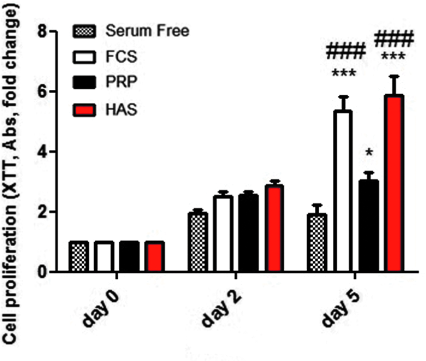
Representation of the mean results from three different experiments with XTT assay, performed in 96-well cell culture plate-based monolayer culture with 2000 hMSC cells per well (10 wells per group), cultured with addition of 10% HAS, 10% PRP, or 10% FCS. Measurements were done: 0, 2, and 5 days after seeding the cells. Proliferation rate is presented as a fold change relative to metabolic activity on day 0. On day 2, it was observed that in all groups the cell number was at least doubled, but without significant difference within the groups. After 5 days, the rapid increase of cell growth was indicated only in FCS and HAS groups and there was a significant difference versus serum-free group and PRP group. Statistical analysis was performed with two-way ANOVA and Bonferroni post-test. * indicates the proliferation rate significantly higher than the serum-free group. Results are expressed as the mean ± SD. *p < 0.05 and ***p < 0.001. # indicates the proliferation rate significantly higher than the PRP group, ###p < 0.001. ANOVA, analysis of variance; FCS, fetal calf serum; HAS, hyperacute serum; hMSCs, human bone marrow-derived mesenchymal stem cells; PRP, platelet-rich plasma; SD, standard deviation.
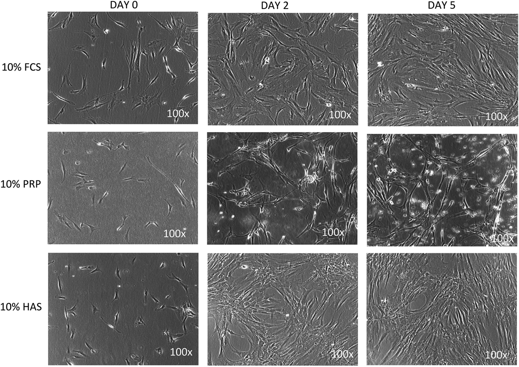
Pictures from microscopic examination of hMSC cell culture after 0, 2, and 5 days with different media supplementation. The morphology of the cells in the PRP group was changed on day 5, whereas in the HAS and FCS groups it remained stable. The pictures also indicate the difference in cell number between the groups. Cells in FCS and HAS groups after 5 days reached ∼80% confluence and in the same time in the PRP group only ≈50% (100× magnification).
Blood derivatives can influence adipogenic differentiation—positive effect of HAS
To determine the influence of different blood-derived products on adipogenic differentiation, cells were cultured with basal medium and adipogenic medium and the following groups were created: 10% FCS, 10% HAS, or 10% PRP. The presence of intracellular lipid droplets was not observed in control groups with basal medium but could be detected in all groups with adipogenic stimulation, but with different intensity (Fig. 3). The greatest intracellular lipid droplet formation was observed in groups with differentiating medium supplemented with HAS or FCS. Based on the microscopic pictures, PRP treatment led to less lipid droplet formation. To quantify the value of the lipid content in the respective groups, a triglyceride assay was performed (Fig. 4). The highest level of triglycerides was detected in the group where cells were treated with HAS and differentiation factors (DF). The mean concentration was two times higher than in the FCS+DF group and three times higher than in the PRP+DF group.

Representative pictures from microscopic examination of hMSC cells after adipogenic differentiation with control groups. Red color indicates the presence of oil red o lysochrome dye. In all groups with differentiating medium, lipid presence was detected, but with different intensity—most abundant in the HAS group and at the lowest estimate in the PRP group. In control groups with basal medium (without DF), lipid droplets were not observed (100× magnification). DF, differentiation factors.

Triglyceride content in different group treatment. The FCS+DF, HAS+DF, and PRP+DF groups were treated with 10% different blood-derived supplementation with adipogenic differentiating medium and FCS, HAS, PRP groups with basal media +10% different blood-derived supplementation. Generally, without the presence of adipogenic factors, the triglyceride content was low and reached a similar level in FCS, PRP, and HAS groups. Most abundant triglyceride concentration was expressed in the HAS+DF group with significant difference in relation to the HAS group as well as to FCS+DF and PRP+DF groups. Interestingly, there was no significant difference between PRP and PRP+DF groups, which can suggest that addition of PRP to the adipogenic media had a negative effect on adipogenesis. Statistical analysis was performed with nonparametric Friedman test and Dunn post-test. * indicates that the triglyceride content is significantly higher between the different treatment groups. Results are expressed as the mean ± SD. * represents p < 0.05, ** represents p < 0.01. # indicates the triglyceride content significantly higher within one treatment group. # represents p < 0.05.
HAS and PRP induce different gene expression
To evaluate whether different blood derivatives influence adipogenic differentiation, the expression of adipocyte-specific genes and other potential lineage-specific changes were screened by quantitative real-time PCR. Groups with adipogenic differentiation media were tested as well as control groups (basal media) (Fig. 5). Peroxisome proliferator-activated receptor gamma (PPARG), fatty acid binding protein 4 (encoded by FABP4 gene), and adiponectin (encoded by the ADIPOQ gene) proteins are typical markers to assess the progress of adipogenic differentiation.20–22 The observations from oil red O staining and triglyceride content measurements were supported by the PCR results. Adipogenic markers were abundant in groups treated with DF supplemented with FCS and HAS, but not with PRP. In all cases, the levels of adipogenic markers were significantly higher in FCS+DF and HAS+DF groups than in the PRP+DF group and also in relation to their respective control groups without differentiating medium. To answer the question whether blood-derived products such as HAS or PRP can prevent cell death, the expression of the apoptotic markers Casp7, Bax, and Bcl-2 was assessed. Interestingly, all markers were upregulated in the groups treated with adipogenic media and supplemented with FCS (FCS+DF). The presence of apoptotic markers in the FCS group was similar to the HAS+DF, HAS, PRP+DF, and PRP groups. The difference in Casp7 and Bax level in FCS+DF was significant in relation to HAS+DF and PRP+DF groups. Surprisingly, dedifferentiated fat (DFAT) markers22–24 were upregulated in the FCS+DF group with significant difference versus HAS+DF and PRP+DF. Col1A1 expression was similar in PRP+DF and FCS+DF groups and it was significantly higher than in the HAS+DF group.
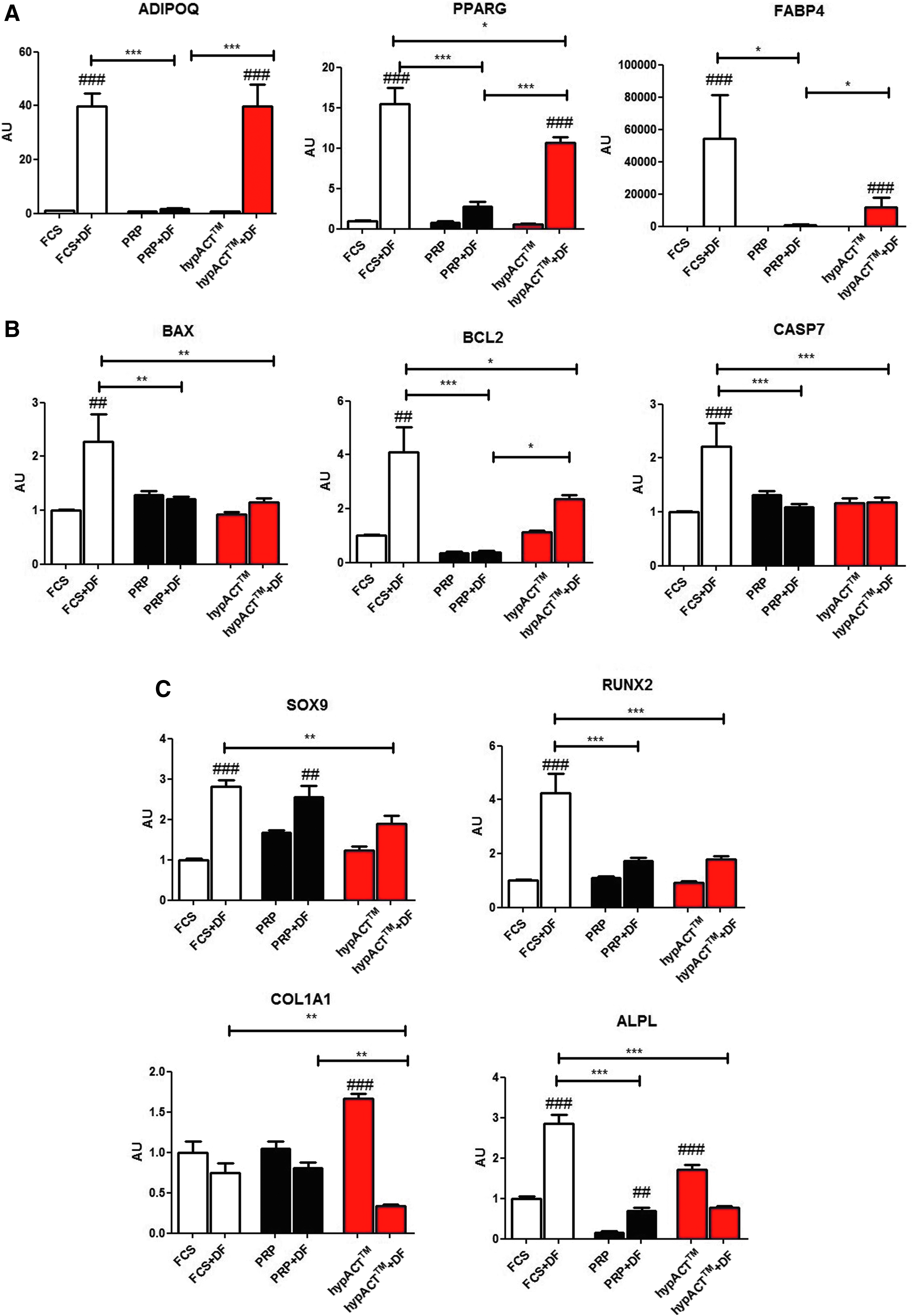
mRNA expression of various genes in different group treatment. In panel
Growth factors and cytokine concentration are different within blood derivatives groups
Blood derivatives were screened for growth factors and cytokines that are known to influence adipogenic differentiation. Serum, plasma, PRP, and HAS were tested, and results were presented as relative values to the positive control for the secondary antibody (Fig. 6). Selected adipokines adiponectin, leptin, and lipocalin20,25–29 were abundant in all blood derivatives, and there was no significant difference in their levels between HAS and PRP groups. Adiponectin and lipocalin were significantly higher in both PRP and HAS in comparison to plasma and serum. Interestingly, after analysis of potential adipogenic supportive growth factors,30–34 no significant differences were observed in the content of IGF-1 and VEGF. The presence of bFGF1 was significantly greater in HAS, but the detection level was very low. Interestingly, among potential adipogenic inhibitors,21,26,35–38 transforming growth factor (TGF)-β was the most abundant in the PRP group and significantly higher than in all other groups.
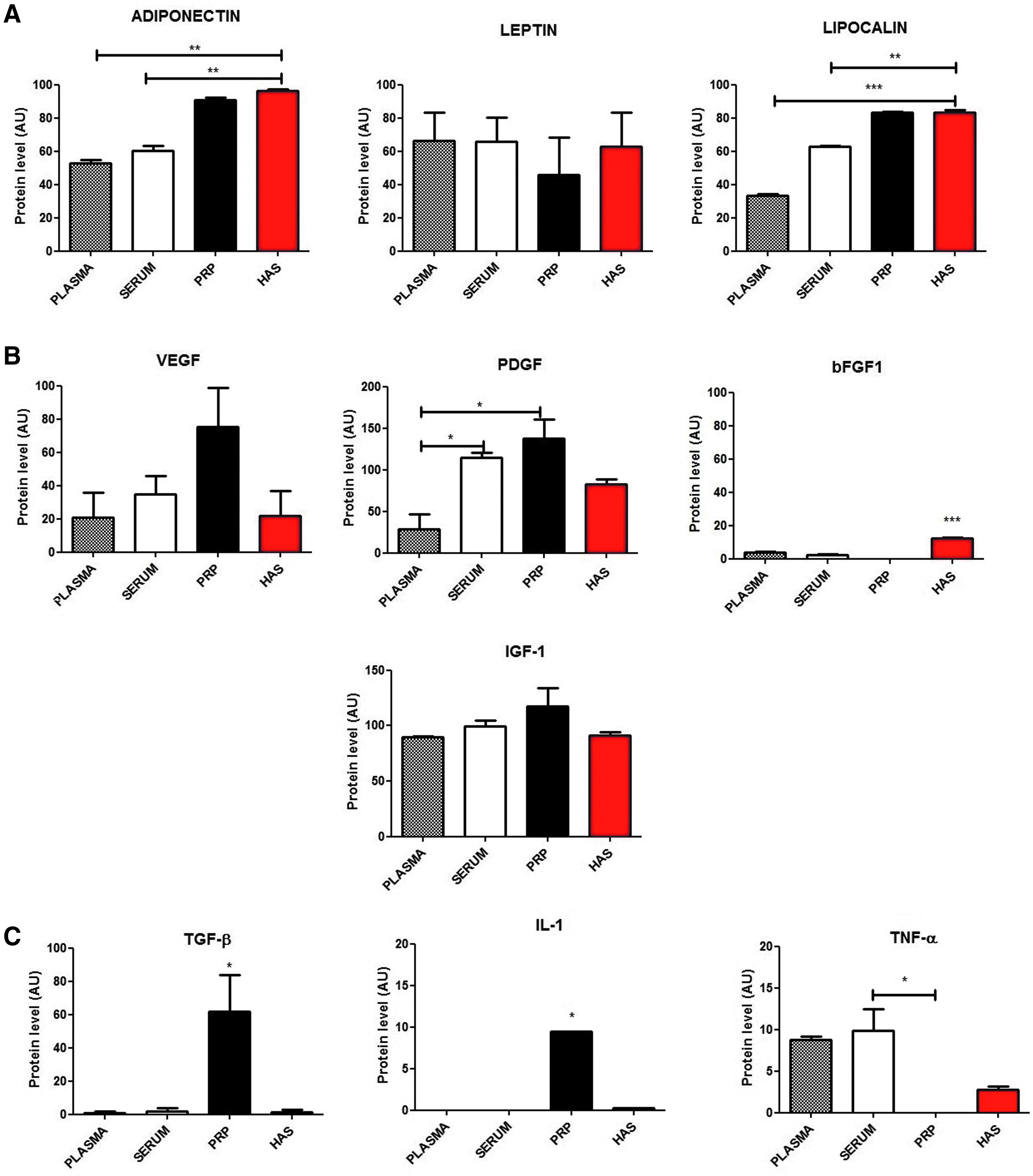
Composition analysis of blood derivatives. Plasma, serum, PRP, and HAS were tested for the relative presence of growth factors and cytokines.
Discussion
Nowadays, fat grafting is not only applied in aesthetic medicine but also has a significant role in reconstructive surgery and regenerative medicine. This treatment can act as an effective solution for patients with irradiated or burned tissues, impaired healing and fibrosis, postcancer reconstruction surgeries, and many other indications. Autologous fat is considered an ideal soft tissue filler, due to its biocompatibility, sufficient availability, and high likelihood of natural integration into the host tissues.39–41 Previous studies have demonstrated that enrichment of fat grafts with ASC improves its quality and viability by enhancing adipogenesis, supporting angiogenesis, and reducing apoptosis.39–44 Therefore, promoting stem cell viability, proliferation, and adipogenic signaling may, in turn, foster the viability of the fat graft. In this study, two different blood derivatives were evaluated for their effects on hMSCs in regard to potential applications in fat grafting procedures. Although bone marrow-derived hMSCs were utilized herein, previous reports have observed similar biological function and clinical performance between these two cell sources.45–47
PRP, a widely used blood product with an extensive documentation about the effects of its action, was studied in comparison to a novel cell- and fibrin-free product—HAS. PRP supplementation has been commonly observed to promote cell proliferation9,10,48–52 with few exceptions53,54; however, there are evident discrepancies about the effects of PRP on adipogenic differentiation. D'Esposito et al., 9 Cervelli et al., 50 and Kocaoemer et al. 51 observed supportive effects of PRP on ASCs proliferation and adipogenic differentiation, whereas Liao et al. 10 and Chignon-Sicard et al. 52 observed that PRP enhances ASC proliferation, but inhibits adipogenic differentiation. Interestingly, Koellensperger et al. 54 concluded that the different blood derivatives (regular human serum, PPP, and PRP) did not significantly affect ASCs doubling time, whereas significant decreases in adipogenic differentiation were observed when the cells were supplemented with PRP. The current study findings support the anti-adipogenic effect of PRP as well as a significant downregulation in metabolic activity of PRP-treated hMSCs in comparison to HAS or FCS supplementation.
The proliferative effect of HAS was compared with FCS as well as with serum-free DMEM and PRP with a fourfold increased platelet concentration. Surprisingly, PRP induced significantly less proliferation in comparison to FCS and HAS. The metabolic activity of hMSCs treated with HAS was nearly twice as high as those treated with PRP, which suggested that HAS was an effective supplement for hMSC proliferation. The effects of PRP can be highly associated with platelet concentration53,55 and since there are no strict concentration requirements for PRP, 13 this may explain the disparate findings observed in the literature. Some reports also described the role of different anticoagulants in PRP properties56,57; however, Fitzpatrick et al. observed large variations regarding the composition and concentration of different factors in PRP samples prepared with four various commercially available kits, 58 even that in all these systems citrate dextrose solution was used as an anticoagulant. However, there is no guarantee that the use of anticoagulants such as heparin, EDTA, or citrate is not affecting biological functions of PRP.
In regard to the anti-adipogenic effects observed herein with PRP, both TGF-β and IL-1 have been reported to inhibit adipogenesis.22,27,35,36,52,59–61 Therefore, considering the presence of TGF-β and IL-1 in PRP, and the absence in HAS, these cytokines may be critical contributors to the differential effects observed between HAS and PRP since significant differences in pro-adipogenic factors were observed only in bFGF level. Enhancing adipogenesis in fat grafts through ASCs enrichment has been found to improve the quality and viability of fat grafts.1,42–44 Based on adipogenic genes expression and triglyceride content, it was assessed that both HAS and FCS stimulate adipogenesis. Therefore, supporting stem cell proliferation and boosting adipogenic differentiation implicates HAS as a more beneficial supplement for fat graft quality and viability than PRP. There are literature reports about mature adipocytes cultured in vitro, which retained the ability to dedifferentiate into fibroblast-like cells, which express Runx2 and Sox9,28–30 critical factors for osteogenesis and chondrogenesis. These DFAT markers were analyzed and the upregulation was observed only in the group supplemented with FCS and adipogenic medium, which suggests that HAS and PRP do not induce dedifferentiation of committed cells. In addition, neither PRP nor HAS induced spontaneous or uncontrolled adipogenic differentiation, which allows such processes to occur more naturally. Interestingly, after microscopic examination, it was observed that hMSCs in the PRP group developed a different morphology—the cells were thinner and more elongated than those treated with FCS or HAS. Chignon-Sicard et al. showed that PRP induce a myofibroblast-like cell phenotype, triggered again by the TGF-β pathway. 52 Considering the work of Plikus et al. and their discovery about manipulating myofibroblasts to differentiate into adipocytes and improving tissue healing without scarring as a result, 62 the question should be posed as to whether the PRP supplementation can be taken into account for fat grafting procedures. Adipose tissue should be considered as a collection of various cells: mature adipocytes, preadipocytes, fibroblasts, endothelial cells, vascular smooth muscle cells, immune cells, and stem cells 41 and its potential supplementation should not disturb a homeostasis or harm any types of cells within the adipogenic niche.
Conclusion
In conclusion, we suggest that in the light of present findings among the two tested blood derivatives with different preparation methods, HAS may be considered for supporting surgical procedures associated with adipose tissue, including fat filling, engrafting, and transplantation. As a next step, in vivo studies have to be conducted to ensure that the observed effects are not limited to the in vitro biological systems. Another important observation of this work is that HAS is a more reliable and standardized blood product and its biological effects can be more predictable than that of PRP not only because of its different growth factor profile but also because of the lack of any added anticoagulants or activators.
Footnotes
Acknowledgment
This work was supported by grants from the FFG Basisprogramme (project no.: 4965429).
Disclosure Statement
The author Z.L. owns stock in a startup company OrthoSera GmbH that holds patents on hyperacute serum. The authors I.H., O.K., and Z.L. are partly employed by OrthoSera GmbH.