
Abstract
Different regenerative medicine approaches for tendon healing exist. Recently, especially gene therapy gained popularity. However, potential mutagenic and immunologic effects might prevent its translation to clinical research. Chemically modified mRNA (cmRNA) might bypass these limitations of gene therapy. Therefore, the purpose of this study was to evaluate the early healing properties of Achilles tendon defects in rats treated with basic fibroblast growth factor (bFGF) cmRNA. Forty male Lewis rats were used for the study and randomly assigned to two study groups: (1) treatment with cmRNA coding for bFGF and (2) noncoding cmRNA control. Protein expression was measured using in vivo bioluminescence imaging at 24, 48, and 72 h, as well as 14 days. Animals were euthanized 2 weeks following surgery. Biomechanical, histological, and immunohistological analyses were performed with the significance level set at p < 0.05. Protein expression was evident for 3 days. At 14 days, bioluminescence imaging revealed only little protein expression. Biomechanically, tendons treated with bFGF cmRNA showed a construct stiffness closer to the healthy contralateral side when compared with the control group (p = 0.034), without any significant differences in terms of load to failure. Hematoxylin and eosin staining detected no side effects of the treatment, as signs of inflammation, or necrosis. Furthermore, it revealed the shape of the nuclei to be more oval in the bFGF group in the tendon midsubstance (p = 0.043) with a reduced cell count (p = 0.035). Immunohistological staining for type I, II, III, and IV collagen did not differ significantly between the two groups. In conclusion, this pilot study demonstrates the feasibility of a novel messenger RNA (mRNA)-based therapy for Achilles tendon defects using chemically modified mRNA coding for bFGF.
Introduction
The incidence of Achilles tendon injuries increases continuously, especially for nonsports-related tears in middle-aged men.1,2 From an etiological perspective, acute traumatic Achilles tendon ruptures due to a sudden deceleration and/or dorsiflexion of the ankle must be differentiated from a spontaneous disruption on the basis of degenerative changes. 3 However, biopsy studies revealed that traumatically ruptured Achilles tendons have signs of tendon degeneration, comparable with the histopathological features of tendinopathic tendons, which consequently reduces tensile properties and increases the risk for a tear. 4
Clinically, it is well known that poor tissue quality of tendon stumps is associated with a higher retear rate following surgical repair. 5 Therefore, it is recommended to debride necrotic or degenerated tissue when a tendon repair is performed. This may lead to a tendon defect and surgical repair is either challenging due to the gap size or (especially in chronic situations) a salvage procedure is indicated.6,7 However, these salvage procedures might be associated with inferior patient-reported outcomes as well as objective function.6–8 Some authors proposed conservative treatment for Achilles tendon ruptures.9,10 Despite good clinical results, the rerupture rate is notably high. 11 Furthermore, it is well known that tendon healing occurs through a fibrovascular scar that has inferior structural properties when compared with a healthy tendon.12,13
To either bridge the tendinous defect or to improve healing of Achilles tendon ruptures, several regenerative medicine approaches have been studied during the last decades. These approaches included administration of different growth factors, cell concentrates, (cell seeded) biological and artificial scaffolds, or gene therapy.14–21 However, despite promising in vitro results, studies on animals reported conflicting findings.14,19,22,23 Therefore, tissue engineering constructs on the basis of both mechanical and chemical stimuli have been introduced with promising in vitro results in terms of paracrine release of growth factors. 24 However, to date, these studies are limited to in vitro results.
When performing gene therapies, limitations include cancerogenicity, inflammatory and immune responses (especially when using viral vectors), or variable vector stability, which make clinical application difficult or impossible. 20
In contrast to DNA-based strategies, messenger RNA (mRNA) is directly translated into protein, which decreases the risk for mutagenesis. However, mRNA is potentially highly immunogenic and unstable. 25 Recently, a chemically modified mRNA (cmRNA) with increased stability and little immunogenicity has been introduced.25,26 While cmRNA coding for bone morphogenetic protein 7 has shown promising results in terms of tendon healing, 27 no study investigated the effect of this cmRNA coding for basic fibroblast growth factor (bFGF) on early Achilles tendon healing as bFGF has been associated with improved tendon healing in terms of cell proliferation and collagen type III synthesis.14,22,28,29
Therefore, the purpose of this study was to evaluate the early healing properties of Achilles tendon defects in rats treated with bFGF cmRNA. It was hypothesized that bFGF cmRNA injection in the tendon stumps improves early tendon healing biomechanically as well as histologically without causing any inflammatory response.
Methods
In forty adult male Lewis rats (>12 weeks; 300–400 g), a complete 2.4-mm full-thickness Achilles tendon defect was created in the right hind limb with an arthroscopic punch at 2 mm proximal to the calcaneal bone, as previously described. 19 Male rats have been chosen as they are not subjected to hormonal fluctuations compared with female animals, which might interfere with tendon healing. The rats were randomly assigned to one of the two study groups: (1) injection of 55 μL cmRNA (Ethris GmbH, Planegg, Germany) coding for bFGF at an mRNA concentration of 1 mg/mL diluted in 5% Hepes-buffered glucose (HBG) in each of the two tendon stumps and (2) controls (injection of 55 μL cmRNA [Ethris GmbH] coding for Luciferase at an mRNA concentration of 1 mg/mL diluted in 5% HBG in each of the two tendon stumps). To stabilize mRNA, enhance translation efficiency, and reduce immunogenicity, chemically modified nucleotides were incorporated.30,31 Specifically, both control mRNA (coding for Luciferase) and bFGF mRNA were produced by adding 25% 2-thio-UTP and 25% 5-methyl-CTP to the in vitro transcription reaction, as previously reported.27,30
Animals and surgical procedure
The study was conducted according to the national legislative requirements following approval from the local animal research committee and the district government of Upper Bavaria (55.2-1-54-2531-55-09).
Adult (12 weeks) male Lewis rats were obtained from Charles River (Sulzfeld, Germany) with a mean weight at surgery of 379.3 ± 31.2 g. Two to four animals were housed together in a standard open-top Makrolon type-IV cage (Techniplast) with autoclaved sawdust bedding and pulp as the nesting material at room temperature. The acclimatization period was 2 weeks. Animals had access to water and food (Altromin), and lighting was controlled by a 12-h on–off cycle.
Surgical procedures were performed under sterile conditions and general anesthesia. General anesthesia was induced by an intramuscular injection of body weight-adapted medetomidine, midazolam, and fentanyl. After shaving and disinfecting, an approximately 10-mm longitudinal skin incision was performed on the medial side over the Achilles tendon. After exposure of the Achilles tendon through blunt dissection, a 2.4-mm arthroscopic punch (Arthrex, Naples, FL) was inserted to fully surround the tendon and paratenon. Then, 110 μL cmRNA (1 mg/mL) coding for either bFGF or Luciferase was injected in a fan-shaped manner proximal and distal to the arthroscopic punch (55 μL proximally and 55 μL distally). Finally, the tendon and paratenon were cut with the arthroscopic punch and the skin was closed with continuous intracutaneous absorbable 4-0 sutures (poligelcaprone; Johnson & Johnson) without repair of the Achilles tendon defect. An OpSite spray-on dressing (Smith & Nephew, Andover, MA) was added. Postoperatively, full weight bearing was allowed without any immobilization devices. Postoperative analgesia using buprenorphine (0.05 mg/kg subcutaneously) and oral metamizole (70 mg/kg) was carried out for 3 days.
Fourteen days following surgery, animals were euthanized with an overdose of pentobarbital (80 mg/kg) injected intracardially under general anesthesia. Both Achilles tendons were exposed and harvested in each animal. Care was taken that the entire tendon and the adjacent triceps surae muscle and calcaneal bone (tendons assigned to histological evaluation) or the entire foot (tendons assigned to biomechanical testing) were harvested as a whole. While the tendons for biomechanical testing were stored at −20°C, samples for histological analysis were stored in methanol.
In vivo bioluminescence imaging
To evaluate protein expression following mRNA injection, in vivo bioluminescence imaging in the control group was performed using a Lumina XR In Vivo Imaging System (Perkin Elmer) 1, 3, and 4 days following surgery and at 14 days in three animals per time point.
Biomechanical testing
Tendons were thawed at room temperature for 12 h before testing. The musculotendinous junction was fixated using a cryoclamp. Liquid nitrogen was used to rigidly fix the muscle and musculotendinous junction to the cryoclamp using a standardized procedure to prevent the tendinous portion of the construct from having clamping effects. The foot of each specimen was placed in a custom-made mounting grid to keep the bone in place, while the tendinous portion was loaded by a uniaxial material testing device (ZwickiLine Z2.5; Zwick Roell, Ulm, Germany), and preloaded with 2 N, followed by an axial load to failure test at a constant speed of 0.16 mm/s. During biomechanical testing, the tendons were moistened with saline.
Histological and immunohistological analysis
First, samples were decalcified using 5% EDTA solution for 2 weeks. Radiographs were obtained to prove whether decalcification was completed. Then, tendons were kept for 24 h in 30%, 20%, and 10% sucrose and phosphate-buffered saline solution. Twelve-micrometer-thick cryosections were obtained from the operated tendons (Leica 1950 cryostat; Leica Biosystems, Wetzlar, Germany). Before staining, the cryosections were stored at −20°C for 2 weeks. Ten healthy contralateral Achilles tendons were used as controls.
Hematoxylin and eosin staining was performed. A semiquantitative scoring system, 32 ranging from 0 (normal) to 3 (markedly abnormal), was applied to evaluate collagen fiber arrangement and alignment, shape of the nuclei, cellularity and vascularity, and collagen staining in the proximal, middle, and distal thirds of the tendon.
For immunohistological analysis, monoclonal antibodies for type I collagen (dilution 1:2000; Sigma, Munich, Germany), type II collagen (dilution 1:200; Developmental Studies Hybridoma Bank, Iowa City, IA), type III collagen (dilution 1:4000; Sigma), type IV collagen (dilution 1:20; Department of Anatomy, LMU Munich, Munich, Germany), and procollagen I (dilution 1:5; Developmental Studies Hybridoma Bank) were used. The applied scoring system ranged from 0 (no staining) to 2 (intense staining). 33
Statistical analysis
For statistical analysis, SPSS® 24.0 (IBM SPSS Statistics) for Mac was used. Data distribution was evaluated with the Shapiro-Wilk test. Quantitative parameters are expressed as mean ± 1 standard deviation and range. Biomechanical data are given as percentages from the contralateral healthy tendon to normalize for intersubject variability. The Mann–Whitney U test was used to compare structural properties and histological and immunohistological data between the two study groups. The significance level was set at p < 0.05.
Results
Animals and macroscopic findings
No infections, wound dehiscence, or other complications were observed. None of the tendons showed discontinuity at 14 days. The macroscopic data are shown in Table 1.
Macroscopic Findings
bFGF, basic fibroblast growth factor; cmRNA, chemically modified messenger RNA.
In vivo bioluminescence imaging
Luciferase activity was measured 1, 3, 4, and 14 days after mRNA application into the tendon defects. Measured levels showed a peak after 1 day with average values of 4.4 × 10⁴ photons/s/cm2/sr and time-dependent slow decrease of activity levels up to 3 days after application, with remaining activity of 9.2 × 10³ photons/s/cm2/sr. After 14 days, Luciferase activity dropped to baseline values (Figs. 1 and 2).
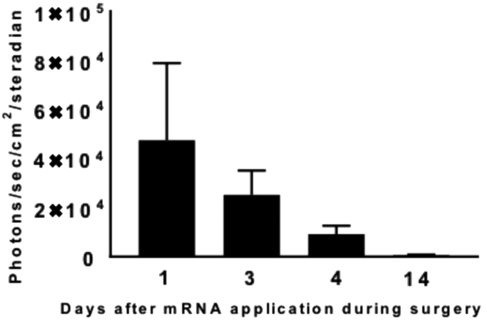
Protein expression in photons/s/sr measured using in vivo bioluminescence imaging. Positive protein expression was observed for 3 days. At day 14, protein expression dropped to baseline values.
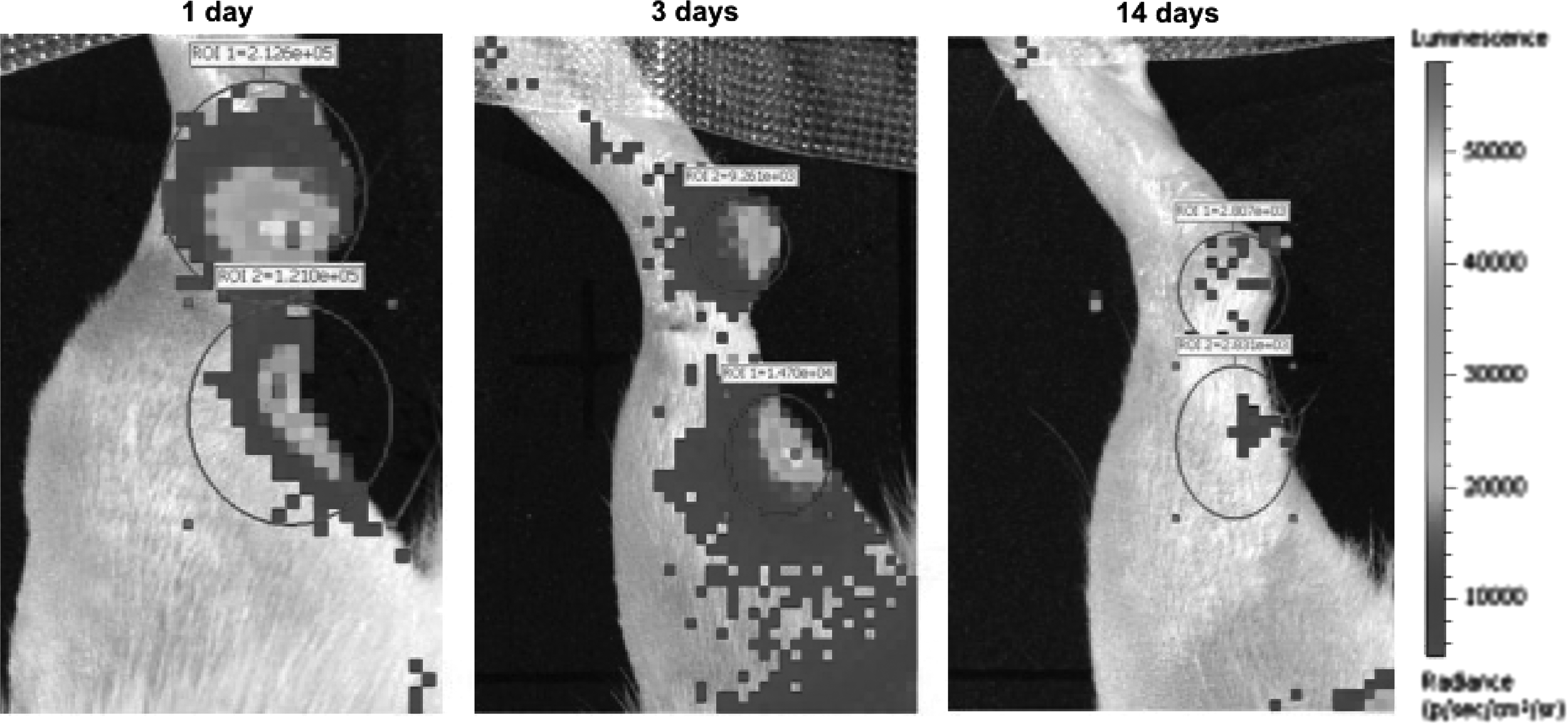
In vivo bioluminescence imaging demonstrated positive protein expression after cmRNA injection for 3 postoperative days. cmRNA, chemically modified messenger RNA.
Biomechanical testing
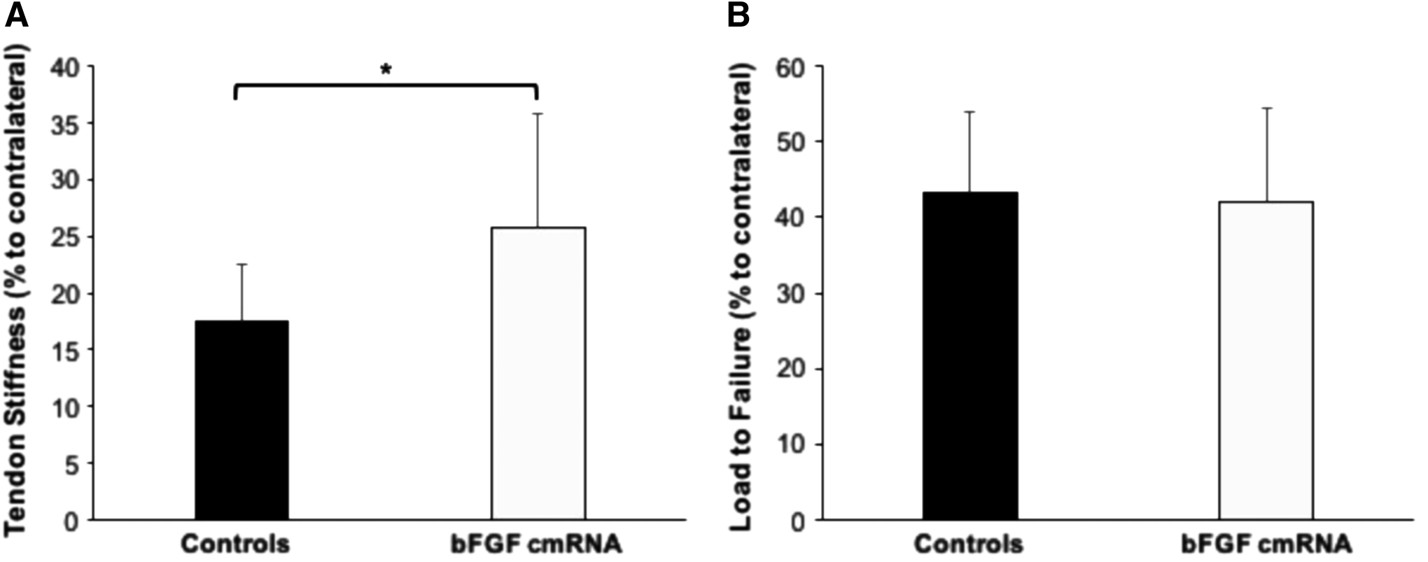
The load to failure was 40% ± 12.3% (range 19.6–61.2) for the contralateral side in the bFGF cmRNA group and 43.2% ± 10.7% (range 30.4–60.6) for controls (p = 0.965; n.s.). Achilles tendon defects treated with cmRNA coding for bFGF were statistically significantly stiffer and closer to values of healthy tendons than untreated controls (25.8% ± 10%, range 10.3–47.5, versus 17.5% ± 5%, range 9.5–25.1; p = 0.034) (Fig. 3).
Histology and immunohistology
Hematoxylin and eosin straining revealed fibrovascular scar tissue in all tendons without evidence of inflammation. The only statistically significant differences between the two groups were for morphology of nuclei in the midsubstance (p = 0.024) and cellularity throughout the entire tendon (p = 0.035). The nuclei in the control group were closer to normal than in the bFGF cmRNA group with a smaller cell count (Fig. 4).
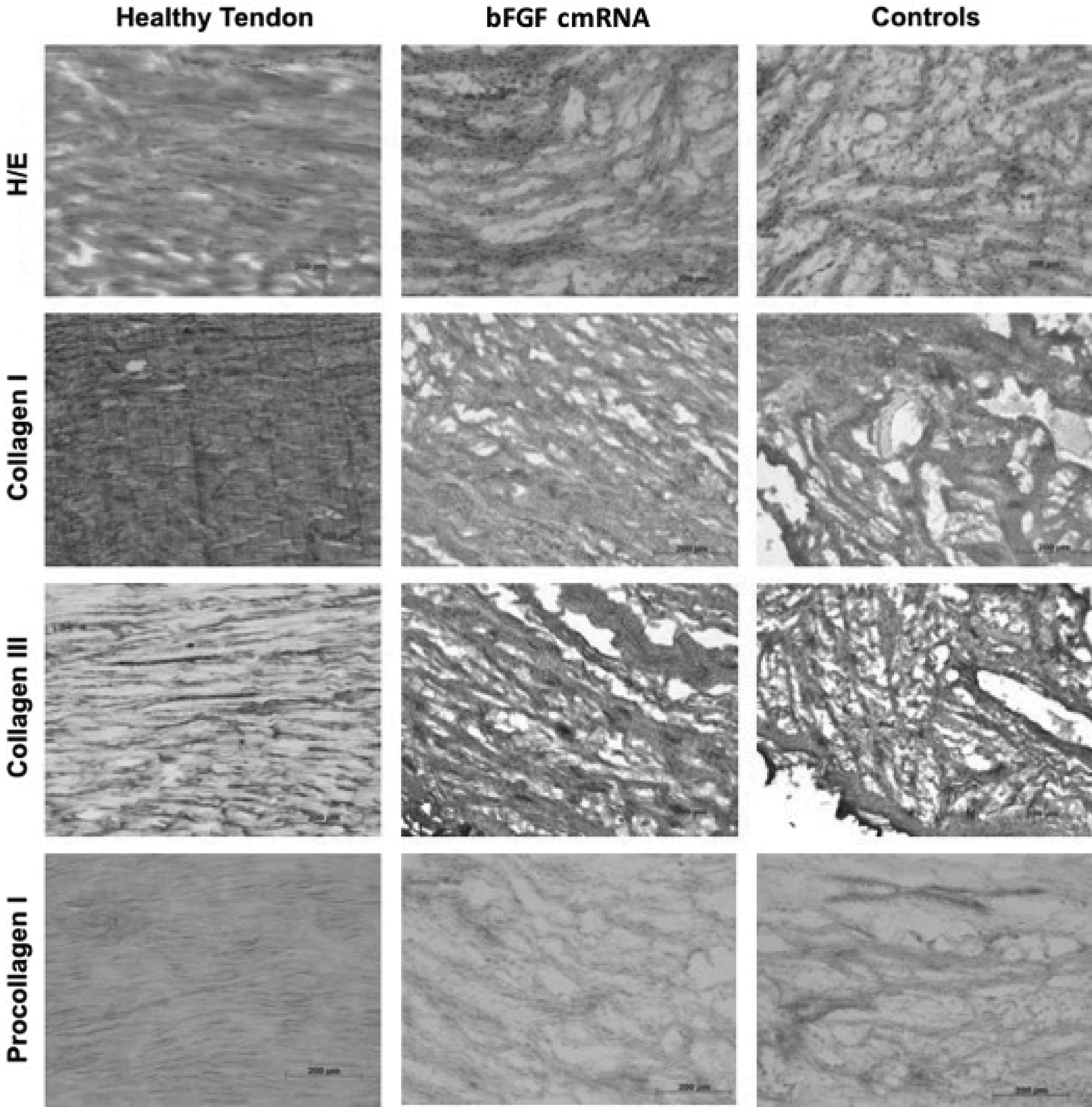
Representative samples of the midsubstance of healthy tendons compared with tendons treated with bFGF cmRNA and controls. Neither H/E staining nor immunohistology revealed statistically significant differences between the two study groups. H/E, hematoxylin/eosin.
The immunohistological analysis revealed no significant difference between staining patterns of collagen types I, II, III, and IV and procollagen I (Table 2).
Histological and Immunohistological Scores for the Tendon Midsubstance for Basic Fibroblast Growth Factor Chemically Modified Messenger RNA and Control Groups
The only statistically significant difference observed was for the shape of nuclei, with a higher score in the bFGF cmRNA group (p = 0.024).
Discussion
The most important finding of the current study is that after injection of a novel stabilized nonimmunogenic mRNA in the healing site of Achilles tendon defects, reporter protein expression can be successfully seen for at least 72 h, indicating successful transfection. However, this has only been analyzed in the control group as in both groups, the same chemical modifications of mRNA have been performed. In vitro data suggested, however, bFGF expression following mRNA transfection for at least 24 h (Supplementary Data and Supplementary Figure S1; Supplementary Data are available online at www.liebertpub.com/tea). None of the animals or tendons had signs of inflammation or other types of immunologic responses. Biomechanically, the construct stiffness in the bFGF cmRNA group was statistically significantly higher compared with controls treated with Luciferase cmRNA. Of note, construct stiffness was remarkably lower as in healthy contralateral tendons. Histologically and immunohistologically, the shape of the nuclei in the midsubstance as well as cellularity was slightly closer to intact tendons in the control group.
Tendon healing occurs through distinct stages, 14 which can be summarized in the inflammatory, formative or proliferation, and remodeling phases. bFGF is involved in all these phases and plays, however, a crucial role during early tendon healing. 14 Chang et al. found in a rabbit flexor tendon injury model that bFGF is upregulated especially during the first 4 weeks following injury. 34 Interestingly, bFGF expression is not only highest during the first 2–4 weeks of tendon healing but it also experiences another peak 8 weeks post tendon injury. 35 As shown in the current study and a previous study, reporter protein expression is upregulated for at least 3–7 days in addition to the natural healing response. 27 However, data of this study suggest that this period of bFGF overexpression might not be sufficient to properly promote early tendon healing. Similarly, Kraus et al. did not find a significant effect by lentiviral application of bFGF. 19 Considering the time course and kinetics of bFGF following tendon injury, 34 sequential application of bFGF might be superior to a single dose.
bFGF is angiogenic 36 and has mitogenic effects on fibroblasts.29,37 Furthermore, it promotes cell migration during early tendon healing. 38 Taking into account the time course of bFGF expression following tendon injury as well as its proliferative effects on fibroblasts, mRNA coding for bFGF has been used in this study.
Chan et al. 29 found in a rat patellar tendon defect model that injection of bFGF 3 days postoperatively increases collagen III expression at 1 week, but not at 2 weeks. The ultimate stress in the defect group was up to 41% of the controls. Similarly, data of the current study failed to find a significant increase of collagen III staining at 2 weeks in tendons treated with bFGF cmRNA, while the load to failure was about 40% of the contralateral healthy tendon. Collagen type III together with procollagen I is an important indicator for tendon regeneration and healing in the early post-traumatic phase. 29 A study by Kraus et al. 19 investigated the effect of mesenchymal stem cells expressing bFGF lentivirally in a rat Achilles tendon defect model. In contrast to the current study, the authors found more procollagen 1 staining after 2 weeks, which was not reflected biomechanically. Of note, the authors discussed potential negative effects of mesenchymal stem cells on structural properties 4 weeks following injection.
Several studies investigated the effects of different growth factors for tendon healing.14,22,39 Recently, especially gene therapeutic approaches have been studied. 20 However, relevant limitations that prevent their clinical application include potential mutagenic and immunogenic effects, especially when using mRNA or viral vectors.20,25 The novel technique of mRNA application used in this study may reduce the risk of immunologic responses.25,26 In fact, no adverse events and histologic evidence of systemic or local inflammation have been observed in the current study.
Data of the current study failed to prove improved tendon healing with mRNA coding for bFGF, which is in line with other negative or conflicting findings in literature.19,22 Reasons for these negative findings may be explained by the variety of growth factors involved in early tendon healing (e.g., bone morphogenetic protein 12, 13, 14, transforming growth factors beta, insulin-like growth factors [IGF], platelet-derived growth factors [PDGFs], and many others).14,22,35,39 It might be preferable to apply a cocktail of different growth factors for early tendon healing since in vitro studies found, for example, a combination of PDGF-BB, IGF-1, and bFGF together promotes tenocyte proliferation more than supplementing each factor individually. 40 Therefore, future studies should aim to combine different growth factors for early tendon regeneration.
Animals in the current study were allowed to fully bear weight immediately postoperatively. It is well known that tenocytes and fibroblasts respond to different mechanical stresses. For example, type III collagen mRNA is upregulated and growth factor concentration is higher with mechanical loading, 14 while immobilization has negative effects on gene expression and tendon healing. 41 Furthermore, the defect model without direct repair might allow a better understanding of tendon healing in rodents.19,42
This controlled laboratory study has limitations. First of all, only one growth factor (bFGF) has been investigated, and a combination of several growth factors might be beneficial to properly improve early tendon healing. 40 However, bFGF is among those growth factors with the highest concentrations following tendon injury. 35 Furthermore, studies on animals and especially rodents should be interpreted carefully since healing and cell metabolism follow a different time course compared with adult humans. However, this study investigated a novel approach of mRNA application with promising results in terms of feasibility and applicability. Further studies are needed to evaluate tendon remodeling at later stages and the effect of a combination of different growth factors.
Conclusion
This pilot study demonstrated the feasibility of a novel mRNA-based therapy for Achilles tendon defects using a stabilized cmRNA coding for bFGF. Protein expression following mRNA injection was present for at least 3 postoperative days in the control group without any evidence of adverse events or local inflammation. Biomechanically, the stiffness of tendons was closer to the healthy contralateral tendon when compared with the control group. However, histologically and immunohistologically, no positive effects could be observed.
Footnotes
Acknowledgment
This study was supported by a research grant from the AGA—Gesellschaft für Arthroskopie und Gelenkchirurgie.
Disclosure Statement
C.P. and C.R. developed the cmRNA and are cofounders of Ethris GmbH. G.H., M.K.A., and J.P.G. are employees of Ethris GmbH. E.H., F.B.I., P.F., S.M., K.G., S.V., A.B.I., and A.S. have no competing financial interests.
References
Supplementary Material
Please find the following supplemental material available below.
For Open Access articles published under a Creative Commons License, all supplemental material carries the same license as the article it is associated with.
For non-Open Access articles published, all supplemental material carries a non-exclusive license, and permission requests for re-use of supplemental material or any part of supplemental material shall be sent directly to the copyright owner as specified in the copyright notice associated with the article.