




Abstract
The physiological reconstruction of artificial vertebral laminae without epidural scar tissue formation or spinal cord compression has been challenging. Mechanical stimulations have been reported to play an important role in bone formation and bone remodeling. In this study, we designed a comparison study to investigate the effect of continuous cerebrospinal fluid pulsation (CSFP) stress on the remodeling of artificial vertebral laminae. Mesenchymal stem cells derived from rabbit umbilical cord Wharton's Jelly were induced for osteogenic differentiation for 3 weeks before seeding on the hydroxyapatite-collagen I scaffolds to construct the tissue-engineered laminae (TEL). TEL were then implanted into the fifth rabbit lumbar vertebrae in both CSFP group (n = 36) and non-CSFP group (n = 36). De novo laminae were examined through histological and radiographic analysis in the 2nd, 4th, 8th, 12th, 16th, and 24th weeks postimplantation. Our results showed that the osteogenic gene expression levels and cancellous microstructure parameters of newborn laminae in the CSFP group reached the peak and the complete newborn laminae formed in the 12th week. Then the osteoclastic gene expression and osteoclast number of newborn laminae in the CSFP group increased greatly in the 12th week, and were significantly higher compared with the non-CSFP group. After 16 weeks of implantation, the arrangement of trabeculae became organized, and the dura surface of newborn laminae in the CSFP group showed similar curvature and smoothness as the native laminae. In conclusion, continuous CSFP stress played an important role in the physiological reconstruction of artificial vertebral laminae by promoting the remodeling abilities of TEL.
Impact Statement
In this study, we designed a comparison study to investigate the effect of continuous cerebrospinal fluid pulsation (CSFP) stress on the remodeling of artificial vertebral laminae, and we found that the continuous CSFP stress played an important role in the physiological reconstruction of artificial vertebral laminae by promoting the remodeling abilities of tissue-engineered laminae (TEL). It deepens our understanding of the in vivo development of TEL, and the impact of biomechanical stimuli on the osteogenesis and remodeling.
Introduction
With the aging of populations and high prevalence of spinal degenerative diseases, total laminectomy and semilaminectomy have been commonly used to treat spinal disorders.1,2 Total laminectomy and semilaminectomy can induce various degrees of scar tissue and adhesion formation in the epidural space, and lead to iatrogenic spine cord or nerve root compression. 3 To avoid the occurrence of epidural scar tissue, various attempts have been clinically applied such as autogenous bone grafting, allogeneic bone, and ceramic artificial bone implantation4–6 However, challenges exist during their clinical applications, including limited supply of autograft bone, donor site injury, immune reaction, and the possibility of spine compression with hard graft at early stage.4–6 Tissue engineering techniques, as an ideal alternative, have been applied to reconstruct artificial epidural fat or vertebral laminae with soft biomaterials to overcome the above challenges.7–9
Our previous studies have successfully applied tissue-engineered techniques to physiologically reconstruct artificial vertebral laminae using mesenchymal stem cells (MSCs) combined with collagen scaffolds or β-tricalcium phosphate bioceramics.7,8 The newborn laminae gradually changed from a flat shape to an arched shape, and had a dura surface as smooth as the normal laminae. The newborn spinal canal also had similar diameter and shape with the normal spinal canal, and formed no new compression on the spine cord or nerve root. In addition, the newborn trabeculae were aligned with the direction of cerebrospinal fluid pulsation (CSFP) stress. 7
CSFP is a continuous pulsation stress caused by heartbeat and respiration, and it changes with cardiac and respiratory rhythms. 10 In man lying horizontally, the pulsation stress at the foramen of Monro is usually less than 50 mm water pressure, of which the cardiac component is about 15 mm and the respiratory component is 35 mm10 As early as in 1892, Dr. Wolff observed that bone grows and remodels in response to the forces that are placed upon it. Mechanical stimulations such as fluid shear stress, pulsation pressure, and cyclical tension have been proven to play important roles in bone regeneration through regulating the proliferation and differentiation of osteoblasts and osteoclasts.11–14 Our previous study has already proved that the biological stimulation of bone defect initiated the early-onset osteogenesis, and CSFP promoted the osteogenesis of de novo laminae during the reconstruction of de novo laminae. 15
Thus, we speculated that CSFP stress might also regulate the remodeling of newborn laminae, and play important roles in its physiological reconstruction in rabbit. We measured the rabbit epidural cerebrospinal fluid pressure at the fifth lumbar vertebrae. Tissue-engineered laminae (TEL) were reconstructed using rabbit umbilical cord Wharton's Jelly-derived MSCs and hydroxyapatite-collagen I scaffold before being implanted into the fifth rabbit lumbar vertebrae. Osteogenesis and osteoclastogenesis in both CSFP and non-CSFP treatment groups were examined for up to 24 weeks postimplantation. At last, we also analyzed the correlation between osteogenic genes and canonical Wnt signaling genes, and between osteoclastic genes and alternative Wnt signaling genes by Pearson's correlation coefficient.
Materials and Methods
TEL construction
The protocol was approved by the Committee on the Ethics of Animal Experiments of Fudan University (No. 20160862A003). The MSCs we used in this study were isolated from rabbit umbilical cord Wharton's Jelly (WJ-MSCs). The isolation, culture, and osteogenic differentiation of WJ-MSCs were described in the previous article. 15 The hydroxyapatite-collagen I scaffold was bought from the Beijing Allgens Medical Science & Technology Co., Ltd. Under sterile condition, the hydroxyapatite-collagen I scaffold was cut to the size of 10 × 8 × 20 mm. After 3 weeks of osteogenic differentiation, osteodifferentiated WJ-MSCs were trypsinized and resuspended in media at a concentration of 1 × 10⁶/mL. Then, 100 μL cell suspensions was pipetted on one side of each scaffold, and after 30 min, 100 μL cell suspensions was pipetted on the other side. All constructs were placed in the osteogenic medium for another week before implantation.
Epidural cerebrospinal fluid pressure measurement
Three rabbits were anesthetized with pentobarbital sodium (1 mL/kg intraperitoneally). A 3-cm skin incision was made in the middle of the lower back. After retracting the muscle to expose the spinous process of the fifth lumbar vertebrae, the exposed spinous processes and interspinous ligaments were removed to clearly expose the laminae. The interlaminar ligament was carefully stripped from the laminae, and the Codman cerebrospinal pressure detector was inserted into the epidural space. Next, the detector was connected to the Codman pressure tester and electrocardiogram monitor (PHILIPS IntelliVue MP50). After 30 min, the pressure gradually stabilized and the pressure value and pulse frequency values were recorded.
Transplantation of TEL into CSFP and non-CSFP rabbit models
This study aimed to compare the osteoblastic and osteoclastic abilities of TEL under different biomechanical conditions of CSFP and non-CSFP rabbit models.
We used 72 of 2-month-old male rabbits weighing 2.25 ± 0.25 kg. The rabbits were randomly divided into 2 groups of 36, which were designated as the CSFP group and the non-CSFP group. The animals were anesthetized with intraperitoneally administered 3% of pentobarbital sodium (1 mL/kg).
For rabbits in the CSFP group, we located the spinous process of fifth lumbar vertebrate by method of anatomical landmark and made a 3-cm longitudinal skin incision on it. The superficial fascia and paraspinal muscle were retracted to clearly expose the spinous process and interspinous ligament. We then removed the exposed spinous processes and interspinous ligaments by rongeur to expose the dura. A bone defect measuring 10 × 8 × 2 mm was created, leaving two fresh cancellous bone end measuring 10 × 2 mm at the left and right direction, and two ligamenta flava stumps at the superior and inferior direction. The TEL were placed in the bone defect and fixed in place at the four corners using a 4-0 polypropylene suture (Prolene; Ethicon, Tokyo, Japan) (Fig. 1A, C).
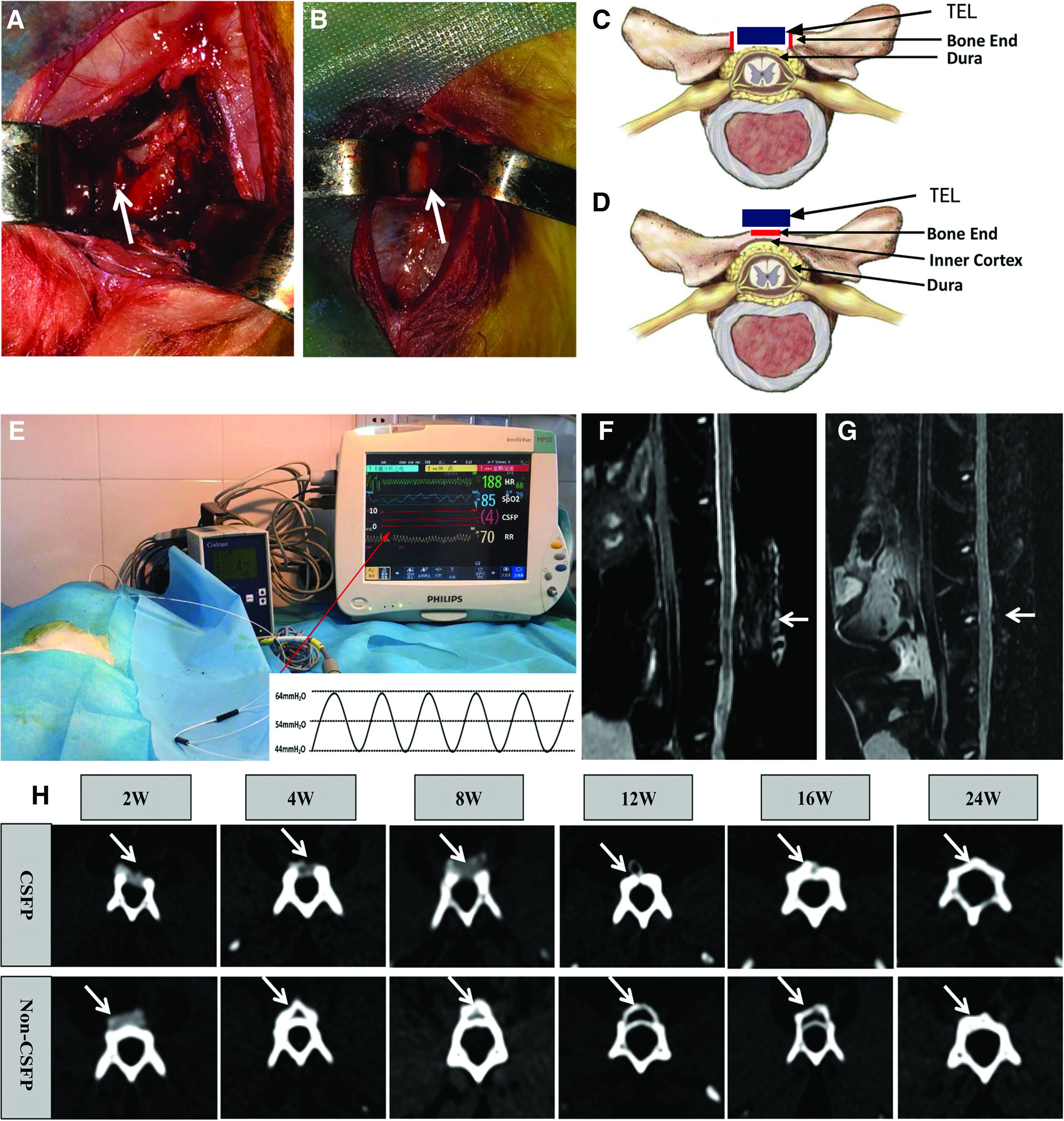
Construction of animal models.
For the non-CSFP group, we removed the spinous processes and interspinous ligaments by rongeur, and removed the adherent muscle with a detacher to clearly expose the laminae. By removing the spinous processes, a cancellous bone end measuring 10 × 1 mm was created. We then enlarged the cancellous end to the size of 10 × 4 mm by abrasive drilling, similar to that of CSFP group (10 × 2 × 2 mm), while preserving the dura surface cortex of laminae. The TEL were then fixed onto the native laminae (Fig. 1B, D).
Then the fenestrated fascia was closed with a 4-0 polyglactin suture (Vicryl; Ethicon). After the operation, sodium penicillin (400,000 U) was administered. All rabbits were housed in separate cages with free access to food and water without immobilization.
Imaging examination
At 2, 4, 8, 12, 16, and 24 weeks after operation, three rabbits of each group were subjected to computed tomography (CT) and magnetic resonance imaging (MRI) to observe the formation of the artificial laminae of the vertebral arch and their relationship with the adjacent tissue.
Micro-CT examination
Three rabbits of each group were sacrificed at 2, 4, 8, 12, 16, and 24 weeks after implantation, and the tissue specimens were harvested and immediately fixed in freshly prepared 4% (w/v) paraformaldehyde. After 2 weeks of fixation, the tissue specimens were preserved in 30% ethanol-phosphate-buffered saline (PBS) solution at 4°C. The specimens were sent to the Shanghai Public Health Clinical Center for micro-CT examination. The scanning method was as follows: 45 μm_24R_18 min; scan parameters: tube voltage, 80 kV; tube current, 450 μA; scanning mode, 360°; scanning time, 18 min; time of exposure, 400 ms; and resolution, and 45.0 × 45.0 × 45.0 μm voxel. For the CSFP group, the newborn bone in the bone defect was analyzed, and for the non-CSFP group, the newborn bone on the native laminae was analyzed, strictly excluding native lamina tissue. After the scanning, a 45.0 × 45.0 × 45.0 μm voxel region of interest was used for three-dimensional reconstruction. Quantitative analysis was performed using the 3D model with the help of GEHC MieroView2.0+ABA software. The analyzed indices included bone volume fraction (BVF), bone volume/total volume (BV/TV), trabecular spacing (Tb.Sp), and trabecular number (Tb.N). After scanning, the tissue specimens were stored in 30% ethanol-PBS solution at 4°C.
Histology examination
After micro-CT examination, the tissues were decalcified with 10% ethylenediaminetetraacetic acid for 4 weeks. Tissue sections measuring 3-μm thick were cut on a microtome and mounted onto glass slides. The sections were processed for routine histological observation by staining with hematoxylin-eosin (HE) and for Von Kossa staining and tartrate-resistant acid phosphatase (TRAP) staining.
Quantitative real-time polymerase chain reaction examination
Using a sterile technique, three rabbits of each group were anesthetized, and the living tissue specimens were harvested at 2, 4, 8, 12, 16, and 24 weeks after implantation and preserved in 1 mL TRIzol® Reagent. The tissue specimens were ground until there was no obvious residual tissue. The total RNA was extracted from the tissue samples using TRIzol Reagent (Life Technologies). All RNA samples were then treated with RNase-free DNase I (Qiagen, Valencia, CA) to digest the genomic DNA. Aliquots of 500 ng total RNA were reverse transcribed into cDNA using the PrimeScript™ RT Master Mix (TaKaRa, Japan). Quantitative real-time polymerase chain reaction (qRT-PCR) was performed using a 7900 Real-Time PCR System (Applied Biosystems, Warrington, United Kingdom) with the Power SYBR Green PCR Master Mix (Applied Biosystems). The relative gene expression was calculated using the following equations: ΔCt = Ct (test genes) − Ct (β-actin); ΔΔCt = ΔCt (newborn bone) − ΔCt (normal laminae); and Fold change = 2−ΔΔCt. The gene-specific primers used for Wnt-3a, Wnt-5a, YAP, ODF, Dkk-1, BMP-2, OPN, RANKL, OPG, Cathepsin K, and β-actin are listed in Table 1.
Sequences of Oligonucleotide Primers Used for Quantitative Real-Time Polymerase Chain Reaction
Statistical analysis
Each experiment was repeated thrice. Statistical analyses were performed using SPSS version 19.0 for Windows. One-way analysis of variance (ANOVA) was used to confirm comparisons of the variables. Multiple comparisons were performed by one-way ANOVA test followed by post hoc contrasts performed by least significant difference test (if the ANOVA results were significant). The correlation index was analyzed by Pearson's correlation coefficient. Significance was identified as a p-value less than 0.05. * Represents p-values <0.05, and ** represents p-values <0.01.
Results
Epidural cerebrospinal pressure determination
Under anesthesia, after 30 min, the cerebrospinal pressure gradually stabilized, and the measured pressure at the epidural space of the fifth lumbar vertebrae ranged from 41 to 54 mm water pressure relative to atmospheric pressure, and the pulsation pressure ranged from 10 to 20 mm water pressure, with a frequency of 3–4 Hz (Fig. 1E).
Construction of CSFP and non-CSFP animal models
CT examination showed that in the CSFP group, the TEL properly located in the bone defect. The newborn laminae grew continually from the bone ends toward the middle, and formed complete newborn orthotopic laminae by the 12th week. After 12 weeks, the dura surface of the newborn laminae gradually transformed from being flat to being arched with a curvature rate similar to that of the dura. This indicated that the osteogenic stage mainly occurred in the first 12 weeks, and then the remodeling stage began, while in the non-CSFP group, the TEL well attached to the native laminae, and the newborn ectopic laminae gradually merged with the native laminae by the 24th week (Fig. 1H).
MRI examination also showed that for subjects in both CSFP and non-CSFP group, there was no evident epidural scar adhesion, spinal cord compression, or intervertebral disc herniation in the targeted vertebrae or adjacent segments (Fig. 1F, G).
Effect of CSFP stress on the remodeling stage of newborn laminae
From the micro-CT scanning, we determined that, in the CSFP group, the trabeculae became more orderly in the 16th and 24th weeks, and the dura surface of newborn laminae showed a curvature and smoothness similar to those of the native laminae. In the non-CSFP group, the newborn laminae gradually merged with the native laminae in the 24th week, with some TEL residual remaining (Fig. 2).
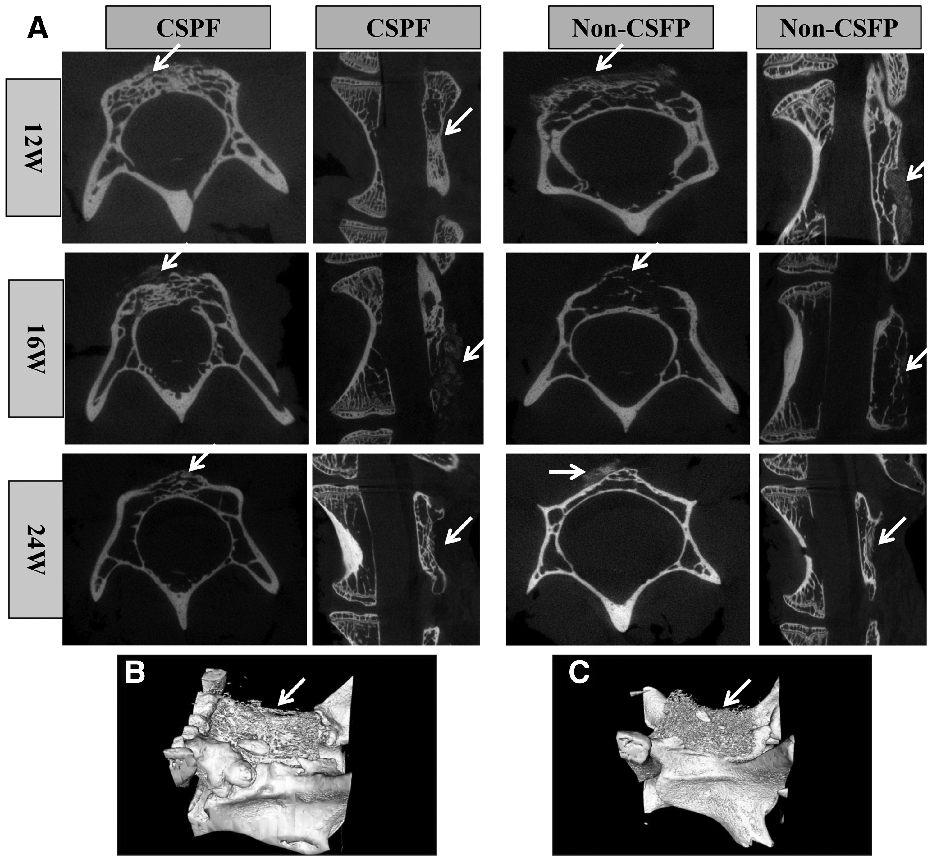
Micro-CT cross-section and sagittal section of the TELs (white arrow) in the CSFP and non-CSFP groups
The values of Tb.N and BV/TV increased slightly, and the Tb.Sp decreased slightly from the 12th week after implantation, only the values of Tb.N of CSFP group were significantly higher than those of non-CSFP group in the 16th week (p > 0.05) (Fig. 3A–C). Three-dimensional system of coordinates was constructed with the cross-section of the spinal canal as the X-axis, coronal plane as the Y-axis, and the sagittal plane as the Z-axis, as shown in Figure 3D. Tb.N values of the three different axes at different time points were analyzed. In the CSFP group, Tb.N values of the three axes were significantly higher than those of the non-CSFP group at all time points, except the 24th week (p < 0.05) (Fig. 3E–G).
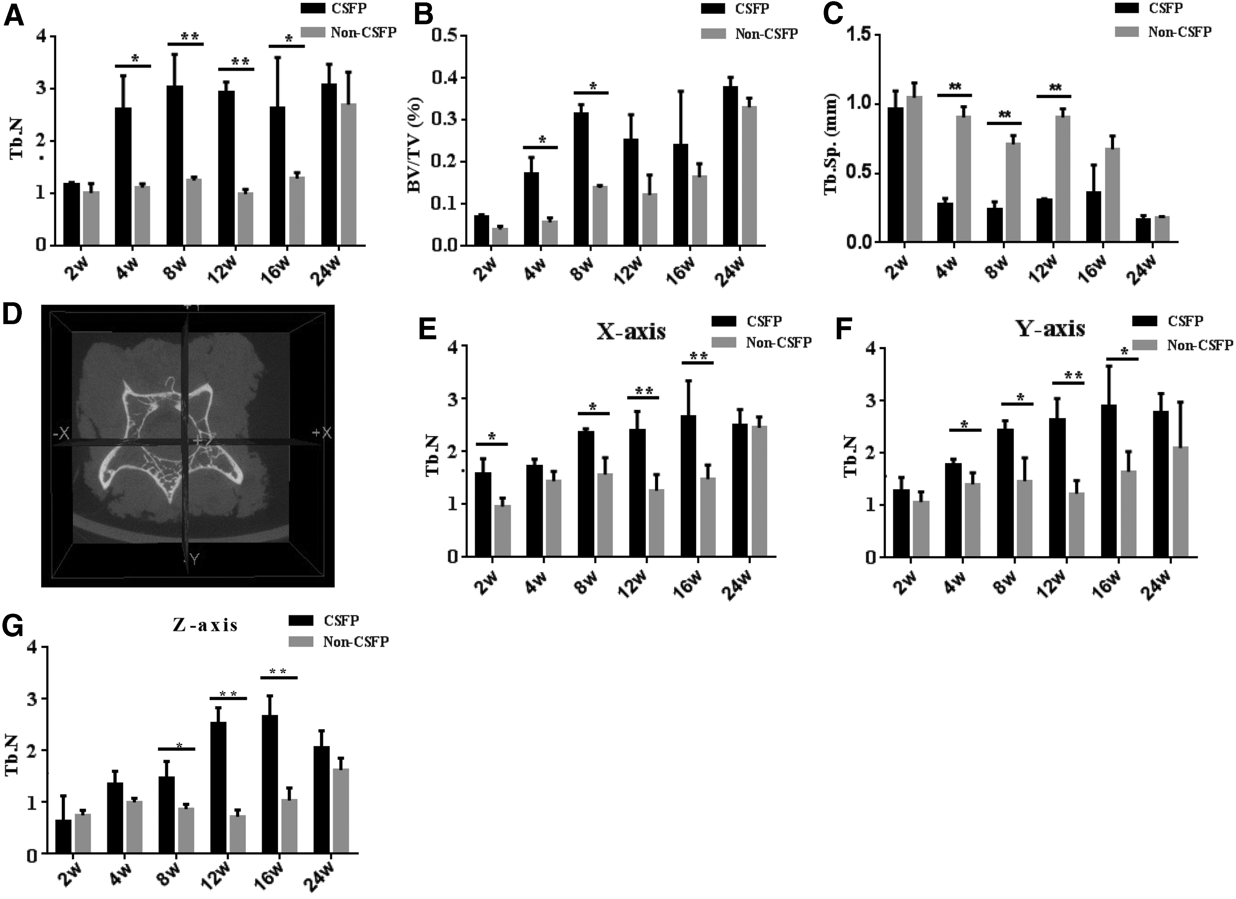
Micro-CT analysis of cancellous microstructure parameters.
HE staining showed the trabeculae in the CSFP group became more orderly and organized in the 16th and 24th weeks, and the dura surface of newborn laminae in the CSFP group showed similar curvature and smoothness as the native laminae by the 24th week (Fig. 4). Von Kossa staining also showed that the trabeculae in both groups increased slightly, the trabeculae in CSFP group became more orderly in the 16th and 24th weeks, and the trabeculae in the non-CSFP group remained disordered in the non-CSFP group (Fig. 5A). There was no significant difference of trabeculae number between two groups in the 16th and 24th weeks as shown by the Von Kossa staining (p > 0.05) (Fig. 5B). qRT-PCR analysis also showed that BMP-2 mRNA expression levels in both groups decreased from the 12th week after implantation, and there was no significant difference between two groups in the 16th and 24th weeks (p > 0.05) (Fig. 5C). The OPN mRNA expression in the CSFP group increased in the 16th week and then declined in the 24th week, and the OPN mRNA expression in the non-CSFP group declined in the 16th and 24th weeks when compared with the 12th week. The OPN mRNA expression levels in the CSFP group were significantly higher than those in the non-CSFP group in the 16th week (p < 0.05) (Fig. 5D).
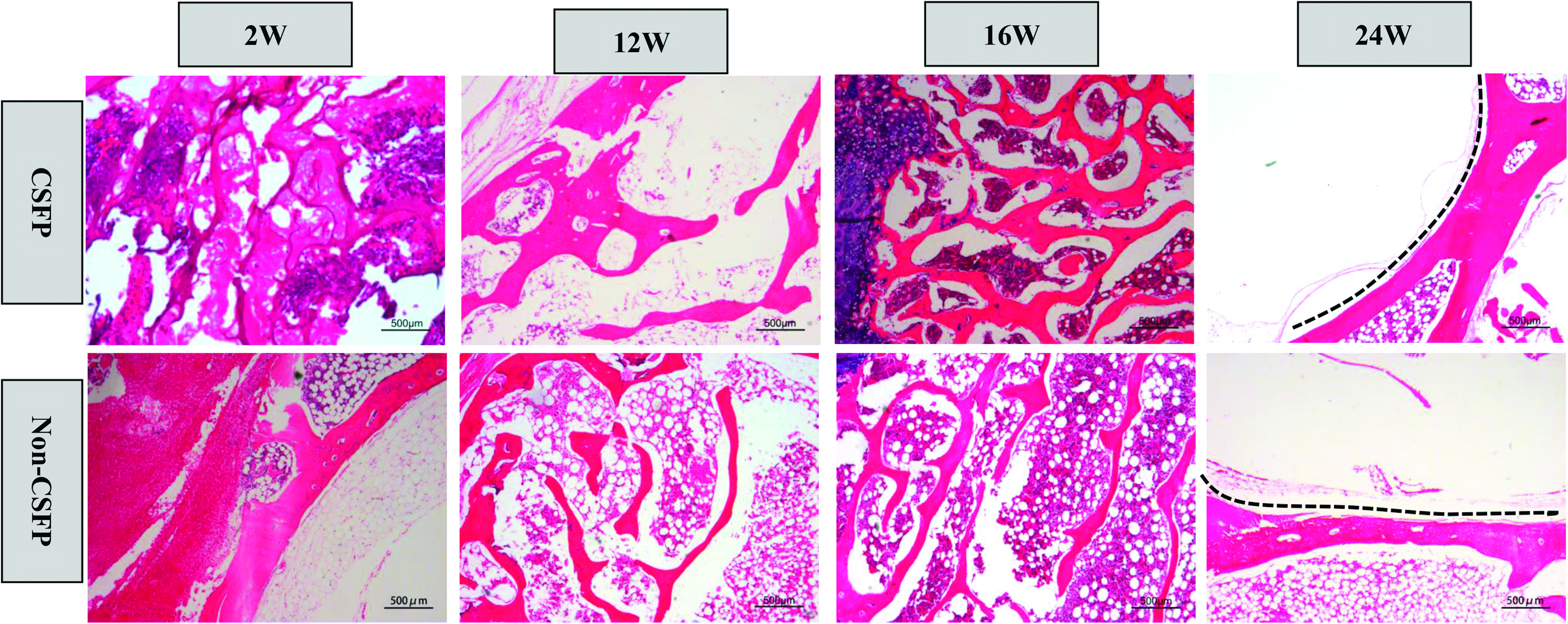
Representative images of HE staining of newborn laminae in the CSFP group and the non-CSFP group showed that both groups experienced disorganized trabeculae formation in the 12th week, and the trabeculae became more orderly and organized in the 16th and 24th week. Some images were adapted from the previous article. 15 The dotted line showed the inner surface of vertebral laminae. HE, hematoxylin-eosin. Color images are available online.

Osteogenesis of newborn laminae.
We also analyzed the monocyte number based on TRAP staining and found that the monocyte number decreased greatly over the first 8 weeks, increased dramatically in the 12th week, and then decreased slightly in the 16th and 24th weeks. The monocyte number of the CSFP group was greater compared with the non-CSFP group, but there was a significant difference in only the 12th week (p < 0.05) (Fig. 6A, B).
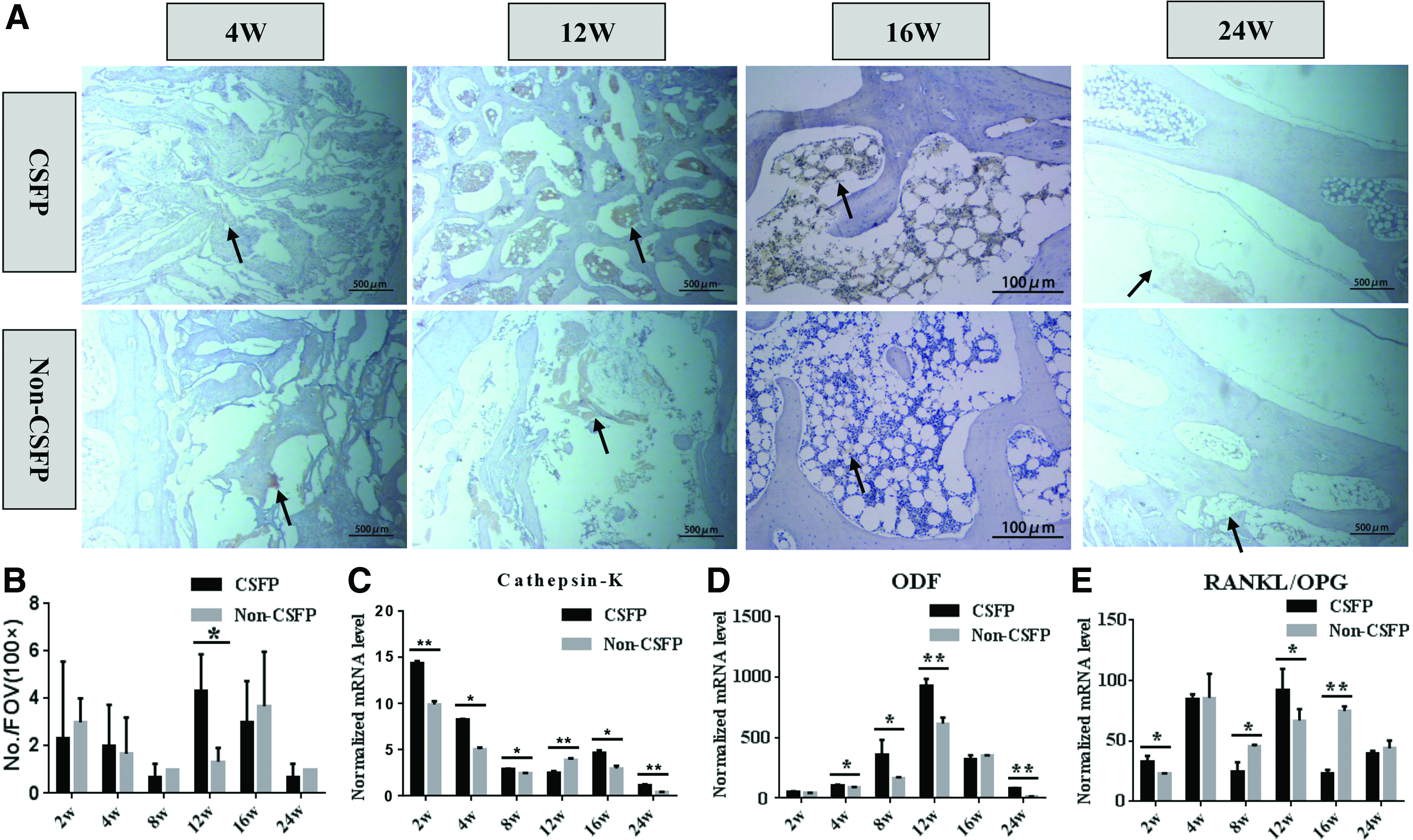
Osteoclastogenesis of newborn laminae.
qRT-PCR analysis showed that Cathepsin K mRNA expression decreased over the first 8 weeks and increased in the 12th week; the Cathepsin K mRNA expression of the CSFP group was higher compared with the non-CSFP group in the 12th week and lower compared with the non-CSFP group in the 16th week (p < 0.05) (Fig. 6C). ODF expression increased greatly in the 12th week and decreased in the 16th week; the ODF mRNA expression of the CSFP group was higher compared with the non-CSFP group in the 4th, 8th, and 12th weeks (p < 0.05) (Fig. 6D). The RANKL (nuclear factor-κ B ligand)/OPG (osteoprotegerin) value decreased in the 8th week and increased in the 12th week in the CSFP group, while the RANKL/OPG value decreased in the 8th week and increased in the 12th and 16th weeks in the non-CSFP group. The RANKL/OPG value of the CSFP group was significantly higher compared with the non-CSFP group in the 2th and 12th weeks and lower compared with the non-CSFP group in 8th and 16th weeks (p < 0.05) (Fig. 6E).
Transition from osteogenic stage to remodeling stage
In the CSFP group, the complete newborn laminae formed by the 12th week (Figs. 1H and 2A), and the trabeculae became more orderly in the 16th and 24th weeks (Figs. 2A, 4, and 5A). The BMP-2 expression reached the maximum (Fig. 5D, E), while the monocyte number (Fig. 6B) and Cathepsin K mRNA expression (Fig. 6C) also increased greatly in the 12th week. The formation of complete newborn laminae and the great increase of osteoclastic abilities suggested that the 12th week is a transition site from osteogenic stage to remodeling stage.
qRT-PCR analysis of canonical and alternative Wnt signaling genes
In the CSFP group, Wnt-3a mRNA expression levels increased over the first 8 weeks, peaked in the 8th week, and then decreased in the 12th week. The Wnt-3a mRNA expression levels in the non-CSFP group increased over the first 12 weeks, peaked in the 12th week, and decreased in the 16th week. The Wnt-3a mRNA expression level in the CSFP group was significantly higher compared with the non-CSFP group in the 8th and 12th weeks (p < 0.05) (Fig. 7A). Dkk-1 expression increased in the first 12 weeks and decreased in the 16th week for both groups, and the Dkk-1 expression level in the CSFP group was higher compared with the non-CSFP group in the 8th, 12th, and 24th weeks and lower compared with the non-CSFP group in the 16th week (p < 0.05) (Fig. 7B). In the CSFP group, Wnt-5a mRNA expression level increased dramatically in the 12th week and then rapidly fell in the 16th week. Wnt-5a expression increased over the first 16 weeks, and decreased in the 24th week; the Wnt-5a expression level of the CSFP group was significantly higher compared with the non-CSFP group in the 12th week, and significantly lower compared with the non-CSFP group in the 16th week (p < 0.05) (Fig. 7C). YAP expression increased over the first 12 weeks and decreased in the 16th week for both groups; the YAP expression level in the CSFP group was higher compared with the non-CSFP group (p < 0.05) (Fig. 7D).
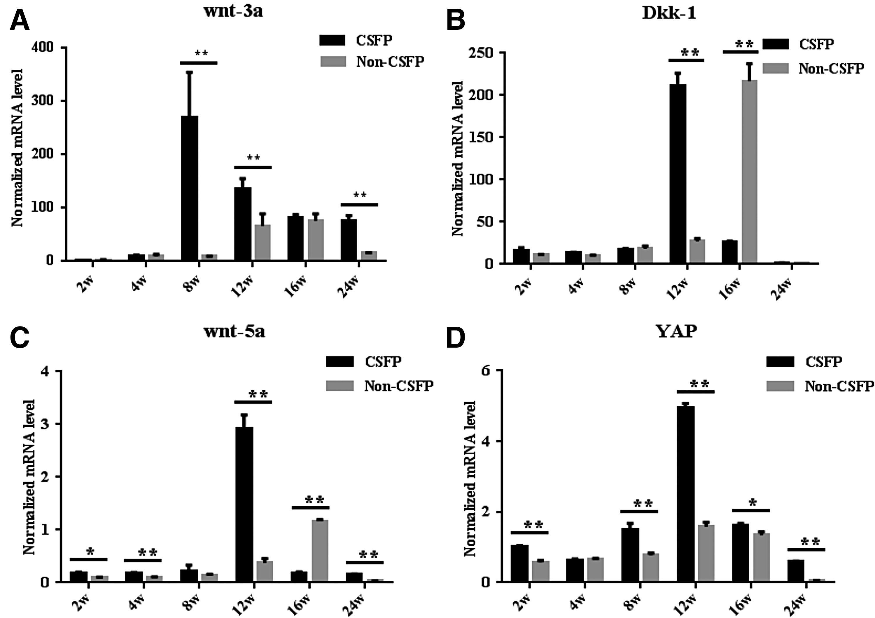
qRT-PCR analysis of Wnt-3a
We also analyzed the correlation between osteogenic genes and canonical Wnt signaling genes, and between osteoclastic genes and alternative signaling genes by Pearson's correlation coefficient. The Pearson's correlation coefficient of BMP-2 and Wnt-3a was 0.554 (p < 0.001). The coefficient of ODF and Wnt-5a was 0.816 (p < 0.001). The coefficient of RANKL and Wnt-5a was 0.886 (p < 0.001). The coefficient of ODF and YAP was 0.904 (p < 0.001). The coefficient of RANKL and YAP was 0.816 (p < 0.001). The coefficient of Dkk-1 and Wnt-3a was −0.337 (p = 0.044). The coefficient of Dkk-1 and Wnt-5a was 0.807 (p < 0.001).
Discussion
In this study, we investigated the role of CSFP in the osteogenesis and remodeling of artificial vertebral laminae by transplanting TEL into CSFP and non-CSFP rabbit models. CSFP rabbit models were created by excising the native laminae, and non-CSFP rabbit models were established by excising the spinous processes and preserving the native laminae.
In the beginning of study design, we meant to construct an animal model only exposed to the mechanical stimulation of CSFP, but the proper CSFP-isolated animal model as a control group that absolutely precluded the disturbance of biological stimulation of bone defect could not be constructed. The reasons are listed below: (i) CSF is the product of basic life activities such as heartbeat and respiration, and is also the requisite for life supporting such as the protection of brain and spine, and transmission of nutrition and waste. 16 Then, we cannot make the non-CSFP model by absolutely insulating the CSF such as kill the animals or spinal cord ligation in the thoracic segment. (ii) Considering the mechanotransduction property and fluidity of tissue fluid,17,18 removing the laminae and interlaminae ligament would release the pressure and no isolation film could absolutely seal the pressure. So creating the CSFP-isolated group by removing the auto-laminae and placing rigid insulating film would severely undermine our experiment design. In addition, the method of excising the native laminae and placing rigid insulating film would also have the threat of spine compression. (iii) The cellular and biochemical factors released from the bone defect would also impact the viability of engineered bone grafts.19–21 To preclude this disturbance, we enlarged the cancellous bone defect in the non-CSFP group to the size of 10 × 4 mm, similar to that of CSFP group (10 × 2 × 2 mm), which assured the similar releasing biochemical factors. Based on the above considerations, it was hard to construct a proper animal model as a control group,; so we chose to design an animal model with the similar biological stimulation, but without the mechanical stimulation of CSFP stress as a comparison group to explore the impact of CSFP stress on the osteogenesis and remodeling of newborn laminae.
The scaffolds we used were hydroxyapatite-collagen I scaffolds composed of nanohydroxyapatite and collagen type I, and were manufactured by mineralization. The structure and composition of these scaffolds are similar to those of human natural cancellous bone, which shows excellent biocompatibility, osteoconductivity, and biodegradability, and its functionality in tissue engineering has been previously demonstrated.22,23 The softness of the scaffolds makes them ideal candidates for the reconstruction of artificial laminae.
Previous studies10,16 showed that intracranial CSFP was closely related with cardiac and respiratory rhythms. In this study, we found that both human and rabbit CSF at the fifth lumbar vertebrae pulsated at the frequency of the respective heartbeat. Consistently, Laitinen and Hsu et al. also found that CSF pulsated inside the spinal canal at the frequency of the heartbeat.24,25 The pulsation pressure of rabbit CSF ranged from 10 to 20 mm water pressure similar to that of human (13–27 mm water pressure), but the frequency was definitely different.
The physiological reconstruction of artificial laminae involves two overlapping stages of osteogenesis and remodeling. Osteogenesis involves the migration and osteodifferentiation of MSCs and the secretion and mineralization of bone matrix, which results in the formation of disorganized trabeculae (woven bone). 26 Remodeling involves the resorption of excessive disorganized trabeculae and regeneration of orderly arranged trabeculae conforming to the stress direction (lamellar bone). 26 In this study, complete newborn laminae formed in the 12th week with a disordered arrangement of trabeculae, corresponding to the osteogenic stage. The trabeculae became orderly arranged in the 24th week, and the dura surface of newborn laminae also presented as smooth and arched as the native laminae by the 24th week, with no reduction of spine canal size or compression of spinal cord, corresponding to the remodeling stage. The key factor underlying bone osteogenesis and remodeling was mechanical stimulation.
During the first 12 weeks after transplantation, CSFP stress mainly promoted the osteogenesis of newborn laminae but also stimulated the relatively low expression of osteoclast differentiation factors, and the overall process favored osteogenesis. In the 12th week, there was a rapid increase in the expression levels of osteogenic genes, and complete de novo laminae formed. It suggested that the biological stimulation of bone defect initiated the early-onset osteogenesis, and CSFP promoted the osteogenesis of de novo laminae during the reconstruction of de novo laminae. 15 In addition, Cathepsin K expression and monocyte number were relatively high in the 2nd and 4th weeks, due to the degradation and resorption of scaffolds, 27 also evidenced by the HE staining (Fig. 4).
Twelve weeks after implantation, the overall variation trend of newborn laminae was inclined to bone remodeling. The remodeling cycle is composed of four sequential phases—activation, resorption, reversal, and formation, requiring the synchronous participation of anabolic (bone forming) and catabolic (bone resorbing) responses. 26 In this phase, the ODF and RANKL/OPG increased greatly, and promoted the proliferation, aggregation, and activation of osteoclasts, as indicated by TRAP staining and the Cathepsin K mRNA expression levels. Cathepsin K expression level and osteoclast number began to increase in the 12th week, while the expression levels of osteoblastic proteins gradually declined. In addition, in the 12th week, the osteoclastic abilities of the CSFP group were significantly higher than those of the non-CSFP group, suggesting the promoting role of CSFP stress in the remodeling of newborn laminae.
The bone remodeling unit is composed of a tightly coupled group of osteoclasts and osteoblasts that sequentially carry out resorption of old bone and formation of new bone. 26 The osteogenic and osteoclastic peaks both occurred in the 12th week for the CSFP group, suggesting that the 12th week was the transition point from the osteogenesis to remodeling and there was some kind of relationship between osteogenesis and remodeling. This is further supported by RANKL/OPG and ODF, which are secreted by the osteoblasts and osteocytes, and modulate the differentiation of osteoclasts and stimulate osteoclast recruitment to bone remodeling areas.28–31 The RANKL/OPG and ODF for the CSFP group were higher than those of non-CSFP group in the 12th week. Therefore, we believed that CSFP stress played an important role in the physiological reconstruction of artificial vertebral laminae by promoting the overall process of osteogenesis and remodeling.
During the regeneration of newborn laminae, bone forming and bone resorbing responses occurred orderly, which require the organized participation of osteoclasts, osteoblasts, and osteocytes embedded in the matrix bone tissue.26,30,32 These participating cells are tightly coordinated by regulatory proteins that interact through complex autocrine and paracrine mechanisms.26,30,32 Previous studies have shown the important role of canonical and alternative Wnt signaling pathways in the osteogenesis and resorption of bone. 29 To have a preliminary exploration of the regulatory mechanism of CSFP stress in the physiological reconstruction of artificial vertebral laminae, we analyzed the mRNA expression levels of canonical and alternative Wnt signaling genes. We found that the expression trend of components of the canonical and alternative Wnt signaling pathways was consistent with the osteogenesis and remodeling of newborn laminae. Furthermore, BMP-2 mRNA expression level was strongly correlated with that of Wnt-3a, and ODF mRNA expression levels correlated with those of Wnt-5a and YAP. Dkk-1 mRNA expression level were also correlated with that of Wnt-5a and Wnt-3a. Dkk-1 is secreted by the osteoblasts, and acts as a downstream molecule of alternative Wnt signaling and as a canonical Wnt signaling inhibitor. 29 The reciprocal interactions between osteoblast- and osteoclast-secreted molecules through the Wnt signaling pathways, forming an integrated negative feedback loop, might contribute to the balance between osteogenesis and remodeling.29,33,34
In conclusion, continuous CSFP stress played an important role in the physiological reconstruction of artificial vertebral laminae by promoting the osteogenic and remodeling processes. The newborn laminae showed similar smoothness and curvature as the native laminae, and did not reduce the volume of spinal canal or compress the spinal cord.
Footnotes
Acknowledgments
The study was supported by National Natural Science Foundation of China (Grant No. 81672179); by Natural Science Foundation of Minhang District, Shanghai (2014 MHZ004); and by the Foundation of Shanghai Municipal Commission of Health and Family Planning (20134305).
Disclosure Statement
Some of the pictures in the Figures 1, 2A, and  have already been used in one article entitled “Biological and Mechanical Factors Promote the Osteogenesis of Rabbit Artificial Vertebral Laminae: A Comparison Study”
15
published in Tissue Engineering Part A (DOI: 10.1089/ten.2017.0426).
have already been used in one article entitled “Biological and Mechanical Factors Promote the Osteogenesis of Rabbit Artificial Vertebral Laminae: A Comparison Study”
15
published in Tissue Engineering Part A (DOI: 10.1089/ten.2017.0426).
There are no other conflicting financial interests.