




Abstract
Because of the lack of donor corneas, an important area of research is the development of bioengineered corneal scaffolds to treat corneal blindness. Decellularized cornea has become a prominent area of research to satisfy the clinical demand. However, the limitation of its application is that a suitable decellularization procedure has not been developed. Organic acids are naturally occurring constituents in animal tissues and plants, and could be safely neutralized into harmless salts. In this study, we developed decellularized porcine corneal (dPC) scaffolds that were prepared by organic acid treatment. Cell removal and intact extracellular matrix preservation were evidenced by histological and biochemical quantitative analysis, and the dPC scaffolds showed porous parallel lamellar microstructure and excellent biomechanical properties. In vitro cell culture demonstrated that the dPC scaffolds had good biocompatibility, and the porous microstructure provided an ideal space for the growth of stroma keratocytes. Moreover, in vivo implantation revealed ideal reepithelialization, stromal recellularization, and complete transparency during the full follow-up period. Thus, dPC scaffolds that were prepared by organic acid treatment could be a promising biological material for use in corneal transplantation.
Impact Statement
This study successfully developed decellularized corneal scaffolds that were prepared by organic acid, which safely exist in animal tissues and plants. The results showed the highly efficient removal of cell debris from porcine corneas, and excellent preservation of optical properties, extracellular matrix (ECM) architecture, and biomolecules. In addition, decellularized corneal scaffolds revealed excellent biocompatibility and recellularization potential in vitro. In an animal model, the transplanted corneas were completely epithelialized, clear, showed no signs of immune response, and effectively supported stromal keratocytes growth. Hence, this could be a promising scaffold material for corneal tissue engineering applications.
Introduction
T
Different synthetic or biological biomaterials have been evaluated for use as corneal substitutes including polyvinyl alcohol, silk, collagen, and amniotic membrane.4–7 However, the difficulty in incorporating cells within a scaffold is a major drawback of all these materials. Despite the development of bioengineered corneal scaffolds, an ideal scaffold for corneal transplantation remains to be found.
A decellularized porcine corneal (dPC) scaffold is thought to be the best candidate as a transplantable corneal substitute because of its native extracellular matrix (ECM) and intrinsic biological components, including glycosaminoglycans (GAGs) and collagen.8,9 In addition, porcine cornea is considered to be an important animal resource because it has similar biological characteristics of a human cornea. 10 One study reported that the decellularization procedure could remove cell membrane epitopes, allogeneic DNA, and a group of damage-associated molecular pattern molecules, and thus decrease the chance of graft rejection. 11 However, the main obstacle of decellularization is the lack of a generally acknowledged protocol.
Previous studies have evaluated physical decellularization methods such as using sodium chloride (NaCl) and freeze–thawing. However, these methods make it difficult to completely remove cell debris, they also require the addition of enzymes to improve decellularization. 12 Another major way to achieve decellularization is using chemical methods such as sodium deoxycholate, sodium dodecyl sulfate (SDS), and Triton X-100. These methods efficiently solubilize the cellular components, but ECM disruption is common, and there are also safety concerns about residual detergents. 13 Acidic methods have been shown to disrupt cells while preserving the ECM. 14 Furthermore, acids can be neutralized into harmless salts, so they are considered ideal for the production of decellularized corneal scaffolds. However, the primary concern with acidic methods is to avoid microstructure degradation and preserve the ECM as much as possible.
In this study, we examined that low-concentration formic acid, acetic acid, and citric acid could be used as decellularization agents. All three are natural constituents in animal tissues and plants, and have been acknowledged for use as food additives by the United States Food and Drug Administration (FDA).15–17 Our aim was to develop an optimal acid protocol for producing dPC scaffolds, and to investigate the scaffold microstructure and biocompatibility produced by this method.
Materials and Methods
Decellularization of porcine corneas
Fresh porcine eyes were obtained from an ISO 22000:2005 certificated slaughterhouse, and the corneas were excised with at least 12-mm diameter rounds. Before the decellularization process, the porcine corneas were washed with phosphate-buffered saline (PBS).
The dissected corneas were then randomly divided into six groups, and decellularized with the following treatments. Group N was fresh native porcine corneas without any treatment. Group FA corneas were immersed in formic acid (FA), 20%, 30%, or 40%. Group AA corneas were immersed in acetic acid (AA), 20%, 30%, or 40%. Group CA corneas were immersed in citric acid (CA) 20%, 30%, or 40% (Sigma-Aldrich). All groups were mechanically agitated on an orbital shaker at 150 rpm at 4°C for 3 days. After each process, the corneal scaffolds were extensively washed with PBS on an orbital shaker at 150 rpm at 4°C, until the final pH value was stable at 7.0. For the supercritical drying (SCD) group, the corneas were immersed in 2 M NaCl for 1 h, followed by ultrapure water for 30 min on an orbital shaker at 150 rpm at 4°C. The corneas were dehydrated through ascending concentrations of ethanol up to pure ethanol, and then placed into a vessel of an SCD system. The treatments were performed at 35°C and 10 MPa, and the samples were treated for 60 min. After that, the corneas were rehydrated by immersing in PBS for 48 h. For the detergent treatment group, the corneas were immersed in SDS 0.1% on an orbital shaker at 150 rpm at 4°C for 24 h. The corneas were then washed for 48 h with PBS.
An 8-mm diameter corneal trephine was then used to remove a corneal specimen for further experimentation. The corneal thickness was controlled to 300 μm by frozen section. The dPC scaffolds were then analyzed, as described in the following sections, and experiments were performed in triplicate. For each experiment, there were at least five independent scaffolds (belonging to the same batch of scaffolds) in a group.
Assessment of the optical property of dPC scaffolds
The native cornea and dPC scaffolds were immersed in glycerol (Sigma-Aldrich) for 15 h to restore corneal clarity before optical evaluations. The spectral transmittance of the corneal scaffolds was determined using a Tecan Sunrise™ Absorbance Microplate Reader (Tecan, Italy), with the light absorbance wavelength ranging from 400 to 700 nm. Data were collected, triplicated, and transferred to percentage by using ultrapure water as baseline control.
Mechanical properties and microstructure of dPC scaffolds
The native corneas and the dPC scaffolds were cut with razor blade punch into a dumbbell shape, with a gauge length of 5 mm, and the thickness of each sample was measured individually. The tensile strength and elastic modulus were examined by Materials Testing System (MTS Systems Corp, Eden Prairie). The test was performed at a crosshead speed of 0.1667 mm/s until complete rupture occurred to obtain the tensile strength. The elastic modulus were calculated from the linear slope of the stress and stain diagram using TextWorks software.
The microstructure of the decellularized corneas was investigated using a scanning electron microscopy (SEM). The specimens were fixed in 4% paraformaldehyde for 8 h, and then washed with 0.2 M cacodylate, followed by washing with ultrapure water. The fixed specimens were dehydrated through ascending concentrations of ethanol up to pure ethanol, and then dried using a critical point dryer. After being coated with gold–palladium in a mini sputter coater and mounted on metal stubs, the specimens were then observed with an SEM (Hitachi SU3500, Japan) at an electron-accelerating voltage of 15 kV.
Biochemical analysis and histological examination
For biochemical analysis, samples were dried and the weight was measured. The DNA content of samples was examined using a DNA isolation kit (Geneaid, Taiwan) and a Qubit™ dsDNA BR Assay Kit (Thermo Fisher Scientific) following the manufacturers' instructions. In brief, samples were weighed and then digested with cell lysis buffer and proteinase K at 60°C for 1 h. After incubation, RNase A and protein removal buffer were added, followed by centrifugation at 16,000 g for 3 min, to form a tight pellet. The supernatant was mixed with isopropanol and centrifuged at 16,000 g for 5 min. After pellet rehydration, the Qubit BR working buffer was added and wavelength 510 nm was measured using a Sunrise light absorbance reader.
To examine the amount of GAGs and total collagen, samples were first digested with papain extraction reagent (0.1 M sodium acetate, 10 mM sodium EDTA, 5 mM
For histological examination, the tissues were fixed in a buffered 4% paraformaldehyde, embedded in paraffin, cut into 4-μm-thick sections, and placed on silane-coated microscope slides. The sections were then stained with hematoxylin and eosin (HE) for examining the presence of cell debris, Alcian blue staining was used for examining GAGs, and Masson's trichrome staining was used for detecting collagen fibers.
Recellularization on dPC scaffolds
L929 fibroblasts propagated in Roswell Park Memorial Institute medium (RPMI; Life Technologies/Thermo Fisher Scientific) with 10% fetal bovine serum were seeded onto the dPC scaffold surface at a density of 1 × 10 4 cells/mL. Cell proliferation was determined on days 1, 3, 5, and 7 by MTT (3-[4,5-dimethylthiazol-2-yl]-2,5 diphenyl tetrazolium bromide) assay. The optical density, at 570 nm, was measured using a Sunrise light absorbance reader.
To examine the proliferation of epithelial cells, human corneal epithelial cells (HCECs) (CRL-11515, ATCC) were seeded onto the dPC scaffold surface at a density of 1 × 10 4 cells/mL, and grown in keratinocyte serum-free medium (Life technologies) with 5 ng/mL human recombinant epithelial growth factor, 0.05 mg/mL bovine pituitary extract, 0.005 mg/mL insulin, and 500 ng/mL hydrocortisone. The proliferation activity of the cells was determined on days 1, 3, and 5 by MTT assay. HCECs grown on the dPC scaffolds were examined with anti-cytokeratin C antibody (anti-CK3) by immunofluorescence staining on day 5. The samples were washed three times with PBS for 5 min each, and then incubated with bovine serum albumin to block the nonspecific sites. Then, the cells were incubated with anti-CK3 (1:50; Abcam) at 4°C overnight. After three washes with PBS for 5 min each, the samples were incubated with Cy3-conjugated IgG (1:200; Invitrogen) and Hoechst staining for 2 h at room temperature. After washing, sections were mounted and examined with a fluorescence microscope.
Rabbit stroma keratocytes, SIRC cell line, were seeded by injection (1 × 10 6 cells in 500 μL) at 10 different points, and then cultured for 3 weeks. The recellularized corneal scaffolds were examined for the rabbit keratocyte marker vimentin (1:200; Abcam) by immunofluorescence staining with frozen section. The immunofluorescence staining protocol was followed as described previously.
dPC scaffolds for corneal xenotransplantation
Eight New Zealand white rabbits were used in accordance with Guidelines for Care and Use of Animals for Research Purposes provided by the National Taiwan University Animal Center, and the experiments were approved by the Ethics Committees for Animal Welfare.
The animals were anesthetized with isoflurane, and deep anterior lamellar keratoplasty (DALK) was performed in the left eye of each rabbit. A 300-μm deep, 8-mm circular corneal defect was made using a Hessburg–Barron vacuum trephine, and the contralateral was used as a control eye. Decellularized corneal scaffolds (30% FA) were placed into the space and fixed with 12 interrupted 10-0 nylon cardinal sutures. Topical steroid, 0.1% betamethasone sodium phosphate, and 0.5% levofloxacin hydrate were instilled in the eye twice daily for 1 week. The eyes were observed for up to 60 days, and examinations included inspection for corneal optical clarity, fluorescein staining for epithelial cell regeneration, and corneal thickness examination. The fluorescein paper strips (Haag Streit) moistened with saline were used, and fluorescence staining on the cornea was examined under a portable slit lamp (KOWASL-17, Japan). Corneal thickness was measured three times at each time point with an iPac Hand-held Pachymeter (Reichert Technologies). On day 60, the rabbits were killed, and the corneas were excised for histological analysis. Histological slides were digitized by using automated imaging system (TissueFAXS Scanning System; Tissuegnostics, Austria).
Statistical analysis
Data were reported as mean and standard deviation. The significance of the data was analyzed by Mann–Whitney U test between the native group and the decellularized groups. Statistical testing was performed using GraphPad Prism (GraphPad Software), and p < 0.001 was considered statistically highly significant.
Results
Transparency, mechanical properties, and microstructure of the dPC scaffolds
The corneas were removed from fresh porcine eye (Fig. 1A) and examined for transparent properties. After washing and the various decellularization procedures the corneal scaffolds became slightly opaque in appearance (Fig. 1B), but transparency was completely recovered using the glycerol deturgescence process (Fig. 1C). The native cornea and dPC scaffolds were examined with light transmittance from 400 to 700 nm. The transmittance of the citric acid group was slightly reduced compared with the other groups, but the differences were not significant (Fig. 1D), indicating no change to the corneal lamellar organization. This result demonstrated that organic acid decellularization did not influence the corneal opacity.
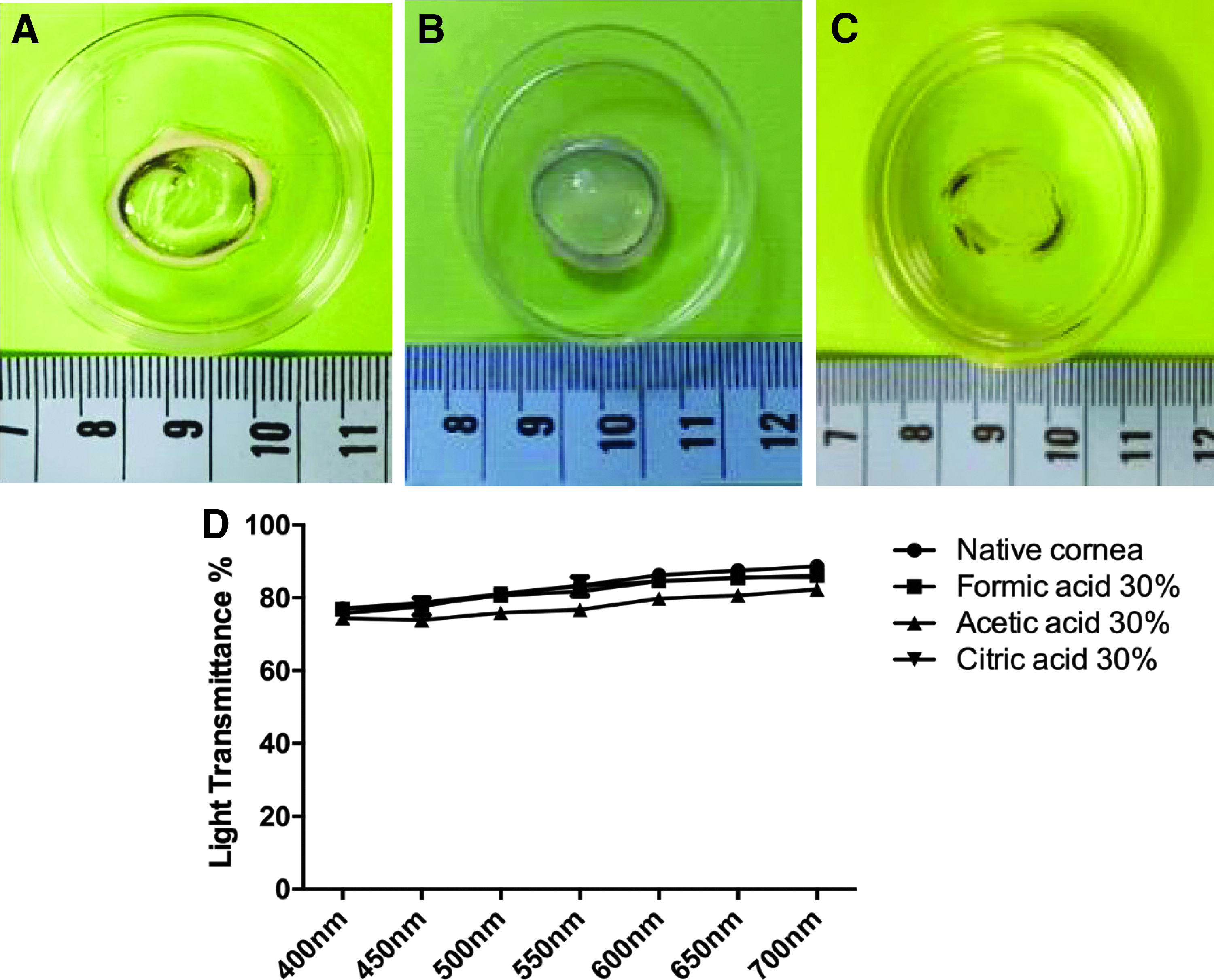
Properties of the dPC scaffolds.
To investigate the strength of the dPC scaffolds, the mechanical properties of each cornea were measured. It was observed that tensile strength of FA and AA dPC groups were slightly less than that of the native cornea, whereas the CA 30%, CA 40%, SCD, and SDS 0.1% groups showed a significant decrease, possibly because of damage of the proteins and some essential materials (Fig. 2A). The elastic modulus data also revealed a significant reduction in the CA 40%, SCD, and SDS 0.1% groups (Fig. 2B).
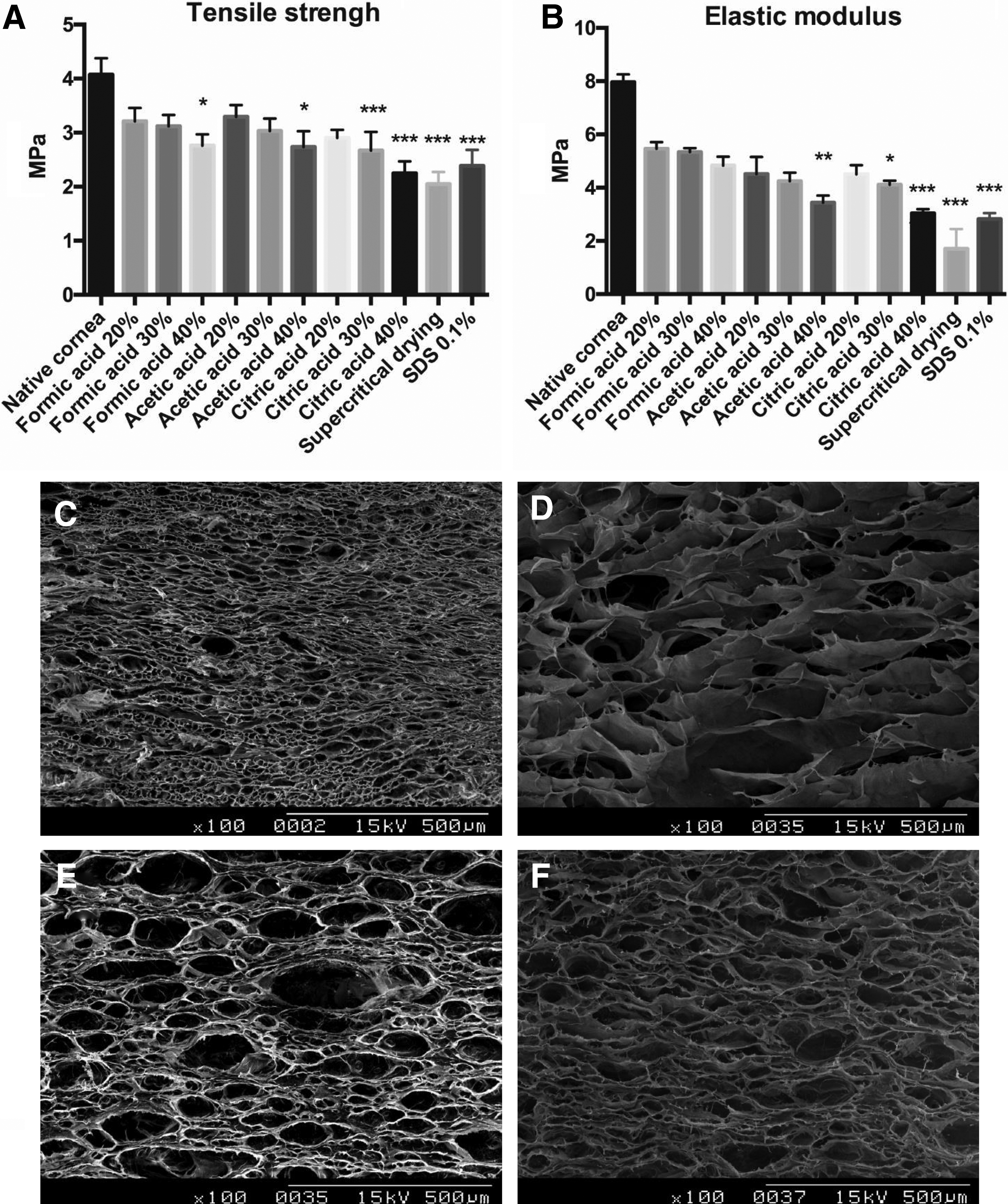
Mechanical properties and microstructure of the dPC scaffolds.
The SEM morphology results showed a uniform parallel lamellar structure in all groups, and the native corneal scaffold was very condensed (Fig. 2C), whereas the pore size of FA, AA, and CA dPC groups had increased and the porosity was uniform (Fig. 2D–F). The porous structure was thought to be beneficial for further cell nutrition, proliferation, and migration.
The efficacy of the decellularization process
The efficiency of eliminating cellular materials was examined by DNA qualification assay and HE staining. The data revealed that the FA, AA, and CA decellularization processes removed cells from the corneal scaffolds. The content of DNA was significantly decreased in the FA 20%, 30%, and 40% groups (1433 ± 166.6, 246 ± 27.6, 159 ± 27.4 ng/mg, respectively), CA 30% and 40% groups (1760 ± 96.3, 620 ± 80.0 ng/mg, respectively), SCD group (1626 ± 261.1 ng/mg), and SDS 0.1% group (840.8 ± 121.2 ng/mg) compared with the native cornea group (6518 ± 258.2 ng/mg) (Fig. 3A). Histology analysis also confirmed the absence of cells observed in the FA and CA groups, but the staining results of the AA group were similar to that of the native group (Fig. 3B–E). This result could be because of the weak decellularized ability of AA treatment.
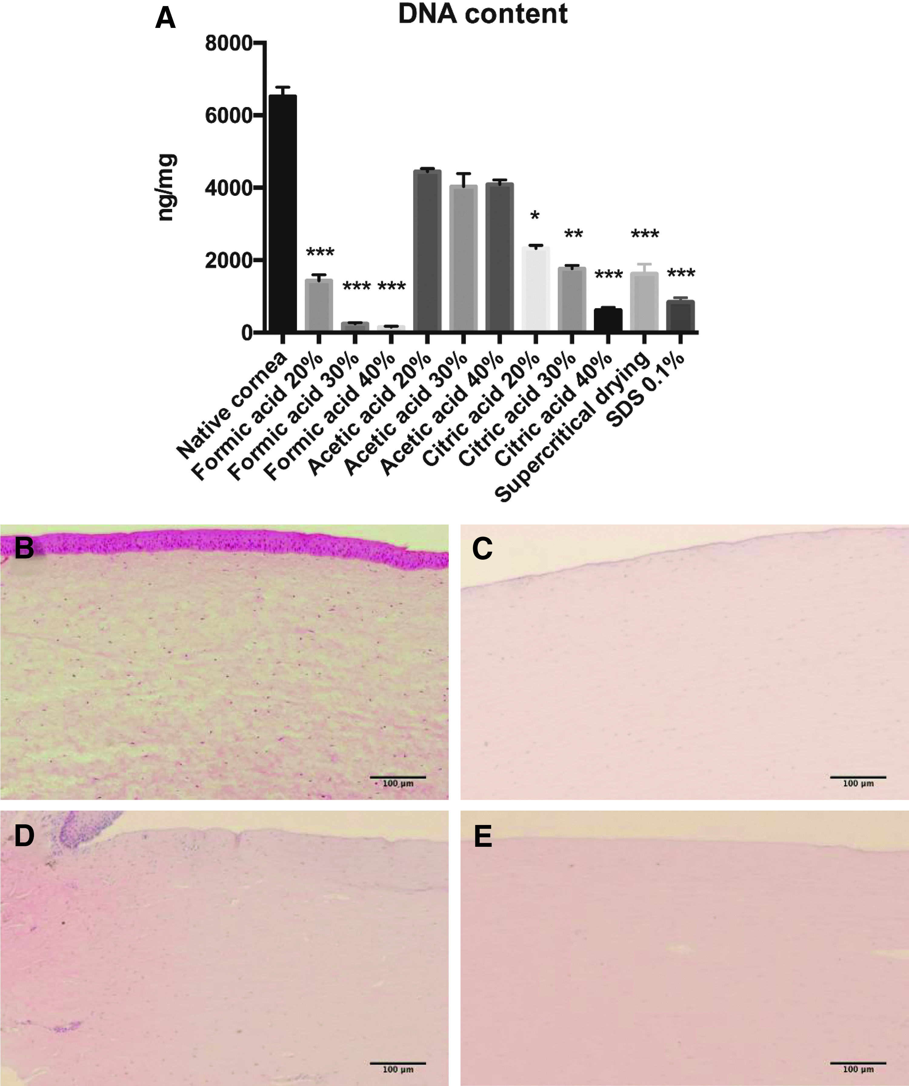
Efficacy of the organic acid decellularized process.
Characterization of ECM components
To evaluate the biochemical properties after the decellularization process, GAGs and collagen of each corneal scaffold were measured. The qualification analysis of GAGs demonstrated a statistically significant decrease in the FA 40%, AA 30%, AA 40%, CA 40%, SCD, and SDS 0.1% groups (44 ± 5.7, 44 ± 5.4, 41 ± 2.6, 48 ± 3.3, 32 ± 1.9, 39 ± 3.1 μg/mg, respectively) compared with the native cornea group (56 ± 2.7 μg/mg), whereas no differences between the other treatment groups and the native cornea group were noted (Fig. 4A). Despite the fact that some of the results showed a decrease, Alcian blue staining still confirmed GAGs preservation of the dPC scaffolds (Fig. 4B–E).

Examination of GAGs content in dPC scaffolds.
Collagen preservation and structure are important for the cornea. The experimental results showed that collagen was slightly decreased after the various decellularization processes, but the CA 40%, SCD, and SDS 0.1% treatments had statistically significant decrease (609 ± 34.5, 575 ± 32.1, 593 ± 12.6 μg/mg, respectively) compared with the native cornea group (817 ± 75.7 μg/mg) (Fig. 5A). Masson's trichrome staining examination showed that the arrangement of collagen fibers were similar between the dPC scaffolds and native corneas (Fig. 5B–E). These results indicated acidic decellularization methods well preserved the ECM and major biological factors.

Examination of collagen content in dPC scaffolds.
In vitro dPC scaffolds recellularization
To assess the biocompatibility and cell proliferation, L929 cells were seeded on the dPC scaffold surface for 7 days. The MTT test showed similar proliferation curves between groups; only the CA group exhibited slightly lower cell viability on day 7 than the other groups, but the differences were not statistically significant (Fig. 6A). There was no sign of toxicity as the data suggested that dPC resulted in excellent biocompatibility. HCECs were cultured in the presence of dPC scaffolds for 5 days to evaluate the growth profile. The result revealed a steady surge in cell proliferation during the cultured period (Fig. 6B). Moreover, immunofluorescence staining demonstrated all the organic acid dPC scaffolds could adequately support growth of HCECs in vitro (Fig. 6C–E).
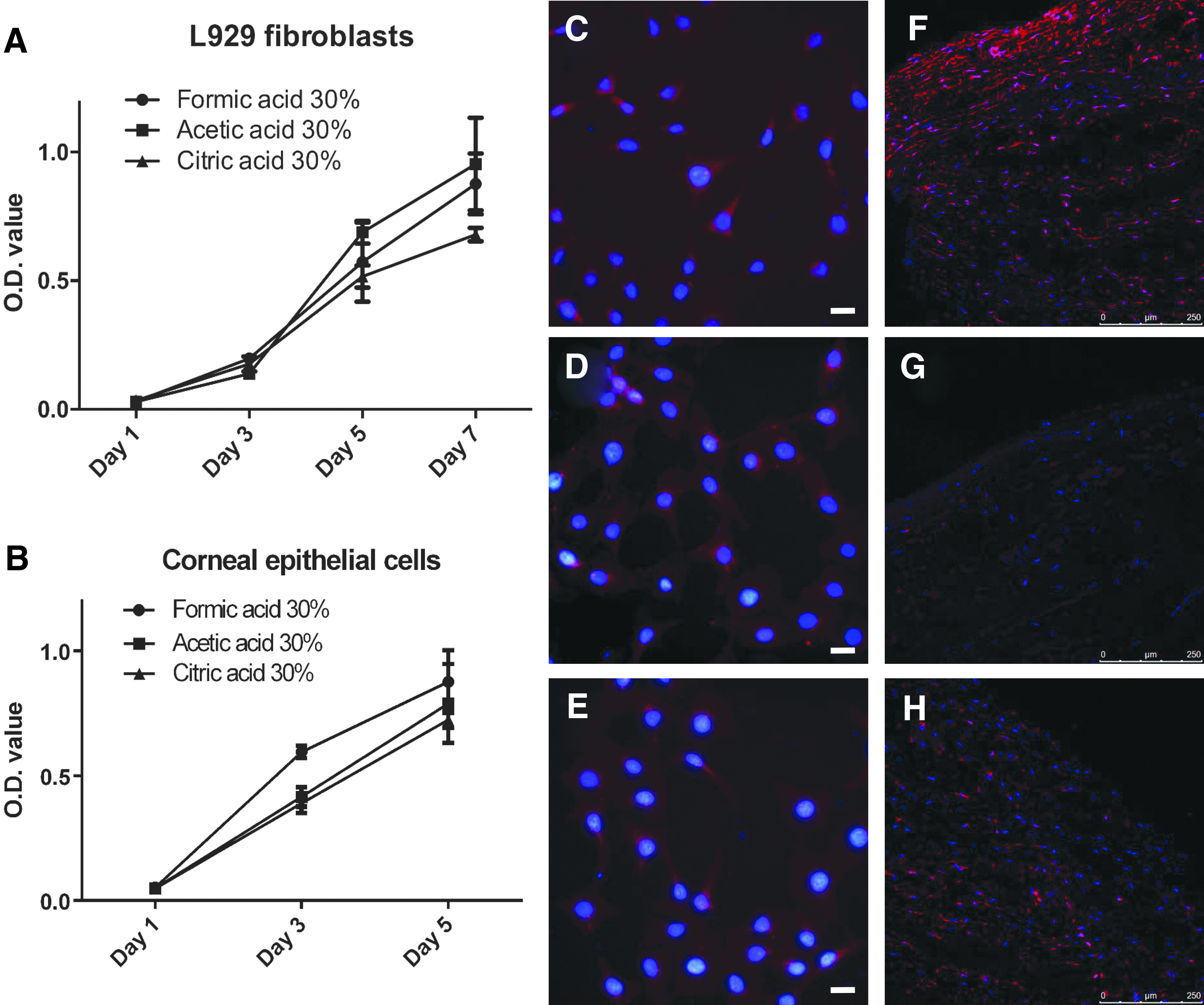
Biocompatibility of the dPC scaffolds.
The dPC scaffolds were injected with rabbit keratocytes and supported keratocytes growth for 3 weeks. DAPI and anti-vimentin staining confirmed that the rabbit keratocytes were distributed well inside the FA decellularized corneal scaffolds (Fig. 6F). In the CA dPC group, some of the cells exhibited the vimentin marker (Fig. 6G), and just a few cells exhibited the vimentin marker in the AA dPC group (Fig. 6H).
Implantation of the dPC scaffolds in vivo
As a whole, FA 30% treatment was superior among the various methods, with greater elimination of cellular debris, and sufficient maintenance of stromal collagen and GAGs contents. Thus, we chose this treatment for further in vivo implantation experiments. After DALK surgery, the implants were slightly hazy on day 1, and were completely transparent after day 7 (Fig. 7). To verify corneal epithelium recovery, the cornea was examined by fluorescein staining. We observed that epithelial cells had migrated toward the central portion of the cornea on day 3, and by day 7 the dPC was totally covered with epithelial cells. During the 60-day observation period, all implants remained transparent, and the epithelium had healed. All rabbits survived without any corneal rejection or neovascularization.
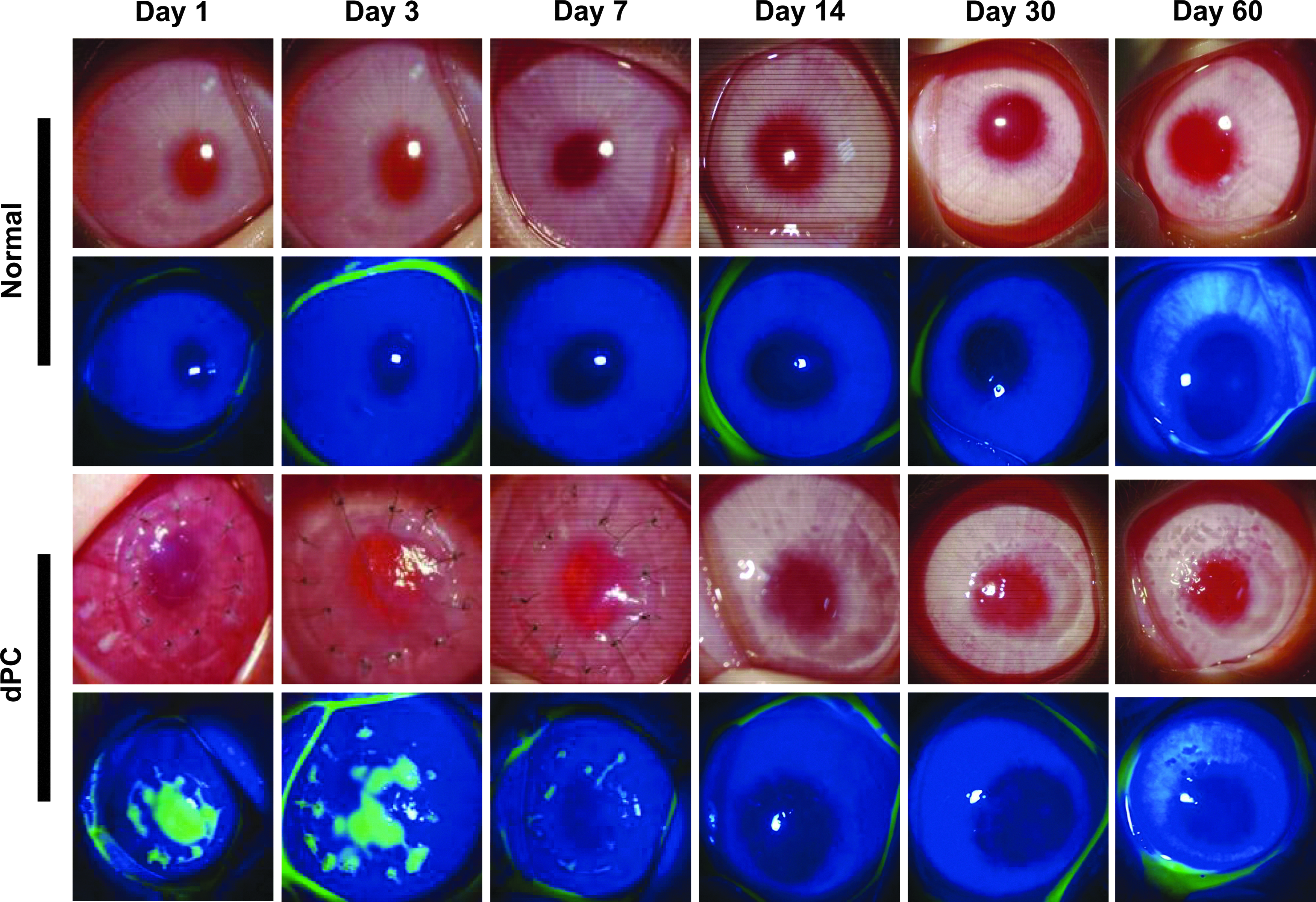
The dPCs were implanted in rabbit eyes by DALK treatment. Slit-lamp photographs and sodium fluorescein staining of rabbit eyes obtained on days 1, 3, 7, 14, 30, and 60.
The presence of the HE staining was detected in the middle portion of the stroma on day 60, showing dPC scaffold integrated very well with host tissue without any obvious gap, vascularization, or any sign of inflammation (Fig. 8B). Moreover, the stromal cells grew very well inside the dPC lamellar structure (Fig. 8D). Corneal thickness analysis revealed that the dPC was slightly thicker than the native cornea group for the first 3 days, but became normal during the following period (Fig. 8E).

HE staining and corneal thickness of rabbit corneas. HE staining of
Discussion
Decellularized corneal scaffolds have been studied for the past few years because they are thought to be the optimal biomaterial to solve the lack of corneal transplantation resources. A suitable decellularized corneal scaffold should have near-complete cell removal, optical clarity, ECM biological preservation, and biocompatibility.18,19 At present, the most widely used approaches to produce dPC involve detergent methods. Although using detergents for decellularization is very effective for cell removal, it is hard to completely eliminate the detergents, which have deleterious effects on ECM and make recellularization more difficult.20,21 Physical methods, such as using osmotic pressure, freeze–thaw cycles, and supercritical fluid extraction to disrupt cells, have also been explored for decellularization.22–24 The benefits of physical methods are its safety and it is easy to maintain the microstructure; however, the ECM is typically compromised, which influences the transparency. In addition, it is difficult to clear the cell debris inside the scaffolds.
In our study, we compared the use of three different organic acids (formic acid, acetic acid, and citric acid) for decellularization. Previous studies have examined acidic treatments to produce decellularized tendon, meniscus, and pericardium. 14 Nevertheless, high acid concentration always leads to changes in the ECM microstructure; acetic acid has poor decellularization ability, which was confirmed in our study. Acetic acid was associated with the presence of cellular residues that were evidenced by the DNA qualification assay and HE staining. On the contrary, we observed that formic acid and citric acid exhibited excellent decellularization efficiency for the removal of cellular and nuclear remnants. FA 30% and FA 40% decreased the DNA level on the corneal scaffold <3%, which is better than results using physical methods or detergent methods in our study and previous studies. 25 In addition, the results of ECM analysis revealed both GAGs and collagen content were not significantly disrupted by FA 20%, FA30%, AA 20%, CA 20%, and CA 30%. Examination of mechanical properties showed no significant difference in most organic acid treatment dPC groups, which suggested no significant degradation and disruption during the decellularized process, consistent with ECM components analysis results.
L929 is a common cell line for evaluating biocompatibility and cytotoxicity of biomaterials.26,27 The MTT assay demonstrated that cells steadily proliferate on dPC scaffolds, which indicated the organic acid treatment protocol had negligible cytotoxicity. The cell proliferation trends in all groups had a similar pattern of increase; only the CA 30% group had a slightly lower proliferation rate than the other groups on day 7, but the difference was not significant. HCECs showed an excellent proliferation profile and attachment on the dPC scaffolds in a 5-day cultured period. In vivo data also revealed rabbit epithelial cells, which might be derived from corneal epithelial stem cells located in the limbus, growing and adhering perfectly, and rabbit corneas were completely protected from sodium fluorescein penetration at 7 days. This result demonstrated that the dPC scaffolds were preferably suitable for epithelial cells to adhere, proliferate, and migrate. Compared with decellularization using the osmotic pressure method, which requires >2 weeks for subsequent complete epithelial closure, 28 our dPC scaffolds had a short reepithelialization time and no epithelial defects were observed.
The main purpose of corneal regeneration is to recover transparency, which is regulated by a uniform lamellar structure and cell remodeling. 29 In our study, SEM images indicated that the microstructure of the dPC had a lamellar morphology consistent with that of the native cornea, but the pore size was larger. This phenomenon might be because of the swelling effect of collagen fibrillar structure. 30 A previous study showed that cells did not migrate toward dPC that was decellularized by SDS during a 3-month follow-up period. 31 Another study also demonstrated that SDS, Triton X-100, or NaCl as decellularization agents produced condensed scaffolds that lacked a porous microstructure, and cells were rarely found inside the scaffolds after rabbit corneal implantation. 32 Therefore, it is necessary to produce large pores for successful cell growth in scaffolds. 33 In our study, we observed rabbit keratocytes grew very well when they were cultured inside the dPC, as evidenced by immunofluorescence staining. In vivo histological examination also showed cells evenly distributed in the dPC, suggesting our dPC had excellent biocompatibility and the porous structure was beneficial for recellularization.
To examine dPC optical properties, we detected visible light transmittance and found no difference when we compared it with the native cornea. This result indicated that dPC maintained an organized microstructure, which is beneficial for in vivo transparency recovery. Previous reports have suggested that the decellularized cornea becomes transparent within 2 weeks after surgery.23,34,35 In our study, in vivo implantation demonstrated that dPC became transparent at 7 days after implantation and maintained transparency without any signs of rejection during the full follow-up.
Conclusion
We found that porcine corneas were optimally decellularized by formic acid. The treatment preserved the optical properties, ECM architecture, and biomolecules, and recellularization potential of the dPC in vitro and in vivo. These results suggest that this organic acid decellularization method could generate a transplantable corneal scaffold with native cornea-like stromal architecture and biological properties. This could be a promising scaffold material for corneal tissue engineering applications.
Footnotes
Acknowledgment
This work was supported by the Ministry of Science and Technology, Taiwan under Grant (MOST105-2221-E-038-003-MY3); and the Council of Agriculture, Taiwan under Grant (106AS-6.2.1-ST-a1).
Disclosure Statement
There is no any potential conflict of interest.