




Abstract
Three-dimensional (3D) printing provides a novel approach to repair bone defects using customized biomimetic tissue scaffolds. To make a bone substitute closest to natural bone structure and composition, two different types of hydroxyapatite, Nano hydroxyapatite (nHA) and deproteinized bovine bone (DBB), were dispersed into collagen (CoL) to prepare the bioink for 3D printing. In doing so, a porous architecture was manufactured with 3D printing technology. The physical and chemical properties of the materials were evaluated, including biocompatibility and effect on the osteogenic differentiation of the human bone marrow-derived mesenchymal stem cells (hBMSCs). The XPS, XRD, FTIR, and the mechanical analysis of the material indicated that the two HA were consistent in their elements, but different in their chemical bonds and crystal phases. The SEM results showed the different surface morphologies of the HA crystals as well as the scaffolds, which would be the main factors affecting the internal porous structure of the scaffold. There were no differences between the two composite scaffolds in cell proliferations. FITC-phalloidin/vinculin/DAPI staining indicated that hBMSCs can adhere well to the 3D-printed surfaces. Alkaline phosphatase (ALP) staining reflected ALP expressed on both of the osteogenic-induction medium (OM) group, but not on proliferation medium (PM) group. The real-time polymerase chain reaction results showed the expression levels of osteogenesis-related genes RUNX2, SOX9, OCN, and COL1A1 in OM group were significantly increased after 7 days compared with the PM group (p < 0.01). The expression of SOX9, OCN, and COL1A1 in nHA/CoL scaffolds was higher than that in CoL scaffolds (p < 0.05). The expression of OCN and COLIA1 in DBB/CoL scaffolds was higher than that in CoL scaffolds (p < 0.05). In conclusion, the physicochemical and biological properties of 3D bioprinted scaffolds consisting of nHA/CoL or DBB/CoL would be well suited for the scaffolds to being a porous customized bone substitute, 3D printing scaffolds would be a prospective candidate for clinical application in future.
Impact Statement
Bone loss due to trauma, inflammation, and surgical processes has posed great difficulty in the aesthetic reconstruction of a functional alveolar bone. Tissue engineering and biomaterials, which can promote alveolar bone regeneration, have become a popular focus of current studies. Three-dimensional (3D) printing provides a novel approach to repair bone defects using customized biomimetic tissue scaffolds. Nano hydroxyapatite (nHA) and deproteinized bovine bone (DBB) are two materials mainly used in clinical practice, particularly DBB are widely used in dentistry and craniomaxillofacial orthosis because of the porosity characteristic. To make a bone substitute closest to natural bone structure and composition, nHA and DBB were dispersed into collagen (CoL) to prepare the bioink for 3D printing. The physicochemical and biological properties between the two 3D printing scaffolds were compared. Both nHA/CoL and DBB/CoL 3D printing scaffold would be promising candidate for the clinical applications in the future.
Introduction
In the past decade, significant advances have been made in three-dimensional (3D) printing technology, and the customized repair of bone defects has been applied successfully in clinics with metal laser printers. 1 The merits of 3D printing scaffolds include low complication risk, short operational time, and good molding effect during surgery.2–4 Compared with conventional metal printing or rapid prototyping, 3D bioprinting has been used to fabricate hydroxyapatite bound by different kinds of adhesive polymer scaffolds (CPS). The biodegradable of these polymer materials are able to guide the bone regeneration in situ.5,6 Among these, low temperature 3D bioprinting (LT-3DP) has been considered to be the most suitable method for bone defects repairs in the future, because it was used to generate a 3D blueprint of the patient's specific defects with high biocompatibility materials that are similar with the natural bone component and construction. 7
As a bone tissue engineering technology, the primary advantage of LT-3DP CPS is the potential to create polymer-mineral composites with enhanced material properties. 8 Also, the LT-3DP has great potential to fabricate tissues with multiple biocomposite materials and cell types, all of which are extremely important for the advancement of bone tissue engineering. Moreover, 3D porous scaffold designs are conducive to cell–matrix interactions and more efficient blood vessels in growth.9,10
To mimic the architecture of bone trabecular in LT-3DP, nano hydroxyapatite (nHA) is a good candidate for an appetite substitute of natural bone because of its high osteoconductive activity. Another alternative is a xenogenous bone, such as the deproteinized bovine bone (DBB), which is morphologically and structurally similar to human cancellous bone. Heat treatment has been suggested as an option to obtain protein-free bovine bone. 11 The crystalline phase composition of sintered bovine bone is similar to natural bone mineral that is composed of hydroxyapatite at about 93wt% and about 7wt% β-tricalcium phosphate (Ca3(PO4)2,β-TCP). 11 The heat-treated bovine bone has an interconnective porous structure (up to about 70vol% porosity) similar to the natural bone, hence allows faster bone in-growth. 12 Moreover, the hydroxylapatite obtained from bone and derived from powder processing has great potential as a bone substitute owing to its excellent biocompatible and osteoconductive properties.13,14
Usually, LT-3DP provides the potential to create composites with synthetic or biological polymers, such as collagen. 15 Type I collagen (CoL-I) is the most abundant structural protein in the human body, and an important component of bone tissue. Collagen is also a critical component of bone extracellular matrix, where it plays important roles in the mineralized tissue's strength and toughness. It has been shown that incorporating collagen into mineralized bone cements could enhance their biomechanical properties. Collagen can induce the mineral deposition, and its surface contains sites that promote osteoblast adsorption and mineral deposition. Moreover, it can effectively enhance the mineralization process and develop new bone in the implanted artificial bone scaffold. 16 This kind of polymer can retain but does not occlude the osteoinductive calcium phosphate component and has osteoconductive and osteoinductive characteristics. 17 For example, collagen incorporation into hand-mixed hydroxyapatite has been shown to improve cellular attachment, viability, proliferation, and activity as well as mechanical properties.17,18
In this study, nHA and DBB were mixed with CoL-I into biocomposite hydrogel, respectively, to create two types of bioinks. To build up a porous bone scaffold within the biomimetic component, as well as the trabecula construction, the two bioinks were printed with the LT-3DP technology. BMSCs were used to observe the topographies and morphologies of cells adherence on the two 3DP scaffolds. The cellular biocompatibility, including the proliferation and osteogenic differentiation, were compared and evaluated. The application of two hydrogels used as “bioinks” in LT-3DP and these biological properties were compared and evaluated.
Materials and Methods
Preparation of DBB
DBB was prepared by calcining bovine cancellous bone following the method of Wang. 19 In brief, the bovine bone was cut into small cubes, and successively being incubated in H2O2 solution and stewed in a NaOH solution to remove collagen and proteins. The bone cubes were then washed with water, immersed in H3PO4 solution, and heated, respectively. This was followed by washing with absolute ethyl alcohol to remove moisture. After that, the bone cubes were air-dried and heated up to 1000°C. Finally the bone cubes were ground and filtered through a 120-screen cloth to obtain DBB particles with a diameter of 105–125 μm. 19
Properties of hydroxyapatite
The nHA (Aladdin) (≥Alad <100 nm particle size) and DBB samples were mounted on conductive adhesive tape and sputter coated with Au, and then observed using a scanning electron microscopy (SEM) (JSM 6700F, Japan) at a magnification between 1000 × and 10,000 × . The composition of the nHA and DBB were investigated by X-ray photoelectron spectroscopy (XPS) (ESCALAB 250Xi USA) using a monochromatic Al Kα source (10 mA, 15 kV).
Preparation of the bioink
The bioinks were divided into three groups, which were, respectively, CoL, nHA/CoL, and DBB/CoL groups. Type I bovine tendon collagen sponge (Collagen Biotechnology Co. Ltd., China) was dispersed in acetic acid of 0.05 mol/mL at a concentration of 3 g/100 mL, then collagen gel was gotten to be the CoL group. nHA and DBB were, respectively, added into the collagen gel with constant stirring at 45 rpm for 12 h at 4°C temperature, the nHA/CoL and DBB/CoL bioink were obtained. 20 The concentration of the HA in biocomposite ink was 0.75 g/100mL.
Three-dimensional bioprinting
The 3D Bioplotter (Envision Tec, Germany) was used to produce the 3D bioprinting scaffolds. For building 3D construction, the cuboid model was designed by Image ware 13.1. The distance between each printed line was 1.5 mm. The hydrogel scaffolds were fabricated by layer-by-layer deposition, and a 15 × 15 × 5 mm3 architecture was gotten (Fig. 1A, B). The parameters of the 3D Bioplotter were determined after repeated printing experimentations to ensure the consistency and repeatability of the 3D bioprinted constructions. Related parameters are presented in Table 1. When each printed scaffold was completed, freeze-drying was conducted to obtain the freeze-dried scaffolds. After that, the scaffolds were crosslinked with 0.2% glutaraldehyde solution (80% ethanol aqueous solution) for 2 h at room temperature, then rinsed in deionized water several times to remove residual glutaraldehyde. The crosslinked scaffolds were put into freeze-dryer again to get the final specimen. The sterile operations were carried out during the processes.
The Printing Parameters of the Three-Dimensional Bioplotter
SEM of the 3D bioprinting scaffold
The freeze-dried scaffolds were coated with Au and investigated with SEM at a magnification between 100 × and 2000 × as the method of “Properties of hydroxyapatite” above.
Fourier transform infrared spectroscopy analysis
The chemical composition of the scaffolds was investigated by Fourier transform infrared spectroscopy (FTIR; Spectrum GX). Each of samples was ground together with KBr at a rate about 1:20 in an agate mortar and compressed to tablets. The spectra ranges were set as 4000 to 400 cm−1 with a resolution of 4 cm−1, and the scan time was about 100 s. Pure collagen sponge served as a reference.
X-ray diffraction analysis
The scaffolds lyophilized were cut into small cubes at 20 × 20 × 2 mm, and gently flattened before analysis. The crystal phase compositions of each scaffold were investigated by X-ray diffraction (XRD, SmartLab X, Japan). The materials XRD spectra were taken using nickel-filtered Cu Kα radiation at 40 kV and 200 mA. Spectra were recorded from 2θ = 5°to 90°at a scanning speed of 15°/min and a step size of 0.02°. nHA and DBB powder served as a reference and was heated to 100°C for 12 h before use.
Mechanical properties of scaffolds
The Young's modulus of the three groups of scaffold were determined using a Universal Testing Machine (Instron 300DX, US). The dimensions of each specimen were measured using a vernier caliper before detection. Then, the scaffolds were placed on the detecting platform and loaded at the rate of 1 mm/min until the scaffolds crushed. The force and displacement data were recorded to generate the stress–strain curves. By measuring the slope of the stress–strain curve in the elastic region, Young's modulus could be calculated. Each group was detected five times, and the results are displayed in the form of mean ± standard deviation.
Evaluation of the biological characteristics of the bioprinting materials
Cell culture
Human bone marrow-derived mesenchymal stem cells (hBMSCs) were purchased from ScienCell (ScienCell) and cultured in 10 cm dishes with Dulbecco's modified Eagle's medium (DMEM) supplemented with 10% (v/v) fetal bovine serum (FBS) in incubator (37°C, 5% CO2 and 100% relative humidity). The medium was changed three times a week until passage 3 (P3). After being digested and counted, the cell suspension was concentrated and suspended in DMEM (10% FBS) to adjust the cell number to 6 × 10⁷/mL. After placing the sterile 3D printing scaffolds in a 24-well culture plate and wetting the materials with proliferation medium (PM) beforehand, each scaffold sample was seeded with 100 μL of cell suspension. After 4 h of cell adhesion to the scaffold in incubator, 900 μL of DMEM (10% FBS) was added to each well. The medium was replaced every other day.
Immunofluorescence staining
The immunofluorescence staining of hBMSCs cytoskeleton was conducted. After 24 h of incubation on each scaffold, the cells were fixed in 4% paraformaldehyde for 10 min at room temperature, permeabilized with 0.1% TritonTMX-100 PBS solution for 5 min, and blocked using 1% bovine serum albumin (BSA) for 30 min at room temperature. Then, the cells were incubated with FITC-phalloidin (Sigma-Aldrich) at a concentration of 5 μg/mL with each well 0.5 mL for 30 min, followed by incubation in DAPI (5 μg/mL) for 5 min in the dark at room temperature. Each incubation was followed by three times of PBS washing. Confocal Laser Scanning Microscope (CLSM, Olympus IX71, Fluoview, Japan) was used to observe the images. 21
Vinculin staining
The vinculin staining of the hBMSCs was conducted to observe the cell adhesion on scaffold. After fixed and permeabilized, 3% hydrogen peroxide was added for 10 min and then 3% goat serum was added for 1 h at 4°C. First antibody of vinculin was added at a scale of 1:200 dilute solutions for the night. Second antibody was added at a scale of 1:500 dilution solutions for 1 h in the dark. With PBS washes after incubation, the cells were counterstained with DAPI for nuclear staining and visualized using a CLSM.
Cell proliferating assay
P3 hBMSCs were counted after digested, centrifuged, and resuspended. The cell suspension was added into each scaffolds, with 1, 5000 cells each well (n = 5). After culturing for 1, 3, 5, and 7 days, cell proliferation was determined using the CCK-8 method. The Optical Density (OD) was measured at 450 nm using a microplate reader (Bio-Kinetics Reader, Bio-Tek Instruments). Cells grown on the CoL scaffold were used as a control. The measurements were performed in triplicate for each group (n = 3). Cell proliferation trends were drawn accodring to cell culture time and the OD value.
Cell osteogenic induction
The hBMSCs cells were cultured on 3D scaffolds with PM for 24 h. Osteogenic induction was conducted. For inducing osteogenesis, osteogenic-induction medium (OM) comprising 10 mM β-glycerophosphate, 100 nM dexamethasone, and 200 μM ascorbic acid was used. The OM was replaced every other day. After 7 days of culturing, alkaline phosphatase (ALP) staining was used to detect the differences of the expression in each group. The cells total RNA was also extracted to test the impact of osteogenesis gene expression differences of each printed scaffold to cells.
ALP staining
The level of ALP activity was examined on day 7 using an ALP kit (Corning Incorporated) according to the manufacturer's protocol. The medium in each well was aspirated and, then, fixed with 95% ethanol for 30 min. Counterstaining consisted of BCIP, NBT, and buffer at a rate of 1:1:38 and was added to each well with a volume of 500 μL for 30 min in the dark. The staining solution was aspirated and washed with PBS three times. Record the results of ALP staining under the inverted phase contrast microscope.
Quantitative real-time polymerase chain reaction assay
Cells cultured on printed 3D constructs for 7 days were analyzed for gene expression (n = 3). Total RNA was extracted using TRIzol reagent (Invitrogen) by following the manufacturer's protocol. SYBR FAST qPCR Kit Master Mix (2BR; KAPA Biosystems) was used for quantitative Real-time PCR, and the HiFiScript Rapid De-Genomic cDNA First-Chain Synthesis Kit (Corning Incorporated) was used for reverse transcription. The target PCR primers for Runt-related transcription factor (RUNX2), Osteocalcin (OCN), SRY-related HMG box gene 9 (SOX9), and Collagen I a1 (COLIA1) were synthesized by Invitrogen (Table 1). β-Actin was used as an internal standard. The cycle threshold values (Ct values) were used to calculate the fold differences by the Ct method (also known as the 2−△△Ct method).
Statistical analysis
Data are represented as the mean ± standard deviation and were analyzed using SPSS 17.0 software. In terms of statistical comparison, this study adopted the t-test and after confirmation of homogeneity of variance, one-way ANOVA as well as the LSD test were performed. The significance level was α = 0.05. *p < 0.05 was considered to indicate statistical significance.
Results
Physical and chemical properties of printed 3D scaffolds
XPS analysis
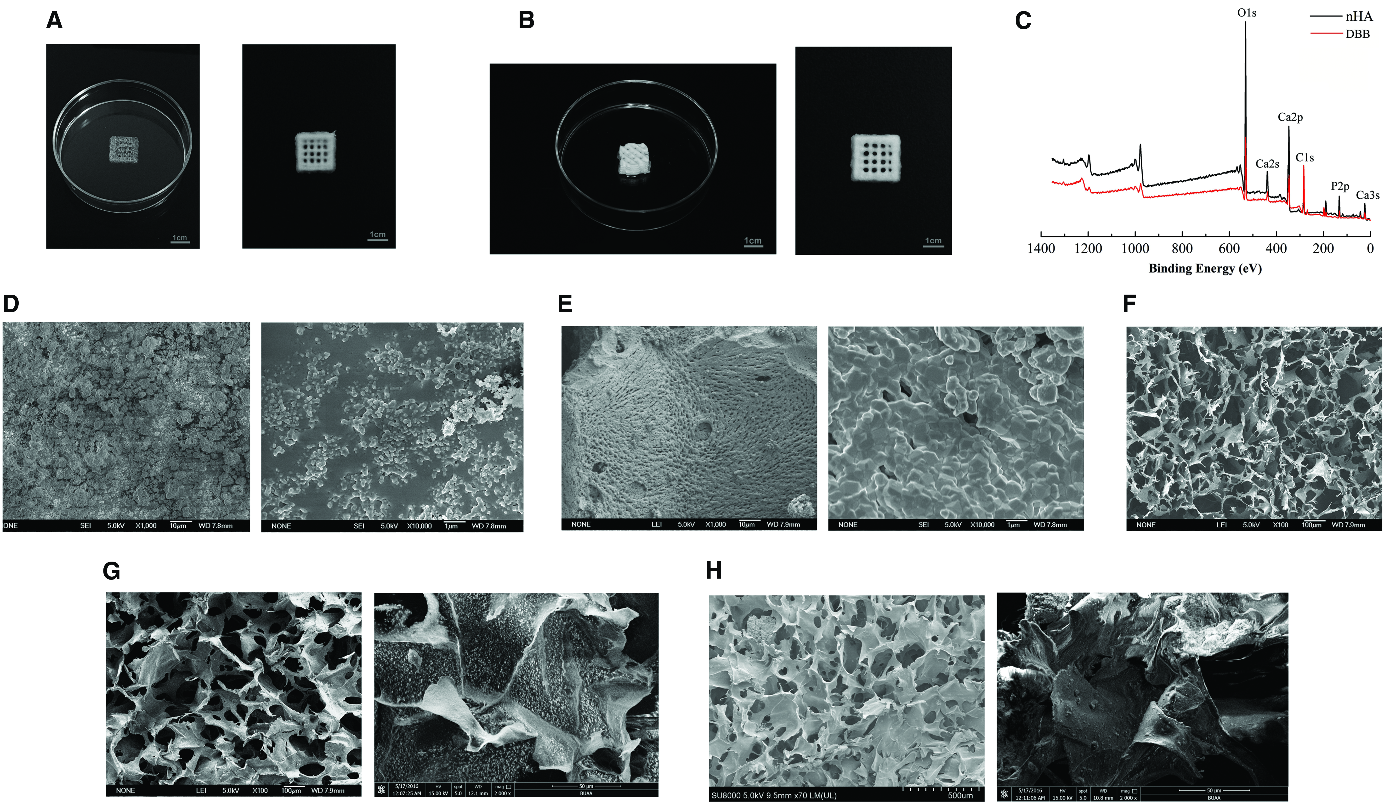
The XPS spectrum in Figure 1C exhibits the characteristic elements of Ca, P, O, and C elements in nHA and DBB crystal. The diffraction peaks were analyzed using CASA XPS software and the Ca/P ratio calculated. The result displayed that Ca/P ratio is 1.67 for nHA and 1.58 for DBB, both of which were consistent with those reported previously.22–26 The Ca/P ratio in DBB is lower than in nHA possibly because the Ca10 (PO4)6(OH) 2 in the calf bone was partially converted to β-Ca3 (PO4)2 during the calcination process, resulting the Ca/P ratio in DBB less than 1.67 in hydroxyapatite.9,10
SEM of nHA and DBB
The surface morphology of the two HA under SEM is shown in Figure 1D and E. A few agglomeration particles were observed in nHA (Fig. 1D). While the surface images of DBB (Fig. 1E) exhibited the evenly distributed micropores, and few interconnecttions between the pores, which is similar with the nature bone trabecula. The micropore diameter from DBB was about 1 μm, which were not presented in nHA. Both nHA and DBB crystals were spherical and have similar diameters in the images inserted to (Fig. 1D, E).
The construction of 3D printed scaffold
The scaffolds printed were fabricated successfully as shown in Figure 1A and B. After printing, a grid-like structure was constructed. The diameter of up and down connected square hole is 600 μm. The DBB scaffolds were more feasible to bottom up a higher construction during the printing (Fig. 1B).
SEM of 3D printed scaffold
The surface morphology of the scaffold after being lyophilized under SEM is shown in Figure 1F–H. All the scaffolds had porous structure, and the pore size was ranged from 100 to 300 μm. The CoL scaffold (Fig. 1F) was presented an irregular lamellar structure and the heterogeneous pore distribution with pore size ranged from 60 to 300 μm, and a little deformation of the pores could be seen on collagen scaffold. The surface morphology of nHA/CoL and DBB/CoL scaffolds (Fig. 1G, H) showed the regular porous structure distributed. The images inserted to (Fig. 1G, H) were magnified to see the details of nHA/CoL and DBB/CoL scaffolds. nHA evenly distributed on the collagen surface due to the small particle, while the DBB particle could be clearly seen in DBB/CoL scaffolds surface.
FTIR analysis
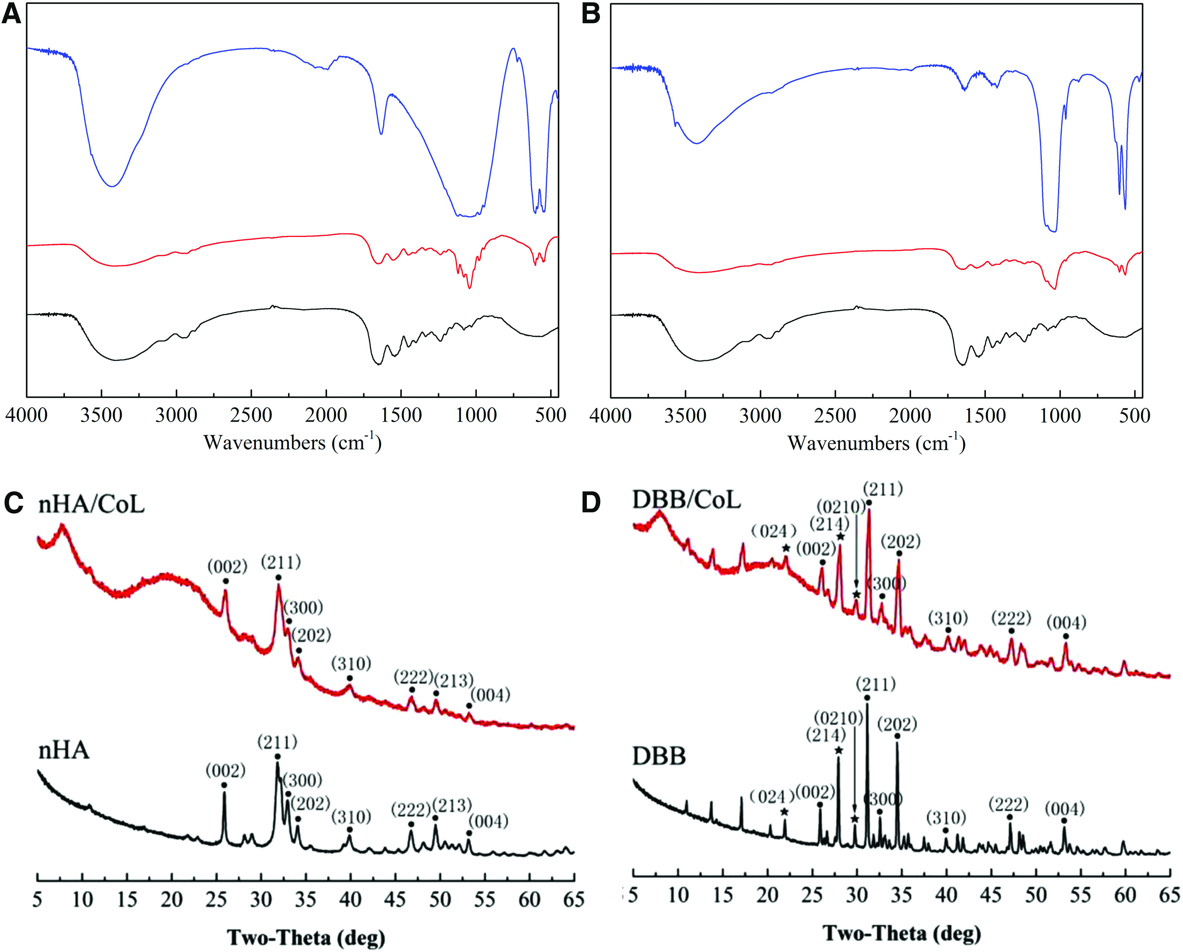
Figure 2A and B show the FTIR spectra of the material and scaffolds. In nHA, the peaks at 3568.2 and 3440.5 cm−1 belong to the OH- stretching vibration of hydroxyl. 27 Furthermore, the major bonds associated with PO43-(1037, 603, and 567 cm−1) appeared in nHA and DBB. 28 The peak at 3330–3428.6 cm−1 belongs to the N = H stretching of Amide A. The peak at 1655 cm−1 is C = O absorption of Amide I, 1550 cm−1 is C-N stretching and C-H bending combination vibration of Amide II, while 1237 cm−1 is consist of C-N stretching, N-H bending and C-C stretching of Amide III.29–31 From the FTIR spectra, the two composite scaffolds are consistent with vibration of PO43− in 1037, 603, and 567 cm−1. However, the peaks at 970 and 1127 cm−1 in DBB/CoL are the PO43- absorption of TCP, 28 which were not presented in nHA/CoL scaffolds.
XRD analysis
The XRD analysis of the material and scaffolds is shown in Figure 2C and D. The crystalline phases of both nHA and DBB were consistent with the phases incorporated in the International Centre for Diffraction Data (ICDD) databases. The analysis of nHA and DBB was based on the Ca10(PO4)6(OH)2 phase (JCPDS No.41-0487).31,32 Peaks at 8° and 21.08°in both scaffolds were the diffraction reflecting the amorphous structure of collagen. 33 The main miller indices for TCP were (024), (214), and (0210), which were included in DBB as well as the DBB/CoL scaffold as shown in Figure 2D. 32
Mechanical properties (Young's Modulus)
Young's Modulus of 3D printed scaffolds was detected as shown in Figure 3. The result clearly indicates that the Young's Modulus was 7.9 ± 0.3 MPa in nHA/CoL group that has significant differences when compared with 3.5 ± 0.4 MPa in CoL group (p < 0.01) and 4.5 ± 0.7 MPa in DBB/CoL group (p < 0.05), respectively. The difference between CoL and DBB/CoL group was not obvious.

Young's moduli of CoL, nHA/CoL, and DBB/CoL group (*p < 0.05; **p < 0.01). The Young's moduli of nHA/CoL was significantly stranger then CoL (**p < 0.01) and DBB/CoL scaffold (*p < 0.05), respectively; DBB/CoL was stranger then CoL scaffold (*p < 0.05).
CLSM analysis of hBMSCs on 3D scaffolds
FITC-phalloidin, vinculin, and DAPI staining (Fig. 4A–C) revealed that hBMSCs were firmly adhered to the scaffolds after 24 h seeding. The FITC-phalloidin and DAPI staining indicated that cells distributed on the skeleton structure and inner sides of the scaffolds. The specific triple helix of the collagen as well as the porous structure of the scaffolds made a strong adsorption of phalloidin leading the scaffolds dyed as green, while the cell microfilaments were not stained, and the pore-size and porosity of scaffolds can be reflected due to the fluorescent staining.

Immunofluorescent staining for DAPI-stained nuclei of cells in 3D constructs after day 1;
Vinculin staining (Fig. 4D–F) showed that hBMSCs cells attached scaffolds well. The cellular pseudopods can be clearly seen in images. Cell adhesive plaques were evenly distributed in the cytoplasm and enriched at the end of cell pseudopodia, which showed enhanced fluorescence signal. The cells on the nHA/CoL and DBB/CoL scaffolds altered to a plate-like morphology in contrast to the spindle morphology observed on CoL scaffolds. The white boxes (Fig. 4E, F) represented nHA and the degradation particles of DBB, respectively. The cells can be seen well grown around the HA parties in scaffolds.
Proliferation of hBMSCs in 3D scaffolds
The cells proliferation in different scaffolds was evaluated by CCK-8 assay at 1, 3, 5, and 7 days after seeding as shown in Figure 5. The OD values of all groups increased over time. The growth curves indicated that the overall growth tendencies in both groups were similar during the culture process. However, the cell proliferation was higher in CoL group than in nHA/CoL and DBB/CoL group at 5th and 7th day (p < 0.05). The cells proliferation on nHA/CoL and DBB/CoL was consistent and had no significant difference (p > 0.05).

hBMSCs proliferation on scaffolds using the CCK8 assay at 1, 3, 5, and 7 days after seeding. hBMSC, human bone marrow-derived mesenchymal stem cell. Color images are available online.
ALP staining
The osteogenic potential of hBMSCs in different scaffolds was evaluated by ALP staining as shown in Figure 6. After 7 days of culturing, the result showed that cells in PM did not display significantly staining (Fig. 6A–C), while cells in OM showed positive ALP staining (Fig. 6D–F). The brown spindle morphology in red dashed box (Fig. 6D–F) represented the expression of ALP in MSCs cells, respectively. Cells in PM showed more obvious proliferation effect, and the numbers of hBMSCs cells in PM were greater than that in OM group.

ALP staining of hBMSCs on scaffolds 7 days after seeding (100 × ).
Osteogenesis-related gene expression in hBMSCs
Osteogenesis-related gene expression could reflect the differentiation of hBMSCs into osteoblasts. After the culture of hBMSCs on three groups of scaffolds for 7 days, the mRNA levels of osteogenesis-related genes of OCN, RUNX2, SOX9, and COLIA1 were detected using real-time polymerase chain reaction (RT-PCR), and the results are shown in Figure 7. As the results display, the osteogenesis-related genes expression in OM group are significantly higher than the parallel group dealt with PM (p < 0.01). In OM group, the expression of SOX9, OCN, and COLIA1 gene in hBMSCs cultured in nHA/CoL scaffolds is significantly higher than that in CoL scaffolds (p < 0.05). The RUNX2 expression in nHA/CoL scaffolds is lower than that in CoL and DBB/CoL scaffolds (p < 0.05). The expression of OCN and COLIA1 in DBB/CoL scaffolds was higher than that in CoL (p < 0.05). Transcription level had no significant difference between the gene of RUNX2 and SOX9 in the OM group in CoL and DBB/CoL scaffolds (p > 0.05). In PM group, there was no obvious difference in OCN expression among the three groups of scaffolds. The expression of RUNX2 and SOX9 was higher in PM group in DBB/CoL scaffolds than the same set of that in CoL and nHA/CoL scaffolds, respectively. At day 7, the expression level of COLIA1 in CoL scaffolds was higher than that in nHA/CoL and DBB/CoL scaffolds in PM group (p < 0.05).
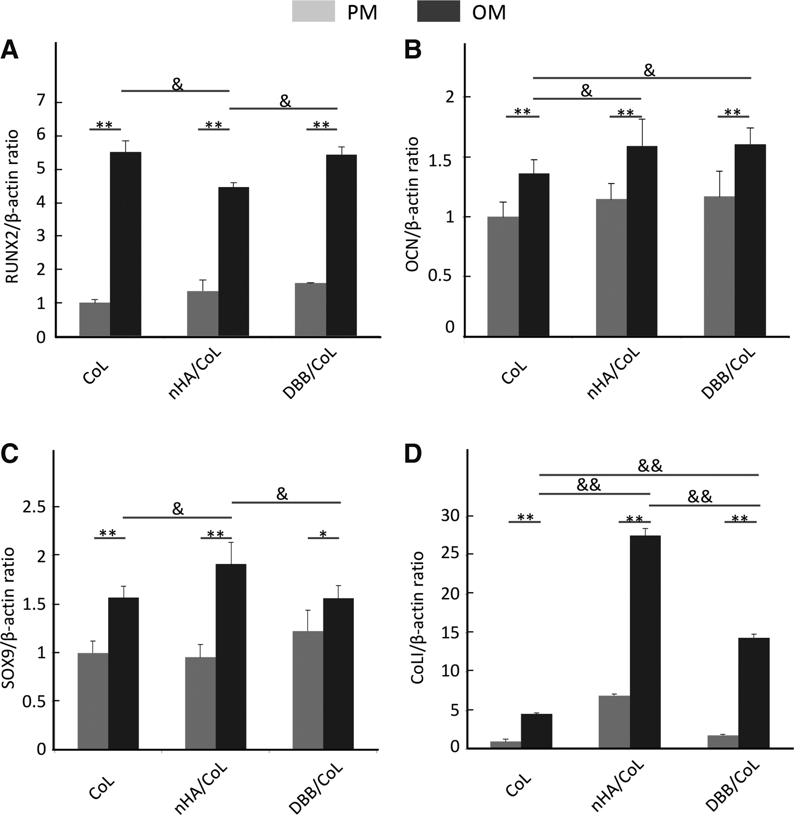
Expression of RUNX2
Discussion
Hydroxyapatite is a primary focus for synthetic bone graft substitutes because they are osteoconductive and provide sufficient mechanical strength. 25 Both nHA and DBB offer several biological advantages as a bone analog, and the HA-collagen composition has been widely applied in clinics as a commercial product.7,9,33–35 Although a variety of materials have been processed into porous 3D scaffolds using various 3D technologies,36–39 the nHA and DBB materials have not previously been mixed with collagen into hydrogel and printed into 3D scaffolds. In this study, two kinds of hydroxyapatite, nHA and DBB, were mixed with collagen, respectively, and printed into specific forms to compare the physical characteristics and the osteogenesis-promoting effects on hBMSCs and to determine a potential candidate for future studies in clinical application.
Three-dimensional bioprinting technology has the advantage of “personal customization” over molding or paste injection, including patient-specific geometries 7 and controlled spatial patterning of drugs or polymers within the scaffold. 8 To achieve this aim, the physical properties of the materials, such as powder granularity, roughness, wettability, or binder viscosity, must satisfy the demands of the complexity of 3D printing, which limits the types of materials that can be printed at low temperature. 40 In this article, a powder of nHA and DBB mixed with Type I collagen was printed into a 3D scaffold, while pure collagen scaffolds served as control group. Based on previous research, glutaraldehyde was applied for crosslinking to enhance the mechanical properties and maintain the structure of the scaffolds. 23
The XPS analysis showed the element composition of the two inorganic materials. The elemental ratio of the two inorganic materials is consistent with what has been previously reported.21,41 The FTIR and XRD analysis confirmed that there is a single ingredient—hydroxyapatite in nHA, while there is not only existence of the HA but also β-TCP in DBB. The DBB degrades and resolves faster compared to HA because of the high biodegradation rate and solubility of β-TCP. 23 With the β-TCP degradation, the calcium ions and phosphate released can promote osteogenesis by favoring the synthesis of osteoinductive growth factors and by upregulating adenosine signaling in phosphate9,42,43 metabolism. Some literature has reported that HA and β-TCP composites have a better osteogenesis effect than using HA only.8,41
Excellent mechanical properties not only provide a mechanical support for bone formation but also make the materials conducive for use in clinical applications. 21 Young's moduli of bioinks were examined before being printed into tissue constructs with the bioprinter. The compressive modulus of the scaffolds is 7.9 ± 0.3 MPa in nHA/CoL group and 4.5 ± 0.7 in DBB/CoL group, both of which match that of cancellous bone (about 2–20MPa).9,32,33 The compressive modulus of nHA/CoL and DBB/CoL scaffolds was higher than that in the group of CoL scaffolds, which illustrates that both nHA and DBB can enhance the compressive modulus of collagen. The phenomenon occurred because of collagen's affinity to Ca2+ as well as crosslinking of using glutaraldehyde, which can increase the mechanics of collagen by increasing its resistance to fibril rotation.23,34 Collagen plays a role in crystallizing and mineralizing templates during the process of HA crystallization, and has the function of guiding the distribution of HA particles along the surface of collagen fibers. 44 Meanwhile, the combination of collagen and Ca2+ in the material have possibility to mineralize the collagen fiber and thus may increase the Young's modulus of the composite scaffolds. 45 nHA has a smaller particle size than DBB, and therefore has a larger surface area, making the Ca2+ easy to dissolve into collagen. Hence, nHA/CoL has higher compressive modulus than the other two groups.
After the composite “bioink” was printed into 3D scaffold by the 3D bioprinter, a specific architecture was built up. After being printed, the 3D scaffold had a grid-like microstructure with pore widths of 600 μm. Due to this porous structure, the DBB scaffolds were more feasible to bottom up a higher construction during the printing. The SEM analysis after lyophilization showed that both of the bioprinted scaffolds have a relatively smooth surface, due to the compression of the tip mold and nozzle. Internally, the scaffold presented the typical characteristics of cancellous bone, a honeycomb microstructure evenly distributed with 100 μm pores. The pores are assumed to be created by the sublimation of the solvent during lyophilization. 46 The high-resolution SEM images of nHA/CoL reflected the uniform particle distribution of nHA, which resulted in a more integrated structure. As to the DBB particles, because of the particle size and the internal pores, BDD/CoL group presented a porosity structure, including the micropores inside the materials and the macropores fabricated by the 3D printers.
A series of studies have demonstrated that the mineral properties, including solubility, chemical composition, and surface morphology were important factors for regulating diverse cell behaviors.36–38 The different properties of the two inorganic parameters may have a diverse effect on cell behavior. The hBMSCs have obvious multipotent differentiation abilities and are the most common cells in bone tissue engineering.7,22,23 In this study, hBMSCs were used to detect the effect of the two inorganic materials on the osteogenic differentiation.
The FITC-phalloidin/PI staining intuitively displayed the growth and distribution of cells on 3D bioprinted constructs. The number of cells in the collagen scaffold group was higher than that of the other two groups. The two sets of printed scaffolds have larger pore size than the collagen group, which caused a lower seeding efficiency, consistent with other research. 41 Assessment of the biosafety of scaffolds is of primary importance. Vinculin staining and CCK-8 assay demonstrated that the bioprinted scaffolds were nontoxic to hBMSCs in vitro. Vinculin staining results proved that vinculin was expressed around almost all cells, which indicated that hBMSCs could show adhesive growth in both the collagen matrix and the composite matrix and illustrated that the adhesion ability was similar on all three types of scaffolds. The CCK-8 assay results indicated that the cells still had proliferative capacity in the bioprinted matrix material. After 3 days of incubation, the cells in collagen group had a significant proliferative effect compared with the composite group. The difference might be caused by the lower seeding efficiency in the nHA/CoL and DBB/CoL group as mentioned above. Moreover, the ions from disintegrated inorganic materials, such as Ca2+ and PO43- and so on, might influence the OD values. ALP staining is an indicator reflecting the ability of osteoblastic differentiation of hBMSCs. After 7 days in OM, hBMSCs showed positive ALP staining, while the PM group did not show significant positive tests results, consistent with the previous report. 23
The genes related to osteogenic differentiation, including RUNX2, OCN, SOX8, and COLIA1, have always been considered an important factor in early osteogenic differentiation, while SOX9 and OCN are marker proteins in late osteogenic differentiation.41,42 CoLI are important components of the ECM. The expression of COLIA1 increased, indicating an increase in the synthesis of nonmineralized ECM, which is an important step in bone formation.9,11 The RT-PCR results indicate that the expression of the osteogenic differentiation-related genes RUNX2, OCN, SOX8, and COLIA1 in the OM groups were significantly higher than that in all the PM groups. The RANKL/OPG balance regulated by osteoclast/osteoblast interactions plays a key role in bone regeneration. The relative expression ratio of RUNX2 in the nHA/Col group was lower than the ratio of CoL and DBB/CoL in the OM group. The OCN and COLIA1 expression in DBB/CoL was significantly higher than in the CoL group. The results in the PM group were consistent with the OM. The osteogenic differentiation-related genes SOX9 and COLIA1 in hBMSCs have higher expression in the nHA/CoL and DBB/CoL groups than in CoL group after 7 days of incubation, especially in the nHA/CoL group, which indicated that Ca2+ released by nHA promoted osteoblast proliferation and the expression of osteogenic genes. 9
From the above, the DBB were originally from natural bone, and the micro- and macroporous structures were a benefit to vascularization during bone defect repair. To obtain the best applicable ratio, and explore the most biomimetic natural bone for clinical application of the printing process, the difficulties of the particle size and irregular shape of this trabecular bone graft were overcome in this study.
Conclusion
Both two composite bioinks, nHA/CoL and DBB/CoL, were used to print biomimic 3D scaffolds successfully. The two kinds of hydroxyapatite composite which favor hBMSCs proliferation and differentiation proved to be promising candidates for a 3D scaffold bioink. A porous architecture was manufactured eventually. Further studies would need to be concentrated on the DBB as a bioink to build up a 3D printing customized bone substitute in clinical application.
Footnotes
Authors' Contributions
Qing Li and Xiongxin Lei equally contributed to this article. Peijun Lyu and Guifeng Zhang are corresponding authors.
Acknowledgment
This work was supported by the National Key Research and Development Program of China (2016YFC1102900).
Disclosure Statement
No competing financial interests exist.