
Abstract
Temporal and spatial presentations of biological cues are critical for tissue engineering. There is a great need in improving the incorporation of bioagent(s) (specifically growth factor(s) [GF(s)]) onto three-dimensional scaffolds. In this study, we developed a process to combine additive manufacturing (AM) technology with acoustic droplet ejection (ADE) technology to control GF distribution. More specifically, we implemented ADE to control the distribution of recombinant human bone morphogenetic protein-2 (rhBMP-2) onto polycaprolactone (PCL)-based tissue engineering constructs (TECs). Three substrates were used in this study: (1) succinimide-terminated PCL (PCL-N-hydroxysuccinimide [NHS]) as model material, (2) alkali-treated PCL (PCL-NaOH) as first control material, and (3) fibrin-coated PCL (PCL-Fibrin) as second control material. It was shown that our process enables a pattern of BMP-2 spots of ∼250 μm in diameter with ∼700 μm center-to-center spacing. An initial concentration of BMP-2 higher than 300 μg/L was required to retain a detectable amount of GF on the substrate after a wash with phosphate-buffered solution. However, to obtain detectable osteogenic differentiation of C2C12 cells, the initial concentration of BMP-2 higher than 750 μg/L was needed. The cells on PCL-NHS samples showed spatial alkaline phosphatase staining correlating with local patterns of BMP-2, although the intensity was lower than the controls (PCL-NaOH and PCL-Fibrin). Our results have demonstrated that the developed AM-ADE process holds great promise in creating TECs with highly controlled GF patterning.
Impact statement
The combined process of additive manufacturing with acoustic droplet ejection to control growth factor (GF) distribution across three-dimensional (3D) porous scaffolds that is presented in this study enables creating 3D tissue engineering constructs with highly controlled GF patterning. Such constructs enable temporal and spatial presentations of biological cues for enhancing cell migration and differentiation and eventually the formation of targeted tissues in vitro and in vivo.
Introduction
One of the fundamental concepts underlying tissue engineering is to incorporate biologically active molecules/agents such as growth factors (GFs) within a scaffold to present these signals at the appropriate time and location that promotes repair and regeneration of tissues by stimulating cellular migration, proliferation, and cellular differentiation.1,2 To improve the efficiency of the functionality of such tissue engineering constructs (TECs), temporally and spatially controlled presentations of biological cues and patterns are needed. 3 More specifically, controlled spatial distribution of the GFs across the constructs in terms of precise position and accurate amount should be enabled. Several methods, including physical entrapment of GFs within porous structures and surface binding of GFs, have been reported for fabrication of GF-loaded TECs.4–6 However, most of the current methods of GF loading are either not in a spatially controlled manner, which can potentially lead to unguided or uncontrolled pathological side effects, or are only applicable for two-dimensional (2D) constructs, which have limited medical relevance in terms of defect repair and tissue regeneration. 7 In addition to fabrication limitations, one major obstacle is to modify the TEC constituent biomaterials or their surface to include chemical or physical binding sites for biomolecules to minimize rapid dissipation of loaded GF via culture medium renewal or body fluid exchange following in vitro cell culture or implantation, respectively.
In this study, we present a novel fabrication process that utilizes additive manufacturing (AM) technology (a.k.a 3D printing) in combination with an acoustic droplet ejection (ADE) system. AM is an automated layer-by-layer fabrication method that enables the control over and access to the internal architecture and composition of the lattices in three-dimensions (3D). ADE is a precise microdispensing system that uses an ultrasound field to eject tiny liquid droplets from an air-liquid interface to a targeted substrate. The combination of AM and ADE technologies (AM-ADE) enables forming 3D lattices with a controlled amount of GF distributed precisely in particular x-y-z locations.
In this study, we utilized our Hybprinter AM machine 8 and Labcyte Echo® ADE system to fabricate 3D TECs with controlled pattern of GF. We used clinically available biodegradable polycaprolactone (PCL)-based materials 9 and bone morphogenetic protein-2 (BMP-2) 10 as a model biomaterial and protein system for osteogenic 3D TECs because PCL has demonstrated many advantages for bone tissue engineering such as being easy and relatively inexpensive to manufacture large range of implants as well as its tailorable degradation kinetics to suit specific implantation sites, 9 and BMP-2 is a potent GF for osteogenesis. 10 In this study, succinimide-terminated PCL was used as the model material for AM of lattice structures because it does not need additional surface treatment and functionalization for GF bonding. Thus, GF can be bonded to this material immediately after forming of each lattice layer during AM process. This feature enables the automation of the AM-ADE. Two other PCL materials with traditional surface functionalizations were used as control for comparison: (1) PCL that was alkali-treated by sodium hydroxide solution and (2) PCL with fibrin coating. To find an appropriate initial concentration of BMP-2 solution, initial studies were conducted on flat 2D substrates before generating 3D tissue engineering structures. Finally, the osteogenic activity of cells was characterized across different scaffold layers in 3D.
Materials and Methods
Materials
Polycaprolactone
Medical-grade PCL pellets (Sigma-Aldrich, St Louis, MO) with density of 1.145 g/cm3 (Mn = 80,000) were used as received. For AM of lattices, PCL pellets were melted and extruded through a 2.2 mm orifice to form filaments. Flat PCL samples were prepared via warming up the material in hot water (60°C) and rolling to <1 mm sheets followed by cutting to 10 × 10 mm2 pieces.
Succinimide-terminated PCL (PCL-N-hydroxysuccinimide)
Succinimide-terminated PCL (PCL-N-hydroxysuccinimide, PCL-NHS) was produced by reacting the hydroxyl end groups of the PCL macromer with the carbonate group of disuccinimidyl carbonate, using methods adapted from previous work.11,12 The purpose of the NHS termination is that this group can be readily substituted by amine groups; in this case, those present in the protein structure. 13 In brief, 10 g PCL and 50 mg of succinic anhydride were reacted in 150 mL dimethylformamide (DMF) for 24 h at 80°C, under constant stirring in a nitrogen atmosphere. The resulting mixture was precipitated in ether and the product was separated by filtration. The product was then redissolved in a flask containing 150 mL DMF. To this solution, 100 mg of N,N′-disuccinimidyl carbonate was added. After purging the reaction mixture with nitrogen, 40 μL Tin (II) 2-ethylhexanoate and triethylamine were added with stirring and the reaction flask was covered with aluminum foil. The reaction was allowed to continue for 24 h at 70°C. The mixture was cooled, precipitated in ether, washed with ethanol, and dried in vacuum. Flat PCL-NHS samples were prepared via warming up the material in hot water (60°C) and rolling to <1 mm sheets followed by cutting to 10 × 10 mm2 pieces. The samples were sealed and stored in dry conditions, covered from light, until further use.
Alkali-treated PCL (PCL-NaOH)
2D flat and 3D lattice PCL samples were surface-treated with 5 M NaOH for 12 h followed by three times washing with Dulbecco's phosphate-buffered saline (DPBS; Life Technologies, Carlsbad, CA) and 30 min drying at room temperature. In this study, we refer to those samples as PCL-NaOH.
PCL with fibrin coating (PCL-Fibrin)
Flat PCL samples of 10 × 10 mm2 were coated with fibrin for further GF printing. In brief, Fibrinogen (Enzyme Research Laboratories, South Bend, IN) was diluted to 100 μg/mL. Each PCL sample was incubated with 3 mL of the diluted solution overnight at room temperature in a 12/24-well tissue culture plate. Subsequently, the fibrinogen solution was aspirated and replaced with 3 mL of PBS followed by replacing with 3 mL of 0.3 M glycine (pH 7.4). Glycine prevents other protein/material from absorbing to any sites on the material that are not occupied/bound with fibrinogen. The samples were left in room temperature for 2 h and then the glycine was replaced with aliquoted stock thrombin (4 U/mL). The samples were incubated at 37°C for 2 h to convert fibrinogen into fibrin and then rinsed three times with PBS and three times with water, 5 min each time, to remove thrombin. The samples were then left under laminar airflow for 30 min to air-dry and then stored at 4°C until use. We refer to those samples as PCL-Fibrin.
Recombinant human bone morphogenetic protein-2
Recombinant human BMP-2 (rhBMP-2) was obtained from Medtronic (Minneapolis, MN) and labeled fluorescently to visualize printed patterns. BMP-2 was labeled with monoreactive NHS ester cyanine 5 dye (Cy5; Pierce Chemical Company/ThermoFisher, Waltham, MA) following the protocol established by Mujumdar et al. 14 The Cy5-BMP-2 bio-inks were diluted in 10 mM sodium phosphate, pH 7.4, to the desired concentrations. PCL and PCL-NHS excited high autofluorescent in UV, blue, and green range and very low autofluorescent in far red. Thus, BMP-2 was tagged with Cy5 to be able to characterize the concentration of conjugated BMP-2 on the samples.
Chemical characterization of PCL-NHS
A proton nuclear magnetic resonance ( 1 H NMR) spectrum of PCL-NHS was recorded using a Varian Inova 300 MHz NMR spectrometer. Samples were dissolved in deuterated chloroform at a concentration of 10 mg/mL, and the spectrum was acquired in a 5 mm NMR tube at room temperature. Pure chloroform was used as an internal standard.
Contact angle measurements
For hydrophilicity characterization, the contact angles for flat PCL-NHS, PCL-NaOH, and PCL-Fibrin and films were determined using a FTA1000 B Class Drop Shape Instrument (First Ten Angstroms, Portsmouth, VA) at room temperature. In brief, flat square samples were attached on a glass slide and the static contact angle of a 0.5 μL Milli-Q water drop in direct contact with the samples' surface was measured. Five samples (n = 5) of each type were used and three droplets on the different locations of the samples' surface were tested.
Degradation
The degradation rate of surface-treated PCL and PCL-NHS samples (10 × 10 mm2 flat samples) was assessed in 10 mL PBS (10 × —Molecular Biology Grade—Corning Cellgro). The degradation rate was determined as the weight loss percentage using the following equation:
where
Fabrication of PCL-based lattices
To form PCL-based lattices, our in-house-built 3D printer (Hybprinter, 8 Fig. 1A) was used. The filaments were fed into Hybprinter where they were melted at 140°C, extruded as 350 μm struts and laid down in two layers with 0/90° patterns (Fig. 1B) to form lattice-shaped structures as shown in Figure 1C. The spacing between the center lines of struts was equal to 500 μm. Three groups of PCL-based lattices, including PCL-NHS, PCL-NaOH, and PCL-Fibrin, were used in the following experiments.

Process of forming PCL-based lattice with printed BMP-2 solution:
BMP-2 deposition
BMP deposition was conducted using the Labcyte Echo 550 liquid handler (Labcyte, Inc., San Jose CA; Fig. 1D). Echo uses ADE technology and focuses ultrasonic acoustic energy at the meniscus of a fluid sample to eject small droplets of liquid and position them precisely onto a substrate suspended above the ejection point15–18 (Fig. 1E, F—detailed description of how echo technology works is found at the Labcyte, Inc. website). Echo can transfer 2.5–25 nL droplets from a 384-well source microplate repeatedly enabling the precise and accurate control of transferring larger volumes.
Fifty spots were patterned in two steps. First, a pattern of 5 × 5 spots with 500 μm spacing was printed. The second pattern of 5 × 5 spots was printed with a shift of 250 μm in both x and y directions to make a total of 50 staggered spots. The second pattern was printed 7 min after the first print to allow the first spots to dry. Trials showed that this drying step was necessary to prevent the merging of the second pattern into the first pattern. A voltage of 5 kV was used for droplet formation.
Physical characterization of printed pattern
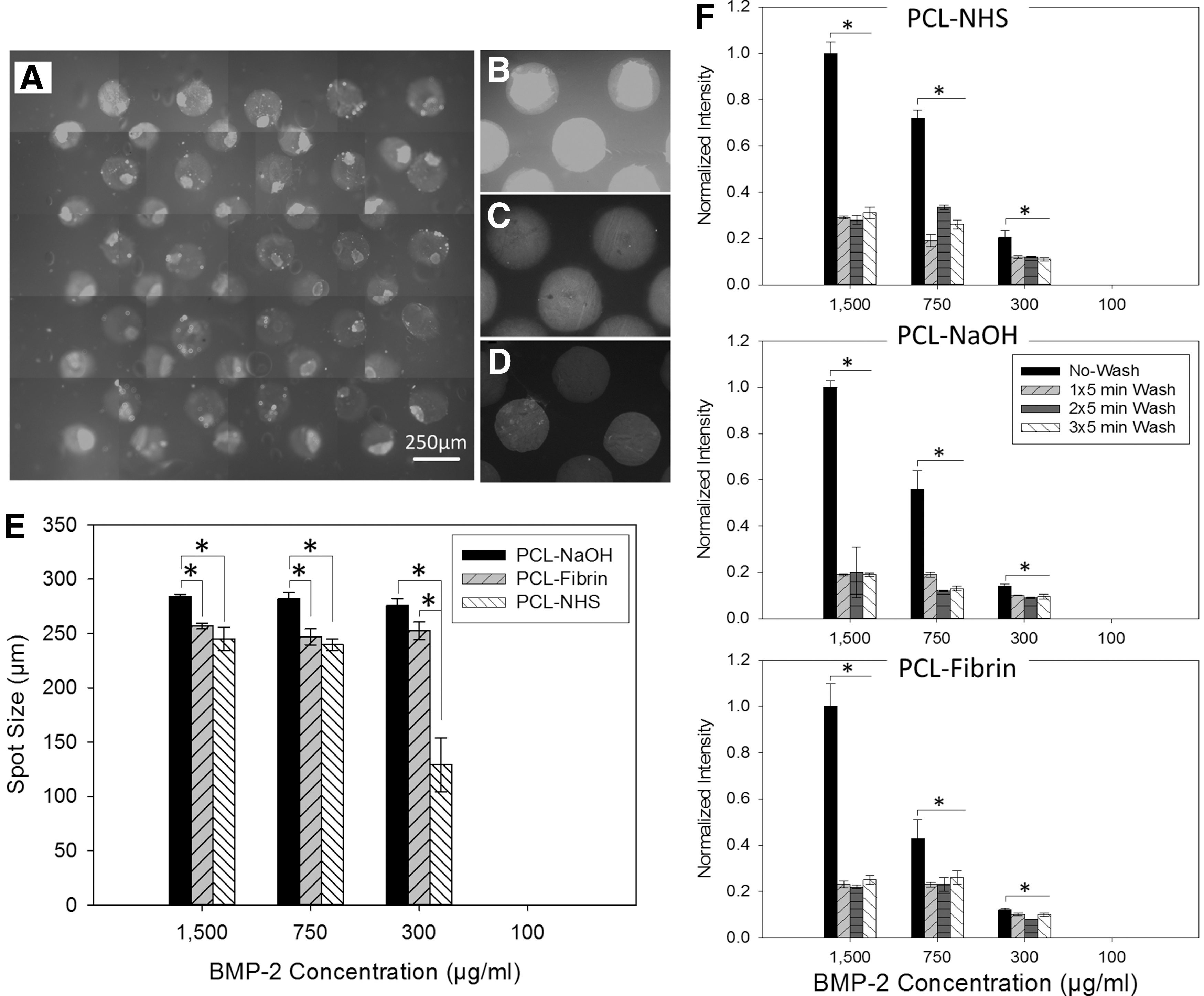
To determine the efficacy of focused acoustic patterning of GFs, droplets containing varying concentrations of DyLight650-conjugated BMP-2 were projected onto thin PCL-NHS, PCL-NaOH, and PCL-Fibrin flat sheets. Before deposition, BMP-2 was thawed from −80°C and diluted to the target concentration in sodium phosphate (pH 7.4). BMP-2 concentrations of 1500, 750, 300, and 100 μg/mL were tested. On each flat sample, 50 droplets of BMP-2 solution were patterned in a scattered configuration. Their fluorescent signal was then imaged using an inverted Zeiss AxioObserver Z1 microscope (Carl Zeiss Microimaging, Thornwood, NY). The microscope was equipped with an X-Cite® Series 120Q metal halide lamp, appropriate filters and an AxioCam MRm camera (Carl Zeiss Microimaging). The microscopic images were taken from the whole area of patterned BMP-2 via an automatic tiling method. Image processing was conducted in MATLAB® to determine the average of the spots diameter. Apparent diameters of the printed BMP-2 spots on flat pieces of all three types of materials were measured and averaged for the 50 spots on three repeat samples (n = 3).
Also, the concentration of tagged BMP-2 spots on PCL-NaOH, PCL-, and PCL-NHS substrates were compared after one time, two times, and three times washing with DPBS, each time for 5 min. To determine the relative concentration, the intensity of collected fluorescent light under the microscope was indicated after each washing step. The values were normalized to the intensity of the highest initial BMP-2 concentration and no-wash condition. The image processing was performed in MATLAB software.
Cell culture
C2C12 mouse myoblast cells were purchased from ATCC (Manassas, VA). The cells were plated at low density, cultured at 37°C in a humidified atmosphere (5% CO2/95% air) in Dulbecco's modified Eagle's medium (DMEM; Gibco® Life Technologies) containing 10% fetal bovine serum (FBS; Gibco Life Technologies) and 1% antibiotic/antimycotic mixture, 5 mL of L-glutamine (200 mM), and sodium pyruvate. The culture medium was changed every 3 days. After the cells reached confluence, they were trypsinized using 0.05% trypsin (Gibco Life Technologies) and maintained in medium until use.
For flat samples PCL-NaOH, PCL-Fibrin, and PCL-NHS, 2D square flat pieces were transferred to 24-well plates and were attached to the bottom of wells using autoclaved Dow Corning 839 Blue Silicone Adhesive-Sealant. C2C12 (P10; ATCC, CRL-1772) cells were cultured at 30,000 cells/sample (for flat samples) in complete medium composed of DMEM supplemented with 10% FBS and 1% antibiotics (penicillin 100 U/mM, streptomycin 0.1 mg/mL; Life Technologies) and 1 μg/mL aprotinin (Sigma-Aldrich) to minimize fibrin degradation.
For lattice samples, four layers of lattice layers with BMP-2 were assembled and integrated using two screw and nuts (as shown in Fig. 1G, H) to form 3D scaffolds. The scaffolds were placed in 24-well plates and C2C12 cells were seeded with the concentration of 100,000 cells/samples and cultured at 37°C in a humidified atmosphere (5% CO2/95% air).
Alkaline phosphatase activity
Alkaline phosphatase of 2D samples
For assessing the bioactivity of patterned BMP-2 on the PCL-based substrates, the expression of alkaline phosphatase (ALP) was assessed. To evaluate the early osteogenic differentiation of the different substrates (PCL-NHS, PCL-NaOH, and PCL-Fibrin), 0.5 × 0.5 cm squares of each were patterned with increasing concentrations of BMP-2 (300, 500, 750, 1000 μg/mL), and the ALP activity of cultured C2C12 cells was measured at day 5 of culture. These concentrations were selected based on the results of the light intensity test (presented in Characterization of BMP-2 Printing/Patterning section): 100 μg/mL was removed, the highest limit was changed from 1500 to 1000 μg/mL, and smaller intervals were used.
Samples were removed from media after 5 days and washed in deionized water before fixation in citrate-acetone-formaldehyde for 30 s. Cells were then stained using the ALP Staining Kit (Sigma-Aldrich) according to the manufacturer's instructions with minor modification described in.19,20 Five samples (n = 5) of each type were used in this test.
ALP of 3D samples
To investigate the early osteogenic differentiation in 3D culture systems, the ALP activity levels of C2C12 cells on four-layer lattices made from PCL-NHS and PCL-NaOH with patterned BMP-2 were measured at day 5 of culture. BMP-2 concentration of 750 μg/mL was selected based on the 2D ALP observations. Based on the results of the test on flat samples that did not show any significant difference between 750 μg/mL PCL-NaOH and PCL-Fibrin samples, the 3D test was conducted only on PCL-NaOH and PCL-NHS samples. Each layer was individually analyzed to quantify the amount of ALP activity/dsDNA. ALP-stained samples were imaged using a stereomicroscope (Leica DFC290 HD with a ring light) with an exposure time of 400 ms, a saturation level of 1, a gamma level of 1, and a 12 × zoom. Five samples (n = 5) of each type were used in this test.
Statistical analysis
The values reported for the dimensional deviation, swelling ratio, mechanical properties, and cell viability are presented as the mean values ± standard deviation. One-way analysis of variance (ANOVA) with t-test (p < 0.05) was used for statistical analysis of data.
Results
Chemical characterization
The NMR spectrum of PCL-NHS is shown in Figure 2A. Chemical shifts at 1.4, 1.6, 2.3, and 4.0 ppm correspond to the methylene hydrogens of the PCL structure. In comparison with NR spectra of PCL, the smaller additional shifts from 2.6 to 2.8 ppm were attributed to the methylene hydrogens of the succinimide end groups. This confirmed the incorporation of succinimide groups to PCL ends.

Chemical and physical characterization of PCL-based materials:
Hydrophilicity characterization
The contact angles for PCL-NHS, PCL-NaOH, and PCL-fibrin were 64° ± 5°, 68° ± 1°, and 71° ± 5°, respectively, as shown in Figure 2B. The differences in contact angles were not statistically significant.
Degradation
The degradation rate of PCL-NHS, PCL-NaOH, and PCL-fibrin materials over 1 week were characterized as shown in Figure 2C. PCL-NHS degraded about 15% in a week, while PCL-NaOH samples showed <1% degradation.
Characterization of BMP-2 printing/patterning
On all three materials, we were able to reproducibly generate precisely located droplets with a diameter of ∼250–275 μm. Figure 3A shows fluorescence images of staggered patterns of the 50 BMP-2 spots on an example PCL-NHS flat sample. Figure 3B–D show representative spots on PCL-NHS, PCL-NaOH, and PCL-Fibrin samples, respectively. The average spot diameters are shown in Figure 3E for different BMP-2 concentrations on the three different substrates. The 100 μg/mL concentration did not leave any recognizable footprint on the samples (only very small spots were observed on PCL-NHS samples). For all the concentrations, droplets formed larger spot size on PCL-NaOH flat substrates. The difference in spot size could be a result of increased nanopores and carboxyl (COOH−) groups on PCL-NaOH surfaces due to alkali treatment although water contact angles (wettability) for the three material substrates were measured nonsignificantly different and all were <90° (∼65°). The spot size reduced to almost half size on PCL-NHS from 750 to 300 μg/mL concentration. This is probably the result of greater hydrophobic interaction between higher concentration of BMP-2 and PCL-NHS surface. Also, of note is the homogeneity of the BMP-2 distribution visually observed in the droplets once they have dried.
Characterization of BMP-2 printing/patterning:
The concentration of fluorescently tagged BMP-2 spots on PCL-NHS, PCL-NaOH, and PCL-Fibrin substrate were compared after one time, two times, and three times washing with DPBS. The fluorescence signal intensity after each washing round was normalized to the intensity of highest initial BMP-2 concentration and no-wash condition, and is shown in Figure 3F. The fluorescence intensity significantly dropped after the first wash for 1500, 750, and 300 μg/mL BMP-2 concentrations, for all material types, but did not change considerably after second and third washes.
ALP
2D flat samples
ALP-stained samples are shown in Figure 4A. ALP activity, dsDNA concentration, and ALP/DNA ratio for flat samples with different concentrations and substrates are shown in Figure 4B and C, respectively. By day 5, both 1000 and 750 μg/mL concentrations demonstrated the statistically highest levels of ALP activity for each substrate when compared to the lower concentrations 500 and 300 μg/mL. No statistically significant difference was observed between 1000 and 750 μg/mL concentrations and between 500 and 300 μg/mL concentrations. Thus, only 750 μg/mL concentration was used for further 3D studies. In addition, on all three substrates, the treatment with the two lowest tested concentrations (500 and 300 μg/mL) demonstrated no significant difference in ALP activity when compared to their untreated controls suggesting that these concentrations were insufficient to generate notable osteogenic differentiation by day 5.

Osteogenic differentiation of C2C12 cells after 5 days culture on two-dimensional flat PCL-NaOH, PCL-Fibrin, PCL-NHA samples, and control well plate with BMP-2 concentrations of 300, 500, 750, and 1000 μg/mL:
There was a significantly lower amount of ALP activity on PCL-NHS substrate than on the PCL-NaOH and PCL-Fibrin substrates when all were exposed to the higher concentrations of printed BMP-2. Moreover, PCL-NaOH and PCL-Fibrin substrates did not demonstrate any significant difference in ALP activity; consequently, only PCL-NaOH was used as control for later 3D studies. All materials demonstrated similar cellular proliferation and dsDNA levels suggesting that the noted differences in ALP expression were not due to different population sizes, but instead to the GF treatment and substrates. This was validated by the fact that all previously made observations were maintained when the ALP activity levels were normalized by dsDNA.
3D lattices
One layer of PCL-NHS and PCL-NaOH lattice that were ALP stained after 5 days culture of C2C12 cells are shown in Figure 5A. The quantified ALP activity and ALP/dsDNA ratios for each material are demonstrated in Figure 5B and C, respectively. The data are presented for each lattice layer (top, upper-mid, lower-mid, and bottom). Figure 5D demonstrates the average of ALP/dsDNA ratio for all the layers of each sample type. PCL-NHS with printed BMP-2 showed about 1/3 of ALP/dsDNA compared to PCL-NaOH lattice with printed BMP-2. The ALP difference between PCL-NHS and PCL-NaOH was consistent in both 2D and 3D structures.

Osteogenic differentiation of C2C12 cells on 3D multilayered lattice scaffolds with patterned BMP-2 of 750 μg/mL concentration. Control samples have no BMP-2:
In terms of overall ALP activity, it was determined that in both materials, there was a significant difference between the treated ( = with BMP-2 pattern) samples and their respective untreated controls at every lattice layer (top, upper middle, lower middle, bottom). This significant difference was additionally corroborated at all lattice layers when ALP activity was controlled for by dsDNA. In addition, when all four layers of PCL-NaOH with BMP-2 samples were compared to their respective counterparts in the PCL-NHS with BMP-2 samples, the former material demonstrated significantly higher levels of activity at all layers.
Discussion
In this work, we presented a fabrication process to develop 3D PCL-based TECs with controlled pattern of bonded BMP-2 via combining AM and ADE technologies. In our previous work, we have demonstrated the potential of PCL-based (a clinically available biopolymer) material for orthopedic tissue engineering and implant applications.8,21–25 Biocompatibility, cell viability, and cell proliferation on PCL material have been tested and reported previously.23,24 AM-made composite of PCL and tricalcium phosphate was successfully studied in vivo as a biodegradable replacement of necrotic femoral head in the presence and absence of bone marrow stromal cells for the treatment of osteonecrosis of the hip.21,26 Also, AM-made PCL-hydrogel structures were formed and tested as templates of macro and microscale vascularized bone TECs.8,27 Both such in vitro and in vivo functionality of the AM-made TECs can be significantly improved via incorporating signaling molecules into the constructs in a 3D controlled manner. For this study, we used a similar base PCL material and the AM method combined with ADE technology, but the process was developed to be applicable for a variety of biomaterials, signaling molecules, and applications.
Of late, advances in biological material patterning technologies utilize automated computer-control processes and enable high-throughput manufacturing of bioagent patterns with accurate control on resolution, repeatability, and uniformity. 28 Although multiple bioagent printing and deposition techniques, including bioplotting/dispensing,29,30 inkjet printing,31–33 laser-based direct write/printing,34–38 bio-electrospraying, 39 and micro-array spotting,40,41 show promise, most of them encounter major challenges such as loss of biomolecules functionality, low cell viability (in case of cell printing), and clogging of ejectors. 42 For instance, inkjet printing,33,43 a relatively fast dispensing method, potentially passes biomolecules throughout small nozzles and orifices where they are exposed to high mechanical and fluid stresses associated with the printing process and, consequently, may experience physical damage. Also, in thermal and piezoelectric drop-on-demand systems, the upper threshold for viscosity of the ink/solution with low density of bioagents (particularly cells) excludes the use of many relatively thick materials (such as hydrogels as a carrier) and limits spatial resolution. 44 Clogging of the nozzle orifice and low reproducibility is another issue reported for such systems.43,45 Thus, the above mentioned methods face major challenges to spatially distribute bioagents in patterns and to scale-up to complex and clinically relevant-sized constructs at such resolutions.
Most recently, contact-free nozzle-less acoustic liquid handling technology has emerged as a promising means to deliver precisely sized droplets of liquid phase that can contain bioagents in solution.18,46 In this technology, there is no contact between the instrument and the liquid; consequently, no pressure, shear, heat, or cross-contamination is applied to the bioagent containing liquid. Therefore, this technology can lead to improved maintaining bioactivity of bioagents, functionality, and assembly accuracy while working at high speed, compared to existing printing methods. Very recently, some researchers have used this technology to study biological and cell interaction in 2D. 46 The Echo® 550 Liquid Handler by Labcyte, a pioneer in developing acoustic material dispensing technology, was the ADE system we used in this study for deposition of GFs. Unlike most of the other technologies, a small amount of bioagent solution (∼12 μL) is enough for the system to work, which reduces the waste of material associated with the process, making it in turn more cost-effective. In addition, the system is designed primarily for high-throughput screening with high speed and accuracy appropriate for scaling.
The physical characterization of the deposited patterns (Fig. 3A–E) demonstrated the potential of the applied ADE technology and system for forming well-controlled patterns of BMP-2 spots on the PCL-based substrates. It should be mentioned that a trial and error approach was needed to optimize configuration and spacing (used here) to create well-defined high-density pattern and prevent merging of droplets upon contacting the targeted material. Data showed that the deposited spot size depends on both material surface condition and the concentration of BMP-2 (Fig. 3E). A spot size of ∼250 μm is in the range that can be located on a single strut of lattices (∼350 μm in width). The repeatability, accuracy, and precision of the deposited patterns were in a reasonable and acceptable range (checked visually) for the ultimate applications such as the formation of growth factor-loaded TECs, although those parameters were not characterized here due to the comparatively lesser level of control on the formation of the 2D and 3D substrates.
Once it was determined that reproducible droplet deposition was possible, the ability for those droplets to maintain their concentration and localization on each material type was determined by monitoring the fluorescent intensity of the droplets after repeated washings (Fig. 3F). After both short and long washing steps (long washing data are not shown here), there was a rapid sharp drop in the observed fluorescent signal followed by a plateau. This drop and plateau effect was seen in all the materials and all the BMP-2 concentrations. Regardless of the initial printed concentration, the signal attenuated to approximately the similar proportion of its maximum intensity (∼10–30%) on all material types. We may conclude that the remaining concentration after the first washing step is probably directly and stably bonded with and immobilized onto the substrate material surface. Further studies are needed to improve the loading capacity such as creating new biomaterials and surface textures for more binding sites and bonding mechanisms.
In this study, three different PCL-based substrate materials were used: succinimide-terminated PCL was used as the main biomaterial developed for AM-ADE process. This material has methylene hydrogen groups that enable chemical bonding with GFs upon AM (melting and deposition) of each lattice layer with no need for additional surface modification. First control material was PCL that was alkali-treated by NaOH solution to provide rougher surface23,24 and potential carboxyl group (COOH−) binding. 47 Second control material was PCL with fibrin coating to provide physical retaining of GFs without the need for covalent cross-linking. Both such control surface treatment methods have been widely used in vitro and in vivo for surface functionalization.21,27,48–52 However, those extra lengthy intermediate steps make it impossible to automate the process of combined AM and ADE. The osteogenic differentiation assessments showed that the deposited signaling molecules resulted in the targeted differentiation of C2C12 cells after 5 days of cell cultures; however, it significantly varied with the BMP-2 concentration (Fig. 4). Although BMP-2 concentration of 300 μg/mL and higher showed stable binding to all three different PCL-based substrates (Fig. 3F), neither samples with 300 μg/mL nor 500 μg/mL concentrations exhibited osteogenic differentiation (Fig. 4A–C). This is probably because of the amounts of BMP-2 retained on the samples after washing or released from the samples are lower than the concentration threshold to trigger osteogenic differentiation. This matter needs further tests to be confirmed.
The 2D ALP tests revealed that the controlled samples PCL-NaOH and PCL-Fibrin with BMP-2 did not show significant difference in osteogenic differentiation for 750 and 1000 μg/mL BMP-2 concentrations. Thus, we removed PCL-Fibrin from the 3D osteogenic tests and conducted such test only with 750 μg/mL concentration. PCL-NHS demonstrated much lower (∼25%) ALP/dsDNA in 2D in comparison with PCL-NaOH. All 3D samples loaded with 750 μg/mL BMP-2 concentration showed significant, positive-ALP staining compared to control samples that determine the effect of printed BMP-2 on guiding C2C12 cells toward an osteogenic lineage across all four layers of the lattices (Fig. 5). The ALP activity per dsDNA for BMP-2 printed PCL-NHS was about one third of PCL-NaOH. The ALP activities did not significantly vary between the layers for both materials. Also, of note is the homogeneity of the BMP-2 distribution visually observed in the droplets once they have dried. In our study, ALP activities were mostly observed on the BMP-2 patterned areas. Such cellular response demonstrated the effectiveness and efficacy of our local patterns for controlled cell response.
It needs to be considered that the osteogenic potential of the constructs are governed by the amount of the GF remained on the substrate material, roughness, and microporosity of the struts. Also, the fact that PCL-NHS exhibited significantly greater degradation rate compared with PCL-NaOH (10 ± 4% vs. 0.3%) during the course of 5 days might have influenced the ALP results: the degradation of the material at the surface might have removed a portion of loaded BMP-2 and consequently reduced the rate of osteogenic differentiation of C2C12 cells. Thus, the degradation rate of the scaffold material needs to be tuned to achieve the desired delivery rate and retention rate of GF for improved tissue healing and regeneration. 2 PCL with relatively slower degradability and with clear intracellular resorption pathways 9 can provide a base for tuning degradation rate for all range of tissue regeneration applications. Thus, a composite of PCL, PCL-NHS, or PCL-TCP with controlled ratio as raw material for AM of TECs may provide more suitable degradation rate and GF delivering rate that will be explored in future work. PCL-NHS alone can be of more interest for the applications that high degradation rate is needed.
In macroscale, the major problem of the currently available GF delivery techniques is that they, while clinically accepted, exhibit poor drug release characteristics: eluting rapidly at a concentration far above physiologically relevant conditions. High concentrations (1.50 mg/mL: Food and Drug Administration [FDA]-approved concentration of rhBMP-2) need to be used to maintain a therapeutically release profile due to the short half-life of biofactors. For instance, the current FDA-approved system is the delivery of rhBMPs simply from a collagen matrix (Infuse™ Bone Graft and absorbable collagen sponge) that has raised concerns over the cost and long-term complications of the therapy associated with the high dosage needed for functionality.6,53 By using an immobilized solid-phase GF patterns onto TECs as what presented in this study, overall lower doses can be more effectively delivered, potentially over longer periods of healing process and only to the tissue of interest to reduce risks of side effects. 33 In this study, we deposited only 0.75 μg/mm2 (1 μL of 750 μg/mL BMP-2 solution for each mm2 of area) of BMP-2 in solid phase on the lattices with a resolution of ∼508 dpi that is significantly less amount needed for commercialized BMP-2 delivery matrices. Based on the data presented in Figure 3F, about 25% of the deposited BMP-2 retains on the PCL-NHS surface, which can be further improved by adding other components such as calcium phosphate bone minerals.
In addition to eliminating the critical limitations for preserving bioactivities and controlling release kinetic rates that are encountered with current tissue engineering drug delivery methods,54,55 a remarkable feature of the utilized method and Echo ADE system is that as many as 384 or more different source materials (e.g., protein, buffer, hydrogel solution, and so on with different concentrations) can be deposited simultaneously without the need to change the reservoir well plate. Thus, multiple bioagents with varying concentration can be incorporated in any certain location of the lattice layers to form bioagent-loaded TECs. In addition, gradient distribution of GF which has a known effect on stimulating the migration of cells is possible via using this system. Specific cells can also be deposited locally with certain signaling molecules via the ADE system. Such features will be explored in future work. The process of layer-by-layer manufacturing of lattice layers while biomolecules are deposited on each layer will be developed in future. Also, additional experiments need to be conducted to investigate the capability of the developed method to generate long-term mineralization both in vitro and in vivo.
Conclusion
This study demonstrated the successful generation of 3D osteoinductive constructs using a combination of AM of PCL-based material and acoustic patterning of BMP-2. The presented process enables rapid production of 3D TECs with customized external geometry and internal architecture with incorporated cell signaling molecule finely tuned in terms of spatial location and concentration. The ADE technology can form a precisely controlled local density of signaling molecules with adequate concentration and high durability across each layer of additively manufactured lattices. Thus, it minimizes the waste of loaded signaling molecules, which are usually costly. The surface-treated PCL showed high affinity for BMP-2. Although PCL-NHS showed less ALP activity compared to NaOH-treated PCL, the affinity of BMP-2 is high enough for automated AM-ADE fabrication of functional TECs. The manually assembled constructs in this study demonstrated the potential of both Hybprinter and Echo acoustic liquid handler for the formation of TECs loaded with spatially patterned GF. Both systems benefit from a flexible design that can be synced enabling the automation of such a fabrication process.
Footnotes
Acknowledgments
The authors express their sincere appreciation to Mark Fischer-Colbrie, Richard Ellson, Joe Olechno, Brent Eaton, and Temo Bandzava from Labcyte, Inc. for providing access to Labcyte laboratory equipment and Echo machine as well as their valuable technical assistance.
Disclosure Statement
No competing financial interests exist.
Funding Information
This work was partially supported by grants from the following agencies: R01AR057837, R01AR074458, R01AR072613, U01AR069395, DOD W911NF-14-1-0545 Defense University Research Instrumentation Program (DURIP), and DoD W81XWH18SBAA1-BA180237.