
Abstract
Stereolithography (SL) has several advantages over traditional biomanufacturing techniques such as fused deposition modeling, including increased speed, accuracy, and efficiency. While SL has been broadly used in tissue engineering for the fabrication of three-dimensional scaffolds that can mimic the in vivo environment for cell growth and tissue regeneration, lithographic printing is usually performed on single-component materials cured with ultraviolet light, severely limiting the versatility and cytocompatibility of such systems. In this study, we report a highly tunable, low-cost photoinitiator system that we used to establish a systematic library of crosslinked materials based on low molecular weight poly(ethylene glycol) diacrylate. We assessed the physicochemical properties, photocrosslinking efficiency, cost performance, and biocompatibility to demonstrate the capability of manufacturing a multimaterial complex tissue scaffold.
Color images are available online.
Impact statement
Stereolithography (SL) has advantages over traditional biomanufacturing techniques, including accuracy and efficiency. While SL has been broadly used for fabricating three-dimensional scaffolds that can mimic the in vivo environment for cell growth and tissue regeneration, lithographic printing is usually performed on single-component materials cured with ultraviolet light, severely limiting the versatility and cytocompatibility of such systems. In this study, we report a highly tunable photoinitiator system and establish a systematic library of crosslinked materials based on poly(ethylene glycol) diacrylate. We assessed the physicochemical properties, photocrosslinking efficiency and biocompatibility to demonstrate the capability of manufacturing a multimaterial complex tissue scaffold.
Introduction
Stereolithography (SL) has been widely used for tissue engineering and regenerative medicine (TE&RM) for the fabrication of biomimetic scaffolds for cell attachment and growth leading to tissue regeneration. 1 Common bioprinting approaches, for example, extrusion-based and inkjet-based systems, including overcoming detrimental effects due to shear stresses emerging at the needle/nozzle of the printer2,3 as well as the need for highly viscous pregel solutions for extrudability and printing. 4 SL-based printing is not encumbered by these limitations due to the fact that pregel solutions are inherently low-viscosity fluids, thus positioning the technique as a strong candidate for the manufacture of high-resolution cell-laden or cell-free scaffolds.1,5
Although SL is a promising and precise method, it still has several limitations. First, most industry and research efforts have focused on the use of ultraviolet (UV)-based photocurable resins with high energy irradiation of UV light. 6 Photoinitiators (PIs) that absorb in the visible range have multiple advantages over their UV counterparts, as visible light not only has greater depth of penetration due to its longer wavelength but is also more biocompatible. PIs can be typically classified as type I or type II with the primary difference being the absence (type I) or presence (type II) of a coinitiator. Type II PIs require the use of an oxidizing agent in combination with a wavelength-specific excitable chromophore.
Type I PIs predominantly function in the UV spectra, whereas type II PIs have the flexibility of operating at various wavelengths based on the selected chromophore. Oxidative stress induced by UV light irradiation may result in damage to cellular DNA, leading to unwarranted immediate and long-term effects, including cancer or accelerated tissue aging.
7
Visible light PIs, while inherently safer compared to commercial UV systems, have yet to be fully explored and optimized for use in
The choice of the visible light PI is generally governed by the requirements of high cytocompatibility and efficiency. Some commercially available visible light PIs exist, including lithium phenyl-2,4,6-trimethylbenzoylphosphinate (LAP), camphorquinone (CQ), Eosin Y, and their derivatives.8–11 Even though they have been shown to exhibit sufficient sensitivity in the absorption of visible light, disadvantages of each persist. For example, the low aqueous solubility of CQ limits its suitability for hydrogels. In addition, little effort has been made in developing a versatile and highly tunable PI system for crosslinking hydrogels with different properties and in the control of crosslinking time or concentration of the PI system components. Furthermore, there is a limited availability of materials for commercial SL printers.
Most current commercial photocurable polymers are expensive, proprietary, and lack systematic study, thus limiting the flexibility and creativity of the end-user. Therefore, there is an urgent need to develop a visible light PI system in an effort to further increase the biocompatibility and versatility of photocurable resins for biomedical applications in
To address the aforementioned shortcomings of commercial SL systems, we have developed a novel type II visible light PI system to increase not only depth penetration but also biocompatibility and tunability. We have also established a systematic library of physicochemical material properties based on low molecular weight poly(ethylene glycol) diacrylate (PEG-DA), considering primarily the photocrosslinking efficiency and biocompatibility. Our new PI system and material library were then used to demonstrate the capability of manufacturing a multimaterial complex tissue scaffold, using a newly developed visible-light stereolithography (VL-SL) printer.
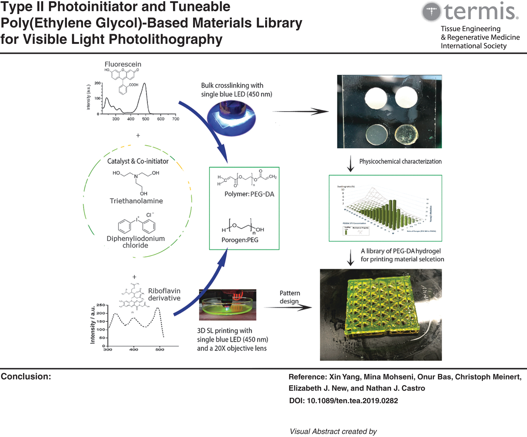
For the current PEG-DA system, we have used a riboflavin or fluorescein (FR) derivative as the chromophore for photoinitiation, triethanolamine (TEA) as coinitiator, and diphenyleneiodonium chloride (DPI) as the catalyst (Fig. 1). This coinitiator and catalyst combination, which is commonly used in photocrosslinking systems, has previously shown promising behavior.6,7 PEG-DA pregel solution was photocrosslinked by irradiation under blue light (450 nm) with a low-energy emitting diode (LED) and evaluated to establish a library of PEG-based hydrogels with highly tunable and reproducible mechanical properties.

Crosslinking process of PEG-DA-based hydrogels with new modular type II PI. PEG-DA, poly(ethylene glycol) diacrylate. Color images are available online.
Riboflavin, also known as vitamin B2, is a water-soluble molecule with absorption bands in both the near-UV and visible region. 12 We have chosen to use tetraacetylated riboflavin due to its superior water solubility. Although previous studies have shown that riboflavin exhibits lower photosensitivity than commercially available water-soluble PIs such as Ciba's Irgacure® 2959, 12 it is still a very promising chromophore for lithographic fabrication of tissue-engineered scaffolds, particularly as it is a naturally occurring molecule and is inherently biocompatible.
FR is a xanthene dye, which is capable of radical generation in aqueous medium, facilitating photopolymerization and crosslinking. 13 To the best of our knowledge, only one study has directly compared the use of FR and riboflavin for bulk crosslinking of cell-laden natural gels, 13 with no translation to application in SL printing, thus prompting our investigations here. To assess the effects of PI composition on reaction kinetics and on the mechanical behavior of PEG-based hydrogels, systems containing riboflavin and FR were both fully characterized and compared.
Researchers seeking to prepare three-dimensional (3D)-printed hydrogel scaffolds to mimic the complex and heterogeneous structures for tissue and organ regeneration are faced with highly specific material constraints. To realize the full potential of SL, it is imperative to integrate the capability of multimaterial printing for functional, end-use applications, and we have therefore used our PI system toward the development and characterization of a highly tunable pregel material library (Fig. 1). Thus, by adjusting the chemical composition of the pregel solution, such as molecular weight/concentration of PEG-DA and ratio of PEG-DA and porogen (PEG), a wide range of mechanical properties could be achieved, making it possible to target tissues varying widely in stiffness, such as muscle and bone.
From a basic science perspective, the physicochemical characterization of a wide range of PEG-DA-based hydrogels helps to establish a greater understanding of the effects of material composition on the mechanical properties that will ultimately satisfy the requirements of a broader spectrum of TE/RM applications.
To further realize our goal of applied multimaterial printing for functional applications, we have also developed a new VL-SL printer utilizing an interchangeable objective lens coupled with LEDs (Fig. 2). The system is based on a low-cost fused filament fabrication (FFF) 3D printer, making it cost-effective and accessible. We examined the feasibility of using this new VL-SL printer for multimaterial scaffold fabrication using our PI/PEG-DA-based hydrogel library.
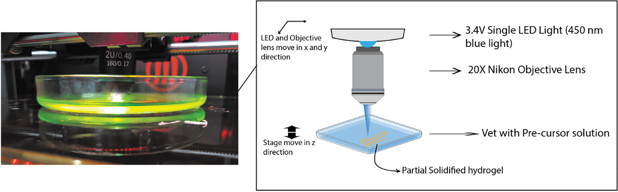
The schematic of table top VL-SL. VL-SL, visible-light stereolithography. Color images are available online.
Materials and Methods
Materials
PEG-DA (Mn = 575), PEG (Mn = 300), TEA, and diphenyleneiodonium chloride (DPI) were purchased from Sigma-Aldrich (St. Louis, MO). PEG (Mn = 8000) was purchased from Fluka Chemie GmbH (Steinheim, Germany). Standard phosphate-buffered saline (PBS) tablets were obtained from Oxoid Limited (Basingstoke, Hampshire, UK). Tetramethylrhodamine-conjugated bovine serum albumin (BSA) was purchased from Life Technologies (Eugene, OR). 2′,3′,4′,5′-Tetraacetyl-N(3)-carboxymethylriboflavin (Ac4Rbf) was synthesized according to standard procedures.14–16 Human lung adenocarcinoma-derived A549 cells were obtained from ATCC® and maintained in Dulbecco's modified Eagle's medium (DMEM; Gibco, Life Technologies, Inc., New York, NY) containing 2% foetal bovine serum (FBS; Gibco, Life Technologies, Inc., New York, NY).
Hydrogel preparation
Solutions of various concentrations of FR and DPI ranging from 50 to 500 μM were prepared in ultrapure water, while Ac4Rbf and DPI were prepared in 1 × PBS. PEG-DA was dissolved separately in the FR-based or Ac4Rbf-based PI over a wide range concentrations from 20%, 40%, 60%, to 80% wt/V. For the groups with porogen, PEG (Mn = 300) and PEG (Mn = 8000) in the ratio with PEG-DA (1:0.4, 1:1, 1:2.5) were then added into the above mixture. 0.2% v/v TEA was subsequently added as a coinitiator. The mixture was then stirred at room temperature until homogeneous. Each precursor solution was transferred into a circular silicone mould consisting of wells with a diameter of 5.0 mm and height of 1.4 mm, and irradiated with a 450 nm single blue LED (1.2 W; LEDSupply, Randolph, VT) for 10 min at a working distance of 48 mm.
Physicochemical characterization of bulk crosslinked hydrogels
Crosslinking efficiency and crosslinking percentage
Real-time Fourier transform infrared (FT-IR) (Nicolet 5700 ATR-FTIR spectrometer; Thermo Fisher Scientific) spectroscopy was used to qualitatively and quantitatively evaluate the reaction kinetics of photocrosslinking. Omnic™ Spectra Software (Thermo Fisher Scientific) was used to analyze the IR absorption spectra. The crosslinking percentage at each time-point was calculated from the change in the absorption peak area (1380–1420 cm−1) corresponding to the appearance of acryl C-H.
Uniaxial compression test
Five replicates from each group were incubated separately in 1 mL 1 × PBS in an oven at 37°C overnight for swelling. The compressive modulus (E) of the bulk crosslinked hydrogels was determined via unconfined uniaxial compression testing (n = 5) on an Instron® Microtester (5848, Instron, Australia) fitted with a nonporous aluminium indenter and a 500 N load cell at a displacement rate of 0.01 mm/s. Compressive moduli of the samples were determined as the slope of the stress-strain plots at a strain of 10–15%.
Swelling ratio
The swelling ratio is defined as the fractional increase in the weight of the hydrogel due to water absorption. 17 Three replicates of each group were submerged in deionized water and incubated statically in an oven at 37°C until equilibrium swelling was achieved. The weight of the hydrogels was measured after 5 min, 10 min, 20 min, 0.5 h, 1 h, 5 h, 1 day, 2 days, and 3 days. After 3 days of incubation, the samples were dried overnight at 37°C. Each sample was weighed after being freeze-dried. The swelling ratio of each hydrogel composite was then determined by the following equation: (<xrefref−type=”other”rid=”eq1”>Eq.1</xref>)
where Ws and Wd are the weight of the swollen hydrogel and the weight of the freeze-dried hydrogel, respectively.
Morphological study
Scanning electron microscopy (SEM) (Zeiss Sigma FESEM) was used to characterize the morphological properties of the samples, including structure and porosity. First, the samples from each group were lyophilized (Alpha 2–4 LD plus, John Morris Scientific) overnight. Then, all samples were immersed in liquid nitrogen for 5 min and cut by scalpel for clear cross section. The dried hydrogels were gold sputter-coated before analyzed by SEM.
Diffusivity
A one-dimensional diffusion model was used to visualize the diffusion properties of the hydrogels. Tetramethylrhodamine-BSA solution was used to study the hydrogel diffusivity. Cylindrical bulk crosslinked samples from each group were placed on a glass slide and 10 μL 0.01 mg/mL rhodamine-BSA solution was placed on the top surface of each sample. After 10 min, excess rhodamine dye was blotted, and a cross-sectioned surface of hydrogel was observed via fluorescence microscopy (Nikon, SMZ25). 18
In vitro cytotoxicity of photoinitiator system
Metabolic activity of A549 cells was assessed using alamarBlue® reagent and spectrophotometry. The percentage of alamarBlue reduction was followed over time. In brief, A549 cells were cultured in DMEM with 2% FBS in 96-well plates at 1000 cells/well for 2 days. The medium was then removed, and cells were treated with 100 μL of optimized concentration for both Ac4Rbf and FR PI system in 1 × PBS (Table 2) in triplicate, to simulate the crosslinking process. The control group consisted of cells treated with 100 μL 1 × PBS. After 30 min incubation, PI solutions were removed. Cells were washed twice with PBS and incubated in 200 μL DMEM at 37°C. Ten microliters alamarBlue reagent was added into each well at day 1, 3, and 7. After 2 h incubation, absorbances at 570 and 590 nm were measured. Cell metabolic activity comparison was calculated as the ratio of absorbance in treated and control groups.
Visible light stereolithography apparatus 3D printer design
The Visible light stereolithography apparatus developed here was based on an existing FFF type 3D printer (Makerbot® Replicator 2 × ). Replicator G 0040 software was used to control printer movement with an effective resolution of 100 μm (x,y,z). The 450 nm single blue LED was coupled and condensed using a 20 × objective lens (Plan Achromatic Objective 20 × [(20/0.40, 160/0.17]) affixed to the existing gantry. A glass Petri dish was affixed to the print bed to function as a vat with a working distance of 3.0 mm between the objective lens and surface of the pregel solution.
Curing characteristics of materials prepared using our VL-SL printer were studied to identify applicable print speed settings for each material and layer height to ensure adequate integration between layers as well as to improve the printing resolution. To do so, a small vat was filled to the rim with each pregel solution. A glass slide was placed on the top of the vat, ensuring contact with the pregel solution. At predefined speeds and operating at full power (3.4 V, 300 mA), the gel adhered to the underside of the glass slide. The cure depth of the adhered gel was measured with a Vernier calliper, as previously described. 19 An optimal working distance of 3.6–3.9 mm was determined and was subsequently used for all printed scaffolds.
Multilayer scaffold design
A four-layer scaffold was designed (Table 1) and fabricated to demonstrate the feasibility of printing multimaterial scaffolds with the newly developed VL-SL and PI system. The material of each layer was chosen from the material library and presented in Table 3. For that purpose, a series of 30 × 30 mm rectangular solids were designed and prepared using Simplify3D with increasing in-fill density (20%, 30%, 40%, and 80%) and in-fill geometries of 0°, 60°, and 120°. The different in-fill densities with different materials provided varying properties, such as mechanical strength and porosity, to each layer. For each specific layer, 3 mL of precursor solution was pipetted into a glass Petri dish, and the Petri dish was placed level on the printer bed. The volume used produced an effective layer thickness of 400 μm.
Scaffold Design
After printing of the superficial zone, the residual pregel solution was removed, and the subsequent pregel solution corresponding to the middle zone was added and printed. The process was repeated with all respective pregel solutions until the finalized scaffolds had been fabricated.
Results and Discussion
Physicochemical characterization of crosslinked hydrogel
Crosslinking efficiency
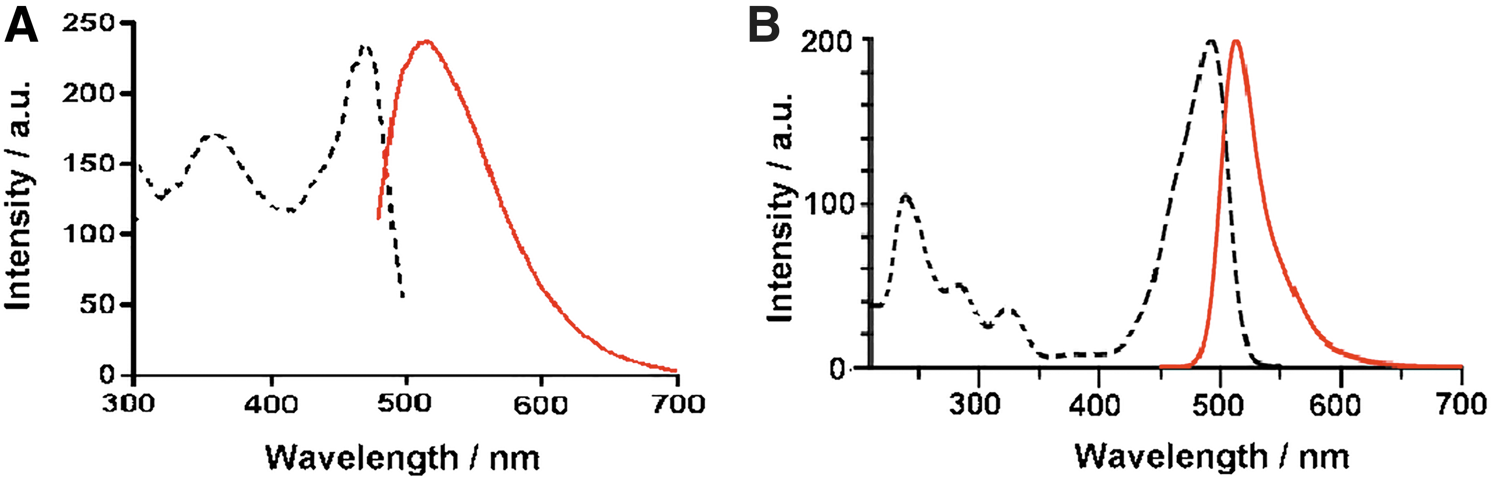
The fluorescence spectrum of Ac4Rbf (Fig. 3A) shows two distinctive excitation peaks in the visible region, with a consistently high excitation throughout the visible region. Due to the broad absorbance peaks, Ac4Rbf was expected to lead to high crosslinking efficiency when excited with 450 nm light. In contrast, only one distinctive absorbance peak was observed in the visible light region for FR, and the peak band appeared narrow. Emission intensities for equimolar concentrations of FR and Ac4Rbf with 450 nm excitation were similar. Due to the higher absorption value at 450 nm of Ac4Rbf, the quantity of light absorption of Ac4Rbf under the same conditions would be larger than FR, which suggests that a higher concentration of FR would be required for the same crosslinking efficiency compared to Ac4Rbf.
The library of PEG-DA hydrogels was successfully crosslinked upon irradiation with 450 nm visible light. The concentration of each component was determined by FT-IR to optimize the crosslinking efficiency and ensure near-complete crosslinking. Figure 4 shows representative dynamic FT-IR spectra of the crosslinking process of 20% wt/V PEG-DA in Ac4Rbf PI solution from time 0 to 10 min under the same irradiation conditions. The decreased intensity of the C-H peak (1380–1420 cm−1) was used as an indicator of double bond consumption in the photocrosslinking process. 20 The reduction in area of this distinct peak was determined and plotted to obtain the percentage and efficiency of crosslinking.

Representative FT-IR spectra for the crosslinking process of 1 mL PI solution containing 20% wt/V PEG-DA and 0.2% V/V TEA, with 125 μM Ac4Rbf and 125 μM DPI at 0 min (blue), 10 s (purple), 2 min (green), and 10 min (red). DPI, diphenyleneiodonium chloride; FT-IR, Fourier transform infrared; PI, photoinitiator; TEA, triethanolamine. Color images are available online.
The concentrations of each component in the PI system was evaluated to optimize the reaction conditions for the proposed PI system are as follows: FR and Riboflavin (50, 125, 250, and 500 μM), DPI (50, 125, 250, and 500 μM), and TEA (0.05%, 0.1%, 0.2% 0.3%, and 0.5% V/V). The novel PI system was developed for SL printing, therefore, both crosslinking efficiency and crosslinking percentage are critical. Based on previous trials with SLA, hydrogels should be crosslinked up to 80% to 90% within 60 s as ideal crosslinking efficiency. By conducting FT-IR experiment with the above permutations and combinations of concentration of each component as described above, the resulting data revealed the most efficient combination of components required to crosslink PEG-DA at the desired rate and conversrion rate (Table 2).
Optimized Concentrations of Each Component in the Visible Light Photoinitiator System for Crosslinking Poly(Ethylene Glycol) Diacrylate Library
3D, three-dimensional; DPI, diphenyleneiodonium chloride; FR, fluorescein; SL, stereolithography; TEA, triethanolamine.
As shown in Figure 5, with optimized concentrations of each component in the PI system, 500 μM of FR and 125 μM Ac4Rbf as PI, respectively, offered similar crosslinking efficiencies for 20% (wt/V) PEG-DA pregel solutions. Both hydrogel precursor solutions achieved 88% crosslinking in ∼30 s, demonstrating high efficiency suitable for high-speed SL printing. After initial optimization of the PI system, the crosslinking efficiency and crosslinking percentage of the PEG-DA and PEG-DA with porogen library was characterized using the same FT-IR method.
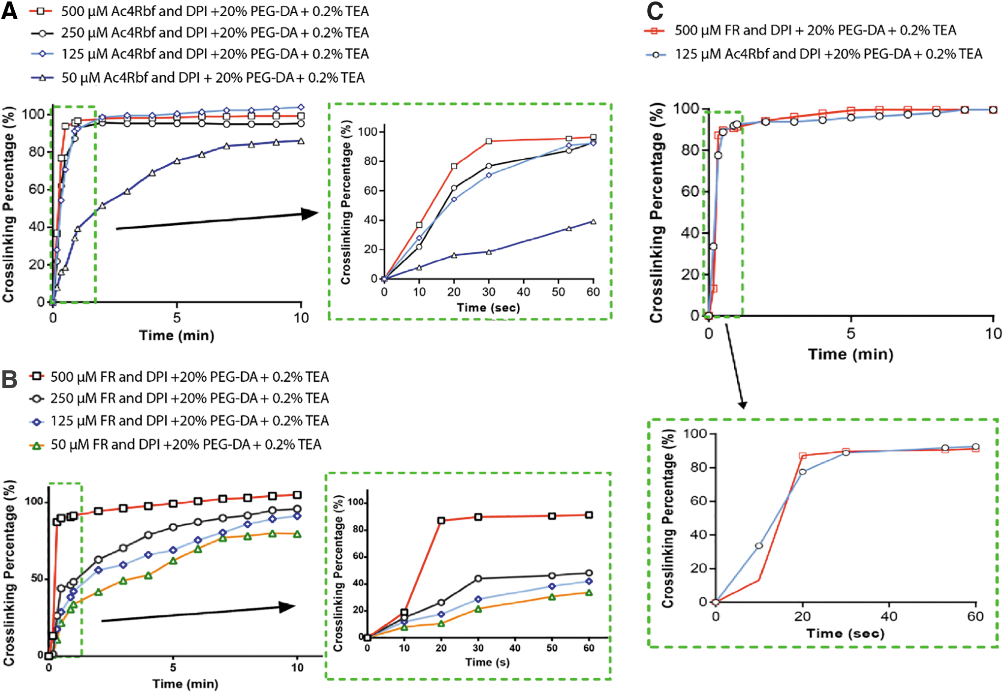
According to Figure 5A and B, by changing the concentration of each component, the kinetics of hydrogel crosslinking can be tailored. Thus, different combinations and concentrations of PI system components can be used to produce hydrogels with different degrees of crosslinking, and hence different properties, meeting the requirements for mimicking soft tissue through to bone for specific applications.
Mechanical properties and swelling ratio
Both mechanical properties and swelling ratio are critical physical parameters of a hydrogel. 21 The compressive stress-strain curves of various groups are presented in Figure 6. The compressive moduli (E), swelling ratio, and stress/strain at break of all hydrogel groups are presented in Figures 7 and 8, respectively. The results showed that stress/strain at break, compressive moduli, and swelling ratio of low-molecular weight PEG-DA hydrogels can be easily modulated by varying the concentration of PEG-DA and ratio of PEG-DA to porogen. The library exhibits a wide range of compressive moduli from 3.57 KPa to 6.4 MPa, which could mimic a wide range of tissues from soft tissue such as brain and adipose tissue to stiffer cancellous bone.18,22,23
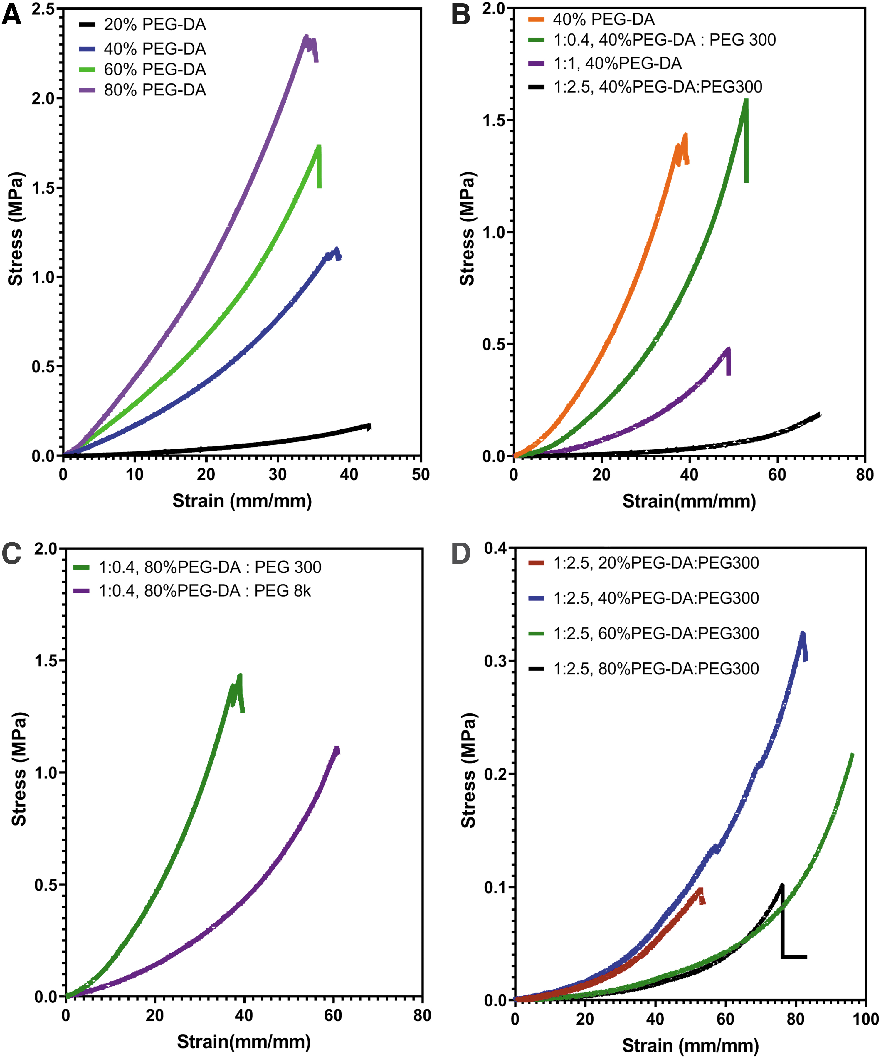
Compressive stress-strain curves of PEG-DA based-hydrogel comparsion among
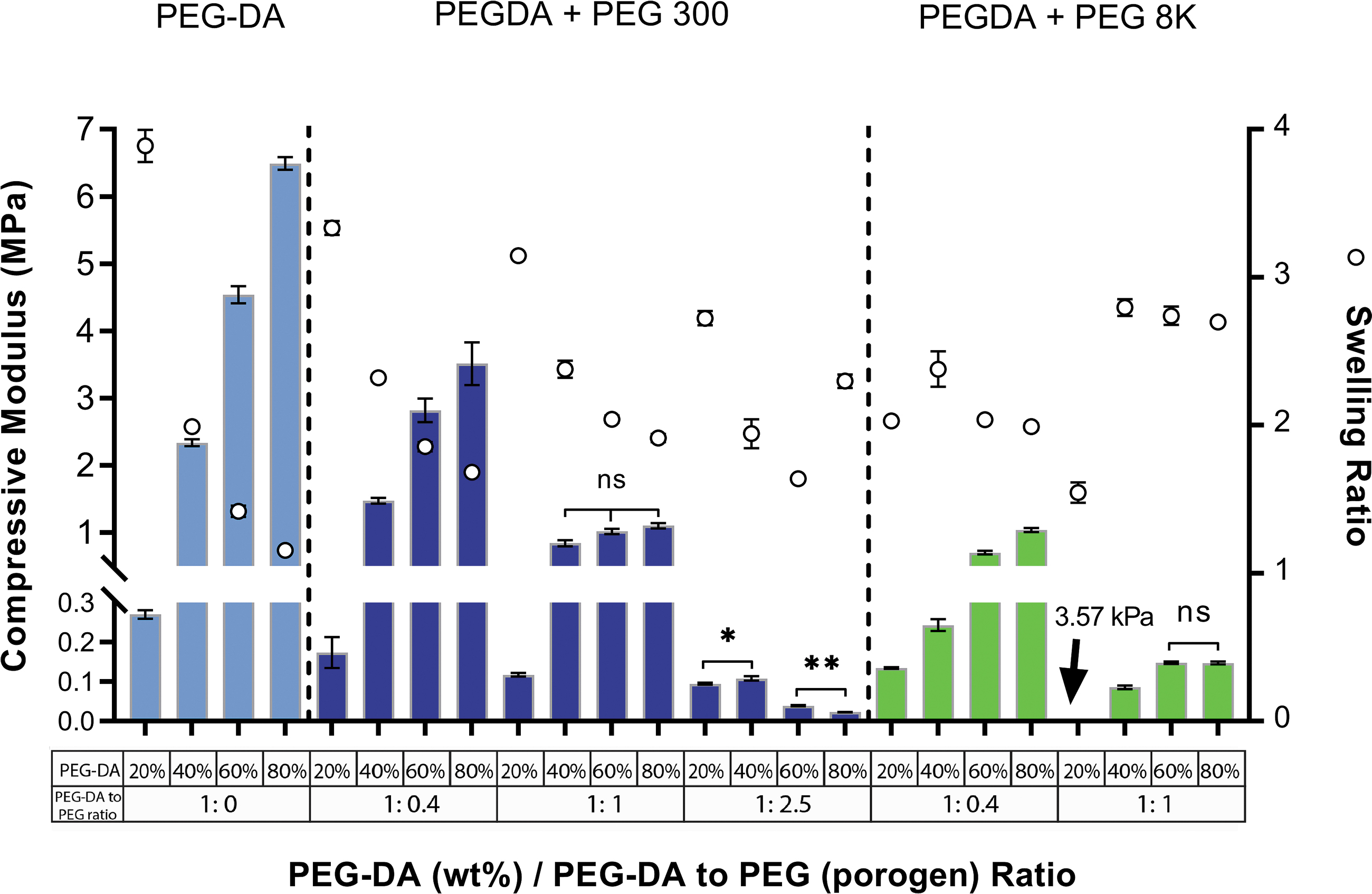
Compressive modulus (E) and swelling ratio (mean + SD) of PEG-DA material library in 1 mL Ac4Rbf PI solution, n = 5. SD, standard deviation. Color images are available online.

Stress and strain at break (mean + SD) of PEG-DA material library in 1mL Ac4 Rbf PI solution, n = 5. * represents p value from 0.01 to 0.05, ** represents p value from 0.001 to 0.01, ns represents p ≥ 0.05. Color images are available online.
As shown in Figures 6 and 7, the compressive moduli (E) of the hydrogels increased with PEG-DA concentration. Through a combination of prolation and subsequent molecular relaxation by removal of the porogen, decreasing material stiffness was achieved. In addition, the molecular weight of porogen also contributed significantly to the reduction of material stiffness. The compression modulus of groups 40 − 80% 1:1 PEG-DA to PEG 300 as well as groups 60% and 80% 1:1 PEG-DA to PEG 300 do not exhibit significant differences, which may be due to similar interactions and common effects of the factors described above.
On the contrary, swellability exhibits an inverse relationship with respect to compressive modulus for increasing PEG-DA:porogen ratios, showing equilibrium swelling ratios ranging from 1.15 (80% [wt/V] PEG-DA in 1 mL Ac4Rbf PI solution) to 3.89 (20% [wt/V] PEG-DA in 1 mL Ac4Rbf PI solution). The capacity to tune the swelling properties of bulk photocrosslinked gels is an important parameter where diffusion and permeability of bioactive molecules are critical for cell encapsulation. 18
Hydrogel morphology
SEM micrographs of lyophilized samples clearly show the morphology of PEG-DA hydrogels prepared with visible light irradiation, demonstrating the effects of porogen and porogen content within a low molecular weight PEG-DA hydrogel (Fig. 9). Pregel PEG-DA content, as well as increasing PEG-DA-to-porogen ratio produced hydrogels with different porosity, pore size, and structure as shown in Figure 9A–I in order of decreasing pore size.

SEM micrographs of lyophilized hydrogels from different groups. Scale bar = 20 μm. All SEM images were taken with EHT = 10 KV.
Both molecular weight and concentration of porogen have a great influence on the overall structure of the porous hydrogel network. 24 It can be inferred and validated that overall porosity, the formation of the porous structure, and pore size are affected by multiple factors such as the concentration of PEG-DA, ratio of PEG-DA to porogen, and the molecular weight of the porogen.
By comparing Figure 9E and F with Figure 9G–I, when the ratio of PEG-DA-to-porogen of similar molecular weight was used, a decrease in pore size ensues with increasing PEG-DA content. By comparing Figure 9F and G, the effect of molecular weight on pore size is significant, with lower molecular weight PEG porogen giving more uniform pores with smaller size. This could be due to a larger proportion of PEG-DA molecules being crosslinked across the terminal ends, forming bridges. Following this logic, when a considerably larger porogen (PEG Mn = 8000) is used, crosslinking ensues linearly akin to radical polymerization, wherein the short-chain polymers extend around the higher molecular weight PEG (Mn = 8000) producing larger pores in a prolative structure (Fig. 9B–D).
For cell encapsulation, adequate diffusion is necessary to maintain the viability of encapsulated cells. 25 In addition, the rate of reaction should also be taken into consideration, as an abrupt change in material stiffness has been shown to negatively affect immediate and extended cell viability and behavior.18,26,27
Diffusivity
One-dimensional diffusion behavior of our PEG-DA materials was evaluated using rhodamine-BSA (Fig. 10). Results show that diffusion depth, which is indicative of the distance travelled after 10 min, increased with decreasing ratio of PEG-DA-to-porogen, irrespective of molecular weight. By comparing Figure 10B1–B4, when ratio of PEG-DA-to-porogen is 1:2.5, diffusion depth increased with increasing concentration of PEG-DA. This characteristic can be attributed to the compact nature and small pore size of crosslinked pure PEG-DA hydrogels compared to the increased pore size and porosity using porogen, as discussed previously, which leads to greater overall permeability. It can, therefore, be inferred that diffusivity is not only related to the pore size but also the pore morphology and interconnectivity.

Fluorescence images of one-dimensional diffusion of rhodamine-conjugated BSA. The thickness of the hydrogel is highlighted by the green dashed line (∼1.6 mm).
In vitro cytotoxicity of photoinitiator system
The relative metabolic activity of A549 was assessed using alamarBlue reagent by spectrophotometry. It was calculated as the percentage difference of absorbance at 570 nm between treated and untreated cells at day 1, 4, and 7 (Fig. 11). These results demonstrate that both PI systems have good cytocompatibility. The metabolic activity increased significantly at day 7 compared with day 1 posttreatment for both optimized PI systems, suggesting that 30 min contact with both optimized PI systems did not cause significant long-term damage to the cells. Cells treated with the Ac4Rbf PI system showed slightly greater viability than the FR PI system, indicating that the Ac4Rbf PI system is more biocompatible under these conditions.
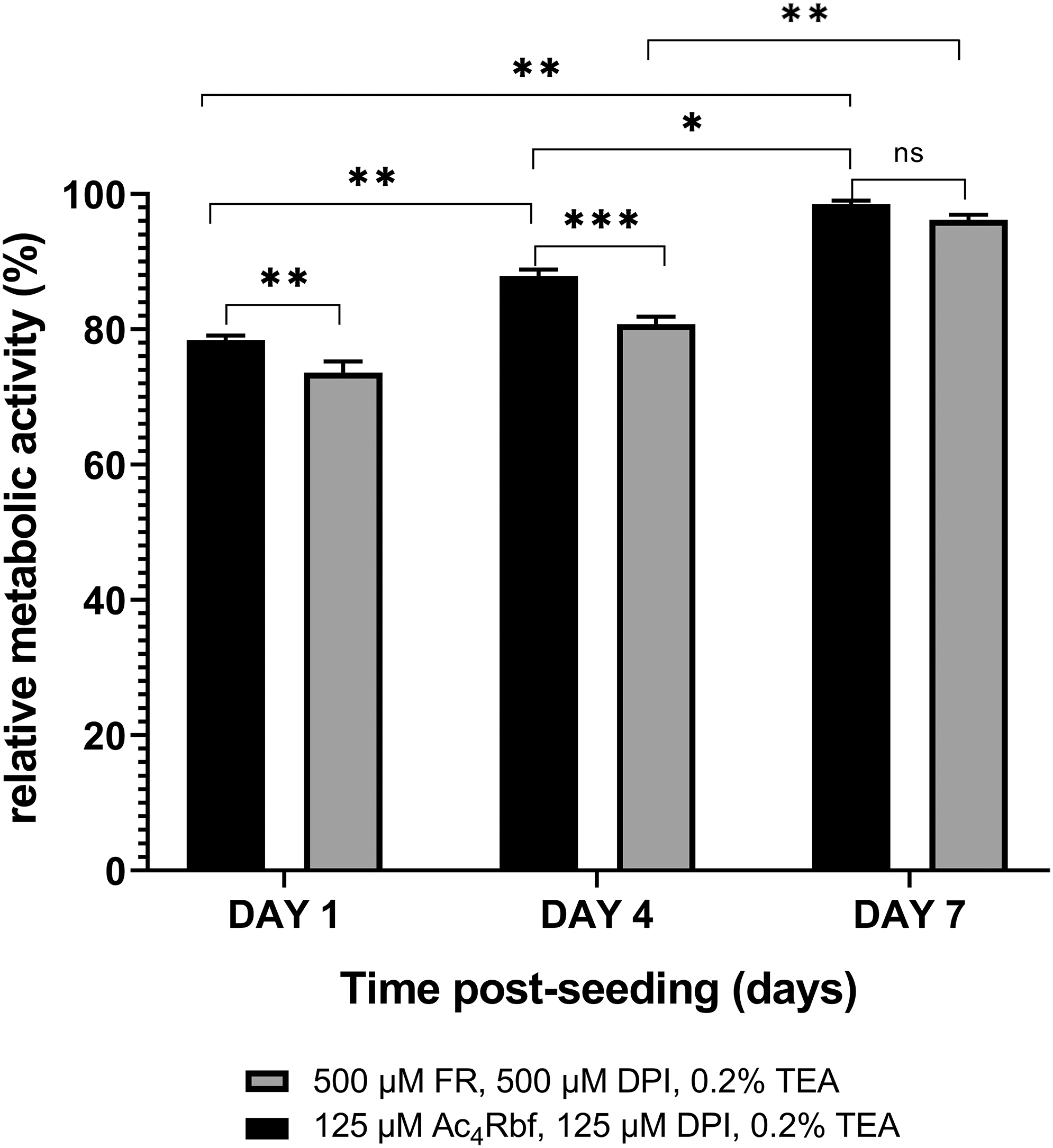
Quantification of A549 cell viability 1, 4, and 7 days after treatment with both optimized PI systems n = 3.
VL-SL 3D printer design and multimaterial scaffold design
The novel type II PI system and VL-SL 3D printing system developed were conducive to rapid fabrication of a photocrosslinkable hydrogel scaffold with efficient fabrication and precise structure. The four materials listed in Table 3 were fabricated as successive layers via our VL-SL, and the cure depth was characterized to identify applicable and efficient printing parameters.
Materials Chosen for Stereolithography Printing as 4 Layered Osteochondral Scaffold and Corresponding Properties
All groups are printed with 500 μM FR, 500 μM DPI, 0.2% V/V TEA.
N/A, not applicable; PEG-DA, poly(ethylene glycol) diacrylate.
All four materials were successfully printed with the newly designed VL-SL system. The compressive moduli of the selected materials, line width, and layer thickness were maintained across the different layers, illustrating the accuracy and high degree of control of the VL-SL printing strategy when combined with our PI system and low-molecular weight PEG-based materials (Table 3). Hydroxyapatite was blended into the precursor solution of the fourth layer, demonstrating the stability of the printing system when nanoparticles are added.
Figure 12 shows both optical micrographs and SEM images of the scaffolds. The structure of each layer was clear with defined edges, which is conducive to consistent pore size and the establishment of a complete structure. As shown in Figure 12, pore sizes increased from the first layer to the fourth layer. Image C1 clearly shows the small pores in the first layer created by the porogen, while surfaces in images A1 and B1 indicated overall smoothness of this layer. SEM images B4 and C4 show a rougher surface compared to others due to the incorporation of Hydroxyapatite which leads to a highly porous geometry inside the hydrogel. As shown in SEM images B5and C5, integration of each layer is adequate while the structure of each layer is diacritical, which indicates the success of multimaterial printing for the designed four-layer scaffold.

Optical micrographs
Conclusion
In this study, we have investigated a new type II visible light photoinitatior system and have used it to prepare a wide range of PEG-DA-based hydrogels with varying properties. We have shown that the PI system, based on riboflavin and FR fluorophores, is highly versatile, enabling tuning of the crosslinking kinetics and speed for different intended applications. For example, cell encapsulation requires slow crosslinking, whereas for additive biomanufacturing, rapid crosslinking is needed.
Using selected materials from the established hydrogel library, a four-layer multimaterial scaffold with a defined pattern was successfully printed by
Overall, the physicochemical characterization of a wide range of PEG-DA-based hydrogels helps to establish a greater understanding of the effects of material composition on the subsequent properties and offers alternative schemes for scaffold design aimed at personalized medicine applications.
Footnotes
Disclosure Statement
No competing financial interests exist.
Funding Information
This study was supported by the Australian Research Council Industrial Transformation Training Centre in Additive Biomanufacturing (IC160100026), Australia.