
Abstract
The positive surgical margins in solid tumors has been a disturbing issue for clinicians. Chemotherapy is an important method to deal with the positive margin. However, systemic chemotherapy is required for long-term administration and has great side effects on health, which cause great pain to the patients. Local administration of slow-release materials provides an opportunity to improve the situation. In this study, we utilized electrospinning technology to create the drug sustained-release materials with nanofibrous structure, which were made from polylactic acid and a certain proportion of chemotherapy drugs (gemcitabine and cisplatin). In vitro release behavior of the drug sustained-release materials were explored by the high-performance liquid chromatography. The antitumor efficacy of the drug sustained-release materials was preliminarily verified in prostate cancer and breast cancer in vitro. Through animal models of breast cancer, the drug sustained-release materials in the treatment of the positive margin has been well documented in vivo, and we also found that the drug sustained-release materials could definitely reduce the liver damage and myelosuppression compared with systemic chemotherapy. In summary, the experimental results showed that the local administration of the drug sustained-release materials could effectively inhibit the growth of the positive incision margins and definitely reduce the partial side effects associated with systemic chemotherapy.
Impact statement
The side effects of systemic chemotherapy in the treatment of positive margins have been troubling clinicians. Local administration of slow-release materials provides an opportunity to reduce the side effects associated with systemic chemotherapy. In this study, it is shown that the local administration of the drug sustained-release materials can effectively deal with the positive incision margins and definitely reduce the partial side effects associated with systemic chemotherapy, and these results confirm that the drug sustained-release materials would be a safer and more effective solution for positive margins in solid tumor surgery.
Introduction
Solid tumors often form definite masses, and clinical treatment mainly involves surgery. 1 However, there are some phenomena that solid tumors cannot be completely removed because they invade the surrounding great vessels or nerve and solid tumors have local metastases that are not visible to the naked eye. Obtaining positive surgical margins for solid tumors has been an issue for clinicians.2,3 This problem is especially prominent in prostate cancer and breast cancer surgery. 4 In terms of prostate cancer surgery, excising too much tissue will cause urinary incontinence. Therefore, the scope of prostate cancer surgery is relatively conservative, which leads to more positive incision margins during the operation.5–7 According to relevant reports, there is no significant difference in the local recurrence rates between breast-conserving surgery and radical surgery in T1–T2 breast cancer. An increasing number of patients choose breast-conserving surgery, which could lead to more positive incision margins during the operation.8–10 Therefore, the positive incision margin during operation has attracted increased attention.
At present, systemic chemotherapy is an important method to deal with the positive margin of solid tumor.11,12 However, the method has unavoidable shortcomings. Systemic chemotherapy confers noticeable side effects on patients, which seriously affects physical and mental health of the patients. Local administration of the slow-release materials provides an opportunity to reduce the side effects associated with systemic chemotherapy. This study aims to construct a kind of sustained-release material loaded with chemotherapy drugs. During the operation, this material is placed at the incision margin, and chemotherapy drugs are continuously released through the sustained-release materials to inhibit the growth of the positive incision margins and remove residual tumors. The local administration of the drug sustained-release materials deals with the potential issues in positive incision margins and may reduce the side effects associated with systemic chemotherapy at the same time.
Electrospinning is currently one of the most widely used nanofiber fabrication processes.13–15 The nanofibers obtained by electrospinning have a large specific surface, which was nearly 1000 times compared with hair filaments. So the electrospinning is considered to be very suitable for the preparation of the drug release materials.14–16 Polylactic acid (PLA) is a new biomaterial with good biocompatibility and biodegradability. The final products metabolized in vivo are water and carbon dioxide, which does not do any harm to people. It is one of the few novel degradable biomaterials approved by the U.S. Food and Drug Administration and widely used in clinical practice. 17 Gemcitabine (GEM) is a cytosine arabinoside analog, which can significantly inhibit tumor cell proliferation. Cisplatin is cytotoxic, which can kill tumor cells effectively. The combination of GEM and cisplatin (CDDP) is a very classic antitumor modality and has been widely used in first-line or second-line therapy for various solid tumors.18,19 Choosing these two anticancer drugs to make materials, the drug sustained-release materials would be widely used in the clinic for the positive surgical margins of a variety of tumors.
Therefore, by using electrospinning techniques, we utilized polylactic acid to create the drug sustained-release materials with a certain proportion of GEM and CDDP.
Materials and Methods
Preparation of the drug sustained-release materials
According to pharmacological principles, the ratio of the half-maximum inhibitory concentration (IC50) of the drugs can be used as the proportion of the combined application in chemotherapy drugs. 20 By the MTT assay (the MTT Cell Proliferation and Cytotoxicity Assay Kit; Beyotime, China), the ratio of the IC50 in the two drugs was taken, and it was used as the ratio of the two drugs loaded in the drug sustained-release system (∼2:1) (Fig. 1a, b).

The efficacy curves of the two antitumor drugs and the process of the material preparation.
To make the drug sustained-release materials, we first dissolved CDDP (0.1 mg) in a glass bottle with 1 mL dimethylformamide solution and dissolved GEM (0.2 mg) in another glass bottle with 9 mL hexafluoroisopropanol (HFIP). Next, when the two drugs are completely dissolved, the two solutions were mixed (while noting any onset of emitting heat) and 1.7 mg PLA was dissolved in the mixed solution (magnetic stirring overnight). The mixed solution was put into a syringe (capacity of 10 mL), and the syringe was placed in the electrospinning machine (Ucalery, China). The parameters of the electrospinning machine were set according to the following values: voltage 15 kV; advancing speed 0.16 mm/min; distance between syringe and receiving plate 18 cm; and room temperature at 24°C (Fig. 1c). The mixed solution became a nanostructured electrospinning through electrostatic field. The nanofibrous materials attached in the receiving plate was put into a vacuum oven for drying (50°C). Two milligrams of PLA was dissolved in 10 mL HFIP to make drug-free nanofibrous materials in the same conditions, which was used as a comparison.
The nanostructure of the drug sustained-release materials was observed under the scanning electron microscope (SEM). Before imaging, the samples were fixed on metal stubs and sputter coated with palladium–platinum–gold for 2 min to increase conductivity. The accelerating voltage of SEM was set to 15 kV. The distribution of fiber diameters was measured from the images using ImageJ software.
Using a hole punch, the drug sustained-release materials were made into round slices with diameters of 4 mm (used in cell and animal experiments) and 10 mm (Used to measure the drug release curve). The low-temperature plasma sterilization was used to disinfect the drug sustained-release materials.
In vitro release behavior of the drug sustained-release materials
The GEM was taken as the detection target and was used to explore the release behavior of the drug sustained-release materials in vitro. Ten round slices of the drug sustained-release materials were weighed and were placed in a 100-mL glass bottle. A 0.9% sodium chloride solution was used as the solvent. The solvent volume is expressed in terms of V0 (V0 = 100 mL), and the solvent was poured into the glass bottle. The glass bottle was placed in the incubator (37°C). At various time points (0.5, 1, 2, 4, 8, 12, 18, 24, 48, and 72 h), 1 mL of the eluent was taken as the sample to measure the drug concentration and 0.9% sodium chloride solution (1 mL) was added to the glass bottle to keep the total volume of 100 mL unchanged.
The concentrations of GEM in the samples at each time point were measured as C1–C10. The sample volume is expressed in terms of Vs (Vs = 1 mL). The cumulative drug release at each time point (M1–M10) were calculated. (M1 = C1 × V0, M2 = C2 × V0 + C1 × Vs, M3 = C3 × V0 + C1 × Vs + C2 × Vs, and so on). The drug release curve was fitted by OriginPro 8.5 software.
High-performance liquid chromatography (HPLC) was used to measure the concentration of GEM in the samples collected at different time points. A C18 column (4.6 cm × 150 mm, 5-μm column) was used for separation and the mobile phase consisted of 0.05 mol/L ammonium acetate buffer (adjusted to pH 5.7 by glacial acetic acid) and methanol at a volume ratio of 90:10. The sample size was 20 μL and the detection wavelength was 268 nm. The peak time and standard curve were measured by the GEM standard.
To calculate the load drug rate of the material, round slices of the drug sustained-release materials were weighed and dissolved in 10 mL chloroform (CHCl3). We mixed 20 mL ultrapure water with CHCl3 solution, and GEM from CHCl3 was transferred to ultrapure water by extraction. The concentration of GEM in ultrapure water was measured by HPLC. The extraction process was repeated until GEM was not detectable in the ultrapure water. The weight of GEM in all ultrapure was calculated. The ratio of the measured value to the theoretical value of GEM was the load drug rate of the materials.
Antitumor effects of the drug sustained-release materials in vitro
The prostate cancer cells (DU145) and the breast cancer cells (4T1-luc) were used in the in vitro experiment. They were cultured by the RPMI-1640 medium (RPMI-1640; Gibco, USA) with 10% fetal bovine serum (Gibco). The two types of cells were divided into four groups, respectively: control group, PLA group, PLA-drug group, and drug group. The control group was without treatment. The PLA group was treated with a round slice of the drug-free electrospinning in medium. The PLA-drug group was treated with a round slice of the drug sustained-release materials in medium. The drug group was treated with 0.8 μg GEM and 0.4 μg CDDP (the approximate as the amount of drug loaded on a piece of the materials). To simulate the metabolic processes of the drugs in vivo, the half of the medium was changed every 24 h. The cells in four groups was collected in the 1st, 2nd, and 3rd day, respectively, after treatment. The expression of Caspase3 was compared among four groups by western blotting (Antibody of Caspase3; BOSTER, China). Moreover, the apoptotic cells were detected using the Annexin-FITC/PI Apoptosis Kit (Annexin-FITC/PI; Vazyme, China).
In vivo animal experiment to investigate the therapeutic effects of the drug sustained-release materials
Forty BALB/c mice, female and 8-week-old, were used in the study. By injecting 0.4 mL the 4T1-luc cell suspension (Concentration: about 5 × 10⁷/mL) into the left groin, tumors were induced. Afterward, the maximum tumor diameter was measured every 2 days. After 16 days, the maximum tumor diameter grew about 10 mm in most mice. The mice with very small tumor diameters (diameter <7 mm) or very large tumor diameters (diameter >13 mm) were screened out and 32 mice were selected to enter the experiment, and they would be equally divided into four groups, randomly.
To simulate positive surgical margins, we removed a portion of the tumor. Before the operation, the tumors were recorded by the Caliper IVIS Lumina II, and the tumor growth was recorded after surgery. The method of tumor imaging is described below. Mice were anesthetized by intraperitoneal injection of 1% pentobarbital (0.06 mL/10 g body weight). Anesthetized mice were intraperitoneally injected with 3% D-Luciferin potassium (0.1 mL/each mouse; Meilunbio, China). The mice were placed in the Caliper IVIS Lumina II system to display the fluorescence intensity in the tumor areas.
On the 3rd day following the tumor imaging, the operation was performed. The mice were anesthetized, and an incision about 10 mm was made along the tumor edge to expose the tumor. Moreover, three quarters of the tumor was removed to simulate the positive incision margins on human patients. The mice were randomly divided into four groups: control group, PLA group, PLA-drug group, and drug group. The wound of mice in control group were directly sutured by wound clip. The mice in PLA group were treated with two round slices of the drug-free materials at the incision edge of the tumor and the wound was sutured. The mice in PLA-drug group were treated with two round slices of the drug sustained-release materials at the incision edge of the tumor and the wound was sutured. The wound of mice in drug group was sutured, and the mice were intraperitoneally injected with GEM (0.04 mg/10 g) + CDDP (0.02 mg/10 g), which was carried out weekly. The whole operation was carried out in a sterile environment on an ultraclean worktable. The mice were given skin disinfection before operation and were intraperitoneally injected with antibiotic drugs (penicillin, 40,000 U/day, intraperitoneal injection) for 3 days after the operation.
According to the Caliper IVIS Lumina II technique, the tumor imaging was recorded at the 3rd day and the 2nd week after operation, respectively. In the 3rd week following operation, the blood of the mice under anesthesia was drawn from the mice's hearts with a syringe. Liver function tests, renal function tests, and hemogram, respectively, were performed. The mice were sacrificed by cervical dislocation under general anesthesia, and the tumors were extracted. The tissue of the tumors was stored at −80°C and extirpated for histological analysis. All animals were conducted according to National Institutes of Health Guidelines for the Care and Use of Laboratory Animals and were supervised by the Ethics Committee of Tongji Medical College.
Statistical analysis
Data collected from the experiments was analyzed using SPSS software (Version 21.0; SPSS, USA). The software of OriginPro8.5 is used for curve fitting. The data conformed to the normal distribution were expressed as the mean ± standard deviation, whereas those that unmatched the normal distribution were described as the median (Q1, Q3). The statistical significance of difference was verified by the independent sample t-test in two groups (the dates of the hemocytes are non-normal data, which is converted logistically to normal data). p-Value <0.05 was considered to be statistically significant.
Results
The results of the MTT assay and the SEM
As shown in Figure 1a and b, the efficacy curves of the two drugs were fitted by Origin Pro 8.5, and the fitting efficiency was adequate (R 2 = 0.9982 and R2 = 0.9976). By the efficacy curves, the IC50 of the two antitumor drugs was calculated. The IC50 of GEM was 0.4228 mg/L and the IC50 of CDDP was 0.2176 mg/L. Therefore, the ratio of GEM to CDDP loaded in the material was about 2:1.
The morphological characteristics of the drug sustained-release materials were evaluated by SEM (Fig. 2a). The images of SEM revealed that the fibers in the material were staggered into a network with uniform thickness, smooth surface, and no obvious beading defects. Quantitatively, the average diameters of fibers in the PLA group and the PLA-drug group were in the range of 0.8770 ± 0.2299 and 0.3587 ± 0.1264 nm, respectively. The homogeneous structure of the drug sustained-release materials provided a foundation for stable drug release.

The morphological characteristics of the materials by means of SEM and the release behavior of the drug sustained-release materials in vitro.
In vitro release behavior of the drug sustained-release materials
The GEM concentration was measured by the external standard method. By measuring the GEM standard (National Drug Reference Standards, China), the retention time of GEM was determined to be about 4.673 min (Fig. 2b) and the standard curve is established (Fig. 2c). When the concentration of GEM was between 0.0625 and 2 μg/mL, the linear relationship of the standard curve was good (R 2 = 0.9999).
The samples taken at different time points were measured (Fig. 2d). The drug-releasing curve of GEM was shown in Figure 2e. We could see that the cumulative release of GEM increased rapidly from 0 to 8 h. The dissolution equilibrium of the material was reached when dissolved for 8 h, and the cumulative release of GEM in solution did not increase significantly over time. The concentration of GEM is about 0.295 μg/mL when reaching the dissolution equilibrium. This suggests that the stable drug concentration is formed in the area around the tumor when the material is put into the body for 8 h.
To calculate the loaded rate of the drugs, the GEM from CHCl3 was transferred to ultrapure water by extraction. The extraction process was repeated 10 times. The concentration of GEM in ultrapure water was measured (Supplementary Fig. S1). The weight of GEM in all ultrapure was calculated as 4.188 μg. The load drug rate of the materials was about 93.7%.
Antitumor effects of the drug sustained-release materials in vitro
Figure 3a and b showed the expression of Caspase3 in four groups of 4T1-luc cells after treatment. In the 1st day, there was significant differences in the expression of Caspase3 between the Control group and the Drug group (p = 0.013). In the 2nd day, the expression of Caspase3 in the PLA-drug group was rapidly reduced and was significantly lower than the PLA group (p < 0.001). In the 3rd day, with the increase of culture time, the expression of Caspase3 decreased continuously in the PLA-drug group, and the expression of Caspase3 in the PLA-drug group was lower compared with the Drug group (p = 0.021).
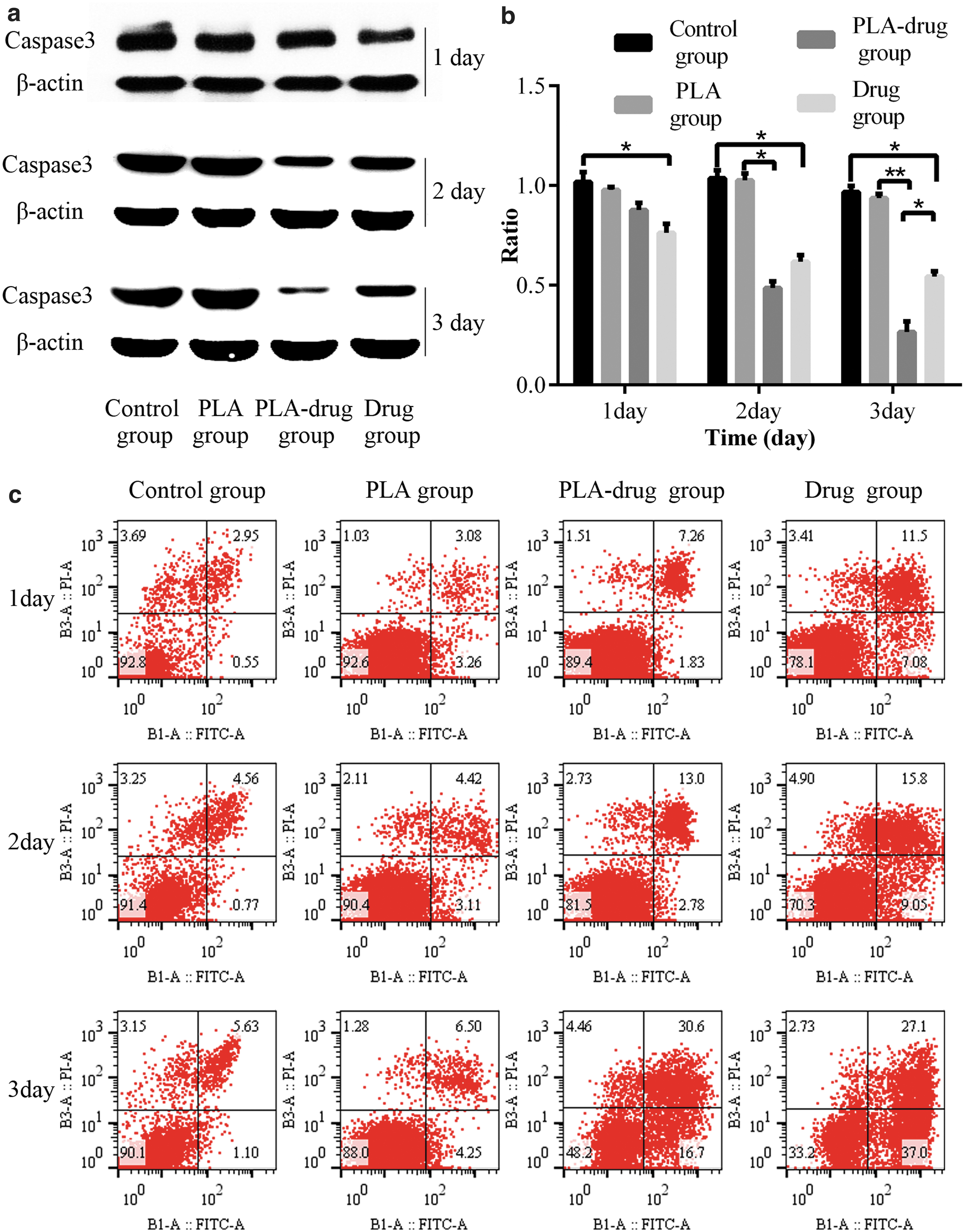
After treating the 4T1 cells in four groups, the expression of Caspase3 and the percentage of apoptotic cells were shown.
As shown in Figure 3c, the apoptotic cells of the 4T1-luc were measured by flow cytometry. The apoptotic cells in the Control group and PLA group had not substantially changed throughout the period of 3 days. The apoptotic cells in the PLA-drug group and Drug group were increasing during the 3 days. On the 3rd day after treatment, the apoptotic cells in the PLA-drug group reached the same level as that in the Drug group, and the apoptosis rates in PLA-drug group and Drug group were 30.6% and 27.1%, respectively.
Figure 4a and b showed the expression of Caspase3 in four groups of the DU145 cells after treatment. Figure 4c showed the apoptotic cells of the DU145 cells after treatment. The expression of Caspase3 and apoptosis was similar to those in the 4T1-luc cells.
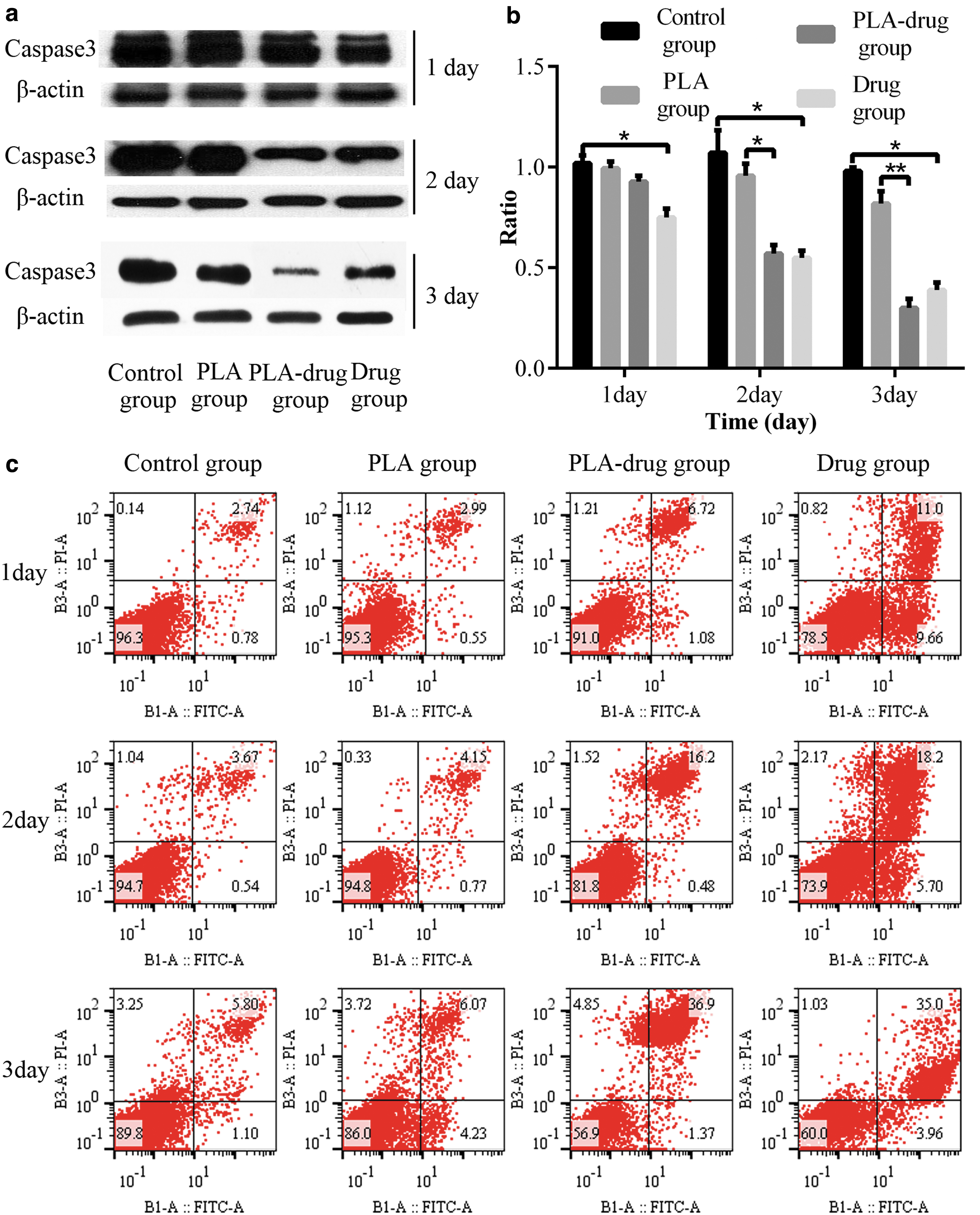
After treating the DU145 cells in four groups, the expression of Caspase3 and the percentage of apoptotic cell were showed.
In the PLA-drug group, the expression of Caspase3 was rapidly reduced during the 3 days. There was significant differences in the expression of Caspase3 between the PLA-drug group and PLA group on the second and 3rd day after treatment (p2 = 0.015, p3 < 0.001). But the difference of protein expression between PLA-drug group and Drug group was not significant on the 3rd day (p = 0.06).
The apoptotic cells of the DU145 cells in the PLA-drug group and Drug group were increasing during the 3 days. The apoptotic cells in the PLA-drug group were similar to that in the drug group on the 3rd day, and the apoptosis rates of the two groups were 36.9% and 35.0%, respectively.
In vivo animal experiment to investigate the therapeutic effects of the drug sustained-release materials
The experiments in vivo mainly explored the antitumor properties of the drug sustained-release materials and the reduction of systemic toxins and side effects compared with systemic chemotherapy. Figure 5a showed the growth of tumors at three points, which were on the 3rd day before surgery, the 3rd day after the surgery, and the 2nd week after surgery. The fluorescence intensity of the tumors in the Control group and PLA group was getting stronger after the surgery and the fluorescence intensity of the tumors in the PLA-drug group and Drug group was getting weaker after the surgery. In the 2nd week after surgery, there were significant differences in the fluorescence intensity between PLA-drug group and PLA group (p = 0.022), Drug group and Control group (p = 0.035), PLA-drug group and Drug group (p = 0.038) (Fig. 5b). The diameter of tumors was measured once a week to observe tumor growth and the volume of tumors was calculated (volume = 0.52 × length × width × width). In the 2nd week after surgery, only the volume of tumors showed significant differences between the PLA-drug group and PLA group (Fig. 5c). In the 2nd week after surgery, there were six remaining mice in the PLA-drug group, Drug group and PLA group, and there were seven remaining mice in the control group.

The growth of tumors was recorded by the fluorescence intensity and the volume of tumors at three time points.
Figure 6a showed the survival curve of experimental animals. The control group had three mice dead on the 3rd, 14th, and 19th day after operation. The PLA group had three mice dead on the 3rd day (two mice) and 17th day after operation. The PLA-drug group had two mice dead within 3 days after operation. Three mice of the Drug group were dead on the 3rd, 8th, and 15th day after operation. In the 3rd week after surgery, the experiment was over and there were six remaining mice in PLA-drug group and five remaining mice in the Control group, Drug group, and PLA group. Figure 6b showed the body weight changes during the 3 weeks after surgery. The average of the body weight in the Control group was low compared with the other groups. But there was no significant difference.

Figure 6c illustrated the general view of the tumors and the rhodamine-stained immunofluorescence histochemistry at the 3rd week after operation. The volume of tumors in the general view was calculated and counted (Fig. 6d). The volume of the tumors in the PLA-drug group (0.18 ± 0.177 cm3) was significantly smaller compared with the PLA group (0.65 ± 0.169 cm3) (p < 0.001). The volume of the tumors in the Drug group (0.35 ± 0.201 cm3) was significantly smaller compared with the Control group (0.89 ± 0.372 cm3) (p = 0.021). The volume of the tumors in the PLA-drug group was smaller compared with the Drug group, but the difference was not significant.
Apoptotic cells can be stained red by Rhodamine, as shown in Figure 6c. The proportion of apoptotic regions in the immunofluorescence histochemistry were compared in four groups (Fig. 6e). The ratio of apoptosis area in the PLA-drug group (0.19 ± 0.013) was significantly larger compared with the PLA group (0.07 ± 0.010) (p < 0.001). The ratio of apoptosis area in the Drug group (0.23 ± 0.011) was significantly larger compared with the Control group (0.06 ± 0.009) (p = 0.021).
The dates of liver function tests, renal function tests, and hemogram are shown in the table (Supplementary Table S1). Figure 7a and b showed the indicators of liver function at the 3rd week after operation. The average of aspartate aminotransferase (AST) was significantly lower in the PLA-drug group (186.76 ± 18.09, U/L) compared with the Drug group (272.08 ± 34.57, U/L) (p = 0.019). The alanine aminotransferase and AST in the Control group were significantly higher compared with the PLA group. In the indicators of renal function (Fig. 7c, d), the mice in the Control group (45.52 ± 7.238, mg/dL) were with significantly higher blood urea nitrogen (BUN) compared with the PLA-drug group (21.76 ± 3.443, mg/dL) (p = 0.014) and the creatinine in the Drug group (53.73 ± 16.739, μmol/L) was significantly higher compared with the Control group (25.54 ± 4.053, μmol/L) (p = 0.014).

The indicators of liver function tests and renal function tests at the 3rd week after operation were compared in four groups.
Figure 8 showed the index of hemogram. The red blood cell in the Drug group [5.53(5.175–5.73), 1012/L] was lower significantly compared with the PLA-drug group [6.8(6.595–6.920), 1012/L] (p = 0.031). The platelets in the Drug group [416(390–491.5), 109/L] was lower significantly compared with the PLA-drug group [751(735–849.5), 109/L] (p = 0.019). Compared with the Control group [331.9(285.0–378.8), 109/L], the white blood cell in the Drug group [180.0(177.2–219.45), 109/L] was significantly lower (p = 0.01).

The indices of hemogram were compared in four groups.
Discussion
In recent years, there has been a shift in the idea of surgery. Simply expanding the scope of surgical resection to reduce recurrence of tumor and improve patient survival has been recognized to have many shortcomings. The comprehensive treatment with surgery as the core has gradually been recognized by clinicians.21,22 Chemotherapy is also more commonly used to treat the residual tumors after surgery.23,24 How to reduce the side effects of systemic chemotherapy is also getting more and more attention. Some studies have shown that the drug toxicity can be effectively alleviated by local administration of slow-release materials.25–28 We explored the effects of the sustained-release materials loaded with antitumor drugs in this study. The results indicated that the sustained-release materials could effectively inhibit tumor growth and partly reduce the side effects of systemic chemotherapy, such as liver damage and myelosuppression.
From the experiments in this study, it was evident that the drug sustained-release materials had good antitumor properties in vitro and in vivo for partial solid tumors. When the 4T1-luc and DU145 cells were treated with the drug sustained-release materials on the 3rd day in vitro, the apoptosis rate of the cells in the PLA-drug group was similar to that in the drug group. This showed that both sustained-release material administration and direct administration may effectively inhibit tumor cells growth. In the experiments in vivo, we reached the same conclusion. As shown in Figure 5a, the fluorescence intensity of the tumors in the PLA group was getting stronger after the surgery, however, the fluorescence intensity of the tumors in the PLA-drug group was getting weaker after the surgery. And the fluorescence intensity of the tumors in the PLA-drug group was weaker compared with the drug group. The rhodamine staining in Figure 6c also showed that the PLA-drug group has more apoptotic tumor cells than the PLA group. The comparison of tumor volume in four groups in Figure 6d also demonstrates this conclusion. All those show that drug sustained-release materials could effectively promote the apoptosis of tumor cells and inhibit tumor growth in vivo. It is possible that local administration may be more effective against cancer than systemic administration.
We also explored whether the drug sustained-release materials could reduce the systemic toxins and side effects compared with systemic chemotherapy. In the survival curve of the mice, we found the fact that the three dead mice in the drug group died the day they received intraperitoneal injection of chemotherapy drugs, which may be related to the side effects of chemotherapy. The time of death of two mice in the PLA-drug group was within 3 days after operation, which may be related to surgical stress or postoperative infection. The time of death of mice in the control group and PLA group were later in the experiment, which might be due to the large size of the tumors. So we can see that the side effects of systemic chemotherapy are significant in mice. We compared the side effects of the different administration methods from three aspects: liver function, renal function, and myelosuppression. Compared with systemic chemotherapy, the mice of the PLA-drug group with locally placed drug sustained-release materials suffered less liver damage and myelosuppression. As shown in Figure 7b, the mice in the PLA-drug group were with significantly lower AST compared with the drug group, which pointed out that the drug sustained-release materials confer less liver damage. As shown in Figure 8, The RBC and PLA in the drug group were significantly lower compared with the PLA-drug group, which pointed out that the drug sustained-release materials can reduce the phenomenon of myelosuppression.
Our research also had some limitations. First, the drug release process of the drug sustained-release materials could not be fully simulated. Lactic acid, the product of PLA hydrolysis, could be metabolized in the human body. This could not be done in vitro. So the drugs in the body are released more quickly. Second, the two indices of liver function in the control group were found to be too high, which was thought to be due to liver metastases. The high BUN in the control group was found to be related to the negative nitrogen balance caused by tumors. Proteins were consumed more in the control group, which led to higher BUN as protein metabolite. All of this could affect the outcome of the experiment. Third, because mice have limited blood, blood samples are used to measure liver function tests, kidney function tests, and hemogram. The changes of drug concentrations in blood were not monitored. Finally, leukocytopenia was evident in both the drug group and the PLA-drug group, which suggested that the drug sustained-release materials could not completely eliminate the side effects of chemotherapeutic drugs. Although there were some drawbacks, the results confirmed that the sustained-release materials could effectively inhibit tumor growth and partly reduce the side effects of systemic chemotherapy.
In recent years, there have been a few of reports on the creation of chemotherapeutic drugs loaded with materials to enhance the efficacy of chemotherapy and reduce systemic toxicity. However, few materials are actually used in clinical practice.29,30 It is important to conduct detailed animal experiments on materials loaded with chemotherapy drugs. In this study, we adequately verified the antitumor effect of the drug sustained-release materials through in vivo and in vitro experiments, and the changes of routine biochemistry and blood routine in experimental animals were investigated. It is hoped that our experiment can accelerate the clinical application of the material.
Conclusion
By electrospinning technology, the drug sustained-release materials were successfully prepared and demonstrated that the material can steadily release the drug in vitro. The local administration of the drug sustained-release materials could effectively inhibit the growth of the positive incision margins and definitely reduce the partial side effects associated with systemic chemotherapy.
Footnotes
References
Supplementary Material
Please find the following supplemental material available below.
For Open Access articles published under a Creative Commons License, all supplemental material carries the same license as the article it is associated with.
For non-Open Access articles published, all supplemental material carries a non-exclusive license, and permission requests for re-use of supplemental material or any part of supplemental material shall be sent directly to the copyright owner as specified in the copyright notice associated with the article.