
Abstract
Microtia is a congenital malformation of the auricle. The conventional therapy for microtia is reconstruction of the auricle by using the patient's own costal cartilage. Because it is invasive to harvest costal cartilages, less invasive ways for auricular reconstruction need to be established. Recent reports have indicated a new method for the production of cartilaginous particles from human induced pluripotent stem cells. To adopt this method to create an auricular-shaped regenerative cartilage, a novel scaffold with the property of a three-dimensional shape memory was created. A scaffold with a three-dimensional shape of auricular frames composed of a helix and an antihelix, which was designed to mimic an auricular framework carved from autologous costal cartilage and transplanted in auricular reconstruction, was prepared, filled with cartilaginous particles, and subcutaneously transplanted in nude rats. The auricular-shaped regenerative cartilage maintained the given shape and cartilage features in vivo for 1 year. Our findings suggest a novel approach for auricular reconstruction.
Impact statement
A recent report that indicated the production of cartilaginous particles from human induced pluripotent stem cells will provide the solution to overcome the limitation of a cell source for cartilage regeneration of large and complicated shapes such as auricles. By combining cartilaginous particles and a novel scaffold with the property of a three-dimensional shape memory, we fabricated auricular frames composed of a helix and an antihelix, which was designed to mimic an auricular framework carved from autologous costal cartilage in auricular reconstruction. Our findings demonstrate a novel approach for auricular reconstruction.
Introduction
Microtia is a congenital malformation of the auricle. Conventional treatment for microtia is transplantation of autologous costal cartilage carved into an auricular frame. Usually, patients wait until the age of 10 when the costal cartilage grows large enough for reconstruction. Because it causes significant donor-site morbidity at the thorax to harvest costal cartilage, less invasive procedures need to be established.
The regenerative medicine of cartilage will be a promising solution to minimize the invasiveness in the treatment for cartilage defects, including microtia. Many groups have reported the fabrication of three-dimensional regenerative cartilage with a good mechanical strength and an appropriate shape by combining cells and a scaffold.1–6
Our group also established an implant-type regenerative cartilage by combining culture-expanded autologous auricular chondrocytes, atelocollagen, and a sponge-like poly-
However, the current technique has several limitations. For example, the number of cells obtained from the harvested auricle is limited. For auricular reconstruction, many more cells are needed than those for the augmentation of the nose dorsum, which increases the invasiveness at the donor site. In addition, similar to many other reported methods, cartilage matures after the implantation, which makes the outcome of this treatment uncertain.
Pluripotent stem cells, namely embryonic stem (ES) cells and induced pluripotent stem (iPS) cells possess unlimited proliferative ability and pluripotency, and they have been intensively studied for their application in regenerative medicine for cartilage.
Wu et al. established a method to differentiate human PSCs into chondrocytes by dissecting the developmental steps of chondrocytes. 9 Loh et al. presented an elegant procedure by which human PSCs can be differentiated into desired cell lineages such as chondrocytes and cardiomyocytes with high purity. By controlling the lineage decision by stimulation and inhibition of key signaling, cells are controlled to differentiate only into specific lineages. 10 Dicks et al. established markers by which chondroprogenitors could be identified within human iPS cells during chondrogenic differentiation. 11 A report by Craft et al. showed that cartilage tissues with a thickness of several-hundred micrometer could be generated in vitro. 12
Recently, an induction technique of cartilaginous particles with a diameter of several millimeter from human-iPS cells was reported.13,14 This technique can solve the issue of limitation in the cell amount, because iPS cells come from an inexhaustible supply, which means that cartilaginous particles can be prepared as much as needed. In addition, mature cartilage is obtained in vitro, which will increase the success rate of the treatment.
In the present research, the technique to regenerate an auricle was established by utilizing cartilaginous particles that originated from two different lines of human iPS cells. To align the particles into an auricular shape with ease, tubular PLA scaffolds were adopted. The optimal opening ratio of the scaffolds and optimal size of the cartilaginous particles were investigated. In addition, the method to apply the shape of the ear frame to the scaffold was established by utilizing the thermo-responsive shape memory. Retention of the shape and the characteristics of the cartilage tissue were evaluated by an implantation assay.
Materials and Methods
Reagents
Dulbecco's modified Eagle's medium: Nutrient Mixture F-12 (DMEM/F12), DMEM (unless otherwise indicated), penicillin and streptomycin, human serum, gelatin, and Mitomycin C were purchased from Sigma-Aldrich (MO). Fibroblast growth factor-2 (FGF-2) was purchased from the Kaken Pharmaceutical Co. (Tokyo, Japan). Insulin was purchased from Novo Nordisk (Bagsvaerd, Denmark). Fetus bovine serum (FBS), knockout serum replacement, nonessential amino acid, 2-mercaptoethanol, Glutamax, Essential 8, ITS-X, and sodium pyruvate were purchased from Thermo Fisher Scientific (MA). Collagenase, basic FGF (bFGF), Y-27632, 4% paraformaldehyde phosphate-buffered solution, and sucrose were purchased from Wako Pure Chemical Industries, Ltd. (Osaka, Japan).
Human auricular chondrocytes
All experiments were conducted with the approval of the ethics committee of the University of Tokyo. Human auricular cartilage, which was to be discarded from a reconstruction operation for microtia, was kindly provided by Dr. Satoru Nagata of the Nagata Microtia and Reconstructive Plastic Surgery Clinic (Saitama, Japan), and his microtia patients gave informed consent to our laboratory's research. Human auricular chondrocytes were isolated from the auricular cartilage and expanded as previously described.2–4
Briefly, the cartilage was minced and digested with 0.3% collagenase for 18 h; the cells were seeded on a type 1 collagen-coated 10-cm dish (AGC Techno Glass Co., Tokyo, Japan) with growth medium (DMEM/F12, 100 U penicillin, 100 μg streptomycin, 100 ng/mL FGF-2, 5 μg/mL insulin, and 5% human serum). Cells were cryopreserved at passage 1 by using CELL BANKER (Nippon Zenyaku Kogyo Co., Fukushima, Japan) and used for the experiments at passage 2.
Pellet preparation of human auricular chondrocytes
Human auricular chondrocytes were mixed in 0.8% collagen type 1 and 3 (Nipponham, Osaka, Japan) at a density of 107 cells/mL, and 20, 50, and 100 μL of the cell suspensions were incubated at 37°C for 2 h for gelatinization. The gelatinized pellets were cultured for 3 weeks in chondrogenic differentiation medium (DMEM/F12, 100 U penicillin, 100 μg streptomycin, 200 ng/mL BMP-2 [COREFRONT, Tokyo, Japan], 5 μg/mL Insulin, and 100 nM T3 [Merck Millipore, Darmstadt, Germany]).
Preparation of SNL feeder cells
SNL feeder cells (Cell Biolabs, Inc., CA.) were cultured according to the protocols of the Center for iPS Cell Research and Application (CiRA). Briefly, the cells were plated on a 0.1% gelatin-coated 10-cm dish and cultured in growth medium (DMEM [Nacali Tesque, Kyoto, Japan], 7% FBS, 50 U penicillin, 50 μg streptomycin). The cells were expanded at the ratio of 1:16 until passage 20, treated with 12 μg/mL Mitomycin C for 2 h and 15 min, and finally cryopreserved until they were used.
Culture of human iPS cells
Human iPS cells (253G1) 15 were purchased from RIKEN BRC (Ibaraki, Japan), and they were cultured on SNL feeder with growth medium (DMEM/F12, 20% nonessential amino acid serum replacement, 50 U penicillin, 50 μg streptomycin, 0.1 mM nonessential amino acid, 0.1 mM 2-mercaptoethanol, 2 mM Glutamax, and 4 ng/mL bFGF). The cells were passaged every 4 or 5 days with treatment of 10 μM Y-27632.
Preparation of cartilaginous particles from human iPS cells
Cartilaginous particles were prepared according to recent reports.13,14 Briefly, human iPS cells were seeded on a six-well plate coated with ES quality Matrigel (Corning, NY) diluted in DMEM/F12. One milliliter of Essential 8 was changed every 2 or 3 days. Approximately 3 weeks later, nodules were formed at the center of the colony. The medium was changed into 1 mL mesodermal differentiation medium (DMEM/F12, 1% FBS, 1% ITS-X, 10 ng/mL Wnt3A [R&D Systems, Inc., Minneapolis], 10 ng/mL Activin A [R&D Systems, Inc.], 50 U penicillin, and 50 μg streptomycin).
After 2 or 3 days, the medium was changed into 1 mL of chondrogenic medium (DMEM, 1% FBS, 1% ITS-X, 50 μg/mL ascorbic acid [Nacali Tesque], 1 mM sodium pyruvate, 10 ng/mL BMP2 [PEPROTECH, NJ], 10 ng/mL TGFβ1 [PEPROTECH], 10 ng/mL GDF5 [PROSPEC, NJ], and 10 ng/mL bFGF). After 14 days from the beginning of the mesodermal differentiation, colonies were scraped from plates and re-plated on noncoated six-well plates with chondrogenic medium without bFGF. Approximately 10 weeks later, the cartilaginous particles matured. Cartilaginous particles from an human leukocyte antigen (HLA) homozygous donor were provided from the Cell Induction and Regulation Field, Department of Clinical Application, CiRA, Kyoto University, under approval of the ethics committees of both Kyoto University and the University of Tokyo.
Agarose mold preparation
Agarose (Takara Bio, Inc., Shiga, Japan) was diluted in phosphate-buffered saline to make a 2% solution, and it was then heated for solation. The soled agarose was molded into a plastic dish, and the impression of the template aluminum wire was acquired by cooling.
PLA scaffold preparation and three-dimensional shape-memory property test
Scaffolds were prepared at the Industrial Technology Center of Fukui Prefecture. Briefly, polylactic acid (PLA) threads were woven to form 3-mm diameter meshed tubes with the opening ratios of 20.3%, 35%, and 55.1% (Fig. 1A). The property of the three-dimensional shape memory was tested by fixing scaffolds with ∼3-mm diameter aluminum wire and aluminum foil, and autoclaving at 121°C for 20 min.

A novel meshed tube PLA scaffold and chondrocyte pellets.
Animals
All animal experiments were conducted with the approval of the animal care and use committee of the University of Tokyo. Male BALB/cAJcl-nu/nu (6-week-old) and F344/N-rnu/rnu (8-week-old) mice were purchased from CLEA Japan (Tokyo, Japan).
Transplantation of regenerative cartilages in mouse
For the transplantation of the auricular chondrocyte pellets, scaffolds were cut to the length of 10 mm and sterilized by autoclaving. Pellets were filled in the scaffolds (5 of 20 μL pellets, 2 of 50 μL pellets, and 1 of 100 μL pellets). Both ends were closed by using titanium vessel ligation clips (Johnson & Johnson, NJ) (Fig. 1B, C).
For transplantation of the cartilaginous particles from human iPS cells (Fig. 3A and Supplementary Fig. S2), they were filled into the scaffold with the opening ratio of 35% cut into ∼15 mm (Fig. 3B). To test the shape retention ability, the shape-memorized scaffold in a quarter arc shape (R = 15 mm) was filled with cartilaginous particles from an HLA homozygous donor (Fig. 4A–C>). They were embedded in a 2% agarose mold to adjust the shape and overnight incubated in DMEM supplemented with 10% FBS, 100 U penicillin, and 100 μg streptomycin (Figs. 3C and 4D).
Transplantation of human iPS cell-originated cartilaginous particles with a scaffold.
Transplantation of a quarter arc-shaped regenerative cartilage
These prepared samples were subcutaneously transplanted on the back of the BALB/cAJcl-nu/nu mouse (Fig. 4C, D). After 8 weeks of transplantation, the mouse was euthanized and the sample was collected (Figs. 1B, C and 3D, E). After collection of the macroscopic findings, the sample was fixed in a 4% paraformaldehyde phosphate-buffered solution at 4°C overnight. Samples were sectioned after paraffin embedding or cryogenically sectioned.
Regenerative cartilage mimicking auricular frame
Scaffolds were three-dimensionally shape memorized to mimic the helix and antihelix as already described (Fig. 5A–C). Cartilaginous particles prepared from the human iPS cells (253G1) or HLA homozygous donor were filled into the scaffolds, embedded in an agarose mold to adjust the shape, incubated overnight, and subcutaneously transplanted on the back of an F344/N-rnu/rnu rat (Figs. 5D–F and 6A). Transplants of the human iPS cells (253G1) were collected 8 weeks after transplantation (Fig. 5G). The transplants prepared with cartilaginous particles from the HLA homozygous donor were macroscopically evaluated at 2, 4, 8, 16, 24, 32, 40 weeks, and a year post-transplantation (Fig. 6A). The transplants were harvested at 8 weeks, 16 weeks, and a year post-transplantation. The experiments were run in triplicate.

Fabrication of the auricular frame-shaped regenerative cartilage

Long-term observation of the auricular frame regenerative cartilage subcutaneously transplanted to F344/N-rnu/rnu
Histological analysis
Paraffin-embedded sections were deparaffinized by a xylene series, and they were stained with hematoxylin (Sakura Finetek Japan Co.) and eosin (Sakura Finetek Japan Co.), 0.05% toluidine blue (Waldeck GmbH & Co. KG, Mü, Germany) solution, or 0.1% safranin O (Waldeck GmbH & Co. KG). The toluidine blue positive areas were evaluated by Image J. 16 Briefly, the scale was set to that of the histology, the threshold was adjusted to select the toluidine blue positive area, and the selected area was measured. For the total area, the outline of the sample was traced and the area was measured.
For Elastica van Gieson staining, Maeda modification resorcin Fuchsine liquid (Muto Pure Chemical Industries, Ltd., Tokyo, Japan), Weigert's Iron Hematoxylin (Muto Pure Chemical Industries, Ltd.), and Wangieson liquid (Muto Pure Chemical Industries, Ltd.) were used. For in situ apoptosis detection, Apoptosis in situ detection kit (Wako Pure Chemical Industries, Ltd.) was used according to the manufacturer's protocol.
For the immunohistochemistry, endogenous peroxidase was inactivated with 3% H2O2 (Wako Pure Chemical Industries, Ltd.) in methanol (Wako Pure Chemical Industries, Ltd.). Sections were blocked with 10% normal goat serum (Nichirei Bioscience, Inc., Tokyo, Japan). Sections were applied as either anti-human vimentin antibody (Abcam, Cambridge, United Kingdom) or anti-type 2 collagen antibody (LSL, Tokyo, Japan) at 4°C overnight. Normal rabbit IgG (Southern Biotech, Birmingham) was applied as the negative control. On the next day, a rabbit IgG goat secondary antibody (Vector Laboratories, CA) was applied followed by ABC (Vector Laboratories); then, the samples were stained with DAB (Vector Laboratories) and hematoxylin.
Mechanical analysis
The mechanical property was evaluated by a Venustron (AXIOM, Fukushima, Japan). Silicon was used for calibration (600–800 kPa). Young's modulus was derived according to the manufacturer's protocol. Briefly, samples were set on a table beneath the probe. The probe was slowly lowered to the sample until it slightly touched and pressed the sample while measuring its tactile. The Young's modulus was derived from these data. The sample was divided into three regions, and the Young's modulus was measured 5 times at each measuring point.
Biochemical analysis
A glycosaminoglycan assay was conducted by using the Blyscan Glycosaminoglycan Assay Kit (Biocolor Ltd., Carrickfergus, United Kingdom) according to the manufacturer's protocol with minor modifications. A protein assay was conducted by using the DC Protein Assay (Bio-Rad, CA).
Briefly, samples before fixation were soaked in 0.8 mL of 0.05 M acetic acid (Wako Pure Chemical Industries, Ltd.); then, they were preserved at −80°C until further analysis. The thawed samples were minced and incubated in 0.1 mL of 10 mg/mL pepsin (Wako Pure Chemical Industries, Ltd.) in 0.05 M acetic acid for 48 h at 4°C. The samples were then treated with 0.1 mL of 10 × TBS, 0.1 mL of 1 mg/mL pancreatic elastase (Wako Pure Chemical Industries, Ltd.); dissolved in 1 × TBS; and incubated overnight at 4°C. The samples were then centrifuged at 9100 g for 5 min, and the supernatant was used for further analysis.
For the glycosaminoglycan assay, 100 μL of the sample supernatant was stained according to the manufacturer's manual. The sample solution was transferred to a 96 microwell plate, and the absorbance was read by a plate reader ARVOX3 (Perkin-Elmer, MA) at 660 nm. For the protein assay, 5 μL of the sample supernatant was processed according to the manufacturer's manual. The sample solution was transferred to a 96 microwell plate, and the absorbance was read with ARVOX3 at 750 nm. The glycosaminoglycan concentrations were corrected with those of the protein.
Statistical analysis
The statistical analysis was performed by using Excel Statistics 2015 (Social Survey Research Information Co., Ltd.). The data were analyzed by using either Fisher's test, Tukey's test, or Dunnett's test. p-Values <0.05 were considered statistically significant.
Results
Transplantation of auricular chondrocyte pellets filled in scaffolds
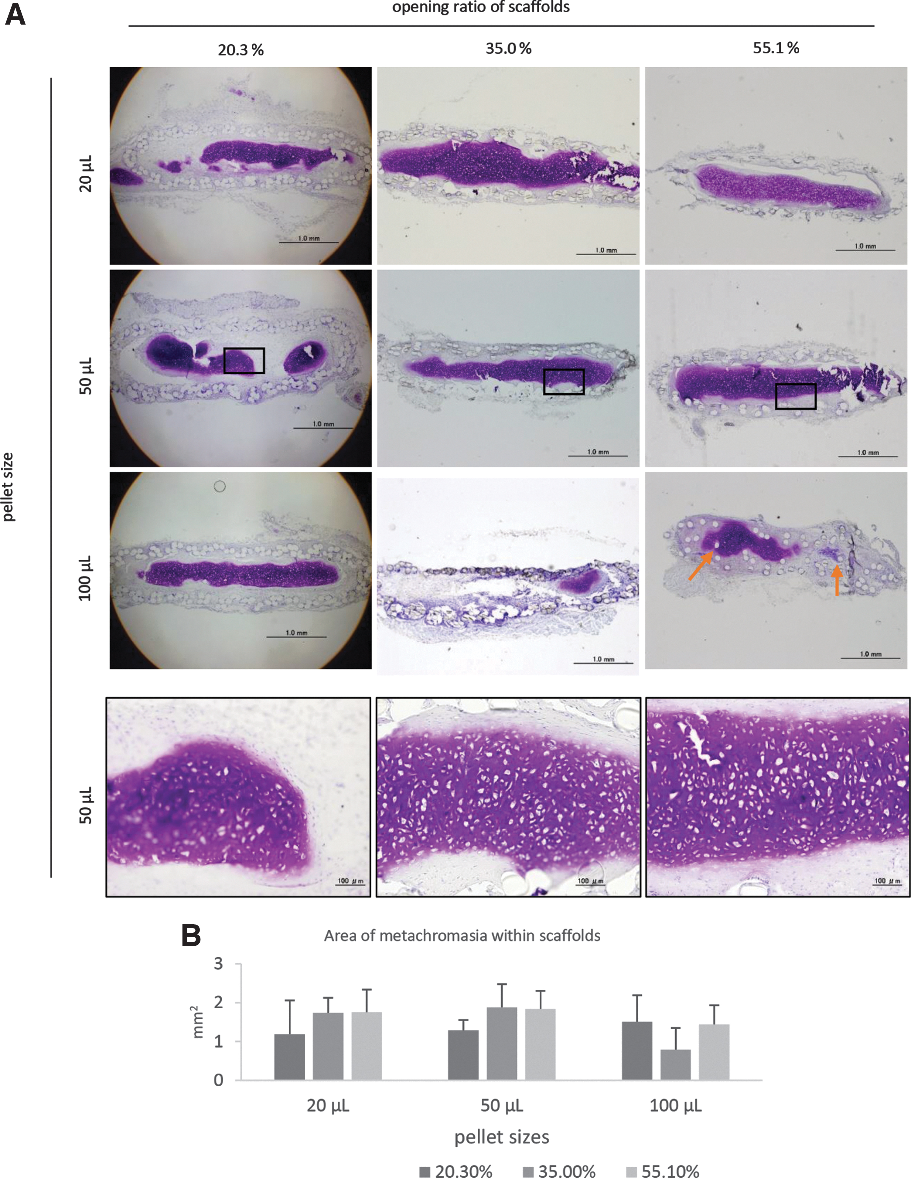
The auricular chondrocyte pellets filled in the PLA mesh scaffolds were transplanted into nude mice to evaluate the appropriate opening ratio of the scaffolds and proper pellet size. After 8 weeks from the transplantation, all the scaffolds remained at the transplantation sites. Regenerative cartilage was observed in all the samples regardless of the pellet size and opening ratio of the scaffolds, indicating that the scaffolds successfully retained the chondrocyte pellets (Fig. 2A). In addition, infiltration or growth into the scaffold was not obvious in each type of scaffold tested.
Histological analysis of auricular chondrocyte pellets with scaffolds after transplantation.
There was no statistical significance regarding the area of metachromasia with toluidine blue staining among the transplants with the different pellet sizes or the opening ratio of the scaffold (Fig. 2B). However, the irregularity of the scaffold shape was observed in samples with the scaffold opening ratio of 55.1% (Fig. 2A indicated by arrow). Taking this into consideration, the scaffold with the opening ratio of 55.1% may not be suitable for further examination.
As for the scaffolds with the opening ratios of 20.3% and 35.0%, although there was no statistical significance among the areas of metachromasia, the scaffold with the opening ratio of 35.0% tended to show a broader area of metachromasia with the pellets sizes of 20 and 50 μL, which were assumed to be used more frequently in further analyses (Fig. 2B). Taking this into consideration, we decided to use the scaffold with the opening ratio of 35.0% for further examinations.
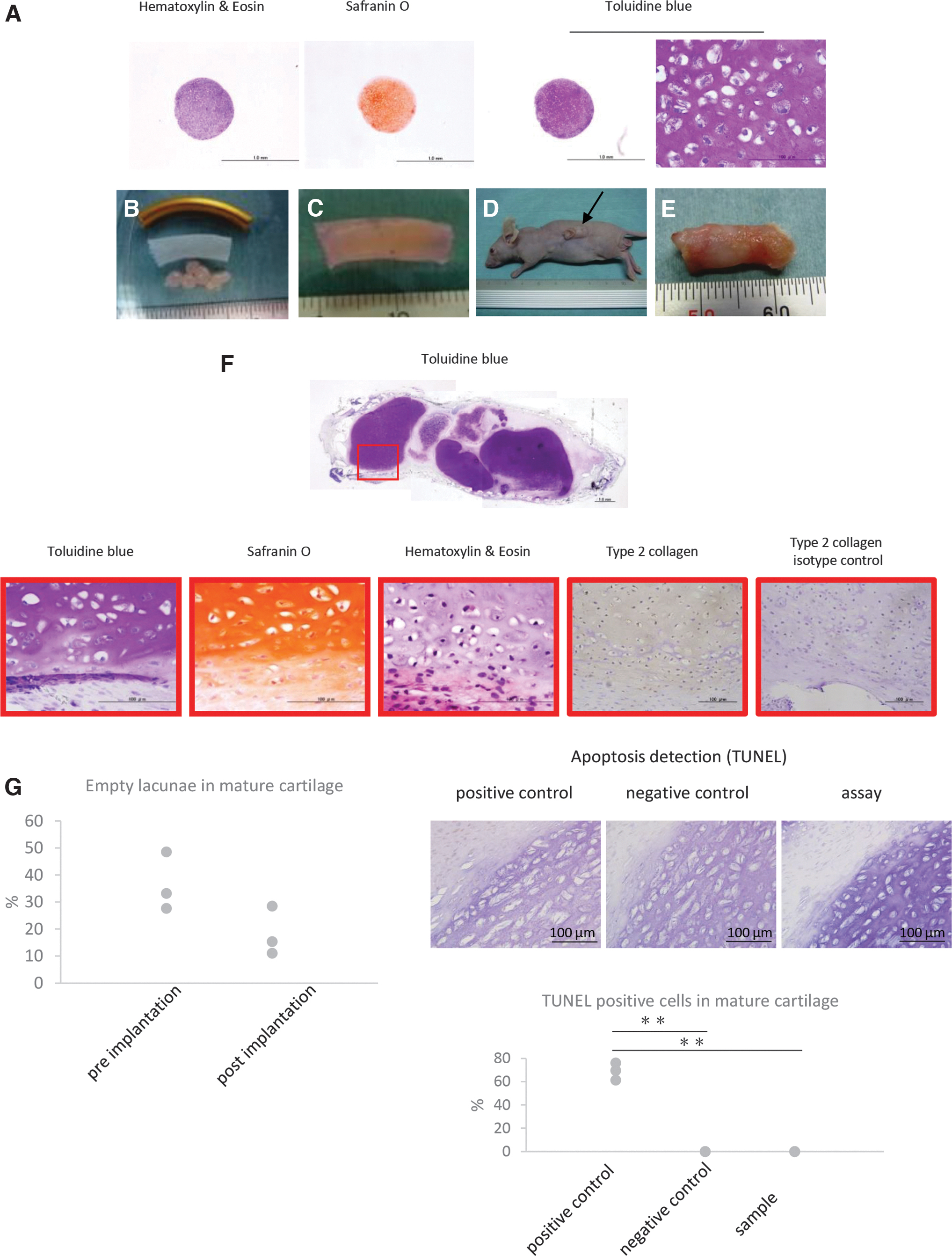
Transplantation of cartilaginous particles filled in scaffold
Using the scaffold with the opening ratio of 35.0%, cartilaginous particles that originated from the human iPS cells were transplanted into nude mice. After 8 weeks, the scaffold retained the cartilaginous particles, which maintained their cartilage features (Fig. 3F). Metachromasia area was 41.681 mm2, which was 82.48273% to the total area.
The rate of empty lacunae in mature cartilage in three representative areas was 18.28% on average whereas that of pre-implanted samples was 36.43%. There was no statistically significant difference between the two groups (Tukey's test, p = 0.0899) (Fig. 3G). The results of in situ apoptosis detection in mature cartilage in three representative areas showed that there was a significant difference between positive control samples and negative control samples (Fisher test p = 0.000001112) and between positive control samples and assay samples (Fisher test p = 0.000001112), whereas no significant difference was detected between negative control samples and assay samples (Fisher test p = 1.0000) (Fig. 3G).
These findings suggest that the opening ratio of the mesh was large enough for substance exchange required for cell survival and cartilage maturation, and yet small enough to retain the cartilaginous particles.
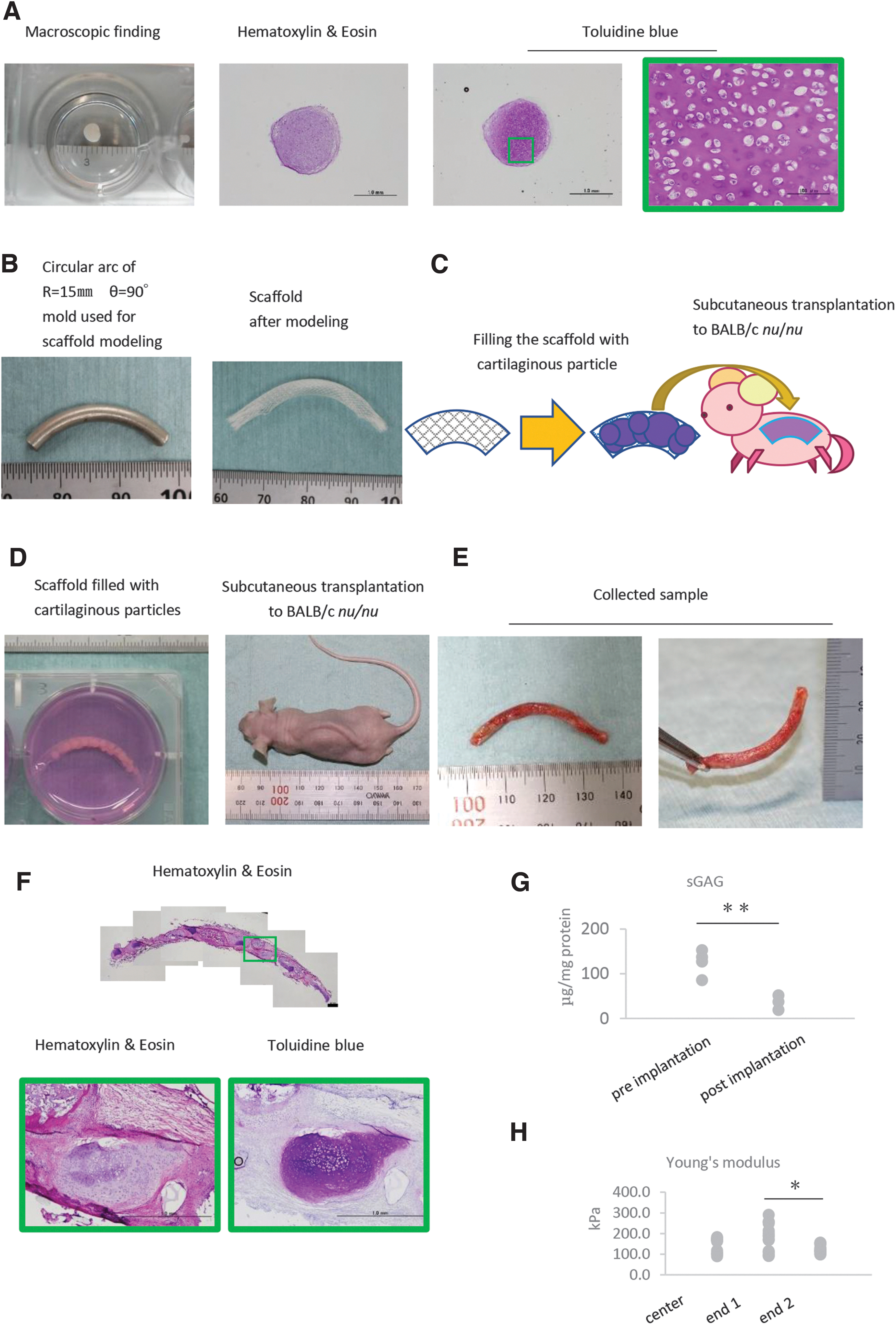
Transplantation of the quarter arc-shaped regenerative cartilage with cartilaginous particles and scaffolds
To examine the shape memorization capacity of the scaffold, quarter arc-shaped scaffolds were prepared by using a metallic wire as the template. The scaffold maintained the given shape after the wire was removed, indicating that the three-dimensional shape was successfully memorized by the scaffold (Fig. 4B). This scaffold filled with cartilaginous particles from human iPS cells was transplanted into nude mice.
After 8 weeks, the harvested transplant maintained the quarter arc shape (Fig. 4E). One end of the transplant did not hang down while holding the other end (Fig. 4E). Histological analysis showed that some cartilaginous particles maintained the cartilage feature in vivo (Fig. 4F). On the other hand, biochemical analysis showed the decrease in the amount of sulfated glycosaminoglycans (sGAG) after transplantation (Tukey's test, p = 0.0049) (Fig. 4G), reflecting, at least in part, the formation of fibrous tissue between cartilaginous particles. The Young's modulus of each end was not significantly different from that of the center (Fisher's test, center vs. end 1; p = 0.3538, center vs. end 2; p = 0.5635), whereas those of both ends were significantly different (Fisher's test, p = 0.0488) (Fig. 4H). These results suggest that the scaffold can retain the given shape for 8 weeks in vivo.
Regenerative cartilage mimicking auricular cartilage frame
To examine the shape retention ability of the scaffold having a much more complex shape, scaffolds were prepared in the shape of the auricular cartilage frame, and they were transplanted into nude rats. Macroscopically, the sample seemed to maintain the given shape for 8 weeks in vivo (Fig. 5G, H). Macroscopic findings of a section of the sample from the yellow square area in Figure 5H showed that a white, smooth, and solid substance filled the scaffold (Fig. 5I). Histological analysis suggested that some part of the sample was cartilage and other tissue surrounding the cartilage was negative for human vimentin but positive for type 1 collagen, suggesting the invasion of connective tissue from the host (Fig. 5J).
These findings suggest that our novel scaffold could memorize the given auricular frame shape, retain cartilaginous particles, and retain the particles' cartilage features in vivo.
Long-term observation of auricular frame mimicked regenerative cartilage in vivo
To examine the stability of the regenerative cartilage over a long term, regenerative cartilages mimicking the auricular framework were subcutaneously transplanted into a nude rat. Regenerative cartilages were prepared by using cartilaginous particles that originated from iPS cells from an HLA homozygous donor, because these are predicted to be used in a future clinical study.
Macroscopically, the transplants mostly maintained their given shape in vivo for as long as 1 year (Fig. 6A). We examined the changes in the height and width of the transplant both before and after transplantation. The average ratio of the width (8 weeks, 16 weeks, or 1 year to pretransplantation) was within 0.96–1.03, and that of the height was within 1.07–1.13 (Fig. 6B). These results suggest that the transplant mostly maintained its given shape in vivo, although the height of the samples slightly increased.
The Young's modulus of transplants was below 1000 kPa in samples of 8 and 16 weeks, whereas it increased at 1 year after transplantation. The values of Young's module of the samples transplanted for 1 year were divided into two groups, one of which was around 1.2 MPa and the other was around 3.6 MPa. There was no statistically significant difference between samples of 8 and 16 weeks post-transplantation (Fisher's test, p = 0.6986), whereas a significant difference was observed between samples of 8 weeks and 1 year (Fisher's test, p = 0.000002201), and between samples of 16 weeks and 1 year (Fisher's test, p = 0.0000003973) (Fig. 7A).

Mechanical and histological analysis of the regenerative cartilage.
A histological analysis indicated that the metachromasic area with toluidine blue staining was positive for type 2 collagen and human vimentin with immunohistochemistry for as long as a year post-transplantation (Fig. 7B and Supplementary Fig. S1). Also, some tissue that showed a low metachromasia was negative for either type 2 collagen or human vimentin, indicating the invasion of the host's cells into the transplant (data not shown). A biochemical analysis indicated that some samples transplanted for 8 weeks had a glycosaminoglycan concentration of around 70 μg/mg protein whereas other samples had <10 μg/mg protein. The majority of the samples transplanted for 16 weeks and 1 year had a glycosaminoglycan concentration of <10 μg/mg protein. The statistical analysis showed that there was a significant difference among samples 8 and 16 weeks post-transplantation (Fisher's test, p = 0.0483), but there was no significant difference among samples 8 weeks and 1 year post transplantation (Fisher's test, p = 0.0561) and samples 16 weeks and 1 year post-transplantation (Fisher's test, p = 0.9423) (Fig. 7C).
These results suggest that some of the transplanted cartilaginous particles maintained their cartilage features for as long as 1 year, whereas invasion of the host's cells as early as 8 weeks post-transplantation decreased the transplant's mechanical properties and glycosaminoglycan concentration. That is to say, the low GAG value could be attributable to the heterogeneity of samples that contained cartilage and noncartilage tissues from the host.
Discussion
In this study, we demonstrated that our novel meshed tube PLA scaffold could retain tissues within and support their survival and maturation. This is due to not only biocompatibility but also the degradability with a lower absorption rate, especially compared with poly lactic-co-glycolic acid (PLGA). Our previous studies indicated that the higher absorption rate of PLGA caused inflammation, which had a negative effect on the cartilage maturation. 1
PLA and its copolymers are reported to hold a mild thermo-responsive shape-memory effect. The thermo-responsive shape-memory effect occurs when materials undergo heat higher than their glass transition temperature or melting temperature, and are then cooled to temperature lower than the glass transition temperature. The glass transition temperature of PLA is around 60°C. 17 This indicated that PLA could have a thermo-responsive shape memory around the autoclave sterilization of 121°C.
Taken together, it is possible to fabricate a novel scaffold with a biocompatibility, ability to retain cartilaginous particles within and to support the survival and maturation of particles, and shape memory simply by heating and cooling.
In the assay to determine the opening ratio of the scaffolds, the pellet sizes were meant to represent the cartilaginous particles of small (1.5–2 mm diameter, theological volume as ideal sphere would be around 20 μL), medium (3–5 mm diameter, volume around 50 μL), and large (6 mm diameter, volume around 100 μL) sizes. As the scaffold was 3-mm diameter meshed tubes, pellet sizes of 20 and 50 μL would represent the sizes of cartilaginous particles that would be used more frequently in further analysis as the maximum size of cartilaginous particles is frequently around 5 mm diameter. We examined three different opening ratios of the scaffolds to evaluate whether any opening ratio was not suitable for cell survival, cartilage maturation, or sample retention. As a result, regenerative cartilage was observed in all the samples.
These results indicate that none of the opening ratios of the scaffolds was too large to retain tissues within or too small for material exchange for all sizes of the pellets. However, the scaffold with an opening ratio of 55.1% sometimes failed to retain the tube shape in vivo, suggesting that the large opening ratio weakened the structure of the scaffold. Therefore, this opening ratio was considered inadequate as the scaffold for regenerative cartilage. Comparing the scaffold with the opening ratio of 20.3% and that with 35.0%, the latter tended to generate a greater cartilage matrix. This result suggested that a larger opening ratio would be beneficial for the efficiency of the material exchange.
With advice from the Industrial Technology Center of Fukui Prefecture, we tested the thermo-responsive shape memory of the PLA scaffold by heating above 100°C for longer than 5 min. Aluminum was chosen as the template for the shape memory, because aluminum is a material with a formability, melting point of around 660°C, and ready availability.
The template for the auricular cartilage was designed to mimic the auricular frame prepared for auricular reconstruction.18,19 We considered it important to mimic the features of the helix and antihelix. We designed the helix with one wire and antihelix with two wires to form the Y shape. As a result, the auricular frame stood out at the back of the F344/N-rnu/rnu rat for as long as 1 year. Also, as shown in Figure 7B, the cartilaginous particles maintained their cartilage features in vivo. Taking these results together, our novel meshed tube scaffold successfully supported the formation of auricular frame regenerative cartilage by cartilaginous particles.
As reported, cartilaginous particles spontaneously integrate when kept in contact with each other in vitro. 20 The same report showed that the integration of particles started from membranous integration on day 7, the cartilaginous integration occurred on day 14 and was completed on day 84. In the preliminary study, we also observed the integration of cartilaginous particles when they maintained contact in the agarose mold for 3 weeks. A histological analysis suggested that the perichondrium-like membrane connected and enclosed the cartilaginous particles (data not shown). We considered that the cartilaginous particles would also fuse together in vivo if they were kept in contact within the meshed tube scaffold. However, as observed in Figure 3F, when the scaffold was totally filled with cartilaginous particles, the scaffold could be easily deformed. To avoid any deformity of the scaffold, the cartilaginous particles were filled in the auricular-shaped scaffold at a slightly lower filling rate.
However, as shown in Figure 7B, a wide area was negative for metachromasia with toluidine blue staining. Some of the area negative for metachromasia in Figure 7B was human vimentin negative fibroblast-like tissue (data not shown).
These results suggest that the host cells migrated into the gaps of the cartilaginous particles, thus hindering their integration.
Heterogeneity of the tissue composed of cartilage and fibrotic tissues could be one explanation for the variation of Young's module. According to previous reports, the mechanical properties of the auricle are 1.66 MPa, 21 those of articular cartilage vary from 20 kPa to 6.44 MPa 22 by zones, and those of the cortical bone are 8.75 GPa 23 and cancellous bone are 4.66 GPa. 23 Young's module of the samples transplanted for 1 year was around 1.2 MPa in some assays and around 3.6 MPa for others, both of which are close to that of the auricle, far weaker than those of the bone, and within the range of those of articular cartilage. It is possible that the value around 3.6 MPa would represent the cartilaginous particles that matured in vivo. On the other hand, the other group with a lower value of 1.2 MPa would be affected by host-derived tissues. These results suggest that there is room for improvement regarding the heterogeneity of the mechanical properties of the transplant. The appropriate filling rate of the cartilaginous particles in the scaffold is to be determined.
To apply this technique for clinical practices, allogenic iPS cells will be used for the preparation of cartilaginous particles, because it is not practical to establish autologous iPS cells for every patient. The immunoreaction is the greatest issue to be overcome. Although cartilage tissues are considered to be immune-privileged, the immunoreaction should be carefully examined during the xenogeneic transplantation of the regenerative cartilage to immunocompetent animals. If the transplantation of the regenerative cartilage evokes an immunoreaction, some measures to avoid the reaction should be established.
In conclusion, an auricular-shaped regenerative cartilage could be prepared by combining the meshed woven scaffold and iPS cell-derived cartilaginous particles. This regenerative cartilage maintained its shape and cartilage properties for 1 year.
Footnotes
Acknowledgments
The authors express their gratitude to Dr. Atsuji Masuda, Dr. Tetsuhiko Murakami, and Ms. Miwa Iwashita of the Industrial Technology Center of Fukui Prefecture. They also express their gratitude to Professor Noriyuki Tsumaki, Dr. Akihiro Yamashita, and everyone who belongs to their laboratory in CiRA, Kyoto University. They also thank everyone who belongs to the Department of Tissue Engineering and Department of Oral-maxillofacial Surgery, Dentistry and Orthodontics of the University of Tokyo.
Disclosure Statement
No competing financial interests exist.
Funding Information
This study was supported by grant-in-aids from the Japan Agency for Medical Research and Development [Center for development of regenerative therapies for cartilage diseases using induced pluripotent stem (iPS)-cell-derived chondrocytes.].
References
Supplementary Material
Please find the following supplemental material available below.
For Open Access articles published under a Creative Commons License, all supplemental material carries the same license as the article it is associated with.
For non-Open Access articles published, all supplemental material carries a non-exclusive license, and permission requests for re-use of supplemental material or any part of supplemental material shall be sent directly to the copyright owner as specified in the copyright notice associated with the article.