
Abstract
Liver disease and the subsequent loss of liver function is an enormous clinical challenge. A severe shortage of donor liver tissue greatly limits patients' options for a timely transplantation. Tissue engineering approaches offer a promising alternative to organ transplantation by engineering artificial implantable tissues. We have established a platform of cell-laden microbeads as basic building blocks to assemble macroscopic tissues via different mechanisms. This modular fabrication strategy possesses great potential for liver tissue engineering in a bottom-up manner. In this study, we encapsulated human hepatocytes into microbeads presenting a favorable microenvironment consisting of collagen and mesenchymal stem cells, and then we perfused the beads in a three-dimensional printed tubular perfusion bioreactor that promoted oxygen and medium diffusion to the impregnated cells. We noted high cell vitality and retention of parenchymal cell functionality for up to 30 days in this culture system. Our engineering-based approach led to the advancement in tissue size and long-term functionality of an artificial liver tissue in vitro.
Impact statement
Liver disease and the subsequent loss of liver function is an enormous clinical challenge. A severe shortage of donor liver tissue greatly limits patients' options for a timely transplantation. Tissue engineering approaches offer a promising alternative to organ transplantation. In this study, we created a novel tubular perfusion system that sustained the survival and function of human liver cells encapsulated in a centimeter-sized three-dimensional construct for up to 30 days. Our study has led to an important advancement in tissue size and long-term functionality of artificial liver grafts, and it opens up exciting possibilities for clinical liver transplantation.
Introduction
Liver disease and the subsequent loss of liver function is an enormous clinical challenge.1–3 Currently, liver transplantation remains the primary treatment for liver failure and is considered the only therapy for patients with end-stage liver diseases.4–6 However, a severe shortage of donor liver tissue at any given time greatly limits patients' options for a timely transplantation, with more than 14,000 individuals awaiting liver transplants in the United States in 2016.7–9 Numerous surgical options have been pursued to expand the availability of livers for transplant, including split liver transplants and living-related partial donor procedures.6,10 Despite these surgical advances and improvements in organ allocation, organ shortages remain acute.
Tissue engineering approaches, which combine stem cells, biocompatible scaffolds, and regenerative agents, offer a promising alternative to organ transplantation to address severe tissue damage. This approach holds great potential to generate ex vivo living liver tissue, both as a model for pathological studies and drug screening, and to engineer functional tissue for transplantation.11,12 For instance, Zhang et al. reported the preservation of liver-specific functions of HepG2 cells when seeded on genipin crosslinked chitosan/gelatin three-dimensional (3D) scaffolds. 13 Likewise, polyhydroxyalkanoate-based scaffold loaded with mesenchymal stem cell (MSC) derived hepatocyte-like cells significantly augmented the functional recovery of liver injury in a mouse model. 14
Our lab has established cell-laden microbeads (microtissues) as basic building blocks to assemble into macroscopic tissues (macrotissues) via cell-driven self-assembly approaches. 15 This modular fabrication strategy possesses great potential for liver tissue engineering in a bottom-up manner, due to the precise control over cell spatial distribution in macroscale and greater flexibility in fitting the clinical needs of customizable volume and architectural features of the grafts.16–18 In this study, we encapsulated human hepatocyte (HH) into microbeads that present a favorable microenvironment consisting of collagen and MSCs to recapitulate proper cell–extracellular matrix and cell–cell interaction, to sustain hepatocyte survival and phenotypic stability.
Sufficient supply of nutrient and oxygen to the parenchymal cells is the key to successful large 3D liver tissue engineering implants in the long term. The thickness of static 3D culture of HH is restricted by the limited passive diffusion of oxygen and nutrients (∼200 μm) from surrounding medium.19,20 To engineer a liver graft in the clinically relevant size (∼1 cm), the liver tissue needs to be sufficiently perfused to augment oxygen and nutrient supply to the center of tissue. In this regard, a wide array of strategies have been developed. Perfusion culture of HepG2/C3A human hepatocarcinoma cell spheroids through a microfluidic device was found to sustain cell vitality for 30 days. 21 Our lab has developed a tubular perfusion system (TPS) bioreactor that enables accelerated oxygen and medium diffusion to the cell-laden beads loaded in the perfusion chamber. 15 In the current study, the same TPS with modification in dimensions and design was employed to perfuse composite microbeads loaded with MSCs and HH.
This study aims at offering a resolution that addresses two major challenges impeding the creation of tissue engineered, clinically favorable liver grafts: (1) impaired long-term HH vitality and functional retention in vitro due to lack of perfusion and (2) limited size of the cell-containing artificial liver graft. In particular, HH and MSCs were encapsulated in microbeads composed of collagen by using a microfluidic device, and they were maintained in a TPS bioreactor with a 3D-printed central vascular channel for consistent perfusion of medium flow up to 30 days. The individual microbeads subsequently fused to form a macroscale, highly robust, and porous liver tissue. The vitality and functionality of the loaded HH were assessed at predetermined time points. Through this study, we have demonstrated the development of a large, one cubic centimeter-sized perfusable liver tissue.
Materials and Methods
Preparation of cell-laden beads
Cryopreserved human hepatocytes (HMCPMS; Thermofisher) were thawed in 37°C water bath and centrifuged in pre-warmed thawing medium. Bone marrow MSCs (MSC-001 RoosterBio) were harvested at passage 3–5. Both cell types were re-suspended in neutralized, ice-cold collagen type I solution at 2.7–3 × 10⁶/mL HH, 0.3 × 10⁶/mL MSC, loaded into 5 mL sterile syringe, and kept on ice until use. Cell suspension was pumped into the primary inlet of the microfluidic device at 50–80 μL/min, and sterile mineral oil (Sigma-Aldrich) was pumped into secondary inlets at 700–800 μL/min in the orthogonal direction to pinch the collagen solution flow and generate droplets behind the junction point. The collagen droplets were thermally crosslinked by submerging the outlet channel in a 37°C water bath under constant flow, and they were then collected at the outlet of the device (Fig. 1A). The collected beads were gently shaken, and allowed to precipitate for 5 min to separate from the oil phase before being washed three times in 1 × phosphate buffered saline (PBS) to remove the residual oil, and then they were maintained in William's E Medium (A1217601; Thermofisher).

Preparation and Characterization of HH-Laden Collagen Beads.
Fabrication of the TPS
SOLIDWORKS (Dassault Systèmes) was used to design the porous vascular channel and the two end caps that supported it within the bioreactor assembly. Following our previous bioreactor works, the vertical channel consists of 600 μm pores angled at 45° to the normal and spaced 1.5 mm apart with five pores in each plane. The channel length was set to 19 mm with an outer diameter of 2.5 mm and a wall thickness of 625 μm. 15 The channel was fitted into the end caps that were designed to fit into luer fittings for a 12.7 mm silicone tubing. To 3D print the designs, parts were exported as .stl files into Magics software (v. 18.21) (EnvisionTec) for the generation of proper supports, followed by importing them into the Perfactory RP software (v. 3.2) (EnvisionTec) to generate the final build files for printing. All parts were 3D printed by using the EnvisionTEC Perfactory IV printer (Fig. 2A, B). The material used for all prints was EShell® 300, a photo-polymerizable, biocompatible resin (EnvisionTec). Post-printing, the parts were carefully removed from the build plate, washed in 100% isopropyl alcohol to remove any uncured resin, and fully cured by using an Otoflash (EnvisionTec). Before cell culture, all parts were sterilized by washing in 100% ethanol for 30 min, followed by exposure to UV light for 30 min, and stored in sterile 1 × PBS until use. Each sterilized central channel was pre-coated with 1 mg/mL fibronectin (F1141; Sigma-Aldrich) overnight, and then they were seeded with 1 × 10⁶ human umbilical vascular endothelial cells (HUVECs) for 3 h with gentle agitation every 20 min before the assembly of the perfusion system. Before the seeding, HUVECs were pre-labeled with an orange fluorescent dye by incubation with Celltracker™ Orange CMRA Dye (Thermofisher) in EGM for 30 min. The distribution of fluorescently labelled HUVECs was visualized by confocal microscopy (Olympus FV3000 laser scanning confocal system) on the next day.

Preparation of Vascular Channel for TPS.
Assembly of the TPS
A transparent, 12.7 mm ID silicone tubing was used as the container for beads perfusion culture. The central vascular channel was mounted between two cap pieces that were integrated into the barb tubing connectors. The void space surrounding the channel was allocated to beads for a total of ∼10 mm in diameter and 12 mm in height. Driven by a peristaltic pump, the culture medium was circulated between a reservoir and perfused through the central vascular channel. The reaction chamber was mounted vertically for clearance of any air bubbles (Fig. 3A, B).

Assembly of TPS and Assessment of Diffusion.
Mass transport analysis
The distribution of nutrients, specifically media-dissolved oxygen and glucose, throughout the aggregated bead construct was simulated via computational analysis by using COMSOL Multiphysics 5.5. For simplicity, a steady-state model of the 2D cross section of the bead construct was used with convection and diffusion-driven transport of the two species. Transport through the collagen beads was primarily diffusion driven with a low permeability (k = 8.9 × 10⁻¹⁷ m2).15,22 The flow rate was set to 6 mL/min. The initial concentration of oxygen and the oxygen consumption rate were approximated to be 0.2 mol/L and 100 fmol/cell/h, 23 with a cell density in the beads set to 2.5 × 10⁶ cells/mL. Similarly, the initial glucose concentration and consumption rate were set to 5 mM and 350 fmol/cell/h, respectively. 24 Finally, the diffusivity of oxygen and glucose through the collagen was approximated as 7.9 × 10⁻¹⁰ and 2.5 × 10⁻¹⁰ m2/s, respectively. 25
Perfusion culture of beads
After a week of incubation in William's E Medium growth promoting hepatocyte medium, the beads were manually loaded into the TPS. Channel porosity was selected to allow only for media exchange and prevent any unwanted transport or escape of the beads. Thirty milliliters of the hepatocyte medium was loaded into a conical tube as the medium reservoir. The medium was circulated by a peristaltic pump at a flow rate of 6 mL/min for 30 days in an incubator. The medium was changed every 10 days (on day 10, 20, and 30), and the conditioned medium was collected and kept frozen until tests. A small portion of the bead aggregate was also obtained at each of these time points for direct evaluation of the seeded cells.
Assessment of impregnated cells
On days 10, 20, and 30 during the perfusion culture, the central channel was extracted from the chamber to collect a small number of beads for the vitality assessment. The calcein-AM and ethidium homodimer-1-based live and dead assay (L3224; Thermofisher) was performed. Isolated cell-laden beads were incubated in the working solution of live and dead reagent (20 μM calcein-AM and 4 μM of ethidium homodimer-1, in growth medium) for 30 min, washed in 1 × PBS for three times, and finally visualized by a confocal fluorescence microscope (Olympus FV3000 laser scanning confocal system). At the end of the perfusion culture (day 30), the beads isolated from the bioreactor were fixed in 10% formalin, permeabilized by 0.1% Triton-100 (X-100; Sigma-Aldrich), blocked with 1% bovine serum albumin, and finally incubated with anti-cytokeratin-18 (CK-18) primary antibody (MA5–12104; Thermofisher) overnight at 4°C under mild agitation. On the next day, the beads were incubated with Alexa Fluor 594-conjugated secondary antibody for 1 h (Thermofisher), washed in 1 × PBS for three times, and finally visualized by a confocal fluorescence microscope (Olympus FV3000 laser scanning confocal system).
Characterization of hepatic functionality
At days 0, 10, 20, and 30 during the perfusion culture, three beads with or without HH were isolated from the device and soaked in serum-free William's E medium for 12 h for albumin secretion. The albumin content in the conditioned medium was quantified by using a human albumin ELISA kit (ab108788; Abcam) according to the manufacturer's protocol, and then it was normalized to the wet weight of beads. Moreover, the medium circulating the TPS was collected and snap frozen at days 10, 20, and 30 for the quantification of cumulative bilirubin secretion from HH every 10 days. In brief, 10 mL of frozen medium was lyophilized and re-dissolved in 500 μL PBS to concentrate the bilirubin content, and then it was colorimetrically quantified by a bilirubin assay kit (MAK126; Sigma-Aldrich) based on the manufacturer's protocol. The output metric was presented as the bilirubin content per milliliter of medium.
Statistics
Data are presented as mean ± standard deviation. The 30-day long-term culture was conducted twice independently. In each test, all samples were treated and analyzed separately in duplicate. One-way analysis of variance with Tukey post hoc test and Student's t-test was performed in Prism 8 (GraphPad) to determine statistical significance. Significance was considered at p < 0.05.
Results
Preparation and characterization of HH-laden collagen beads
HH-laden beads were prepared by using a microfluidic system developed in our lab (Fig. 1A). The cell-laden collagen beads were sufficiently cured in the 37°C water bath when collected at the outlet of the device, as they could withstand the repetitive agitation during washing to remove the residual mineral oil. The HH-laden collagen beads prepared in this manner displayed homogenous size and spherical shape, as well as an even distribution of cells within each bead regardless of the presence of MSCs as nonparenchymal supportive cells (Fig. 1B). No significant difference in diameter distribution or average diameter was found between HH-free and HH-containing beads (Fig. 1C, 2.77 ± 0.23 mm vs. 2.83 ± 0.27 mm, p = 0.09).
Preparation of vascular channel for TPS
The porous central vascular channel was designed in SOLIDWORKS (Dassault Systèmes) along with the two end caps that supported the channel piece within the bioreactor assembly (Fig. 2A, left). All parts were 3D printed by using the EnvisionTEC Perfactory IV printer with great fidelity to the 3D model (Fig. 2A, right). To mimic the endothelium of the microvasculature in liver tissue, the central channel was bio-functionalized with HUVECs (Fig. 2B, left). On the next day, confocal microscopy imaging revealed a highly confluent monolayer of HUVECs pre-labeled by CellTracker Orange that adhered to the outer and inner wall of the channel without blocking the micro-pores on the construct (Fig. 2B, right).
Assembly of TPS and assessment of diffusion
Figure 3A depicts the schematic of a fully assembled perfusion chamber and the direction of medium flow driven by a peristaltic pump. The gross appearance of the device is shown in Figure 3B, which highlights the central position of vascular channel surrounded by beads in the side and top views. When traveling through the vascular channel, the media diffused through the pores to access the void space rapidly and sustain the cells within the beads. This process was computationally evaluated by using COMSOL Multiphysics to determine the steady-state concentrations of oxygen and glucose throughout the system. As expected, we observe a decrease in concentration of both species in a radially outward direction from the central channel (Fig. 3C), as cells continue to consume the nutrients. Nevertheless, the minimum concentration of oxygen in the construct was roughly 0.1 mol/m3, whereas that of glucose was 4 mM, which is significantly higher than the level leading to hypoxic or nutrient-deficient conditions. To validate the outcome of simulation, the bioreactor was perfused with a red dye solution in the presence of cell-free collagen beads. A series of pictures were taken at multiple time points to demonstrate the progress of diffusion of the red dye from the central channel (Fig. 3D). Four hours after the start of perfusion, the chamber was evenly filled with the red dye with no observable gradient, indicating steady state and a complete perfusion. Taken together, we have created a novel perfusion culture system with augmented nutrient and oxygen diffusion that is potentially capable of sustaining thick, engineered grafts in vitro.
Characterization of cell vitality and functionality
MSC-laden beads with or without HH were placed in the perfusion chamber and subjected to a 30-day perfusion. The culture chamber was accessed every 10 days to collect a small number of beads for the vitality assessment. Interestingly, the beads spontaneously aggregated around the central channel and displayed a relatively strong integrity that could withstand multiple relocation processes (Fig. 4, inset). Live and dead staining on the beads revealed a consistently high percentage of live cells throughout the entire culture span without any noticeable fluctuation in both groups (Fig. 4).

Characterization of Cell Vitality. Live and dead staining on the beads revealed a consistently high percentage of live cells throughout the entire culture span in both groups. Interestingly, the beads spontaneously aggregated around the channel (inset).
The secretion profile of key hepatic compounds from HH was assessed to reflect the retention of parenchymal cell functionality. In particular, an equal number of beads from each group were collected at days 0, 10, 20, and 30, and they were soaked in serum-free medium for 12 h for albumin secretion. The albumin content in the conditioned medium was quantified by enzyme-linked immunosorbent assay (ELISA) afterward (Fig. 5A), and it was normalized to the wet weight of beads. The quantitative analysis indicated a nonsignificant (p = 0.17) drop in albumin secretion on day 10 compared with day 0 control, which was then restored to a level comparable to day 0 at later time points (Fig. 5B, n = 4). The albumin content of the HH-free beads isolated from the same perfusion regimen was negligible. Moreover, the amount of bilirubin cumulatively secreted into the perfusing medium by HH was evaluated every 10 days (Fig. 5C). We noted a relatively consistent level of bilirubin throughout the culture span (Fig. 5D, n = 4). The bilirubin content in the HH-free group was not detected.

Characterization of Cell Functionality.
In addition to the secretion profile, we also interrogated the hepatic phenotype of impregnated cells. The immunofluorescent staining for CK-18, a widely used and important surface marker of matured hepatocytes, revealed abundant positively stained cells at the end of the perfusion culture (Fig. 6). This result indicates the retention of HH phenotype for at least 30 days ex vivo in our culture platform. These findings substantiate the hypothesized capability of our perfusion system to sustain the bio-functionality and phenotype of HH in centimeter-sized 3D culture for up to 30 days.
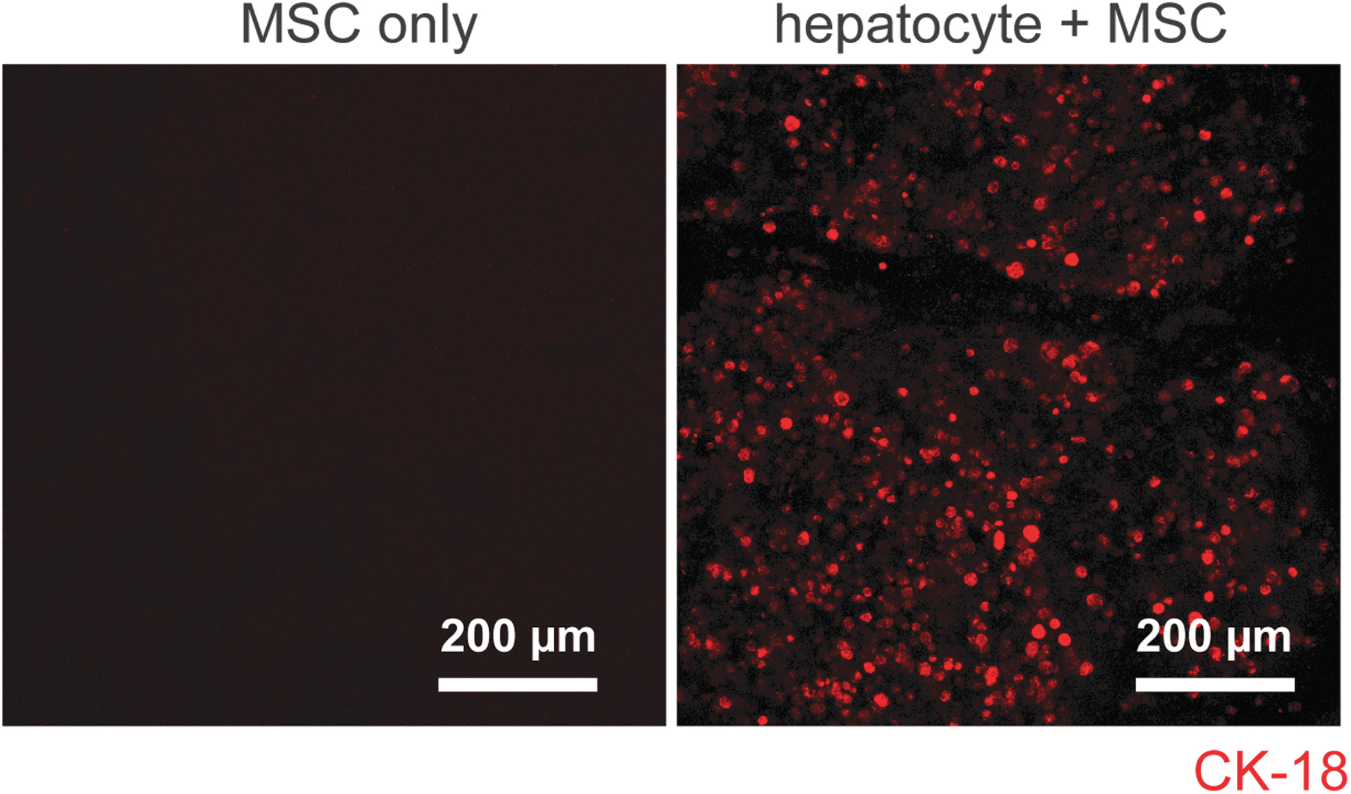
Characterization of HH Phenotype. The immunofluorescent staining for CK-18, a widely used surface marker of matured hepatocytes, revealed abundant positively stained cells at the end of the 30-day perfusion culture. CK-18, cytokeratin-18.
Discussion
Liver is the core organ of human body for metabolism, detoxification, homeostasis, protein synthesis, and bile production, performing more than 500 biochemical processes.26–28 To date, the clinical capacity to cure severe liver damage and disease is greatly hindered by the shortage of donor liver tissue. This urgent challenge has spurred the vast development of tissue engineering approaches that aims at either augmenting the self-regeneration of diseased liver or creating a functional and implantable artificial liver graft as an alternative for donor tissue.29–32
In our attempt to develop a thick, functional liver graft with native cellular components, we first aim at addressing the organization of a medium-delivering system/vascular-like structure in the engineered tissue for sufficient mass transfer. In our study, a 3D-printed vascular channel with tunable dimensions and porosity was created and coupled with a TPS to enable rapid oxygen and nutrient diffusion inside the liver construct. COMSOL Multiphysics was employed to investigate the diffusion of oxygen and nutrients throughout the aggregated constructs in an effort to optimize the channel dimensions. 15 This output metric, when orchestrated with practical factors such as the printing fidelity and mechanical strength of the device, has greatly accelerated the process of our device development. The in vitro outcomes of our perfusion culture suggest enhanced mass transfer under dynamic conditions, resulting in a prolonged cell survival as indicated by live and dead staining, as well as the preservation of key hepatic functionality such as albumin synthesis and bilirubin secretion.
The improved survival and functionality of HH maintained in our TPS enabled us to create a centimeter-sized liver construct for the first time. The diameter and height of the liver graft produced in our TPS is 12 and 10 mm, respectively. We also attempted to recapitulate the favorable microenvironment that sustains long-term hepatocyte stability and liver-specific functions. Collagen type I was employed due to its widely reported supportive role in preserving the hepatic phenotype.33–35 Moreover, the MSCs were loaded into the beads at a predetermined optimal ratio to facilitate the bioactivity of HH.36,37
Constructing a functional vasculature is the key to successful engineered liver tissue. Previous studies have shown various remarkable approaches to accomplish this goal to supply adequate nutrient and oxygen to the hepatocytes and enable anastomosis to host tissues by microsurgery. For instance, engineered liver tissue was preimplanted in angiogenic sites for vascularization by surrounding host tissue before therapeutic application.38–40 Compared with this method, our 3D-printed channel serves as a pre-existing vasculature that perfuses the encapsulated hepatocytes without any additional in vivo implantation or incubation. Another widely reported vascularization strategy entails the co-culture between hepatocyte and vascular endothelial cells.41,42 As opposed to static co-culture, the HUVECs seeded in the central channel of our device are patterned in a 3D tubular shape to mimic the endothelium. Moreover, the HUVECs were experiencing consistent shear stress caused by medium flow, which is a known mechanical stimulus that activates the cells to rapidly invade/sprout into the surrounding microbeads.43–45
Unlike the human primary hepatocytes used in this study, the human induced pluripotent stem cells (hiPSCs) is an inexhaustible cell source enabling autologous cell transplantation; thereby mitigating the challenge of limited availability of autologous parenchymal cells and the adverse effects of immune rejection against allogenic tissue.46,47 A wide spectrum of studies have provided insights on the liver-related lineage commitment of hiPSCs via various strategies, including hepatocyte-like cells,48–50 MSCs, 51 and vascular endothelial cells. 52 Using iPSC-derived hepatic cells for the liver construct developed in this study will be our next milestone to reach that will further augment the translational potential of our device as alternative liver transplants, as well as a novel patient-specific drug screening platform for precision medicine. For instance, the hepatotoxicity/adverse hepatic response to any drug given to specific patient can be tested in our culture system by probing cytochrome P450 (CYP450) enzymatic activities. 50
In conclusion, in this study we created a novel 3D-printed perfusion construct that led to the centimeter-sized engineered liver tissue stable in cell vitality, phenotype, and functionality for 1 month in vitro. This study sheds light on the potential of 3D printing and bioreactor culture to advance novel tissue engineering-based solutions to the clinical challenge of liver treatment.
Footnotes
Acknowledgment
The authors gratefully acknowledge Dr. Sijie Hao, Core Facility Manager of Fischell Department of Bioengineering at University of Maryland, for his technical support for this study.
Disclosure Statement
No competing financial interests exist.
Funding Information
This work was supported by the National Institute of Biomedical Imaging and Bioengineering/National Institutes of Health (NIBIB/NIH) Center for Engineering Complex Tissues (P41 EB023833). This work was partly supported by the A. James Clark School of Engineering at University of Maryland (MPact Challenge).