




Abstract
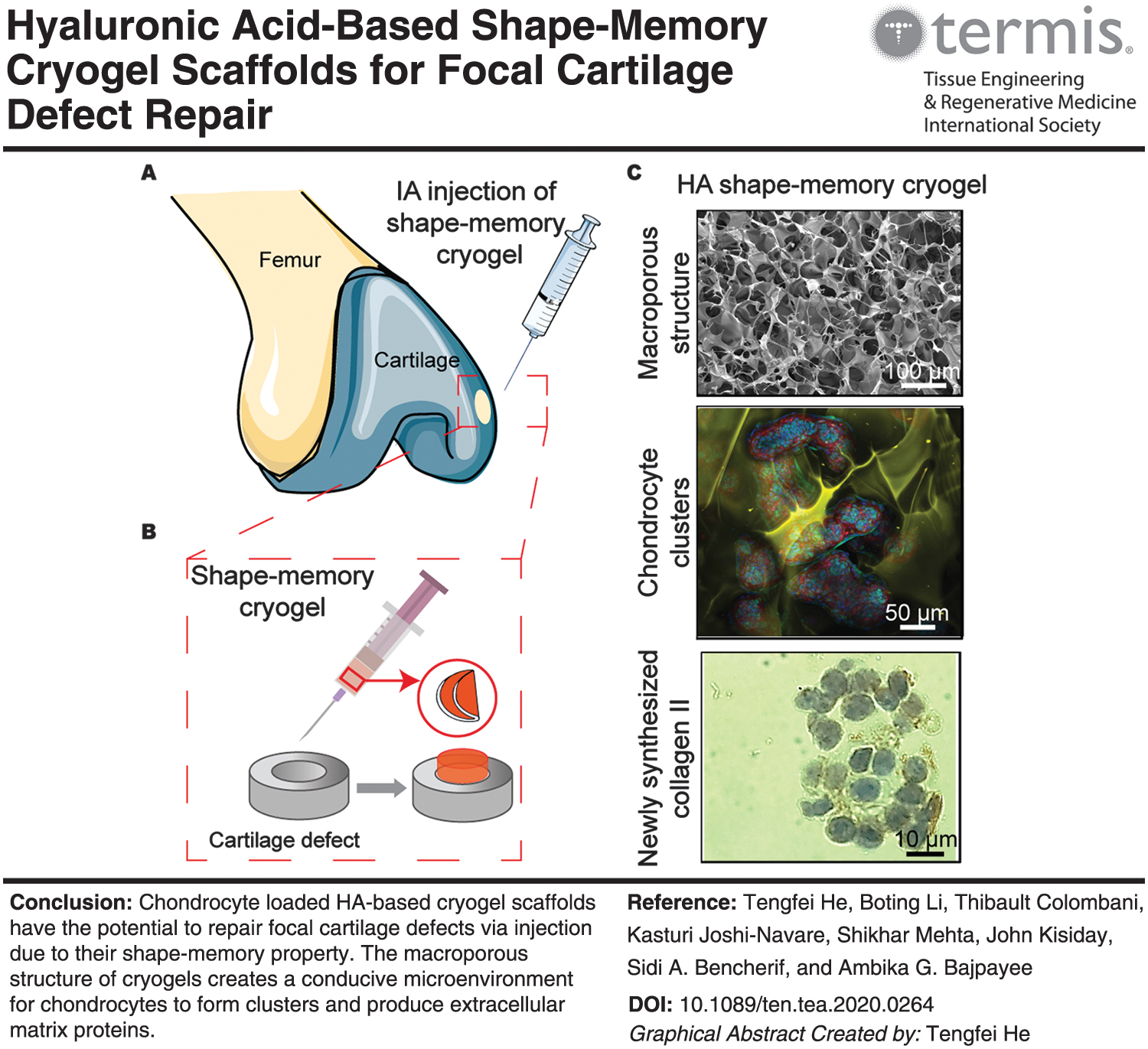
Traumatic joint injuries can result in significant cartilage defects, which can greatly increase the risk of osteoarthritis development. Due to the limited self-healing capacity of avascular cartilage, tissue engineering approaches are required for filling defects and promoting cartilage regeneration. Current approaches utilize invasive surgical procedures for extraction and implantation of autologous chondrocytes; therefore, injectable biomaterials have gained interest to minimize the risk of infection as well as patient pain and discomfort. In this study, we engineered biomimetic, hyaluronic acid (HA)-based cryogel scaffolds that possess shape-memory properties as they contract and regain their shape after syringe injection to noninvasively fill cartilage defects. The cryogels, fabricated with HA and glycidyl methacrylate at −20°C, resulted in an elastic, macroporous, and highly interconnected network that provided a conducive microenvironment for chondrocytes to remain viable and metabolically active after injection through a syringe needle. Chondrocytes seeded within cryogels and cultured for 15 days exhibited enhanced cell proliferation, metabolism, and production of cartilage extracellular matrix glycosaminoglycans compared with HA-based hydrogels. Furthermore, immunohistochemical staining revealed production of collagen type II from chondrocyte-seeded cryogels, indicating the maintenance of cell phenotype. These results demonstrate the potential of chondrocyte-seeded, HA-based, injectable cryogel scaffolds to promote regeneration of cartilage tissue for nonsurgically invasive defect repair.
Impact statement
Hyaluronic acid-based shape-memory cryogels provide a conducive microenvironment for chondrocyte adhesion, proliferation, and matrix biosynthesis for use in repair of cartilage defects. Due to their sponge-like elastic properties, cryogels can fully recover their original shape back after injection while not impacting metabolism or viability of encapsulated cells. Clinically, they provide an opportunity for filling focal cartilage defects by using a single, minimally invasive injection of a cell encapsulating biocompatible three-dimensional scaffold that can return to its original structure to fit the defect geometry and enable matrix regeneration.
Color images are available online.
Introduction
Traumatic joint injuries can result in focal cartilage defects that regenerate poorly because of a lack of blood supply and low chondrocyte density in cartilage. 1 Cartilage defects, if left untreated, can cause the tissue to lose its mechanical integrity, while the joint loses lubrication between bones, ultimately leading to severe pain and disability. 2 At present, the most promising Food and Drug Administration-approved treatment for regeneration of cartilage defects is MACI®—a matrix-applied implantation of autologous chondrocytes cultured on a porcine collagen membrane. 3 Despite the clinical improvement it has provided, this technique has a number of limitations. Primarily, MACI requires two surgical procedures—an arthroscopy for chondrocyte collection and a mini-arthrotomy for implantation. Second, the membrane used for culturing cells is comprised of collagen types I and III derived from porcine tissue that increases the risk of an immune response after implantation. 3 Thus, an improved design that can limit the number of surgeries or bypass surgical implantations, while improving biocompatibility, is desirable for cartilage defect repair.
Hydrogels (e.g., PEG, alginate, self-assembling peptides, hyaluronic acid [HA], collagen) have been extensively studied as conventional three-dimensional (3D) scaffolds for their biocompatible characteristics, mimicking the extracellular matrix (ECM).4–11 However, hydrogels often exhibit a quasi-nonporous network that limits cell proliferation, mechanical flexibility, and diffusion of nutrients, oxygen, and cellular waste.12–14 Implantation of preformed hydrogels typically requires invasive surgical procedures such as arthrotomic surgery15,16; thus, injectable hydrogels have gained interest as a method for filling defects because of their minimally invasive nature.17–20 However, owing to the fact that a pregelation liquid has to be delivered to the defect, issues with gelation time can cause leakage to adjacent tissues or body fluids resulting in potential toxicity or changes in tissue mechanical properties.9,21–24 Thus, injectable scaffolds possessing shape-memory properties (the ability to contort within the needle and then recover to the original shape of the defect immediately) are desired for maintaining mechanical properties and for preventing leakage from occurring.4,13,25–31
Cryogels, a subclass of hydrogel, formed from crystallization at freezing temperatures, have been shown to offer shape-memory properties and have several advantageous features such as a macroporous architecture with highly interconnected pores (for unrestricted flow of nutrients and matrix proteins), and adequate mechanical strength and swelling capacity (for shape memory and integrity).13,24,25 These properties allow for efficient flow of nutrients and cellular trafficking that in turn facilitate tissue integration. 32 Cryogels can be fabricated using naturally derived polymers, thus reducing risk of toxicity. For example, HA-based cryogels are biocompatible and biodegradable, thereby limiting an immune response and allowing for the scaffold to be replaced by repaired tissue. 2 Therefore, because of their injectable shape-memory properties and their biocompatibility, the use of cryogels as cell-seeding scaffolds for cartilage defect repair is promising.33,34
In this study, we engineered macroporous biomaterials with shape-memory properties to provide a conducive environment for chondrocyte adhesion, proliferation, and matrix formation. We first synthesized HA-based cryogel scaffolds with and without RGD, a cell adhesion peptide, to evaluate cell–cryogel interactions, and then test their mechanical properties for comparison with conventional HA-based hydrogels. We then seeded primary chondrocytes within the gels to provide the cells with a physiologically relevant microenvironment for in vitro culture. We hypothesize that (1) chondrocytes infused within HA-based cryogels will remain metabolically active after injection through a small-pore needle and (2) that the macroporous and highly interconnected structure of cryogels will provide an unhindered environment for cell growth and diffusion of oxygen and nutrients, thereby enhancing chondrocyte metabolism and matrix production compared with that of mesoporous HA-based hydrogels.
Materials and Methods
Materials
Ammonium persulfate (APS), N,N,N′,N′-tetramethylethylenediamine (TEMED), HA, glycidyl methacrylate (GM), dimethylmethylene dye (DMMB), resazurin sodium salt, Griess reagent, chloramine T and 4-(dimethylamino) benzaldehyde (DMAB), paraformaldehyde (PFA), Triton™ X-100, 4′,6-diamidino-2-phenylindole (DAPI), and other salts were purchased from Sigma-Aldrich (St. Louis, MO). Acrylate-PEG-N-hydroxysuccinimide (Acrylate-PEG-Succinimidyl Valerate, molecular weight: 3,400) was purchased from Laysan Bio, Inc. Lithium phenyl-2,4,6-trimethylbenzoylphosphinate (LAP) was purchased from Biobots. Trypan blue and high-glucose Dulbecco's modification of Eagle's medium (DMEM) were purchased from Corning (Corning, NY). Fetal bovine serum (FBS) was purchased from GE Healthcare (Chicago, IL). HEPES, nonessential amino acids (NEAA), penicillin–streptomycin antibiotic-antimycotic (PSA),
Fabrication of HA-based cryogels and hydrogels
Chemical modification of HA
HA was conjugated to GM to incorporate polymerizable methacrylate residues along the polymer backbone. 35 The resulting product, hyaluronic acid glycidyl methacrylate (HAGM), was characterized by 1 H-NMR as previously described. 25 Rhodamine-labeled HAGM (R-HAGM) was prepared by reacting NHS-rhodamine with amine-terminated HAGM (NH2-HAGM) based on a method previously reported. 25 R-HAGM was incorporated into hydrogels and cryogels to stain the polymer network for imaging using confocal microscopy. G4RGDSP (containing the RGD sequence) was conjugated to acrylate-PEG-N-hydroxysuccinimide to form acrylate-PEG-G4RGDSP (APR), as previously reported. 25 This approach allows functionalized G4RGDSP to be covalently incorporated into the gels (hydrogels and cryogels) during polymerization to promote cell adhesion.
Synthesis of HA cryogels and hydrogels
HA-based cryogels were prepared with 3.2% (w/v) HAGM and either 0.8% (w/v; ∼2 mM) APR (Cryo RGD+) or 0.8% (w/v) methoxy-PEG-acrylate (MPA; Cryo RGD−) based on methods previously described (Fig. 1A). 25 In brief, the polymers were first dissolved in precooled deionized (DI) water. Next, 14% (w/v) APS (20 μL/mL) and 7% (v/v) TEMED (10 μL/mL) were added and mixed well. Next, the prepolymer solution was transferred into cold and sterile Teflon cylindrical molds (6 mm diameter × 1.5 mm height) and incubated overnight at −20°C to allow for cryogelation. The next day, Cryo RGD+ and Cryo RGD− were thawed at room temperature to remove ice crystals, then washed and stored at 4°C in DI water for future use.
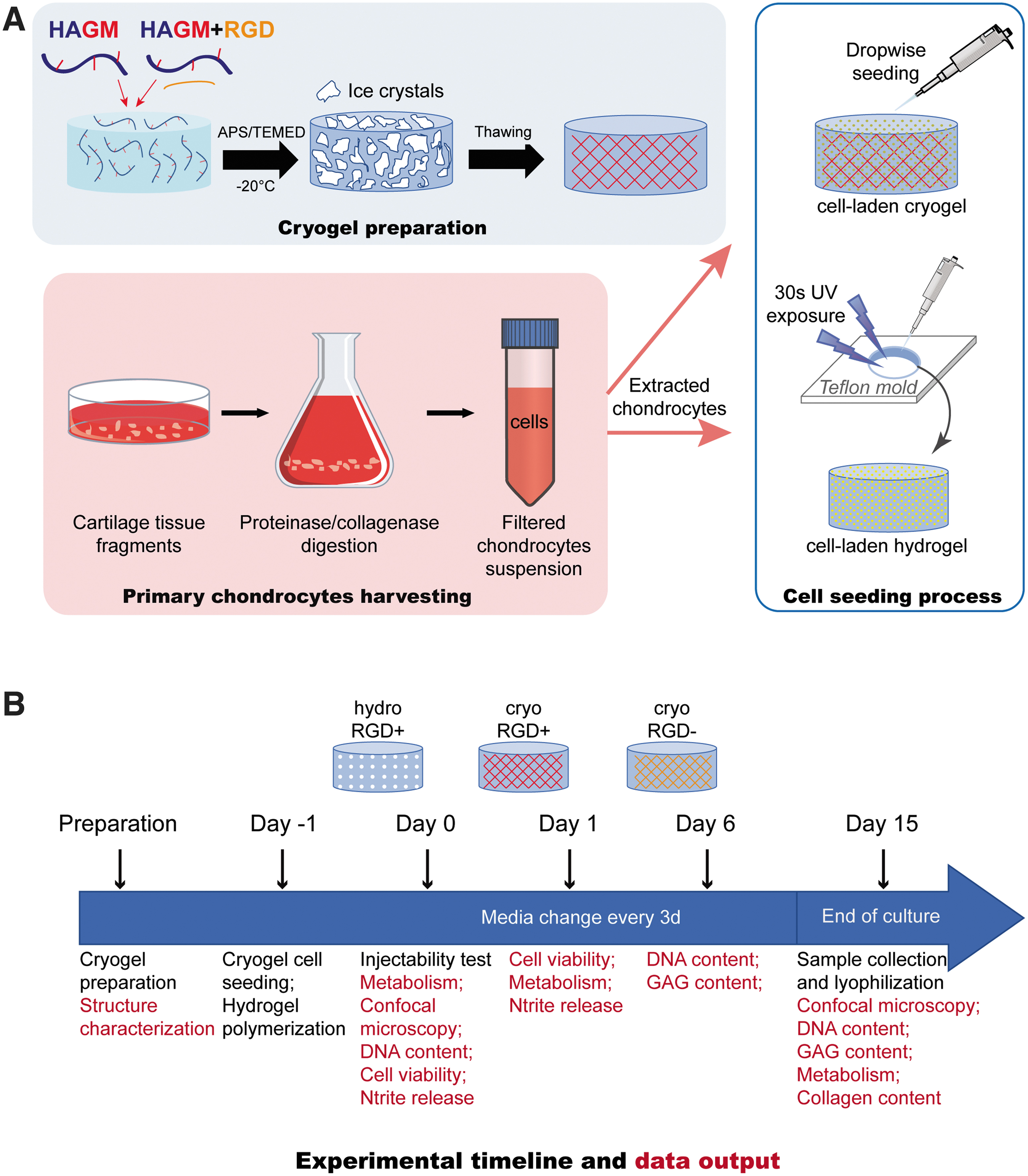
Fabrication of gels and experimental timeline.
HA-based hydrogels (Hydro RGD+) were fabricated by first mixing 3.2% (w/v) HAGM, 0.8% (w/v) APR, 0.5% (w/v) LAP photoinitiator in DI water. Next, the prepolymer solution was transferred into Teflon cylindrical molds (6 mm diameter × 1.5 mm height) and photopolymerized for 30 s under ultraviolet (UV) light (405 nm) with a 10 W UV lamp. To fabricate cell-laden hydrogels, Hydro RGD+, were also photopolymerized with primary bovine chondrocytes as described in the Primary chondrocyte isolation, seeding in gels, and culture Section.
Scanning electron microscopy imaging and characterization of mechanical properties
For scanning electron microscopy (SEM) analysis, cryogels were lyophilized and sputter coated with platinum/palladium up to 5 nm thickness. 35 Gels were imaged using a Hitachi S-4800 scanning electron microscope (Hitachi High-Technology Corporation) at 3 KV and 10 μA.
Swelling ratio (QM) and pore connectivity were determined for further characterization.
35
For swelling ratio, gels were swelled in phosphate-buffered saline (PBS) at 37°C under gentle shaking for 1 day to reach equilibrium. Then, the wet weights of gels were measured after removal of excess surface liquid. The gels were lyophilized and weighed to get the dry weight. The swelling ratio, QM, was calculated by using the following equation:
The Pore connectivity was calculated as follows:
where wet weight is the weight of fully hydrated gels, and wicked weight is obtained by measuring the gel weight after the free water is wicked away using absorbent wipes from the gel surface.
Bulk compressive elastic modulus of gels was examined by using stress-relaxation testing on an ElectroForce 5500 mechanical testing apparatus (TA Instruments). Cryogels were subjected to multiple successive stress-relaxation cycles of 10% strain per minute, followed by static holds of 800 s, 35 whereas hydrogels were subjected to a single cycle at the same strain rate. For each cycle, the compression displacement and load of the scaffolds were recorded with WinTest7 software. Equilibrium stress values were extracted from relaxation curves using a custom MATLAB script adapted from previous methods, 36 and the modulus was calculated from the linear region of equilibrium stress–strain curves with 10% strain rate for 10 cycles per minute.
Primary chondrocyte isolation, seeding in gels, and culture
Cartilage slices were obtained from immature bovine femoral condyles 37 using a scalpel and then placed in a Petri dish containing PBS (Fig. 1A). Slices were then chopped using a sterile blade before digesting in 50 mL of 2 mg/mL pronase solution in culture medium for 1 h at 37°C and in 50 mL of 0.25 mg/mL collagenase II solution in culture medium overnight at 37°C in a spinner flask. Isolated chondrocytes were filtered from the rest of tissue using 70 μm pore-size cell strainer and further purified using a 40 μm cell strainer, which was then rinsed twice using sterile PBS. Chondrocytes were then resuspended in culture media containing high-glucose DMEM, 10% FBS, 1% HEPES, 1% NEAA, 1% antibiotic-antimycotic, 0.4% proline, and 0.4% ascorbic acid. Viable primary chondrocytes were counted using Trypan blue. On the same day, cells were seeded into cryogels and hydrogels as described hereunder.
Before cell seeding, cryogels were first sanitized with 70% ethanol for 30 min and subsequently washed several times with sterile PBS. Next, cells were seeded dropwise into two types of cryogels (Cryo RGD+ or Cryo RGD−): 10 μL of cell suspension containing 8 × 10⁵ cells in the culture media were seeded into cryogels in a dropwise manner in a 48-well plate and kept for 30 min in a 37°C, 5% CO2 environment before adding culture media. Hydro RGD+ containing cells were fabricated by first mixing cells (8 × 10⁵ cells) with the polymer and LAP photo-initiator in complete cell culture medium. Next, the cell-containing polymer solution was transferred into a cylindrical mold (6 mm diameter × 1.5 mm height). Cell-laden hydrogels were then formed by photopolymerization under UV light (6.9 mW/cm2, 320–390 nm) for 30 s.
Cryogels and hydrogels were cultured in 500 μL of culture media at 37°C in a 5% CO2 environment for 15 or 21 days. Media was changed every 3 days and used for a variety of biochemical analysis to measure nitrite release, DNA content, cell metabolic activity, glycosaminoglycans (GAGs), and collagen content in gels (Fig. 1B) after 15 days, which is described in the Biochemical analysis Section. Gels were fixed for histology and immunostaining as described in the Histology and immunohistochemistry Section.
Injectability test
Cell-laden cryogels (Cryo RGD+; 2 × 10⁵ cells/construct) were prepared and incubated overnight in culture media to allow for cell adhesion to the polymer walls. Cryogels were then loaded individually into syringes containing 50 μL of culture media, 35 and then injected through a hypodermic 16-gauge needle. Next, syringe-injected cell-laden cryogels were cultured for another 24 h and the cell viability, cell metabolism, and nitrite release were assessed and compared with a control group (noninjected). Cryo RGD− was not evaluated because of its similar mechanical structure and composition with Cryo RGD+, whereas Hydrogel RGD+ was not injectable. Cryo RGD+ was also injected through a 16-gauge needle and deposited directly into an ex vivo bovine articular cartilage defect (6 mm diameter × 1.5 mm thickness) for evaluation of shape-memory property (Supplementary Video S1). The defect was created by punching a full thickness 6 mm diameter core into a 9 mm diameter × 1.5 mm thick condylar cartilage explant.
Biochemical analysis
Media was changed every 3 days throughout the culture duration. At days 0 and 15, cell-laden gels were incubated in media containing 1 × resazurin sodium salt (AlamarBlue assay) for 3 h in dark at 37°C and 5% CO2. Then, the cell metabolic activity was quantified following the manufacturer's recommendation. For injectability test, nitrite released into the media was measured using the Griess assay. In brief, equal volumes of Griess reagent and culture media collected were mixed and incubated at room temperature for 15 min. Next, the absorbance at 540 nm was measured using a microplate reader. Sodium nitrite was used as a standard. 38
At the end of culture, gel samples were frozen at −80°C and then lyophilized. Gels were then digested in 500 μL of 5% w/v proteinase K for 2 h at 57°C in 1 M Tris buffer (pH = 8.0). DNA content in gels was analyzed using the PicoGreen assay following the manufacture's protocol. The total GAG content in gels was measured using the DMMB assay. 39 Total collagen content was determined using the hydroxyproline assay. 38
Cell viability and cytoskeleton staining in gels using confocal microscopy
At days 0 and 15, cell-laden cryogels and hydrogels were washed with 1 mL of PBS and subsequently incubated for 15 min in the presence of a ViaQuant Fixable Far-Red Dead Cell Staining Kit according to the manufacturer's instructions. Next, the gels were rinsed with 1 mL of PBS, fixed with 500 μL of 4% PFA for 20 min at room temperature in dark, and washed with 1 mL of PBS. Next, the cells were permeabilized using 500 μL of PBS supplemented with 0.1% Triton X-100 for 5 min, then stained with DAPI (nucleus, blue) and Alexa Fluor 488-phalloidin (cytoskeleton, green). Confocal microscopy images were obtained using a Leica TCS SP5 X WILL confocal microscope (Buffalo Grove, IL), and processed using ImageJ. Percentage of cell viability was defined as the fraction of number of viable cells to the total number of cells. For each condition, one representative stacked image was recorded with a 5 μm separation in between z-stacks. Cryogels were stained with rhodamine during fabrication as described previously. 35
Histology and immunohistochemistry
Cell-laden gels at day 21 were fixed in 4% formalin, embedded in 0.75% agarose for ease of handling, dehydrated in a graded series of ethanol and xylenes, and embedded in paraffin. Transverse sections of gels were taken at 6 μm thickness. Sections were then immunostained with anti-collagen II mouse monoclonal antibody (1:700) (GB12021; ServiceBio) before an HRP-labeled goat anti-mouse secondary antibody (G1214; ServiceBio) was used (1:2000). Using 3,3′-diaminobenzidine, sections were stained to develop a strong signal. For nuclei staining, sections were further stained with hematoxylin. Stained sections were imaged using a 3D scanner (MIDI II HISTECH).
Statistical methods
Data are presented as mean ± standard deviation. For all studies, n = 3–5 gels per condition were used and experiments were repeated at least three times. Confocal images shown are representative of three samples per condition. For comparisons between different treatment conditions, Tukey's honest significant difference test was used. p < 0.05 was considered statistically significant.
Experiment
Synthesis and characterization of gels
Gelation of HAGM (with or without RGD) at −20°C resulted in ice crystal formation (Fig. 1A). After thawing at RT, 3D cryogel scaffolds with an interconnected macroporous architecture were created. Using SEM, an average pore size of 50 ± 0.5 μm was determined for “dry” cryogels (preswelling) both with and without RGD (Fig. 2A), confirming their macroporous architecture. As given in Figure 2B, Hydro RGD+ had the highest swelling ratio (QM = 50.2 ± 2.6), whereas Cryo RGD+ and Cryo RGD− exhibited 2× lower swelling ratios compared with Hydro RGD+ (QM = 22 ± 3.7 and 31 ± 2.6, respectively). In addition, the degree of pore connectivity for Cryo RGD+ and Cryo RGD− (83.5 ± 2.8 and 85.0 ± 0.8, respectively) was approximately seven times higher than that of Hydro RGD+ (Fig. 2C). The mechanical stiffness for Cryo RGD+ and Cryo RGD− was 4.8 ± 0.3 and 5.5 ± 0.4 kPa, respectively, both of which were significantly lower than that of hydrogels, which measured at 65 ± 10 kPa (Fig. 2D). Cryogels, thus, exhibited a macroporous network with high pore connectivity and low swelling.

Mechanical characterization of gels.
Injectability of cryogels
The cell-laden cryogels restored their original shape rapidly, fitting into the same size (6 mm diameter × 1.5 mm thickness) cylindrical defect in a cartilage explant after being injected through a 16-gauge needle (Fig. 3A, B). This feature demonstrates their shape-memory properties (Supplementary Video S1). In addition, chondrocytes encapsulated within cryogels remained metabolically active and viable (Fig. 3C) (same as untreated control) and showed no inflammatory response after their injection through the 16-gauge needle, as evidenced by the similar levels of nitrite release (Fig. 3D).

Maintenance of biological activity after injection.
Biological response
After 15 days of culture, cryogels did not show a significant reduction in percentage of chondrocyte viability compared with day 0, indicating that the cryogel had a microenvironment that was conducive to maintaining chondrocyte health over a 2-week period (Fig. 4A). Hydro RGD+ showed a slight decrease in cell viability from 83.8% to 73.7% over 15 days, although this difference was not significant.

Biochemical analysis of gels.
In addition, chondrocytes in cryogels exhibited higher cell proliferation rates (up to 1.5× greater) than hydrogels over 15 days (Fig. 4B). By day 15, at least 2× higher GAG content was measured in cryogels compared with hydrogels, whether the data were normalized by DNA content or not (Fig. 4C, D). Furthermore, a greater percentage of GAGs was found to be retained within the cryogels compared with Hydro RGD+, indicating the superior conducive microenvironment for matrix production provided by cryogels (Supplementary Fig. S1). In addition, there was no significant difference in total collagen content in cryogels or hydrogels by day 15 (Fig. 4E). Chondrocyte metabolism in all three types of gels increased significantly by day 15 compared with that at day 0 (p < 0.003); cell metabolism in cryogels was 2× higher compared with that in hydrogels at day 15 (Fig. 4F), indicating its superior microenvironment for chondrocytes.
These data suggest that cryogels provide a superior microenvironment for maintaining chondrocyte phenotype, while supporting cell growth and promoting matrix regeneration. The presence of RGD in cryogels, however, did not significantly enhance the number of chondrocytes that remained adhered within the scaffold after 24 h of cell seeding (Supplementary Fig. S2). In addition, the presence of RGD did not improve biosynthesis rates as shown by no difference in GAG or collagen content in the two types of cryogels by day 15.
Cell attachment, survival, and spreading in the gels
Chondrocytes in Hydro RGD+ remained dispersed over 15 days, exhibiting less cell–cell interaction and no characteristics of forming an intricate tissue-like structure (Fig. 5). When cultured in RGD-containing and RGD-free cryogels, chondrocytes exhibited different behaviors. Chondrocytes seeded in both types of cryogels were homogenously distributed and simultaneously formed cellular aggregates at day 0. For Cryo RGD+, cells interacted with the polymer network and displayed more of a spindle-shaped morphology by day 15 rather than maintaining their initial spheroid-like shape. On the contrary, chondrocytes in Cryo RGD− self-organized and formed large 3D cylindrical organoids throughout the construct by day 15. The organoids displayed a sophisticated architecture with spherical cells in the center surrounded by stretched spindle-shaped cells interacting with the polymer walls.
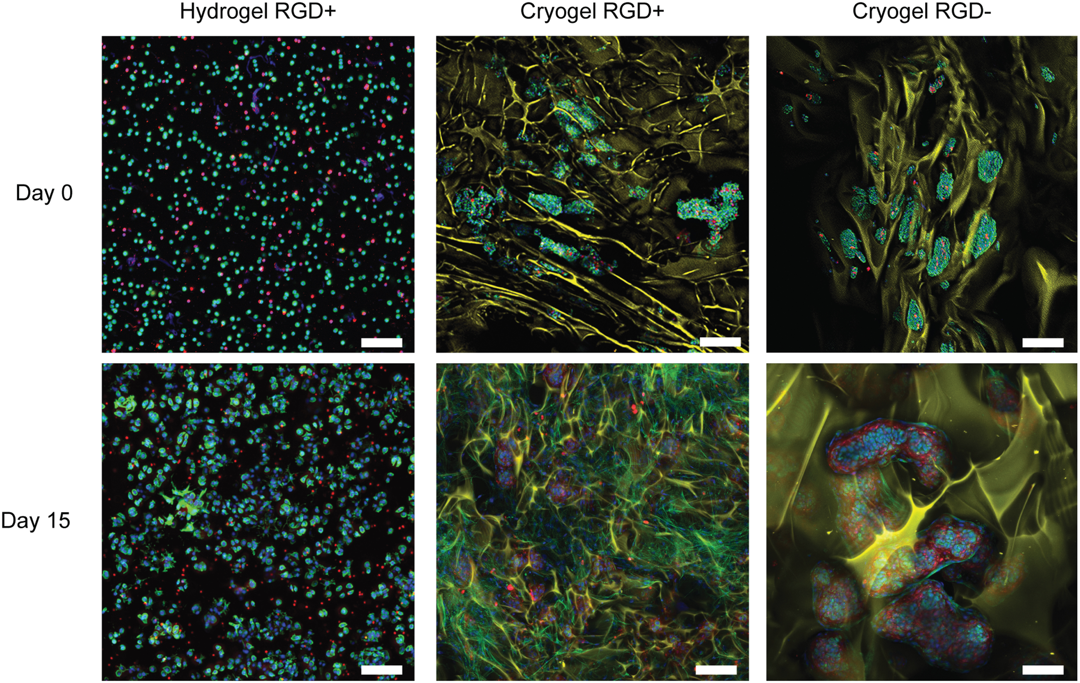
Confocal microscopy of gels. Confocal microscopy images taken at days 0 and 15 for each gel. Blue: nucleus; green: actin; yellow: cryogels; red: dead cells. Scale bar = 50 μm. Color images are available online.
Histological and immunohistochemical analysis
The three gel types were assessed by immunohistochemistry for collagen type II content after 21 days of culture (Fig. 6). Collagen type II staining indicated that both hydrogels and cryogels stimulated chondrocytes for the synthesis of de novo collagen as evidenced by the brown color. Dark blue nuclei staining of Hydro RGD+ was not visible because of its mesoporous structure (pore size approximately 2–50 nm), which hinders the penetration of hematoxylin, whereas the macroporous structure of cryogels (pore size ∼50 μm) allowed for vivid dark blue and brown staining of the nuclei and collagen II, respectively. Total collagen levels did not show any difference between Cryo RGD+ and Cryo RGD−, thereby supporting the data from the biological analysis (Fig. 4E).

Collagen staining of cryogels. Immunohistochemical analysis of Hydro RGD+, Cryo RGD+, and Cryo RGD− at day 21. Sections stained with hematoxylin (nuclei) and immunostained for collagen type II (brown color). Scale bar = 20 μm. Black arrows indicate collagen II deposited by chondrocytes. Dark blue hematoxylin staining for cell nuclei is not visible for chondrocytes in Hydro RGD+ owing to its mesoporous structure with a pore size approximately 2–50 nm that hinders the penetration of the dye. Color images are available online.
Discussion
In this study, we have designed syringe-injectable, HA-based shape-memory cryogels that provide a conducive microenvironment for chondrocyte adhesion, proliferation, and matrix biosynthesis for use in the repair of cartilage defects. These shape-memory cryogels provide an opportunity for improving the standard of care as they require only a single, minimally invasive injection of a biocompatible 3D scaffold that can return to its original structure (fitting to cartilage defect size and shape) after injection. 40 The large interconnected macropores of cryogels (∼50 μm) (Fig. 2A), may provide the unobstructed transport of nutrients, solutes, and waste as evidenced by the increased cell metabolism and GAG synthesis from chondrocytes over 15 days. In addition, their high pore interconnectivity is able to enhance the regulation of cell adhesion, viability, proliferation, and metabolism while providing the mechanical strength required for injectability and shape-memory properties. 39 The interconnected macropores of the designed cryogels also aid in recreating the tissue microenvironment for chondrocyte growth and function, while providing compositional similarity with the native cartilage ECM because of its HA-based nature. These characteristics have also been previously shown to improve ECM protein production for tissue engineering.32,41
Although cryogels have been designed with different types of polymers (i.e., HA, chitosan, gelatin, chondroitin sulfate, PEG, collagen, alginate, agarose and their blends) for various tissue engineering applications, cartilage defect repair remains a largely unexplored area.2,4,32,33,41–45 Gupta et al. designed a cell-free chitosan–agarose–gelatin cryogel scaffold that was able to promote the regeneration of cartilage in a rabbit osteochondral defect. 4 However, their non-injectable cryogels required surgical implantation, most likely owing to their high modulus (∼44 kPa) and poor compressibility. A chondrocyte-seeded gelatin-chondroitin-6-sulfate-hyaluronan cryogel scaffold designed by Kuo et al. also regenerated cartilage in rabbit osteochondral defects. 43 However, the scaffold's ability to retain its structure after injection was not evaluated. Recently, a gelatin-HA-PEG acrylate cryogel was shown to be repeatedly injectable and stretchable for use in adipose tissue engineering applications. 46 Thus, this is the first time that injectable shape-memory cryogels have been considered for their ability to promote cartilage regeneration for defect repair.
HA is a naturally occurring polysaccharide that has been widely studied and used for tissue engineering. 25 It is the polymer backbone of aggrecan—the primary proteoglycan that comprises the dense protein meshwork existing in cartilage tissue, and it is also the main solid component of synovial fluid. 47 Furthermore, chondrocytes have been shown to efficiently adhere to HA owing to the presence of CD44 receptors on the cell surface, which can enhance chondrocyte proliferation and matrix synthesis. 48 Therefore, HA is an ideal candidate biomaterial for cartilage tissue regeneration. 49 However, the function of HA is limited because of the in vivo enzymatic degradation caused by multiple metabolic pathways 50 ; therefore strategies are required to crosslink chemically modified HA to decrease degradation. 51 Previous studies have added methacrylate derivatives to HA to create photopolymerizable HAGM macromers for further crosslinking. The crosslinked networks of such conjugates have been reported to have high biocompatibility, low inflammatory response, and considerable vascularization at an optimized polymer concentration.52,53
The cryogels used in this study were previously optimized for the degree of methacrylation (∼30%) and HAGM macromonomer concentration (3.2% w/v) to enable shape memory after injection. 25 In this study, HAGM is utilized as the main component of the polymer for both hydrogels and cryogels. In hydrogels, chondrocytes were encapsulated during the photopolymerization process while they were seeded dropwise on the preformed cryogels. This way, the relatively low Young's modulus (5.5 ± 0.4 kPa), combined with high crosslink density and interconnected macropores, make cryogels injectable with shape-recovering ability. Although the cryogels possess shape memory, it is important to note that the gels are poroelastic in nature, as a result of fluid flow through the matrix.
Our biological analysis shows that the HAGM cryogel can provide a superior microenvironment for chondrocytes compared with the hydrogel. After syringe injection of cell-seeded cryogels, cells exhibited similar metabolic activity and viability as before injection (Fig. 3C, D), which is consistent with previously reported findings.25,35 Shape-memory cryogels can therefore be used as a tissue-engineered construct to be injected into chondral defects without surgical implantation.
Its highly interconnected macroporous structure enables efficient transport of nutrients, waste, and macromolecules (i.e., matrix proteins), thereby providing ideal physical support for chondrocytes to adhere, remain functional, and synthesize their own ECM (GAGs and collagen). Thus, these cryogels allow for the complete integration of implanted 3D substrates compared with conventional hydrogels (Fig. 4B, D, E). Conversely, our previous work has shown that conventional hydrogels possess a mesoporous structure (pore size: approximately 2–50 nm) with low pore interconnectivity (Fig. 2C), which limits cellular infiltration and ease of injectability, 24 further emphasizing the superior physical support provided by cryogels to chondrocytes.
To provide a bioactive surface for chondrocytes to proliferate, adhesion peptides (RGD) were grafted to the cryogels.25,35 However, the cell-substrate interaction performed similarly in HAGM cryogels with or without RGD likely owing to the interactions between CD44 receptors present on the surface of chondrocytes and the HA component of the gel structure. This strong bioadhesion may overcome the adhesion effects expected from the presence of RGD peptide,27,35 resulting in similar DNA content between Cryo RGD+ and Cryo RGD−. It is important to note that under compression (during injection), presence of RGD may influence long-term cellular adhesion and production of GAG and collagen, therefore warranting a future study to investigate the long-term effects postinjection. Of interest, chondrocytes were able to form large spheroids with fibroid matrix connecting to the polymer walls in cryogels without RGD, whereas chondrocytes formed smaller spheroids in the presence of RGD (Fig. 5). The presence of integrin-binding ligands (RGD) can enhance cell interaction with the synthesized ECM and polymer walls, leading to a reduced number of cells in clusters. These chondrocyte clusters are involved in the reparative process owing to the overexpression of RUNX1, a hematopoietic lineage determining transcription factor, which likely upregulates the expression of aggrecan and lubricin, a chondroprotective molecule.54,55 In addition, it has been shown that chondrocyte clusters form to initiate the repair response in a partial thickness rabbit cartilage defect model, 56 confirming the cells' chondroprogenitor phenotype. 55 Therefore, in our study, the clusters formed in cryogels may contribute to the enhanced cell proliferation, metabolism, and production of cartilage ECM.
A limitation of this study was the exclusion of hydrogels without RGD, as the primary objective was to evaluate chondrocyte behavior in cryogels compared with hydrogels after injection. Furthermore, there were no significant differences in chondrocyte proliferation, metabolism, or matrix production between Cryo RGD+ and Cryo RGD−, despite their improvement over hydrogels. Previous studies have also indicated that presence of RGD in PEG hydrogels positively influences cartilage matrix synthesis and gene expression; however, only in response to dynamic mechanical stimulation. 57 Thus, because our gels were not subjected to mechanical loading, we would expect to see similar trends between hydrogels with or without RGD.
In our study, we evaluated the microenvironment for chondrocytes, however clinically, success for chondrocyte-based tissue regeneration has been limited. A phase III trial was recently launched with NeoCart, a product similar to MACI, however with a bovine type I collagen matrix as opposed to porcine collagen mix of type I and III. The trial however did not meet its primary endpoint of change in physical functioning and pain. 58 Hyalograft C, a hyaluronan-based scaffold seeded with autologous chondrocytes, was investigated in prospective clinical trials with 5–12 years follow-ups, showing clinical improvement in patients at 32 ± 12 years old, with single and multiple defects (<4 cm2); however, its use in salvage cases was not recommended. Concerns surrounding the manufacturing process and uncertainty of a positive benefit-risk balance eventually lead to the withdrawal of the product from the European market. 59 Thus, MACI remains as the only scaffold-based product for repairing cartilage defects.
The limited success of these trials and the risks and high costs associated with extraction and culture of autologous chondrocytes has resulted in the increased use of alternative cell types such as mesenchymal stem cells (MSCs) for cartilage regeneration. Injectable, shape-memory cryogels designed similar to ours have been used for delivering bioluminescent murine MSCs to mice through subcutaneous injection, resulting in enhanced survival and higher local retention compared with free cells. 25 MSCs are advantageous as they can be sourced from several areas including bone marrow, adipose tissue, and umbilical cord using a syringe, and therefore, do not require surgery for collection. 60 Thus, combined with the injectable nature of cryogels, there is the potential to treat cartilage defects with MSCs entirely free of surgery, which would not only reduce patient discomfort but would also lower the risk of infection and incurred costs. 61 Furthermore, induced pluripotent stem cells (IPSCs) which can be derived from somatic cells, have also shown the ability to differentiate into chondrocytes, and therefore have the potential to be seeded into our cryogels for cartilage defect repair. 62 Future studies will investigate the use of cryogels seeded with either chondrocytes, MSCs or IPSCs for in vivo cartilage defect implantation.
It is important to note that for cartilage regeneration, chondrogenic differentiation of MSCs and IPSCs must be induced to produce hyaline-like cartilage. Therefore, these cells require treatment with factors such as TGF-β or Kartogenin for sustained periods of time to promote chondrogenic differentiation.63,64 This can be achieved using drug delivery systems that can enable charge-based interactions between the negatively charged hyaluronan matrix and positively charged drug carriers such as multi-arm Avidin that can provide controlled drug release.65–73 Combination of different cell sources, biomaterials, and/or growth stimulators are directions to be considered going forward in cartilage tissue engineering. In addition, bioadhesives may be required to keep the gels tightly secured within the cartilage defect site after their placement such that they can sustain cyclic mechanical loading pressures through the regeneration period, which will be incorporated in future design and tested using animal models. Optimization of cell harvesting and growth, improvements in ECM production, and reduction of cost from medical operation should be the focus of future research. 61
Conclusion
HA-based cryogels loaded with primary chondrocytes remain viable and metabolically active, while recovering their shape after injection. Compared with HA-based hydrogels, cryogels provide a more conducive microenvironment for cell adhesion, proliferation, and matrix biosynthesis. The shape-memory feature of these cryogels allows for them to be injected into the joint space through syringe for the nonsurgically invasive treatment and repair of cartilage defects. Use of these cryogels with various cell types and phenotype promoters can enhance cartilage tissue regeneration and allows for a broad range of applications in regeneration of other tissues of the body.
Footnotes
Authors' Contributions
T.H., B.L., T.C., K.J.-N., S.M., J.K., S.A.B., A.G.B. all contributed to the design of experiments and preparation and review of this article.
Acknowledgment
The authors thank Professor Samuel Chung for providing access to their microscopes for histological imaging.
Disclosure Statement
The authors have no competing financial interests to disclose.
Funding Information
This work was supported by the United States Department of Defense through the Congressionally Directed Medical Research Programs (CDMRP; W81XWH-17-1-0085); and National Science Foundation (DMR 1847843).

References
Supplementary Material
Please find the following supplemental material available below.
For Open Access articles published under a Creative Commons License, all supplemental material carries the same license as the article it is associated with.
For non-Open Access articles published, all supplemental material carries a non-exclusive license, and permission requests for re-use of supplemental material or any part of supplemental material shall be sent directly to the copyright owner as specified in the copyright notice associated with the article.
