




Abstract
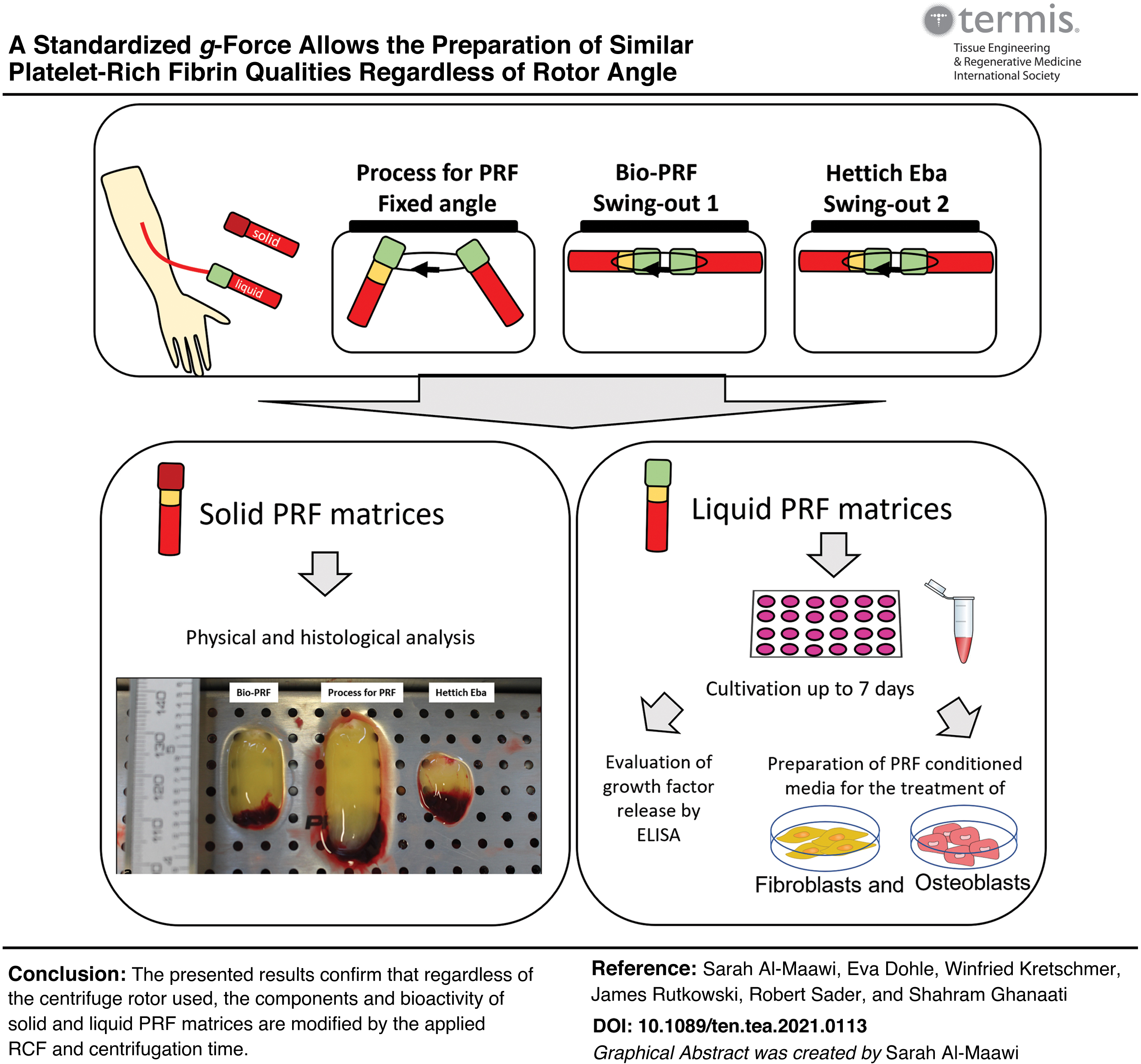
Platelet-rich fibrin (PRF) is an autologous blood concentrate that supports tissue regeneration. The effect of the centrifuge rotor angle in the fabrication of PRF is still not fully elucidated. The hypothesis of this study is: When applying the same g-force (relative centrifugal force [RCF]) and centrifugation time, PRF components and bioactivity are not modified using either a swing-out rotor or a fixed angle rotor. For this purpose, peripheral blood samples (from five donors) were used to gain solid (710 ×g, 8 min) and liquid (44 ×g, 8 min) PRF matrices using three different centrifuges (one fixed angle as a control and two different swing-out rotor centrifuges). The physical characteristics of the solid PRF were measured to evaluate the clot formation and cellular distribution. The liquid PRF was used to evaluate the cell number, bioactivity, and influence on primary human osteoblasts (pOBs) and primary human fibroblasts (pHFs) in vitro. Solid PRF clots were significantly larger in the group of fixed rotor centrifuges compared with either of the two evaluated swing-out rotor centrifuges. No differences were observed when evaluating the cellular distribution within the solid PRF. No statistically significant differences were documented in the cell's density in liquid PRF samples (platelets, lymphocytes, neutrophils, eosinophils, and basophils) among the differently gained PRF samples. No statistically significant differences were documented for the released growth factors (vascular endothelial growth factor, epidermal growth factor, and transforming growth factor beta 1) over 7 days. pOBs and pHFs viability after treatment with PRF conditioned media showed no statistically significant differences between the evaluated groups. However, the number of adherent cells treated with PRF obtained with the use of the fixed angle rotor was significantly higher when compared with those treated with PRF obtained by using the swing-out rotors. The presented results confirm that regardless of the centrifuge rotor used, the components and bioactivity of solid and liquid PRF matrices are modified by the applied RCF and centrifugation time. These findings are of great importance for highlighting the essential role of adapting the centrifugation protocols when using different centrifuges and to correctly report the used centrifugation protocols in scientific research to allow for reproducible results.
Impact statement
Platelet-rich fibrin (PRF) is prepared from autologous peripheral blood and is widely applied in research and clinical treatments. The centrifugation parameters used during the preparation of PRF directly influence its components and bioactivity. By using a standardized protocol, the present study demonstrated that adapting various centrifuges to a standardized relative centrifugal force and centrifugation protocol resulted in reproducible PRF matrices with similar bioactivity, regardless of the centrifuge rotor angle. These findings underline the necessity to carefully adapt and correctly report the used centrifuge and centrifugation protocols in scientific research to allow reproducible results.
Color images are available online.
Introduction
Blood concentrate systems utilizing peripheral blood provide a promising autogenous bioactive additive to support tissue regeneration in various clinical fields.1,2 The main purpose for obtaining blood concentrates is to improve surgical healing by utilizing the regenerative and bioactive components of the peripheral blood. 3
Platelet-rich fibrin (PRF) was introduced as a second-generation blood concentrate that does not require the addition of any external substances or anticoagulants. The application of PRF in different fields of regenerative medicine, such as orthopedics, 4 sport medicine, 5 aesthetics, 6 as well as oral and maxillofacial surgery, 1 exhibited promising clinical outcomes since its introduction. It aims to gain a fibrin scaffold enriched with platelets, leukocytes, and the growth factors contained within these cells. The PRF can be generated in either a solid matrix or a liquid matrix according to the clinical need. 7 However, the irregularities regarding the use of PRF in the clinic differ with the clinician's educational background and geographic region (e.g., Europe vs. United States) of practice, which makes its implementation in the surgical routine complicated and inconsistent results. There is a lack of standardization of PRF preparation protocols, which makes the introduction and reproduction of the clinical outcomes difficult.
Different parameters have a direct influence on the bioactivity and components of PRF such as: (i) applied relative centrifugal force (RCF), (ii) centrifugation time, (iii) blood collection tube surface, and (iv) initial whole blood volume used for the preparation of PRF. 8 In the past decade, a series of studies were performed to analyze the influence of the centrifugation process on the resulting PRF matrix components and bioactivity. It was demonstrated that the applied RCF plays an essential role in modulating the characteristics of the PRF matrix.9–12
The first centrifugation protocol was introduced in 2001 and recommended the use of a rather high RCF of ∼700 ×g applied by a fixed angle rotor centrifuge. 13 Histological analysis of PRF matrices centrifuged with a high RCF showed an accumulated distribution of the blood cells (especially leukocytes) within the interface between the PRF clot and the red blood cell phase. However, when using a lower RCF (208 ×g), a different pattern was observed. For solid PRF matrices, lower RCF demonstrated a cellular distribution that was more evenly distributed throughout the matrix. 9
Based on these findings, a low-speed centrifugation concept (LSCC) was introduced to outline the role of the applied RCF during the centrifugation of PRF matrices in a more standardized design. 8 A systematic analysis using the same centrifugation time, but different RCF demonstrated that PRF matrices, centrifuged at a high RCF exhibited a significantly lower number of blood cells (platelets, leukocytes, and subtypes) compared with those centrifuged using a low RCF.14,15 The growth factor release from PRF matrices was significantly higher when a low RCF was applied. 16 Furthermore, various cellular responses to differently centrifuged PRF matrices were also evident in vivo. A recent study analyzed the cellular reaction and integration pattern of solid PRF matrices centrifuged with either a high (710 ×g) RCF or a medium (208 ×g) RCF. The results demonstrated that PRF matrices centrifuged using a medium RCF demonstrated a significantly higher in vivo vascularization pattern in comparison to those centrifuged with a high RCF. 17
Additionally, medium RCF PRF matrices attracted a significantly higher number of host cells compared with high RCF PRF. 17 These findings highlighted the importance of understanding the characteristics and properties of the used PRF matrix and aimed to standardize the protocols used in clinical treatment.
Recently, a further approach was addressed to optimize the centrifugation development of PRF matrices by utilizing a swing-out rotor centrifuge and was referred to as horizontal centrifugation. 18 This approach suggested that the bioactivity of the resulting PRF matrix is enhanced by means of horizontal centrifugation, which allows for a more controlled and gentle separation of the blood cells compared with existing fixed angle centrifuges. 18 A recent study performed a histological observational study comparing PRF matrices obtained using either a swing-out rotor or a fixed angle rotor. The findings described a more uniform separation of the blood cells when a swing-out rotor centrifuge was applied. 19 However, to date, still little is known about the characteristics, components, and bioactivity of PRF matrices obtained using the swing-out rotor.
Therefore, the present study aimed to test the hypothesis whether PRF components and bioactivity are modified using a swing-out rotor centrifuge compared with a fixed angle rotor, when applying the same RCF and centrifugation time in three different investigated centrifuges. For this purpose, differently centrifuged solid and liquid PRF matrices were characterized and analyzed in vitro.
Materials and Methods
PRF preparation
The use of PRF samples from healthy donors was approved by the responsible Ethics Commission of the Goethe University, Germany (265/17), and was performed in accordance with the principles of informed consent. PRF was prepared according to the LSCC, as previously described.14,15 Blood was obtained from five healthy donors in two steps. For the preparation of solid PRF, three tubes of 10 mL (A-PRF, Process for PRF, Nice, France) were obtained from each donor. The tubes were immediately placed in three different centrifuges exhibiting either a fixed angle rotor or a swing-out rotor. All centrifuges were set to the same RCF of ≈710 ×g and centrifugation lasted for 8 min as described in Table 1 in detail. After centrifugation, the blood was separated and the formed PRF clot was collected using a surgical forceps. The PRF clot was separated gently from the red blood phase by peeling if it was attached. The collected PRF clots were further characterized as described in the section “PRF characterization”.
Detailed Description of the Centrifugation Parameters and the Centrifuge Characteristics Used in This Study
EBA; PRF, platelet-rich fibrin; RCF, relative centrifugal force; RPM, revolutions per minute.
For the preparation of liquid PRF, three tubes of 10 mL (i-PRF, Process for PRF) were collected from each donor and placed immediately in different centrifuges exhibiting either a fixed angle rotor or a swing-out rotor. The blood was centrifuged using a RCF of ∼50 ×g for 8 min, as described in Table 1. After centrifugation, the upper phase containing liquid PRF was collected by means of a syringe and transferred to a new tube for further characterization. Liquid PRF samples (400 μL) were placed in a multiwell cell culture plate and incubated for 30 min in 37°C until a PRF clot was formed in all groups. Thereafter, 800 μL of cell culture media (Roswell Park Memorial Institute [RPMI]) supplemented with 1% streptomycin/penicillin antibiotics was incubated in a cell culture incubator at 37°C in a humidified atmosphere. The supernatants (PRF conditioned cell culture media) were collected after 1, 3, 5, and 7 days, and fresh media (800 μL) was added at each time point and used for subsequent cell culture experiments and for the determination of growth factor release.
PRF characterization
The physical characteristics of the solid PRF matrices were measured to evaluate the PRF clot formation and cellular distribution. The liquid PRF samples were used to evaluate the cell number, bioactivity, and influence on primary human osteoblasts (pOBs) and primary human fibroblasts (pHFs) in vitro.
Analysis of the physical parameter of the solid PRF clots
The length and weight of the different solid PRF clots were determined immediately after harvesting. The length was measured by means of a digital caliper (Alpha tools BAHAG AG, Mannheim, Germany). The weight of the PRF clots was determined by a laboratory medical scale (Discovery, OHAUS Europe GmbH, Nanikon, Switzerland). The measurements are calculated in mean and standard deviation.
Complete blood count
Liquid PRF samples were used for complete blood count to determine the number of the different blood cells within the different PRF matrices, as previously described.14–16,18
Therefore, the samples were directly transferred to ethylenediaminetetraacetic acid tubes (S-Monovette, Servoprax GmbH, Wesel, Germany), and analysis was performed at the specialized medical laboratory according to standardized protocols (Laborarztpraxis, Frankfurt am Main, Germany) using a hematological analysis system (Sysmex XN 1000, Norderstedt, Germany).
Histological evaluation
After physical characterization of the solid PRF matrices, the PRF clots were fixed for 24 h using 4% formaldehyde (ROTI®; Histofix, Germany). Subsequently, the samples were treated as previously described. 20 The samples were then embedded in paraffin. Sections of 3–4 μm were then performed using a rotary microtome (Leica RM2255, Wetzlar, Germany) and stained using hematoxylin and eosin staining for general evaluation of cell distribution. Additional sections were stained using specific immunohistochemical antibodies to identify platelets (CD61) and leukocytes (CD45). Autostainer (Lab Vision™ Autostainer 360; Thermo Scientific) was followed according to previously published methods.7,16 CD-61 antibody (1:2000 [ab225742]) and CD-45 antibody (1:50 [ab10558]) were used as primary antibodies. Anti-mouse secondary antibody (HRP UltraVision Quanto Detection System; Thermo Fisher Scientific) and chromogenic visualization were achieved using aminoethyl carbazole peroxidase (Dako).
Qualitative histological analysis was performed using a light microscope (Nikon Eclipse 80i; Nikon, Tokyo, Japan) equipped with a Nikon DS-Fi1 digital camera and a Nikon Digital sight unit DS-U3 (Nikon) to capture representative histological images.
Quantification of growth factor release
The collected supernatants of the differently gained liquid PRF matrices were stored at −80°C until growth factor concentrations were determined by means of enzyme-linked immunosorbent assay (ELISA). The concentration of vascular endothelial growth factor (VEGF), epidermal growth factor (EGF), and transforming growth factor beta 1 (TGF-β1) was measured using DuoSet® ELISA Development Systems according to the manufacturer's protocol (R&D Systems). The optical density was measured using a microplate reader (Tecan, Crailsheim, Germany) at a wavelength of 450 nm.
Cell culture experiments
Primary cells that were used for this study were obtained from anonymized excess tissue, and their application was in accordance with the principle of informed consent and the guidelines of the responsible Ethics Commission of the Goethe University, Hessen, Germany. Excess tissue in form of cancellous bone or oral soft tissue of otherwise healthy donors was obtained from the operating rooms of the Department of Maxillofacial and Plastic Surgery (Goethe University, Frankfurt Am Main, Germany) and was used for the isolation of human primary cells.
pOBs were isolated from fragmented cancellous bone following previously published protocol.21,22 The isolated cells were cultured in Dulbecco's modified Eagle medium Nutrient Mixture F-12 (DMEM; Sigma–Aldrich, St. Louis, MO) supplemented with 10% fetal bovine serum (FBS; Biochrom, Berlin, Germany) and 1% penicillin/streptomycin (P/S; Sigma–Aldrich) at 37°C in a humidified atmosphere.
pHF were isolated according to previously established protocol.23,24 Human fibroblasts were cultured in Dulbecco's modified minimum essential medium (DMEM; Biochrom) supplemented with 1% P/S (Sigma–Aldrich) and 10% FBS (Biochrom). The cells were grown and expanded in a ratio of 1:2 and were used in this experiment up to passage 4.
Preparation of PRF conditioned cell culture media
Aliquots of liquid PRF supernatants of each experimental group (obtained in the “PRF preparation” section) and harvesting time point were used to prepare PRF conditioned cell culture media. Within each group, supernatant of each harvesting time point was pooled using standardized proportions (100 μL of each time point) to obtain three different PRF conditioned cell culture media that were used for the subsequent cell culture experiments.
Cell adhesion assay
pOBs and pHFs were washed using phosphate-buffered saline (PBS), detached using trypsin, and centrifuged to obtain a cell pellet. The cell pellets were homogenized in different PRF conditioned cell culture media gained from the liquid PRFs and set in a density of 5000 cells per 100 μL. The cells were seeded in a 96-well cell culture plate in triplicates for each donor (5000 cells per well). The cells were allowed to adhere for 24 h by means of incubation at 37°C in a humidified atmosphere. Thereafter, the cells were washed using PBS and fixed in 4% formaldehyde (ROTI Histofix) before staining with DAPI (4′,6-diamidino-2-phenylindole) to visualize adherent cells of each experimental group. Cells cultured in RPMI +1% P/S + 10% fetal calf serum (FCS, control 1) and RPMI +1% P/S without FCS (control 2) were considered control groups. Cell adhesion analysis was performed using a fluorescence microscope (Nikon Eclipse TS100, Düsseldorf, Germany). For the quantification of adherent cells, five pictures of 100 × magnification were analyzed for each well, and the mean of cell number was used for statistical analysis.
Cell viability assay
For evaluation of possible effects of PRF gained from different centrifuges on cell viability of pOBs and pHFs, an extraction assay was performed. pOBs and pHFs, cells were pre-seeded in a 96-well cell culture plate in a density of 5000 cells per well for all groups. The cells were allowed to adhere for 24 h by incubation at 37°C in a humidified atmosphere. After 24 h, the cells were washed and treated with the same volume (100 μL per well) of different extraction media prepared from the differently centrifuged PRF (liquid PRF conditioned cell culture media) for 7 days. Media were changed every other day. Cells cultured in RPMI +1% P/S + 10% FCS (control 1) and RPMI +1% P/S without FCS (control 2) were considered control groups. Effect of different extraction media on cell viability was then analyzed using an 3-(4,5-Dimethylthiazol-2-yl)-5-(3-carboxymethoxyphenyl)-2-(4-sulfophenyl)-2H-tetrazolium (MTS) assay after 3 and 7 days of cultivation of cells in the PRF conditioned media and was compared with controls (cell culture medium with/without FCS).
After 3 and 7 days of cultivation, cell viability was assessed using an MTS assay. Therefore, a CellTiter 96® AQueous One Solution Cell Proliferation Assay Kit (Promega, Madison, WI) was used following the manufacturer's instructions. Briefly, 20 μL of MTS solution was added to each well containing the sample in 100 μL of culture medium and incubated for 1 h at 37°C before the absorbance was measured at 490 nm with a plate reader (Infinite M200, Tecan). Blanks consisted of 100 μL of media with 20 μL of MTS solution incubated on plastic plates. All experiments were performed in triplicate.
Statistical analyses
The number of donors was considered based on previous studies.10,15,16,18,22,25,26 All experiments were performed with at least five different donors. The data are presented as mean values ± standard deviation. Statistical significance was evaluated using one-way analysis of variance with Tukey's multiple comparison test (α = 0.05). Statistical analyses were performed with GraphPad Prism 8 (GraphPad Software, Inc., La Jolla, CA), and differences were considered significant when p < 0.05*, p < 0.01**, or p < 0.001***.
Results
Characterization of differently prepared solid PRF clots using high RCF (710 ×g)
Directly after centrifugation, PRF clot formation took place in all evaluated samples. However, the physical appearance of the PRF clots differed markedly (Fig. 1a). PRF clot length measurements revealed the greatest length in the group of fixed angle centrifuge Process for PRF, which was significantly higher compared with the groups of horizontal centrifugations Bio-PRF (*p < 0.05) and Hettich EBA (*p < 0.05) (Fig. 1b).
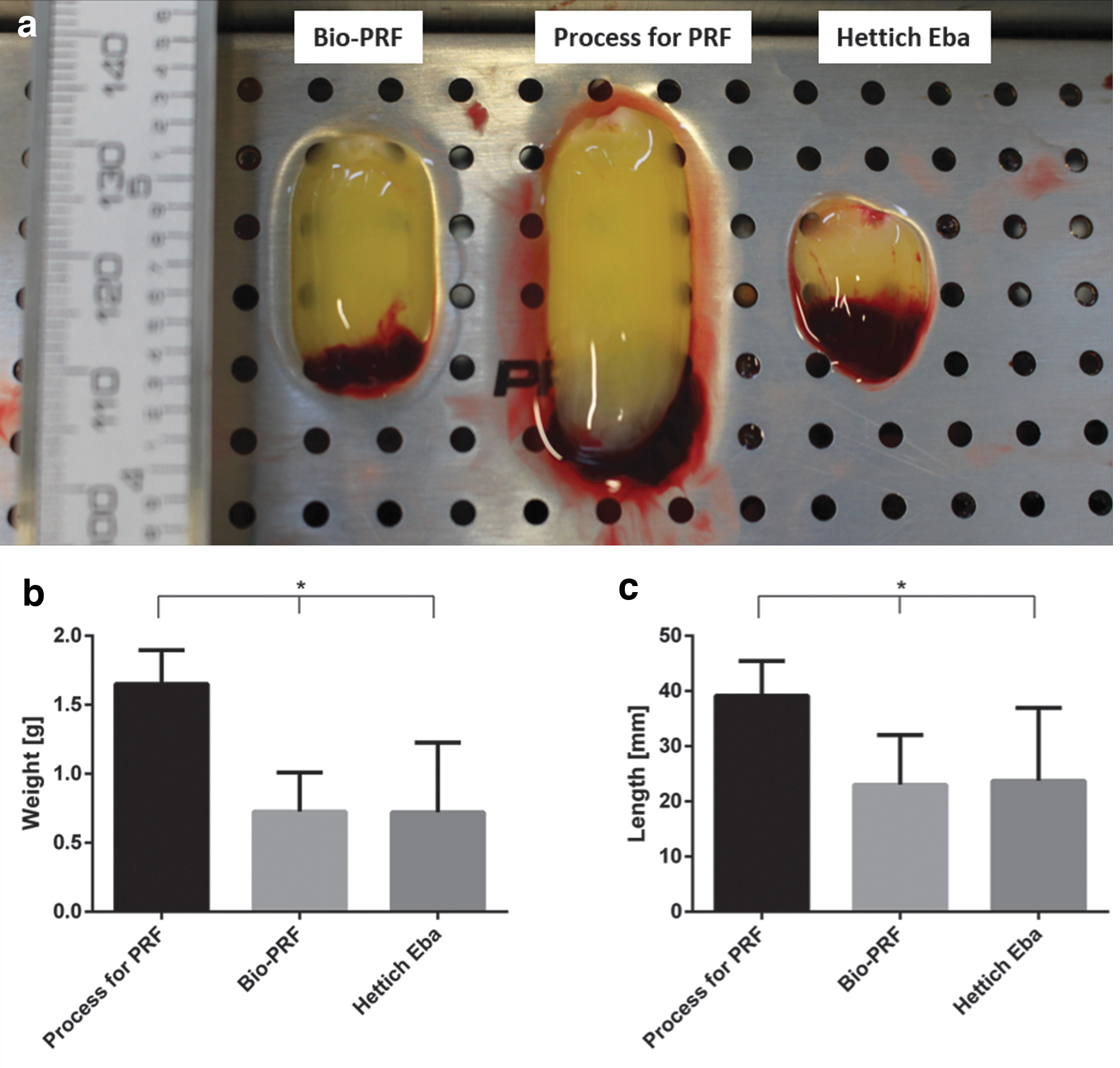
Physical characterization of the solid PRF clots centrifuged using a high RCF of 710 g for 8 min.
Similar results were documented by the determination of the PRF clot weight. Thereby, PRF clots that were prepared using a fixed angle centrifuge (Process for PRF) showed significantly higher weight compared with those centrifuged by horizontal centrifugation Bio-PRF (*p < 0.05) and Hettich EBA (*p < 0.05) (Fig. 1c).
Histological analysis of the cellular distribution in differently prepared solid PRF clots
All analyzed samples exhibited similar histological structure of the fibrin matrix. Visible differences were detected when analyzing the interface between the fibrin matrix and the buffy coat (Fig. 2). Samples centrifuged with the fixed angle centrifuge (Process for PRF) showed a diagonal interface. By contrast, samples that were obtained using the swing-out rotor horizontal centrifuges (Bio-PRF and Hettich EBA) showed a horizontal interface. However, blood cells such as platelets and leukocytes accumulated near the buffy coat in all analyzed samples (Fig. 2a, e, and i). The cellular distribution was characterized throughout the different parts of the PRF clots. Platelets were identified by means of anti-CD-61 antibodies. The platelet distribution was accumulated near the buffy coat in all samples without any observable differences between the examined samples. The number of platelets was reduced in the peripheral lower part, compared with the upper part in all evaluated samples (Fig. 2d–l). Leukocytes were targeted by anti-CD-45 antibody. They were frequently observed in the upper parts of the PRF clots near the buffy coat in all evaluated samples. Their distribution was less common in the peripheral area toward the lower parts of the PRF clots. These patterns were evident in all evaluated samples, regardless of the centrifuge used (Fig. 3a–i).

Immunohistological analysis of solid PRF clots centrifuged using a high RCF of 710 ×g for 8 min using anti CD-61 antibody to target platelets in the samples gained by:
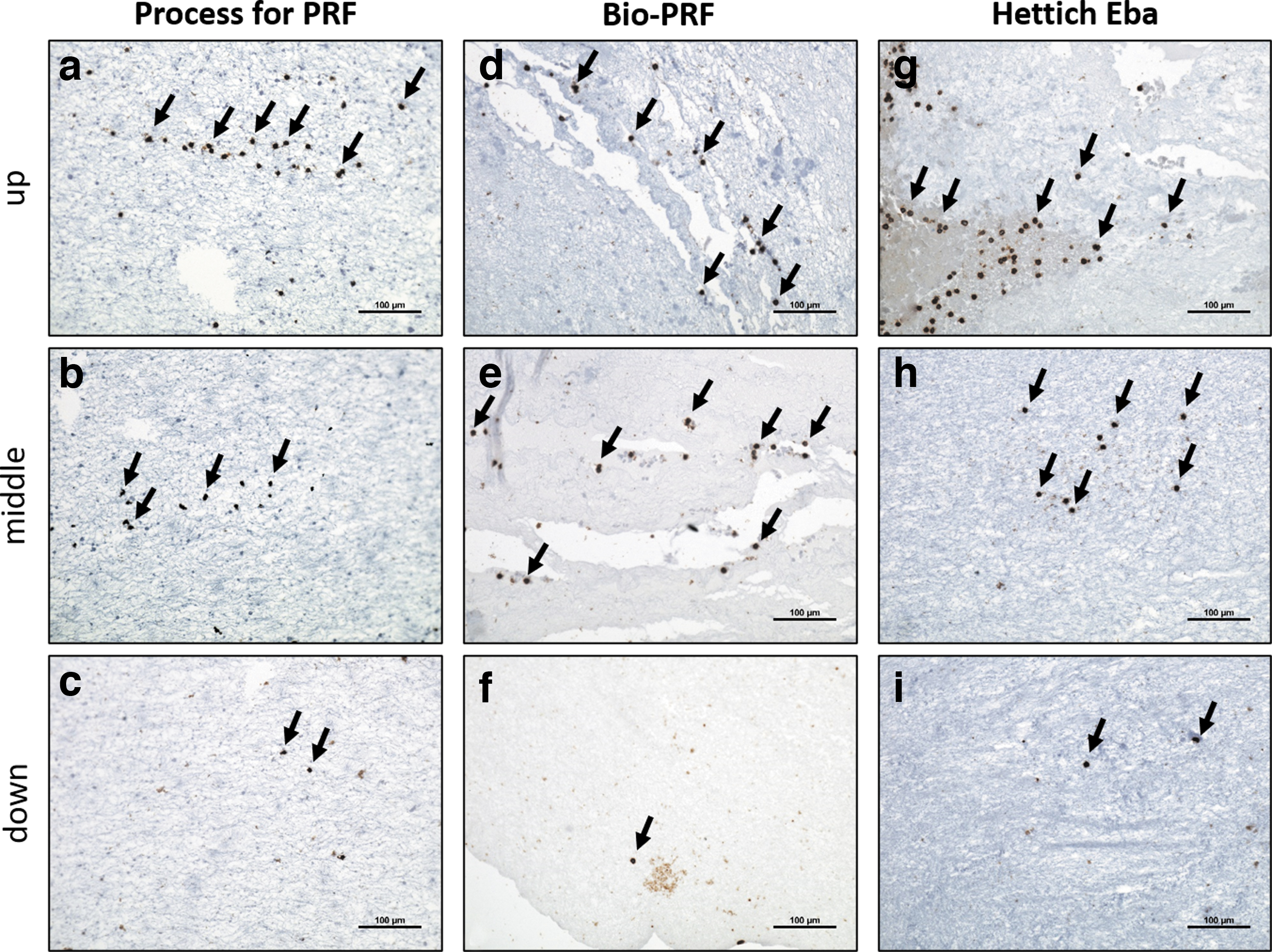
Immunohistological analysis of the solid PRF clots centrifuged at high RCF of 710 g for 8 min and using anti CD-45 antibody to target platelets in the samples gained by:
Characterization of differently prepared liquid PRF samples using low RCF (44–51 g)
The volume of the obtained liquid PRF was different in each experimental group. Even though the same RCF was set at all centrifuges, samples that were centrifuged using a fixed rotor centrifuge Process for PRF resulted in a significantly higher volume compared with those centrifuged using a swing-out rotor horizontal centrifuge Bio-PRF (**p < 0.01) and Hettich EBA (*p < 0.01) (Fig. 4a and b). The cell density determined by complete blood count for the different blood cells showed the same pattern in all evaluated groups. The cell density of platelets, lymphocytes, monocytes, neutrophils, and eosinophils was similar in all evaluated samples without statistically significant differences (Fig. 4c–g).
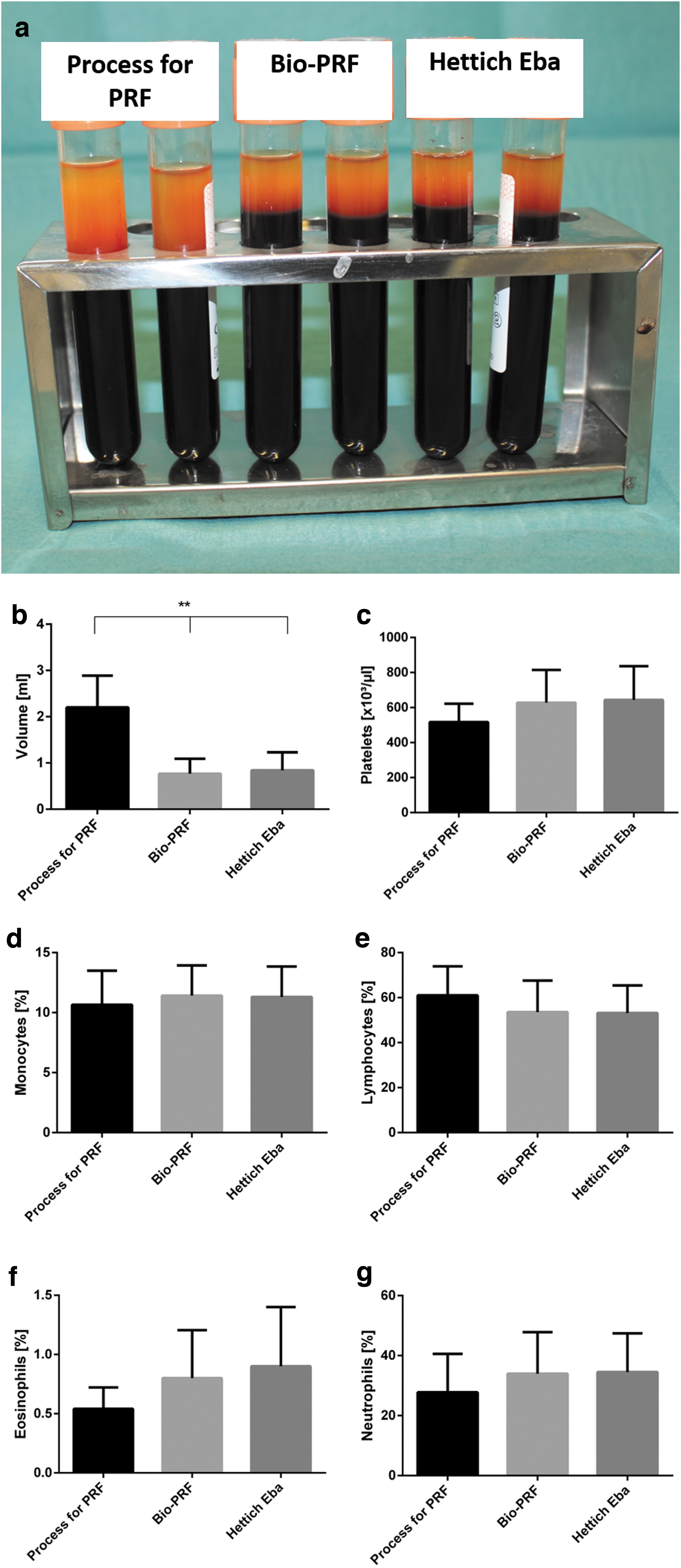
Characterization of liquid PRF samples centrifuged at low RCF of 44–51 g for 8 min.
Bioactivity of differently prepared liquid PRF samples
The release of the growth factors VEGF, TGF-β1, and EGF were analyzed in the supernatants of differently centrifuged liquid PRF samples over 7 days. The release of VEGF was similar in all evaluated groups after 1 day. Interestingly, after 3 days, liquid PRF samples centrifuged using a fixed angle centrifuge Process for PRF released significantly lower VEGF levels when compared with the swing-out rotor horizontal centrifuges Bio-PRF (****p < 0.001) and Hettich EBA (*p < 0.01). However, no statistically significant differences were found between Bio-PRF and Hettich EBA regarding VEGF release from the samples. However, this effect subsided after 5 days. Thereby, no statistically significant differences were found between the evaluated group after 5 and 7 days (Fig. 5a). The accumulated VEGF release demonstrated comparable concentrations without statistically significant differences (Fig. 5b).

Growth factor release of liquid PRF samples centrifuged using a low RCF of 44–51 g for 8 min.
The release of TGF-β1 was significantly higher in the group of the swing-out rotor centrifuge Bio-PRF compared with the fixed rotor centrifuge Process for PRF (*p < 0.05). However, with time progress, this difference was not consistently observable. Therefore, no statistically significant differences were found between the evaluated groups on day 3, 5, and 7 (Fig. 5c). The accumulated TGF-β1 was not significantly different in the evaluated groups over 7 days (Fig. 5d). Similarly, the release of EGF was comparable in all evaluated groups and showed no statistically significant differences at any time point (Fig. 5e). This phenomenon was also observed when analyzing the accumulated EGF concentrations over 7 days. No statistically significant difference was detected at any time point (Fig. 5f).
Influence of differently prepared liquid PRF on cell viability and adhesion in vitro
Primary human cells pOBs and pHFs were cultured in PRF conditioned media of differently centrifuged liquid PRF samples. The adhesion assay was performed after 24 h of cultivation. The cells treated with PRF conditioned media generated from liquid PRF centrifuged by fixed angle rotor Process for PRF showed a significantly higher number of adherent pOBs compared with the swing-out rotor horizontal centrifuges Bio-PRF (*p < 0.05) and Hettich EBA (*p < 0.05) (Fig. 6a–e and m). Additionally, the Process for PRF group exhibited a significantly higher number of adherent pOBs compared with the control groups 10% FCS (**p < 0.01) and without FCS (**p < 0.01).

Influence of PRF conditioned media:
Similar results were documented for the pHFs. In this case, the highest number of adherent cells was also detected in the group of Process for PRF and was significantly higher compared with the swing-out rotor horizontal centrifuges Bio-PRF (*p < 0.05) and Hettich EBA (*p < 0.05) (Fig. 6f–j). Moreover, a significantly higher number of adherent pHFs was observed in the group of Process for PRF compared with both control groups with 10% FCS (**p < 0.01) and without FCS (**p < 0.01) (Fig. 6o and p).
MTS assay was used to evaluate the cellular viability on day 3 and 7 after treatment. No statistically significant differences were found between the evaluated groups at any time point. However, the control group without FCS was significantly lower compared with all other groups (p < 0.05) (Fig. 6k–n).
Discussion
Recently, the influence of the centrifugation parameter on the components and bioactivity of blood concentrates was thoroughly described.14,27 Aiming to standardize the clinically applied centrifugation protocols, a systematic analysis outlined the importance of the applied RCF in the preparation of PRF matrices. 15 Additionally, several studies analyzed the use of different centrifuges in the preparation of PRF. 25 Moreover, other recent studies pointed out an additional parameter that may influence the bioactivity and composition of PRF, which is related to the rotor angle (i.e., fixed angle or swing-out angle). 18 The impact of the rotor angulation is thus still not fully elucidated. Therefore, the present study characterized the variety of centrifuged PRF matrices and analyzed their regenerative capacity in vitro. The aim of the study was to test the hypothesis that PRF components and PRF bioactivity are not modified using a swing-out rotor centrifuge compared with a fixed angle rotor, when applying the same RCF and centrifugation time.
Physical examination of the generated PRF matrices showed rather small PRF clot formation in the groups centrifuged using a swing-out rotor centrifuge, although the calculated applied RCF was similar in all groups. These results agree with a recent study that analyzed the time of preparation and bioactivity of PRF matrices centrifuged, using either a fixed rotor or a swing-out rotor by adapting the RCF. The results showed statistically significant higher weight of PRF clots that were centrifuged using fixed angle rotors. 28 Similar results were observed in a recent histological study, although the authors did not perform quantitative measurements of the resulted PRF clots. 19 These observations may be explained by the liquid phase shift during the centrifugation process. Thereby, the use of fixed angle rotor seems to reveal higher amounts of fibrin, which is entrapped in the resultant PRF clot compared with the horizontal centrifugation using the swing-out rotor.
Histological and immunohistological analyses revealed a similar fibrin structure within all evaluated groups as well as a comparable distribution pattern of the blood cells within the solid PRF clots. These results are understandably expectable, when considering the similarly applied relatively high RCF in all groups (710 ×g) and a constant centrifugation time of 8 min. Therefore, these findings highlight that the applied RCF is decisive for the cellular distribution within the PRF clots regardless of the used rotor angle. The findings are supported by previous studies analyzing the influence of the applied RCF.9,10,29 However, other investigators reported a more unified distribution pattern when PRF was centrifuged using a swing-out rotor centrifuge. 19 They also reported that cells were accumulated to the edge of the matrix in the group of fixed rotor centrifuge, which was not evident in the current study. Fujioka-Kobayashi et al. preferred the more even cellular distribution obtained from the swing-out rotor to the uneven cellular distribution of the fixed rotor angle. However, they fail to mention that they did not use standardized protocols in their study. 19 Although they set both centrifuges to ∼700 ×g, they used different centrifugation times, that is, 8 min for the swing-out rotor centrifuge and 12 min for the fixed angle centrifuge. 19 In this context, the observed effect of a rather uniform distribution, as described by Fujioka-Kobayashi et al., may have resulted by the different centrifugation time and not the rotor angle. This explanation would be consistent to the data documented by the present study.
The complete blood count of different blood cells in the resulted liquid PRF did not show statistically significant differences for the cell types evaluated in this study. Miron et al. reported an enhanced leukocyte number when using horizontal centrifugation compared with the fixed angle centrifuge. 18 However, the data presented in that study were not collected systematically. Thereby, the mentioned study used different centrifugation protocols (RCF and time) as well as different centrifuges (fixed angle vs. swing-out rotor). 18 Therefore, no solid scientific explanation may be drawn by the data of that study, as too many variables were included in the study. No further studies in the literature were found that analyzed the number of blood cells in liquid PRF matrices centrifuged using the same centrifugation protocol (RCF and time), with differing rotor angles. The present study did not reveal any significant differences in the evaluated cell densities. Thereby, the observed results in the present study suggest that the yield number of cells within the PRF matrices is not influenced by the rotor angle, but rather by the centrifugation parameters such as RCF and time. These suggestions were previously shown in various studies.9,14,16
Various growth factors released from the differently generated liquid PRFs were evaluated at four different time points. No statistically significant differences were observed either at the different time points or in the accumulated concentrations after 7 days. Significantly higher concentrations were observed only on single early time points but subsided as the time progressed. These observations agree with a previously published study, which evaluated the bioactivity of differently centrifuged solid PRF clots in vitro. 30 The observed results in this present study are supported by similar results found in other studies that evaluated the preparation of solid PRF matrices using different centrifuges, but the same RCF.28,29 Therefore, the previously observed results using solid PRF matrices are confirmed by the present study that examined liquid PRF matrices. The present study highlights that adapting different centrifuges to the same centrifugation protocol results in reproducible PRF matrices with similar bioactivity, regardless of the centrifuge or rotor type used.
PRF is clinically used for different indications to promote the regeneration of soft tissue and bone. 2 To evaluate PRF in supporting the formation of bone, pOBs were used to investigate potential bone regeneration. Similarly, pHFs were considered a model to analyze soft tissue regeneration following treatment with PRF conditioned media. Cells were incubated with PRF conditioned media for up to 7 days. The authors decided to use PRF conditioned media rather than using whole PRF samples to allow a standardized analysis of the evaluated regenerative cells (i.e., pOBs and pHFs) and to exclude the possible effect that may have occurred by the cells included within PRF (especially leukocytes). The results showed that all evaluated PRF matrices exhibited a comparable effect on cell viability when compared with the control group (10% FCS). These observations highlight the potential role of PRF to serve as a human supplement for human primary cell culture that is not inferior to FBS. Moreover, these authors observed the results that agreed with the measured concentrations of the differently prepared PRF matrices. Thereby, no statistically significant difference was observed between the evaluated groups. However, cell adhesion showed significantly higher number of adherent cells in the group treated with the fixed angle centrifuge compared with the other evaluated groups (swing-out rotor). These findings raise the question to whether there are further undetected factors that may have been influenced by the different centrifuge rotors. It can be speculated that this group contained a higher amount of fibrin, similar to the results found in the solid PRF clots. This may have led to an enhanced amount of soluble fibrinogen or fibrin degradation products in the used PRF conditioned media. Both parameters have been shown to contribute to cell adhesion.31,32 Another factor that may have been modified is the release of soluble adhesion molecules such as intercellular adhesion molecule-1 (ICAM-1), which is involved in enhancing cell adhesion. 23 ICAM-1 is known to be released from blood cells and was evidenced in the supernatants of PRF matrices. 22 However, these parameters were not analyzed in the present study. Thereby, further evaluations are needed. Additionally, the present study considered five different donors for the evaluation of differently centrifuged PRF matrices. Further studies using a higher number of donors may be needed to verify these findings.
Within the limitation of the present study, the presented data showed that PRF matrices are not modified by the type of the centrifuge rotor angle used, but rather by the applied RCF and centrifugation time. Thereby, PRF matrices that were processed using similar RCF, regardless of rotor angle used, show no significant difference concerning their cellular components or bioactivity. Also, the present study stressed the importance of reporting on the centrifugation protocols used in scientific studies, including the applied RCF, type of centrifuge used, and the centrifugation time to allow reproducible results.
Conclusions
The present study tested the hypothesis of whether PRF components and bioactivity are modified using a swing-out rotor centrifuge compared with a fixed angle rotor, when applying the same RCF and centrifugation time. The presented results confirm that the components and bioactivity of solid and liquid PRF matrices are modified by the applied RCF and centrifugation time, regardless of the centrifuge rotor angle. These findings are of great importance highlighting the essential role of adapting the centrifugation protocols when using different centrifuges and to correctly report the centrifugation protocols used in scientific research to allow reproducible results.
Footnotes
Acknowledgments
The authors would like to thank Ms. Verena Hoffmann for her excellent technical assistance and the voluntary donors for providing blood samples to be used for research.
Disclosure Statement
No competing financial interests exist.
Funding Information
The study was funded by the authors' own funds.