




Abstract
Transforming growth factor beta 2 (TGFβ2) is a pleiotropic growth factor that plays a vital role in smooth muscle cell (SMC) function. Our prior in vitro work has shown that SMC response can be modulated with TGFβ2 stimulation in a dose dependent manner. In particular, we have shown that increasing concentrations of TGFβ2 shift SMCs from a migratory to a synthetic behavior. In this work, electrospun compliance-matched and hypocompliant TGFβ2-eluting tissue engineered vascular grafts (TEVGs) were implanted into Sprague Dawley rats for 5 days to observe SMC population and collagen production. TEVGs were fabricated using a combined computational and experimental approach that varied the ratio of gelatin:polycaprolactone to be either compliance matched or twice as stiff as rat aorta (hypocompliant). TGFβ2 concentrations of 0, 10, 100 ng/mg were added to both graft types (n = 3 in each group) and imaged in vivo using ultrasound. Histological markers (SMC, macrophage, collagen, and elastin) were evaluated following explanation at 5 days. In vivo ultrasound showed that compliance-matched TEVGs became stiffer as TGFβ2 increased (100 ng/mg TEVGs compared to rat aorta, p < 0.01), while all hypocompliant grafts remained stiffer than control rat aorta. In vivo velocity and diameter were also not significantly different than control vessels. The compliance-matched 10 ng/mg group had an elevated SMC signal (myosin heavy chain) compared to the 0 and 100 ng/mg grafts (p = 0.0009 and 0.0006). Compliance-matched TEVGs containing 100 ng/mg TGFβ2 had an increase in collagen production (p < 0.01), general immune response (p < 0.05), and a decrease in SMC population to the 0 and 10 ng/mg groups. All hypocompliant groups were found to be similar, suggesting a lower rate of TGFβ2 release in these TEVGs. Our results suggest that TGFβ2 can modulate in vivo SMC phenotype over an acute implantation period, which is consistent with our prior in vitro work. To the author's knowledge, this is the first in vivo rat study that evaluates a TGFβ2-eluting TEVG.
Impact statement
TGFβ2 affects the SMCs in a vascular graft.
Introduction
The primary cell in the media of mature vasculature and in developing functional tissue engineered vascular grafts (TEVGs) is the smooth muscle cell (SMC). These cells are integral for the production of collagen in the vessel and the regulation of vascular tone. The behavior and function of SMCs are governed by several factors, including the local mechanical environment, extracellular matrix (ECM) composition and stiffness, and biochemical stimulation.
It is therefore not surprising that changes in the compliance of an implanted TEVG, which would alter the local chemical and mechanical environment, will influence the phenotype, inflammatory response, and overall patency of TEVGs.1–3 The TEVG compliance will also directly influence the hemodynamic environment in and around the implant, often leading to flow disturbances and potential loss of patency.4–7 The lack of the long term success of synthetic vascular grafts (DACRON, ePTFE) and (stiff) saphenous veins in bypass procedures reinforces this idea.
As a result, researchers have shifted to using native biopolymers and more sophisticated synthetic polymers due to their tunable mechanical properties, biocompatibility, degradability, and chemical versatility.8–10 As a particular example, polymers such as polycaprolactone (PCL) and gelatin have received more attention due to their high mechanical strength and ability to promote cellular infiltration, respectively.1,11,12 In fact, it has recently been shown that modulating compliance of a TEVG using ratios of PCL and gelatin does indeed alter the subsequent remodeling of the graft after only 1 month in vivo. 1
Controlling the composition and degradation of a TEVG also offers the unique opportunity to direct SMC response through the integration and elution of bioactive molecules from the scaffold. Fully biodegradable TEVGs require their matrices to be replaced by host SMCs and native ECM. As such, the ideal eluted bioactive molecule would recruit SMCs into the TEVG early on and subsequently promote ECM synthesis as the graft degrades. The timing and rates of graft degradation ECM production should be tuned to allow the TEVG to remain compliance matched to the adjacent host vasculature throughout the remodeling process. One molecule of interest is transforming growth factor beta 2 (TGFβ2), which is a pleiotropic cytokine that regulates cell cycle, cell differentiation, cell growth, cell death, and ECM deposition/organization.13,14 TGFβ2 is known to be critical for the growth and development of cardiovascular tissues as its absence leads to serious cardiovascular dysfunction.15–17
Recently, it has shown that the migration of SMCs can be increased by eluting a low concentration of TGFβ2 (0.1 ng/mg) to promote cellular migration, while higher concentrations arrest cellular proliferation/migration and promote collagen production. 18 This low TGFβ2 concentration has also shown the ability to promote SMC proliferation/migration on a 3D TEVG scaffold after 5 days of incubation. 19 Thus, incorporating TGFβ2 into a TEVG could promote earlier SMC proliferation/migration into the graft in vivo.
The goal of this study was therefore to determine if SMC infiltration, migration, and proliferation could be manipulated over an acute 5-day period using increasing levels of TGFβ2 (0, 10, and 100 ng/mg). This was investigated in compliance-matched and hypercompliant (stiff) TEVGs as fabricated using our integrated computational and experimental approach. 20 Early assessment of graft function was assessed using ultrasound and host response quantified using explant histology.
Methods
Optimization routine for TEVG formulation
An overview of the study is provided in Figure 1. Compliance-matched and hypocompliant TEVGs were formulated using our integrated computational/experimental approach that predicts alternating layer thickness and gelatin:PCL (G:P) ratio of our graft.20,21 Briefly, the material constants were collected for a Fung-type constitutive model based on biaxial mechanical testing of 20G:80P, 50G:50P, and 80G:20P tubular constructs using our microbiaxial optomechanical device.22–27 These properties were used in a four-node, reduced-integration, axisymmetric finite element model in Abaqus (Dassault Systemes Simulia, France) in combination with an optimization scheme in Matlab (MathWorks, Inc.). The model's open design parameters were G:P ratio and the thickness of each of the paired layers. The constrained parameters for each construct were inner diameter, number of paired layers, and total thickness. The output of this approach created an alternating bilayered construct consisting of 12 total layers that was compliance-matched and hypocompliant to rat aortic values with approximately 65% and 30% gelatin, respectively. The TEVG formulations are found in Table 1.
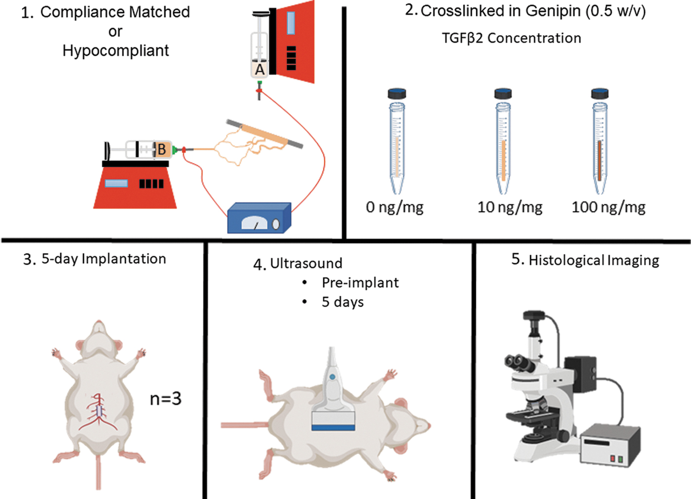
Overview of TGFβ2 study. (1) Compliance-matched and hypocompliant constructs were fabricated using a biaxial electrospun method that incorporated TGFβ2 concentrations at 0, 10, 100 ng/mg. (2) Constructs were crosslinked in 0.5% w/v genipin for 24 h at 37°C. (3) The TEVGs (n = 3 for each group) were implanted in the abdominal aorta of a Sprague Dawley rat for 5 days. (4) Before surgery and 5 days after implantation the TEVG was imaged using ultrasound. (5) Histological markers were used to evaluate host response. TEVG, tissue engineered vascular graft; TEVGs, tissue engineered vascular grafts; TGFβ2, Transforming growth factor beta 2.
Polymeric Formulations, Layer Thickness, Crosslinking Concentrations, and Transforming Growth Factor Beta 2 Concentration
G, gelatin; PCL, polycaprolactone.
Graft material and fabrication
The constructs were fabricated using a biaxial syringe setup with IME electrospinning device that included a climate-controlled chamber and gas shield to prevent buildup (Waalre, Netherlands). For this process, gelatin extracted from porcine skin (Sigma-Aldrich, St. Louis, MO) and PCL with a molecular weight of 80,000 (Sigma-Aldrich) were dissolved in 1,1,1,3,3,3-hexafluoro-2-propanol (Sigma-Aldrich) to create a 10% w/v solution. The polymeric formulations for biaxial electrospinning were 75:25 and 50:50 G:P and 20:80 and 35:65 G:P for the compliance-matched and hypocompliant constructs, respectively.
The polymeric solutions were loaded into 5 mL BD syringes (BD Franklin Lakes, NJ), and lyophilized recombinant human TGFβ2 (RD systems) was reconstituted with 4 mM HCL containing 1 mg/mL bovine serum albumin (BSA) (Sigma-Aldrich). TGFβ2 was added to each syringe based on the ratio of total gelatin percentage of the two formulations. For example, one construct layer had a syringe filled with a 75% and the other a 50% gelatin concentration; thus, this ratio of 3:2 was used as the amount of TGFβ2 mass to assign to each syringe. The polymer blends and TGFβ2 were mixed to create a final TGFβ2 mass of 0, 10, or 100 ng per mg of scaffold. 18
Once mixed for 24 h, the electrospinning solutions were both loaded onto separate computer-controlled syringe pumps and connected to IME electrospinning device through 1 mm PTFE tubing. The electrospinning parameters were as follows: working distance of 10 cm, dispense rate of 50 μL/min (both syringes), and 15-kV voltage. Constructs were spun onto a 1.1 mm diameter steel rod rotating at 300 mm/s with a tip translation of 300 m/s. The climate and humidity were set to 25°C and 30%, respectively. Before creating the construct, each syringe pump was primed and checked for solution extrusion. The higher gelatin formulation was spun from the vertical nozzle to help prevent buildup on the tip. Each layer was extruded sequentially which took about 4 min per layer to allow dissipation of the line pressure. Following the completion of the electrospinning process, the constructs were soaked and shaken in 0.5% (w/v) genipin (Wako Chemicals USA, Inc.) in 200 proof ethanol for 24 h at 37°C to crosslink gelatin. 20 Following crosslinking, the constructs were then rinsed with 200 proof ethanol and implanted within 24 h.
Rat aortic implantation
The TEVGs were implanted into the abdominal aorta of 8-week-old Sprague Dawley rats for 5 days. The compliance-matched and hypocompliant TEVG contained 0, 10, or 100 ng/mg TGFβ2 concentration with n = 3 in each group for a total of 18 animals. Each experimental group consisted of two males and one female or vice versa. The interpositional abdominal aortic surgical procedure followed closely the protocol described by Niepionce and recently used by our group.1,28 To begin the surgery, each rat was anesthetized with isoflurane (3% for induction and 1% for maintenance) for the duration of the study. Next, the fur was removed using NairTM with a paper towel, and a 3–4 cm incision with a No. 10 scalpel was made on the medial abdominal wall. The small intestines and cecum were removed from the abdominal cavity and covered with heparin soaked gauze to prevent dehydration and clotting. The aorta was isolated (1 cm above the bifurcation) from connective tissue, side branches were ligated and transected, and two 30 g clips were applied to the bifurcation and iliolumbar artery to arrest blood flow. The aorta was transected, the lumen of the vessel was irrigated by saline, and the anastomosis was initiated at the proximal region of the vessel with stay sutures using a 10-0 nylon placed at the 3 and 9 o'clock positions. Three or four stitches were added to the anterior wall, and then the vessel was rotated 180° clockwise with a similar amount of stitches added to the posterior wall.
A similar procedure was performed on the distal anastomosis. Before the last suture was placed, the lumen of the implant was irrigated with saline. The clips were then removed, and the vessel was assessed for patency using a perfusion test. If bleeding occurred, pressure was applied to the suture line until bleeding stopped. On occasion, an extra suture would be placed on a distal suture if bleeding persisted. In general, each anastomosis had around 7–10 stitches per end. After completion of the anastomosis, the small intestines and cecum were returned to the abdominal cavity, the rectus abdominis was closed using an interrupted 4-0 absorbable polyfilament suture, and skin was stapled closed.
Upon completion of the surgery, each rat received two daily intramuscular injections of 0.5 mg/kg buprenorphine and 100 mg/kg cefuroxime for the first 3 days to minimize pain and prevent bacterial infections, respectively. All solid medication was pulverized, mixed with jelly, put into a small ice cream cone, and fed to the rat. Inflammation was minimized by feeding the rat carprofen (5 mg/kg) at 24 and 48 h postoperatively. Aspirin (pain reliever) and dipyridamole (antiplatelet agent) were also fed to the rat at a concentration of 200 and 250 mg/kg for duration of the study. After 5 days, the graft was recovered by euthanizing the rat and fixed in a 4% paraformaldehyde solution in phosphate buffered saline (PBS) (Thermo Fisher Scientific, Waltham, MA).
Measurements and quantification of outcomes
In vivo ultrasound
Graft patency, diameter, compliance, and maximum blood velocity were collected using high frequency ultrasound (VEVO 2100 FUJIFILM VisualSonics, Inc., Canada) with a MS400 30 MHz probe. The rats were imaged for up to 1 week before surgery and 5 days postimplantation. Ultrasound data were collected by anesthetizing the rat with isoflurane (3% for induction and 1% for maintenance), removing the fur with Nair Hair Removal (Church & Dwight), and imaging the abdominal aorta cross-sectionally and longitudinally. In general, cross-sectional imaging was used for identifying the aorta and patency, while diameter/compliance measurements and velocity were calculated longitudinally. Patency was determined using ultrasound to observe flow through the entirety of the implant. All ultrasound measurements and calculations were determined using the Vevo Lab software version 1.7.1. In vivo compliance was calculated from the inner minimum and maximum longitudinal diameters:
The 50 mmhg in the denominator of the compliance equation was constant for all animals and represented the difference in theoretical pressure. The inner minimum and maximum diameters were then averaged to determine the diameter of the vessel. Using Doppler ultrasound, the maximum velocity of three waveforms per rat was used and adjusted for probe angle. 29 All ultrasound data were checked for normality using a Shapiro–Wilk test and compared to healthy rat aorta using a one-way analysis of variance (ANOVA) with Dunnett's post hoc testing. The length of the construct was assessed using a one-way ANOVA with a Tukey post hoc analysis.
Immunohistochemistry and fluorescent imaging
All TEVGs were evaluated for cellular markers and construct remodeling. Each explant was cut in half, separated into proximal and distal portions, frozen in O.C.T. medium, and cut into 10 μm thick sections using a Microm HM550 cryostat microtome. The proximal portion of the sample was cut longitudinally, and the distal part was cut cross-sectionally at the mid-graft location.
For immunohistochemistry, samples were rehydrated in PBS and then soaked in 1% sodium dodecyl sulfate (Sigma-Aldrich) in PBS for 15 min. The samples were next permeabilized in 0.3% Triton X-100 (Sigma-Aldrich) for 15 min and blocked with 0.5% BSA (Sigma-Aldrich), 0.3 M glycine (Thermo Fisher Scientific), and 20% goat serum (Thermo Fisher Scientific) for 1 h. Following blocking, all samples were incubated with primary antibodies overnight at 4°C. α-Smooth muscle actin (αSMA), calponin (CAL), and smooth muscle myosin heavy chain (MHC) were used to detect SMCs. Highly proliferative cells were labeled with Ki67, and the samples were also labeled for elastin. The inflammatory response was determined using CD68, CD86, and CD163 for pan, M1, and M2 macrophage phenotypes, respectively.
All primary antibodies were used at a ratio of 1:100 and purchased from Abcam. After incubation and washing, the samples were incubated for 1 h at room temperature with Alexa Fluor 488 Goat Anti-Rabbit IgG (Abcam, 1:1000) except for αSMA, which was incubated with Alexa Fluor 488 Donkey anti-goat IgG (Abcam, 1:1000). Each sample was washed and counterstained with 4′,6-diamidino-2-phenylindole (DAPI) mounting medium (Abcam).
Cellular markers were evaluated using a Nikon 90i Eclipse fluorescence microscope. Each section was imaged entirely using DAPI (excitation at 359 nm; emission at 461), fluorescein isothiocyanate (FITC) (excitation at 495 nm; emission at 519 nm), and Cy5 (excitation at 647; emission at 665) emission channels using a Nikon Plan Apo 10 × /0.45 objective. Cell nuclei, implant, and marker were identified in DAPI, Cy5 (autofluorescence from genipin), and FITC channel, respectively. The exposure time for the FITC channel was consistent between all samples of the same marker to quantitatively assess difference between samples.
Multiphoton imaging
Collagen was imaged using second harmonic generation using the Pitt Advanced Intravital Microscope at the University of Pittsburgh for the cross-sectionally and longitudinally sectioned samples. For this setup, an Olympus BX51 upright scanning microscope (Olympus, Tokyo, Japan) was coupled into a 120-fs tunable pulsed laser (INSIGHT DS+DUAL, Newport, Irving CA) and Olympus XLUMPLFL 20 × water immersion objective (NA 0.9). Collagen fibers were imaged at 780 nm and collected with a bandpass filter 377/50 nm with a power of 118–152 mW using second harmonic generation. DAPI and autofluorescence were also imaged at 780 nm with a power of 25–31 mW and collected with 525/50 nm and 620/60 nm bandpass filter, respectively. The signal was collected over multiple 499 × 499 μm fields of view with a 5 μm z-step size through the entire thickness of the sample. Mosaic images of each channel (377, 525, and 620 nm) were converted into maximum intensity projections and used for image processing.
For processing, the images were used to calculate abluminal number of cells, construct thickness, and total collagen. The construct thickness was measured for each explant and compared using a one-way ANOVA. The cells inside the construct and in the abluminal region were calculated using the ImageJ (Fiji) particle function. Percent collagen in the samples was calculated the same as the antibody image processing discussed in the section below 155 (Image Processing section).
Image processing
Fluorescent signal was collected using a Nikon 90i Eclipse fluorescence microscope in three channels DAPI, FITC, and Cy5, which were faux colored with blue for cell nuclei channel, green for implant channel (green), and red for the antibody or collagen channel, respectively. All images were processed using Matlab to determine antibody signal differences between each group. FITC signal was binarized to remove background noise and identify the pixels of interest (POI). The amount of signal per sample was calculated by dividing the POI by the total number of pixels (POI/TP) in a certain area. For collagen specifically, the sample had a high amount of noise which also required the collagen image to be despeckled. All marker calculations were done for the proximal and middle sections. The markers for each group 0, 10, 100 ng/mg were checked for normality and compared using a one-way ANOVA with Tukey post hoc testing. Due to the limited sample size in each group (n = 1 or 2, male or female), sex differences in each group were unable to be tested.
Results
Construct characterization
Previous work quantified the compliance-matched and hypocompliant TEVG burst pressure, suture retention, and compliance, which were 504 ± 164 and 1435 ± 107 mmHg, 11 ± 2 and 43.5 ± 15 g, and matched compliance and twice as stiff as rat aorta, respectively. 1 The overall release profile of TGFβ2 from the compliance-matched and hypocompliant construct was evaluated in Ardila et al. with around 1–1.5 and 0.05% of the total growth factor released from each construct by 5 days. 19
For the surgery, no complications or adverse events occurred with all animals surviving the duration of the study. Each surgery generally lasted around one and a half hours with the anastomosis time taking ∼50 min. The average implant length for the compliance-matched constructs 0, 10, and 100 ng/mg was 7.7 ± 0.7, 6.9 ± 0.2, and 6.6 ± 0.8 mm, respectively, with no difference between groups. The hypocompliant TEVG average length for 0, 10, and 100 ng/mg was 6 ± 0.5, 5.7 ± 0.6, and 6.4 ± 0.3 mm, respectively, with no difference between groups. The 100 ng/mg compliance-matched construct thickness was found to be thinner than the 10 ng/mg construct (59 ± 6 vs. 74 ± 4 μm, p = 0.04) with no difference in relation to the 0 ng/mg construct (71 ± 4 μm), Figure 3. The thickness of hypocompliant constructs was all similar.
Ultrasound
Ultrasound imaging confirmed patency in all groups throughout the duration of the study. The compliance-matched constructs were found to decrease in compliance as the concentration of TGFβ2 increased. Specifically, the 100 ng/mg group was considerably stiffer than the native rat aorta, 17 ± 2 × 10⁻⁴ (0 ng/mg) to 11 ± 1 × 10⁻⁴ mmHg−1 (100 ng/mg). The diameter and maximum velocity for all compliance-matched groups did not differ from native rat aorta tissue. In general, the average implant diameter and velocity was around 1000 μm and 700 mm/s, respectively. These results can be found in Figure 2.
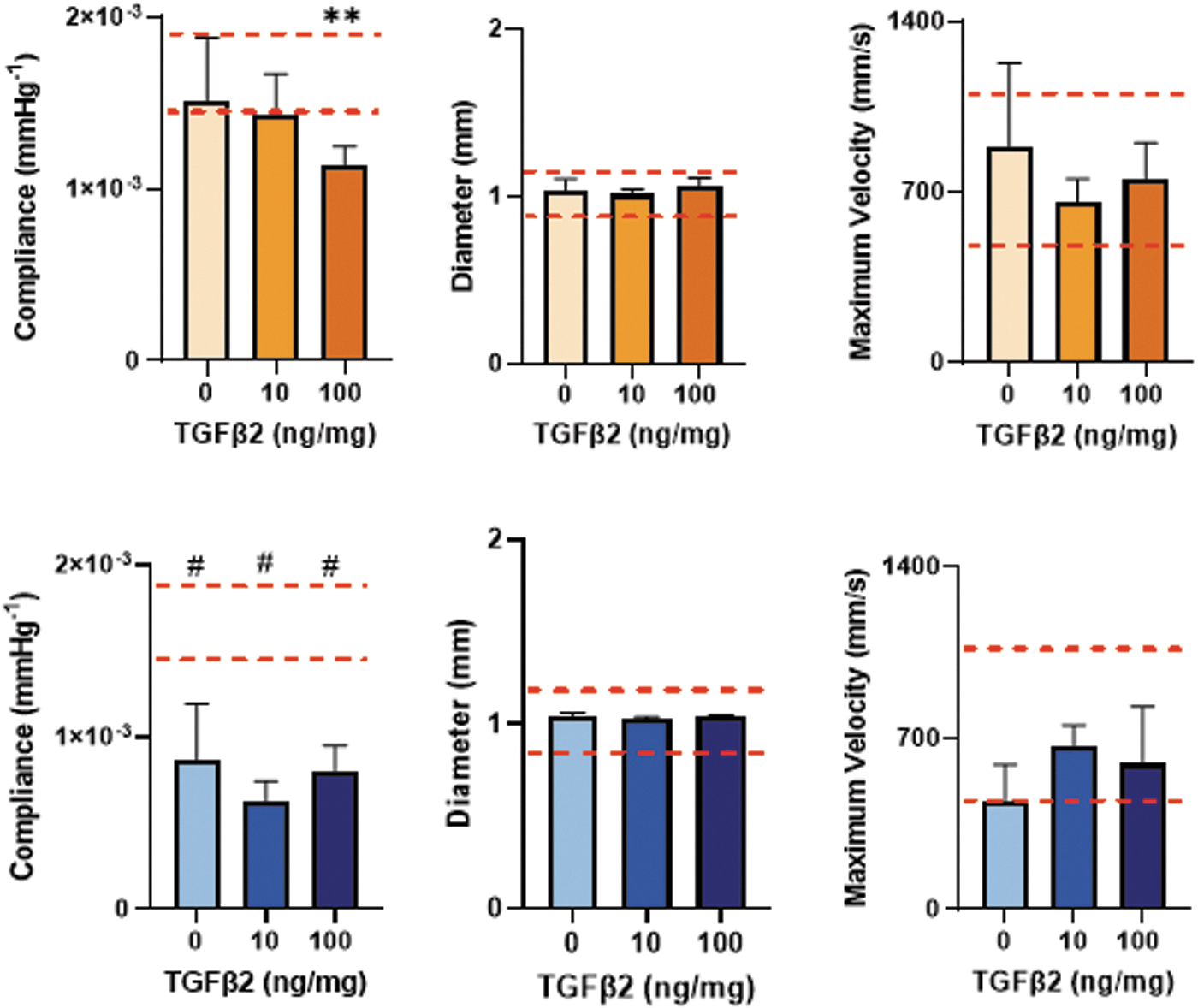
Compliance, diameter, and maximum velocity of 0, 10, 100 ng/mg of constructs. Orange and blue bars indicate compliance-matched and hypocompliant TEVG implants, respectively. The red dashed lines represent rat aorta.**p < 0.01 and #p < 0.0001 in reference to rat aorta.
The compliance of the hypocompliant grafts for 0, 10, 100 ng/mg was 8.7 ± 3 × 10⁻⁴, 6.3 ± 1 × 10⁻⁴, 8 ± 2 × 10⁻⁴ mmHg−1 with each graft being significantly stiffer than native rat aorta, p < 0.0001 for all comparisons. The diameter 190 and velocity of the hypocompliant TEVGs were similar to that of native rat aorta (Fig. 2).
Multiphoton imaging
Multiphoton images representing the hypocompliant (blue) and compliance (orange) TEVGs for the 0, 10, 100 TGFβ2 ng/mg 5-day explant groups are shown in Figure 3 with green being the construct and blue cell nuclei. The overall cell count (lumen, implant, and abluminal) for the compliance-matched implant was generally similar between the 0 and 100 ng/mg groups with the least amount of cells in the 10 ng/mg group. Cellular infiltration decreased in the compliance-matched TEVG with increasing concentrations of TGFβ2, 0 versus 100 ng/mg (3600 ± 1700 vs. 770 ± 300 cells, p < 0.05). TGFβ2 had the opposite effect on the abluminal cell count in compliance-matched TEVGs with the 100 ng/mg construct having the most cells compared to the 10 ng/mg construct (6820 ± 1670 vs. 200 ± 140 cells, p < 0.05). For the hypocompliant TEVGs, cellular infiltration into the construct and abluminal region was found to be similar between all constructs.
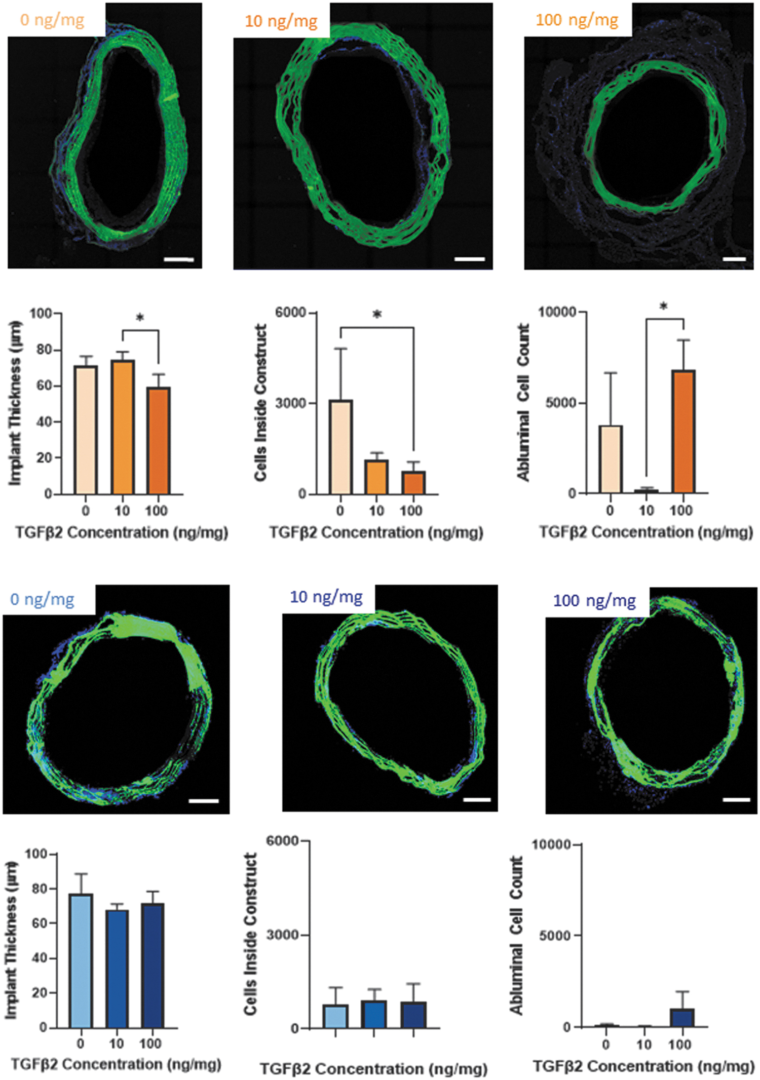
Compliance, diameter, and maximum velocity of 0, 10, 100 ng/mg of constructs. Orange and blue bar graphs indicate compliance-matched and hypocompliant TEVG implants, respectively. Scale bar is 100 μm. *p < 0.05 and **p < 0.01 compared to rat aorta.
Immunohistochemistry
Longitudinal and cross-sectional slices of the samples were stained for SMCs, macrophages, and ECM products with the results shown in Figures 4 and 5. The 5 day proximal portion of the compliance-matched explant showed that the 100 ng/mg group had a reduction in SMCs but an increase in CD68 (M1 macrophage) and collagen. There was also a dramatic increase in MHC of the 10 ng/mg compared to 0 and 100 ng/mg in the compliance-matched grafts. In contrast, the hypocompliant constructs showed no differences in the proximal and middle location of the grafts for all markers and ECM products, Supplementary Figures S1 and S2.
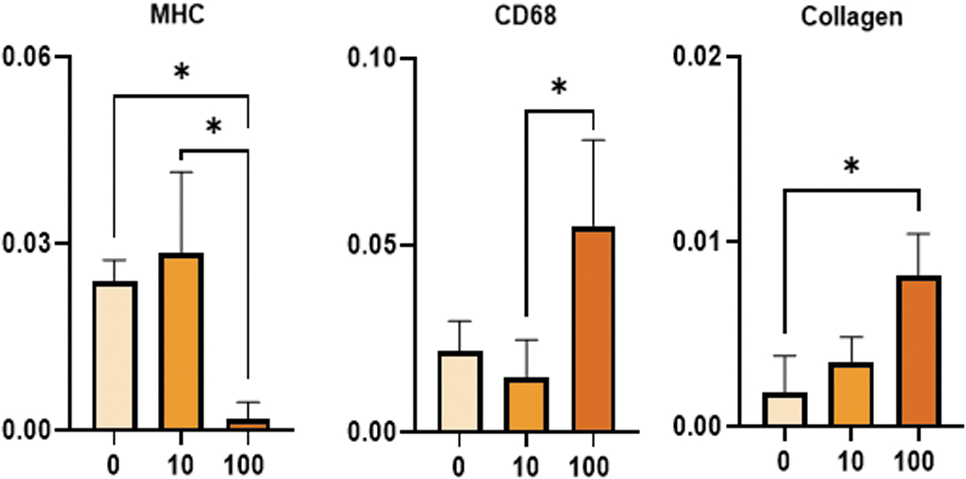
Compliance-matched proximal immunofluorescence images and immunohistochemistry results. The TEVG, nuclei, and each marker are green, blue, and red, respectively. Immunohistochemistry quantification of the signal from MHC, pan macrophages (CD68), collagen, αSMC, CAL, Ki67, M1 (CD86), M2 (CD163), and elastin. *p < 0.05; **p < 0.01; X axis = TGFβ2 concentration (ng/mg); Y axis = POI/total pixels; Scale bar is 50 μm. CAL, calponin; MHC, myosin heavy chain; POI, pixels of interest; SMC, smooth muscle cell.
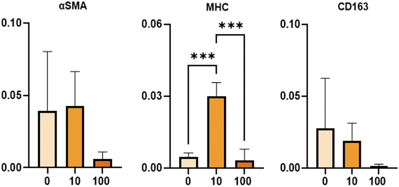
Compliance-matched midgraft immunofluorescence images and IHC results. Construct is green, cell nuclei are blue, and marker is red. Immunohistochemistry quantification of the signal form αSMC, MHC, M2 (CD163), CAL, Ki67, pan macrophage (CD68), M1 (CD86), elastin, and collagen. *p < 0.05; ***p < 0.001; X axis = TGFβ2 concentration (ng/mg); Y axis = POI/total pixels; Scale bar is 50 μm. IHC, immunohistochemistry; αSMC, α smooth muscle cell.
For the proximal location of the compliance-matched grafts (Fig. 4), the SMC population was measured using αSMA, CAL, and MHC with differences only occurring in the MHC marker. Specifically, there was a decrease in MHC signal in the 100 ng/mg group compared to the 0 and 10 ng/mg (0.0019 ± 0.002 vs. 0.024 ± 0.003 and 0.029 ± 0.013 POI/TP, p < 0.05). The Ki67 label did not show any differences between the groups. The definitive markers CD86 and CD163 displayed no differences between groups, but the general macrophage marker CD68 was increased in the 100 versus 10 ng/mg (0.055 ± 0.23 vs. 0.015 ± 0.001 POI/TP, p < 0.04). ECM production of collagen increased with increasing concentrations of TGFβ2, 0, 10, and 100 ng/mg (0.002 ± 0.002 to 0.003 ± 0.001 to 0.008 ± 0.002 POI/TP, p < 0.05, 0–100). Overall elastin amounts in the ECM were found to be similar between all groups. The remaining markers not shown in Figure 4 are found in Supplementary Figure S3.
For the middle location of the compliance-matched grafts, MHC again was the only SMC marker to show differences between groups with a drastic increase in signal for 10 ng/mg group compared to the 0 and 100 ng/mg groups (0.03 ± 0.006 vs. 0.005 ± 0.002 and 0.003 ± 0.005 POI/TP). No other differences were found in the macrophage, Ki67, and ECM markers, which are further highlighted in Figure 5. The remaining markers not shown in Figure 5 are found in Supplementary Figure S3.
Discussion
The incorporation of TGFβ2 in compliance-matched TEVGs (faster degrading scaffold) increased the SMC population and collagen production over an acute 5-day period. Specifically, low TGFβ2 concentrations (10 ng/mg) caused an increase in the SMC population/infiltration at both the proximal and middle locations of the graft. The high concentration of TGFβ2 (100 ng/mg) in the compliance-matched grafts increased collagen production in the proximal region, halted SMC proliferation/infiltration, and caused the TEVG to stiffen. The hypocompliant TEVGs (slower degrading scaffolds) displayed no difference in cellular population and ECM production with any TGFβ2 concentration. These results demonstrate that the elution of TGFβ2 from a TEVG can promote in vivo SMC recruitment and collagen production after only an acute 5-day implantation period.
TGFβ2 and the TGFβ family are key signaling molecules that regulate SMC activity depending on concentration.18,30–33 At low concentrations, the TGFβ family has been found to increase SMC proliferation through the promotion of platelet derived growth factor (PDGF) which in turn stimulates DNA synthesis.32,34 These SMCs are found in more immature vessels which have a higher rate of proliferation and migrate much faster across a substrate. 35 At higher TGFβ concentrations, PDGF expression is limited, which arrests SMC proliferation and leads to an increase in ECM production. 32 These results have been confirmed in SMCs exposed to high levels of TGFβ1 that induced collagen synthesis (Kubota el al). 36 These SMCs are found in a mature vessel where it regulates vascular tone and ECM production and minimizes cellular migration. 37
In the context of this work and TGFβ2, Ardila et al. demonstrated in vitro that using low (5 ng/mL) exogenous TGFβ2 concentrations in electrospun gelatin:fibrinogen scaffolds promoted SMC proliferation and collagen production, respectively. 18 Our laboratory has taken the next step and implanted TGFβ2-eluting, compliance-matched constructs into a rat model for 5 days showing similar results with increased SMCs (MHC marker) at low concentrations and higher collagen content at 100 ng/mg. Using this knowledge, the elution of TGFβ2 from the TEVG could be modified to promote SMC migration and collagen deposition subsequently. For example, Jhunjhunwala et al. demonstrated that TGFβ1 could be released at low levels for 3 weeks followed by an increased release until around 50 days using microparticle encapsulation. 38 This microparticle release profile could also be transferred to TGFβ2 and is currently being evaluated in our laboratory.
A substantial portion of TEVG research is focused on SMCs; however, the immune response to the implant plays an equally important role in the success of the TEVG. Immune responses to the TEVG are broken down into the inflammatory and wound healing response.39,40 The inflammatory response which is closely tied to M1 macrophages (CD86) promotes encapsulation of the tissue, destruction of foreign bodies, and impeded growth. 40 While it is necessary to have this inflammatory response early on to increase the cellular population around the implant and protect the body against foreign material, a prolonged or exaggerated M1 immune reaction can be detrimental to the TEVG. 41 Thus, promoting these M1 macrophages to move toward the M2 phenotype (CD163) encourages cellular growth around the implant, ECM production, and neovessel formation. 42
In this study, the results demonstrate the initial response to the implant with only noticeable differences in macrophages shown in the CD68 marker (generalized macrophage marker) of the 100 ng/mg compliance-matched group in the proximal location. These results suggest that increasing TGFβ2 promotes an exaggerated immune response, which has also been shown to occur in other TGFβ-influenced environments. 43 Prior work in our laboratory has also evaluated immune cells at a longer time point. Specifically, the 0 ng/mg compliance-matched implant has been previously implanted in Sprague Dawley rats for 1 month and compared to a hypocompliant control TEVG. The M2/M1 ratio of macrophages in the 0 ng/mg compliance-matched grafts was found to be higher than in the hypocompliant grafts. 1 This suggests that the compliance of the implant is more suitable for desired TEVG remodeling. While there are no noticeable differences in M1 and M2 population at 5 days in the compliance-matched construct, we anticipate that this M2/M1 ratio may carry over in the 10 and 100 ng/mg groups in future studies. Longer time points and larger sample size will need to occur before there is a clear understanding of the longer term effects of TGFβ2 on TEVG remodeling.
One major difference between the compliance-matched and the hypocompliant grafts was the degradation rate of the scaffold. 19 Previous work has shown that the compliance-matched construct degrades twice as much as the hypocompliant graft in 1 month. 1 This degradation of the scaffold is vital for the release of TGFβ2. Past in vitro work has shown that in 5 days the compliance-matched TEVG can elute about 20–30 times more TGFβ2 than the hypocompliant grafts. 19 Thus, the reduced degradation/elution of hypocompliant constructs is believed to be the reason why there were no differences between any of the hypocompliant groups in this work.
This is surprising considering that the 100 ng/mg hypocompliant group has an order of magnitude more TGFβ2 than the 10 ng/mg group. Another reason for the lack of TGFβ2 release from both scaffolds may emanate from the genipin crosslinking procedure. Each construct is submerged in 0.5% genipin in 100% ethanol at 37° and shaken for 24 h. During this procedure, any loose or unbound TGFβ2 may be eluted into the genipin solution which would reduce the amount of growth factor available. A potential solution to prevent the loss of TGFβ2 is to explore alternative crosslinking procedures such as using ultraviolet light, vapor, chemical, or plasma treatment. 44 Future testing could incorporate one of these crosslinking methods to prevent the potential loss of TGFβ2 during the fabrication process.
One unexpected result from this work was the decreased compliance in the 100 ng/mg compliance-matched construct compared to rat aorta. Specifically, this graft stiffening comes as even more of a surprise due to the acute nature of the implantation period and the decrease in implant thickness of the 100 ng/mg compliance-matched construct (60 μm) in reference to the 0 and 100 ng/mg groups. This decrease of compliance is related to the increase of cells in the abluminal region of the 100 ng/mg group. All 100 ng/mg compliance-matched samples had large abluminal populations compared to the vastly varying abluminal cellular populations of the 0 and 10 ng/mg TEVGs.
To further understand these results, our laboratory has also investigated the 100 ng/mg compliance-matched grafts at a 4-week implant (data not shown) and found that this decrease of compliance also led to an increase in luminal thickness and cellular alignment compared to 0 ng/mg at 4 weeks. As these results and previous literature suggest, TGFβ can be atheroprotective and atherogenic depending on the cellular environment.18,45 The increase in alignment demonstrates that high TGFβ2 concentrations may promote a contractile SMC but could also encourage intimal hyperplasia. This increase of intimal hyperplasia could be related to the endothelial-to-mesenchymal transition of endothelial cells in the TEVG. From previous work, high TGFβ concentrations in mouse endothelial cells cause cells to become more leaky, transition to a mesenchymal state, and lead to increased atherosclerotic lesion markers.46,47 Controlling the concentration and release of TGFβ2 from compliance-matched TEVGs will be ideal for maintenance of compliance as the graft is remodeled in vivo.
Due to the higher degradation rate and release of TGFβ-2 from the compliance matched (CM) scaffolds, one may expect to see a convective effect along the length of the scaffold (differences in proximal vs. middle sections due to convection). Using a preliminary qualitative assessment, we did not see differences in any outcome between proximal and middle locations. This is perhaps not surprising given the acute implantation period. Future investigations, at longer time points, should be used to further investigate these effects.
All TEVGs remained patent throughout the time course of this work. These initial results suggest that the inclusion of TGFβ2 does not affect in vivo patency, velocity, or diameter over a 5-day period. This is consistent with our prior work showing that the velocity, diameter, and compliance of 0 ng/mg implanted TEVGs remained similar over a 4-week implantation period. 1 The only drawback from our previous study was that both compliance-matched and hypocompliant grafts clotted in 2/7 and 1/7 of the trials, respectively (no significant differences between groups). It should be noted that the hemodynamics, implant length, and end-to-end anastomotic approach in the rat model exhibit differences between the eventual clinical needs of a human implant. This will need to be examined in future work. The rat end-to-end interpositional model used here served as a high throughput model to test these preliminary hypotheses and is commonly used.
The major limitation of this work was the narrow scope of this study. Further TGFβ2 studies are needed to fine tune in vivo TGFβ2 release such as concentration, elution profile, sample size, study duration, and timing. Modifying these variables in future work will help provide a better understanding of the effects of TGFβ2. Our current working hypothesis of TGFβ2 release is that a low concentration will promote early SMC migration, and a subsequent higher dose will promote ECM production that will stabilize the compliance of our fully biodegradable TEVG. While these results are promising, other factors will have to be taken into account to find the ideal TGFβ2 release profile for a particular animal species, side effects of TGFβ2, graft composition (as shown in our hypocompliant group), and several other factors. The acute response of the host to TGFβ2 in this study will hopefully spawn additional future research exploring the next generation of TEVGs that elute bioactive factors to improve functional in vivo performance.
Another limitation of this work was our experimental design. Due to the limited sample size and composition of the scaffolds, the CM and hypocompliant (HYPO) grafts were not able to be compared. These groups were chosen as they represent the current (HYPO) and possibly the future “gold standard” (CM) in vascular graft design.48,49 These HYPO or hypocompliant grafts have led to compliance mismatch (graft and native vessel), which has been known to cause hemodynamical changes, thrombosis, and stenosis.4–6 Due to the short nature of this study, no difference in intimal hyperplasia or thrombosis was seen in the HYPO group. As for the compliance-matched grafts, our prior work has shown that CM grafts promote a more positive remodeling response over a 1-month implantation period. 1
While TGFβ and its family of cytokines have been given significant attention in the literature, studies that focus on the exogenous delivery of the TGFβ2 isoform specifically are limited. This is important to note as there may be fundamental differences in the binding between TGFβ 1 and 2, and the actions of TGFβ1 may not always relate directly to TGFβ2.50,51 For example, the RGD (Arg-Gly-Asp) integrin binding region found in the LAP of TGFβ1 and TGFβ3 is lacking in TGFβ2, which suggests that it may have a different method of activation. 52 This may play a role in functional differences during cardiovascular development, as heterozygous TGFβ1 knockout mice function similarly to wild type animals, whereas TGFβ2 mice present with aortic root dissection. 53 In terms of vascular remodeling, TGFβ is again a regulator of cellular migration/proliferation at low concentrations and prevents movement at higher concentrations. 45 This growth factor not only affects SMCs but also endothelial cells, macrophages, and lymphocytes.
The exact timing and concentration of TGFβ elution are key for success or failure in vivo. For example, high TGFβ concentrations early may prevent endothelial cell migration into the implant, thus increasing the likelihood of a possible thrombotic event. 54 In contrast, a higher TGFβ concentration later may promote collagen development in the TEVGs. Elevated TGFβ levels may not be ideal after the initial implantation once the vessel has matured. Specifically, the upregulation of TGFβ can promote an increase in collagen production to stiffen the vessel and lead to calcification. 45 These results have been demonstrated in a rat and a rabbit model with the addition of exogenous TGFβ or after a vascular injury.55,56 Thrombotic events may occur more frequently, as well as TGFβ1 is less efficient at clearing thrombosis formation due to an increase in plasminogen activator inhibitor activity. 57 Keeping all of these factors in mind, the delivery of TGFβ will need to be tightly controlled for a desired long-term functional outcome.
To the author's knowledge, this is the first study to assess in vivo TGFβ2 release from compliance-matched and hypocompliant TEVGs. Historically, TGFβ2 has been found to be a potent SMC phenotype modulator, which was supported in this work. As TGFβ2 concentrations increase in the compliance-matched constructs the production of collagen is elevated, the SMC population is reduced, the TEVG becomes less compliant, and the growth factor causes a larger immune response. In contrast, the lower concentration of TGFβ2 in the compliance-matched construct increases SMC population and proliferation. The degradation of the scaffold is key to TGFβ2 elution as the slower degrading hypocompliant construct had no effect on the cellular environment or implant. We believe that this work demonstrates that TGFβ2 incorporated into a TEVG is beneficial for SMC migration, proliferation, and collagen production and will inspire new related work in the vascular tissue engineering field.
Footnotes
Acknowledgments
The authors thank Dr. Rob Kellar for his advice on immunohistochemistry staining (Ki67), the Dr. David Vorp laboratory for the use of their cryotome, and credit Biorender.com for some images in Figure 1. Portions of this article were used in Kenneth Furdella's dissertation and can be found at  .
.
Authors' Contribution
All authors contributed to the word presented in this article.
Disclosure Statement
No competing financial interests exist.
Funding Information
This research was funded by the NIH, grant 1R56HL136517-01. Support was also provided by National Institute of Biomedical Imaging and Bioengineering Award T32EB003392 (BiRM). The small animal imaging system (Vevo 2100) was supported by the NIH grant 1S10RR027383-01.
References
Supplementary Material
Please find the following supplemental material available below.
For Open Access articles published under a Creative Commons License, all supplemental material carries the same license as the article it is associated with.
For non-Open Access articles published, all supplemental material carries a non-exclusive license, and permission requests for re-use of supplemental material or any part of supplemental material shall be sent directly to the copyright owner as specified in the copyright notice associated with the article.
