




Abstract
Although choledochojejunostomy is the standard technique for biliary reconstruction, there are various associated problems that need to be solved such as reflux cholangitis. Interposition with an artificial bile duct (ABD) to replace the resected bile duct maintains a physiological conduit for bile and may solve this problem. This study investigated the usefulness of an ABD made of gelatin hydrogel nonwoven fabric (GHNF). GHNF was prepared by the solution blow spinning method. The migration and activity of murine fibroblast L929 cells were examined in GHNF sheets. L929 cells migrated into GHNF sheets, where they proliferated and synthesized collagen, suggesting GHNF is a promising scaffold for bile duct regeneration. ABDs made of GHNF were implanted in place of resected bile duct segments in rats. The rats were killed at 2, 6, and 12 weeks postimplantation. The implantation site was histologically evaluated for bile duct regeneration. At postoperative 2 weeks, migrating cells were observed in the ABD pores. The implanted ABD was mostly degraded and replaced by collagen fibers at 6 weeks. Ki67-positive bile duct epithelial cells appeared within the implanted ABD. These were most abundant within the central part of the ABD after 6 weeks. The percentages of Ki67-positive cells were 31.7 ± 9.1% in the experimental group and 0.8 ± 0.6% in the sham operation group at 6 weeks (p < 0.05), indicating that mature biliary epithelial cells at the stump proliferated to regenerate the biliary epithelium. Biliary epithelial cells had almost completely covered the bile duct lumen at 12 weeks (epithelialization ratios: 10.4 ± 6.9% at 2 weeks, 93.1 ± 5.1% at 6 weeks, 99.2 ± 1.6% at 12 weeks). The regenerated epithelium was positive for the bile duct epithelium marker cytokeratin 19. Bile duct regeneration was accompanied by angiogenesis, as evidenced by the appearance of CD31-positive vascular structures. Capillaries were induced 2 weeks after implantation. The number of capillaries reached a maximum at 6 weeks and decreased to the same level as that of normal bile ducts at 12 weeks. These results showed that an ABD of GHNF contributed to successful bile duct regeneration in rats by facilitating the cell migration required for extracellular matrix synthesis, angiogenesis, and epithelialization.
Impact Statement
Development of an artificial bile duct (ABD) enables physiological biliary reconstruction and may solve clinical problems associated with choledochojejunostomy. In this study, we created ABDs with gelatin hydrogel nonwoven fabric and implanted them in place of resected bile duct in rats. We evaluated the process of bile duct regeneration as well as decomposition of the ABD and demonstrated successful regeneration of resected bile duct, highlighting the possibility of this novel biliary reconstruction method to replace choledochojejunostomy.
Introduction
Bile duct resection is performed as a surgical treatment for various biliary disorders such as cholangiocarcinoma, congenital biliary dilatation, and benign biliary strictures resulting from trauma, Mirrizi syndrome, or iatrogenic bile duct injury. Choledochojejunostomy is the standard technique of biliary reconstruction after bile duct resection; however, it is associated with various problems that need to be overcome including the complexity of the procedure, risk of anastomotic leakage and stenosis, reflux cholangitis, and carcinogenesis.1,2 In addition, endoscopic intervention in the anastomotic site or intrahepatic bile duct becomes more challenging after choledochojejunostomy.
Interposition of an artificial bile duct (ABD) in place of the resected extrahepatic bile duct could reconstruct the physiological biliary tract, which may solve various problems associated with choledochojejunostomy. Attempts at developing ABD have been made for a long time. 3 Autologous tissue,4,5 nondegradable materials, and degradable materials have been used as ABD materials.6–8 However, an ABD has not been successfully put into clinical use yet. This is in contrast to the fact that artificial blood vessels have been in practical use for decades and are indispensable for cardiovascular surgery.
Gelatin is a natural macromolecule consisting of collagen that has good biocompatibility, degradability, and low immunogenicity. Gelatin has been used clinically as a temporal embolization material (Spongel® or Gelpart®), making it advantageous when considering clinical applications. We have recently developed nonwoven fabrics made of gelatin (gelatin hydrogel nonwoven fabrics [GHNF]) and demonstrated their utility as scaffolds that facilitate cell migration and maintain cellular functions. 9 Thus, we expect GHNF to be promising materials for ABD. In this study, we generated an ABD using GHNF and evaluated its feasibility for biliary reconstruction in a rat model.
Materials and Methods
Preparing the GHNF and polypropylene nonwoven fabric sheets and ABD
The GHNF and polypropylene nonwoven fabric (PPNF) sheets and ABD made of GHNF were supplied by The Japan Wool Textile Co., Ltd (Hyogo, Japan). GHNF sheets were prepared by the previously reported solution blow spinning method. 9 Polypropylene (PP) was selected as a control for the in vitro experiments because PP has a cell adhesion capacity and structural characteristics (fiber diameter and density) similar to those of GHNF when processed into a nonwoven fabric. The GHNF sheet was punched out into disks of 8 mm in diameter and the PPNF sheet was punched out into disks of 11 mm in diameter. The ABD was made by the same method as the GHNF with some modifications. To acquire sufficient strength for anastomosis, the density was increased by spinning at a speed ∼18-fold faster than for GHNF sheet preparation and keeping in wet during lamination. The gelatin density of the GHNF and the ABD were 0.26 ± 0.01 mg/mm3 and 0.73 ± 0.03 mg/mm3, respectively. All biomaterials were sterilized with ethylene oxide gas.
Measuring the material properties of GHNF and PPNF sheets and ABD
The weight of the dry and swollen GHNF and the volume of swollen GHNF were measured. The porosity of GHNF was calculated by the following formula: [(WGHNF – WG)/VGHNF] × 100 (%), where WGHNF is weight of swollen GHNF, WG is weight of swollen gelatin (calculated as 3.1-fold increase over the weight of dry GHNF), and VGHNF is volume of swollen GHNF. The porosity of the PPNF sheet was calculated by the same method. The density of PP was 0.9 g/cm3. The fiber diameter was measured with the ImageJ program (Public Domain Image Processing Program, National Institutes of Health, Bethesda, MD) from an image taken with an optical microscope (BZ-X710; Keyence Co. Ltd., Tokyo, Japan). The stiffness of swollen GHNF sheets and ABD were measured with a creep meter (RE2-33005C; Yamaden, Bunkyo-ku, Tokyo, Japan). Stress was measured up to a strain of 50%.
Fibroblast culture
Murine fibroblast L929 cells were cultured in Dulbecco's modified Eagle medium (Thermo Fisher Scientific, Waltham, MA) supplemented with 10% fetal bovine serum (Thermo Fisher Scientific) and 1% penicillin/streptomycin (Nacalai Tesque, Kyoto, Japan) at 37°C. L929 cells at 90% confluence were detached with 0.25% trypsin-ethylenediaminetetraacetic acid (Nacalai Tesque) to obtain a cell suspension.
Preparing fibroblast aggregates for migration assays on GHNF sheets and ABD
Cell aggregates were prepared as described by Nii et al. 10 In brief, the L929 cell suspension (5 × 10³ cells/mL, 200 μL) was seeded onto a polyvinyl alcohol-coated (degree of polymerization: 1800; saponification: 88 mol%; Unichika, Tokyo, Japan), round-bottomed 96-well plate and cultured for 3 days to prepare cell aggregates.
The prepared aggregates were seeded onto swollen GHNF sheets, cultured, and then collected at the designated timepoints (12, 24, 48, and 72 h). Similarly, cell aggregates were seeded onto the ABD, and then collected at the same timepoints.
Cell culture in the GHNF and PPNF sheets and ABD
A L929 cell suspension (10 × 106 cells/mL, 20 μL) were seeded onto swollen GHNF and PPNF sheets with 0.1 mM
ABD implantation in model rats
Female Sprague-Dawley rats (Charles River, Kanagawa, Japan) weighing 260 ± 11.8 g were used in this experiment. The animals were anesthetized by inhaling 1.5% isoflurane. Blood samples were collected from the tail vein. Abdominal midline laparotomy was performed, and the common bile duct was exposed. A 1–2 mm segment of the common bile duct was resected. Each stump of the bile duct and swollen ABD (4 mm in length) were anastomosed with 10-0 nylon (Crownjun, Ichikawa, Chiba, Japan) under a microscope. A polyethylene tube (SP-8; Natsume, Yushima, Tokyo, Japan) was placed as a stent in the lumen. In the sham group, only abdominal midline laparotomy was performed after blood sampling from the tail vein. Rats were fed a standard chow diet with free access to tap water. ABD group rats were killed at 2, 6, and 12 weeks after implantation, and the sham group rats were killed at 12 weeks. Blood samples were collected from the tail vein and the inferior vena cava, and the bile duct was collected.
The institutional animal research committee of Kyoto University approved this experimental protocol (Medkyo-19170). All experiments were conducted in accordance with the Guidelines for the Care and Use of Laboratory Animals from the National Institutes of Health.
Histology
Cell aggregates on GHNF sheets and ABD were fixed with a 4% paraformaldehyde (PFA) solution, embedded in optimal cutting temperature compound, and frozen. Frozen samples were cut into 6-μm thick sections by a cryotome (CM3050S; Leica Microsystems, Wetzlar, Germany). Hematoxylin and eosin (HE) staining was performed on each sample. Bile ducts were harvested from all rats used in the animal experiments, fixed with a 4% PFA solution, embedded in paraffin, and sectioned into 4-μm thick sections. HE staining was performed on each sample. Prepared specimens were examined under an optical microscope.
Sirius red staining
Sections of frozen tissue were stained with the Sirius Red/Fast Green Collagen Staining Kit (Chondrex, Woodinville, WA), and sections of paraffin-embedded tissue were stained with the staining solution (Sirius Red 500 mg/Van Gieson solution P, 500 mL).
Immunofluorescence
After permeabilization and blocking, sections were incubated overnight at 4°C with primary antibodies. Then the sections were incubated for 1 h with labeled isotype-specific secondary antibodies (AlexaFluor, Invitrogen, Life Technologies Ltd, Paisley, United Kingdom) and counterstained with 4,6-diamidino-2-phenylindole. The antibodies used for immunofluorescence are described in Supplementary Table S1.
Immunohistochemistry
After deparaffinization, antigen retrieval and blocking of endogenous peroxidase activity were performed. Sections were incubated overnight at 4°C with primary antibodies, incubated with horseradish peroxidase-labeled polymer (Agilent Technologies, Santa Clara, CA) for 20 min, colored with 3,3′-diaminobenzidine (Agilent Technologies), and counterstained with hematoxylin. The antibodies used for immunofluorescence are described in Supplementary Table S1.
Quantitative real-time polymerase chain reaction
L929 cells cultured in GHNF sheets for 1 week were collected and total RNA was extracted with the RNeasy Plus Mini Kit (Qiagen, Hilden, Germany) according to the manufacturer's instructions. Total RNA was reverse transcribed to complementary DNA using a high-capacity complementary DNA reverse transcription kit (Applied Biosystems, Foster City, CA). The mRNA levels were analyzed by quantitative real-time polymerase chain reaction (qPCR) using a SYBR green master reaction mix on a StepOnePlus system (Applied Biosystems). β-actin was used for standardization. The primer sequences used for qPCR are listed in Supplementary Table S2. A calibration curve was prepared with the control samples, and a relative quantification method was performed. The control group was statically cultured L929 cells in medium supplemented with ascorbic acid on the polyethylene dish for 3 days.
Imaging and quantification of histology
To quantify the number of migrating cells into GHNF sheets from aggregates, the number of cells in the scaffold was counted using ImageJ (Supplementary Fig. S1a). To quantify cell localization within the GHNF and PPNF sheets, a cross-section of the samples was divided into outside and inside area, and the cell localized area was quantified using ImageJ (Supplementary Fig. S1b). The bile duct regeneration ratio was calculated by the following formula: [(4 – a)/4] × 100 (%), where 4 is the length of the implanted ABD, and a is the distance of bile duct epithelial defect.
To quantify the number of capillaries, five high-power magnification (400 × ) images of CD31 immunohistochemical staining were taken. The number of capillaries were counted, and the CD31-positive area was analyzed by ImageJ. The number of Ki67-positive cells were also quantified in the same manner.
Statistical analysis
All data are presented as mean ± standard deviation. Differences between groups were compared using the Mann–Whitney U-test. A value of p < 0.05 was considered statistically significant. JMP Pro 14 Software (SAS Institute, Cary, NC) was used for all statistical analyses.
Results
Measurements of the structural parameters of GHNF sheets
The macroscopic appearance of GHNF sheets in the dry and saturated swollen states are given in Figure 1a and b, respectively. The swelling ratio of GHNF sheets, which was calculated as the weight of the swollen GHNF sheet divided by the weight of the dry GHNF sheet, was 10.1 ± 0.05. The porosity of GHNF sheets was 66.7 ± 4.4%. The microscopic appearance of a GHNF sheet is given in Figure 1c. The mean fiber diameter of the GHNF sheet was 48.7 ± 6.6 μm. When the stiffness of the GHNF sheet was measured by creep mater, the stress was 7.6 ± 1.4 kPa at 30% strain and 20.6 ± 2.7 kPa at 50% strain.
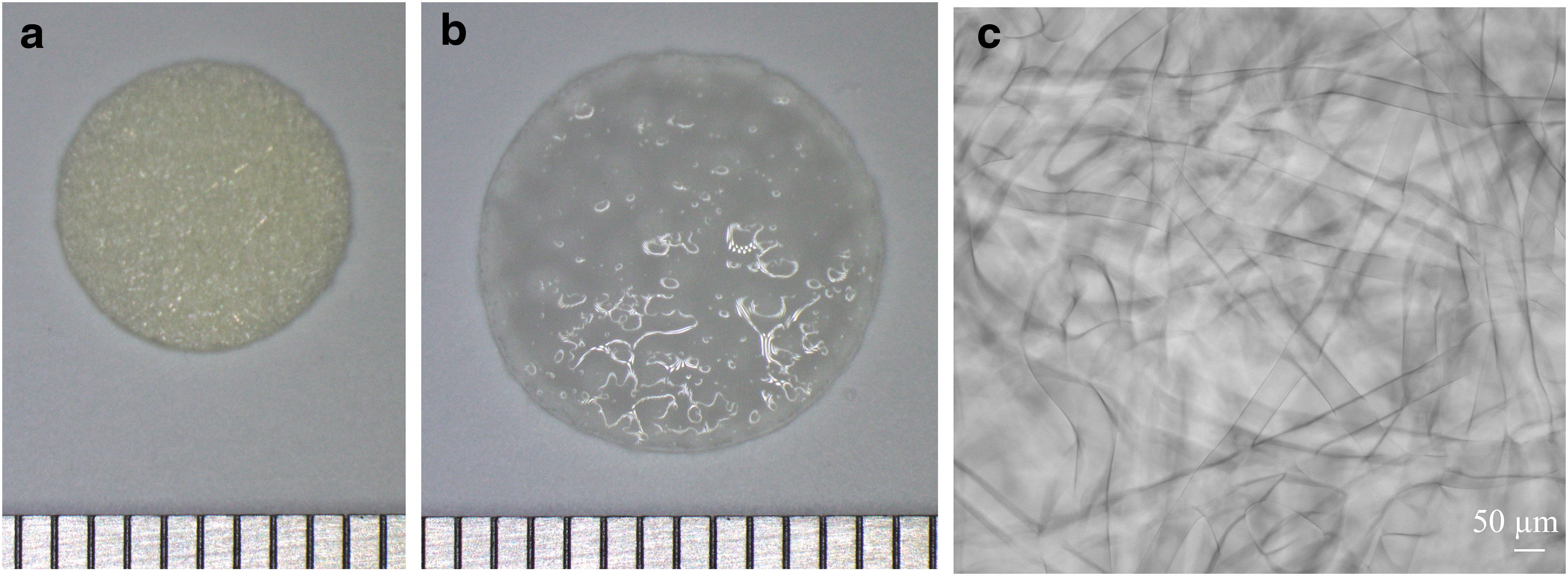
Macroscopic and microscopic appearance of the GHNF sheet.
Migration assays on GHNF sheets
Migration assays were performed to investigate if gelatin hydrogel was a suitable environment for cell infiltration. When L929 cell aggregates were seeded onto GHNF sheets, the cells migrated into the GHNF sheet around the cell aggregates (Fig. 2a). The number of cells that migrated into the GHNF sheet increased over time (Fig. 2b). The cells migrating into the GHNF sheet were positive for Ki67, demonstrating that they maintained proliferative ability (Fig. 2c). L929 cells cultured in GHNF sheets had higher collagen mRNA expression than those cultured on plastic dishes (Fig. 2d).
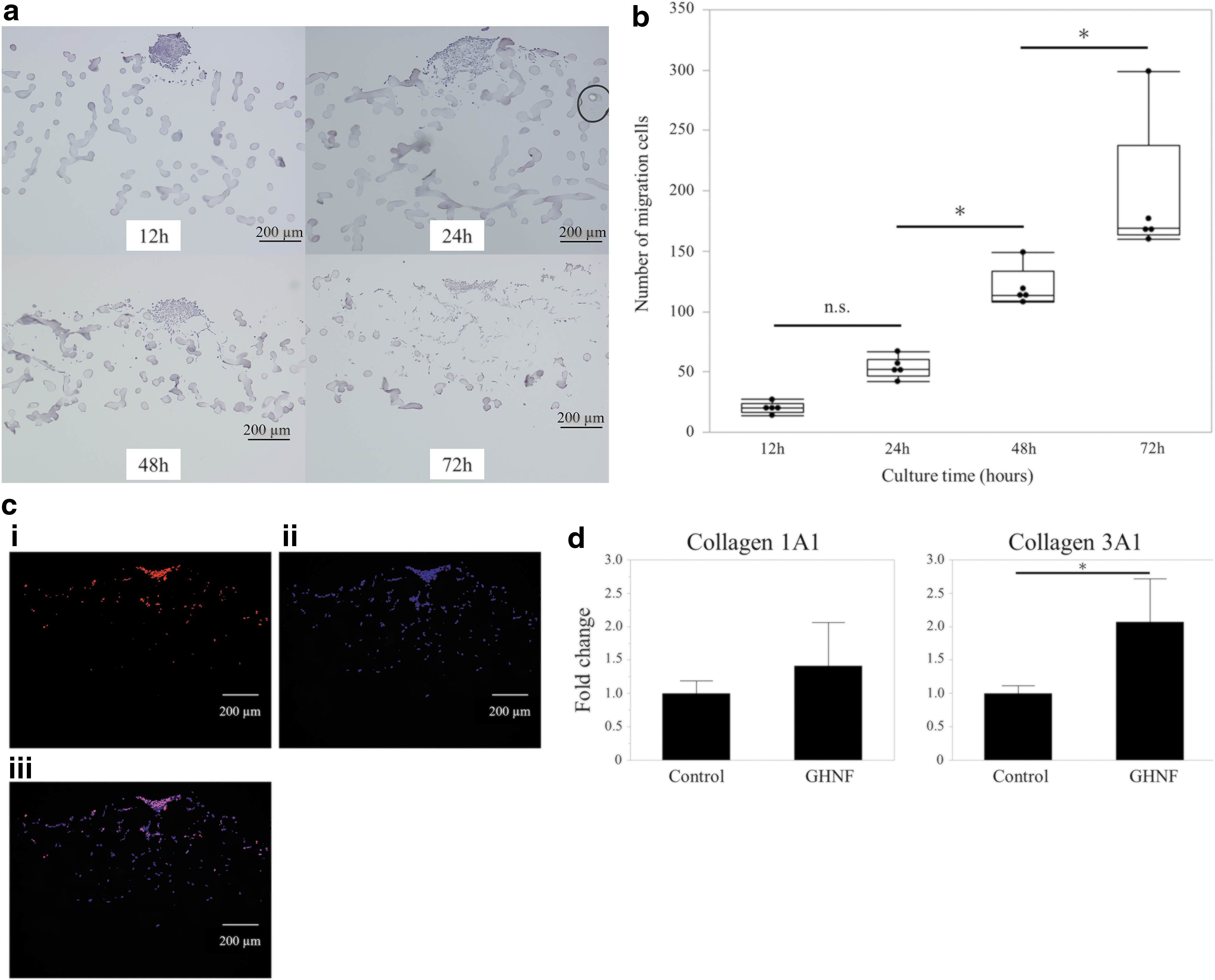
Migration and maintenance of cellular functions by murine L929 cells in the GHNF sheet.
Comparing the localization of cells cultured in GHNF and PPNF sheets
We next evaluated whether cells were present inside the GHNF sheet. PPNF sheets with similar porosity (61.4 ± 0.87%) and fiber diameter (49.4 ± 5.6 μm) were used as a control (Fig. 3a). Cells proliferated in both the GHNF and PPNF scaffolds, but there were significantly more cells present inside the GHNF sheet than the PPNF sheet (Fig. 3b, c). The diffusion limit of oxygen and nutrients made the culture conditions worse inside the scaffold than the surface. The presence of more cells inside the GHNF sheet suggested that the GHNF sheet had advantages over PPNF in terms of cell survival.
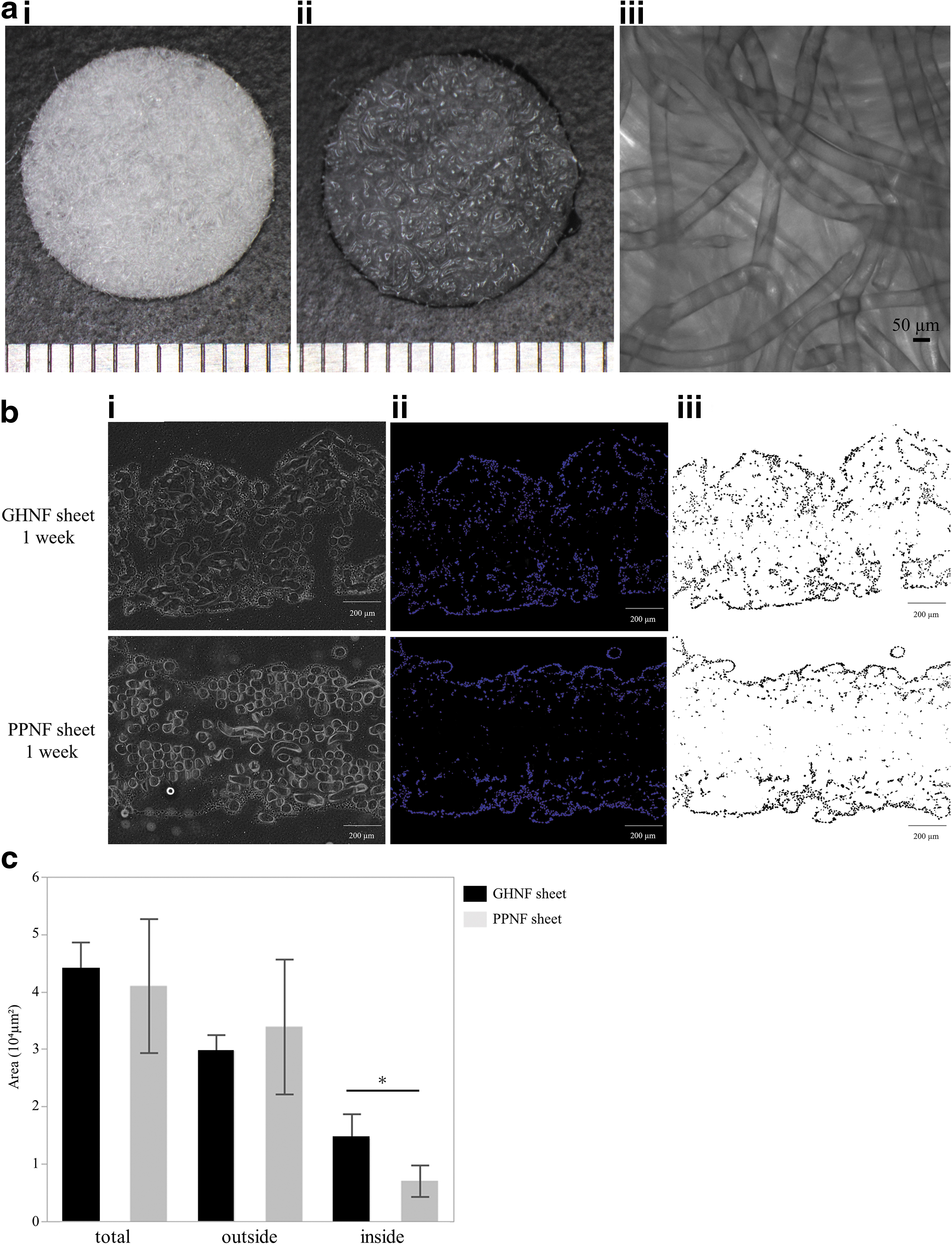
Comparison of cellular localization in the cultured GHNF and PPNF sheets.
Measuring the stiffness of GHNF sheets, ABD, and the structural parameters of ABD
After confirming the suitability of GHNF sheets as a scaffold that induces cell migration, we created an ABD using GHNF. As the GHNF sheets used in previous experiments were not strong enough for anastomosis (data not shown), the ABD was prepared with an increased gelatin density. The macroscopic appearance of the ABD is given in Figure 4a. The swollen ABD had an inner diameter of 0.8 mm and a wall thickness of 1.0–1.1 mm. When compressed with the creep meter, the ABD showed a stress of ∼8.5-fold greater than that of the GHNF sheet at 30% strain and ∼9.4-fold greater than that of the GHNF sheet at 50% strain, indicating greater strength than the GHNF sheet. The ABD was not easily torn and had sufficient strength for anastomosis (data not shown). The swelling ratio of the ABD was 4.6 ± 0.12. The porosity of the ABD was 23.6 ± 1.25%. The microscopic findings of the ABD are given in Figure 4a. The fiber diameter of the ABD was 40.6 ± 5.5 μm.
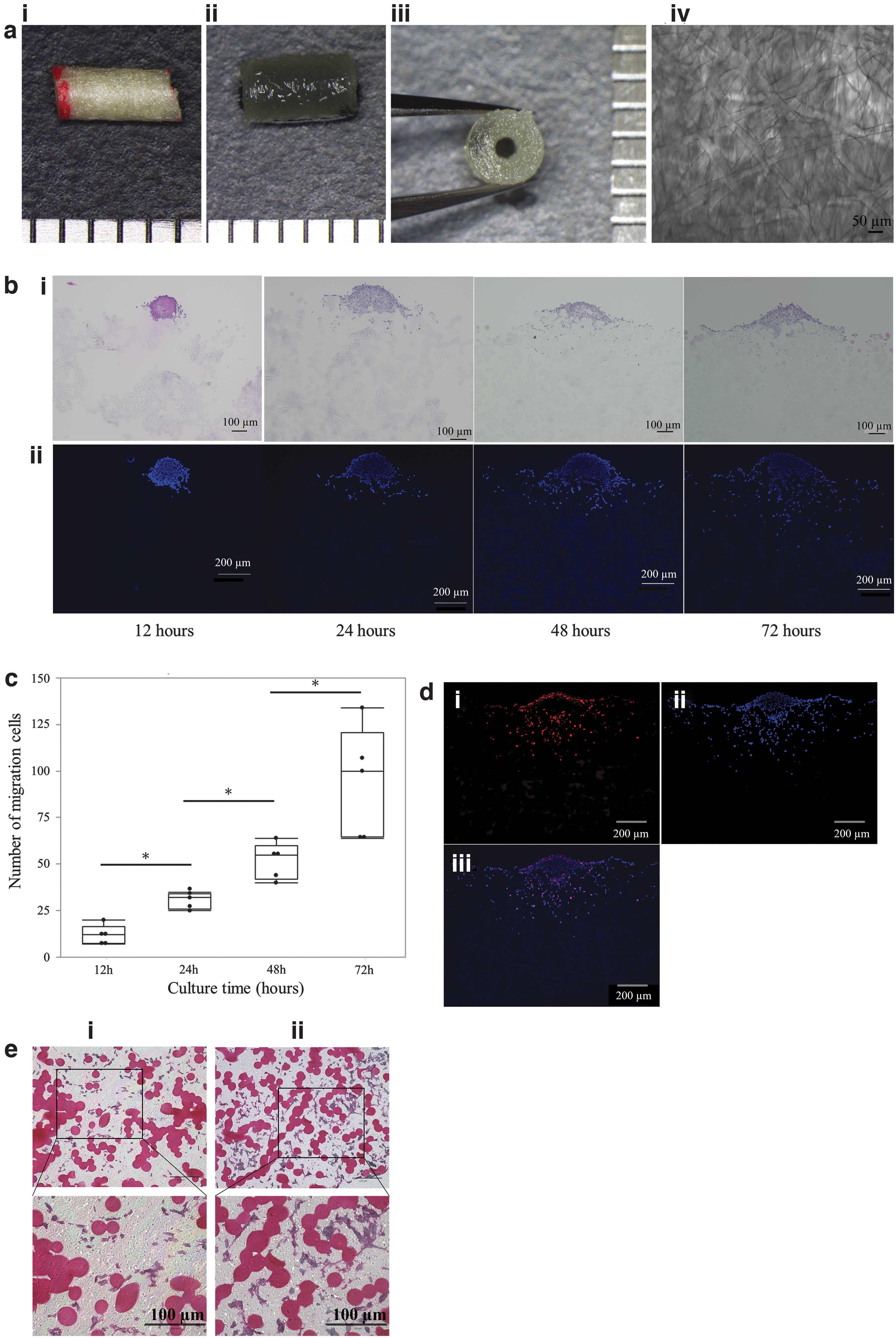
Cell migration and maintenance of cellular functions in the ABD.
Cell migration and maintenance of cellular functions in the ABD
Similar experiments were performed to verify whether the characteristics of the GHFN sheet were maintained in the ABD, which had a higher density and lower porosity than the GHFN sheet. The cell aggregate was seeded onto the ABD in the same manner as the GHNF sheet. The number of the cells that migrated into the ABD increased over time, although it was decreased compared with the GHNF sheet (Fig. 4b, c). The migrating cells in the ABD were Ki67 positive (Fig. 4d), indicating a similar proliferation ability in ABD as in the GHNF sheet. When L929 cells were cultured for 1 week in ABD, collagen secretion (positive for Sirius red staining) was observed around the cells (Fig. 4e). After culture for 2 weeks, the cells proliferated and the secreted collagen around the cells became more evident (Fig. 4e), indicating that ability to synthesize extracellular matrix was maintained in ABD.
ABD implantation model in rats
Figure 5a provides the gross appearance of the rat bile duct and a resected portion of the bile duct. ABD was interposed in place of the resected bile duct in the experimental group (Fig. 5a). General status of the rats was maintained until killing, as indicated by similar weight gain and serum biochemistry data as the sham group (Table 1).
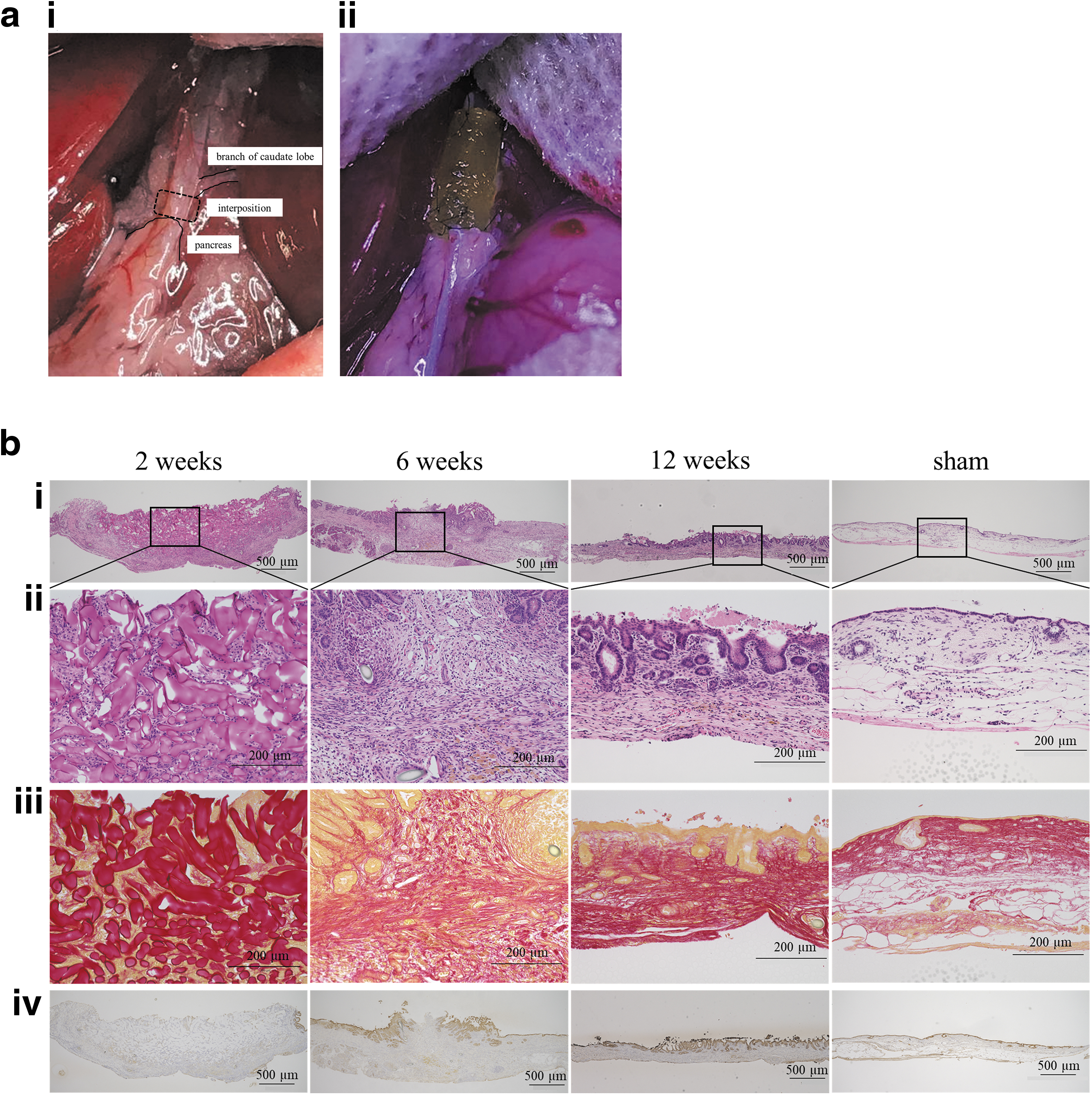
Surgical findings of ABD implantation and histopathology of the ABD implantation site and liver over time.
Weight Gain and Biochemical Values of Preoperative and 12-Week Postoperative Serum in the Artificial Bile Duct and Sham Groups
n = 5 (sham) and n = 5 (ABD); data are presented as mean ± standard deviation, Mann–Whitney U-test.
ABD, artificial bile duct; ALP, alkaline phosphatase; ALT, alanine aminotransferase; AST, aspartate transaminase; T-Bil, total bilirubin.
Figure 5b provides the histological changes at the bile duct implantation site over time. Two weeks postoperatively, Sirius red staining revealed that most of the implanted ABD remained (Fig. 5b[iii]), and cells had migrated into pores of the ABD (Fig. 5b[ii]). Cytokeratin 19 (CK19) is one of the markers specific to bile duct epithelium. 11 The ABD was mostly not covered by bile duct epithelium, as indicated by few CK19-positive epithelial cells. At 6 weeks, the ABD was mostly degraded and replaced by collagen fibers (Fig. 5b[iii]). Epithelialization with CK19-positive bile duct epithelial cells was observed (Fig. 5b[iv]). CK19-positive epithelial cells were present on both ends and deficient in the center, suggesting that epithelialization initiated at the edges and progressed toward the center. At 12 weeks, epithelialization was almost completed, and the bile duct was thoroughly regenerated. However, the bile duct epithelium showed a tall columnar shape, which was slightly different from the flat epithelium in the sham group. Epithelialization ratios at 2, 6, and 12 weeks were 10.4 ± 6.9%, 93.1 ± 5.1%, and 99.2 ± 1.6%, respectively (Table 2).
Quantitative Evaluation of Bile Duct Epithelial Regeneration
Data are presented as mean ± standard deviation.
Angiogenesis in the ABD of the rat implantation model
CD31 is expressed in vascular endothelial cells and is useful for evaluating vascular structure. 12 Two weeks after implantation, angiogenesis was observed in the ABD, as indicated by the presence of CD31-positive vascular structures (Fig. 6a, b). Angiogenesis was most intense at 6 weeks postimplantation and decreased at 12 weeks compared with 6 weeks (Fig. 6c). Capillaries of different sizes were formed at 6 weeks. On the contrary, at 12 weeks, the size difference of the capillaries was adjusted, and the thickness of endothelial cells was increased, and blood vessels with a stable structure were observed.
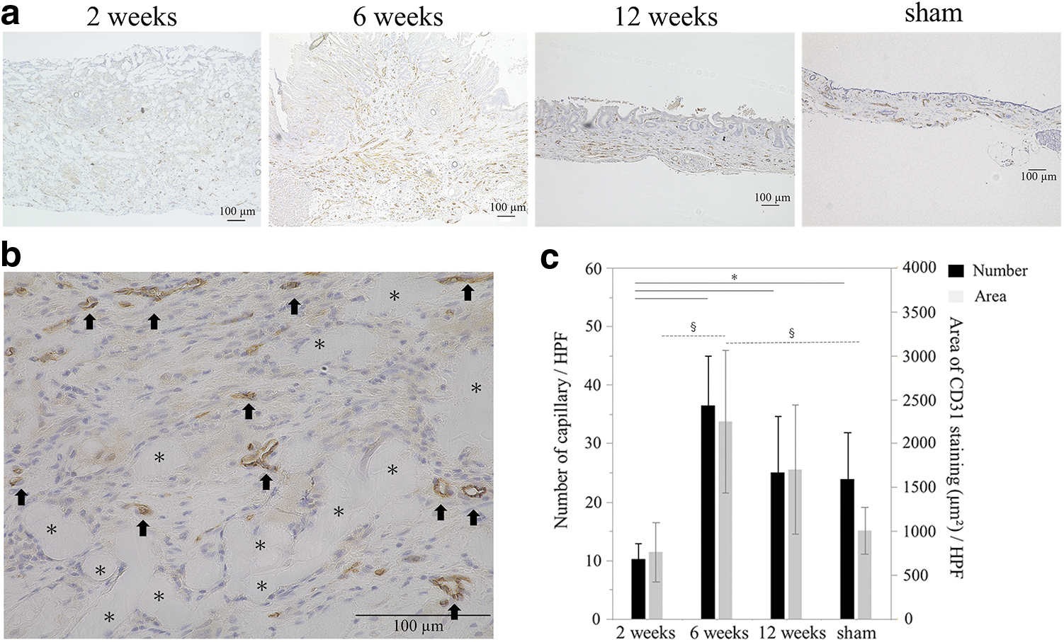
Angiogenesis within the ABD rat implantation model.
Proliferation of the biliary epithelium in the rat implantation model
Two weeks after implantation, few Ki67-positive bile duct epithelial cells were found (Fig. 7a). Many Ki67-positive cells were found at 6 weeks after implantation and were abundant in the central part of the implantation site (Fig. 7b). At 12 weeks postimplantation the number of Ki67-positive cells had decreased, suggesting that regeneration of the bile duct epithelium had passed the peak (Fig. 7c).

Proliferation of biliary epithelial cells in the rat implantation model.
Discussion
Biliary reconstruction using an ABD interposed in the excised part of the bile duct may solve various problems related to choledochojejunostomy such as reflux cholangitis. In this study, we demonstrated a successful regeneration of a neo-bile duct with an implanted ABD in rats. The process of degrading the implanted ABD and regenerating the bile duct was shown over time, which was corroborated by the capacity of the GHNF to accept cell migration in vitro.
In previous studies, various materials have been used for ABD including autologous tissue such as blood vessels and ureters,4,5 nondegradable materials such as polytetrafluoroethylene, 8 degradable materials such as polylactic acid (PLA) and collagen,6,7 and bioengineered materials. 13 Nondegradable materials have achieved limited success because they remain as foreign substances in the bile duct. 8 Autologous tissue and bioengineered materials require extra time and effort for preparation. Degradable materials that permit cell migration and gradually decompose without leaving residuals in the bile duct appear to be the most convenient and promising for these purposes.
Gelatin is a biodegradable material that is enzymatically decomposed in vivo and used as various biomedical materials owing to its high biocompatibility. 14 In previous studies, the biodegradable synthetic polymers PLA and polyglycolic acid (PGA) have been used for ABD.6,15 However, they generate acids upon degradation by hydrolysis, causing toxic pH changes.16,17 Degraded PLA particles could also cause inflammation. 18 Unlike PLA or PGA, gelatin is enzymatically degraded and does not cause inflammation. These characteristics prompted us to investigate the possibility of using gelatin as a substrate for ABD.
Fibroblasts play an essential role in wound healing by secreting extracellular matrix, especially collagen. 19 Thus the capacity of scaffolds to accept cell migration into their three-dimensional (3D) structure is assumed to be an important requirement for successful bile duct regeneration. In this study, when fibroblast L929 cell aggregates were cultured on the GHNF sheet, the cells migrated from the cell aggregate into the sheet. The number of migrating cells into the deeper portion of the GHNF sheet was higher than that of the PPNF sheet, suggesting that the GHNF sheet permitted better oxygen and nutrients supply than the PPNF sheet. 20 Cell proliferation was observed in the GHNF sheet, as shown by Ki67 staining. Collagen mRNA expression was also detected and was even higher than that of the controls, possibly because of 3D culture. It has been reported that GHNF may reduce the diffusion limit of nutrients and oxygen. 9 Because the GHNF sheet contributed to more internal nutrients and oxygen supply, there were more cells inside the GHNF sheet compared with the PPNF sheet. Our data suggest that GHNF is a promising scaffold for bile duct regeneration in terms of cell migration, proliferation, and enhanced collagen synthesis. However, the GHNF sheet had insufficient anastomotic strength; thus, we prepared an ABD with increased gelatin density. The mechanical properties of this ABD were sufficiently improved for anastomosis. The porosity of ABD decreased in exchange for the increased strength; however, cell migration was also confirmed in the ABD by the migration assay.
Few studies using biodegradable materials have evaluated the process of degradation over time. Previous studies have demonstrated regeneration of the bile duct wall or bile duct epithelium was not achieved in 2 weeks 15 and required 6 weeks at the earliest. 6 As a preliminary experiment, we subcutaneously implanted GHNF in rats to evaluate its degradability with different thermal cross-linking conditions and found thermal cross-linking at 140°C for 48 h was ideal (data not shown).
The ABD was successfully implanted in rats and appeared to function well without sign of cholestasis or liver dysfunction. Most of the past studies on ABD have shown only final status after bile duct regeneration was complete.5,8 Previous studies have not explored how the bile duct regenerated. In this study, we evaluated the degradation of the implanted ABD and regeneration of the bile duct over time. At 2 weeks after implantation, cell migration was observed in the ABD, which was in the process of degradation. At 6 weeks after implantation, the ABD was almost completely degraded, the wall of the bile duct was connected, and the bile duct epithelium appeared to regenerate from both sides of the bile duct stump. At 12 weeks after implantation, the bile duct epithelium uniformly covered the lumen. This histological transition suggested that the fibroblasts and bile duct epithelial cells necessary for bile duct regeneration had migrated from both sides of the ABD.
An adequate blood supply is essential for bile duct regeneration. 21 This study showed angiogenesis in the implanted ABD, as evidenced by CD31-positive ductular structures. This was achieved without the need for bioactive molecules, which are often used to induce angiogenesis in tissue engineering.22,23 Bioactive molecules are secreted by macrophages, neutrophils, fibroblasts, and others cell types. 24 It is speculated that the cells migrating into the ABD secrete the angiogenic factors that induce blood vessels in the GHNF. 25
An abundant expression of Ki67 in the biliary epithelium suggested interesting implications for the process of regeneration of the bile duct epithelium. Ki67 was highly expressed in the epithelium at 6 weeks after implantation and were concentrated near the center of the crypt with a tubular villous structure. This was very similar to the findings seen in the proliferative zone in the process of healing gastric ulcers. 26 Although little is known about extrahepatic bile duct homeostasis and regeneration, a study on a bile duct injury model in mice suggested that the peribiliary glands were a stem cell niche for bile duct epithelium progenitors. 27 However, this study suggests that mature bile duct epithelium at the stump of the bile duct proliferated to regenerate the bile duct and questioned the need for bioengineered ABD as reported in the previous study. 28 Our hypothesis is supported by another previous study that showed no advantage in a bioengineered ABD compared with a simple ABD prepared from a biodegradable material. 6
This study suggests that the GHNF ABD allows cells to migrate from both ends of anastomosis, which subsequently promoted extracellular matrix synthesis, angiogenesis, and epithelialization. It is possible that this series of events took place in synchronization with the degradation of ABD, resulting in successful bile duct regeneration.
There are some limitations to be acknowledged in this study.
First, the length of the ABD used in our experiments was only 4 mm. Longer defects are expected to need more time for the bile duct to regenerate, raising a concern that the ABD used in our study may decompose too quickly.
Second, in this study, the bile duct stent was indwelled until killing, so it has not been verified whether the patency is maintained after removal. We tried to establish a model in which the stent is removed after a few weeks but were unsuccessful because of technical difficulties.
Third, we did not conduct a control in vivo experiment in which another material was utilized for the ABD. It would have been most appropriate to use PPNF as a control in the ABD implantation experiments as well as in the in vitro experiments. In fact, we performed preliminary experiments of subcutaneous implantation of GHNF and PPNF in rats. GHNF was mostly degraded at 4 weeks with no scar formation around the implant and no persistent inflammation. However, inflammatory cells were infiltrated around and inside the PPNF at 4 weeks (data not shown). We thought that the PPNF-induced inflammation may be owing to its nondegradable nature, and thus it would not be suitable for ABD. Based on these findings, we did not perform a comparative experiment with PPNF in the ABD implant experiment, and the sham data were used as the control.
Fourth, in rats, the gallbladder is not present and the sphincter of duodenal papilla is underdeveloped. 29 Because of these anatomical differences, intraluminal pressure of the bile duct may be lower in rats than in humans,30,31 which may make anastomotic leakage less likely to occur in rats. Taking these limitations into consideration, verification in larger animals is needed before human testing.
Conclusion
In conclusion, we demonstrated successful bile duct regeneration in rats using an ABD made of GHNF. GHNF is a promising material for constructing ABD, and further research in larger animals is needed for future clinical applications.
Footnotes
Acknowledgments
The authors thank The Japan Wool Textile Co., Ltd (Hyogo, Japan) for providing the GHNF sheets, PPNF sheets, and ABD. The authors also thank James P. Mahaffey, PhD, and H. Nikki March, PhD, from Edanz for editing a draft of this article.
Authors' Contributions
Y.U., K.T., S.U., Y.T., and E.H. were involved in the conception and design of study. Y.U., D.N., L.X., N.H.M., Y.K., K.Y., and H.F. were involved in the acquisition of data. Y.U., K.T., T.Y., T.N., G.Y., Y.K., S.S., T.T., K.I., and Y.T. were involved in the analysis and/or interpretation of data. Y.U. and K.T. were involved in the drafting the article. Y.T. and E.H. were involved revising the article critically for important intellectual content. All authors critically revised the report, commented on drafts of the article, and approved the final report.
Disclosure Statement
Y.U. and K.T. receive research support of the providing materials from The Japan Wool Textile Co., Ltd. in this research.
Y.T. receives research support from The Japan Wool Textile Co., Ltd.
All the other authors declare that they have no conflict of interest.
Funding Information
This research was supported by the Cooperative Research Program (Joint Usage/Research Center program) of the Institute for Frontier Life and Medical Sciences, Kyoto University, the Translational Research Seeds A 113 from the Japanese Agency for Medical Research and Development, and JSPS KAKENHI (Grant No. JP20K09006).

References
Supplementary Material
Please find the following supplemental material available below.
For Open Access articles published under a Creative Commons License, all supplemental material carries the same license as the article it is associated with.
For non-Open Access articles published, all supplemental material carries a non-exclusive license, and permission requests for re-use of supplemental material or any part of supplemental material shall be sent directly to the copyright owner as specified in the copyright notice associated with the article.
