




Abstract
Calcium phosphates (CaPs) and silicate-based bioglasses have been extensively studied since the early 1970s due to their unique capacity to bind to host bone, which led to their clinical translation and commercialization in the 1980s. Since the mid-1990s, researchers have synthesized nanoscale CaP and silicate-based particles of increased specific surface area, chemical reactivity, and solubility, which offer specific advantages compared to their bulk counterparts. This review provides a critical perspective on the history and emerging trends of these two classes of ceramic nanoparticles. Their synthesis and functional properties in terms of particle composition, size, shape, charge, dispersion, and toxicity are discussed as a function of relevant processing parameters. Specifically, emerging trends such as the influence of ion doping and mesoporosity on the biological and pharmaceutical performance of these nanoparticles are reviewed in more detail. Finally, a broad comparative overview is provided on the physicochemical properties and applicability of CaP and silicate-based nanoparticles within the fields of (i) local delivery of therapeutic agents, (ii) functionalization of biomaterial scaffolds or implant coatings, and (iii) bioimaging applications.
Impact statement
This review provides a critical perspective on the history and emerging trends of the two main classes of bioceramic nanoparticles, that is, calcium phosphate (CaP) and silicate-based nanoparticles. While most reviews in literature focus on either CaP or silicate-based nanoparticles, our review evaluates both classes of bioceramic nanoparticles simultaneously. This combined review offers the opportunity to analyze differences and similarities with respect to the historic development and emerging trends within both fields of bioceramics research.
Introduction
Bioceramics are defined as ceramic materials that are biocompatible. This biocompatibility ranges from inert and nondegradable oxide ceramics such as alumina to (bio)chemically more reactive and soluble bioceramics, including calcium phosphate (CaP) and silicate-based bioglasses. These latter two have been investigated most frequently in biomedical research because of their capacity to bind to host bone, also coined as “bioactivity” by Hench. 1
The first studies on the use of CaP bioceramics for treatment of bone diseases (i.e., rickets) were reported in the 18th century (1797). 2 More than a century later the first implantation of a laboratory-made CaP formulation was reported in 1920 by Fred Albee, who used a suspension of tricalcium phosphate to repair surgically created fractures in rabbit bones (Fig. 1). 3 Almost 50 years later, hydroxyapatite (HA) powder was hot-pressed into dense and premade implants. This investigation is one of the earliest articles on the fabrication of man-made bulk CaP implants, 4 which paved the way for extensive research and development of CaP implants from the 1970s onward. These extensive R&D efforts led to the first clinical application of synthetic bulk CaP bioceramics in dentistry in 1975. 5 Since then, CaP bioceramics gradually evolved as one of the two main classes of clinically applicable bioceramics in addition to silicate-based bioceramics. These CaP bioceramics were rapidly introduced in various new clinical application areas, including bone-reconstructive surgery in dentistry and orthopedics, as well as other medical applications such as tooth pastes, dermal fillers, and formulations for soft tissue regeneration.

First reported implantation of laboratory-made calcium phosphate formulations in humans by Albee. 3
From the early 1980s onward, bulk HA blocks and coatings were used in dentistry to support bone augmentation procedures. Subsequently, HA implants were introduced in orthopedics for bone defect augmentation and coatings on metallic prostheses. 6 Commercialization of CaP (mainly HA) bioceramics in the dental and orthopedic markets was pioneered by Jarcho in the United States, de Groot in Europe, and Aoki in Japan. 2 Soon after the emergence of tissue engineering as a new research area in the early 1990s, CaP bioceramics were already introduced as scaffold materials in 1994. 7 Since then, research on CaP bulk ceramics has rapidly expanded, which formed the basis for clinical application of an ever-increasing number of CaP bioceramics in various forms such as blocks, granules, and cements.
In 1971 Larry Hench discovered that silicate-based glasses composed of SiO2, CaO, phosphorus pentoxide (P2O5), and Na2O were degradable and exhibited tissue-bonding capabilities, especially with regard to bone tissue. 1 This composition was later termed 45S5 Bioglass® and has since then been used in millions of patients to treat bone defects. Currently, bioactive glasses (BGs) are frequently used for many clinical applications such as dental fillings, orthopedic coatings, drug delivery carriers, biocomposites, and tissue engineering scaffolds.8–10
When used in association with bioceramics such as glass nanoparticles, the term “bioactivity” often refers to the capacity to form hydroxycarbonated apatite (the major inorganic component of bone) on the surface of BGs when exposed to simulated body fluids (e.g., Kokubo's simulated body fluid). The first BGs were synthesized using melt-quenching, which involves melting of oxides at elevated temperatures (above 1300°C) followed by cooling to room temperature at a very high rate to prevent crystallization and obtain a vitreous amorphous material. Siloxane bonds (Si-O-Si) form the main network of BGs, but other network formers such as P2O5 and boron trioxide can also be used. Network modifiers such as calcium and sodium disrupt these networks, thereby creating an open glass structure, which is an important prerequisite of BGs. The bioactivity and other functional properties of BGs strongly depend on their composition, where CaO-rich compositions are most frequently used as bone grafts as they promote mineralization.11,12
In the 1990s, sol–gel chemistry was first used for the production of BGs. 13 The sol–gel process is a wet-chemical approach where silica precursors (e.g. tetraethyl orthosilicate) are hydrolyzed and condensed in the presence of a catalyst and metal ions. 14 To create BGs, the materials are thermally treated after drying at lower temperatures than required for traditional melt-quenching methods (i.e., at temperatures of around 700°C). Another advantage of the sol–gel method entails the fact that organic structure-directing agents can easily be added to control the shape and dispersity of the BGs.
Generally, reviews in literature cover either CaP15–21 or silicate-based nanoparticles.12,14,22–25 The current review, on the other hand, evaluates both classes of bioceramic nanoparticles. As such, this review discusses similarities and differences with respect to the historic development and emerging trends within both fields of bioceramic research.
CaP Nanoparticles for Medical Applications
From bulk CaP bioceramics to CaP nanoparticles
From a chemical perspective, calcium (ortho) phosphates are a family of salts of the tribasic phosphoric acid H3PO4, which can form various CaP phases that contain H2PO42−, HPO42−, or PO43− ions. CaP phases containing both HPO42− and PO43− ions occur in the mineral phase of bones and teeth. Phase diagrams show the thermodynamically stable phases (e.g., mono/di/tri/tetra/octacalcium phosphate and HA) and provide an indication of the theoretical conditions required for their specific synthesis. However, the actual phase that forms under any given conditions is often dictated by kinetic rather than thermodynamic conditions. 26 HA, or more specifically carbonate apatite, is the most abundant inorganic phase in humans. 27 This abundance derives from the fact that apatites are thermodynamically the most stable CaP phase with the lowest solubility at physiological pH. The name “apatite” derives from the Greek απαταω (Eng. to deceive) since this mineral was frequently confused with other minerals such as aquamarine, amethyst, and so on. 26 Generally, the apatite structure is very tolerant for ionic substitutions. From the elements of the Periodic Table more than half can be stored in the apatite lattice, which is a unique property of CaP ceramics compared to less tolerant mineral compounds such as calcium carbonate or calcium sulfate. 28 This tolerance for ionic substitutions has been exploited to modify apatitic bioceramics with various therapeutic ions such as Ag+, Sr2+, Mg2+, or Zn2+ cations or SeO32− anions. 29
A nanomaterial can be defined as “any form of a material that is composed of discrete functional parts, many of which have one or more dimensions of the order of 100 nm or less.” However, biomedical engineering has gradually expanded this definition up to a critical component size limit of less than 1 μm. 30 Consequently, nanomaterials in biomedical research are often composed of submicron sized components, which do not meet the criteria for nanomaterials in nonmedical fields of material science. Generally, surface-to-volume ratios and dissolution rates increase with decreasing particle size. The very high surface-to-volume ratio of nanoparticles renders them much more chemically reactive than bulk CaP bioceramics, which generally display higher crystallinities and lower reactivities due to sintering treatments at elevated temperatures. However, the enhanced chemical reactivity of CaP nanoparticles also causes self-aggregation into larger particles in solutions. This self-aggregation diminishes the various benefits that are associated with nanoparticles, but aggregation can be reduced by introducing repulsive interactions (e.g., electrostatic or steric hindrance) between the nanoparticles.31,32
Biological apatites are characterized by (i) small nanosized dimensions, (ii) low crystallinity, and (iii) substoichiometry. Although the nanosized dimensions of platelet-shaped apatite crystals in bone mineral were known much earlier (platelet thickness ∼3 nm), systematic research on synthesis and characterization of dispersed CaP nanoparticles for medical use was initiated only in 1994 by pioneering groups in The Netherlands and France (Fig. 2).33,34 Since then, research on applications of CaP nanoparticles in biomedicine has rapidly expanded in view of their high surface-to-volume ratio. Generally, CaP nanoparticles offer many advantages for medical applications15–17,35–37:
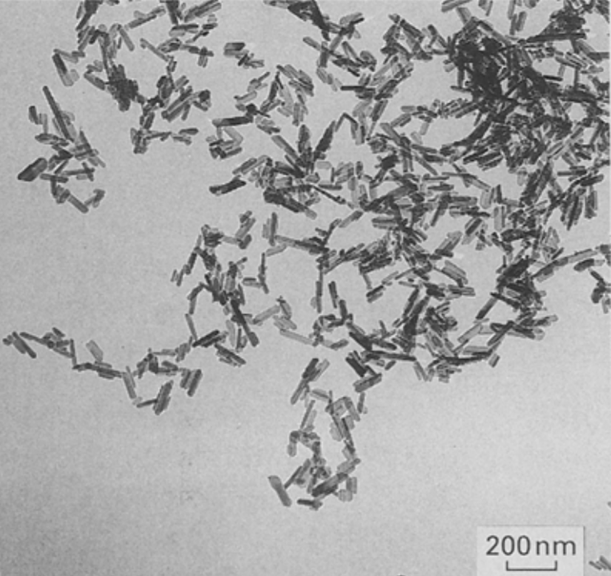
Calcium phosphate nanoparticles for medical use synthesized in 1994 by Li et al. 33
Mechanical strength and stiffness: CaP nanoparticles can be used for reinforcement of weaker polymers due to their strength and stiffness.
Low toxicity: CaP bioceramics are generally recognized as biocompatible biomaterials. Although CaP nanoparticles may display a toxicological profile that differs considerably from bulk CaP bioceramics, the toxicity of CaP nanoparticles is considered to be low. 37
Environment-friendly production: Different from many other organic and inorganic nanoparticles, CaP nanoparticles can be produced at industrial scale using green and inexpensive wet-chemical production methods that have a low environmental impact. 36
Chemical and biological reactivity: CaP nanoparticles can be used as tools to modulate cell functions such as adhesion, migration, proliferation, and differentiation.
Bioactivity: CaP bioceramics, including CaP nanoparticles, are defined as bioactive biomaterials since they are able to induce the formation of apatitic layers on their surface upon soaking in solutions that mimic body fluids. This capacity has been suggested to contribute to in vivo bonding to bone tissue, although this direct correlation is increasingly questioned. 38
High affinity to proteins and nucleic acids: proteins and nucleic acids strongly bind to CaP nanoparticles in view of their high specific surface area and chemical affinity. This favorable feature determines their biocompatibility and allows loading of therapeutic biomolecules.
Protection of sensitive biomolecules: the high affinity of proteins and nucleic acids to CaP nanoparticles offers protection against premature in vivo degradation.
Nanoparticle targeting: CaP nanoparticles can be targeted toward specific cells through active targeting mechanisms. 16
Cellular uptake: CaP nanoparticles can be internalized by various cell types (in contrast to CaP bulk bioceramics), which enable intracellular drug delivery.
Fast degradation: Compared to many other inorganic nanoparticles, CaP nanoparticles degrade faster, leading to release of calcium and phosphate ions that are abundantly present in cells and physiological fluids such as blood.
pH-dependent solubility: CaP nanoparticles are stable at physiological pH of blood (pH 7.4) but dissolve rapidly under acidic conditions as found in tumors, infected tissues, and endosomes/lysosomes. This feature renders CaP nanoparticles suitable as pH-responsive nanocarrier for (intra)cellular delivery of drugs for disease treatment.
Facile doping: Due to their extremely flexible crystal lattice, CaP nanoparticles can be easily doped with numerous dopants, which can enhance their protein adsorption capacity or increase their therapeutic efficacy by releasing antibacterial or anticancer ions.
Diagnostic efficacy: CaP nanoparticles can be functionalized with a wide range of compounds to facilitate their use in bioimaging applications.
Theranostic efficacy: CaP nanoparticles can be easily endowed with multiple functionalities, for instance, by combining therapeutic and diagnostic functionalities for theranostic applications.
Synthesis and functional properties of CaP nanoparticles
Various CaP phases, including mono/di/tricalcium phosphates, have been synthesized into nanoparticulate form, but the vast majority of studies on CaP nanoparticles deal with HA nanoparticles as this CaP phase resembles the apatitic mineral phase of bone and teeth most closely. Since the first publications on synthetic CaP nanoparticles appeared in 1994,33,34 numerous strategies toward synthesis of CaP nanoparticles with tunable properties have been developed, which have been reviewed elsewhere.15–19 Generally, CaP nanoparticles can be synthesized by either (i) wet-chemical methods such as chemical precipitation, hydrothermal treatment, sol–gel synthesis, and microemulsion synthesis or (ii) dry fabrication methods such as flame-spray pyrolysis of pulsed laser ablation. While dry methods typically produce large amounts of highly crystalline nanoparticles, these high-temperature techniques offer less control over important particle properties than wet-chemical methods. In addition, they do not allow for loading of sensitive biomolecules due to their high processing temperatures. Consequently, wet-chemical methods (involving simple reactions between calcium and phosphate precursors under aqueous conditions at low temperatures) have emerged as the most attractive approach toward synthesis of CaP nanoparticles.16,17 Advantages of CaP nanoparticle production by chemical precipitation and hydrothermal treatment include their cost-effectiveness, environment-friendliness, and industrial scalability. Sol–gel and microemulsion syntheses, on the other hand, offer more control over particle size and agglomeration due to the use of surfactants in microemulsions, which can act as templates for particle formation. 18 Usually, CaP nanoparticles are prepared from synthetic precursors, but a recent trend involves preparation of CaP nanoparticles from biogenic sources such as mammalian and fish bones or calcium carbonate derived from biogenic sources such as shells, coral, and algae. 39 This approach enables the incorporation of dopants such as carbonate, strontium, and magnesium in a simple and cost-effective manner.
The functional performance for various medical applications strongly depends on particle properties, including shape, charge, agglomeration/dispersion, and size. Therefore, extensive R&D efforts have been carried out during the past two decades to unravel the relationship between processing parameters and physicochemical particle properties. Generally, independent control over these properties is still notoriously challenging and often impossible, which complicates decoupling of the influence of single particle properties on their biological, mechanical, and pharmaceutical performance in medical applications. Nevertheless, significant progress has been made regarding dimensional control over wet-chemical synthesis of CaP nanoparticle with tunable shape, ranging from 1D geometries such as needles/rods/whiskers/fibers through 2D geometries, including plates and sheets, to 3D shapes such as spheres or even complex geometries (flowers, spherulites, dumbbells, etc.). 18 Similarly, since the zeta potential of unmodified as-prepared CaP nanoparticles is relatively low (typically < ±15 mV), 36 several adsorptive surface modification strategies have been successfully developed to increase CaP nanoparticle charge and dispersion and reduce particle agglomeration. To this end, simple adsorption of charged macromolecules, ionic dispersants (e.g., citrate), 31 or cationic lipid bilayers (lipid-coated CaP nanoparticles) 32 improves colloidal stability of CaP nanoparticles by introducing repulsive electrostatic interparticle interactions. Colloidal stability is crucial for any application of CaP nanoparticles, since extensive agglomeration of nanoparticles in general compromises the benefits associated with the use of nanoparticles due to the loss of specific surface area.
Independent control over CaP nanoparticle size has received relatively little research attention, but additives such as citrate have been successfully used also to allow for control and stabilization of particle size. 16 In addition to conventional nanoparticle properties such as dimensions and charge, properties of CaP nanoparticles such as magnetic properties have gained considerable research interest as well during the past decade. This progress has been mainly facilitated by the facile and effective manner by which CaP nanoparticles can be doped with magnetic ions such as iron. 40
Both physical and chemical properties of CaP nanoparticles determine their toxicological profile, which has been studied and reviewed extensively during the past decade.37,41 General conclusions on the effect of single particle properties are very difficult to draw due to the interrelated nature of particle properties such as size and shape. Consequently, contrasting findings on the effect of particle shape have been reported in literature. For instance, needle-shaped CaP nanoparticles were reported to have a higher toxicity toward epithelial cells or osteoblasts compared to CaP nanoparticles with other shapes such as rods and spheres.42,43 In contrast, Xu et al. found that spherical CaP nanoparticles can be more hazardous to osteoblasts than needle-like CaP nanoparticles. 44 Epple recently reviewed potential health risks of CaP nanoparticles as a function of the most relevant nanoparticle properties, including nanoparticle bulk and surface composition, size, shape, charge, dispersion, and solubility. 37 Highest in vitro toxicities were generally observed for unfunctionalized CaP nanoparticles that had a strong tendency to agglomerate. Subsequent sedimentation of micron-sized particles typically resulted in high local particle concentrations and enhanced cellular uptake, which induced cell death by excessive intracellular calcium concentrations. He concluded that CaP nanoparticles possess a low cytotoxicity, which mainly depends on the overall release concentrations of calcium ions rather than on single particle properties such as size and shape. 37
Overall, it should be emphasized that the added value of CaP nanoparticles for various medical applications depends on numerous strongly interrelated primary particle parameters (most notably composition, size, shape, charge, agglomeration, and crystallinity), the separate effects of which on functional performance are very hard—if not impossible—to decouple. These primary particle properties jointly determine their degradation rate, toxicological profile, and ultimately their overall mechanical, biological, pharmaceutical, and/or diagnostic properties. Consequently, overly simplistic statements on either “good” or “poor” performance of CaP nanoparticles should be regarded with caution, since evaluation of CaP nanoparticles for medical applications requires a scientifically more sound and sophisticated approach.
Applications of CaP nanoparticles
Based on the advantages described above, CaP nanoparticles have been used for a plethora of applications. Generally, CaP nanoparticles have made a major impact in the areas of (i) local delivery of therapeutic agents, (ii) functionalization of biomaterial scaffolds or implant coatings, and (iii) bioimaging (Fig. 3). These application areas will be separately discussed in the following subsections.
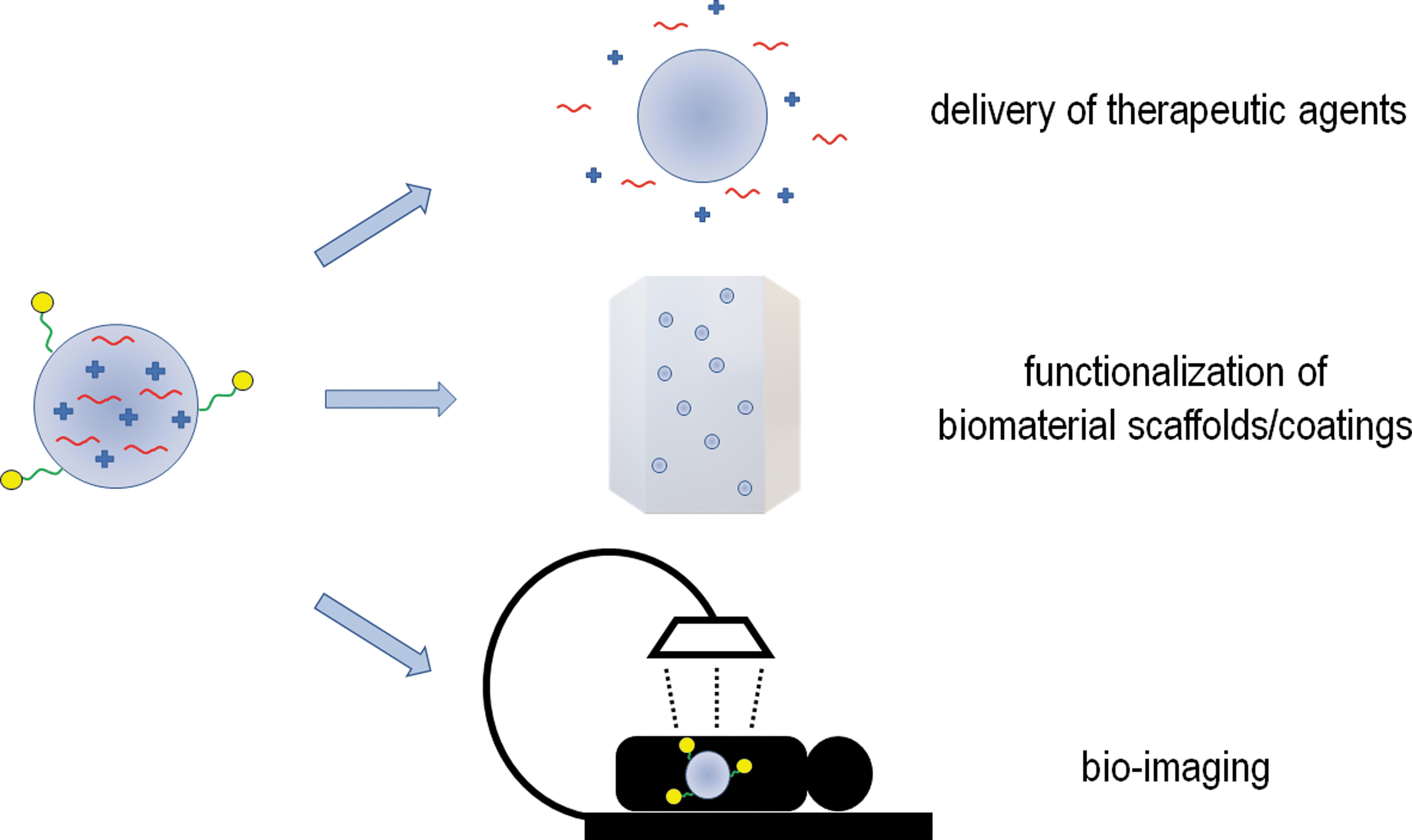
Main biomedical application areas of calcium phosphate and silicate-based nanoparticles.
CaP nanoparticles for delivery of therapeutic agents
During the past two decades, CaP nanoparticles have been mainly used for delivery of therapeutic agents. Traditionally, these therapeutic approaches focused on regeneration of bone tissue by delivery of osteogenic ions (e.g., Sr2+), proteins (e.g., bone morphogenetic protein-2 [BMP2]), peptides, nucleic acids (plasmid DNA, siRNA, microRNA), and other biomolecules.16,35,45,46 BMP2 delivery has been investigated extensively since the 2000s as this growth factor is generally recognized as the most potent osteogenic biomolecule. Nevertheless, the use of BMP2 for clinical applications has been questioned since 2011 due to safety and efficacy concerns related to overdosing due to spatiotemporally uncontrolled BMP2 delivery from commercially available collagen sponges. 47 CaP nanoparticle-mediated delivery of osteogenic nucleic acids (e.g., plasmid DNA encoding for BMP2) might overcome the risks of recombinant protein delivery. In fact, CaP nanoparticles were already successfully used for transfection of DNA in 197348 owing to their high binding affinity to nucleic acids through electrostatic interactions formed between calcium ions in CaP nanoparticles and phosphate groups in nucleic acids. Moreover, CaP nanoparticles were shown to be readily internalized by various cell types through endocytosis. However, DNA delivery faces severe obstacles toward translation into the clinic since DNA can randomly insert into the host genome, which might induce cancer. Therefore, RNA delivery (siRNA, miRNA) from CaP nanoparticles has gradually gained interest at the expense of research on plasmid DNA delivery since the 2010s.35,45,46
Generally, biomedical use of CaP nanoparticles has recently progressed beyond bone regeneration, most notably for the use of CaP nanoparticles for cancer therapy by enabling (i) cancer imaging (diagnostics), (ii) delivery of anticancer agents (therapeutics), or (iii) a combination thereof (theranostics).20,21 To maximize the therapeutic efficacy of anticancer agents from locally delivered CaP nanoparticles, targeted nanomedicine offers new opportunities by turning nonspecific nanoparticles such as as-prepared CaP nanoparticles into tumor-targeted ones. Although as-prepared CaP nanoparticles are claimed to target tumors through the Enhanced Permeation and Retention effect, this type of passive targeting is not very efficient and does not occur outside tumors, 16 so its clinical relevance is currently questioned. Therefore, various tumor-targeting strategies have been developed for CaP nanoparticles during the past decades. These tumor-targeting strategies are based on decoration of the surface of CaP nanoparticles with active tumor-targeting moieties, including antibodies and antibody derivatives, peptides, aptamers, transferrin, carbohydrates, and small organic molecules. These tumor-targeting moieties can be conjugated to CaP nanocarriers using simple adsorption or more complex conjugation strategies such as click chemistry and nucleotide hybridization. 16
CaP nanoparticles have been reported to exhibit intrinsic chemotherapeutic efficacy due to intracellular calcium release, increased generation of reactive oxygen species, and inhibition of protein synthesis following degradation of CaP nanoparticles in endolysosomal compartments.21,49,50 In addition, it was shown that this antitumor activity of HA nanoparticles increases with decreasing particle size. Nevertheless, this therapeutic efficacy of CaP nanoparticles is often not strong and specific enough to completely eradicate tumors. Therefore, the chemotherapeutic efficacy of CaP nanoparticles is usually upgraded by loading of inorganic dopants such as selenite anions 51 and/or established anticancer drugs such as doxorubicin, methotrexate, or platinum-based drugs. 16
Besides their application in cancer therapy, CaP nanoparticles are also increasingly recognized as promising candidates for treatment of bone infections.29,52,53 Their antibacterial activity can be attributed to (i) the buffering effect against infection-induced acidity, (ii) release of antibacterial dopants such as Ag+, Cu2+, Zn2+, or SeO32−, (iii) release of antibacterial biomolecules such as antibiotics and antimicrobial peptides, or (iv) codelivery of multiple antibacterial agents. In addition, CaP nanoparticles have also been successfully used for vaccination and immunization by binding antigens and/or adjuvants efficiently to their surface, thereby inducing immunomodulatory effect binding.17,54
Overall, we envisage that the scope and number of pharmaceutical applications of CaP nanoparticles will significantly broaden during the coming decades due to their evident benefits for controlled delivery of numerous therapeutic agents. As such, the highly versatile nature of CaP nanoparticles will undoubtedly open up entirely new application areas beyond traditional hard tissue regeneration, such as treatment of heart failure by means of peptide-loaded CaP nanoparticles. 55
CaP nanoparticles for functionalization of coatings and bulk biomaterials
CaP nanoparticles have been used as component for 2D coatings and 3D bulk biomaterials for hard tissue replacement in dentistry and orthopedics for more than three decades aiming to (simultaneously) improve their mechanical and biological performance.
Numerous approaches have been explored to deposit coatings composed of CaP nanoparticles onto implant surfaces, including techniques such as electrophoretic deposition, 56 electrospraying,57,58 discrete crystalline deposition, 59 and immersion deposition.60,61 Favorable effects of such CaP nanoparticle implant coatings prepared by immersion deposition and subsequent heating were reported by the Wennerberg group in Sweden.60,61 However, other studies by the Jansen group in The Netherlands did not observe such beneficial effects of implant coatings made of electrosprayed CaP nanoparticles.62,63 These contrasting results emphasize that the efficacy of CaP coating techniques should be based on thorough characterization of coating properties such as coating adhesion, crystallinity, solubility, and roughness.
Although CaP nanoparticles can be directly applied to bone defects in highly agglomerated pastes and putties, these pastes lack mechanical strength and confinement. By combining a tough and viscoelastic polymeric matrix with hard and stiff particulate inorganic fillers, 3D composite biomaterials can be produced combining the best of both worlds. Generally, since ceramics are brittle as indicated by their low tensile strength, HA should be used in nanoparticulate instead of fibrous form. 30 Similar to bone, where the dispersed apatitic phase is present as 2D-platelet nanoparticles, irregularly shaped HA nanoparticles offer the highest degree of reinforcement of composite biomaterials due to enhanced mechanical entanglement with surrounding polymer chains. 64 Biologically, the limited osteocompatibility of most polymeric matrices can be overcome by incorporation of osteogenic CaP nanoparticles into these polymeric matrices. These nanoparticles improve the osteogenic capacity of the resulting polymers by (i) enhancing protein adsorption, (ii) increasing surface roughness and hydrophilicity, (iii) releasing calcium and phosphate ions, and (iv) stiffening composite matrices. All of these factors are known to stimulate bone formation by stimulation of stem cell differentiation into the osteogenic lineage.65–67
CaP nanoparticles for bioimaging applications
With the advancement of modern imaging modalities such as computed tomography (CT), fluorescence imaging, magnetic resonance imaging (MRI), positron emission tomography (PET), and single photon emission computed tomography (SPECT), the functionalization of CaP nanoparticles with traceable probes to enable bioimaging has emerged since the 2000s as a new application area.17,50,68 Owing to their facile doping and/or surface functionalization, CaP nanoparticles have been functionalized with fluorescent lanthanide ions (Ce3+, Eu3+, Tb3+, Yb3+) or fluorescent organic dyes (e.g., fluorescein isothiocyanate, indocyanine green) to allow for fluorescence imaging of CaP nanoparticles in vitro and in vivo.16,50 In addition, these fluorescent CaP nanoparticles can also be used as cell-labeling agent. However, optical labeling methods are limited due to poor tissue penetration, even in the near-infrared region. Therefore, CaP nanoparticles have also been functionalized with PET radioisotopes 18 F- (replacing hydroxyl groups in the apatitic crystal lattice) or 68 Ga (covalently coupled to HA-binding groups such as bisphosphonates 68 or the chelator dodecane tetraacetic acid). 69 PET-CT imaging is a highly valuable tool to monitor biodistribution of systemically or locally administered CaP nanoparticles. In addition, CaP nanoparticles have been functionalized with 99mTc-radioisotopes to study their biodistribution using SPECT-CT imaging. Both of these nuclear imaging techniques revealed that CaP nanoparticles are often found in the lungs, liver, and spleen following intravenous injection.17,68,69 Interestingly, citrate-functionalized CaP nanoparticles did not accumulate in the lungs but were mainly detected in the liver and spleen, which was attributed to their enhanced colloidal stability. 68
The extremely flexible crystal lattice of CaP nanoparticles also allows to simultaneously functionalize them with both therapeutic ions and biomolecules, as well as probes for bioimaging. The resulting theranostic nanoparticles combine therapeutic with diagnostic potential. For instance, CaP nanoparticles were successfully doped with silver (Ag), gadolinium, and iron ions to enhance their antibacterial efficacy and allow for multimodal CT and MRI bioimaging. 70 To this end, lipid-coated CaP nanoparticles are particularly attractive for the development of theranostic nanoparticles since these particles can be functionalized using their CaP core, as well as their lipid coatings. 71
Miscellaneous applications of CaP nanoparticles
CaP nanoparticles have been mainly used in three main medical application areas as highlighted above, that is, (i) delivery of therapeutic agents, (ii) functionalization of coatings and bulk scaffolds, and (iii) bioimaging. Besides these traditional medical application areas, miscellaneous applications of CaP nanoparticles include the following.72–76
Dentistry and oral care72,73: CaP nanoparticles are frequently used in dentistry and oral care as remineralizing, desensitizing, and anticaries agents in products such as bleaching gels and tooth pastes. These applications rely on the well-known capacity of CaP nanoparticles to repair minute defects in enamel, occlude dentinal tubules, stimulate remineralization, and prevent demineralization of teeth.
Skin care74,75: CaP nanoparticles are used for skin care in (i) sunscreens (to protect against ultraviolet radiation), (ii) skin cleaners (to remove sebum, dead skin, dirt, and bacteria), (iii) topical drug delivery (to limit skin penetration of drugs), and (iv) cosmetics (to improve their functional properties such as biocompatibility, color, adhesiveness, and smoothness).
Hair care 74 : CaP nanoparticles are increasingly considered as delivery vehicle for dyes in hair care products due to their capacity to encapsulate water-insoluble compounds such as dyes.
Deodorants 74 : Owing to their strong adsorptive capacity, CaP nanoparticles have also been applied as effective adsorbent for malodorous compounds to improve the deodorizing efficacy of deodorants.
Soft tissue regeneration 76 : CaP bioceramics have been mainly used in orthopedics and dentistry for applications involving contact with hard tissues, but these nanoparticles can also be applied in contact with soft tissues upon (i) cancer therapy (as delivery vehicle for anticancer drugs), (ii) wound repair (as delivery vehicle for soft tissue-regenerative ions or drugs), (iii) hemostasis (as delivery vehicle for antifibrinolytic drugs), (iv) blood transfusion (as cryoprotectant for erythrocytes), and (v) peripheral nerve regeneration (as source of calcium ions which are required for axonal regeneration).
Silicate-Based Nanoparticles for Medical Applications
From bulk bioglass to bioactive glass nanoparticles
Sol–gel mediated controlled synthesis of BGs was initiated in the 1990s and led to the development of bioactive glass nanoparticles (BGNs) with sizes ranging between 20 and 800 nm, where the synthesis conditions (e.g., pH, solvent, and so on) can be used to modulate BGN morphology and size. 14 BGNs exhibit attractive properties for various biomedical applications due to their large surface area and small size. Specifically, they are small enough to be internalized by cells allowing intracellular release of therapeutic ions for drug delivery applications. Their small size also renders BGN promising building blocks for incorporation within hydrogels to create composite materials with improved mechanical properties and enhanced protein adsorption.77,78 Moreover, the larger surface area of BGNs has enhanced their bioactivity by accelerating degradation and ion release compared to BGs. 14 This fast ion release can also effectively promote tissue regenerative processes such as osteogenesis and angiogenesis.79,80 In summary, BGNs are particularly attractive for bone regenerative applications since they (i) degrade in a controlled manner, (ii) exhibit bone-bonding properties, and (iii) promote tissue regeneration.
Mesoporous bioactive glass nanoparticles
Until the early 1990s, only dense silicate-based nanoparticles were available for biomaterial research. In 2004 the sol–gel method was first used together with organic structure-directing agents (i.e., surfactants) to form mesoporous bioactive glass nanoparticles (MBGNs). 81 MBGNs are based on compositions such as SiO2-CaO, SiO2-P2O5, or CaO-SiO2-P2O5 and contain a highly ordered mesoporous structure with pore sizes ranging from 5 to 20 nm (Fig. 4). 82 Like BGNs, MBGNs themselves can affect regenerative cellular processes such as differentiation and angiogenesis, primarily by the release of its ionic components (namely silica, calcium, and phosphate). A major challenge in the preparation of MBGNs involves the fact that the synthesis protocol, composition, and type of precursors all influence the incorporation of ions in the glass matrix, which determines particle shape, size, and morphology. 83 For example, a recent systemic study comparing 24 different glass compositions concluded that formation of ordered mesopores requires a molar silica content of 60% or higher, independent of the amount of CaO and P2O5 present in the glass. 84 Another study investigated how addition timing of calcium precursors affected the morphology, dispersity, and composition of BGNs and concluded that delayed addition of the calcium precursor resulted in more regular, homogenous, and better dispersed nanoparticles. 85 However, a persistent challenge in this field remains compositional control for both MBGNs and BGNs; there is a discrepancy between the intended composition and the actual one obtained after the nanoparticle synthesis, where ion incorporation levels are consistently lower than intended. This is problematic since released phosphorus, calcium, and additional ion species are responsible for the regenerative efficacy of the nanoparticles. A systematic study which carefully reviewed relevant synthesis parameters such as solvent selection, stirring, and the order of reagent additions has led to optimized synthesis, which can overcome this discrepancy. 86
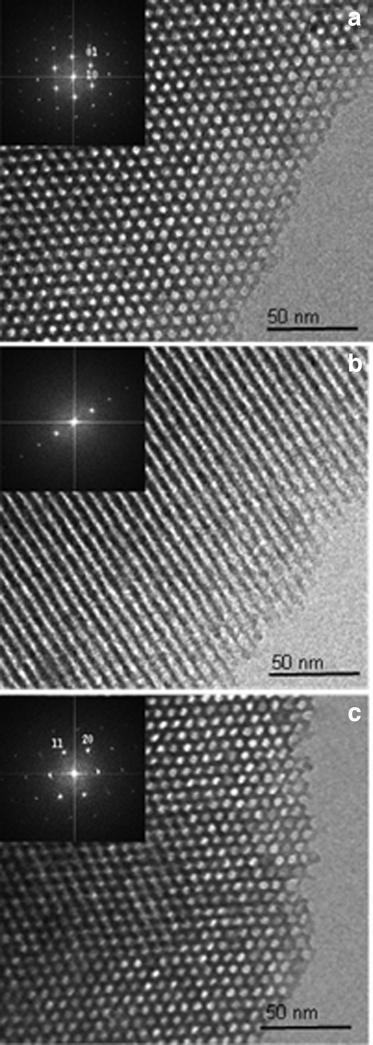
TEM images and Fourier transform patterns of MBGNs synthesized in 2006 for biomedical purposes recorded
Although MBGNs are used in many biomedical applications, MBGNs have made the biggest impact in the field of drug delivery. 24 This popularity is attributed to the fact that the MBGN matrix can be easily modified with therapeutic ions, while their mesoporous structure can also be simultaneously used for codelivery of (organic) drugs. Similar to other BGs, MBGNs are particularly suitable for application in the bone regeneration field. Their large surface area and high pore volume further enhance their surface reactivity, improving their bioactivity with respect to apatite mineralization and bone-bonding properties compared to conventional BGs and BGNs. Moreover, like BGNs, MBGNs themselves can affect regenerative cellular processes such as differentiation and angiogenesis, primarily by the release of its ionic components (namely silica, calcium, and phosphate). 24
Their use for the delivery of bioinorganic ions and (co)delivery of biomolecules in bone tissue engineering and their application in other areas, including soft tissue regeneration and tumor therapy, will be separately discussed in the following subsections.
MBGNs for delivery of therapeutic agents
Recent trends in the field of MBGN research involve doping of the silica matrix of MBGNs with additional bioactive ions to improve their bone regenerative capabilities. Examples of such ions include lithium, strontium, zinc, and iron, which are commonly incorporated as inorganic salts together with MBGN precursors. 24 Ion incorporation often leads to synergistic effects in the osteogenic capabilities and bioactive behavior of MBGNs, although the biological processes they stimulate may differ. For example, a recent study comparing MBGNs doped with manganese, copper, or zinc showed that while manganese had a small therapeutic window, copper doping improved bone marrow stromal cell (BMSC) viability and increased alkaline phosphatase (ALP) activity to nondoped MBGNs. 87 In contrast, zinc doping of the same MBGNs led to an upregulation of extracellular matrix related genes. Recently, emerging trends also focus on other types of ions in the silica matrix, such as cerium, gallium, and boron, to endow MBGNs with additional features such as antioxidant or immune-modulatory properties. 24 Ion incorporation, however, further complicates MBGN synthesis, since it can lead to disorganization of the mesoporous structure and impair control over particle size and shape.88,89 Moreover, MBGN glass composition and the extent of ion loading also impact, to a large degree, the degradation and in situ mineralization capabilities of the MBGNs. Consequently, bioactive and regenerative properties of MBGNs are largely intertwined. 84 Although synthesis and processing of MBGNs are extremely versatile and considerably optimized during the past decade, clear limitations have also been identified regarding incorporation of ions since excessive ion incorporation might compromise MBGN morphology, shape, dispersity, and composition. Regardless, their clinical relevance is evident as there are many studies reporting on the positive effects of MBGNs on regenerative processes. To ensure their future clinical use, it is however of crucial importance that protocols will be developed that ensure sufficient control over MBGN composition, shape, and dispersity upon synthesis at industrial scale.
To further improve their regenerative potential, the mesopores of MBGNs can be used to carry and release organic biomolecules such as growth factors, anti-inflammatory agents, or antimicrobial agents. 24 Several studies have reported on such dual use of MBGNs, where therapeutic ions are incorporated in the matrix and the mesopores are loaded with additional organic biomolecules. For example, MBGNs have been used to deliver genetic material such as siRNA or BMP2 plasmid DNA delivery to effectively promote osteogenesis.90,91 To review all MBGN ion doping and drug delivery strategies is outside of the current scope of this review since these strategies have already been excellently reviewed elsewhere, 24 but the breath of examples clearly demonstrates the potential use of MBGNs in (bone) regenerative therapies. Most of the research focusing on MBGNs study their application as single particle suspensions, but other applications have been explored as well. For example, MBGNs and BGNs have been used as coatings on bioinert metals or ceramics to improve the mineralization and bone bonding capabilities. 10 MBGNs and BGNs have also been combined with other biomaterials, such as polymers, hydrogels, and ceramics, to develop composite materials with additional properties or improved functionalities. 92
Miscellaneous applications of MBGNs
The use of MBGNs is currently being expanded to other fields such as soft tissue regeneration, cancer therapy, and nanomedicine, mainly owing to their strong potential for drug delivery. For example, MBGN matrix can be doped with elements that have antitumor efficacy while simultaneously loading the mesopores with chemotherapeutic agents for cancer treatment. Examples include calcium doped MBGNs showing synergistic anticancer activity with doxorubicin, a known chemotherapeutic. 93 Due to such ion incorporation, the nanoparticles exhibit increased biodegradability, which further improves their efficacy. A more recent trend includes the development of magnetic MBGNs by iron doping for hyperthermia treatment of tumors. 94 These magnetic MBGNs are exposed to an alternating magnetic field to generate heat, creating localized thermal damage in the tumor region. After tumor removal, the bioactivity of MBGNs can stimulate bone regenerative processes, providing these nanoparticles with a multifunctional purpose. Other recent applications include the incorporation of radioactive lanthanides for in situ tumor radiotherapy 95 and bioimaging applications by the incorporation of rare earth elements, such as europium, which render the nanoparticles luminescent. 96 Finally, the use of ion-doped MBGNs as antibacterial agents is actively being investigated due to increasing number of antibiotic-resistant bacterial strains. MBGNs incorporating metal ions with well-known antibacterial properties, such as silver, copper, and zinc, have led to multifunctional constructs for antibacterial application. 25
The use of MBGNs for soft tissue regeneration is still in its infancy, which is remarkable in view of their biocompatibility and potential to deliver biomolecules such as growth factors and genetic material together with therapeutic ions. Promising examples include the use of MBGNs for wound healing and skin regeneration. In this study, ion release was used to stimulate angiogenesis, modulate the inflammatory response, or exert antibacterial effects.97–99 On the contrary, the use of macroscale BGs in soft tissue regeneration is already generally established, as reflected by applications in wound healing, muscle, and nerve repair, as well as cardiac regeneration. 100 Commercial products containing silver doped phosphate glass have already been marketed as dressing for wound healing (Antimicrobial Arglaes® film, Antimicrobial Arglaes Island; Medline) or topical powders to prevent infections (Arglaes powder; Medline). The advantages of using BGNs for soft tissue engineering are evident; the increased bioactivity and faster release of ions compared to BGs can be beneficial for several approaches. Some reports already showed favorable properties caused by nanosized BGs in soft tissue regeneration applications. For example, ointments incorporating BGNs healed wounds more quickly and efficiently compared to similar ointments prepared with commercially available microsized Bioglass particles. 101 In combination with polymers to obtain composite devices for peripheral nerve regeneration, the use of nanofibers can improve the mechanical properties of the polymer and release of therapeutic ions to enhance nerve repair. 102 Evidently, BGNs and MBGNs show strong promise for use in soft tissues.
Mesoporous silica nanoparticles
Mesoporous silica nanoparticles (MSNs) were first synthesized in 1992 for nonbiomedical application areas by Mobil Research and Development Corporation from aluminosilicate gels. Unlike (M)BGNs, MSNs are purely silica-based nanoparticles with typical sizes ranging between 20 and 200 nm. Various particle morphologies have been synthesized (spheres, ovals, rods), while their internal structure is mesoporous (Fig. 5).22,23,103 Their synthesis route generally involves the hydrolysis and condensation of silica precursors in the presence of a micelle template, which acts as a structure-directing agent. After template removal, silica nanoparticles are obtained with a uniform and highly ordered mesoporous structure (diameters that range from ∼2 to 10 nm) and an extremely large surface area. MSNs allow close control over surface and core shell functionalizations; functional groups can be introduced within the nanoparticles by condensing different silica starting products in a layer-by-layer approach. This feature renders MSNs highly versatile for postfunctionalization—and as such suitable for drug delivery and bioimaging applications. Their popularity also derives from their large pore volume, which provides space for biomolecule loading, as well as their large surface area, which can be selectively modified to enable responsive drug delivery and cellular targeting. 23
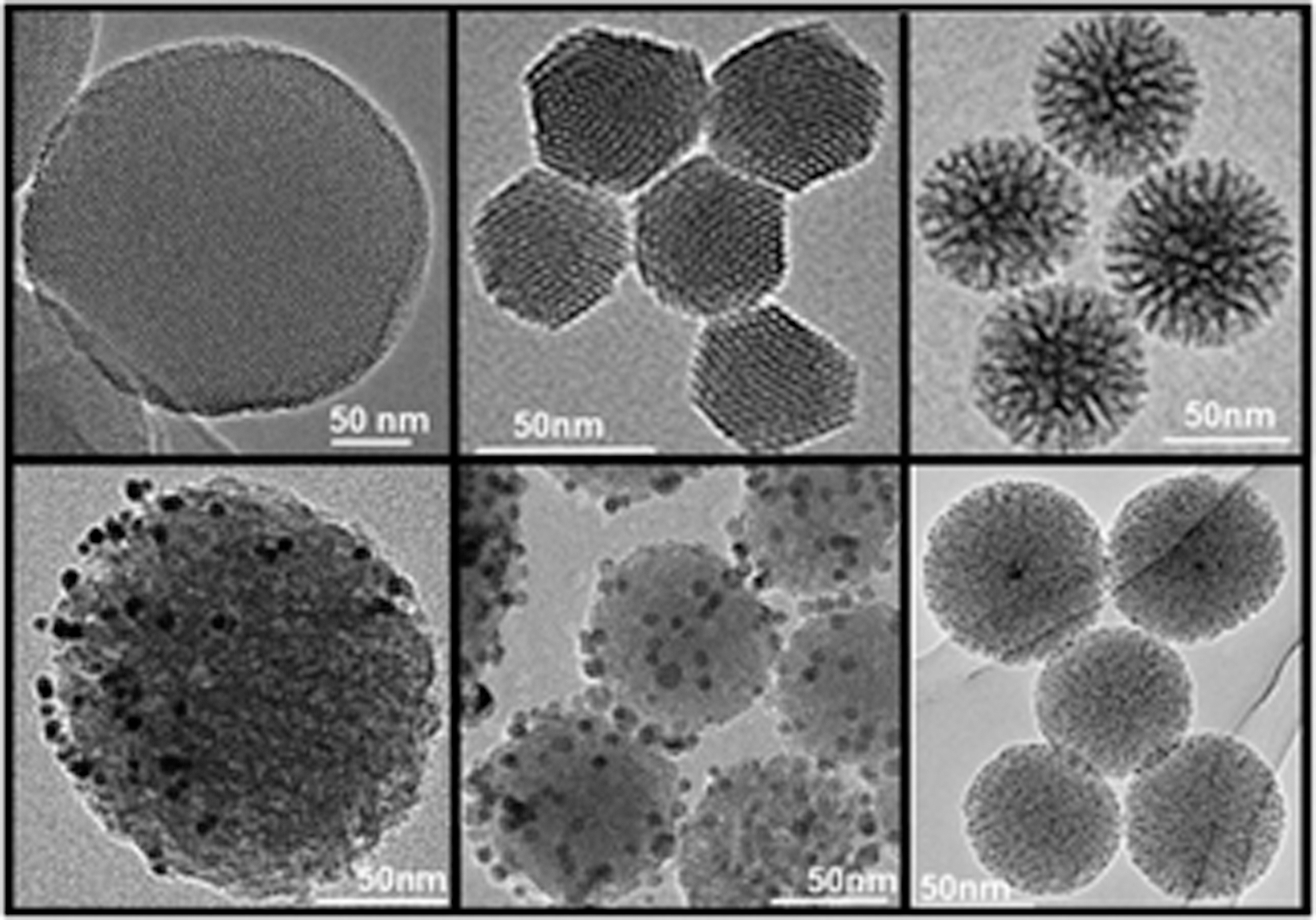
TEM images of MSNs with different sizes, morphologies, and pore sizes (upper panel) and MSNs functionalized with metal nanoparticles (lower panel). 23 MSN, mesoporous silica nanoparticle.
The vast majority of studies on MSNs focus on their use for targeted delivery of therapeutics, particularly in the cancer research field. 104 Currently, applications beyond cancer are actively explored as well. For instance, MSNs are increasingly considered for use in the field of regenerative medicine. MSNs display an intrinsic bioactivity; silica ions that are released due to MSN degradation are claimed to promote regenerative processes such as osteogenesis and angiogenesis, 105 but also nonbiodegradable MSNs exhibit osteogenic and angiogenic properties.106–108 Due to these intrinsic properties and their “hard” physical appearance, most applications within regenerative medicine focus on the development of MSNs for bone or cartilage regeneration, but other application areas are also being investigated. Generally, MSNs have made their main impact on the biomaterial community for (i) delivery of therapeutic agents, (ii) functionalization of biomaterial scaffolds or implant coatings, and (iii) bioimaging (Fig. 3). Each of these three application areas will be separately discussed in the following subsections.
MSNs for delivery of therapeutic agents
MSNs have made the strongest impact on the regenerative medicine field as versatile platforms for biomolecule delivery. Therapeutic agents such as growth factors, ions, DNA, peptides, or other biomolecules are promising strategies to promote tissue regeneration. However, direct administration of these therapeutics is associated with many drawbacks, including nonspecificity, degradation, and poor cellular uptake. MSNs can improve the pharmacokinetics of such agents by allowing the incorporation of large amounts of cargo within their core structure, providing protection from degradation. In addition, MSNs can be easily surface modified to allow tissue targeting and controlled drug delivery, leading to lower effective doses and lowered risk of adverse effects. Like BGs, the silica matrix itself can be used to deliver therapeutic ions. Although the number of articles on ion doping of MSNs is still minor compared to the BG community, several reports describe doping of the MSN silica network with ions, including strontium, lithium, europium, or copper for tissue regeneration purposes.109–112 Inorganic ion doping in the silica matrix of MSNs has also been used to modulate the immune environment by internalization of immune cells and subsequent favorable modulation of cytokine secretion.113,114 While BGNs and MBGNs are biodegradable without any ion doping, MSNs are generally quite stable and require network impurities or responsive bonds in the silica matrix to allow (faster) biodegradation. 115 Ion doping can function as a network impurity, thereby increasing the biodegradability of MSNs and release of therapeutic ions. This was recently demonstrated for calcium-doped MSNs, which allowed fast dissolution at lower pH as found in endosomes, facilitating cytosolic delivery of genetic material. 116 Ion doping of MSNs is advantageous since these ions can be doped within the silica matrix while maintaining their mesoporous structure and surface chemistry, thus allowing encapsulation of additional biomolecules and simultaneous modification of surface chemistry.
MSNs allow for simultaneous delivery of multiple biomolecules, which can enhance their therapeutic efficacy. For example, dual delivery systems based on dexamethasone and BMP2 could significantly stimulate osteoblast differentiation and promote new bone synthesis in vivo.117,118 This approach has also been used to create biodegradable MSNs that stimulated both osteogenesis and angiogenesis processes. 119 In these approaches, the MSNs are surface modified with a protein or peptide, where the relatively small osteogenic molecule is incorporated in the mesopores. The larger biomolecule (e.g., BMP2) is attached to the MSN surface in view of the small pore size within MSNs (typically between 2 and 10 nm), restricting the size of the cargo that can be incorporated in the mesopores. Using the surface to carry larger biomolecules can have additional advantages such as the promotion of cellular uptake. 120 Nevertheless, this strategy also comes along with disadvantages, including the fact that the surface- attached biomolecules are more prone to degradation, while additional surface modifications (e.g., for cell targeting) are hard to perform effectively. Methods to enlarge the mesopores or create a hollow core to allow efficient encapsulation of proteins or genetic material represent another possibility to deliver large biomolecules, which has been explored within the cancer therapy field, 121 but yet to a much lesser extent in regenerative medicine. Moreover, new developments are frequently reported in other fields of nanomedicine such as gatekeeping properties to allow control over drug release kinetics or targeting ligands for cell and tissue targeting. These developments are still early stage within the field of regenerative medicine. When this knowledge will become fully utilized in regenerative medicine, we anticipate that MSNs can make a more pronounced contribution to this field by allowing controlled, on-demand, and targeted tissue delivery of biomolecules that can guide tissue-regenerative processes.
MSNs for functionalization of coatings and bulk biomaterials
Within regenerative medicine, MSNs are also greatly contributing to the development of composite biomaterials and implants used for replacement, regeneration, and repair of damaged tissues. MSNs can be immobilized on surfaces such as implants mainly to promote tissue integration or reduce the risk for infections. Several approaches demonstrate the added value of coatings comprising MSNs to alter surface chemistries and to allow local and sustained cargo delivery to steer the behavior of attached stem cells, osteoblasts, or osteoclasts.122–124 The incorporation of MSNs within other biomaterials such as polymers, hydrogels, and ceramics can also be used to create composite materials with modified properties. The main benefit of incorporating MSNs is to improve the mechanical properties of mechanically weak polymers, expanding their applicability to stiffer organs and tissues.125–134 MSNs are generally incorporated during hydrogel formation, where they act as inert filler components in the polymeric network or as physical crosslinkers through interactions of the polymer with the MSN surface. In such materials, the nanoparticles do not actively take part in the hydrogel network formation, but rather act as structure reinforcements where higher MSN amounts lead to mechanically stronger hydrogels. Several studies have investigated the incorporation of MSNs as covalent crosslinkers, which however reported contrasting results. Where MSN-assisted crosslinking significantly improved the mechanical properties compared to adding MSNs as filler components, 135 another study reported that MSN crosslinking led to reduced mechanical properties compared to the pristine hydrogel. 126 As such, it should be realized that the polymeric backbone, method of incorporation, as well as amount of MSN, jointly determine the effect of MSNs on the overall mechanical properties of the nanocomposites. Consequently, these parameters should be carefully studied in future.
Incorporation of MSNs in biomaterials can also improve the adhesion, spreading, and metabolic activity of encapsulated cells. Several studies have reported that incorporating MSNs into the polymeric matrix can enhance cellular adhesion and spreading. For example, Yang et al. reported that the incorporation of MSNs into PEGDA hydrogels increased rat bone marrow stem cell adhesion and spreading, 132 and Gaharwar et al. showed that PEG hydrogels containing MSNs improved cell adhesion and spreading in a dose-dependent manner. 127 To further enhance the bioactivity of the nanocomposites, currently explored directions in this field include MSN-based delivery of antibiotics, cytokines, or osteogenic molecules.126,134,136,137 In summary, this field is still in its infancy where investigations mainly focus on the relationship between MSN incorporation and resulting mechanical and/or biological performance. In future, this specific field should confirm its impact on various application areas within regenerative medicine. Areas of high potential impact include the use of MSN nanocomposites as self-healing injectables, tissue adhesives, and as bioprinted scaffolds for application in stiffer organs and tissues.
MSNs for bioimaging applications
The use of MSNs for bioimaging applications (including cell tracing) is a third major application area within regenerative medicine. The ability to trace and characterize cells in vivo has received considerable attention in recent years. MSNs can be structurally modified to be used as effective stem cell tracking tools as they are stable, exhibit unique optical properties, and do not require genetic modification of the stem cells for visualization. Although not intrinsically optically active, MSNs can be engineered to acquire magnetic and/or optical properties by incorporation of fluorescent dyes, radioactive compounds, or smaller metallic nanoparticles within their matrix. 138 MSNs offer advantages over other (optically active) nanoparticles, since they are biocompatible and their surface can be easily modified, for example, to improve stem cell labeling capabilities or reduce undesired interactions within the cells. 106 The incorporation of fluorescent dyes within the silica matrix has been a popular approach to improve the signal strength and avoid dye degradation or bleaching compared to using the dye by itself. Similar to research on CaP nanoparticles, current trends in MSN research approaches focus on the development of MSNs that can be imaged using multiple imaging techniques (i.e., multimodal), for example, by combining fluorescence, ultrasound, and MRI techniques simultaneously. 139
MSNs also hold promise for theranostic approaches that combine both imaging and drug delivery 138 to trace and simultaneously modulate stem cell behavior in vivo.139,140 Stem cell tracing using MSNs is in the most early stage of development, and we foresee many potential breakthroughs in this area. MSNs have already made a significant impact in the tumor imaging field, where they are being translated for clinical use in humans as diagnostic PET imaging probes. 141 These emerging developments in the cancer field will hopefully lead to faster translation of MSNs as useful bioimaging tools for regenerative medicine as well.
Comparison Between CaP and Silicate-Based Nanoparticles
Although CaP and silicate-based nanoparticles are currently being explored for similar application areas as highlighted above, their synthesis and physicochemical properties differ considerably. The main differences between both classes of bioceramic nanoparticles are briefly summarized below:
Synthesis: CaP nanoparticles are most frequently prepared using wet-chemical procedures without heat treatments at elevated temperatures. On the contrary, synthesis of silicate-based nanoparticles often requires heat (calcination) treatments at elevated temperatures, for instance, to remove impurities and remove structure-directing templates. Such heat treatments impede loading of heat-sensitive biomolecules during nanoparticle synthesis. Size and geometry: Sizes of CaP and silicate-based nanoparticles both range between tens to hundreds of nanometers. However, aspect ratios of silicate-based nanoparticles, which are most frequently spherical, are typically lower than those of CaP nanoparticles, which are more often synthesized as geometries with higher aspect ratios such as needles/rods/whiskers/etc. Porosity: Synthesis of mesoporous silicate-based nanoparticles has become mainstream, whereas reports on synthesis of mesoporous CaP nanoparticles are still scarce.
142
Therefore, the mesoporosity of many silicate-based nanoparticles can be regarded as an additional tool to improve control over (post)loading and release of biomolecules. Charge: Unmodified as-prepared CaP nanoparticles are usually relatively neutral,
36
whereas unmodified silicate-based nanoparticles are typically negatively charged. Consequently, CaP nanoparticles are often surface modified to enhance their surface charge and reduce particle agglomeration.31,32 Crystallinity and degradation: CaP nanoparticles are typically crystalline, while silicate-based nanoparticles are usually amorphous. Since the crystallinity degree of CaP nanoparticles can be adjusted by various parameters (e.g., processing temperature, hydrothermal synthesis, addition of dopants, and so on), the crystalline nature of most CaP nanoparticles is an additional tool to control their degradability and bioactivity. Nevertheless, the degradability of silicate-based nanoparticles can also be effectively controlled by tuning their composition through the incorporation of network modifiers such as calcium and sodium. Upon their degradation, CaP nanoparticles mainly release calcium and phosphate ions, which are present in serum at millimolar concentrations. Silicate-based nanoparticles, on the other hand, mainly release silicate ions as main ionic constituent. These ions are present at much lower concentrations (approximately micromolar range) in serum.
Conclusion and Outlook
Both CaP and silicate-based nanoparticles have had—and will continue to have—an enormous impact on the ever-evolving field of regenerative medicine. During the past decades tremendous progress has been made regarding synthesis of these nanoparticles with tunable composition, dimension, dispersion, ion/biomolecule-releasing kinetics, and degradability. Their favorable biocompatibility has been confirmed in numerous studies, and recent studies have indicated that CaP and silicate-based nanoparticles are generally characterized by a low cytotoxicity as determined by ion release rates rather than physical particle properties such as size and shape. 19 Nevertheless, caution should always be taken when translating bioactive bioceramic nanoparticles toward the clinics. The toxicological profile of nanoscale bioceramics can be entirely different from their bulk counterparts due to their enhanced reactivity, which warrants extensive research on the basic relationship between nanoparticle properties and their toxicology and biocompatibility.
We foresee that both CaP and silicate-based nanoparticles will be increasingly used to functionalize polymeric biomaterials of inferior biocompatibility and functionality, which is one of the most rapidly expanding research areas within regenerative medicine. For these applications, CaP and silica-based nanoparticles can help to overcome several shortcomings of currently available polymeric biomaterials such as hydrogels. Most notably, these nanoparticles will be instrumental to improve the poor mechanical performance and lack of bioactivity of many natural and synthetic hydrogels. Mechanically stiff and strong bioceramics nanoparticles can provide an elegant solution for this problem by simultaneously reinforcing gels, improving their bioactivity, and enabling both intra- and extracellular delivery of biomolecules to steer tissue regenerative processes. Finally, their potential use as multimodal imaging and theranostic tools will impact the field of regenerative medicine at large as novel toolbox to trace and modulate stem cells in vivo.
So far, the application of CaP and silicate-based nanoparticles has been mainly limited to the field of hard tissue regeneration. Although this application area is an obvious choice considering that these nanoparticles are “hard” materials with intrinsic bone-regenerative capabilities, recent studies indicate that their applicability can be expanded beyond hard tissues toward soft tissues to promote important biological processes such as cell adhesion, proliferation, and differentiation. The opportunity to exploit bioceramic nanoparticles for tunable and sustained delivery of multiple biomolecules will enhance the soft tissue-healing capacity of these biomolecule-loaded biomaterials even further. Several studies have already been published on the use of CaP and silicate-based nanoparticles for soft tissue regeneration, including wound healing, cardiac regeneration, nerve regeneration, and intestinal tissue engineering. However, to further the development of bioceramic nanoparticles for soft tissue applications, extensive systematic studies are required to optimize material properties for soft tissue regeneration by preventing the mismatch in functional properties between hard biomaterials and surrounding soft tissues.
Research on CaP, mesoporous silica, and (mesoporous) bioglass nanoparticles has developed and evolved almost independently from each other, which occasionally even led to competition rather than collaboration between these potentially synergistic fields. Generally, crossing borders between historically separated research areas within the bioceramics community will propel the maturation of bioactive bioceramic nanoparticle research forward. For instance, while MSNs have been mainly considered for cancer therapy, BGNs have traditionally found widespread usage in regenerative medicine. Thousands of articles have been dedicated to surface modifications of MSNs to enable on-demand and local release of drugs, but this available knowledge on these highly sophisticated drug delivery strategies has not yet been fully utilized in regenerative medicine for the application of nanoparticles made of CaP or (mesoporous) BG. Similarly, within the field of (mesoporous) BG nanoparticles, research has heavily focused on tuning the incorporation and release of inorganic therapeutic ions. These efforts have resulted in a wealth of knowledge on numerous ion incorporation strategies and their effects on bioactivity and biodegradation, but this knowledge remains largely untapped to date within the mesoporous silica community. Moreover, valuable knowledge on the therapeutic effects of ion release from CaP versus (mesoporous) BG nanoparticles is hardly shared. Finally, in-depth knowledge on surface modifications of mesoporous silica can be easily translated to (mesoporous) BGNs, where largely similar synthesis protocols are used. Interestingly, novel cross-disciplinary strategies have recently been reported on the synthesis of composite nanoparticles based on CaPs and silicate-based glasses with superior functional properties compared to their individual components.143,144 We envisage that implementation of such cross-disciplinary strategies will provide the bioceramic nanoparticle community with sophisticated tools to develop the next generation of bioceramic nanoparticles allowing superior control over biological processes governing tissue regeneration.
Footnotes
Authors' Contributions
All authors actively contributed to the article and approved the final version of the article.
S.v.R.: conceptualization, writing of original draft, review, and editing.
K.d.G.: conceptualization, review, and editing.
S.C.G.L.: conceptualization, writing, review, and editing.
Acknowledgments
The authors thank the Netherlands Society for Biomaterials and Tissue Engineering (NBTE) for the kind invitation to write this review.
Disclosure Statement
No competing financial interests exist.
Funding Information
No funding was received for this article.