




Abstract
Periodontitis is an oral disease caused by bacterial infection that has stages according to the severity of tissue destruction. The advanced stage of periodontitis presents irreversible destruction of soft and hard tissues, which finally results in loss of teeth. When conventional treatment modalities show limited results, tissue regeneration therapy is required in patients with advanced periodontitis. In the present study, we aimed to evaluate the effect of bone marrow-derived mesenchymal stem cells (BM-MSCs) delivering bone morphogenetic protein 7 (BMP7) on tissue regeneration in a periodontitis model. BMP7 is a member of the BMP family that shows bone-forming ability; however, BMPs rapid clearing and degradation and unproven efficacy make it difficult to apply it in clinical dentistry. To overcome this, we established BMP7-expressing engineered BM-MSCs (BMP7-eBMSCs) that showed superior osteogenic differentiation potential when subcutaneously transplanted with a biphasic calcium phosphate scaffold into immunocompromised mice. Furthermore, the efficacy of BMP7-eBMSC transplantation for periodontal tissue regeneration was evaluated in a rat ligature-induced periodontitis model. Upon measuring two-dimensional and three-dimensional amounts of regenerated alveolar bone using microcomputed tomography, the amounts were found to be significantly higher in the BMP7-eBMSC transplantation group than in the eBMSC transplantation group. Most importantly, fibrous periodontal ligament (PDL) tissue regeneration was also achieved upon BMP7-eBMSC transplantation, which was evaluated by calculating the modified relative connective tissue attachment. The amount of connective tissue attachment in the BMP7-eBMSC transplantation group was significantly higher than that in the ligature-induced periodontitis group, although the increase was comparable between the BMP7-eBMSC and human PDL stem cell transplantation groups. Taken together, our results suggested that sustainable release of BMP7 induces periodontal tissue regeneration and that transplantation of BMP7-eBMSCs is a feasible treatment option for periodontal regeneration.
Impact Statement
Periodontitis is the second most common human dental disease affecting chronic systemic diseases. Despite the tremendous efforts trying to cure the damaged periodontal tissues using tissue engineering technologies, a definitive regenerative method has not been in consensus. Researchers are seeking more feasible and abundant source of mesenchymal stem cells (MSCs), and furthermore, how to use reliable growth factors under more efficient control are the issues to be solved. In this study, we aimed to evaluate the effect of bone morphogenetic protein 7 (BMP7) gene delivering bone marrow-derived MSCs on periodontal tissue regeneration to evaluate the efficacy of BMP7 and engineered BMSCs for periodontal tissue regeneration.
Introduction
Periodontitis is an oral disease caused by bacterial infection, the clinical features of which include inflamed soft tissue and damaged hard tissue surrounding the teeth. These tissues include the gingiva, periodontal ligaments (PDLs), and alveolar bone, and the integrity of the tissues is essential for the maintenance of dental structures and masticatory function. 1 Once the periodontal tissues are damaged irreversibly by advanced periodontitis, teeth lose support from the surrounding tissue, resulting in loss of teeth. Therefore, the provision of timely treatment for periodontitis and regeneration of the destroyed periodontal complex have been paramount goals for dental professionals and clinical researchers.
Periodontitis has stages according to the severity of tissue damage. Early periodontitis with mild loss of PDL attachment can be treated by means of nonsurgical debridement. 2 Those includes scaling and root planning, which are the conventional periodontal therapy involving removal of dental plaque and calculus, and cementum or dentine that is impregnated with calculus, toxins, or microorganisms. However, advanced-stage periodontitis presents alveolar bone destruction and tooth loss, which results in limited outcomes of conventional clinical approaches. Therefore, several tissue engineering methods have been developed for complete periodontal tissue regeneration. 3 For example, guided tissue regeneration is one of the methods introduced in the 1980s that has shown favorable clinical outcomes. 4 Enamel matrix derivatives and various growth factors have also shown similar results.5–8 Nonetheless, the efficiency and durability of the newer regenerative methods vary widely, 9 and one of the possible reasons for limited results is the lack of number or regenerative capacity of the endogenous cells present in the periodontal tissue.1,10,11
Therefore, in recent years, stem cell therapy has been applied in the damaged tissues, to overcome the regenerative capacity of the endogenous cells. Mesenchymal stem cells (MSCs), one of the most widely studied adult stem cells, have been used for periodontal tissue regeneration because of their high growth rate and multipotent differentiation potential.12–14 Human periodontal ligament stem cells (hPDLSCs) have shown notable effects on periodontal tissue regeneration, 15 not only because of their identical origin from dental tissues but also because of their easy accessibility and high regeneration potential. 16
However, hPDLSCs require primary culture from the extracted tooth, which is often limited in terms of the cell quantity, resulting in difficulties in commercial facilitation and clinical application. In contrast, bone marrow-derived mesenchymal stem cells (BM-MSCs) can be acquired in relatively abundant amounts, owing to which they serve as the most frequently used MSCs in tissue regeneration studies. Therefore, cell therapy using BM-MSCs has been applied for periodontal tissue regeneration, 17 although the quantity and quality of the regenerated periodontal tissues need to be further evaluated.
To improve the quality of the regenerated periodontal complex, various growth factors, including bone morphogenetic protein (BMP), have been tested. Among the BMP family, BMP2 and BMP7 have shown osteogenic induction properties17–20 and have been studied in relation to jaw alveolar bone and periodontal tissue regeneration.21,22 The cementum, a key compartment of the periodontal complex, which is the outermost layer of the hard tissue surrounding the dental roots, is responsible for PDL attachment. Therefore, regeneration of damaged cementum using BMP2 or BMP7 has become a critical goal in periodontal regeneration studies.
While BMP2 has been shown to have negative effects,12,23 BMP7 or osteogenic protein-1 (OP-1) has so far been shown to safely regenerate the cementum.21,24,25 Since BMPs undergo rapid clearance and decomposition by means of enzymatic actions, leading to their short half-life, 26 a high dose of BMP is required to obtain an effective concentration in vivo. 27 Ironically, a high dose of BMP can cause adverse effects such as swelling, seroma, or increased cancer risk 28 ; therefore, controlling the dose, duration, and spatial distribution of BMP is essential in determining the success of BMP therapy.
Recently, cell-based gene therapy has been proposed to utilize growth factors more efficiently in tissue engineering. Different types of gene therapy have been proposed, such as direct gene delivery, direct vector introduction (in vivo), or transplantation of genetically modified cells where a gene was transferred (ex vivo). 29 In the last method, the engineered cells sustainably release growth factors such as BMPs through the gene delivery system, resulting in the prevention of degradation and excessive release. 30
In this study, we hypothesized that transplantation of BMP7 gene-delivering BM-MSCs may induce periodontal tissue regeneration in periodontitis. BMP7 expression in BM-MSCs was controlled by means of a cell-based gene delivery system and applied to the ligature-induced periodontitis rat model, which mimics chronic periodontitis, as described in a previous study. 31 The regeneration capacity of the genetically engineered BMP7-expressing BM-MSCs was compared with that of hPDLSCs to evaluate the efficacy of these MSCs in future clinical applications.
Materials and Methods
Isolation and culture of hPDLSCs
hPDLSCs were isolated from the human periodontal ligaments of premolars extracted from a healthy patient who visited the Department of Periodontology, Jeonbuk National University Hospital (Jeonju, Republic of Korea), after obtaining written informed consent (approved by the Institutional Review Board of Jeonbuk National University Hospital, CUH 2015-06-038-001). The detailed methods are described in the Supplementary Material.
Establishment of BMP7-eBMSCs
Immortalized engineered BM-MSCs (eBMSCs) were prepared by transfecting lentiviral vectors encoding c-Myc and human telomere reverse transcriptase genes, which were amplified using Lenti-X™ cells (Clontech Laboratories, Palo Alto, CA). Transfection of interested genes was performed using the pBD lentiviral vectors (SL BiGen, Inc., Republic of Korea), and the CMV promoter was used for genes requiring continuous expression for immortalization, and the tetracycline response element promoter was used for genes requiring expression control such as BMP7 to regulate its expression. The transduced gene was stably inserted into the genome, and it was confirmed by polymerase chain reaction to confirm the transgene from the extracted genomic DNA. To generate BMP7-expressing eBMSCs (BMP7-eBMSCs), a human BMP7-encoding lentiviral vector was transduced into eBMSCs, which were seeded using the limiting dilution method and isolated into single clones. An appropriate clone was then selected based on BMP7 protein expression, proliferation rate, and other BMSC phenotypes.
Before in vivo administration, cryopreserved eBMSCs and BMP7-eBMSCs were pretreated with 100 Gy of gamma irradiation using a high-level gamma irradiation device (MDS Nordion, Ottawa, ON, Canada) at the Korea Atomic Energy Research Institute, Advanced Radiation Technology Institute (Daejeon, Republic of Korea). The irradiation doses were optimized as the amount for cessation of cellular proliferation but continuous secretion of the BMP7 protein. The eBMSCs and BMP7-eBMSCs were kept at a temperature below −50°C using specially fabricated devices during irradiation.
Subcutaneous ectopic transplantation of BMP7-eBMSCs in vivo: histological and histometric analysis
A total of 15 immunocompromised male mice (9-week-old BALB/c nude mice; Orient Bio, Seongnam, Republic of Korea) were used. Subcutaneous ectopic transplantation was conducted using a biphasic calcium phosphate (BCP) scaffold alone or one loaded with MSCs (5 × 10⁶ cells), as previously described. 32 Two ectopic dorsal sites in each mouse were randomly assigned to the following experimental groups (n = 10 per group): BCP alone (group 1), eBMSCs with BCP (group 2), and BMP7-eBMSCs with BCP (group 3). Histological and histometric analyses were performed in the transplanted area, after the animals were euthanized 8 weeks after transplantation.
Local injection of BMP7-eBMSCs and hPDLSCs in a ligature-induced periodontitis model: microcomputed tomography, histological, and histometric analyses
Ligature-induced periodontitis was made in Sprague-Dawley rats (Orient Bio). The ligation was removed, and stem cells were injected into the rats after 4 weeks of periodontitis induction. The disease sites of rats were randomly assigned to the following groups (n = 40 total; eight sites per group): normal periodontium control (group a), ligature-induced periodontitis (group b), ligature-induced periodontitis with eBMSC transplantation (group c), ligature-induced periodontitis with BMP7-eBMSC transplantation (group d), and ligature-induced periodontitis with hPDLSC transplantation (group e). For the eBMSC, BMP7-eBMSC, and hPDLSC transplantation groups, injections were administered on each palatal side of the maxillary second molar. Cells (2 × 10⁶) in 30 μL serum-free alpha minimum essential medium were injected at each site using a 30-G syringe (Hamilton, Reno, NV). Rats were sacrificed 8 weeks after injection, and tissue regeneration was analyzed using microcomputed tomography (micro-CT), histological, and histometric methods.
Statistical analysis
The Kruskal–Wallis test was performed to determine the statistical differences among three or more experimental groups. The statistical difference between the two groups was confirmed using the Mann–Whitney U test with Bonferroni correction. Statistical significance was set at p < 0.05. Statistical data were processed using SPSS 20.0 (IBM, Chicago, IL).
Supplementary Material
Detailed Materials and Methods are described in the Supplementary Material.
Results
Establishment and characterization of BMP7-eBMSCs and hPDLSCs
Human BM-MSCs were cultured and prepared for generating eBMSCs as described in the Materials and Methods section (Fig. 1, right). To evaluate whether the eBMSCs present any altered phenotypes after viral vector transduction, the morphology of the cells was observed under a microscope during serial subcultures. The eBMSCs in passages 10 and 28 did not show differences in cellular morphology for over 100 days (Fig. 2a). Furthermore, when the population doubling levels were compared between BM-MSCs and BMP7-eBMSCs, there was no significant difference between them by day 25 of culture (Fig. 2b). In contrast to the decreased proliferation rate of BM-MSCs, BMP7-eBMSCs proliferated for a long period, at a steady rate, as expected (Fig. 2b). Karyotype analysis also showed that there were no apparent cytogenetic abnormalities in BMP7-eBMSCs (Supplementary Fig. S1). Importantly, BMP7 was fairly expressed in the BMP7-eBMSCs, with the amount of BMP7 protein released by BMP7-eBMSCs measured at 1.45 ng/mL/day, whereas BM-MSCs released almost none (Fig. 2c).

Schematic diagram of the MSC preparation process as well as the in vitro and in vivo experiments.

MSC characterization and BMP7 expression in BMP7-eBMSCs.
To characterize the differentiation potentials of BMP7-eBMSCs, adipogenic, osteogenic, and chondrogenic differentiation conditions were established and the various cell types, MSCs, BM-MSCs, eBMSCs, BMP7-eBMSCs, and hPDLSCs, were compared (Fig. 2d). The eBMSCs and BMP7-eBMSCs presented comparable osteogenic and chondrogenic differentiation potentials as those of BM-MSCs and hPDLSCs. They showed adipogenic differentiation potential, although their differentiation was limited (Fig. 2d). To further identify the MSC characteristics of the BMP7-eBMSCs, expression levels of MSC surface markers such as CD44, CD73, CD90, and CD105 were analyzed using flow cytometry (Fig. 2e, left).
The BMP7-eBMSCs expressed CD44, CD73, and CD105 at high levels, and CD90 at intermediate levels, which confirmed their MSC identity. Interestingly, hPDLSCs expressed CD44, CD73, and CD90 at high levels but expressed less CD105 than BMP7-eBMSCs. When BMP7-eBMSCs were stained with the antibody cocktail of negative MSC surface markers, including CD11b, CD19, CD34, CD45, and HLA-DR, the cells were found to be negative for all (Fig. 2e, right). Taken together, the BMP7-eBMSCs are functional MSCs expressing BMP7 protein ex vivo.
BMP7-overexpressing BM-MSCs facilitate new bone formation in vivo
First, we tested whether BMP7-eBMSCs have an increased osteogenic differentiation potential in vivo compared with eBMSCs. For this, the BCP scaffold was transplanted subcutaneously into immunocompromised mice, and 8 weeks after transplantation, the mice were sacrificed, and histological analysis was performed on them (Fig. 3). Hematoxylin and eosin staining of the transplanted tissues showed that the formation of the new mineralized tissue was strikingly different among the three groups. Indeed, deposition of mineralized tissue was overtly present in group 3, especially at the surface of the BCP scaffold and inter-scaffold space, whereas fibrous tissues were prevalent without mineralization in groups 1 and 2 (Fig. 3a). Interestingly, the embedded osteocyte-like cells were only observed in the newly formed bony tissues in the BMP7-eBMSCs group (group 3; indicated using arrows), and reversal lines were observed in the new mineralized tissue (Fig. 3a, c, f).

Histological analysis of the subcutaneous ectopic transplantation of BMP7-eBMSCs in vivo.
In the histometric analysis, we calculated the ratio of the new mineralized tissue area to the total transplanted area (Fig. 3b), which was 0.012% ± 0.012%, 0.015% ± 0.040%, and 2.7% ± 2.0% (mean ± standard deviation) in groups 1, 2, and 3, respectively. Therefore, ectopic transplantation of the BCP scaffold with BMP7-eBMSCs resulted in significantly higher new mineralized tissue formation, which indicated that BMP7 overexpression positively affected osteogenic differentiation of eBMSCs (p < 0.001).
BMP7-eBMSC transplantation potentiates new bone formation in the ligature-induced periodontitis model
Next, the therapeutic efficacy of BMP7-eBMSCs in an experimental periodontitis model was tested in vivo. First, the osteogenic regenerative potential of BMP7-eBMSCs was compared under five different conditions, groups a–e, as described in the Materials and Methods section. Since cell therapy using PDLSCs has shown excellent results in periodontal tissue regeneration, the hPDLSC transplantation group (group e) was set up as a positive control.
In the cross-sectional and three-dimensional (3D)-reconstructed micro-CT images, an intact alveolar bone was observed in the normal periodontium (group a), whereas resorption of the alveolar bone around the maxillary second molar was observed in the ligature-induced periodontitis group (group b) (Fig. 4a). Although regeneration of the alveolar bone was shown in all the transplantation groups, that is, groups c–e, the ratios of the regenerated bone height were significantly different among the five groups: 82% ± 1% (group a), 44% ± 3% (group b), 46% ± 3% (group c), 51% ± 1% (group d), and 58% ± 7% (group e).
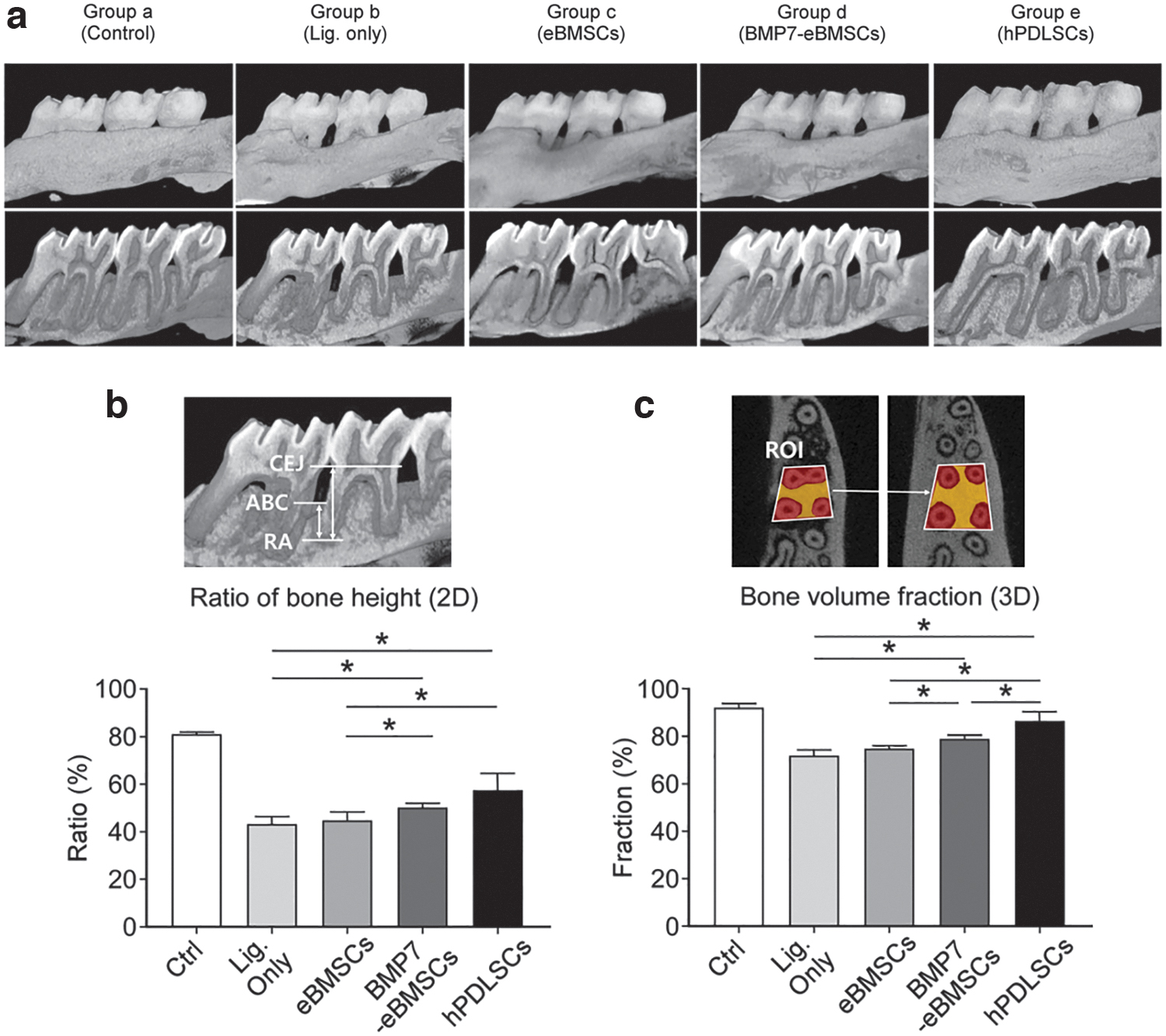
Micro-CT analysis of new bone formation upon BMP7-eBMSC transplantation in a ligature-induced periodontitis model.
The regenerated bone volume (BV) was also calculated in the 3D-reconstructed views (Fig. 4b), and the relative BV in each group was as follows: 92% ± 3% (group a), 72% ± 3% (group b), 74% ± 2% (group c), 79% ± 2% (group d), and 86% ± 5% (group e). The BMP7-eBMSCs (group d) showed significantly increased bone height and larger BV compared with the ligature only (group b) and eBMSC transplantation (group c) groups, although the BV was significantly lower than those in the hPDLSC transplantation group (group e), indicating relatively fair efficacy of BMP7-eBMSCs in alveolar bone regeneration (p < 0.05) (Fig. 4).
BMP7-eBMSC transplantation regenerates the periodontal complex in the ligature-induced periodontitis model
The periodontal complex is composed of cementum, PDL, and alveolar bone, and thus, there is a need for complete regeneration of these elements for successful MSC therapy. Histological and histometric analyses were performed to evaluate if BMP7-eBMSC transplantation promotes regeneration of the periodontal complex. In low-magnification images (30 × ), the junctional epithelium (JE) and cementum layer of the root appeared to be intact in the normal periodontium (group a) (Fig. 5a). However, recession of the gingival tissue, accumulation of inflammatory immune cells with damaged JE, and loss of cementum and alveolar bone were observed in the ligature-induced periodontitis group (group b) (Fig. 5a; triangle).
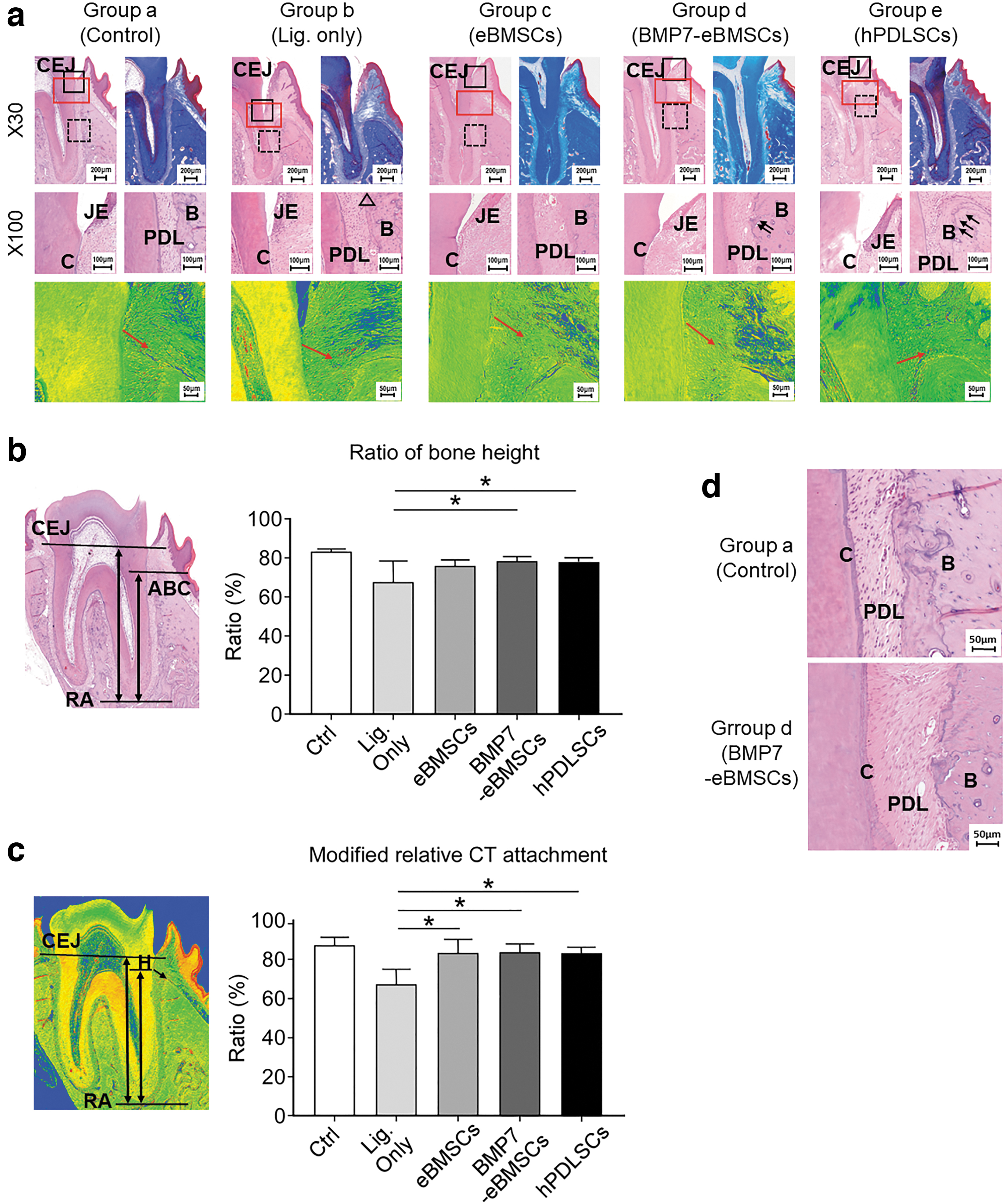
Histological and histometric analysis of BMP7-eBMSC transplantation in the ligature-induced periodontitis model.
Micro-CT analysis confirmed that new alveolar bone was formed in the three MSC transplantation groups (groups c–e), but the amounts of regenerated bone were different among the groups in the histometric analysis (Fig. 5b). The alveolar bone height ratio calculated in the longitudinal section was as follows: 83% ± 1% (group a), 68% ± 12% (group b), 75% ± 3% (group c), 77% ± 1% (group d), and 77% ± 2% (group e). Statistical analysis showed that the bone heights in groups d and e were significantly higher than those in the ligature-induced periodontitis group (group b), indicating that these two groups promoted alveolar bone regeneration. However, the eBMSC transplantation group (group c) did not show a significant difference (Fig. 5b).
Importantly, the MSC transplantation groups, that is, groups c–e, showed regeneration of fibrous ligament tissue without bony ankylosis (Fig. 5a). In the BMP7-eBMSC group (group e), collagen fibers were aligned obliquely to the root surface, indicating PDL-like tissue regeneration (Fig. 5a, d). Furthermore, group e showed a lower boundary of the JE located near the cementoenamel junction, and a reversal line was observed in the alveolar bone, an indicator of bone regeneration. Mechanistically, the modified relative connective tissue attachment ratio was calculated, and the values for the different groups were as follows: 87% ± 4% (group a), 68% ± 7% (group b), 83% ± 7% (group c), 84% ± 4% (group d), and 84% ± 3% (group e). Groups c–e showed significant increases in connective tissue attachment compared with group b (p < 0.05), but there was no statistically significant difference among the three MSC transplantation groups (Fig. 5c).
Discussion
The present study aimed to investigate the regenerative potential of BMP7-eBMSCs for periodontal tissue regeneration, compared with that of hPDLSCs. The MSC characteristics of BMP7-eBMSCs and their osteogenic differentiation potential were confirmed by means of in vitro experiments and subcutaneous ectopic transplantation in vivo. Local injection of BMP7-eBMSCs into the ligature-induced periodontitis rat model resulted in periodontal tissue regeneration comparable to that upon local injection of hPDLSCs.
In our study, both eBMSCs and BMP7-eBMSCs were pretreated with high-level gamma irradiation before being locally transplanted. A high dose of radiation enables activation of the cell apoptosis pathway, resulting in the inhibition of cell proliferation. 33 This may prevent tumor formation resulting from excessive stem cell proliferation in MSCs. 34 However, it may also impair the osteogenic differentiation of BM-MSCs. 35 This could be a possible explanation for the insignificant bone regeneration in the eBMSCs with BCP (group 2) and eBMSC (group c) groups in the ectopic subcutaneous transplantation experiments. Unlike our results, previous studies showed a significant amount of new bone formation 36 mediated by BM-MSCs. Another possible explanation for the insignificant bone formation mediated by eBMSCs is the variation in cell donors or different stem cell clones from a single donor. The select cells in the immortalization process may have an inferior function to induce in vivo osteogenesis. Nevertheless, it is noteworthy that BMP7 released by BMP7-eBMSCs induced significant osteogenesis.
However, a relatively higher amount of new bone formation was observed upon ectopic transplantation of BMP7-eBMSCs with a BCP scaffold. Consistent with our results, previous studies have also reported that BMP7 could generate bone in various animal models, although the concentration of BMP7 required for bone formation is still under debate.37–39 It has been reported that a nanogram level of BMP7 shows osteogenic effects in in vitro cell culture, whereas a microgram dosage is required to induce bone regeneration at the clinical stage. 38 In the present study, 1.45 ng/mL/day of BMP7 was released by BMP7-eBMSCs in vitro, which is a relatively low quantity compared with that reported in previous studies. In addition, transplantation of the cells showed fair outcomes of alveolar bone regeneration in an animal model of periodontitis. We speculate that this was due to the continuous release of BMP7, although it gradually decreases over time, which is consistent with a previous result that genetically modified hPDLSCs expressing BMP2 exhibited the greatest amount of new bone formation. 32
Histological findings in the present study indicated that regeneration of cementum-like tissue occurred in the BMP7-eBMSCs group (group d), which is consistent with the findings of other studies.21,25 However, the lack of quantitative analysis of regenerated cementum is one of the limitations of this study. Our study also showed that ankylosis did not occur in the BMP7-eBMSCs group (group d). Instead, regenerated PDLs were arranged relatively perpendicular to the root surface, as shown by the higher ratio of the modified connective tissue attachment. This result follows a previous study where OP-1 (BMP7) was used for periodontal wound healing in surgically created supra-alveolar class III furcation defects in the canine model. 40 Our study confirmed that BMP7-eBMSCs did not induce ankylosis in periodontal regeneration, which is different from the findings of studies on BMP2. 12
Although growth factors and eBMSCs are promising treatment technologies, viral transfection is still not safe, in addition to which MSC engineering costs are high and the process is complex. 20 Moreover, pathogen contamination may occur during cell culture, which can induce infection. Possible karyotypic instability and gene mutations after prolonged culture may also limit the usefulness of this strategy. Above all, the direct osteogenic and regenerative effects of BMP7 need to be further studied in vitro and in vivo using BMP7 antagonist or blocking antibodies. Therefore, for the application of BMP7-eBMSCs in clinical practice, there is a need for further research to overcome these drawbacks.
Conclusions
Collectively, this study confirmed the characteristics of stem cells in BMP7-eBMSCs and periodontal tissue regeneration in a ligature-induced periodontitis rat model. Although there are several limitations, the method offers a promising strategy for the treatment of periodontitis.
Footnotes
Authors' Contributions
Y.-H.J. collected, analyzed, and interpreted the data and drafted the article. J.-Y.P. contributed to data interpretation, drafted the article, and critically revised the article. H.-J.K. collected and analyzed the data and drafted the article. S.M.L. conceived the ideas, collected the data, and drafted the article. S.-H.K. interpreted data and critically revised the article. J.-H.Y. conceived the ideas, designed and approved the study, collected and interpreted the data, and critically revised the article. All authors gave final approval and agree to be accountable for all aspects of the work.
Disclosure Statement
No competing financial interests exist.
Funding Information
This work was supported by the National Research Foundation of Korea (NRF) grant funded by the Korea government (MSIT) (NRF-2019R1A2C1086515).

References
Supplementary Material
Please find the following supplemental material available below.
For Open Access articles published under a Creative Commons License, all supplemental material carries the same license as the article it is associated with.
For non-Open Access articles published, all supplemental material carries a non-exclusive license, and permission requests for re-use of supplemental material or any part of supplemental material shall be sent directly to the copyright owner as specified in the copyright notice associated with the article.
