




Abstract
Tissue engineering scaffolds are often made from the decellularization of tissues. The decellularization of tissues caused by prolonged contact with aqueous detergents might harm the microstructure and leave cytotoxic residues. In this research, we developed a new technique to use supercritical carbon dioxide (Sc-CO2)-based decellularization for porcine nerve tissue. The effect of decellularization was analyzed by histological examination, including Hematoxylin and Eosin, Masson's Trichrome staining, and 4′,6-diamidino-2-phenylindole staining. Moreover, biochemical analysis of the decellularized tissues was also performed by measuring DNA content, amount of collagen, and glycosaminoglycans (GAGs) after decellularization. The results showed that the tissue structure was preserved, cells were removed, and the essential components of extracellular matrix, such as collagen fibers, elastin fibers, and GAG fibers, remained after decellularization. In addition, the DNA content was decreased compared with native tissue, and the concentration of collagen and GAGs in the decellularized nerve tissue was the same as in native tissue. The in vivo experiment in the rat model showed that after 6 months of decellularized nerve implantation, the sciatic function index was confirmed to recover in decellularized nerve. Morphological analysis displayed a range of infiltrated cells in the decellularized nerve, similar to that in native tissue, and the number of Schwann cells that play essential for motor function and sensory in the decellularized nerve was confirmed. These findings indicate that tissue decellularization using Sc-CO2 has been successfully used in tissue engineering.
Impact statement
The nervous system is pivotal in regulating bodily functions, encompassing a complex network of central and peripheral regions. Injuries to these systems can lead to substantial mental and physical disabilities, profoundly impacting health care systems. Utilizing a novel approach involving supercritical carbon dioxide for decellularization, this research focuses on creating acellular scaffolds with intact neuronal structures. The promising outcomes of this technique, demonstrated through mechanical testing, histological analyses, and transplantation on the rat model, offer new prospects for human nerve tissue transplantation.
Introduction
The nervous system is critical in controlling and regulating bodily functions and comprises a complex network of central and peripheral regions. Therefore, central or peripheral nervous system injuries can lead to various mental or physical disabilities, resulting in significant clinical and economic burdens. According to recent statistics, there were ∼28 million central nervous system injuries (e.g., traumatic brain injury and spinal cord injury) and 20 million peripheral nerve injuries in the United States alone.1–9 Microsurgeons face significant challenges when attempting to repair damaged nerves, as nerve healing is a complex process and using autogenous nerve grafts is limited due to donor site morbidity, inadequate tissue, or donor and recipient size incompatibility.10–12
Tissue engineering has emerged as a promising solution for creating artificial nerve tissues that can be implanted into patients. Ideally, artificial nerve tissues should be acellular scaffolds that preserve the ultrastructure and extracellular matrix (ECM) components of native tissues.2,13,14 The process of creating such scaffolds is known as decellularization. Decellularized extracellular matrices, such as the paravertebral muscle matrix in the spinal cord, have been created from tissues outside the intended target location.
Decellularization is commonly used to create acellular scaffolds using physical, chemical, or biological methods. 2 However, when choosing the optimal decellularization method, tissue supplies, desired morphology of the end product, and treatment target region must all be considered.15–18 Several techniques have been developed and successfully utilized for effective decellularization, with the absence of an immunogenic response being a common success criterion.15,17,19–22
Detergent-based decellularization is a commonly used process in which detergents disrupt cell–cell and cell–matrix interactions while maintaining the structural integrity of the matrix, resulting in nonimmunogenic decellularized tissues.23,24 The three categories of detergents are ionic (cationic and anionic), nonionic, and zwitterionic/amphoteric chemicals. Sodium dodecyl sulfate, a detergent widely used in decellularization, effectively reduces DNA in tissues. However, it can also cause a significant decrease in desirable matrix components and biomolecules, such as growth factors and hormones, which are important for bioactive scaffolds.25–27 The addition of an enzymatic treatment may be beneficial for eliminating unwanted biological components. Enzymatic decellularization, which uses enzymes to alter and remove antigenic material and reduce immunogenicity, can be employed with chemical (detergent) decellularization to reduce cytotoxicity.28–30 Various enzymes, often used as additives to detergents to enhance antigen removal, can be used in decellularization procedures, such as Trypsin/EDTA.
Mechanical delamination or multiple freeze–thaw cycles can efficiently remove dense cell areas or lyse cells, requiring less severe chemical treatments.31,32 However, they are often used in combination with chemical or enzymatic approaches as they are unable to remove genetic material from a scaffold following cell lysis. Furthermore, excessive physical decellularization may alter the mechanical characteristics of the scaffold by disrupting the natural ECM ultrastructure.
Supercritical carbon dioxide (Sc-CO2) is an emerging approach for decellularization that offers advantages over traditional methods that use detergents or enzymes.33–38 Sc-CO2 is nontoxic, nonflammable, and relatively inert, with good solvent characteristics and a low critical temperature (31.1°C), enabling it to function at physiological temperatures. Sc-CO2 has been used in various biomedical applications, including extraction of physiologically important chemicals, pasteurization, and sterilizing synthetic and natural biomaterials. Sc-CO2 has also been extensively employed to develop tissue engineering scaffolds from synthetic biomaterials, such as polymer foaming, with no significant loss of scaffold bioactivity.
This study utilized the advantages of Sc-CO2-based decellularization to prepare porcine nerve tissue for transplantation. Comprehensive assessments included physical characterization tests, histological and immunofluorescence (IF) staining methods, DNA qualification, western blot analysis, and in vivo experiments (Fig. 1). These multifaceted analyses collectively substantiated the acellular nature and viability for the transplantation of the decellularized nerve tissue.
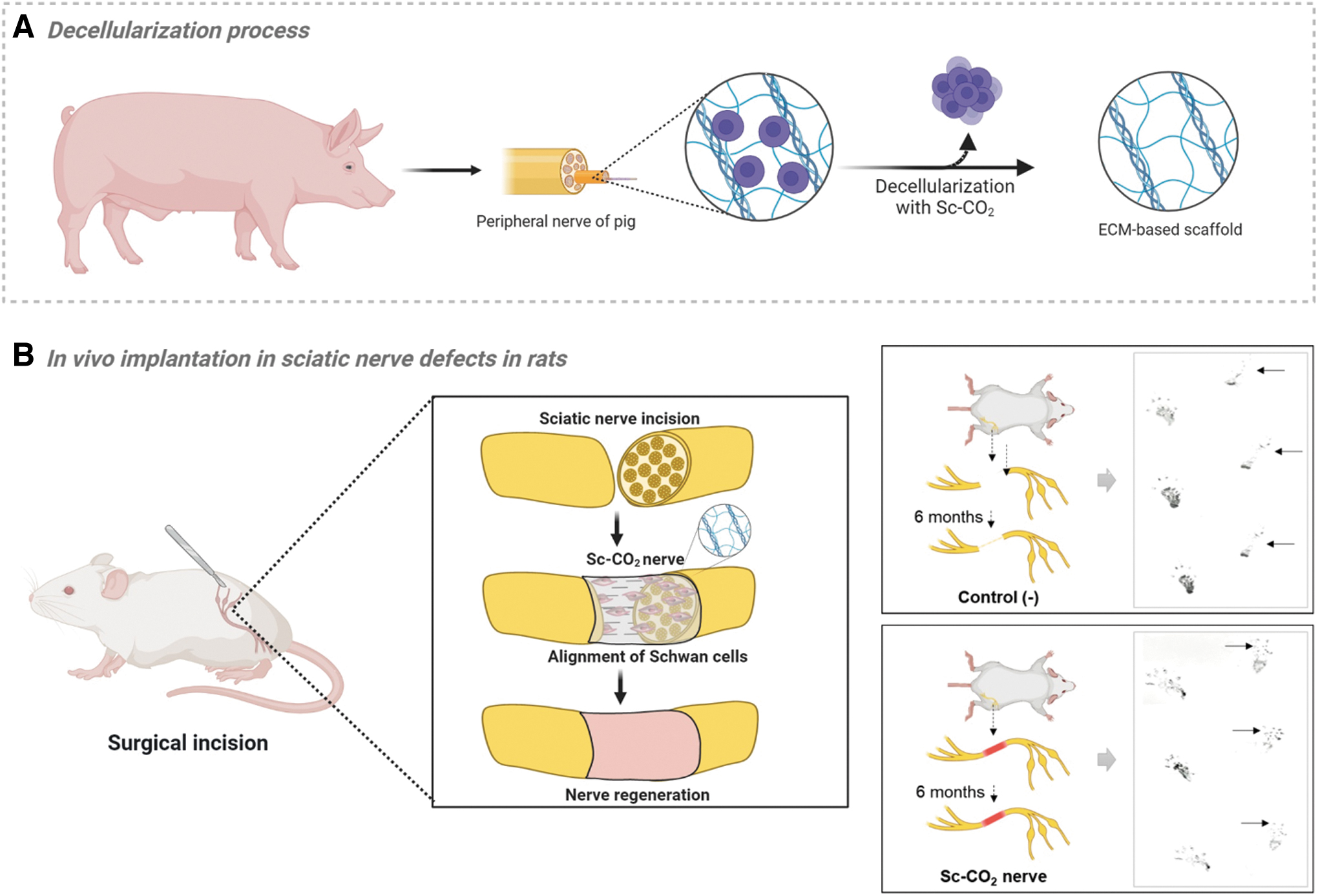
Schematic illustration.
Materials and Methods
Decellularization with supercritical CO2
Porcine peripheral nerve tissue with a diameter of about 2 mm was obtained from a livestock company. The nerve tissue was carefully selected, removed the remaining fat and muscle tissue, then soaked in sterile 1 × phosphate-buffered saline (PBS), and washed for ∼30 min. Supercritical (Sc) CO2 extraction was performed using homemade equipment with a 0.3-L cylindrical vessel. The nerve tissue was soaked in 1 × PBS with 1% antibiotics overnight on a 30 rpm rocker. A gas flowmeter regulated the CO2 flow, and cosolvents (EtOH) were added to the vessel. The pressure applied during the Sc-CO2 extraction was between 200 and 300 bar, and the processing time was ∼3 h at 35°C (Fig. 2). After the extraction, the nerve tissue was washed twice with 1 × PBS for 15 min on a 30 rpm rocker and stored at −80°C for further analysis. Gamma-ray sterilization (25 kGy, commissioned by Greenpiatech) was used for tissue sterilization.
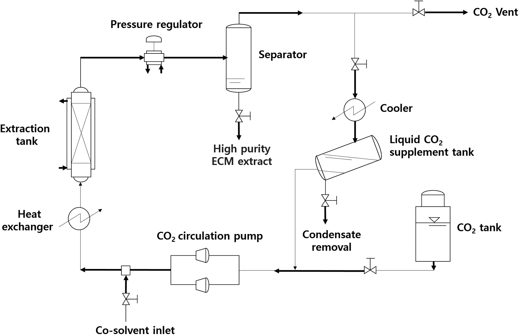
Schematic of the supercritical carbon dioxide extraction system.
DNA quantification
DNA was extracted by the DNeasy Blood and Tissue Kit (Qiagen, Germany). In brief, 20 mg of tissue from each sample was ground and digested with extraction solution and proteinase K at 56°C. Subsequently, DNA was extracted using a DNeasy mini-column after being precipitated by 200 μL of 100% ethanol. The extracted DNA was eluted, and the total amount was quantified using NanoDrop at 260 nm and Qubit 3.0.
Western blot
The western blot experiment was conducted for Major Histocompatibility Complex (MHC) class I, MHC class II, β-actin, and Laminin. Samples were homogenized in PROPREPTM with protein inhibitors, then centrifuged at 14,000 g for 15 min at 4°C. Supernatants were collected, and protein concentration was determined using the bicinchoninic acid method. Electrophoresis and transfer to nitrocellulose membrane followed. After blocking with 5% nonfat dry skim milk, membranes were incubated overnight at 4°C with primary antibodies. A horseradish peroxidase-conjugated secondary antibody was applied for 2 h at room temperature. Protein expression was visualized using a C-DiGit Blot Scanner (Li-Cor).
Physical characterization
The samples' tensile strength was measured at a dimension of 1 × 3 × 10 mm at a strain rate of 0.5 mm/s by a Universal testing machine (Sur TA, Chemilab. Co., Ltd.). The samples were localized and fixed at a strain rate of 0.5 mm/s; they were pulled toward two sides until totally broke.
Histological analysis and IF staining
After decellularization, the tissues were immersed in 10% formaldehyde solution for 24 h, embedded in paraffin, and cut into 5-μm-thick slides. Histological analysis was performed with Hematoxylin and Eosin (H&E) staining, 4′,6-diamidino-2-phenylindole (DAPI) staining, and Masson's Trichrome (MT) staining. For IF staining, sections were heated at 60°C for 1 h, dehydrated, and blocked with 4% BSA in 1 × PBS buffer at room temperature for 1 h. Anti-S100 beta antibody (1:500, ab52642; Abcam), and anti-Myelin Basic Protein (MBP) (1:200, sc-271524; Santa Cruz) were applied at 4°C overnight. Following this, rabbit-anti-mouse IgG Alex Fluor 488-conjugated secondary antibody (1:1000 dilution, cat. no.: A-11059; Invitrogen) was used at room temperature for 1 h. Nuclei were counterstained with DAPI and coverslips mounted. Imaging was done using a Zeiss LMS 710 confocal microscope, with fluorescence signals analyzed using ImageJ software.
ECM quantification
The Sircol Insoluble Collagen Kit (Cat. No.: S2000; Biocolor) was used to determine the collagen concentration in tissue samples. Briefly, 50 mg wet weight (∼26.5 mg dry weight of Native sample, 15.6 mg dry weight of Sc-CO2-treated sample) each tissue sample was fragmented at 65°C for 3 h, and the supernatant was collected by centrifugation at 12,000 rpm at 4°C. After mixing with Sircol dye reagent and centrifugation, pellets were washed, resuspended, and collagen content was measured at 550 nm.
For hyaluronic acid (HA) assay, 1 g of each tissue sample was washed and homogenized with 1 × PBS buffer and then collected the supernatant after centrifugation at 15,000 rpm for 10 min. Fifty microliters of supernatant mixed with biotin-labeled antibody solution, followed by HRP-Streptavidin Conjugate solution and 3,3′,5,5′-tetramethylbenzidine substrate incubation. HA was quantified by measuring optical density at 450 nm.
Cytotoxicity assay
Sc-CO2 nerve tissues were immersed with 1 × PBS buffer containing antibiotic for 24 h and then incubated in Dulbecco's modified Eagle medium culture containing 10% fetal bovine serum (FBS) and 1% penicillin–streptomycin (PS) at 37°C, 5% CO2 for 24 h. NIH-3T3 cells with 4 × 10⁴ cells/mL were seeded into a 24-well plate; the plate was divided into two groups: the control group, and Sc-CO2 group. The control group cells were cultured in the completed medium, while the Sc-CO2 group involved cells cultured in wells containing the Sc-CO2 nerve tissues. The cultures were maintained for 24 and 48°h at 37°C, 5% CO2. Cell viability was quantified by 3-(4,5-dimethylthiazol-2-yl) −2,5-diphenyltetrazolium bromide (MTT) assay.
Cytocompatibility analysis
Decellularized nerve tissues were washed with 1 × PBS buffer containing antibiotic for 24 h, then incubated in DMEM culture containing 10% FBS and 1% PS at 37°C, 5% CO2 for 24 h. The Sc-CO2 nerve tissues were placed in each well in 48-well plate. Subsequently, the NIH-3T3 cells and Schwann cell (RSC96), each at a density of 4 × 10⁴ cells/mL, were seeded into these wells and proliferated for up to 11 days. Then, tissues were immersed with 10% formalin for 24 h at 4°C and embedded in a paraffin block before H&E staining with tissue slices. Cell infiltration into nerve tissues was confirmed by photographing under microscopes.
Animal experiment
Animal experiments were approved by the Institutional Animal Care and Use Committee of Seoul National University Bundang Hospital (approval number: BA-2110-329-008-01). Adult male Sprague Dawley rats, 8 weeks of age (BioOrient Company, Seongnam, Korea), weighing 200 and 350 g were kept in groups of two with free food and drink accession. The rats were maintained in a controlled environment under specific pathogen-free conditions, with 12-h light/12-h dark cycle, relative humidity of 55%, and a temperature of 24°C.
Evaluation of host immune response to Sc-CO2 nerve tissues
Fifteen rats were randomly divided into three distinct groups: the native group, the Sc-CO2 nerve group, and the autogenous nerve. After being anesthetized with Isoflurane, the dorsal hair of the area along the connecting line between the spine and the anterior superior iliac spine was removed. The subcutaneous graft implantation site was disinfected and cut 1 cm along the sagittal position at the center of this area. The respective grafts were then carefully placed under the skin of the rats. In the autologous nerve group, each rat's sciatic nerve was extracted and reimplanted subcutaneously, serving as an autologous graft. Four weeks postsurgery, the rats from each group were sacrificed to facilitate the analysis of immune response. IF staining was performed, utilizing specific antibodies to detect the presence of immune cells. The antibodies used included rabbit anti-CD4 (1:4000, ab237722; Abcam) for identifying CD4+ T cells, mouse anti-CD8 (1:200, ab33786; Abcam) for CD8+ T cells, and mouse anti-CD68 (1:3000, ab955; Abcam) for macrophages.
In vivo implantation in rat sciatic nerve defect
On the day of surgery, rats were randomly divided into two groups: control (n = 5) and supercritical carbon dioxide-treated nerve (Sc-CO2 nerve, n = 5).
Both groups were anesthetized with Isoflurane. The surgical region was created by shaving the dorsal hair of the left hind limb, disinfecting the area, and making an incision in the skin along the intermuscular space. The fat and connective tissue around the nerve was removed and separated from the muscle space. In the Sc-CO2 group, a decellularized porcine nerve segment of the same length (15 mm) was used to substitute the dissected nerve segment. A microsurgical technique was performed to connect the decellularized nerve matrix to adjacent nerve tissue. The outer membranes of the two nerve stumps were sutured and anchored at both ends of the graft by 8/0 sutures with needles under the microscope. A 15-mm-long nerve segment was removed without a graft in the negative control group. The subcutaneous fascia layer and skin were sutured after hemostasis. The rats were carefully monitored during recovery and allowed to drink and eat freely after the operation.
After 6 months of transplantation, the rats were examined by walking track analysis and calculating Sciatic Functional Index (SFI), then were sacrificed for histological examination and IF staining.
Walking track analysis
Rats had undergone prior training before surgery to walk up a wooden board (covered with paper) leading into a cage with the ink-marked left and right hind paws. During the testing session, three consecutive footprints were collected from each limb, enabling the acquisition of pertinent measurements for the SFI calculation by the following equation:
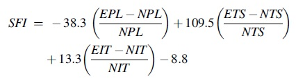
Where PL (Print Length) is the distance from the heel to the third toe; TS (Toe Spread) is the distance from the first to the fifth toe; IT (Intermediate Toe spread) is the distance from the second to the fourth toe; E is experimental hindfoot, and N is nonexperimental hindfoot.
Statistical analysis
The data were displayed as mean value ± standard error bar using GraphPad Prism software (GraphPad Software, Inc., San Diego, CA) with at least three independent experiments. Statistical analysis was performed using Statistical software for data science (STATA) software (version 18/SE; StataCorp). Nonparametric tests were employed to assess group differences in measurement data that did not follow a normal distribution and the small sample size. The Kruskal–Wallis test was used for comparing differences within multiple groups, while the Wilcoxon rank-sum test was used to compare the differences between two groups. A p-value <0.05 indicates that the difference between groups was statistically significant (Supplementary Data S1).
Results
Histological evaluation and ECM analysis of decellularized nerve tissues
Histological staining was performed to investigate the morphological alterations and cell removal within the Sc-CO2-treated nerve tissue. In the context of H&E staining, Figure 3A–C illustrates the presence of various cells (blue dots) within the endoneurium of the native tissue. In contrast, the Sc-CO2-treated nerve tissue displays fewer cells than the native tissue (Fig. 3D–F). Notably, the morphology of the Sc-CO2-treated nerve tissue closely resembles that of the native tissue. The persistence of residual cellular nuclei after nerve tissue decellularization was assessed through DAPI staining. The outcome reveals a spectrum of cellular nuclei (blue dots) within the native tissue (Fig. 3M, N), whereas the Sc-CO2-treated nerve tissue exhibits a diminished number of blue-stained nuclei in comparison (Fig. 3O, P).
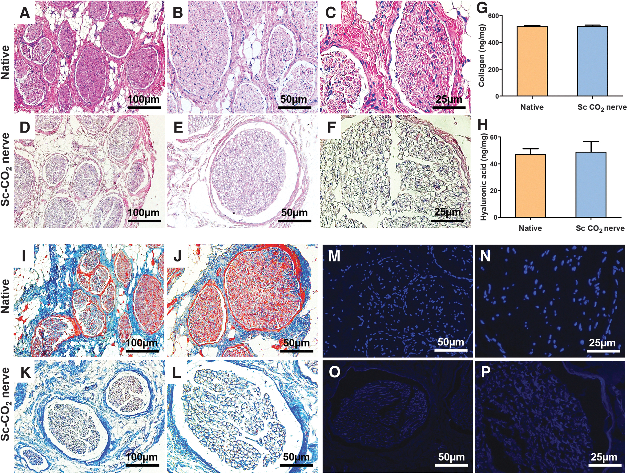
Histological evaluation and determination of the amount of HA and collagen of decellularized nerve tissues. H&E staining images of Native tissue
MT-staining images of the nerve tissues also reveal disparities between the native sample (Fig. 3I, J) and the Sc-CO2-treated sample (Fig. 3K, L). The collagen scaffolds (depicted in blue) in both native and Sc-CO2-treated nerve tissues display similar characteristics. Quantitative analysis of collagen content further underscores the resemblance of collagen levels between the native and Sc-CO2-treated nerve tissues (Fig. 3G). HA, a vital non-sulfated glycosaminoglycan (GAG) contributing to connective and neural tissue, was quantified in native and Sc-CO2-treated nerve tissues. The measured HA quantities in the two tissues were closely aligned: 23.52 ± 2.16 ng/mg in the native tissue and 24.34 ± 4.02 ng/mg in the Sc-CO2-treated nerve tissue, respectively (Fig. 3H). The histological staining, HA, and collagen analysis results suggest that the Sc-CO2 decellularization method eliminates cellular elements while preserving the tissue scaffold crucial for nerve regeneration.
Quantification of DNA and western blot analysis after decellularization
The NanoDrop analysis indicated a significantly lower DNA concentration in Sc-CO2-treated nerve tissue (25.0 ± 0.0 ng/mg) compared with native tissue (162.0 ± 1.0 ng/mg) (Fig. 4A). The Qubit 3.0 results also observed this significant difference, with DNA concentrations of 1.0 ± 0.0 ng/mg in Sc-CO2-treated nerve tissue and 144.0 ± 2.0 ng/mg in native tissue (Fig. 4B). DNA content was verified through electrophoresis on a 1.5% agarose gel to complement these findings. The result confirmed the absence of all DNA fragments of 200 bp or smaller, which could potentially trigger an immune response in the Sc-CO2-treated nerve tissue, whereas they were observed in the native tissue (Fig. 4C). Besides, western blot results revealed that MHC class I, MHC class II, and the cytoskeletal protein β-actin, identifiable in the original tissue, were not detected in the decellularized tissue. However, Laminin, an ECM protein crucial for promoting cell growth, remained preserved after the decellularization process (Fig. 4D).
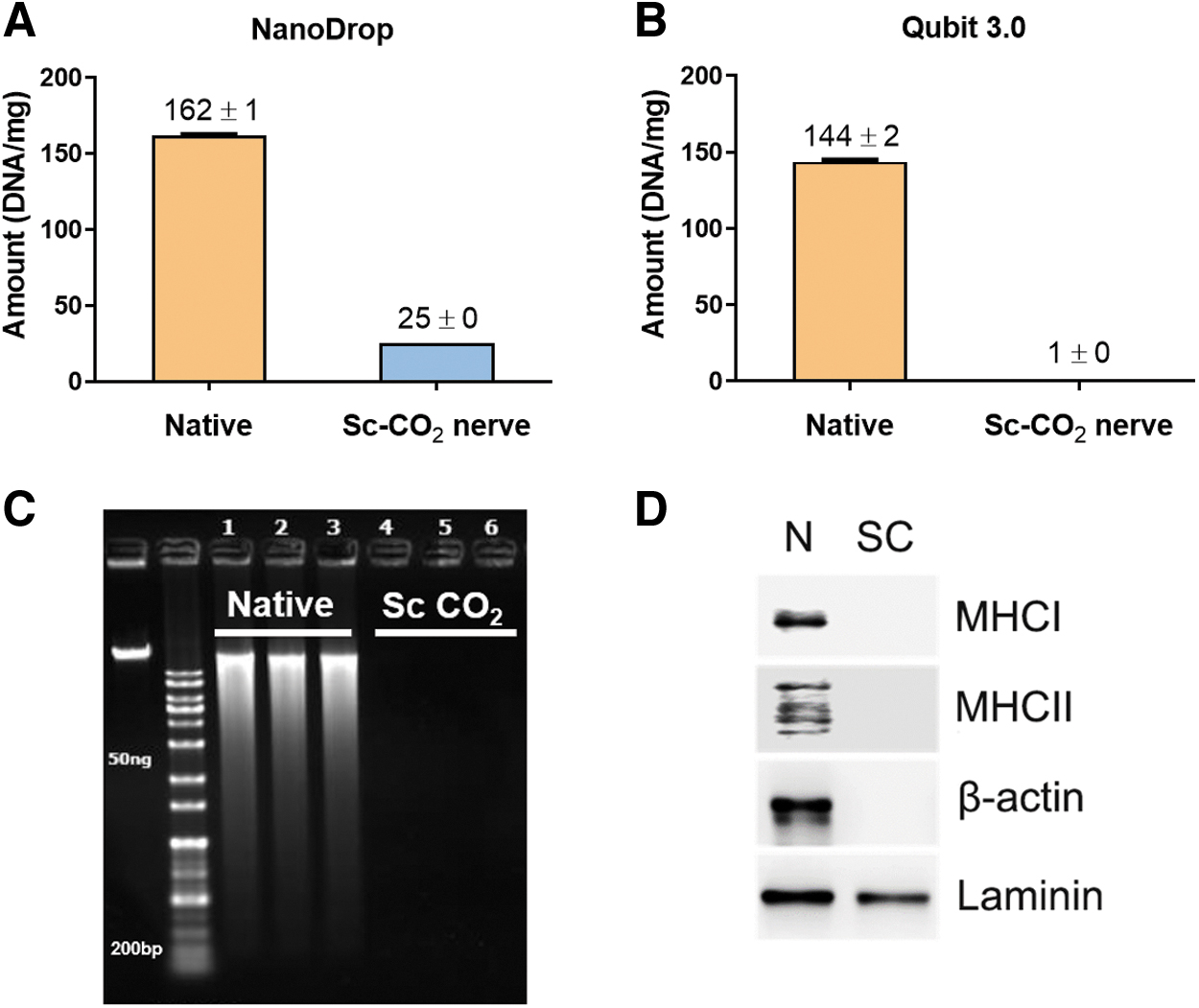
DNA quantification and western blot analysis.
Physical characterization
To evaluate the mechanical and physical properties of the nerves after decellularization, tensile strength measurement was conducted, as shown in Figure 5C. The tensile strength profiles demonstrated that the native nerve had a slightly higher Young's modulus value (0.0274 MPa) than the Sc-CO2 sample (0.0198 MPa), representing a softer texture of the Sc-CO2 decellularized nerve. These changes were further confirmed by dynamic frequency sweep (Fig. 5A) and strain sweep (Fig. 5B), in which the Sc-CO2 decellularized nerve displayed a decline in storage modulus compared with the native sample. Consequently, the strain sweep data showed a depletion in the sustaining strain of the Sc-CO2 decellularized sample to the native one.
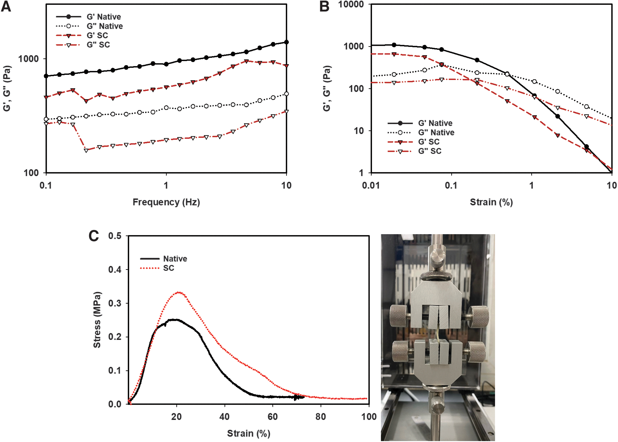
Physical characterization.
Cytotoxicity assessment and cytocompatibility analysis
The MTT assay examined the viability of the NIH-3T3 cells exposed to Sc-CO2 nerve tissues. After 24 and 48 h incubation, the Sc-CO2 sample displays higher cell viability values than the control group (Fig. 6A). Additionally, an investigation into cell infiltration within the Sc-CO2 nerve tissue was conducted using NIH-3T3 and RSC96 cells to assess the tissue's potential to support cell growth after implant surgery of Sc-CO2 nerve tissue. H&E staining image shows that, after 11 days of seeding, fibroblast cells and the Schwann cell successfully infiltrated the scaffold of the Sc-CO2 nerve tissue (Fig. 6B, C, D). The MTT and H&E staining results indicate that the Sc-CO2 nerve tissue was noncytotoxic and capable of supporting new cell growth.
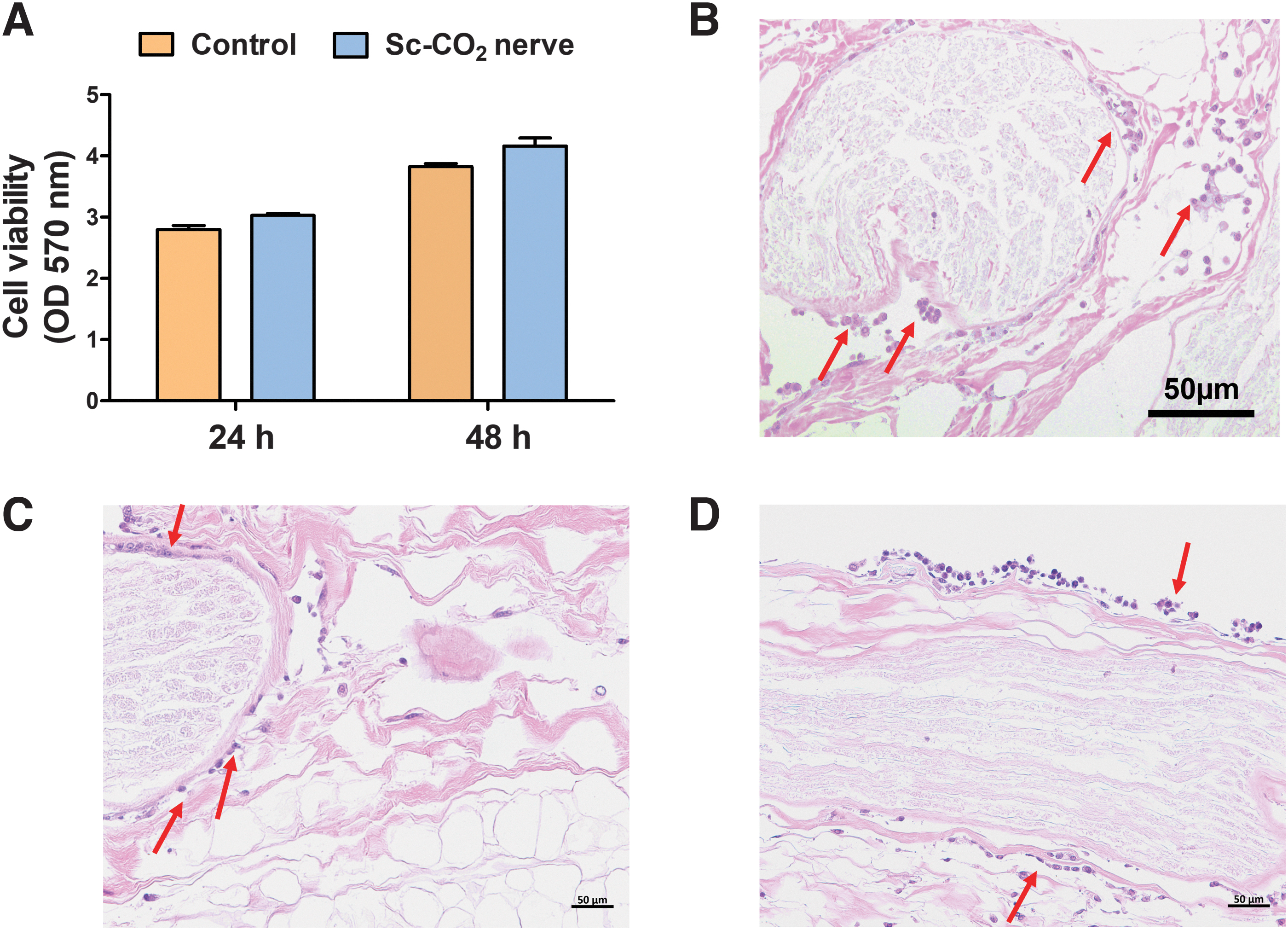
Cytotoxicity and cytocompatibility test.
Analysis of immune response to decellularized porcine nerve after subcutaneous implantation
Four weeks after subcutaneous implantation, Sc-CO2-treated nerve tissues were assessed for immune response, a standard timeframe informed by prior studies as optimal for observing post-implantation inflammatory reactions. The immune response was quantified by measuring the presence of CD4+ and CD8+ T cells, along with macrophages using CD68 marker. The expression levels of these markers were significantly reduced in the Sc-CO2 group when compared with native tissues, demonstrating a comparable profile to the autologous nerve group (Supplemenatary Fig. S1). The immune response analysis conducted 4 weeks postimplantation reveals that decellularized nerves treated with Sc-CO2 are less likely to induce the cytotoxic activity of T lymphocytes and macrophage-mediated degradation, thus reinforcing their potential for in vivo usage due to their reduced likelihood to provoke a substantial inflammatory immune reaction.
Walking track analysis
Motor function was evaluated in the sixth month through walking analysis to calculate SFI for each rat. Walking tracks of the rats with defected sciatic (Fig. 7A) and the rats implanted with Sc-CO2 nerve tissue (Fig. 7B) were collected, subsequently comparing their SFIs (Fig. 7C). Figure 7C demonstrates a substantially higher average SFI for the decellularized nerve group (averaging at −41.55 ± 3.27) than the negative control group (averaging at −62.47 ± 3.62). This finding indicates the successful regeneration of motor function in rats with implanted decellularized nerves, emphasizing superior recovery compared with the negative control group.
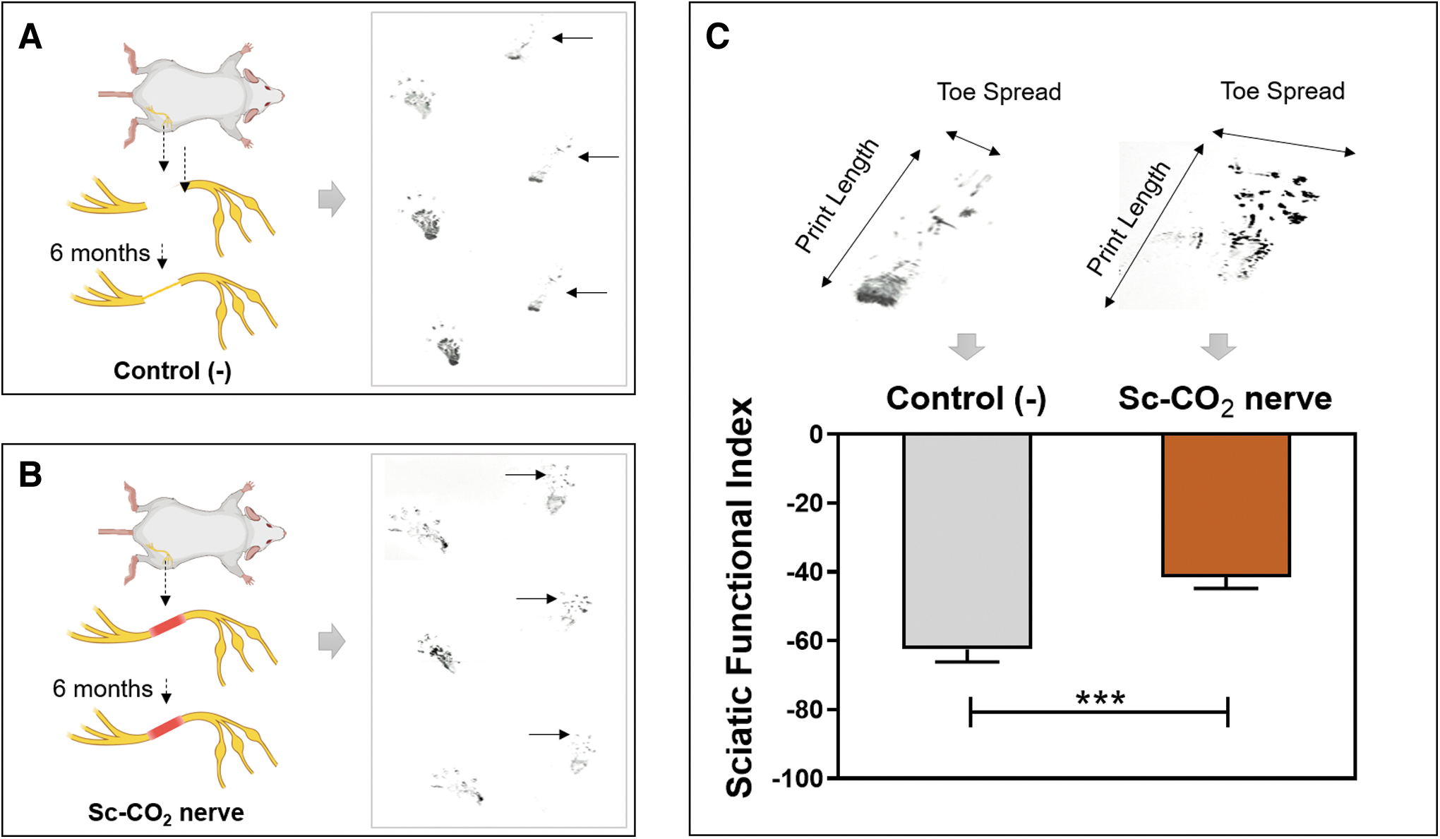
Walking track analysis.
In vivo histological evaluation of implanted nerve
After walking track analysis, the rats were sacrificed, and the tissues from the normal group, the negative control group (a 15 mm defective sciatic), and Sc-CO2-treated group were collected to be examined by the H&E and DAPI staining. In the tissue of the normal group, the nerve morphology remained unchanged (Fig. 8A), and there were numerous positive cells (bright spots) in the scaffold (Fig. 8D). In contrast, the negative control tissue exhibited a distinctly different scaffold structure than the normal nerve tissue (Fig. 8B), and the presence of positive cells was noticeably reduced (Fig. 8E). Notably, the structure of Sc-CO2 nerve tissue and the number of cells in this sample were similar to the normal nerve tissue (Fig. 8C, F).
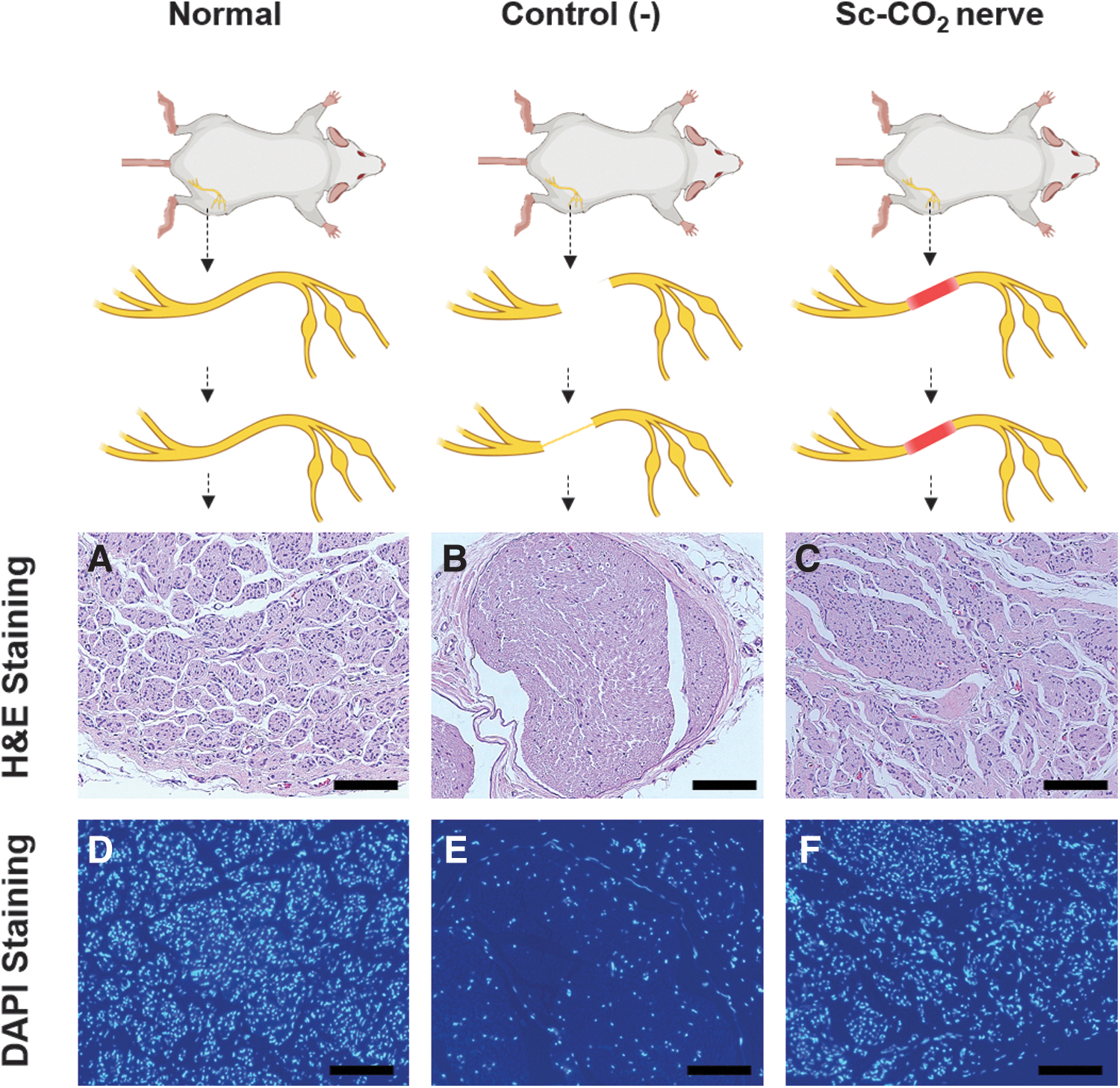
Histological analysis of decellularized tissue after 6 months of implantation.
IF staining with S100β and MBP antibodies
Within the peripheral nervous system, Schwann cells are crucial for maintaining and regenerating of motor and sensory neurons. Reflecting this, S100β IF staining conducted 6 months postoperatively indicated the presence of Schwann cells in decellularized grafts. The staining intensity for S100β was highest in normal nerve tissue, followed by Sc-CO2-treated and negative control nerve tissues (Fig. 9A). Parallel to these findings, IF staining for MBP- a major constituent of the myelin sheath also showed a similar trend (Fig. 9C). Quantitative analysis revealed that both S100β and MBP intensity in the Sc-CO2-treated nerves was significantly greater than in the negative control, and considerably lower than the normal sample (Fig. 9B, D). This result indicated that Sc-CO2-treated nerve tissues provide a suitable microenvironment for the infiltration, functional activity of Schwann cells and myelin sheath formation, facilitating effective nerve reconstruction.
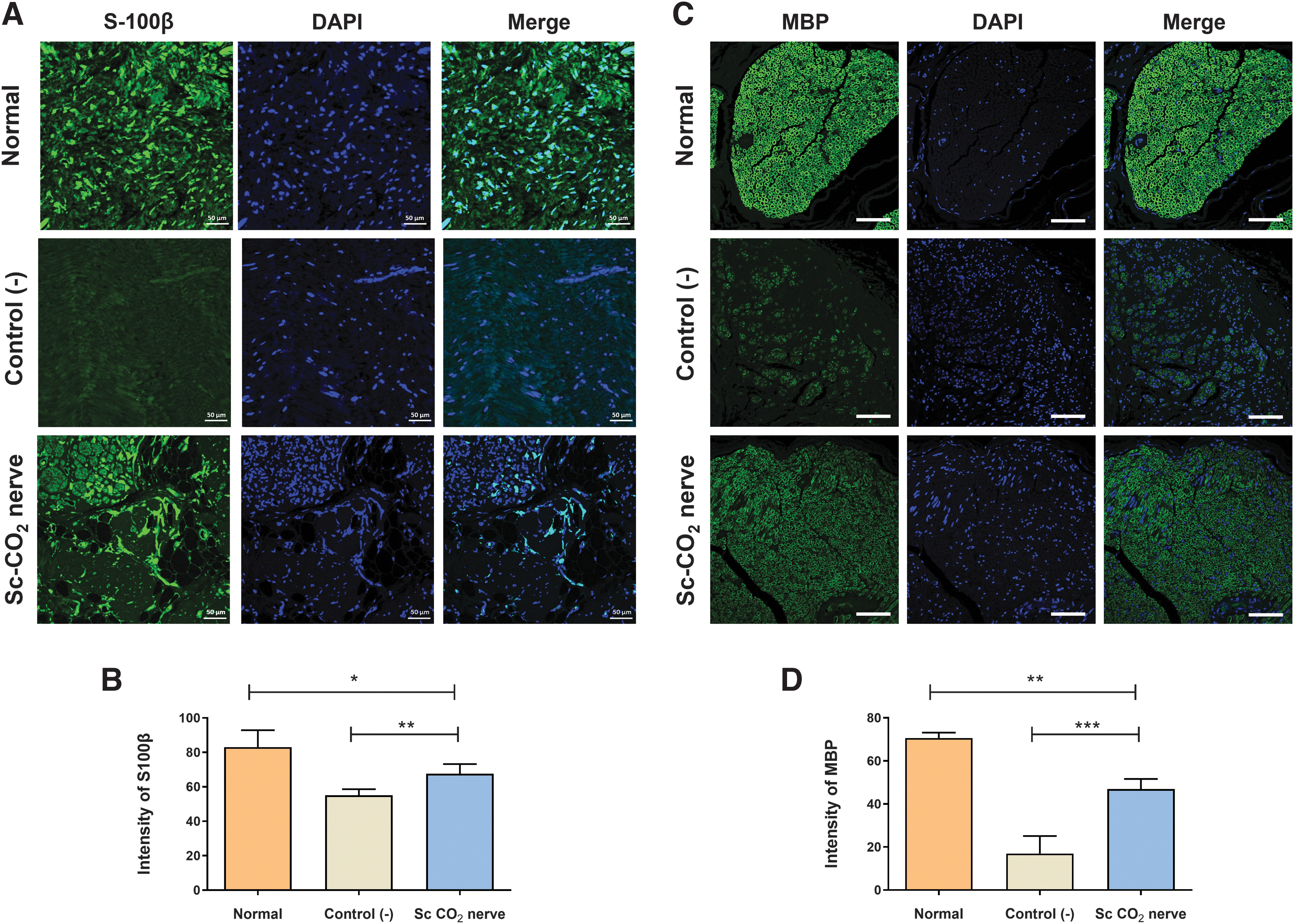
Immunofluorescence staining.
Discussion
Tissue decellularization aims to preserve the intrinsic properties of the ECM while eliminating cellular components and immunogenic factors to mitigate adverse immune responses and inflammation post-transplantation. Various decellularization methods have been reported, including physical techniques (freeze–thaw cycles, immersion, agitation, and direct pressure),39–41 chemical approaches (ionic, nonionic, alcohol, alkaline, and acidic treatments, chelating and zwitterionic detergents), 42 and biological strategies (antibiotics or enzymatic agents).30,43,44 In this study, we developed an innovative tissue decellularization technique utilizing Sc-CO2, successfully eliminating cellular components while maintaining the integrity of neuronal frameworks and basal membranes.
A key objective of decellularization is to remove native cells and genetic materials from the ECM while maintaining its structural, biochemical, and biomechanical properties. Although xenogeneic-origin ECM has proven effective for tissue repair scaffolding, the preparation method can significantly influence the host's remodeling response.40,45 The decellularized ECM can then be repopulated with the patient's cells to generate customized tissues. 46 Histological examination with H&E staining and DAPI staining in our study demonstrated that most native cells were removed from Sc-CO2-treated nerve tissues compared with the native tissues. The efficacy of decellularization of eliminating cells and immunogenic components was also investigated by analyzing the remaining DNA45,47 and western blot analysis. Decellularized tissues with less than 50 ng double-stranded DNA/mg tissue dry weight and less than 200 bp DNA bp fragment length remaining in the decellularized tissues are regarded as successful, because the remaining high concentration of DNA from the scaffold can induce an inflammatory reaction after implant.42,48,49
Additionally, the protein MHC class I, MHC class II, and β-actin, which are major immunogenic proteins that cause acute immune rejection by immune T cells after transplantation, 50 are absent in those tissues, indicating that the decellularization process effectively removed cells and nuclei.
In addition, H&E staining images show the morphology of Sc-CO2 decellularized tissues being substantially preserved relative to the native tissues, and the MT staining revealed comparable collagen fiber density in both Sc-CO2-treated and native nerve tissue. Collagen is a versatile biomaterial due to its broad applicability and numerous properties, including biocompatibility, biodegradability, and accessibility. 51 As the primary structural protein in animal and human hard and soft tissues, collagen plays a crucial role in maintaining the biological and structural integrity of the ECM while providing physical support to tissues. 52 GAGs, the polysaccharide component of proteoglycans, represent one of the two primary classes of biomolecules in the ECM and play vital roles in various cellular processes, including cell growth, proliferation, adhesion, wound repair, and anticoagulation. 53 To evaluate the preservation of these components, the biochemical analysis was used to measure GAG and collagen concentrations in Sc-CO2-treated tissues by the HA ELISA Kit and a Sircol insoluble collagen assay.
The concentrations of GAGs and collagen in decellularized tissues were comparable to those in native tissue, indicating successful decellularization and intact ECM components.
On the other hand, it is essential to consider that residual cytotoxic reagents in the decellularized nerve matrix during the decellularization process might inhibit the regeneration of resident cells within the matrix. 54 This toxicity could also obstruct stem cell recolonization of the decellularized neural matrix in tissue engineering applications. 55 Our results demonstrated that nerve matrix decellularization did not exhibit cytotoxic effects, suggesting that the decellularized nerve matrix is devoid of cytotoxic leachables and suitable for use in regenerative medicine.
Especially in vivo rat model, 6 months postimplantation of decellularized nerve tissue, motor function evaluations revealed that sciatic nerve function had been restored. Histological and DAPI staining analyses demonstrated cell repopulation within the decellularized nerve postimplantation. In the negative control group (15 mm of nerve tissue was cut, and the nerve tissue was naturally regenerated after 6 months) the number of cells was higher than and comparable to the normal group.56,57 This observation suggests that decellularized nerve tissue is more conducive to cell infiltration and retention due to the absence of myelin and exposed basal lamina tubes. Furthermore, the decellularized nerve tissue may not only support cellular infiltration but also promotes the production of bioactive molecules essential for Schwann cell function and myelin reconstruction.58–60 The results of IF staining showed that the intensity of S-100β, a Schwann cell biomarker, and MBP, a key component of the myelin sheath in the decellularized nerve graft, was significantly higher than in the negative control group.
The presence of Schwann cells and the significant increase in myelin presence, as indicated by S100β and MBP staining, point toward a successful repopulation and remyelination within the grafts. Schwann cells, crucial for neuron maintenance and regeneration, alongside the enhanced myelin sheath formation, underline the grafts' ability to support nerve function restoration. 61 This finding indicates that structural integrity and composition of the decellularized grafts provide a conducive environment for Schwann cell activity and myelin sheath reconstruction, a conclusion further supported by walking track analysis data collected 6 months after decellularized nerve implantation.
The SFI was developed to assess functional impairments in mammals by analyzing changes in footprints following injury. Animals without nerve defects predominantly walk on their toes, while those with nerve damage struggle to support their weight and tend to place their whole foot down, resulting in a longer print length, narrower toe spreads, and intermediate toe spreads compared with a normal print. An SFI score of −100 indicates complete impairment, while a score of 0 indicates normal function. 62 Our results reveal a significantly higher average SFI in the decellularized nerve group compared with the negative control group. These data optimistically show that the Sc-CO2-assisted xenograft has the potential to support nerve regeneration.
Conclusion
In this study, nerve tissue decellularization using Sc-CO2 demonstrated the absence of immunogenic components while essential ECM components, such as collagen, GAGs, Laminin, were preserved. Furthermore, in vivo experiments confirmed that the decellularized nerve exhibited robust regeneration and restoration of nerve function. The results suggest a promising decellularization method using supercritical fluid, paving the way for clinical application in nerve transplantation.
Footnotes
Authors' Contributions
L.T.T.L.: methodology, investigation, data curation, formal analysis, visualization, validation, and writing original draft. P.N.C.: methodology, investigation, data curation, visualization, validation, and writing original draft. X.T.T.: data curation, visualization, and writing––review and editing. N.N.G.: investigation, data curation, and visualization. N.V.L.: investigation and data curation. S.Y.N.: conceptualization, supervision, methodology, writing––review and editing, funding acquisition, and project administration. C.Y.H.: conceptualization, supervision, methodology, writing––review and editing, funding acquisition, and project administration.
Disclosure Statement
No competing financial interests exist.
Funding Information
This work was supported by the Ministry of Trade, Industry, and Energy, the Korean government (Project No. 1415169283), and a grant of the Korea Health Technology R&D Project through the Korea Health Industry Development Institute (KHIDI), funded by the Ministry of Health & Welfare, Republic of Korea (grant number: HR22C1363).

References
Supplementary Material
Please find the following supplemental material available below.
For Open Access articles published under a Creative Commons License, all supplemental material carries the same license as the article it is associated with.
For non-Open Access articles published, all supplemental material carries a non-exclusive license, and permission requests for re-use of supplemental material or any part of supplemental material shall be sent directly to the copyright owner as specified in the copyright notice associated with the article.
