




Abstract
In this study, we develop a bio-based and bioactive nanofibrous patch based on bacterial cellulose (BC) and chitin nanofibrils (CNs) using an ionic liquid as a solvent for BC, aimed at tympanic membrane (TM) repair. Electrospun BC nanofiber meshes were produced via electrospinning, and surface-modified with CNs using electrospray. The rheology of the BC/ionic liquid system was investigated. The obtained CN/BC meshes underwent comprehensive morphological, physicochemical, and mechanical characterization. Cytotoxicity tests were conducted using L929 mouse fibroblasts, revealing a cell viability of 97.8%. In vivo tests on rabbit skin demonstrated that the patches were nonirritating. Furthermore, the CN/BC fiber meshes were tested in vitro using human dermal keratinocytes (HaCaT cells) and human umbilical vein endothelial cells as model cells for TM perforation healing. Both cell types demonstrated successful growth on these scaffolds. The presence of CNs resulted in improved indirect antimicrobial activity of the electrospun fiber meshes. HaCaT cells exhibited an upregulated mRNA expression at 6 and 24 h of key proinflammatory cytokines crucial for the wound healing process, indicating the potential benefits of CNs in the healing response. Overall, this study presents a natural and eco-sustainable fiber mesh with great promise for applications in TM repair, leveraging the synergistic effects of BC and CNs to possibly enhance tissue regeneration and healing.
Impact statement
Repair of tympanic membrane perforations following chronic otitis media is a main clinical issue in otologic surgery, where the underlying infection obstacles self-healing. To address this challenge, our study proposes a bio-based patch made of nanoscale carbohydrate materials (i.e., bacterial cellulose electrospun fibers and chitin nanofibrils) processed via green solvents. The scaffold is nonirritating in vivo, and cytocompatible with fibroblasts, endothelial cells, and keratinocytes. In epithelial cells, it stimulates the expression of the antimicrobial peptide human beta defensin 2, with a pathway of cytokine expression compatible with the wound healing process. Therefore, it could be applied with unsolved infective pathology.
Introduction
Tympanic membrane (TM) is a key tissue component in hearing. Anatomically, it is thin layer of connective tissue that divides the outer from the middle ear and primes the conductive transmission of sounds across the ear, synergistically with the three auditory ossicles. 1 Perforation is the most common illness of the TM, which may require surgical procedures in case of extensive damage. 2 Owing to the complex anatomy and function of ear, an ideal treatment of TM perforation should consider that the wound healing failure can be supported by an infectious and chronically inflamed tissue environment. Therefore, in case of a TM perforation derived from chronic infection (e.g., chronic otitis media), a functional healing could be better supported by a scaffold able to provide an optimal biocompatibility in this harsh environment. 3
To date, different types of biological materials, such as silk, paper, collagen, chitosan, Gelfoam®, or auto/allografts from other tissues, have clinically been used as patches or replacements to repair the eardrum. 2 However, obtaining a successful TM treatment with optimal functional outcomes is still challenging. For example, the currently used graft materials cannot allow the hearing function to be fully recovered. On the contrary, in chronically inflamed ears, graft tissues are often resorbed; therefore, a long-term structural TM healing may not be reached. 4 To fill this gap, nanotech strategies based on electrospinning and electrospray involving biopolymers, have recently been offered as new routes for reconstructing TM under the tissue engineering paradigm.5,6 Micro/nanofibrous scaffolds can mimic the fibrillar part of the natural extracellular matrix of connective tissues, such as the pars tensa of the eardrum lamina propria, which plays a vital role in cell migration, adhesion, and colonization.7,8 Electrospinning is a versatile method for producing ultrafine fibers by one-step process based on the drawing of a solution or a melt under a strong electrostatic field, in which working and environmental parameter control allows the desired fiber morphology and size to be obtained. 9
Cellulose is a biopolymer that has recently gained attention in tissue engineering applications owing to its high compatibility with different cell types and good mechanical properties. 2 The nanofibrous structures composed of electrospun cellulose fibers generally show several interesting features, such as high surface-to-volume ratio, high porosity with small pore size, and high tensile mechanical properties, which make them suitable candidates for different biomedical applications, including wound healing.10,11 On the contrary, electrospinning cellulose is challenging, because the strong inter-and intramolecular interactions of hydrogen bonding and rigid backbone structure of cellulose inherently limit its solubility in common volatile solvents suitable for this technique.12,13
Many efforts have been performed to find effective and possibly eco-friendly solvents to spin cellulose. Ionic liquids are organic salts characterized by a melting point lower than 100°C and their structural features provide unique chemical environments, which allow the dissolution of several organic and inorganic species via specific chemical interactions. Therefore, ionic liquids provide a superior dissolving capability for certain biopolymers, such as keratin or cellulose, which are difficult to dissolve in conventional solvents. 14 Although the solubility of cellulose achieved in ionic liquids can be suitable to perform the electrospinning procedure, owing to the high viscosity of the resulting solution and nonvolatile nature these solvents, electrospinning of a cellulose/ionic liquids solution is not easy to perform. The solvent needs to be removed completely from the fibers as soon as after collection, which is typically performed using a coagulation bath. 15
In addition to solution parameters, namely, concentration and viscosity, the application of a cosolvent to the ionic liquid can modulate the viscosity and surface tension of the solvent by reducing the entanglement density of the system; therefore, the electrical conductivity of the solution and consequently the processability of cellulose are increased.16,17 Among the different types of cellulose to obtain electrospun bacterial cellulose (BC) nanofibers suitable for a TM patch, here we focused on BC, as a highly pure cellulose source produced using biotech fermenters.
To provide the BC fiber mesh with bioactive properties, fundamental to activate the wound healing process, it is possible to decorate the nanofiber surface with nanoparticle suspensions using electrospray. 18 The latter is typically performed via an electrospinning system using a critical range of polymer concentration in the solution, combined with other variations in parameters, which lead to a spray regimen.7,19 Chitin is the second most abundant natural polysaccharide, found in the shells of crustaceans, cuticles of insects, and cell walls of fungi, which can be naturally degraded by the body enzymes. When fibrillated on the nanoscale, chitin loses its relevant inflammatory and allergenic character and becomes capable to efficiently interact with many cellular compounds in biological tissues. 20 Antimicrobial activity of chitin nanofibrils (CNs) as a function of the pH has been also demonstrated.
In this study, we aimed at investigating the potential applications of electrosprayed CNs/electrospun BC fiber mesh, as a functional nonwoven patch for TM repair. BC nanofibers were produced in an optimal ionic liquid solution and electrospinning conditions, and CNs were selected to modulate the immune response of epithelial cells without compromising their regenerative capability. CNs were electrosprayed on the surface of BC nanofibers, and the samples were characterized according to morphology, physicochemical and mechanical properties. The CN/BC fibrous meshes underwent in vitro cytocompatibility assessment using rat fibroblasts (L929) and in vivo skin irritation evaluation on rabbits. Ultimately, cell culture assays were performed using human umbilical vein endothelial cells (i.e., HUVECs) and human dermal keratinocytes (i.e., HaCaT cells) to assess cytocompatibility of the BC/CN scaffolds in cellular models relevant for TM healing, which included the evaluation of the inflammatory and innate immune response of HaCaT cells.
Having an anti-inflammatory, yet durable, TM patch that also allows tissue regeneration, would improve the current outcomes in myringoplasty.
Methods
Materials
Dry and wet BC from Gluconacetobacter xylinus were provided by Roy's Lab, University of Sheffield, Sheffield, United Kingdom. 1-Butyl-3-methylimidazolium acetate ([Bmim]OAc; code: 39952), dimethyl sulfoxide (DMSO), glacial acetic acid, Dulbecco's modified Eagle medium (DMEM), Roswell Park Memorial Institute (RPMI) medium, trypsin, penicillin and streptomycin (PEN-STREP; 10,000 penicillin units and 10 mg streptomycin in 1 mL of 0.9% sodium chloride [NaCl]),
Absolute ethanol was obtained by BioOptica Milano S.p.A. (Milan, Italy). LC Fast Start DNA Master SYBR Green kit and TRizol were obtained from Roche Applied Science (Euroclone S.p.A., Pero, Italy). Mouse fibroblasts (L929 cell line), DMEM, fetal bovine serum (FBS) and Live/Dead kit (i.e., calcein AM and propidium iodide) were bought from Gibco (by Life Technologies, ThermoFisher Scientific, Waltham, MA). Trypsin ethylenediaminetetraacetic acid (EDTA) 1%, diflucan, and ciprofloxacin were bought from Euroclone S.p.A. Phosphate-buffered saline (PBS) was supplied by Lonza (Basel, Switzerland). Endothelial cell growth medium was purchased from Promocell Company.
BC solution preparation and characterization
A predetermined solid piece of BC was stored in an oven at 60°C to remove any humidity. The 3 w% solution preparation was performed using a stepwise procedure consisting of adding BC to [Bmim]OAc, under magnetic stirrer for 2 h at 75°C before the addition of DMSO. Thereafter, DMSO was added ([Bmim]OAc:DMSO, 1:3 w/w). The solution was left at 75°C overnight under stirring to reach homogeneity. To evaluate the viscosity of BC solution as a function of the applied shear rate, a rotational viscometer (RM 100; Lamy Rheology, France) was used at room temperature. The rotational viscometer consists of a rotating element of defined shape immersed inside a cylinder containing the solution. The rotating element is set in motion by varying the speed (in revolutions per minute [rpm]) over time: the shear stress applied on the material allows the measurement of the viscosity. Moreover, dynamic viscosity was evaluated as function of the shear rate.
BC electrospinning
The solutions were electrospun using an electrospinning bench apparatus (Linari Engineering s.r.l., Pisa, Italy) equipped with a rotating collector, and provided with a static bath filled with distilled water (d-water). The static bath was positioned to be entered by the rotating collector, set at velocity of 50 rpm with ground charge and placed at 9 cm from the needle tip (21G × 3/4″). A flow rate of 0.3 mL/h and a voltage range of 20–23 kV were used. After the electrospinning was completed, the sample was left overnight inside the coagulation bath, filled with fresh d-water to remove all traces of ionic liquid from the fibrous BC mesh. All the curves demonstrated shear thinning behavior, namely, the viscosity decreases as the shear rate increases. That indicates that all the dissolved cellulose solutions are non-Newtonian fluids.
The schematic in Figure 1 illustrates the formation of a Taylor cone and jet for three different solutions: a normal polymer solution, a BC solution in [Bmim]OAc, and a BC solution in DMSO:[Bmim]OAc (3:1 w/w). Contrary to the typical principle of Taylor cone and continuous jet formation during electrospinning of a normal polymer solution (Fig. 1, lens a), using only an ionic liquid as a solvent resulted in a stretched droplet on the needle tip instead of a Taylor cone and a noncontinuous jet (Fig. 1, lens b). However, the addition of DMSO as a cosolvent led to the formation of a Taylor cone and continuous jet during electrospinning (Fig. 1, lens c).

Schematic showing the formation of a Taylor cone and jet. Possible outcomes are reported in lenses: three different solutions gave rise to (lens a) a normal polymer solution, (lens b) a BC solution in [Bmim]OAc, and (lens c) a BC solution in DMSO:[Bmim]OAc (3:1 w/w). BC, bacterial cellulose; [Bmim]OAc, 1-butyl-3-methylimidazolium acetate; DMSO, dimethyl sulfoxide. Color images are available online.
Electrospray of CN suspension
CNs were used at 0.52 w% in aqueous acetic acid and d-water (50:50 w/w). The suspension was magnetically stirred for 3 h until it appeared uniform, and then was electrosprayed using an electrospinning bench apparatus (Linari Engineering s.r.l.) for 20 min using a grounded static collector covered by an aluminum foil placed at 10 cm from the needle tip. A flow rate of 0.298 mL/h and a voltage of 15 kV were applied. Using the same process parameters, the CN suspension was electrosprayed onto the previously produced BC fiber mesh for 5 h.
Morphological characterization of BC fiber meshes
Morphological analysis of the samples was performed using field emission scanning electron microscopy (FE-SEM) with FEI FEG-Quanta 450 instrument (Field Electron and Ion Co., Hillsboro, OR). The samples were sputtered with gold or platinum for analysis. SEM micrographs were acquired at different magnifications to visualize the details of interest. ImageJ software (version 1.52t) was used to evaluate the size of nanofibrils and fibers. The average of 100 measurements was recorded for each sample (n = 100). ImageJ analysis method was used to evaluate size and size distribution of the mesh pores. 6 In brief, SEM images (n = 3) were selected. By using ImageJ software, the micrographs were changed to binary colors representing the fibers in white and the void in black. SEM micrographs at 2500 × and 10,000 × magnifications were used to evaluate pore size in the range of 30–3300 nm.
To estimate the negative spaces, a threshold tool was applied. The numerical values of the selection parameters were set to give the best visual representation retracing the negative spaces between the reticulum of the fibers, by comparison with the original images. The areas of void interspaces were measured by the software. Thereafter, the pore equivalent diameters were calculated assuming the pore sections to be circles (i.e., the pores to be cylinders). Finally, the data were converted from pixels to microns using an automatic scale bar for calibration.
Physicochemical characterization
Fourier transformed infrared spectroscopy (FTIR) using Nicolet T380 instrument (ThermoFisher Scientific) equipped with a Smart ITX attenuated total reflectance (ATR) with a diamond plate was used for chemical structure characterization of both solid chitin-based substances and electrospun/electrosprayed samples.
X-ray diffraction (XRD) analysis was carried out on air-dried and freeze-dried virgin BC, electrospun BC, and electrospun BC functionalized with chitin using a PANalytical Aeris diffractometer (Malvern Panalytical, Malvern, United Kingdom) in flat plate geometry. Data were collected from 10° to 100° in θ-θ mode with a primary beam slit size of 0.6 mm, a detector step size of 0.02° and a count time of 0.1 s. To determine the sample crystallinity, profile analysis was carried out with peak-fitting program (OriginPro) using a poly5 line shape for the baseline. Crystallinity index (CrI) was calculated from the XRD intensity data following the Segal method (Eq. 1)
21
:
where I200 is the maximum intensity of crystalline region at 2θ (∼22.8°), and Iam is the intensity of the amorphous region of the wide-angle XRD curves at 2θ (∼18°).
Mechanical characterization
The mechanical properties of the samples were evaluated by a mechanical tester (INSTRON 5500R) equipped with MERLIN software (INSTRON; Norwood, MA). The load cell had a maximum capacity of 100 N, the crosshead speed was 5 mm/min, and the gauge length (2 cm) of the electrospun fiber meshes was determined by the gap between the parallel strips of the frame. An approximate cross-section area of each mesh was measured using a thickness gauge. Representative meshes were tested and the results of three replicates were reported as mean and standard deviation (SD).
Cytotoxicity evaluation
In vitro cytotoxicity test was performed by KiaNanoBioVista Laboratory (Tehran, Iran) accredited laboratory for medical device testing, and was based on ISO 10993-5:2009 Biological Evaluation of Medical Devices—Part 5: Tests for In Vitro Cytotoxicity. The samples have been produced based on ISO 10993-12:2021 Biological Evaluation of Medical Devices—Part 12: Sample Preparation and Reference.
Under laminar flow hood in aseptic condition, the samples were filled with extraction solution (RPMI culture medium). The sample extract was obtained by overfilling the test sample in RPMI to reach a surface/volume ratio of 3 cm2/mL. The extract of the negative and positive control was prepared by immersing HDPE in RPMI, to reach a surface/volume ratio of 3 cm2/mL and immersing latex in RPMI, to reach a surface/volume ratio of 6 cm2/mL, respectively. Then all samples were incubated at 37°C for 4 h in dynamic condition. Extraction solutions were used immediately after preparation.
The test has been carried out using the cellular line L929 that has a high capacity to proliferate, as recommended in ISO 10993-5. Cell cultures were grown until a confluent monolayer in flask and then a cell suspension (1 × 10⁴) in RPMI culture medium containing 10% FBS, 1% PEN-STREP was seeded inside 96-well plates (with six repetitions; n = 6). The plate was placed in incubator containing 5% CO2 atmosphere at 37°C. After 24 h, when the cells were completely attached to the bottom of plates, the culture medium was replaced with the samples extract and incubated at 37°C for 24 h.
At the end of incubation, the extracts were removed and MTT solution (1 mg/mL) was added to each well. After 2 h, MTT solution was replaced with isopropanol to generate purple. Negative controls and positive controls were prepared at the same time and submitted to the same process of the samples. The color intensity was measured using an ELISA reader at 570 nm wavelength (BioTek Epoch microplate spectrophotometer; Agilent Technologies, Santa Clara, CA). The cell viability rate (%) was calculated as in Equation 2, considering that the wells containing more living cells demonstrated higher optical density (OD):
Acceptance criteria for this test were as follows: mean OD of negative controls ≥0.2; SD of the same sample ≤15%; viability of the positive control ≥50%. The sample is considered to have a noncytotoxic potential if cell viability is reduced to ≥70%.
Irritation test
Irritation test was performed by KiaNanoBioVista Laboratory accredited laboratory for medical device testing, and was based on the following standards: ISO 10993-10:2021 Biological Evaluation of Medical Devices—Part 10: Tests for Irritation and Skin Sensitization, ISO 10993-12:2021 Biological Evaluation of Medical Devices—Part 12: Sample Preparation and Reference Materials, and ISO 10993-02:2022 Biological Evaluation of Medical Devices—Part 02: Animal Welfare Requirements.
Three white male rabbits with the weight of ∼3 kg were used based on recommendation of ISO 10993-10. Each rabbit was caged in cages of 70 × 70 × 47 cm3. The housing room was lighted with fluorescent lamps and maintained with cycles of 12 h of light and 12 h of dark. Room temperature (19°C) and relative humidity (RH; 35–40%), with air change rate of 10 times per hour, were regulated by a conditioning plant and were monitored daily. Each cage has been identified via a tag. The cages and the housing room were cleaned and disinfected periodically. Animals were fed with standard pellet complete diet supplied by the authorized breeder. Purified water was supplied ad libitum. Before allocation to the study, animals were kept in quarantine for 1 week.
During this period, they were observed daily. At the end of the quarantine week, animals were carefully examined to evaluate their suitability for the study. Animals with healthy intact skin allocated to the study were selected randomly from those suitable, available at the time. The test sample of 0.5 mL was applied directly to each test skin site. The hydrophobic test sample did not moisten before application. NaCl injection (0.9%) was prepared as a negative control at the same time and same process of the test sample. Experimental design consisted of three animals treated with the test sample and negative control on both sides of spinal column. Animal skin was prepared as follows: 24 h before the treatment, fur of the animals back on both sides of the spinal column was clipped approximately over 10 × 15 cm2 wide area, avoiding mechanical irritation and trauma.
The samples were prepared for application as follows: Site A: 0.5 mL test sample was applied to the 2.5 × 2.5 cm2 absorbent gauze patches; Site B: 0.5 mL negative control was applied to the 2.5 × 2.5 cm2 absorbent gauze patches; Site C: Test sample patch; and Site D: negative control patch. Application sites were covered with a bandage for 4 h. At the end of the contact time the dressings were removed, and the positions of the sites were marked with permanent ink. The residual test material was washed with lukewarm water and carefully dried. Treated and control skin sites were evaluated for erythema and edema at 1, 24, and 72 h. The results were analyzed according to the following score system (Table 1).
Scoring System for Skin Reaction (Maximum Total Score for Irritation = 8)
The primary irritation score for an animal is calculated by dividing the sum of all the scores by 6 (two test/observation sites, three time points). Overall mean score for each test sample and negative control was determined for three animals and divided by 3. The primary irritation score was obtained by subtracting the negative control score from the sample score. The appropriate response category was reported by comparing primary irritation score with the categories of irritation response given in Table 2. The requirement of the test is met if the final test sample score is 1 or less.
Irritation Response Categories in a Rabbit
Cytocompatibility with human cells
In vitro tests with human cells (HaCaT cells and HUVECs) were performed to assess the patch suitability for wound healing. Immortalized human keratinocytes (HaCaT cells) and HUVECs were used to seed the scaffolds. The −80°C frozen cells were quickly thawed at 37°C, centrifuged to remove cryoprotection agents, and suspended in cell-specific complete culture media. HaCaT cells were suspended in DMEM supplemented with 10% FBS, 1% PEN-STREP, 1%
Cells were detached with trypsin EDTA, centrifuged for 5 min at 1500 rpm, and counted. Samples (diameter of 8 mm, n = 3) were sterilized overnight in absolute ethanol, then rinsed three times with PBS containing 3 × antibiotics and 3% antifungals. HaCaT cells or HUVECs were seeded on the scaffolds at a density of 15 × 104 cells/scaffold in 20 μL DMEM supplemented with sterile-filtered 2% (w/v) gelatin/water solution inside 24-well plates, in a humidified incubator set at 37°C in 95% air and 5% CO2 for 1 h to ensure cell adhesion. AlamarBlue® test was performed at 24 and 72 h to monitor cell metabolic activity, as from the manufacturer's instructions. We used the protocol for cell proliferation, that is, calculated against negative controls, with the same numericity as samples, which are obtained by adding the dye solution to the scaffolds without any cells. In brief, AlamarBlue incorporates a redox indicator that changes its color according to cell metabolic activity. Samples and blank controls were incubated for 3 h at 37°C with the AlamarBlue dye diluted in culture media according to the manufacturer's instructions. At each time point, 100 μL of supernatant taken from sample or control was loaded in 96-well plates; excess supernatant was removed from the cultures and replaced with fresh culture media. The supernatants were analyzed with a spectrophotometer Victor 3 (PerkinElmer, Waltham, MA), using a double wavelength reading at 570 and 600 nm. Finally, the reduced percentage of the dye (ABred%) was calculated by correlating the absorbance values and the molar extinction coefficients of the dye at the selected wavelengths. The calculation is performed using Equation 3, in which λ = absorbance, s = sample, and c = negative control.
At the endpoint, cell viability was assayed with Live/Dead. This assay is a two-color test capable of simultaneously determining live and dead cells, based on cellular esterase activity and cell membrane integrity. Living cells have an intact cell membrane and a high esterase activity that can be stained by calcein AM. In fact, live cells can convert the nonfluorescent calcein AM to green-fluorescent calcein, after acetoxymethyl ester hydrolysis by intracellular esterases. Ethidium homodimer-1 (EthD-1) instead enters cells with damaged membranes, binds to nucleic acids, and produces a bright red light, which then identifies dead cells. EthD-1 cannot cross healthy cell membranes.
The samples were washed with sterile PBS, and each of them was incubated with 1 μL calcein AM and 0.5 μL of EthD-1 diluted in 1 mL PBS solution for 30 min at 37°C in the dark. The imaging results were captured using a digital camera and an inverted microscope equipped with a fluorescein isothiocyanate (FITC) and tetramethylrhoda mine (TRITC) filters (Nikon Eclipse CI; Nikon Instruments, Amsterdam, The Netherlands).
SEM was used to analyze scaffold morphology, cell attachment, and cell diffusion on the scaffolds. After imaging, the samples were fixed in 1% neutral-buffered formalin for 10 min at 4°C, washed in PBS, dehydrated in 70% (v/v) ethanol/water 30 min, and dried in a vacuumed oven at 37°C overnight. The next day the samples were sectioned to allow the external and internal vision and mounted on a specific aluminum stub; subsequently, they were sputter-coated with gold using S150B Sputter Coater (Edwards High Vacuum International, West Sussex, United Kingdom) and analyzed via SEM (EM-30N; Coxem, Daejeon, South Korea).
Immune and inflammatory response evaluation
Immortalized HaCaT cells were used as a model of TM epithelial cells to investigate the immune and inflammatory response owing to the interaction with the patch. Cells were cultured in DMEM supplemented with 1% PEN-STREP, 1%
At the end of the experiment, the mRNA was extracted from the cells and the levels of expression of the proinflammatory cytokines interleukin (IL)-1β, IL-1α, IL-6, IL-8, tumor necrosis factor alpha (TNF-α), and antimicrobial peptide human beta defensin 2 (HBD-2) were evaluated by real-time reverse transcriptase polymer chain reaction (RT-PCR). In brief, the total RNA was isolated with TRizol, and 1 μL of RNA was reverse-transcribed into complementary DNA (cDNA) using random hexamer primers at 42°C for 45 min, according to the manufacturer's instructions. RT-PCR was carried out with the LC Fast Start DNA Master SYBR Green kit using 2 μL of cDNA, corresponding to 10 ng of total RNA in a 20 μL final volume, 3 mM MgCl2, and 0.5 μM sense and antisense primers (Table 3). The results were normalized by the expression of the same cytokine in untreated cells, as a control (ctrl).
Real-Time Reverse Transcriptase–Polymer Chain Reaction Primer Sequence and Operating Conditions for HaCaT Cells
HBD-2, human beta defensin 2; IL, interleukin; TNF-α, tumor necrosis factor alpha.
Statistical analysis
To evaluate AlamarBlue assay outcomes, Student's t-test for paired samples was performed in comparisons of the same samples during culture time, whereas homoskedastic or heteroskedastic t test was used for comparisons between independent samples, based on the variance analysis. To evaluate mRNA expression toward untreated cells (controls; ctrl) Student's t-test was applied. Probability (p) values <0.05 were considered as statistically significant differences.
Experimental
BC viscosity for spinning process
The viscosity of BC solutions was investigated, as a main property for electrospinning processing. The changes of steady shear viscosity with shear rate for BC solution in [Bmim]OAc and BC solution in DMSO:[Bmim]OAc (3:1 w/w) are given in Figure 2.

Changes of steady shear viscosity (η) as functions of shear rate (
Characterization of BC electrospun fibers
The morphology of the BC fibers produced at collector speed of 50 rpm was analyzed via SEM (Fig. 3A, B). Uniform continuous cellulose fibers were successfully obtained. Application of rotating collector partially soaked inside d-water bath with rotating speed of 50 rpm led to the formation of partially aligned fibers with size of 228 ± 77 nm. The fiber size is thus approaching nanofiber range (purely, d ≤ 100 nm), still being considered ultrafine fibers. The imaged pores were all ≤2 μm, with predominance (i.e., 88%) ≤1.2 μm (Fig. 4).

SEM micrographs of electrospun BC fibers produced at 50 rpm, imaged at 10 kV and different magnifications:
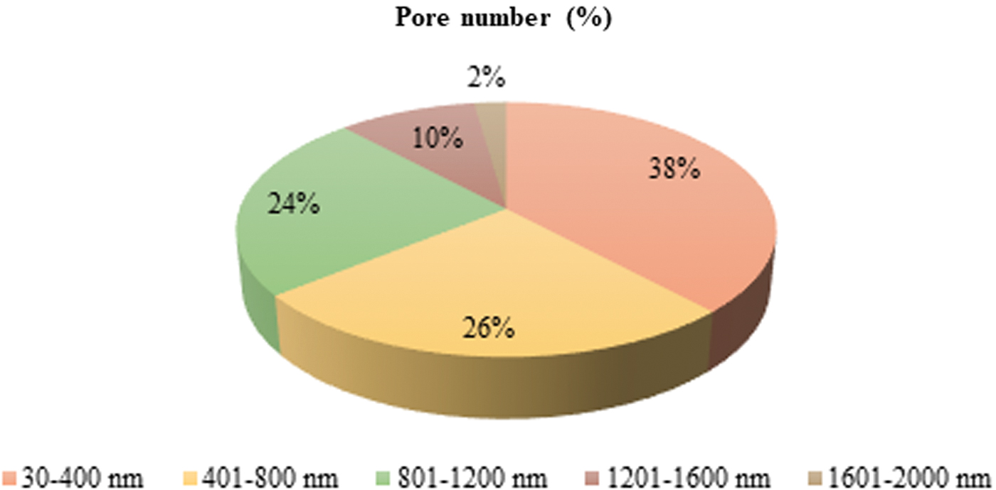
Pore size distribution of electrospun BC fiber meshes. The pie graph shows the results of pore number distribution across size ranges classes performed via ImageJ analysis. Color images are available online.
Characterization of CN/BC fiber meshes
CNs were uniformly electrosprayed using an aluminum foil as a substrate (Fig. 3C, D), and showed an average size (i.e., main dimension) of 180 ± 47 nm. In addition, using the BC electrospun meshes as substrates, CNs were homogeneously detected at the surface of the fibers (Fig. 3E, F).
FTIR analysis was performed over pristine BC, electrospun BC fibers, dried pristine CNs, and CNs-coated on the surface of electrospun BC fibers to check for CN presence via modifications in the chemical composition following the different manufacturing processes (Fig. 5). All samples were characterized by FTIR in ATR mode. The characteristic bands of BC, including 3334 cm−1 attributable to O–H stretching (i.e., hydroxyl groups) of cellulose I, 2884 cm−1 attributable to CH2 asymmetric stretching, 1155 cm−1 attributable to asymmetric stretching C–O–C, CH deformation, 892 cm−1 attributable to CH out of plane bending vibration and 1110 cm−1, assigned to C−O−C stretching within an anhydroglucose ring were also observed on spectrum of BC fibers.
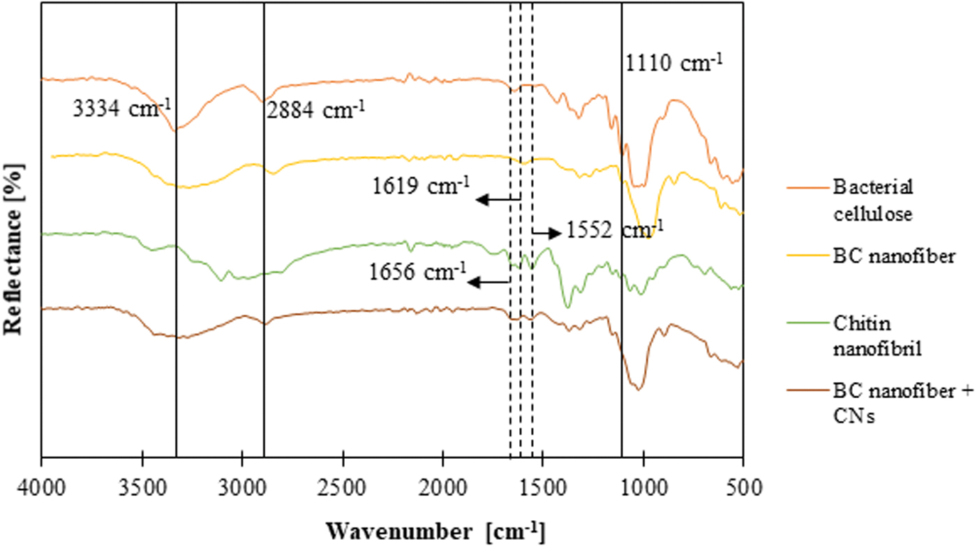
Fourier-transform infrared spectroscopy spectra of pristine BC, electrospun BC nanofiber, pristine CNs and BC electrospun nanofibers coated with electrosprayed CNs. Color images are available online.
The obtained results demonstrated that the solvent system and electrospinning did not affect BC structure. The characteristic bands of CN are 1010 and 1070 cm−1, typical of C–O stretching, 1552 cm−1 attributed to amide II, 1619 and 1656 cm−1 attributed to amide I, 2874 cm−1 attributed to C–H stretching, 3102 and 3256 cm−1 attributed to N–H stretching of the amide and amine groups, and 3439 cm−1 attributable to O–H stretching. The main characteristic bands of CN (1552, 1619 and 1656 cm−1) were also observed on CN-coated BC fiber spectrum. These observations confirmed the presence of CNs on the scaffold surfaces.
BC fibers harvested from the bacteria, BC fibers produced by electrospinning, and BC fibers coated with CNs were characterized by wide-angle XRD spectroscopy to investigate the crystallinity of the material and the effect of electrospinning. Air-dried BC fibers harvested from the bacteria showed the characteristic peaks of type I cellulose at 14.4° (1ī0), 16.7° (110), and 22.5° (200) 22 (Fig. 6A). Electrospun BC fibers, with and without CN functionalization, were tested to investigate how the processing technique affects the crystal structure of the material. Although no difference was introduced by the surface functionalization on the crystal composition of the samples, the electrospun BC had a mainly cellulose II structure compared with the BC fibers harvested from the bacteria, with characteristic peaks at ∼2θ = 12.0° (101), 20.0° (10ī) (shoulder peak), and 21.5° (002) (Fig. 6B).

X-ray diffraction patterns of
The crystallinity percentage of the electrospun BC was overall lower than the BC harvested from the bacteria, confirming that cellulose I to cellulose II crystalline polymorph transformation had occurred, as reported in a previous study.23,24 CrI was calculated for all the samples to discover the relative amount of crystalline material in BC. 25 Although CrI was found to be high for the BC fibers harvested from the bacteria, electrospun BCs resulted in significantly lower values probably because of the destruction of hydrogen bonding forces when pure BC is exposed to the action of solvents. 23 It is also known that the traditional cellulose type II structure model describes cellulose chains, as containing both crystalline (ordered) and amorphous (less ordered) regions.25,26 Results for crystallinity percentage and CrI are summarized in Table 4.
Crystallinity and Crystallinity Index Values Obtained for the Differently Processed Bacterial Cellulose Samples
BC, bacterial cellulose; CN, chitin nanofibril; CrI, crystallinity index.
The BC electrospun fiber mesh thickness was 90 ± 14 μm. The Young's modulus of BC fiber mesh was 2.48 ± 0.62 MPa, calculated by removing the initial slipping region, that is, within a strain range 0.8%–1.8%.
Biological characterization
The cytotoxicity evaluation was performed according to ISO 10993-5:2009. None abnormalities in extract appearance were observed, that is, no signs of particles, clouding, discoloring, or chemical precipitation were recorded. The extract was not filtered before use. Based on the results, the test sample was considered noncytotoxic (Table 5).
Viability of L929 Cells with Extracts from Electrospun Bacterial Cellulose Fibers (Indirect Cytotoxicity Test)
Irritation tests were performed on rabbits according ISO 10993-12:2021. All animals survived, and no abnormal signs were observed during the study. According to the observation, the primary irritation score for the test sample was calculated to be 0, as given in Table 6. Based on the results, the response of the test sample was categorized as negligible under the test condition; the test sample CN-coated BC fiber patch is considered nonirritating.
Results of Irritation Test Conducted on Rats According to ISO 10993-12:2021
To understand the suitability of CN/BC fiber meshes to be used for wound healing, in vitro experiments were performed using human dermal keratinocytes (HaCaT cells) and HUVECs. The AlamarBlue test was performed to assess the metabolic activity of the cells cultured on BC fiber and CN-coated BC fiber meshes (Fig. 7). HaCaT cells cultured on BC fibers showed an increased metabolic activity (p < 0.001) between 1 and 3 days of culture. In addition, on day 3 HaCaT cell metabolic activity on BC fibers was superior as compared with both the same cells cultured on CN-coated BC fibers (p < 0.0001) and HUVECs cultured on BC fibers (p < 0.0001).
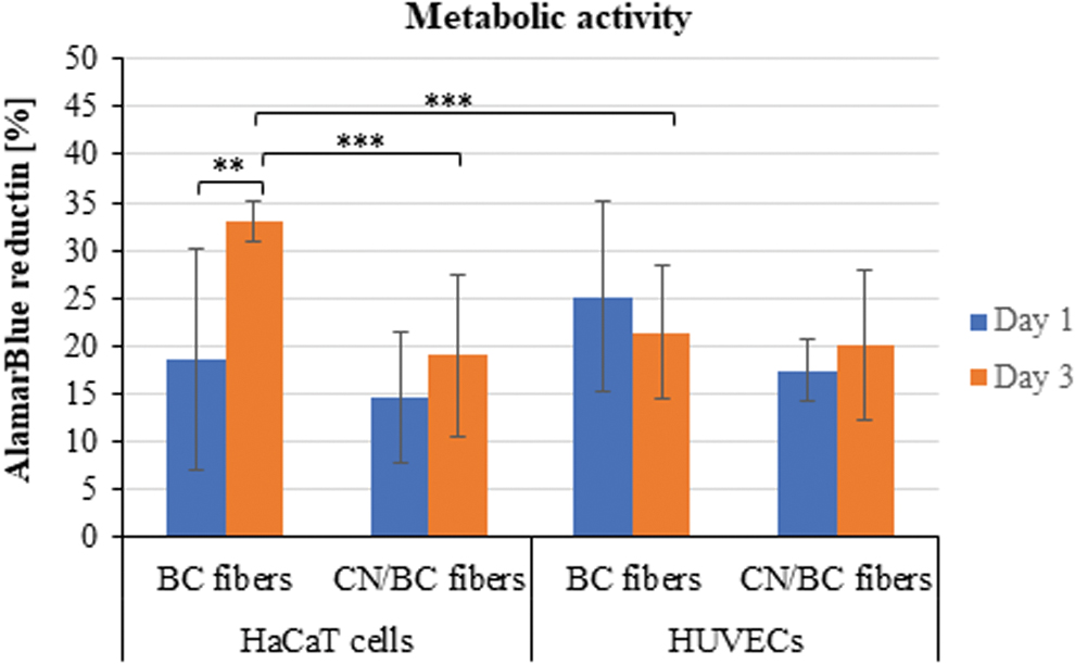
Bar graph showing the metabolic activity (mean ± standard deviation) of HaCaT cells and HUVECs cultured on BC fibers and CN-coated BC fibers for 3 days; **p < 0.001, ***p < 0.0001. HUVECs, human umbilical vein endothelial cells. Color images are available online.
At the endpoint, imaging analyses were performed to assess cell morphology and function (Figs. 8 and 9). The results of the Live/Dead test showed that in all the constructs live cells highly predominate over few dead cells (Fig. 8). In particular, HaCaT cells were able to form a quite uniform layer on the uncoated and CN-coated BC fibers (Fig. 8A, B). HUVECs were found to be on different z-planes in the fibrous samples (Fig. 8C, D). The SEM images confirmed a good colonization of the fibers for both cell types, which in uncoated BC fibers gave rise to many layers of cells that covered the top surface (Fig. 9A, C).
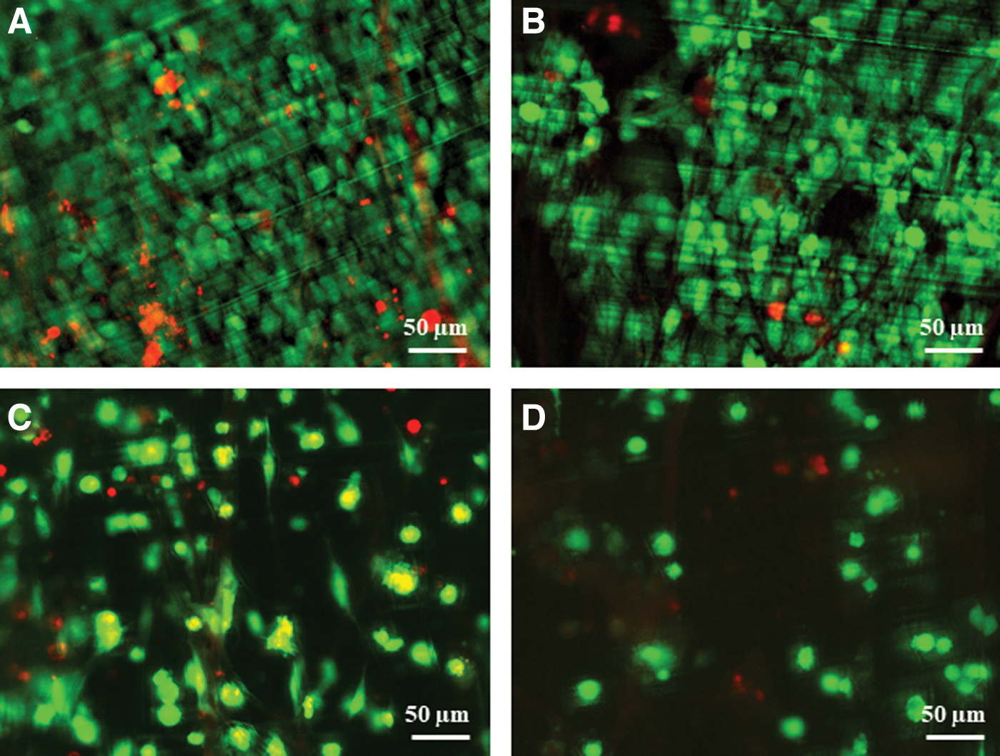
Live/Dead assay fluorescence micrographs showing
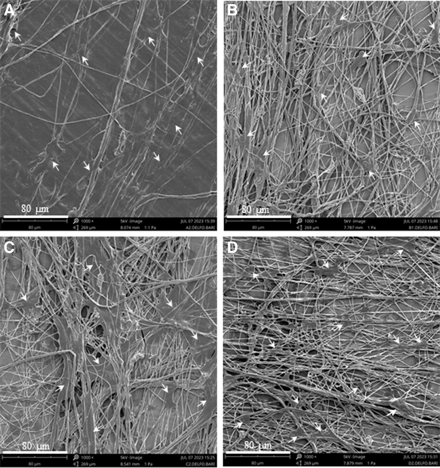
SEM micrographs showing
Figure 10 provides the results of quantitative RT-PCR related to different cytokines involved in the inflammatory response of HaCaT cells after 6 and 24 h of exposure uncoated and CN-coated BC electrospun fibers. The outcomes show that all the samples possess a proinflammatory activity, in fact they are capable to strongly upregulate the expression of IL-6, IL-8, and (at 24 h) IL-1α, and IL-1β, mostly in the presence of CNs. In addition, CN-coated BC fibers were uniquely able to upregulate the expression of HBD-2. Therefore, it can be hypothesized that this type of samples is endowed with indirect antibacterial activity. Significantly, after an increase at 6 h, TNF-α, which is a powerful proinflammatory marker, returned to basal levels in 24 h.
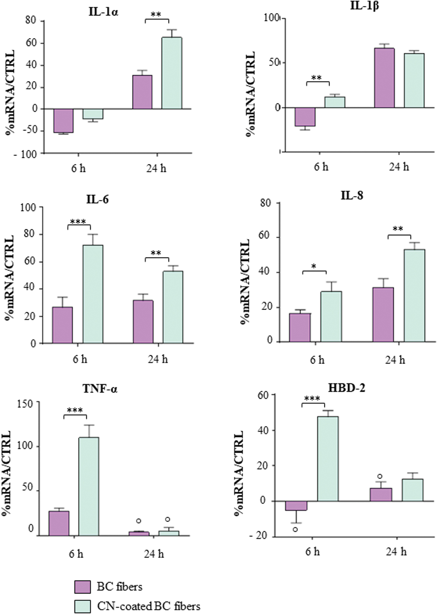
Bar graphs showing the results of real-time reverse transcriptase–polymer chain reaction related to different cytokines involved in the inflammatory response of HaCaT cells after being exposed for 6 and 24 h to uncoated and CN-coated BC electrospun fiber. The results were reported as percentage (%) normalized by the basal expression of those cytokines in untreated cells, as controls (CTRL). °p, Nonsignificant with respect to CTRL; *p < 0.05, **p < 0.001, ***p < 0.0001. Color images are available online.
Discussion
TM perforations are a pervasive clinical problem in otology. Purulent secretion, physical external trauma, or infections such as chronic otitis media often result in TM perforation, which may be by accompanied by long-term hearing damage and conductive hearing loss. 27 Depending on the extent of TM perforation, different otologic reconstructive surgeries are currently performed using autologous tissues, including autologous temporal fascia or fat.28,29 Although surgery can use successful strategies to replace the wounded eardrum, pathology recurrence usually happens owing to the presence of an inflamed ear as a result of chronic infections, which challenge the stability of biomaterial replacements. 30 Long-term structural middle ear restoration may not be obtained because graft tissues are often resorbed owing to inflammatory and mechanical issues. Hence, a tissue engineering approach based on different long-term biodegradable polymers has been proposed.6,31,32
For small perforations, healing can be induced by a tympanic patch that supports cell migration. In clinical practice, diverse materials are used for this purpose, spanning from hyaluronic acid, to fat and paper. Recently, electrospun polymeric fiber meshes have been proposed for TM healing either as patches (for small perforations) and scaffolds (for large perforations, up to full replacement). 33
In this study, we hypothesized that an electrospun fiber mesh based on BC could be an ideal wound-healing patch for TM perforation owing to some specific characteristics of this biopolymer, including high purity, wettability, wide biocompatibility, and transparency (i.e., requested by otosurgeons to monitor TM healing), as well as other properties related to electrospun materials, such as a very large surface area to volume ratio, high porosity with adequate pore size, and tunable mechanical properties.34,35 In addition, owing to the key role of acute versus chronic inflammation in would healing, we propose CNs to produce a bioactive patch able to modulate inflammatory signals and possibly stimulate native immune response in epithelial cells. Having a patch that could fulfill the optimal regenerative and functional requirements could offer a relevant step forward in myringoplasty.
Recently, nanostructured cellulose types have become a subject of intense interest, because they can combine the general nature of cellulose with specific features of nanoscale materials, thus offering improved properties of interest. Electrospinning is a straightforward process to produce continuous cellulose fibers approaching the nanoscale size. 36 BC fibers are harvested from bacteria as an extracellular product; however, these fibers, as collected, do not allow any control over physical properties, such as size, morphology, porosity, pore size, fiber orientation, which instead can be reached if BC fibers are postprocessed by electrospinning. 37 Compared with a typical BC mesh harvested from bacteria, the electrospun BC mesh exhibited a more uniform and evenly dispersed fiber structure, greater porosity, and better water retention capability. 23
We used [Bmim]OAc, an ionic liquid, as a green solvent for solubilizing cellulose. 38 The use of ionic liquids for cellulose dissolution stems from their unique properties that interact with the strong hydrogen bonds of polysaccharides and facilitate the electrospinning process to produce fibers with tunable characteristics. 39 Cellulose molecules have strong hydrogen bonding interactions that cause them to aggregate, even at dilute concentrations. 40 The shear thinning observed at low shear rates differs from that observed in many liquid crystalline polymeric systems, in which a transition of liquid crystalline phase occurs.41,42
It has been demonstrated that cellulose and ionic liquids can combine to form dynamic clusters within solutions. Therefore, the initial shear thinning observed in our study (e.g., range = 1–30 s−1) was linked to the gradual disruption of the physical network formed by the hydrogen bonds of cellulose or ionic liquid between the clusters. A Newtonian fluid region (i.e., constant viscosity) can thus be observed at higher shear rates (e.g., range = 40–100 s−1). We estimated the shear rate (
in which R is the needle inner radius, V is the fluid volume, t is the time; therefore, F is the flow rate.
Based on the applied shear rate of 92.72 s−1 in our apparatus, it can be inferred that the BC solution exhibits a constant and low viscosity. Although cellulose could dissolve in [Bmim]OAc very well, the resultant high viscosity and nonvolatility of the solution, made the electrospinning process hard to control for continuous jets and fiber production. In the common case of polymeric solutions, the jets remain stable against capillary perturbations, but they can experience bending instability owing to the electric field, a phenomenon commonly observed during the electrospinning process. 43 If the viscosity of the solution is too high, the bending instability is suppressed, and instead of forming continuous fibers, the polymer jet breaks up into droplets, which results in the formation of beads instead of fibers.
Typically, when the viscosity of cellulose solution rises, it leads to an increase in the chain overlap or entanglement between cellulose molecules up to a critical point at which the jet elongation is facilitated, and the ejected solution is prevented from breaking up. However, the viscosity should not be so high to completely suppress capillary instability, which happened for cellulose solution dissolved only in [Bmim]OAc. 36 Second, the poor electrical conductivity of ionic liquids can cause the electric field to be distorted, leading to a loss of control over fiber formation. To address these issues, researchers have explored various methods such as adjusting the solution concentration, adding cosolvents or surfactants, optimizing the electrospinning parameters, ultimately using alternative solvents or processing techniques to improve the spinnability of cellulose in ionic liquids.16,44
In this study, we used DMSO as a cosolvent with the aim to modulate the solution viscosity, reduce the entanglement density of the system, increase the electrical conductivity, improve spinning of the solution, and finally increase the removal of the solvent. The presence of cosolvent can also strongly affect the surface tension of solution. 36 We found that the BC solution showed a typical shear-thinning behavior at high shear rates.
Since [Bmim]OAc did not evaporate between the electrospinning spinneret and the collector, a custom-designed spinning procedure was used, in which the cellulose fibers were deposited into a coagulant bath containing d-water to recover the fibers and remove the ionic liquid. The high polarity of the coagulant governs the hydrophobic interactions between polymer chains during regeneration. 45 Coagulation in water with higher polarity, mainly leads to cellulose type-II formation, whereas coagulation in alcohols predominantly generates noncrystalline structures. In our system, continuous and uniform BC fibers were obtained. An increased alignment of fibers was obtained thanks to the application of rotating collector inside the d-water bath.
The poor solubility in physiological media as well as the absence of β-glucanase enzymes that break down large polysaccharides via hydrolysis in humans make BC a potentially suitable candidate for durable biomedical application and functionality. 46 BC is very well tolerated in the human body; however, it does not inherently possess bioactive properties, such as immune-modulatory activity. 47 Therefore, BC composites with nanomaterials and other polymers have been developed to improve these aspects. Recently, CNs have received a lot of attention in the skincare area for their green character, as well as superior antibacterial and immunomodulatory properties.7,47,48
Application of CNs in biomedical structures, such as otologic devices would be convenient, because CNs are biodegradable by resident enzymes, and there is no risk of accumulating in organs. Mota et al. explored the use of CNs as a nanofiller for poly(ethylene oxide terephthalate)-poly(butylene terephthalate) (PEOT-PBT) copolymer in the production of electrospun and fused deposition modeling-based TM scaffolds suitable for clinical use. 49 The nanocomposite substrates showed excellent oto- and cytocompatibility, which confirmed their biological relevance in perforated eardrum reconstruction. These findings are in line with those reported earlier by Danti et al., who showed a significant indirect antimicrobial activity on HaCaT cells exerted by crustacean-derived CNs, in comparison with mushroom-derived CNs, when electrosprayed onto PEOT-PBT electrospun fiber scaffolds developed for TM repair. 3
In this study, we used a similar strategy to provide CN availability in the middle ear, induce a suitable immune response, and fight inflammatory states. We used electrospray to coat the surface of the fibrous mesh with CNs, to have them located, and available at the surface, where they would provide biological activity in the short term after application, as previously proposed for skincare.7,50 In addition, the mucoadhesive properties of chitin make CN-decorated materials valid candidates as TM patches. The produced BC fibers were successfully decorated by CNs, as confirmed by the FTIR spectra, and resulted nonaggregated, as shown by SEM analysis. This result is not straightforward, because the specific back layer material affects the deposition of nanoparticles during electrospray.
In some areas, the deposited CNs generated a surface layer on the top of the fibers, thus reducing the available porosity for cell penetration. However, because CNs are biodegradable and not chemically bound to the fibers, it is expected that this initial difference in scaffolds' morphology will not affect the regenerative behavior of the patch.
One of the main challenges for the development of cellulosic structures is the comprehensive understanding and the predictability of crystallization, which effects on chemical stability of the cellulose regenerated from ionic liquids. 51 Some studies stated relatively low crystallinity of cellulose regenerated from ionic liquids, which may affect mechanical properties. 52 The native cellulose generally is polymorph type-I (i.e., cellulose I) with the highest amount of intermolecular and intramolecular hydrogen bonds. The crystalline polymorph cellulose I transforms into a polymorph type-II after being dissolved in an ionic liquid. 53 Indeed, during the dissolution procedure, the ionic liquid molecules interpenetrate to the gap between the sheets of hydrogen bond in cellulose I, leading to a slight lattice expansion.17,54 It has also been found that electrospinning of cellulose solution in ionic liquids gives rise to an increase in the amorphous phase and transfer of polymorph type-I into polymorph type-II cellulose, which corroborates our results. 44
The mechanical properties of TM are crucial for its function in hearing and protecting the delicate structures of the middle ear. The elasticity and mechanical strength of the artificial patch should be matched with the TM mechanical properties for sound conduction. 55 The tensile strength of TM is estimated to be ∼4–5 MPa. 56 Different values (e.g., 1–400 MPa), have been reported for the Young's modulus of the human TM, depending also on applied method. 57 A Young's modulus value of 4.38 MPa has recently been reported for PEOT-PBT based electrospun TM scaffolds. 5
We performed preliminary in vitro indirect cytotoxicity and in vivo skin irritation assessment according to ISO 10993. These outcomes demonstrated that produced materials were cytocompatible with no sign of skin irritation in rabbits. Therefore, we conducted in vitro tests using human cells. Specifically, we chose HUVECs and HaCaT cells to investigate the specific cytocompatibility with endothelial and epithelial cells, both involved in the wound healing process at short times (i.e., 72 h). In addition, HaCaT cells were used to address the role exerted by uncoated and CN-coated BC fiber meshes in inflammatory and innate immune response at 6 and 24 h, because epithelial cells are known to contribute to healing by secreting several cytokines and defensins. 58
The cytocompatibility assays showed that uncoated and CN-coated BC fiber meshes could support HaCaT cell and HUVEC adhesion. After 3 days, uncoated BC fibers displayed the highest metabolic activity of HaCaT cells, whereas no statistically significant differences could be observed with both cell types in uncoated and CN-coated formulations. All the fibrous samples were densely populated by cells, which were predominantly live. In this short time, HaCaT cells could form a cellular layer on both the types of BC fiber meshes, as shown by SEM micrographs. Both uncoated and CN-coated fiber meshes exerted an initial proinflammatory activity on epithelial cells, by upregulating a panel of cytokines involved in the wound healing process, which was significantly higher in CN-coated BC fiber samples.
CN displayed a role in the wound healing process, by modulating the expression of the proinflammatory cytokine TNF-α, which was remarkably upregulated at 6 h and reported to basal level at 24 h. Wound healing is a complex biological process, which is described by a series of events primed by acute inflammation, and followed reepithelialization, matrix formation, tissue repair, and remodeling, in which biomaterials can play a dominant role. 59 The initial phase occurring in the first days actually drives the healing process by stimulating the necessary production of proinflammatory cytokines. Among all, TNF-α and IL-1 represent the actuators of proinflammatory response and are immediately released by keratinocytes. TNF-α is an essential mediator of inflammation; it promotes different responses by the vascular system, for example, the recruitment of immune cells, 60 and on the contrary, TNF-α stimulates fibroblast growth factor production, thus helping the reepithelialization process. 61
Through its two isoforms α and β, IL-1 activates a local antimicrobial pattern in epithelial cells, which concurs to the upregulation of IL-6 and/or IL-8. The former helps recruiting the immune cells and inducing their differentiation, whereas the latter is also involved in the angiogenic process.62,63 In a second phase, at longer times, inflammation must stop to allow tissue regeneration. Basing on the obtained outcomes, we observed that electrospun BC meshes can act at early stages of the wound healing process.
The last molecule investigated was HBD-2, which highlights if these patches could promote indirect (i.e., cell mediated) antibacterial activity. Defensins are small cationic peptides that can be synthesized by epithelial cells upon injury, and have been largely studied, because they possess biocidal activity toward several microorganisms, such as bacteria and fungi. 64 Recently, some microstructured materials have been discovered to induce HBD-2 expression in dermal keratinocytes, which is considered a very promising strategy in the management of complex wounds. 65 In our findings, only CN-coated BC fiber meshes were able to induce a significant HBD-2 upregulated expression in HaCaT cells, as shown by RT-PCR analysis, seen at 6 h and still significant after 24 h. This result is supportive of the application of CN-coated BC patch in TM perforations sustained by underlying infections, such as otitis.
Although these first in vitro and preliminary in vivo studies look promising, future investigations need to assess the regenerative and proper immune response capacity in an in vivo model of eardrum perforation, to provide a substantial proof for device validation.
Availing natural nanoforms of biopolymers, such as BC and CN, and using less impacting solvents would allow sustainable, yet effective, nanotech products to reach bedside practice as an alternative to current TM patches available on the market, such as paper, collagen, and others, which are combined with silver or antibiotics to improve the clinical outcome.
Conclusion
The treatment of TM perforations remains a challenging task, particularly in cases of chronic infections that hinder the long-term stability of graft materials. This article explored a novel tissue engineering approach using nanotech strategies to develop a functional TM patch to promote optimal wound healing and tissue regeneration. Electrospinning of BC solution was successfully achieved using [Bmim]OAc as a green solvent, thus overcoming the limitations posed by cellulose poor solubility in conventional solvents. By optimizing the electrospinning parameters and introducing DMSO as a cosolvent, we obtained continuous and uniform BC nanofibers. To enhance the immunomodulatory and regenerative properties of the BC fiber mesh, CNs were electrosprayed onto the surface of BC fiber meshes.
The presence of CNs demonstrated the ability to induce indirect antimicrobial properties by enhancing the expression of HBD-2 in HaCaT cells, and promoted modulation of the proinflammatory cytokines, making the developed patch a promising candidate for supporting the wound healing process in TM perforations. In vitro cytocompatibility studies using mouse L929 fibroblasts, as well as human dermal keratinocytes HaCaT cells, and HUVECs confirmed the cytocompatibility of both uncoated and CN-coated BC fiber meshes. In vivo tests on rabbit skin showed no irritation after 24 h.
In summary, the combination of electrospun BC nanofibers with electrosprayed CNs offers a promising approach for the development of an ideal TM patch that addresses the challenges faced by current graft materials. This innovative biomaterial has the potential to significantly improve the outcomes of myringoplasty procedures, providing both optimal regenerative capabilities and functional support for TM healing. Future studies and clinical trials are warranted to further validate the effectiveness of this nanotech-based approach in restoring hearing function and providing long-term structural TM healing.
Footnotes
Acknowledgments
The authors thank Dr. Delfo D'Alessandro (University of Pisa) for his technical support in biological analysis and Dr. Andrea Mele (University of Sheffield) for his technical support to X-ray diffraction analysis. Linari Engineering s.r.l. is acknowledged for electrospinning equipment support. The Centre for Instrumentation Sharing–University of Pisa is acknowledged for SEM analysis.
Authors' Contributions
B.A.: Conceptualization, data curation, supporting, funding acquisition, investigation, project administration, writing—original draft, writing—review and editing; A.R.: investigation; A.F.: investigation, methodology; T.M.: investigation, methodology, visualization, writing—original draft; C.R.: investigation, methodology; M.H.: visualization; L.G.: methodology; G.D.: methodology, resources, software, supervision; R.B.: supervision, visualization; M.L.: supervision; I.R.: investigation, methodology, resources; S.D.: formal analysis, resources, validation, writing—original draft, writing—review and editing; A.L.: resources. All authors reviewed the article and agreed to its published version.
Authors' Confirmation Statement
Ms. A.R., Dr. R.B., and Dr. M.L. are from Amirkabir University of Technology (Tehran, Iran), where education and research are the primary functions.
Disclosure Statement
The authors declare no competing interests.
Funding Information
Industrialization Center for Applied Nanotechnology and Iran Nanotechnology Innovation Council are gratefully acknowledged for supporting this study (NanoCell project).