




Abstract
A combination of hydrogels and stem cell spheroids has been used to engineer three-dimensional (3D) osteochondral tissue, but precise zonal control directing cell fate within the hydrogel remains a challenge. In this study, we developed a composite spheroid-laden bilayer hydrogel to imitate osteochondral tissue by spatially controlled differentiation of human adipose-derived stem cells. Meticulous optimization of the spheroid-size and mechanical strength of gelatin methacryloyl (GelMA) hydrogel enables the cells to homogeneously sprout within the hydrogel. Moreover, fibers immobilizing transforming growth factor beta-1 (TGF-β1) or bone morphogenetic protein-2 (BMP-2) were incorporated within the spheroids, which induced chondrogenic or osteogenic differentiation of cells in general media, respectively. The spheroids-filled GelMA solution was crosslinked to create the bilayer hydrogel, which demonstrated a strong interfacial adhesion between the two layers. The cell sprouting enhanced the adhesion of each hydrogel, demonstrated by increase in tensile strength from 4.8 ± 0.4 to 6.9 ± 1.2 MPa after 14 days of culture. Importantly, the spatially confined delivery of BMP-2 within the spheroids increased mineral deposition and more than threefold enhanced osteogenic genes of cells in the bone layer while the cells induced by TGF-β1 signals were apparently differentiated into chondrocytes within the cartilage layer. The results suggest that our composite spheroid-laden hydrogel could be used for the biofabrication of osteochondral tissue, which can be applied to engineer other complex tissues by delivery of appropriate biomolecules.
Impact statement
This research developed a bilayer hydrogel encapsulating two types of composite spheroids, with each layer engineered to induce osteogenic or chondrogenic differentiation. The hydrogel was prepared by adjusting the size of spheroids and mechanical strength of hydrogel to provide a desired microenvironment of encapsulated cells, leading to homogeneous sprouting. Spatially confined delivery of osteogenic or chondrogenic signals in each layer enabled the specific differentiation to each layer. Thus, our spheroid-laden bilayer hydrogel could be a potential model or therapeutic platform as three-dimensional (3D) osteochondral tissue.
Introduction
Global demand for orthopedic replacements has been increasing every year due to the aging population, and tissue engineering approaches that mimic complex characteristics of natural tissues have been burgeoning to meet this demand. 1 In particular, the pathologies associated with osteoarthritis often led to lesions throughout osteochondral tissue, and thus, a three-dimensional (3D) tissue recapitulating its fundamental structural features and biological functions is needed.1,2
Osteochondral tissue is composed of cartilage and subchondral bone, which support the elastic lubrication of joints and the strength of human body, respectively. 3 Biphasic constructs have been designed to mimic this hierarchical structure using biomaterials, cells, and signaling molecules. 4 For example, combinations of soft/elastic materials as a cartilage layer and highly mineralized and mechanically strong materials as a bone layer have been investigated. 2 The cartilage or bone-related cells were seeded, and then chondrogenic or osteogenic signaling molecules were delivered into the different layers of biphasic structure. 5 However, meticulously engineering 3D osteochondral constructs with specific zonal characteristics of cartilage and subchondral bone remains a challenge due to technical difficulties in effective delivery of signaling molecules, and ensuring adequate interfacial adhesion between the two distinct layers.2,4,5
Cell-compatible hydrogels such as collagen, fibrin, and gelatin methacryloyl (GelMA), and hyaluronic acid have been used to engineer osteochondral constructs by encapsulating various cell types.6,7 However, the encapsulated cells often show limited differentiation capability due to low cell viability and lack of paracrine signaling.4,7–9 For example, a GelMA hydrogel encapsulating human adipose-derived stem cells (hADSCs) and a poly(ethylene glycol) (PEG) hydrogel incorporating mesenchymal progenitor cells showed minimal deposition of extracellular matrix (ECM).8,9 Recently, spheroids, self-assembled by multiple cells, demonstrated enhanced cell to cell interactions and paracrine signaling, which have been encapsulated within hydrogels to address the limitations. 10 For example, chondrocyte or bone marrow-derived stem cell (BMSC) spheroids improved chondrogenic or osteogenic ECM deposition, respectively, within a hydrogel.11,12
However, spheroids were often hindered from sprouting and proliferating within hydrogel, leading to poor in vitro differentiation and deformation of structure, which might be explained by suboptimal mechanical properties of hydrogel, disrupting cell/gel matrix interactions and ECM remodeling capacity of the cells.10–12 In addition, failure to take into account the size of spheroids often resulted in their sedimentation within hydrogel during crosslinking processes, impairing normal tissue structure formation and producing atypical tissue with flat or islet-like morphology. 13 Therefore, mechanical microenvironment of hydrogel and homogeneous encapsulation of spheroids should be delicately engineered to guide cell behavior in a hydrogel.
Various signaling molecules, such as growth factors, biominerals, or small chemicals, have been used to guide the fate of stem cells within a hydrogel. 4 Despite positive results in previous works, stem cell activity was varied or affected by the delivery modality. 7 For example, a bilayer hydrogel encapsulating mesenchymal stem cells (MSCs) serially cultured with chondrogenic and osteogenic conditioned media showed insignificant differentiation to each lineage. 14 Coacervate microspheres releasing insulin-like growth factor 1 was co-encapsulated with hADSCs in gelatin-based hydrogel, but hard to generate cartilaginous ECMs in vitro. 15 The results indicate that indirect interactions between the encapsulated cells and signaling molecules reduced induction of stem cells and the off-targeted molecules may cause dedifferentiation.2,15,16 Meanwhile, composite spheroids assembled with signaling molecules have been developed and demonstrated effectiveness in leading to lineage-dependent differentiation, as well as spatially confined delivery of inductive signals within a spheroid. 17
For example, composite spheroids of adipose-derived stem cells and gelatin microparticles loaded with transforming growth factor beta-3 (TGF-β3) and fragmented fibers immobilized with biominerals enhanced chondrogenic or osteogenic gene expression better than when the same signals were provided in medium18–20 ; encapsulating composite spheroids assembled with signaling molecules within hydrogels could enable both stable localization of spheroids and spatially confined signal delivery. However, current studies are inadequate to define optimal microenvironment for a hydrogel that could contain such spheroids and prevent dedifferentiation without assistance of external biomolecules.21,22
To engineer a biphasic structure that mimics osteochondral tissue, chondro- and osteoconductive materials have been attached using several methods, including ionic interactions, physical adhesion, and pasting through chemical glues (e.g., cyanoacrylate). 5 However, the different characteristics of two layers with regard to stiffness, size, and irregular compartments made earlier constructs vulnerable to phase separation, tissue delamination, and unstable localization.16,23,24 Meanwhile, a bilayer hydrogel could be a potential tool to prevent phase separation because a hydrogel can be formed by sequential crosslinking of bottom and top layers with similar mechanical properties and pore sizes without apertures. 25 For example, Liu et al. prepared three different types of bilayer hydrogel combining different concentrations of glycol chitosan and sodium alginate, and they demonstrated stable positioning without a division of layers in all groups. 26
Despite the advantage of a bilayer hydrogel, adhesion force between the layers is still far from satisfactory, and composite spheroid-laden bilayer hydrogels have barely been investigated, and thus it is necessary to characterize cellular responses, such as viability, zone-specific differentiation, and efficiency in delivering signaling molecules, within the hydrogel microenvironment.25,26
In this study, we developed GelMA hydrogels encapsulating hADSC spheroids and investigated their size and mechanical properties on sprouting and ECM remodeling ability of incorporated cells. We prepared spheroids incorporating fibers immobilized with osteo- or chondroinductive growth factors, and delicately investigated the effects of delivery modalities of growth factors on spatially confined osteogenic or chondrogenic differentiation of hADSCs. Finally, by using sequential crosslinking of each layer, we developed a bilayer hydrogel encapsulating the composite spheroids under desired size of spheroid, mechanical strength of hydrogel, and delivery modality of inductive factors.
Materials and Methods
Materials
Poly(ι-lactic acid) (PLLA; 5.7–8.5 dL/g intrinsic viscosity, Mn 350–500 kDa) was purchased from Samyang (Seoul, Korea). The 1,1,1,3,3,3-Hexafuloro-2-propanol (HFIP) and 4% paraformaldehyde were acquired from FUJIFILM Wako Pure Chemical Corporation (Osaka, Japan), and Tris-HCl was acquired from Alfa Aesar (Haverhill, MA). Ethylenediamine (EDA), isopropyl alcohol (IPA), dopamine hydrochloride, fluorescein isothiocyanate (FITC), 2-hydroxy-2-methylpropiophenone, an ApopTag® Peroxidase In Situ Apoptosis Detection Kit, sodium bicarbonate (NaHCO3), sodium carbonate (Na2CO3), dimethyl sulfoxide (DMSO), Alizarin Red S, Alcian Blue solution, and Trypan Blue solution were purchased from Sigma-Aldrich (St. Louis, MO).
For cell culture and hydrogel, hADSCs, basal medium, fetal bovine serum (FBS), and penicillin/streptomycin (P/S) were purchased from CEFOBIO (Seoul, Korea). Ultra-low cell-adhesive micro-well plates, Labsphero™, were obtained from Labtolab (Daejeon, Korea). Phosphate-buffered saline (PBS), its ion-free version (Dulbecco's phosphate-buffered saline; DPBS), and trypsin/ethylenediaminetetraacetic acid were purchased from Welgene (Gyeongsan, Korea).
GelMA (Gelatin from porcine skin, type A, gel strength: ∼300 bloom, degree of methacrylation: >90%) was acquired from 3D Materials (Busan, Korea), and ultraviolet (UV) spot curing system was from UV SMT (Bucheon, Korea). Growth factors, bone morphogenetic protein-2 (BMP-2), and transforming growth factor beta-1 (TGF-β1), were obtained from PeproTech (Rocky Hill, NJ).
For the in vitro assays, rhodamine/phalloidin, a LIVE/DEAD® Viability Kit, and CellROX green reagent were purchased from Invitrogen (Carlsbad, CA). Diamidino-2-phenylindole (DAPI) was obtained from Vector Laboratories (Burlingame, CA). Hematoxylin and Eosin (H&E) solution was purchased from BBC Biochemical (Mount Vernon, MA). A Zeba Spin desalting column was purchased from Thermo Scientific (Waltham, MA). Primary antibodies (anti-runt-related transcription factor 2 [RUNX2], osteopontin [OPN], SRY-box transcription factor 9 [SOX9], aggrecan, collagen type 1a [Col1a], and collagen type 2a [Col2a]) and enzyme-linked immunosorbent assay (ELISA) kits (BMP-2, bone sialoprotein [BSP], TGF-β1, and TGF-β3) were purchased from Abcam (Cambridge, United Kingdom).
Secondary anti-mouse/rabbit immunoglobulin G (IgG) biotin-conjugated antibody was obtained from Sigma-Aldrich, and FITC-streptavidin was purchased from eBioscience (San Diego, CA).
Components of real-time quantitative polymerase chain reaction (RT-qPCR; the RNeasy Mini Kit, Maxime RT Premix, and SYBR Premix Ex-Taq) were purchased from Qiagen (Valencia, CA), Intron Biotechnology (Seoul, Korea), and TaKaRa Bio (Otsu, Japan), respectively.
Preparation of composite spheroids
To synthesize the segmented fibers, aligned nanofiber sheets were synthesized by electrospinning (5 mL/h, 23-G needle, 12–14 keV) 10 mL of 3% PLLA solution in HFIP. The collected nanofiber sheets were dried at room temperature for 24 h and cut into four pieces. The pieces were embedded in frozen section compound, frozen at −70°C, and cryosectioned using a Cryostat cryocut microtome (Leica Biosystems GmbH, Wetzlar, Germany). The sectioned fibers, 60–100 μm in length, were selectively collected by sieving, treated with 10% (v/v) EDA in IPA for 1 h, and sequentially washed with IPA, 70% ethanol, and distilled water (DW). For polydopamine (PD) coating, the fibers were immersed in a 2 mg/mL dopamine hydrochloride solution (Tris-HCl buffer, pH 8.5) for 20 min. The PD-coated fibers were sequentially sterilized with 70% ethanol, DW, and UV light before use. Morphology of the fibers was observed by Field emission scanning electron microscopy (FE-SEM; Hitachi S 4800 FE-SEM; Hitachi, Tokyo, Japan).
For sample preparation, the fibers were completely dried at 37°C, attached to a scanning electron microscopy (SEM) holder, and coated with platinum by ion sputter (6 nm/s, 10–15 nm in thickness). Length of each fiber was quantified using ImageJ software (National Institutes of Health, Stapleton, NE).
The hADSCs were cultured in basal medium with 10% FBS and 0.5% P/S under standard culture conditions (5% CO2 and 37°C). Cells at passage number 4–5 were used for all experiments, and the medium was refreshed every 2 days. We placed 1 × 10⁶ hADSCs and 125 μg of fibers in a LabSphero with 6000 wells (167 cells and 0.0208 μg of fibers per well); 2.5 × 10³ hADSCs and 0.3125 μg of fibers were placed in one well of an ultra-unattached cell plate (SPL Life Science, Pocheon, Korea); and 5 × 10³ hADSCs and 0.625 μg of fibers were placed in the same plate to prepare small, medium, and large spheroids, respectively. The fibers and cells formed a spheroid after 24 h of incubation.
Phase-contrast images of the spheroids were captured using an optical microscope (CKX41; Olympus, Tokyo, Japan), and area of each spheroid was measured from the images. FITC-labeled fibers were prepared by diluting 68 μg/mL of FITC powder in PLLA solution before electrospinning to visualize the incorporation of fibers in spheroids. Small spheroids incorporating the FITC-labeled fibers were cultured for 24 h and fixed in 4% paraformaldehyde for 1 h, washed three times with PBS, and then stained with rhodamine/phalloidin (1:200 in PBS) for 1 h at 37°C.
Each sample was mounted with DAPI mounting medium (nuclei staining) and observed using a confocal laser microscope (TCS SP5, Leica Biosystems GmbH).
Encapsulation of spheroids in hydrogel
GelMA was dissolved in DPBS (7.5% w/v), incubated for 2 h at 37°C, and mixed with 2-hydroxy-2-methylpropiophenone (photo initiator, 0.1% v/v) after filtering (0.2 μm pores). Then, for engineering one hydrogel, 100 μL of hydrogel solution encapsulating 240, 16, and 8 of small, middle, and large spheroids, respectively (total number of cells in a hydrogel is equally 40,000 cells for all groups), were casted in a polytetrafluoroethylene (PTFE) mold (8 mm in diameter, 2 mm in depth, and cylindrical shape). The spheroids were gently agitated in hydrogel solution, and the solution was then immediately irradiated with UV light (350 mW/cm2, 10 s, 7 cm of distance from each sample) for gelation. The spheroid-laden hydrogels were cultured for 24 h and then fixed in 4% paraformaldehyde for 20 min, dehydrated by immersion in sequential steps of gradient ethanol (70–100%) and xylene, and embedded in paraffin blocks. The paraffin blocks were horizontally sectioned into 10 μm slices using a microtome (Leica Biosystems GmbH) for H&E staining.
The sectioned specimens were hydrated by following the dehydration steps in reverse (xylene, 100–70% ethanol, and water), stained with Hematoxylin for 2 min and with Eosin for 6 min, dehydrated, and mounted. The H&E-stained images were captured using a phase-contrast microscope (Eclipse Ti2; Nikon, Tokyo, Japan). Vertically sectioned hydrogels were stained with calcein AM for 30 min and observed using a fluorescence microscope (Eclipse Ti2; Nikon) to visualize the distribution of spheroids in hydrogels. The positions of spheroids in each gel were divided from bottom to top (0–20%, 20–40%, 40–60%, 60–80%, and 80–100%), and the number of spheroids in each position was quantified.
The small spheroids were used for further experiments. Spheroid-encapsulating hydrogels with various UV exposure times (10, 30, and 60 s) were prepared following the aforementioned gelation process, and optical images of each hydrogel were captured using a camera. After 1 and 14 days of culture, storage modulus of each hydrogel was measured using a rheometer (HR10; TA Instruments, New Castle, DE) under 0.4 N of axial force and 1% strain, and weight of each sample was measured before and after freeze drying to calculate swelling ratio by the following formula: Swelling ratio (w/w) = (weight of swollen hydrogel − weight of dried hydrogel)/weight of dried hydrogel [(Ws − Wd)/Wd]. Phase-contrast images of each hydrogel with spheroids were captured with the optical microscope, and each sample was subjected to H&E and calcein AM staining as described above. Sprouting length from an encapsulated spheroid on each calcein AM-stained image was measured using ImageJ software. Internal structure of each hydrogel and cells from the encapsulated spheroids were observed by FE-SEM.
Before gene expression analysis, each hydrogel was dissolved in collagenase solution (100 CDU/mL, CDU = collagen digestion units) for 1 h at 37°C. The supernatant was discarded after centrifugation (1200 rpm, 3 min), and messenger RNA (mRNA) was extracted and purified using the RNeasy Mini Kit. Concentration of mRNA was measured using a nano-spectrometer (NanoDrop 2000; Thermo Scientific, Wilmington, DE).
Complementary DNA (cDNA) was synthesized by adding 1 μg of mRNA and diethyl pyrocarbonate (DEPC)-treated water to a cDNA Polymerization Kit (total volume: 20 μL; Maxime RT Premix Kit; Intron Biotechnology). Then, 2 μL of the cDNA solution was mixed with 0.4 μL of SYBR reagents, 0.4 μL of ROX reference dye (50 × ), 6.8 μL of DEPC-treated RNA-free water, and 0.4 μL of target-specific primers for the gene of interest, and the samples were processed for RT-qPCR (StepOnePlus Real-Time PCR System; Applied Biosystems, Foster City, CA), with amplification during 40 cycles of annealing at 95.0°C for 15 s, extension at 60.0°C for 60 s, and a melting curve stage at 60.0°C to 95.0°C in increments of 0.5°C per 5 s.
The primer sequences were as follows: rho-associated protein kinase 1 (ROCK1): forward, 5′-GAA GCT CGA GAG AAG GCT GA-3′, reverse, 5′-TTG TCT GCC TCA AAT GCT TG-3′; mitogen-activated protein kinase 1 (MAPK1): forward, 5′-CCA GAC CAT GAT CAC ACA GG-3′, reverse, 5′-CTG GAA AGA TGG GCC TGT TA-3′; matrix metallopeptidase 2 (MMP2): forward, 5′-ATG ACA GCT GCA CCA CTG AG-3′, reverse, 5′-ATT TGT TGC CCA GGA AAG TG-3′; matrix metallopeptidase 9 (MMP9): forward, 5′-TTG ACA GCG ACA AGA AGT GG-3′, reverse, 5′-GCC ATT CAC GTC GTC CTT AT-3′; and housekeeping gene GAPDH: forward, 5′-GTC AGT GGT GGA CCT GAC CT-3′, reverse, 5′-TGC TGT AGC CAA ATT CGT TG-3′. The relative gene expression from gels treated with UV for 10 and 30 s was normalized to that from the 60 s group. DNA assay was performed for each spheroid-encapsulating hydrogel after 1 or 14 days of culture.
The hydrogels at each time point were lysed in 300 μL of radioimmune precipitation assay (RIPA) lysis buffer (150 mM NaCl, 1% Triton X-100, 1% sodium deoxycholate, 0.1% SDS, 150 mM Tris-HCl, pH 7.2), ground using a homogenizer, and reacted with working reagents from a Quant-iT PicoGreen dsDNA Assay Kit (Invitrogen). A spectrophotometer (Varioskan LUX; Thermo Scientific, Waltham, MA) was used to detect fluorescent intensities at an excitation wavelength of 480 nm and emission wavelength of 520 nm. For terminal deoxynucleotidyl transferase dUTP nick end labeling (TUNEL) assay, the hydrogel samples were embedded in paraffin blocks by following the procedure described above and cut into 10 μm slices. The specimens were hydrated, and DNA fragments were stained using an ApopTag Fluorescein In Situ Apoptosis Detection Kit and a fluorescence microscope. Intracellular oxidative stress level of encapsulated spheroids was analyzed by incubating the hydrogel samples in CellROX staining solution (5 μM in culture medium) for 1 h at 37°C and observing fluorescence with the fluorescence microscope.
TUNEL-positive nuclei and relative CellROX intensity were measured using Photoshop CS6 software (Adobe, San Jose, CA).
Immobilization of growth factors on the fiber
We agitated 1 mg of PD-coated fibers with BMP-2 or TGF-β1 solution (10 μg/mL in Tris-HCl buffer, 4°C, overnight) to synthesize BMP-2- and TGF-β1-immobilized fibers. FE-SEM was used to observe the surface morphology of uncoated fiber, PD-coated fiber, BMP-2-immobilized fiber, and TGF-β1-immobilized fiber, which were named Fiber, PD, BMP-2, and TGF-β1, respectively. To visualize the immobilization of growth factors, BMP-2 or TGF-β1 was dissolved in a reaction buffer (3 μg/mL in 500 mM carbonate, pH 9.5) and reacted with FITC solution (1 mg/mL of FITC powder in DMSO) for 1 h at room temperature to chemically conjugate the BMP-2 and TGF-β1 with the FITC. A Zeba desalt spin column (89889; Thermo Scientific, Rockford, IL) was used to remove the unreacted FITC, and the FITC-labeled growth factors were coated onto PD-coated fibers following the same immobilization procedure. Fluorescent images were then captured using the fluorescence microscope, and phase-contrast images of the same spots were used for background.
The amount of each growth factor in the supernatant after immobilization process was measured using a BMP-2 or TGF-β1 ELISA Kit, and the values were subtracted from the initial amount of growth factor provided to calculate the coating efficiency. High-resolution X-ray photoelectron spectroscopy (Theta Probe Base System; Thermo Scientific, Waltham, MA) was analyzed to characterize surface chemical composition of each fiber.
In vitro osteogenic and chondrogenic differentiation of hADSCs from spheroids within hydrogel
Different groups of hydrogels encapsulating spheroids were prepared using various BMP-2 delivering methods (GFS: without growth factors, GBS-1: 50 ng of BMP-2 diluted in the culturing medium, GBS-2: 50 ng of BMP-2 physically blended in the hydrogel, and GBS-3: 50 ng of BMP-2-immobilized fibers in the spheroids). The hydrogels were cultured for 7, 14, and 21 days and prepared for RT-qPCR analysis following the processes described above. The primer sequences were as follows: RUNX2: forward, 5′-GCA GTT CCC AAG CAT TTC AT-3′, reverse, 5′-CAC TCT GGC TTT GGG AAG AG-3′; Osterix (OSX): forward, 5′-TAA TGG GCT CCT TTC ACC TG-3′, reverse: 5′-CAC TGG GCA GAC AGT CAG AA-3′; Osteocalcin (OCN): forward, 5′-GTG CAG AGT CCA GCA AAG GT-3′, reverse, 5′-TCA GCC AAC TCG TCA CAG TC-3′; OPN: forward, 5′-TGA AAC GAG TCA GCT GGA TG3′, reverse, 5′-TGA AAT TCA TGG CTG TGG AA-3′. Each group was cultured for 14 days, embedded in paraffin, and then sectioned into 10 μm slices following the processes given above.
For immunohistochemistry (IHC) staining, the sectioned hydrogels were hydrated and reacted with anti-RUNX2 and anti-OPN solution (1:100 in blocking buffer, 5% FBS and 0.1% Tween-20 in PBS) at 4°C for 24 h, followed by reactions with anti-mouse IgG biotin-conjugated secondary antibodies (1:100 in PBS) at 37°C for 2 h and FITC-conjugated streptavidin tertiary antibodies (1:100 in PBS) at 37°C for 2 h. The stained samples were then mounted with DAPI mounting medium, and images were obtained using the fluorescent microscope. RUNX2- and OPN-stained DAPI signals were counted to quantify the number of RUNX2- and OPN-positive nuclei. Deposition of mineral calcium was visualized by staining whole hydrogel and then staining histologically sectioned specimen with Alizarin Red S. The samples were fixed in 4% paraformaldehyde for 20 min, hydrated, and immersed in Alizarin Red S working solution (2% in DW, pH 4.2) for 5 min. The images were obtained using the optical microscope.
Deposited calcium ions were extracted by incubating the gels in 300 μL of 0.6 N HCl buffer for 24 h at 37°C, and 5 μL of the supernatant was added to 200 μL of working reagent from the QuantiChrom Calcium Assay Kit (Bioassay Systems, Hayward, CA). Optical densities from each sample were measured using the spectrophotometer at 612 nm. A large spheroid (4 × 10⁴ of hADSCs and 5 μg of BMP-2 immobilized fibers) was placed in a six-well culture plate and cultured with growth medium for 14 days to investigate osteogenic differentiation of cells sprouting from a spheroid containing BMP-2-immobilized fibers. RUNX2 IHC staining was performed by following the previously stated procedures. Images were captured with the fluorescent microscope, and RUNX2-positive cells that spread from a spheroid in designated areas were counted (distance from a spheroid A: 0–1.3 mm, B: 1.3–2.6 mm, and C: 2.6–3.9 mm). Phase-contrast images of the same spots were also captured.
Different groups of hydrogels for encapsulating spheroids were also prepared using various TGF-β1 delivering methods (GFS: without growth factors, GTS-1: 50 ng of TGF-β1 diluted in the culturing medium, GTS-2: 50 ng of TGF-β1 physically blended in the hydrogel, and GTS-3: 50 ng of TGF-β1-immobilized fibers in the spheroids). The hydrogels were cultured for 7, 14, and 21 days and prepared for RT-qPCR analysis following the processes described above. The primer sequences were as follows: SOX9: forward, 5′-GCG GAG GAA GTC GGT GAA GA-3′, reverse, 5′-GTA GAC GGG TTG TTC CCA GT-3′; aggrecan: forward, 5′ -ACA GCT GGG GAC ATT AGT GG-3′, reverse, 5′-GTG GAA TGC AGA GGT GGT TT-3′; chondroadherin (CHAD): forward, 5′-ACC TGG ACC ACA ACA AGG TC-3′, reverse, 5′-TAG CTG GAC AGC TGG TTC CT-3′; and cartilage oligomeric matrix protein (COMP): forward, 5′-AGG ACA ACT GCG TGA CTG TG-3′, reverse, 5′-GTG TCC TTT TGG TCG TCG TT-3′. Each group was cultured for 21 days, embedded in paraffin, and sectioned following the aforementioned processes.
For IHC staining, the sectioned hydrogels were hydrated and reacted with anti-aggrecan and anti-SOX9 solution (1:100 in blocking buffer, 5% FBS, and 0.1% Tween-20 in PBS) at 4°C for 24 h, followed by the IHC reactions described in the previous paragraph. The images were obtained using the fluorescent microscope. Aggrecan- and SOX9-stained DAPI signals were counted to quantify the number of aggrecan- and SOX9-positive nuclei.
The deposition of GAGs was visualized by staining the whole hydrogel and histologically sectioned specimens (paraffin embedding process, 10 μm slices) with Alcian Blue solution. The samples were fixed in 4% paraformaldehyde for 20 min, hydrated, and immersed in Alcian Blue solution (1% in 3% acetic acid, pH 2.5) for 30 min. The images were obtained with the optical microscope. Deposited GAGs were extracted by lysing the gels in 300 μL of RIPA buffer at 4°C for 24 h, and then 20 μL of supernatant was mixed with 200 μL of working reagent (16.0 mg of dimethylmethylene blue [DMMB], 3.0 g of glycine, 1.6 g of NaCl, 95.0 mL of 0.1 M acetic acid in 1 L of DW). Optical densities of each sample were measured using the spectrophotometer at 525 nm.
A large spheroid (4 × 10⁴ hADSCs and 5 μg of TGF-β1-immobilized fibers) was placed in a six-well culture plate and cultured with growth medium for 21 days to investigate chondrogenic differentiation of cells sprouting from a spheroid containing TGF-β1-immobilized fibers. SOX9 IHC staining was performed by following the previously stated procedures. The images were captured using the fluorescent microscope, and SOX9-positive cells spread from a spheroid were counted in designated areas (distance from a spheroid A: 0–1.3 mm, B: 1.3–2.6 mm, and C: 2.6–3.9 mm). Phase-contrast images of the same spots were also captured.
Preparation and characterization of 3D bilayer hydrogel
A PTFE mold for a 3D biphasic hydrogel was newly designed (cylindrical shape, 8 mm in diameter, 4 mm in height: double the height of a monolayer hydrogel). We sequentially put 100 μL of GelMA solution and spheroids into the molds for gelation of the first and second layers, and each layer was UV irradiated for 5, 7, or 10 s. Trypan Blue dye was mixed with hydrogel solution for the first layer to distinguish the layers. Storage moduli of whole biphasic hydrogels and each separate layer were measured using the rheometer.
The biphasic hydrogels were cultured in growth medium for 1 and 14 days, and then they were vertically sectioned for analysis. H&E staining and calcein AM staining of the specimens followed the methods given in previous paragraphs. The interface of bilayer hydrogels was evaluated for uniaxial tensile (pull-off test) and horizontal shear (lap-shear test) strength using a universal testing machine (UNITEST M1; TESTONE Co., Ltd., Sihueng, South Korea) with a 100 N cell. For each test, both sides of the hydrogels were attached to customized acrylic plates (1 × 1 cm and 1 × 4 cm) using 10 μL of commercial cyanoacrylate (Loctite 401; Henkel).
The complexes were fixed into a metal holder within the universal testing machine after 10 min. Tensile and shear stresses were increased at 1 mm/min until the interface of bilayer hydrogel was separated.
In vitro differentiation of hADSCs from spheroids within the bilayer hydrogel
Biphasic hydrogel encapsulating spheroids after treatment with 7 s of UV irradiation for the first and second layers were used for further experiments. Experimental groups were as follows: Biphasic hydrogel composed of GTS-2 and GBS-2 and biphasic hydrogel composed of GTS-3 and GBS-3. The biphasic hydrogels were cultured for 21 days to investigate osteogenic and chondrogenic differentiation of encapsulated cells. Following the Alizarin Red S or Alcian Blue staining procedures described above, whole bilayer hydrogels, and the specimens after paraffin embedding and sectioning of the hydrogels were stained to visualize macroscopic and internal staining images of the bilayer hydrogel. The images were obtained using the digital camera and optical microscope. For RT-qPCR analysis, the biphasic layers were divided into individual layers by sectioning middle area (GBS-2 and GTS-2, and GBS-3 and GTS-3). The osteogenic or chondrogenic gene expression of separated layers from each group was analyzed.
The same primers mentioned in the previous paragraph were used, and the primers for collagens were as follows: Col1a: forward, 5′-ACA GGG CTC TAA TGA TGT TGA-3′ and reverse, 5′-AGG CGT GAT GGC TTA TTT GT-3′; Col2a: forward, 5′-ACA GGG CTC TAA TGA TGT TGA-3′ and reverse, 5′-AGG CGT GAT GGC TTA TTT GT-3′. The sectioned specimens were also used for IHC staining of Col1a and Col2a, and the images were captured using the fluorescent microscope. Col1a- and Col2a-stained DAPI signals were counted to quantify the number of Col1a- and Col2a-positive nuclei. The staining and RT-qPCR followed the same procedures described above. The media culturing each biphasic hydrogel were collected at 1, 4, 7, 10, 14, and 21 days, and the amount of BSP and TGF-β3 in the obtained media was measured using each ELISA Kit.
Statistical analysis
All quantitative data were directly calculated and expressed without preprocessing as the mean ± standard deviation at least three independent experiments. GraphPad Prism 7.0 software (La Jolla, CA) was used to perform one- or two-way analysis of variance followed by Tukey's honestly significant difference test for all investigations, except the experiments comparing two groups, which were analyzed by Student's t-test. Significances are denoted as p < 0.05, p < 0.01, and p < 0.001 (p-values <0.05 were considered significant), and all the sample sizes (n) were calculated from at least triplicate samples (n ≥ 3). All sample sizes and statistical significance are independently denoted in each figure legend.
Results
We developed a bilayer GelMA hydrogel with favorable interface integration, and each layer encapsulated hADSC spheroids incorporating either BMP-2- or TGF-β1-immobilized fibers. Our ultimate goal was to resemble spatial complexity of osteochondral tissue in composition, structure, and function (Fig. 1). Optimal size of the spheroids, UV exposure time, and delivery of dual growth factors enabled homogeneous proliferation of hADSCs from the encapsulated spheroids and spatially confined osteogenic or chondrogenic differentiation. Furthermore, lineage-specific differentiation of each layer was stably maintained after in vivo transplantation, and host-cell infiltration and ECM remodeling were enhanced.
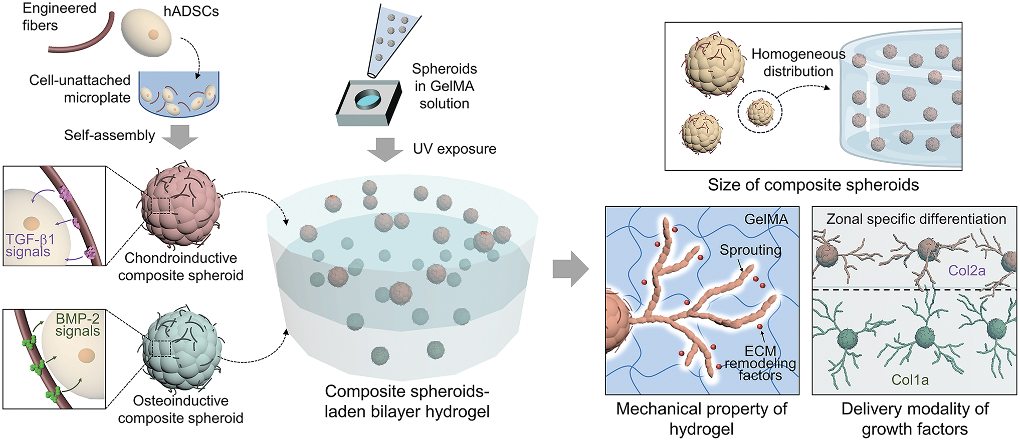
Schematic illustration of the formation of a bilayer hydrogel laden with composite spheroids. The two types of spheroids, each incorporating fibers with immobilized chondroinductive or osteoinductive growth factor, were formed by self-assembly and encapsulated within each layer of the GelMA bilayer hydrogel through sequential UV exposure. The optimal microenvironment for cellular behavior and the spatially confined differentiation of cells encapsulated within the hydrogel were investigated using in vitro analyses. GelMA, gelatin methacryloyl; UV, ultraviolet. Color images are available online.
Preparation and encapsulation of composite spheroids within a GelMA hydrogel
FE-SEM images show the morphology of segmented fibrils (Fig. 2a) of an average length of 57.1 μm (Fig. 2b). All composite spheroids were spherical, irrespective of their size (Fig. 2c, d). The area and diameter of small, middle, and large spheroids were measured and found to be 0.07 ± 0.01, 0.06 ± 0.01, and 0.11 ± 0.04 mm2 (Fig. 2d), and 85 ± 5, 272 ± 16, 379 ± 32 μm (data not shown), respectively. A confocal image of the small spheroid demonstrates homogeneous distribution of fibers and hADSCs within the spheroid (Fig. 2e). The spheroids were encapsulated within a GelMA hydrogel under UV exposure (10 s) (Fig. 2f), and H&E staining showed stable localization of spheroid within the hydrogel by forming the number of cell–matrix binding (red arrows) between the hydrogel matrix and encapsulated spheroids (Fig. 2g). Calcein AM staining of vertically sectioned spheroid-laden hydrogels shows homogeneous distribution of the small spheroids in hydrogel, whereas the medium and large spheroids subsided to the bottom of hydrogel during gelation (Fig. 2h). The only small spheroids were evenly positioned throughout hydrogel (Fig. 2i).
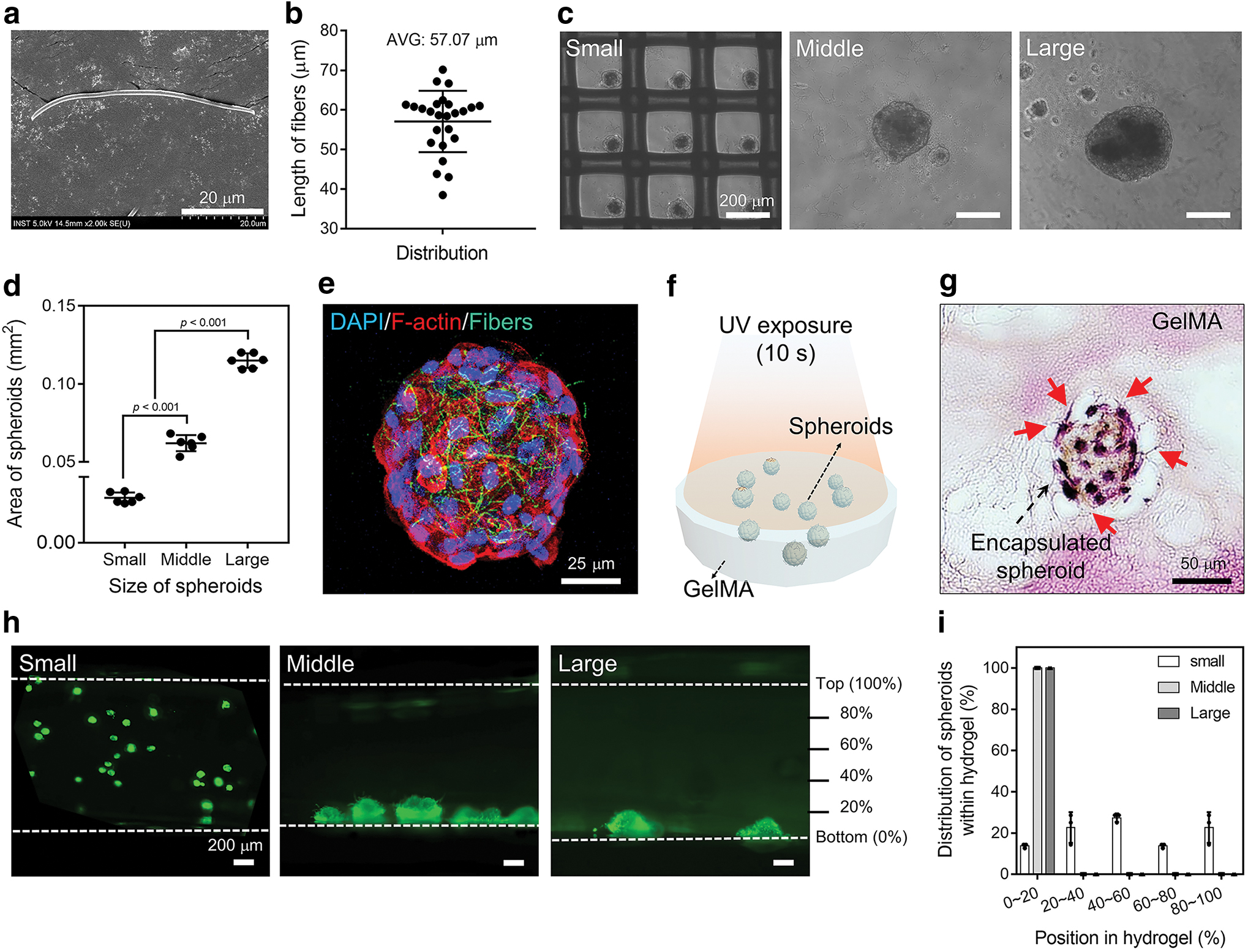
Preparation and encapsulation of composite spheroids within the GelMA hydrogel.
Macroscopic images of the spheroid-laden hydrogels were shown in Supplementary Figure S1. Thus, the small spheroids (6829 ± 815 μm2 of area; Fig. 2d) were used for further investigations.
Characterization of in vitro encapsulated spheroids within the hydrogel depending on UV exposure time
Optical images of each hydrogel show the same cylindrical shape and transparency regardless of UV exposure time (Supplementary Fig. S2a). Storage modulus of the spheroid-laden hydrogels under 10 s of UV exposure was 0.5 ± 0.1 kPa, which was significantly lower than that under 30 and 60 s of UV exposure (1.3 ± 0.1 and 1.5 ± 0.1 kPa, respectively), and no change in modulus was found during 14 days of incubation (Fig. 3a). Swelling ratio of the 10 s UV-exposed hydrogels was greater than that with 30 or 60 s of exposure, and the ratios did not change significantly for 14 days (Fig. 3b). Internal pore structure of the hydrogel with 10 s of UV exposure was larger than with exposure for 30 or 60 s, and cell sprouting from an encapsulated spheroid was observed only in the hydrogel with 10 s of UV exposure (Supplementary Fig. S2b, c). Intensive cell sprouting from the spheroids was found only within the hydrogel with the lowest mechanical strength (10 s of UV exposure) (Fig. 3c). Sprouting cells in the 10 s group filled most of hydrogel area and elongated through hydrogel matrix, which was not found in the other groups (Fig. 3c).
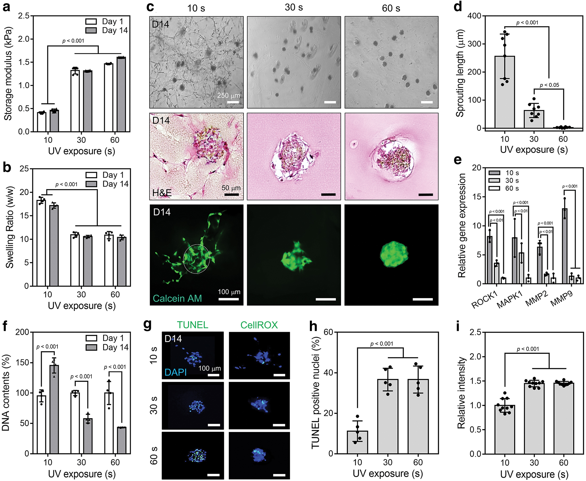
In vitro behavior of cells encapsulated within hydrogels depending on the UV exposure time.
Sprouting lengths within the hydrogels were 256.3 ± 79.3, 63.0 ± 25.3, and 2.4 ± 2.0 μm with 10, 30, and 60 s of UV exposure, respectively (Fig. 3d). RT-qPCR investigation demonstrated that the cells in hydrogel with 10 s of UV exposure showed significantly better ECM remodeling gene expression than those in the other groups (Fig. 3e). For example, expression of ROCK1 and MMP9 from the cells within hydrogel with 10 s of UV exposure was 8.2 ± 1.1 and 13.0 ± 1.7 times greater, respectively, than from those within the 60 s group (Fig. 3e). Furthermore, the cells from spheroids within the lower mechanical-strength hydrogel were more proliferative than those within hydrogels from the other groups (Fig. 3f).
Similarly, the fewer signals from TUNEL-positive nuclei and CellROX were found in hydrogel with 10 s of UV exposure than in the other groups (Fig. 3g). Only 11.2% ± 5.1% of TUNEL-positive nuclei per spheroid and 1.5 times less CellROX-positive fluorescence were detected in the hydrogel with 10 s of UV exposure (Fig. 3h, i). Taken together, the results indicate that the lower mechanical strength in hydrogel enhanced cell sprouting from spheroids, ECM remodeling, and proliferation of hADSCs resistant to apoptosis.
Therefore, the hydrogel with 0.5 kPa of storage modulus was used for further investigations.
Immobilization of growth factors on the fibers
FE-SEM images of the fibers show a roughened surface after PD coating and an even distribution of microscopic granules after BMP-2 or TGF-β1 immobilization (Fig. 4a). The use of FITC-conjugated BMP-2- or TGF-β1-immobilized fibers revealed that the growth factors were homogeneously bound on fiber surfaces, and high-magnification images indicate that the factors were homogeneously distributed on each strand (Fig. 4b). Coating efficiencies of BMP-2 and TGF-β1 on the fibers were 88.5% ± 8.0% and 99.8% ± 0.0%, respectively (Fig. 4c). X-ray photoelectron spectroscopy analysis demonstrated that carbon (C1s, 288 eV) and oxygen (O1s, 533 eV) peaks were common to all fibers, whereas nitrogen (N1s, 399 eV) peak was generated after the PD coating, and sulfur (S2p, 163 eV) peak was specifically found on the BMP-2- or TGF-β1-immobilized fibers (Fig. 4d). High resolution of N1s and S2p peaks for the fibers indicate that the N1s peaks were clearly present in PD, BMP-2, and TGF-β1 groups, and the S2p peaks were found only in BMP-2 and TGF-β1 groups (Fig. 4e).
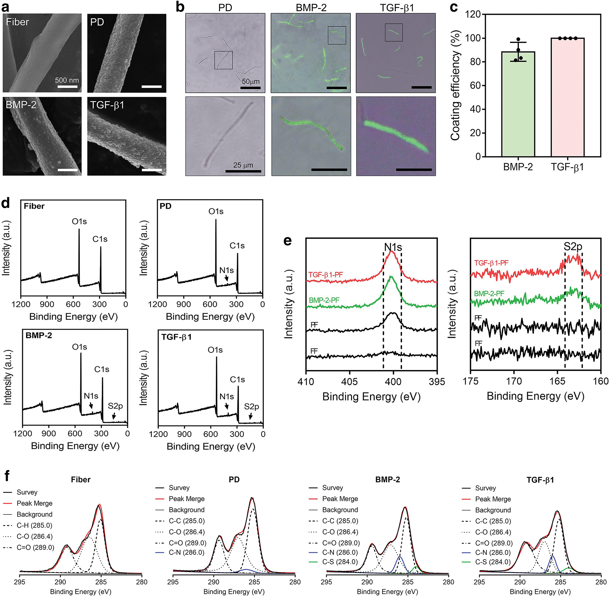
Characterization of the BMP-2- and TGF-β1-immobilized fibers.
Similarly, high-resolution carbon spectrum indicates that C-C (285.0 eV), C-O (286.4 eV), and C = O (289.0 eV) were found in all groups, whereas C-N (286.0 eV) was found only after the PD coating, and C-S (284.0 eV) was found only after the immobilization of growth factors (Fig. 4f). The results indicate that growth factors were evenly and stably immobilized on the PD-coated fibers with reasonable efficiency.
In vitro osteogenic and chondrogenic differentiation of hADSCs from spheroids within the hydrogel
A schematic illustration shows different delivery modalities of BMP-2 to the spheroid-laden hydrogels (GFS: without growth factors, GBS-1: 50 ng of BMP-2 diluted in culturing medium, GBS-2: 50 ng of BMP-2 physically blended in hydrogel, and GBS-3: 50 ng of BMP-2-immobilized fibers in spheroids) (Fig. 5a). Expression of early osteogenic markers, RUNX2 and OSX, by the encapsulated hADSCs was significantly enhanced within GBS-2 and GBS-3 groups, compared with GFS and GBS-1 groups, after 7 days of culturing, and only GBS-3 group showed significantly enhanced osteogenic gene expression, compared with the other groups, after 14 and 21 days (Fig. 5b). For example, the cells within GBS-3 hydrogels had 7.4 ± 0.9, 7.8 ± 0.7, 10.0 ± 0.8, and 5.8 ± 1.0 times more RUNX2, OSX, OCN, and OPN expression, respectively, than those within GFS hydrogels after 14 days (Fig. 5b). Similar trends were found in IHC-stained images for quantified ratio of RUNX2- and OPN-positive nuclei per spheroid in GBS-3 compared with the other groups (Fig. 5c, d).
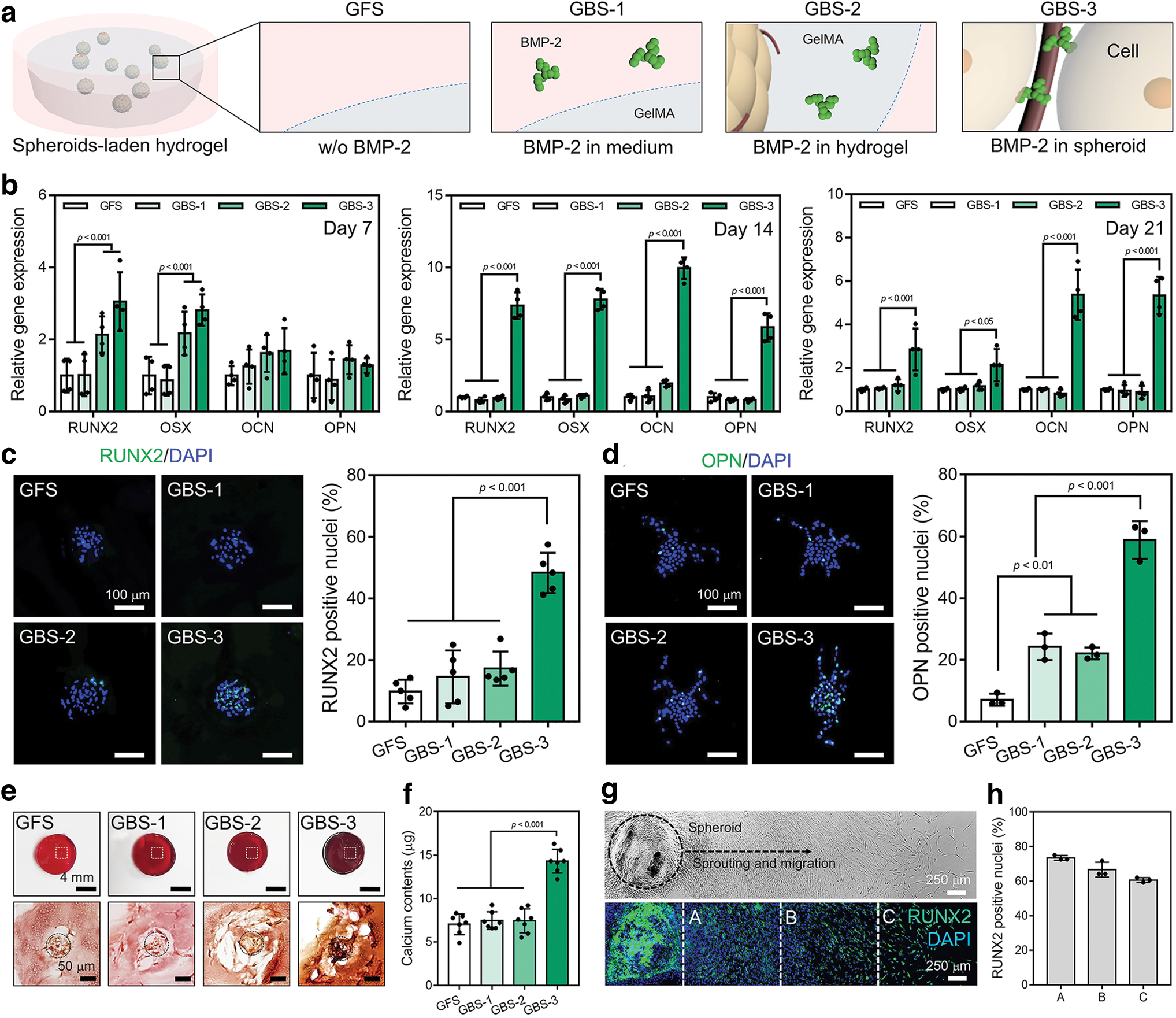
In vitro osteogenic differentiation of hADSCs from spheroids within hydrogels.
After 7 days of culture, the IHC staining results demonstrated the significantly greater number of RUNX2-positive and OPN-positive cells within GBS-3 than the other groups and GFS group, respectively (Supplementary Fig. S3a, b).
A greater intensity of reddish color from Alizarin Red S staining was found throughout GBS-3 hydrogel, compared with the other gels, and deposition of calcium granules (dark red) was also found in cross-sectioned GBS-3 hydrogel (Fig. 5e). The greatest amount of mineral calcium (14.3 ± 1.4 μg) was deposited in GBS-3 hydrogel (Fig. 5f). The cells sprouting from a spheroid incorporating BMP-2-immobilized fibers were positively stained for RUNX2, and the ratio of RUNX2-positive nuclei was statistically same in A, B, and C compartments (Fig. 5g, h).
A schematic illustration shows different delivery modalities of TGF-β1 to the spheroid-laden hydrogels (GFS: without growth factors, GTS-1: 50 ng of TGF-β1 diluted in culturing medium, GTS-2: 50 ng of TGF-β1 physically blended in hydrogel, and GTS-3: 50 ng of TGF-β1-immobilized fibers in spheroids) (Fig. 6a). Similar to the results for osteogenic differentiation, analyses for chondrogenesis demonstrated that GTS-3 hydrogel produced greater efficiency of differentiation among cells than the other groups (Fig. 6b–f). In RT-qPCR investigations, GTS-3 hydrogels showed significantly enhanced SOX9 compared with the other groups after 7 days, and expression of all tested genes was significantly higher in the cells within GTS-3 hydrogels than in those within the other hydrogels after 21 days (Fig. 6b). More SOX9 and aggrecan-positive nuclei were detected in the IHC-stained images from GTS-3 hydrogels than in those from the other groups (Fig. 6c, d).
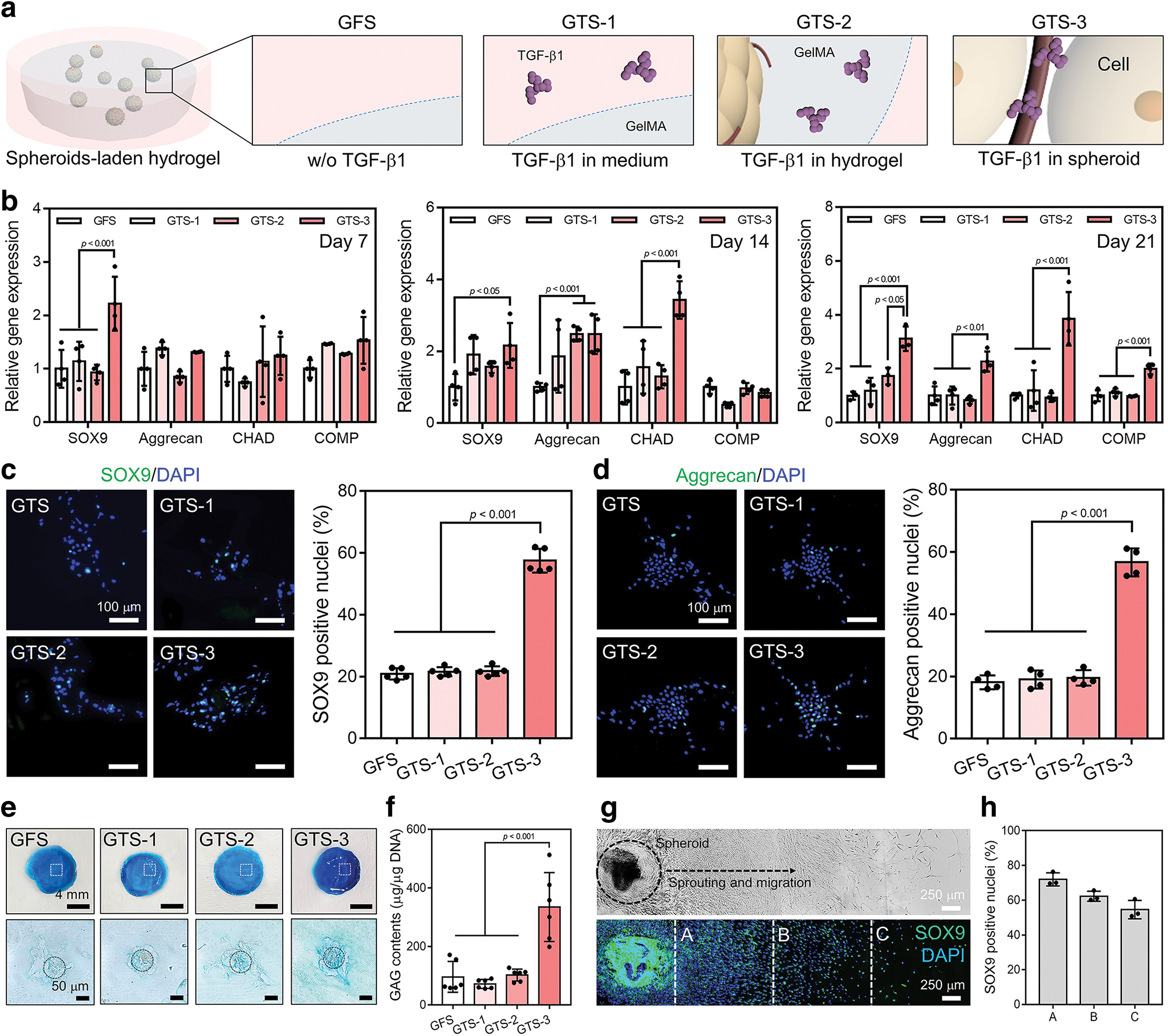
In vitro chondrogenic differentiation of hADSCs from spheroids within the hydrogels.
Alcian Blue staining showed a richer blue color for GTS-3 than for the other hydrogels, and cross-sectioned GTS-3 revealed GAG formation around the encapsulated spheroid (bright blue) (Fig. 6e); 334.7 ± 117.6 μg of GAG was measured in GTS-3, compared with 96.0 ± 52.7, 71.8 ± 15.5, and 102.4 ± 20.3 μg in GFS, GTS-1, and GTS-2 groups, respectively (Fig. 6f). IHC staining for hADSCs sprouting from a spheroid incorporating TGF-β1-immobilized fibers showed that the cells were evenly positive for SOX9 (Fig. 6g, h). Collectively, the results suggest that BMP-2 or TGF-β1 delivery within each composite spheroid by using the PD-coated segmented fibrils effectively and specifically directed osteogenic or chondrogenic differentiation, respectively, of hADSCs in hydrogel, and that differentiation was enhanced compared with the control methods, in which growth factors were added to the medium or physically blended into hydrogel.
Preparation and characterization of 3D bilayer hydrogel
A schematic illustration shows sequential procedures used to prepare the bilayer hydrogels (Fig. 7a). Crosslinking of hydrogels under 5 s of UV exposure did not produce completely discrete bilayers, but the other bilayer hydrogels were hierarchically arranged with compatible interface integration (Fig. 7b). Storage moduli of the bilayer hydrogels encapsulating spheroids increased from 0.3 ± 0.0 to 0.7 ± 0.0 kPa with 5 and 10 s of UV exposure on each layer, respectively (Fig. 7c). Storage moduli of top and bottom layers (7 or 10 s of UV exposure) were the same as overall storage modulus for each hydrogel (Supplementary Fig. S4a). H&E-stained images reveal greater cell sprouting from the encapsulated spheroids in each layer with 7 s of UV exposure than with 10 s (Fig. 7d). The spheroids were homogeneously encapsulated within both bilayer hydrogels (Supplementary Fig. S4b), but after 14 days of culture, sprouting of living cells was observed only in 7 s group (Fig. 7e). Collectively, our results suggest that sprouting from spheroids was achieved within the bilayer hydrogel with a modulus of 0.5 kPa.
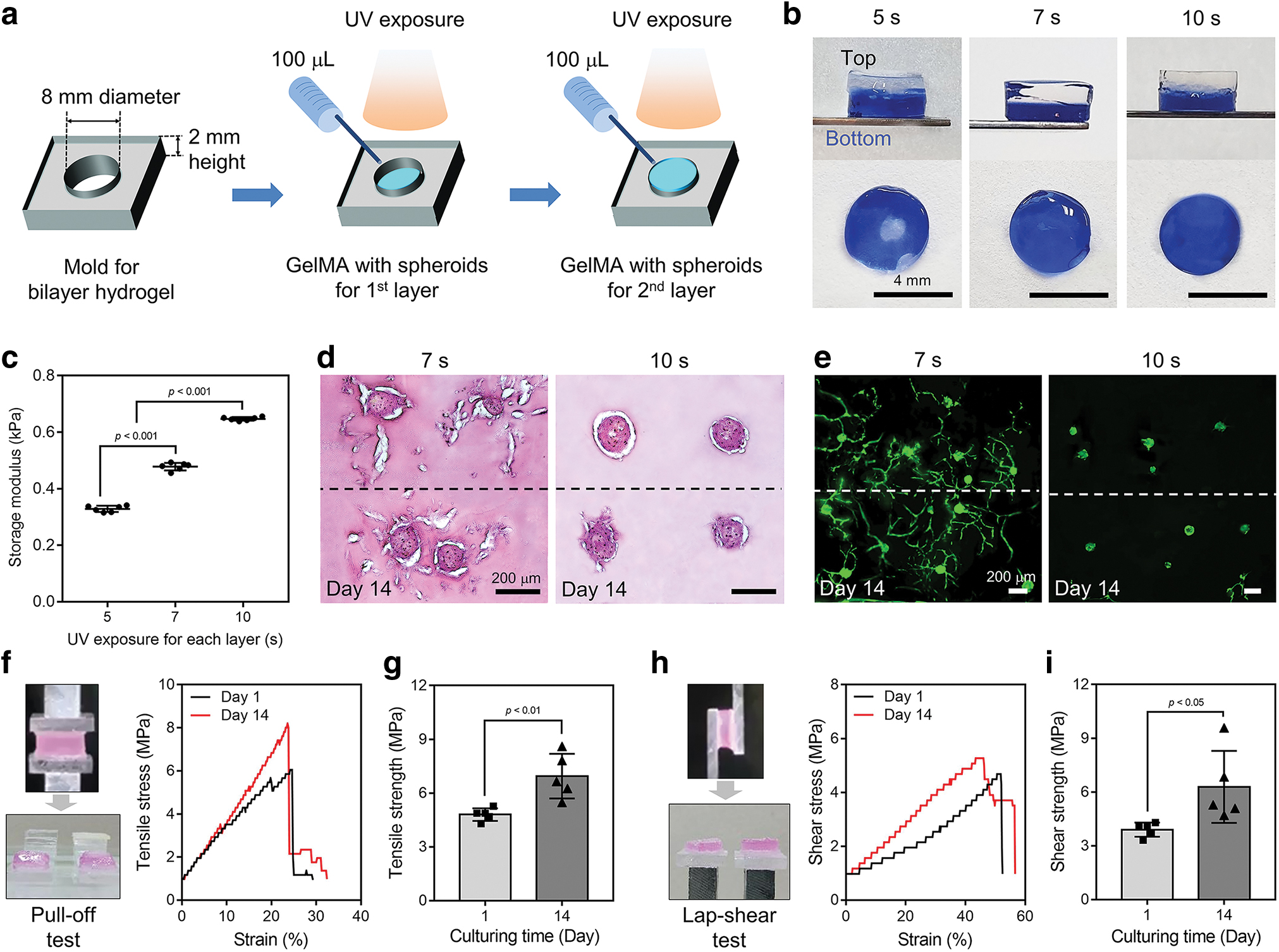
Preparation and characterization of 3D bilayer hydrogels.
We then examined interface integration using pull-off and lap-shear tests, which measure resistance to uniaxial and shear stress, respectively (Supplementary Fig. S4c). Intermediate area of the bilayer was separated by uniaxial force in pull-off test for the bilayer hydrogel with 7 s of UV exposure at each layer, and tensile strength increased from 4.8 ± 0.4 (1 day) to 7.0 ± 1.3 MPa after culturing for 14 days (Fig. 7f, g). Similar results were obtained from lap-shear test (Fig. 7h). Calculated shear strength of the bilayer hydrogel increased from 3.9 ± 0.4 (1 day) to 6.3 ± 2.0 MPa (14 days) (Fig. 7i). The results indicate that the bilayer hydrogel was well crosslinked with strong interfacial adhesion during preparation, which was improved during 14 days of in vitro culture, possibly due to intensive cell sprouting (Fig. 7f–i).
In vitro differentiation of hADSCs from spheroids within the bilayer hydrogel
We next confirmed zonal osteogenic or chondrogenic differentiation of hADSCs induced by instructive signals in each layer by culturing entire hydrogel in a general medium. Macroscopic image of bilayer hydrogel after Alizarin Red S staining was strongly positive for GBS-3 layer, and the spheroids within layer showed granulated calcium minerals whereas those within GTS-3 layer were weakly stained (Fig. 8a). In contrast, Alcian Blue staining of showed a greater intensity of blue color for GTS-3 layer, and the spheroids within layer clearly revealed GAGs while those within GBS-3 were barely stained (Fig. 8b).
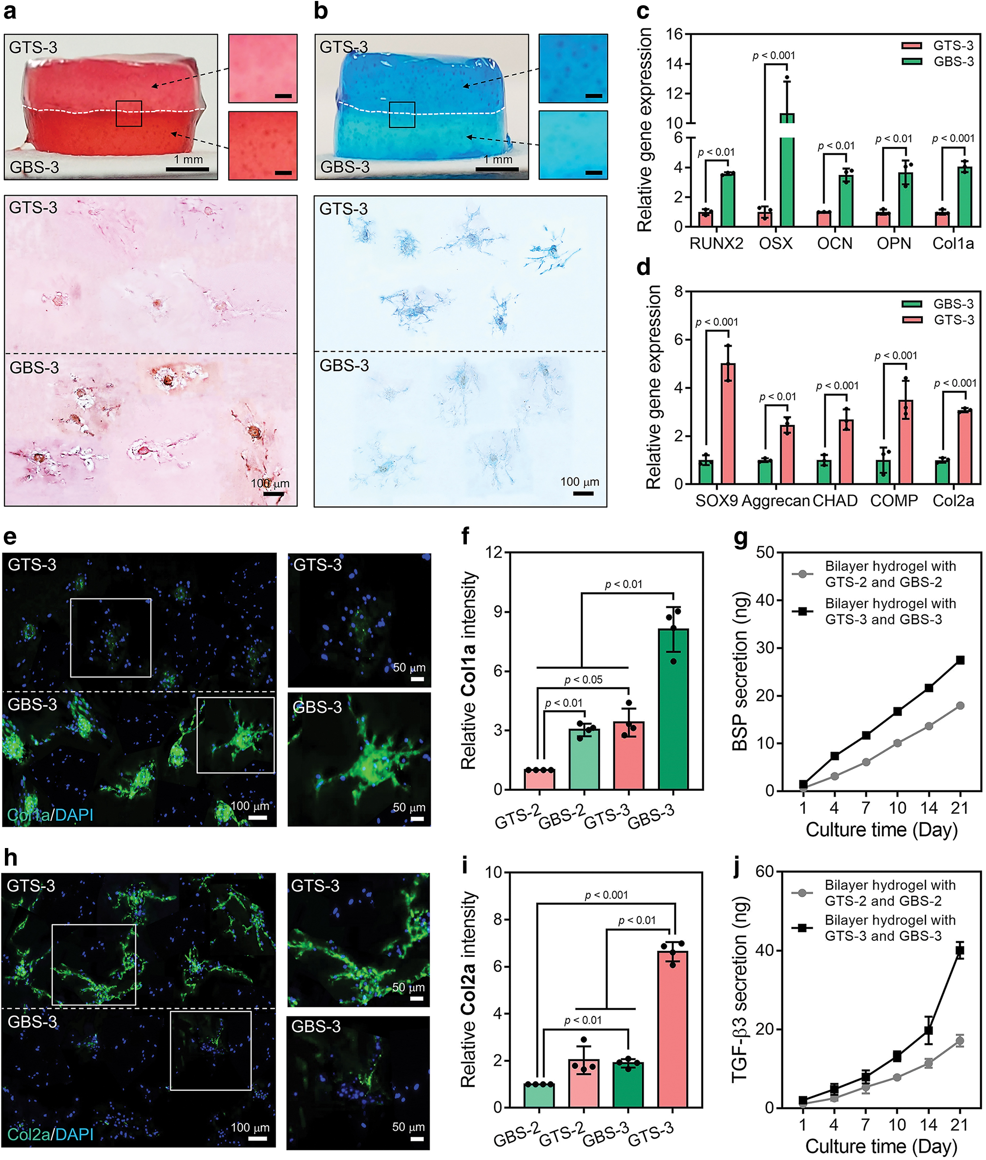
In vitro differentiation of hADSCs from spheroids within the bilayer hydrogel.
The spheroids within each GTS-2 and GBS-2 layer similarly demonstrated weak intensity of colors and insignificant color differences from each layer (Supplementary Fig. S5a). The expression of osteogenic markers increased significantly in the cells within GBS-3 layer, whereas expression of chondrogenic markers was significantly upregulated within the GTS-3 layer (Fig. 8c, d). However, the expression of osteogenic and chondrogenic genes by the cells within GTS-2 and GBS-2 layers did not differ significantly, except for OSX and OCN (Supplementary Fig. S5b).
In addition, IHC staining of collagens and an ELISA for the bilayer hydrogel composed of GTS-3 and GBS-3 clearly showed distinct characteristics of each layer (Fig. 8e–j), whereas the characteristics were rarely detected in the bilayer hydrogel composed of GTS-2 and GBS-2 (Supplementary Fig. S5c). Most cells in GBS-3 were positive for Col1a, and high-resolution image clearly shows elongated Col1a, whereas the signals in GTS-3 layer were weak (Fig. 8e). Quantified fluorescent intensity was greatest in the GBS-3 layer (Fig. 8f).
ELISA demonstrated that the amount of BSP secreted from the bilayer hydrogel composed of GTS-3 and GBS-3 for 21 days was 27.5 ± 0.7 ng, which was significantly greater than that secreted from the GTS-2 and GBS-2 bilayer hydrogel (18.0 ± 0.2 ng) (Fig. 8g). Similarly, Col2a signals were found only from cells and spheroids in the GTS-3 layer and were barely detected in the GBS-3 layer (Fig. 8h). The strongest fluorescent intensity was found in the cells in GTS-3 layer (Fig. 8i), and the amount of TGF-β3 secreted from the bilayer hydrogel composed of GTS-3 and GBS-3 for 21 days (40.1 ± 2.1 ng) was greater than that secreted from the GTS-2 and GBS-2 bilayer hydrogel (17.1 ± 1.5 ng) (Fig. 8j).
Taken together, our results suggest that spatially confined delivery of growth factors within the spheroids encapsulated in each layer (GTS-3 and GBS-3) induced zone-dependent differentiation (chondrogenic or osteogenic) of hADSCs without dedifferentiation within bilayer hydrogel structure (Fig. 8). On the other hand, physically blended growth factors in the GTS-2 and GBS-2 bilayer hydrogel showed weak induction of encapsulated cells (Fig. 8 and Supplementary Fig. S5).
Discussion
In this study, we developed a composite spheroid-laden bilayer hydrogel with spatially confined induction of stem cells toward chondrogenesis or osteogenesis at each layer to mimic zonal organization of osteochondral tissue that mimics native cartilage and subchondral bone. The controlled UV crosslinking of a GelMA hydrogel provided a microenvironment able to support cellular activity of the spheroids, such as sprouting, proliferation, and ECM remodeling. A potent chondrogenic or osteogenic growth factor was immobilized onto the surfaces of synthetic fibers that were then combined with hADSCs to form multicellular composite spheroids that stimulated sufficient in vitro zonal differentiation of hADSCs in each layer without dedifferentiation. Importantly, in addition to engineering osteochondral tissue, this approach offers flexibility for designing specific instructive signals, cell types, and mechanically adaptable hydrogels that can ensure a desired cellular phenotype when engineering other complex tissues.
Most anchorage-dependent cells spontaneously form spheroidal aggregates through strong cell to cell and cell to ECM interactions, and spheroids have various advantages in mimicking native tissue microenvironments, making them a great candidate for tissue engineering. 27 Recently, assembly of cells with ECM-mimicking materials such as nano/microparticles or fibers has been reported to generate composite spheroids that can provide signals directly to the cells within spheroids and mitigate diffusional limitations of oxygen or gas. 28 We have previously developed micrometer-scale fibers coated with growth factors and demonstrated their efficacy in improving cell viability and modulating lineage-dependent differentiation of stem cells for bone tissue regeneration.29,30 In this work, we similarly achieved the stable integration of hADSCs and fibers and newly confirmed homogeneous distribution of the fibers within each spheroid regardless of its size by 3D immunostaining (Fig. 2c–e). Delivery of spheroids is challenging due to evasion or severe aggregation of spheroids, which results in impaired 3D tissue morphology. 31
Therefore, several engineering strategies, including Kenzan method, spheroid positioning into a microchamber, and encapsulation in hydrogels, have been developed for stable in vivo localization of spheroids.31–33 In particular, hydrogels are convenient and suitable for encapsulating spheroids because they are compatible with in vivo 3D microenvironment and offer flexible modulation of their mechanical properties. 34 However, heterogeneously distributed spheroids within a hydrogel induced abnormal tissue formation.12,35 For example, sedimentation of MG-63 cell spheroids in a GelMA hydrogel produced abnormal two-dimensional (2D) sheet-like tissue, and BMSC spheroids localized on the marginal area of a hydrogel-formed immature bone.12,35 In this study, homogeneous distribution of spheroids in the hydrogel during crosslinking was carefully considered, and an appropriate size was found for in vitro culture of spheroid-laden hydrogels (Fig. 2h, i). Similarly, it was previously reported that smaller spheroids were advantageous due to their relatively low loading and faster permeability in a viscous microenvironment.36,37
GelMA hydrogels have been used for various tissue engineering applications because their mechanical strength can be conveniently modulated by varying their photocrosslinking and chain reactions of radicals propagated under UV light.38,39 We also found that the storage moduli of GelMA hydrogels encapsulating spheroids increased as a function of UV exposure time (Fig. 3a). However, stiff, highly polymerized hydrogels were reported to restrain binding between cell-binding motif in gelatin and the encapsulated cells, which reduced cellular sprouting and ECM remodeling capacity. 40 Consistently, the hADSCs in GelMA hydrogel with lower mechanical strength in this study showed improved sprouting and ECM remodeling capacity compared with those in hydrogels with stronger moduli (Fig. 3a–e). Generally, UV exposure of <500 mW/cm2 for 60 s is not detrimental to cell survival within a hydrogel. 39 However, migration and sprouting of cells within a hydrogel are pivotal for their viability and proliferation.41,42 It was reported that encapsulated cells adopted more adhesion-ligand clustering, shape change, and proliferation of cells within hydrogels with weaker mechanical strength. 41
Similar to previous investigations, we found that viability and proliferation of hADSCs from spheroids within the GelMA hydrogel with lower mechanical strength were significantly enhanced (Fig. 3f–i). Taken together, homogeneous distribution and sprouting of cells within the hydrogel, which were modulated by the size of spheroids and the mechanical strength of hydrogel, were important for viability, proliferation, ECM remodeling, and guidance of stem cell differentiation.
BMP-2 and TGF-β1 are well-known growth factors that induce osteogenic and chondrogenic differentiation of stem cells, respectively.43,44 However, delivery of the growth factors to cells encapsulated within hydrogels is challenging due to the large size of the proteins relative to the mesh size of a hydrogel network, which makes passive penetration difficult without coordinated degradation of the hydrogels. 45 Moreover, intraspheroidal penetration is another barrier because of cell compaction on the surface of a spheroid. 46 In contrast, engineered nano/microfibers or particles within composite spheroids can effectively and homogeneously modulate cell functions within a spheroid.13,46 In our previous study, we demonstrated that 100 ng of fibers immobilized with BMP-2 or TGF-β3 doubled osteogenic or chondrogenic differentiation, respectively, when they were delivered as a composite spheroid, compared with spheroids cultured with medium containing the same amount of growth factors. 47
In this study, we proved the effects of BMP-2 or TGF-β1 immobilized fibers within each composite spheroid, more precisely, by comparing the method with the other conventional delivering modalities of growth factors (freeform in medium or blended in hydrogel).12,48,49 As a result, hADSCs from the composite spheroids incorporating each fiber were effectively differentiated into osteogenic or chondrogenic lineages within hydrogels (GBS-3 or GTS-3 groups), which revealed the more than three times increased each gene expression than the other methods after 21 days (Figs. 5b–d and 6b–d). Our results suggest that modality for delivering growth factors to spheroids is critical for controlling cellular behavior, and our growth factor-immobilized fibers can be flexibly custom designed to guide stem cell differentiation, as well as sprouting, without addition of supplements to medium during in vitro culture (Figs. 5 and 6).
Bilayer hydrogel can recapitulate hierarchical structure of natural complex tissues, but optimization of mechanical strength of each layer is important. 4 Two layers of hydrogel could intermingle due to their weak mechanical properties, and resulting translocation of initially given inductive factors could lead to undesired differentiation of encapsulated cells. 4 In the case of GelMA, insufficient photoinitiation and chain polymerization due to short UV exposure can limit the sol/gel transition and produce weak mechanical properties. 50 The bilayer hydrogel with 5 s of UV exposure at each layer showed intermingled layers and a low storage modulus, and it was not able to spatially separate chondrogenic and osteogenic layers (Fig. 7b, c). Meanwhile, a bilayer hydrogel with mechanical properties that are too strong could restrict proliferation and sprouting of encapsulated spheroids.51,52 Importantly, mechanical properties of a bilayer hydrogel should be reoptimized from those of a monolayer hydrogel because bottom layer is exposed to UV radiation twice, and top layer is exposed to UV light from a shorter distance. 51
Huang group reported a bilayer hydrogel encapsulating BMSCs and found that they did not sprout in the stiffer bottom layer and did sprout in the softer upper layer. 52 In this study, we found that a bilayer hydrogel with 7 s of UV exposure at each layer had a proper storage modulus (0.5 kPa) for cell sprouting as similar to that of single-layer hydrogel with 10 s of UV exposure (Figs. 3a and 7c and Supplementary Fig. S4a). Despite several previous studies utilizing bilayer hydrogels for engineering 3D osteochondral tissue, cells with limited sprouting and ECM remodeling ability were unable to fill a volumetric 3D space of hydrogel. 53 This is due to the lack of investigation into the response of cells or spheroids within hydrogel depending on the change of hydrogel mechanical property. On the other hand, the hADSC spheroids within GBS-3 and GTS-3 with 0.5 kPa of storage modulus were strongly sprouting and uniformly filled the volume of hydrogel with secreted collagens (Fig. 8e, h).
Natural osteochondral tissue is composed of cartilage and subchondral bone with distinct biological and biochemical characteristics in terms of parenchymal cells and ECM molecules. 4 Several biphasic constructs using various materials, including hydrogels (e.g., PEG, alginate) and synthetic polymer-based scaffolds (e.g., polycaprolactone [PCL], poly(lactic-co-glycolic acid) [PLGA]), and various processes for each layer, such as hydrophobic or hydrophilic adhesion, physical blending, or pasting with biochemical glues, have been designed to mimic 3D osteochondral tissue. 5 However, biphasic constructs composed of distinct materials are generally subject to stronger attractive interactions within each layer than at the bridging interface between them, and the irregular layering of materials has resulted in delamination and apertures between layers under even weak shear stress.54,55 In contrast, sequentially laid bilayer hydrogels polymerized by UV crosslinking appeared to avoid the delamination problem. 5
Our bilayer hydrogel with 7 s of UV exposure on each layer showed strong interfacial strength, withstanding 4.8 and 3.9 MPa of tensile and shear stress, respectively (Fig. 7f–i), which appears to be sufficient to prevent delamination in vivo. 56 It should be noted that the interfacial strength became stronger during 14 days of culture (Fig. 7g, i). These results indicate that cell–matrix adhesion might have increased the interfacial adhesion of the bilayer hydrogel.57,58 Conventional methods for engineering zonal organization include serial culturing of biphasic constructs in chondrogenic and osteogenic media, placing inductive molecules onto each layer by immobilization, gel encapsulation, and surficial coating.4,5 Generally, the cells induced in osteogenic or chondrogenic ways differentiated in the desired manner in each layer; however, after long-term culture, severe dedifferentiation and osteochondrosis or ossified cartilage have commonly been found at the interface, possibly due to off-target inductive molecules.14,16
In contrast, our results imply that combining a bilayer structure with spheroids containing fibers immobilized with potent growth factors could hold promise for engineering 3D osteochondral tissue without the dedifferentiation problem (Fig. 8). The macroscopic images of bilayer hydrogel showed that each GTS-3 and GBS-3 layer was also lightly stained with Alizarin Red S and Alcian Blue, respectively, because the staining solution was soaked in hydrogel despite several washing steps (Fig. 8a, b). However, the high-magnified images clearly showed that the red colored dots were specifically found only for GBS-3 layer, while the blue dots were for GTS-3 layer (Fig. 8a, b). Each staining of cross-sectioned specimens also showed that red signals in GBS-3 layer were from the mineralized granules of BMP-2 fiber-incorporated spheroids, and the blue signals in GTS-3 layer were from the GAGs of TGF-β1-incorporated spheroids (Fig. 8a, b). To demonstrate osteogenic or chondrogenic differentiation of hADSCs under in vivo environment, we subcutaneously implanted the spheroid-laden bilayer hydrogels into mice (Supplementary Fig. S6a. (The animal management procedures were approved by the Institutional Review Board (IRB) (HYU-2019-11-017) and Institutional Animal Care and Use Committee (IACUC) of Hanyang University (HY-IACUC-2022-0081A))).
All the spheroids were positively stained for HNA, and more importantly, the positive staining for OPN or SOX9 was found only in GBS-3 or GTS-3 layer, respectively, indicating spatially confined osteogenic or chondrogenic differentiation of hADSCs under in vivo microenvironment (Supplementary Fig. S6b, c). However, unlike in vitro, the restricted sprouting of hADSCs from spheroids was observed. These results could be potentially attributed to the lack of nutrient supply or disruption of vasculature after transplantation. Therefore, further investigation is necessary to determine how to enhance in vivo cellular activity.
In summary, we developed the composite spheroid-laden bilayer hydrogel made of hADSCs and appropriate inductive growth factors to mimic osteochondral tissue organization. Unlike previous studies, our approach is the novel attempt so far to modulate the distribution of spheroids, sprouting, and ECM remodeling ability of cells depending on mechanical property of hydrogel, and delivery modality of growth factors in a platform, and succeeded to design 3D osteochondral tissue model having zonal specific characteristics. Our engineered hydrogel with composite spheroids could be applied to the formation of various complex tissues by varying the signaling molecules delivered. Nevertheless, the regenerative effects of the constructs, not for the 3D tissue model, needs further investigations about in vivo variables, which were the absence of vasculatures, discontinuous oxygen and serum supply to transplanted hydrogel in in vivo microenvironment, and interactions with host tissue,59,60 and thus, the improvement of in vivo cellular activity from the composite spheroid-laden hydrogel will be addressed for our future studies.
Conclusions
In this study, we designed a bilayer GelMA hydrogel encapsulating two types of composite spheroids to mimic natural osteochondral tissue. Our meticulous investigation of cellular behavior within the hydrogel, such as positioning, sprouting, ECM remodeling, differentiation, and interfacial adhesion allowed us to successfully reconstruct 3D osteochondral tissue with the specific zonal characteristics of cartilage and subchondral bone while addressing the issues that remained in previous studies. The size of the spheroids and the mechanical strength of the hydrogel were characterized to provide an optimal microenvironment for the encapsulated cells and enable the homogeneous distribution and sprouting of spheroids within the hydrogel. The fiber-mediated TGF-β1 and BMP-2 delivery in each composite spheroid effectively delivered chondrogenic or osteogenic signals, respectively, to the cells within the hydrogel while allowing spatially confined induction in each layer without dedifferentiation. Sequential gelation produced a highly cell-proliferative microenvironment in the bilayer hydrogel, which was strongly integrated at the interface without delamination.
Thus, our composite spheroid-laden bilayer hydrogel could be a potential 3D model for osteochondral tissue, and the scientific techniques used in this study could be used to design other advanced, complex 3D tissues that possess multiple characteristics.
Data Availability
Data will be made available on request.
Footnotes
Authors' Contributions
J.L.: conceptualization, formal analysis, methodology, and writing—original draft. E.L.: data curation, formal analysis, and writing—original draft. S.J.H.: formal analysis. J.I.K.: formal analysis. K.M.P.: methodology and validation. H.B.: data curation. S.L.: visualization. E.K.: visualization. H.S.: supervision and writing—review and editing.
Disclosure Statement
The authors declare that they have no known competing financial interests or personal relationships that could have appeared to influence the work reported in this article.
Funding Information
This research was supported by Basic Science Research Program through the National Research Foundation of Korea (NRF) funded by the Ministry of Education (MOE; Grant No. 2021R1A6A3A01086376), the NRF grant funded by the Korean government (MEST; Grant No. RS-2023-00207983), the Korean Fund of Regenerative Medicine (KFRM) grant funded by the Korean government (the Ministry of Science and ICT, the Ministry of Health & Welfare; Grant No. 22A0105L1), and the BK21 FOUR (Brain Korea 21 Fostering Outstanding Universities for Research) funded by the MOE (Korea).

References
Supplementary Material
Please find the following supplemental material available below.
For Open Access articles published under a Creative Commons License, all supplemental material carries the same license as the article it is associated with.
For non-Open Access articles published, all supplemental material carries a non-exclusive license, and permission requests for re-use of supplemental material or any part of supplemental material shall be sent directly to the copyright owner as specified in the copyright notice associated with the article.
