




Abstract
From a literary perspective, the concept of tissue engineering and regenerative medicine dates back several thousand years. However, from a scientific aspect, the current state of the field owns its initial origin to the discovery of cell culture methods and the ability to maintain cells outside the body in the early 1900s, to later discoveries surrounding stem cells. The science of biomaterials evolved more recently, from the use of degradable natural biomaterials in the 1970's to artificial biomaterials in the 1980s, and bioprinting hydrogels this century. Tissue engineering, originally involving the combination of cells and biomaterials, owes its roots to the early attempts in the 1960s to create artificial skin grafts as temporary wound covers for burn patients. Much has transpired since, with an increasing number of technologies reaching patients. Academia, industry, government agencies, societies, and nonprofit organizations have all played a role in advancing the field to where it is today. This overview, presented at the Rice Short Course on Advances in Tissue Engineering, highlights some of the historical aspects, as well as past and future challenges and opportunities. At the current pace of discovery, the field is poised to continue its exponential growth.
Before I start my overview of “Thirty Years of Tissue Engineering,” I would like to recognize the 30 years of the course at Rice. This was the first short course in tissue engineering, and is the longest running course in the field. It has been a standard—one that everybody looks forward to every year—and many individuals in the field have taken this course as trainees, and then returned to lecture as their careers advanced. A big thanks to Dr. Mikos for running this course.
I think it is important to start with a brief history of the origins of regenerative medicine. The first potential reference to regenerative medicine may be in the Book of Genesis from circa 1400 BC, “The Lord, breathed a deep sleep on the man and while he was asleep, he took out one of his ribs and closed up its place with flesh. The Lord God then built up into a woman the rib that he had taken from the man.” This may represent the very first written example of anesthesia, surgery, and cloning.
Organ regeneration was also depicted in Greek mythology. Prometheus, an immortal Titan in Greek mythology, stole fire and gave it to humanity, defying the will of the Gods. As punishment, Zeus decreed that he was to be bound to a rock where an eagle would feast on his liver every day and his liver would regenerate itself every night, leading to continuous torture. This myth suggests that the fund of knowledge regarding the ability of livers to regenerate was available circa 800 BC.
If we were to choose a scientific origin for the field of tissue engineering, we could start with the ability to culture cells. Initially, cell culture did not mean expansion, but rather just maintaining cells in media so they could be studied. In fact, most human derived cells could not be expanded just a few decades ago, and still cannot be done for some, such as specific pancreatic or liver cells.
The first person to report the successful cultivation of cells was Dr. Ross Harrison in 1907. He cultivated frog nerve fibers for several weeks in lymph fluid freshly drawn from an adult frog. He collaborated with Dr. Alexis Carrel, a surgeon from Lyon, France, who received the Nobel Prize in medicine for devising methods to suture blood vessels. Carrel was intrigued with growing tissues and organs. In 1909, he sent one of his proteges, Montrose Burrows, to work under Harrison's supervision. Burrows found that lymph was not useful for cultivating warm-blooded animal cells and used plasma instead, a related method used today with serum.
Alexis Carrel went on to design the cell culture flasks (Fig. 1) that are the predecessors for what we use today, and in 1912, showed that cultivation of connective tissue from chick fetuses is possible long term with regular medium exchange. He later partnered with Charles Lindbergh, world-renowned for having completed the first solo flight across the Atlantic, to create pumps that would keep tissues alive. They published a book called The Culture of Organs back in 1938. This work paved the way for the first organ transplant in 1954 by Dr. Joseph Murray in Boston when a patient received his twin bother's kidney. This was a major event for the field of medicine, as there was no option available for patients with end-stage organ failure before that.
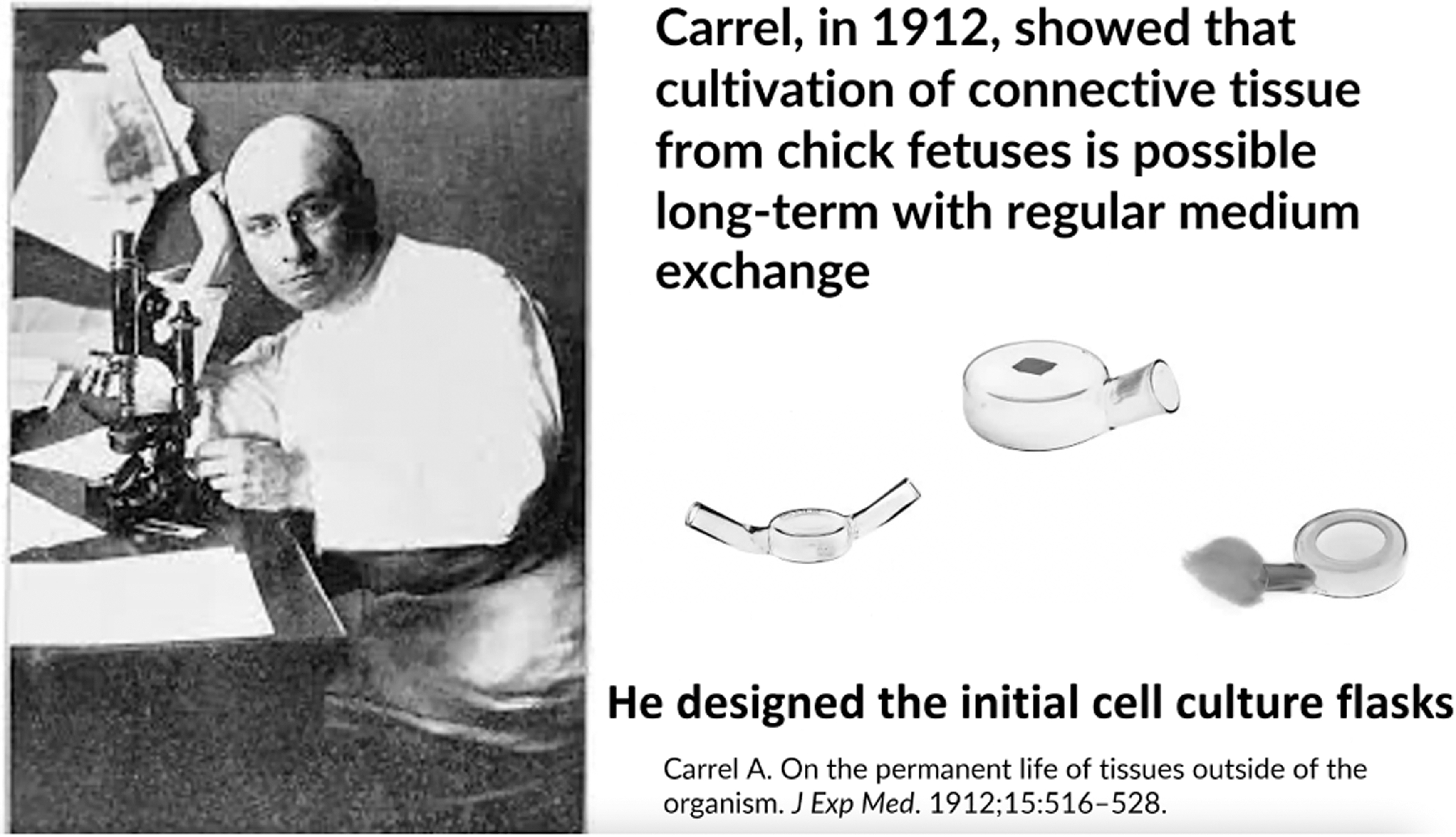
Alexis Carrell, a pioneer with human cell culture, designed the basic principles for culture flasks still used today.
The concept of using alternatives to native tissue for healing originated with burn surgeon John Burke at the Massachusetts General Hospital. The standard treatment to close a burn wound is to harvest skin from an unburned portion of the body and graft it to the damaged areas. For 50 percent or greater burns, there is not enough healthy skin to graft. Dr. Buke established the world's first frozen skin bank in 1969 by storing cadaver skin. However, cadaver allografts are only a temporary solution.
The cadaver skin used to cover the burn wound is removed as soon as the patient develops additional donor sites, which occurs when the initially removed healthy skin site heals and grows new skin. This is a long process and burn patients can remain in the hospital for months as these treatments continue. Burke partnered with Massachusetts Institute of Technology (MIT) Engineering Professor Ioannis Yannas to create an artificial skin cover. By 1973, they had the concept for their bilayer skin wound product, now known as Integra® (Integra Lifesciences, Princeton, NJ, USA). In 1981, Burke and Yannas published their first report using the skin wound dressing on 10 patients. This product did not involve cells, but used natural extracellular matrix proteins.
Simultaneously, Harvard cell biologist Howard Green was working with surgeons Greg Gallico and Nicholas O'Connor. Dr. Green started growing keratinocytes by taking a skin biopsy and creating cell layers that were applied with a petroleum-based gauze. Because these were cells alone, and just keratinocytes without the other five cell types present in skin, a prior graft was typically needed in the wound bed. In 1985, they published the first use of epithelial sheets as wound dressings, which would later be marketed as Epicel®.
Also, from MIT, Dr. Eugene Bell started to seed bovine collagen gels with neonatally derived allogeneic fibroblasts, and thereafter keratinocytes, referring them to contracted dermal equivalents. This technology, marketed as Apligraf® (Basel, Switzerland), represented the first use of a scaffold with cells in patients. Because the cells were not autologous, generated from the same patient, it also was a temporary graft used for skin protection until further autologous native skin grafts could be obtained from the injured patient.
In 1983, Dr. Joachim Thüroff and Dr. Emil Tanagho showed the feasibility of producing grafts from cultured smooth muscle cells on an absorbable synthetic polyglycolic acid mesh. They used scanning electron microscopy to show that they could cover the synthetic fabric fully with these cells. This marked the first time that cells were used with synthetically derived biomaterials.
The term tissue engineering was coined in 1987, several years after biologic skin dressings had been placed in patients, at a committee meeting of the National Science Foundation. The first scientific meeting titled “tissue engineering” was held in Lake Tahoe in 1988, where scientists like Dr. Robert Nerem were in attendance. Dr. Bob Nerem soon after went on to establish the Georgia Tech Institute for Bioengineering and Biosciences, which largely focused on tissue engineering.
The term tissue engineering was further defined by MIT engineer Robert Langer and Harvard surgeon Joseph Vacanti in 1993, as “a field that applies the principles of engineering and life sciences towards the development of biological substitutes that restore, maintain, or improve tissue function or whole organs.” They also advanced the use of artificial biodegradable scaffolds.
In 1994, the first society for the field was formed, the Tissue Engineering Society. The original charter listed seven members: The founding Presidents, Robert Langer, Charles and Joseph Vacanti, and Governors Anthony Atala, Linda (Cima) Griffith, Mark Randolph, and Joseph Upton, all in Boston at the time. In 1995, the founding Board of Governors decided to start the journal “Tissue Engineering.” It was launched with Editors Tony Mikos and Charles A. Vacanti. The field continued to evolve, and what started as one journal became three journals under a part A, B, and C structure, now under the leadership of both Drs. Tony Mikos and John Fisher. These journals have become a staple for the field. Subsequently, both Asia and Europe formed their own tissue engineering societies. Over the next decade, in conjunction with the Asian and European Societies, TES would evolve and reorganize to become TESi (Tissue Engineering Society, International).
In 1999, Bill Haseltine first coined the term regenerative medicine at a conference in Lake Como, Italy. The original definition “includes all interventions that restore the body to normal function, whether injured by trauma, damaged by disease, or worn by time.” In 2000, Bill Haseltine and this author founded the Regenerative Medicine Society and the Journal for Regenerative Medicine. In 2005, the Regenerative Medicine Society and TESi merged to form TERMIS (the Tissue Engineering and Regenerative Medicine International Society), facilitated by Alan Russell, the TESi President at the time. That same year, the Regenerative Medicine Foundation was formed as a nonprofit entity that would promote research, advocate for patients, and gather all stakeholders on behalf of the field. The Regenerative Medicine Foundation now runs the World Stem Cell Summit.
The tissue engineering industry started taking off in the 1990s, and it was mostly around the area of skin replacement therapies. Various companies were formed, such as Integra Life Sciences, marketing their Dermal Template product, Advanced Tissue Sciences, providing a dermal equivalent made from dermal fibroblasts and polymeric scaffolds, Organogenesis, which produced Apligraf, and Genzyme, which provided Epicel. At the same time, the role of the Food and Drug Administration (FDA) started to change. In the 1990s, there were basically two centers of potential relevance to the field, the Center for Devices and Radiological Health (CDRH), which regulated wound-healing products, and the Center for Biologic Evaluation and Research (CBER), which regulated biological products.
It is interesting to note that the initial tissue-engineered products that reached patients did not require FDA approval because there was no mechanism for these products to be regulated. This included the Epicel technology from Dr. Howard Green for surface wound coverage. As the role of the FDA evolved, and the new oversight language included tissue engineering products, the therapies that had already reached patients had to go back for further regulatory approval. Over the years, the FDA definition for more than minimally invasive manipulated changed numerous times, becoming stricter. Initially, tissue-engineered products were being regulated through CDRH, and currently, all cell-based engineered products are regulated through CBER.
What transpired with the tissue engineering companies that took off in the 1990s? Unfortunately, they did not do very well. Advanced Tissue Sciences and Organogenesis declared bankruptcy. Genzyme stopped producing their products. Others met the same fate. Why? Basically, the technologies were much more difficult to produce and manufacture than initially thought. There were prolonged technology development timelines, the production and sale costs were high, and there were end user and reimbursement challenges. Also, markets were often miscalculated. For example, payors were reluctant to reimburse the high cost of temporary skin substitutes, which served as a wound cover until the patient's own skin was available for harvesting and permanent placement, when stored cadaver skin served a similar purpose and was typically available for free in academic medical centers where most patients were managed.
What was the state of Tissue Engineering 30 years ago, back in 1994? At that point, human embryonic stem cells had not yet been discovered, Dolly the sheep had not yet been cloned, most normal primary human cells could not be grown or expanded outside the body, and the term regenerative medicine had not even been coined. Tissue engineering was still in its infancy. If we fast forward, the sheep Dolly was cloned in 1996, human embryonic stem cells were discovered in 1998, tissue-engineered organs were first implanted inside a patient in 1999, and stem cells derived from amniotic fluid and placenta were discovered in 2007 (Fig. 2).

A partial list of firsts in Regenerative Medicine. 1 (Reproduced with Permission from Elsevier.)
Funding from various federal agencies, such as the NIH and NSF, started to become more available for basic research. The Department of Defense played a major role in advancing the tissue engineering field in the United States. In 2003, Col. John Vandre, head of Combat Casualty Care at the U.S. Army Medical Research and Materiel Command at Fort Detrick, the research headquarter for the Armed Services, approached us after seeing a presentation on tissue engineering, and decided to spearhead an effort to fund work to advance therapies for wounded warriors.
The Commanding General, Lester Martinez-Lopez, visited the Laboratory for Tissue Engineering in Boston, and this facilitated the initial funding in 2003 through Boston Children's Hospital before the laboratory moved to Winston, Salem, North Carolina, to launch the Wake Forest Institute for Regenerative Medicine (WFIRM) in 2004. In 2005, additional funds were allocated through the Regenerative Medicine Foundation, creating the Soldier Treatment and Regeneration Consortium, an effort coordinated by Col. Vandre, and led by WFIRM, in collaboration with the McGowan Institute in Pittsburgh, PA. This program was the predecessor to what later became the Armed Forces Institute for Regenerative Medicine (AFIRM).
In 2006, Colonel John Holcomb, commanding officer of the U.S. Army Institute for Surgical Research in Fort Houston, in San Antonio, Texas, defined that the major need for the wounded warrior was skin replacement. Burns were a major cause of death for injured warriors, and there was still no permanent engineered skin replacement available, just temporary wound management measures that used biomaterials, cells, or both. Also, in 2006, Col. Vandre and Ft. Detrick Commanding General, Eric Schoomaker, who later that year became the U.S. Army Surgeon General, brought together partners that included the Department of the Army, the Navy, the Air Force, the Marine Corps, the National Institutes of Health, and the Veterans Administration, all coming together to fund the AFIRM to advance therapies for wounded warriors. AFIRM I had four major research areas, primarily focusing on skin regeneration and wound healing, as well as extremity and craniofacial regeneration.
There were over 20 grant applications for the $42.5M consortium, and two sets of partnering institutions reached the final round, the Wake Forest-Pittsburgh (WFP) consortium and the Rutgers-Cleveland Clinic (RCC) consortium. The WFP consortium was selected for funding. The WFIRM had been working with the White House on their stem cell policy at the time due to their discovery of Amnion multipotent stem cells and made a request through the president's Chief Domestic Policy Advisor, Karl Zinsmeister, and staff member Christopher Papagianis to possibly fund the second consortium. The thought behind the request was that if more funding was made available, more researchers could enter the field and transfer therapies to the clinic.
The White House facilitated the funding of the second consortium, with a doubling of the funding through their budget to $85M, and both consortia worked together through a Department of Defense joint effort to advance the field. The initial expectation of AFIRM I was to have one patient ready to be treated by the end of the 5-year performance period. By the time the first AFIRM program was completed 5 years later, 10 clinical trials had been initiated with over 260 patients treated in the field of tissue engineering in technologies aimed at wounded warriors.
Both consortia merged for the second award. AFIRM II, managed by WFIRM from 2013 to 2022, with 60 projects across 37 institutions, received $75 million of funding. The program was expanded to five major areas (Fig. 3). The program leaders were Tony Mikos and Mark Wong for Craniomaxillofacial Regeneration, Maria Siemionow and Andy Lee for Composite Tissue Allotransplantation, Ken Gregory and Bob Goldberg for Extremity Regeneration, John Jackson and Tom Lue for Genitourinary Reconstruction, and Geoff Gurtner and Richard Clark for Skin Regeneration.

The AFIRM, focused on five research categories, and funded by the Department of Defense, served as a vehicle to translate technologies from the bench to the bedside. AFIRM, Armed Forces Institute for Regenerative Medicine.
Kacey Marra served as the Enabling Technologies leader, James Yoo served as the Chief Scientific Officer, and initially Rocky Tuan, then replaced by Bill Wagner, served as Co-Director, working with this author as the Director. Over 20 technologies out of AFIRM II transitioned to human clinical trials. The newest AFIRM consortium launched in 2023. Due to the efforts of AFIRM, skin regeneration was eventually removed from the Department of Defense's priority need list because of several skin and wound healing technologies developed through the consortium that reached the clinic, including tissue-engineered skin replacement products that were permanent rather than temporary for burn patients.
As the tissue engineering field naturally advanced from research to clinical application, there was a growing interest in manufacturing. The U.S. Department of Health and Human Services released a report showing that regenerative medicine would be the standard of care for replacing tissue and organ systems in the human body in the 21st century. Looking back at why some technologies failed in the1990s, it was obvious that scale-up manufacturing was a roadblock. The government started to look at manufacturing in a broad sense and how to optimize the process. It was observed that U.S. scientists are very good at innovation, but as innovative products needed to be manufactured, they typically go abroad. Examples of this include the battery, computer, and chip industries. The best strategy for new technology would be to have product innovation occur in concert with manufacturing innovation.
During the 2014 annual meeting of the Regenerative Medicine Foundation in Berkeley, CA, a half a day premeeting session was held, and an ensuing roadmap was created to advance regenerative medicine manufacturing. It was through this industry-led effort that a consortium was formed in 2014, called the Regenerative Medicine Manufacturing Innovation Consortium, which was the first organized effort in the field created to advance manufacturing and scale-up know-how. It was recognized that a vehicle was needed by which various stakeholders representing industry, academia, nonprofit, and government representatives could collaborate, and subsequently, the RegenMed Development Organization (ReMDO) was formed as a 501(c)(3) nonprofit dedicated to accelerating the discovery of regenerative medicine therapies across the whole spectrum.
The consortium, managed by ReMDO, meets regularly to look at the needs of industry and where resources should be focused for collaborative projects. The industry-led surveys measure which projects would have the highest impact for moving the field forward, typically in the precompetitive space (Fig. 4). For example, as 3D printing had an increasing role in the field, bioinks were recognized as being an essential component for manufacturing, and an industry-academic-government partnership was initiated to advance this area through joint funding by industry, academic partners, and the federal government through the Medical Technology Enterprise Consortium.

The Industry-led Regenerative Medicine Manufacturing Innovation Consortium, the first effort dedicated to advancing production know-how and managed by the ReMDO, meets regularly to conduct surveys, discuss priorities, and coordinate collaborative projects. ReMDO, RegenMed Development Organization.
The knowledge generated through the Regenerative Medicine Manufacturing Innovation Consortium was shared among its members, and ReMDO recognized that to serve the field in general, information on manufacturing know-how should be widely disseminated. As a result, ReMDO started the Regenerative Medicine Manufacturing Society, with open membership to interested parties. Focus groups are continuously working on surveys and roadmaps, and share results through periodic webinars, in-person meetings, and publications. Stem Cells Translational Medicine is the official journal of the society.
As the manufacturing industry grows in the field, a scarcity of trained personnel, from technicians to PhDs, is becoming a challenge. A ready and able workforce is essential for a healthy manufacturing ecosystem. The first national effort for workforce development in the field of regenerative medicine was initiated by Forsyth Technology Community College in Winston Salem, North Carolina, through an initial grant from the Department of Labor in 2004. The initiative continues with added funding from the National Sciences Foundation (Fig. 5). Various other national programs on Workforce Development are active, such as one led by the University of Texas, Austin. It would be important to keep growing these efforts.

An example of an ongoing Workforce Development Program funded by the National Science Foundation, focused on high schools to technical schools and PhD granting programs to train the next generation of individuals, who can be prepared to enter the regenerative medicine field.
Another component that is important in advancing the field is increasing clinical trials. Industry agrees that the number one challenge in getting products through the regulatory approval process is patient enrollment. The systems in place within corporations and health centers are cumbersome, with a lack of coordinated efforts for ethical review boards, legal and financial expectations, and patient availability. To help alleviate these challenges, ReMDO launched a new program, a Regenerative Medicine Clinical Trial Catalyst Program, where patient enrollment through a single review process allows patient recruitment from a national network of ∼70 hospitals and over 1000 outpatient centers.
What is the current state of the field? We started from the early beginning with methods to expand cells and the development of first natural and then artificial biomaterials. There were many hiccups along the way as researchers from multiple disciplines learned how to navigate technology development. Today, the product market is better defined. The FDA guidelines are well established with more products now receiving FDA approval. There are renewed investment resources with increased numbers of capital entities investing in the field. There are multiple manufacturing efforts, including the Regenerative Manufacturing Innovation Consortium, the Advanced Regenerative Manufacturing Institute, and the Regenerative Medicine Manufacturing Society. There are several regenerative medicine workforce development programs that are regional and national. What we have seen over the last 30 years is the development of a more efficient regenerative medicine ecosystem.
It took many individuals working together and moving forward to get where we are today. How do we recognize those individuals who contributed to the field? There are so many that it is hard to recognize them all, but we can turn to TERMIS to name some of them. The TERMIS International Fellows of Tissue Engineering and Regenerative Medicine program was created to recognize leaders who played a role in shaping the field to where it is today, either scientifically or through contributions to the professional society. These individuals are chosen by their peers, as new members are added yearly. About 50 individuals have been recognized to date (Fig. 6). Of course, there are many others not yet recognized, who have advanced the knowledge base for those who followed to get to where we are today, everyone standing on the shoulders of others.
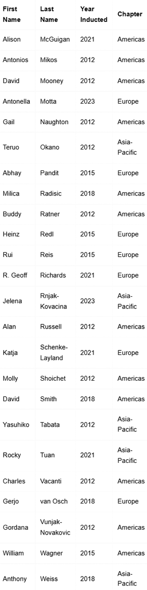
International Fellows of Tissue Engineering and Regenerative Medicine recognized by their peers as contributors to the field, either through research or the Tissue Engineering and Regenerative Medicine International Society.
Certainly, there is a bright future ahead for our field as it continues to evolve. It will be amazing to see where the field will be in 30 years; the one constant is often change, but I predict that the seminal Advances in Tissue Engineering Short Course at Rice University will still be around training future generations—a fitting legacy to our colleague and friend, Tony Mikos
Obtaining clinical release criteria that will lead to reliable and consistent results has been an issue, although it's now much improved. Another barrier is the cost for both development and product sale. We must do better to reduce costs. I believe the tissue engineering industry is already benefiting from the engineered meat industry, as they explore ways to create better media, cell expansion, and production at reduced prices.
Dr. Sackner-Bernstein generated interest working within the FDA and other federal agencies and along with Jon Mogford, Director of the Defense Sciences Office of DARPA, was among the first to promote funding for what is now referenced as tissue/body on-a-chip technologies. As a result, DARPA, the NIH, and DTRA announced grant opportunities within months of each other starting in 2011. In the last few years, the organoid/chip field has greatly expanded. For example, our institute is using the organoid technology to create organ tissue equivalents and body-on-a-chip systems for drug, chemical, and biological testing, as well as personalized medicine, such as predicting the best therapy for individual cancer patients. The field has grown extensively, and many different entities and individuals are currently doing great work in this area.
Footnotes
Disclosure Statement
No competing financial interests exist.
Funding Information
No funding was received for this article.