




Abstract
In the realm of in situ cartilage engineering, the targeted delivery of both cells and hydrogel materials to the site of a defect serves to directly stimulate chondral repair. Although the in situ application of stem cell-laden soft hydrogels to tissue defects holds great promise for cartilage regeneration, a significant challenge lies in overcoming the inherent limitation of these soft hydrogels, which must attain mechanical properties akin to the native tissue to withstand physiological loading. We therefore developed a system where a gelatin methacryloyl hydrogel laden with human adipose-derived mesenchymal stem cells is combined with a secondary structure to provide bulk mechanical reinforcement. In this study, we used the negative embodied sacrificial template 3D printing technique to generate eight different lattice-based reinforcement structures made of polycaprolactone, which ranged in porosity from 80% to 90% with stiffnesses from 28 ± 5 kPa to 2853 ± 236 kPa. The most promising of these designs, the hex prism edge, was combined with the cellular hydrogel and retained a stable stiffness over 41 days of chondrogenic differentiation. There was no significant difference between the hydrogel-only and hydrogel scaffold group in the sulfated glycosaminoglycan production (340.46 ± 13.32 µg and 338.92 ± 47.33 µg, respectively) or Type II Collagen gene expression. As such, the use of negative printing represents a promising solution for the integration of bulk reinforcement without losing the ability to produce new chondrogenic matrix.
Impact Statement
The physiological environment of the knee joint includes repeated loading; therefore, the development of a cartilage regeneration strategy needs to consider the mechanical properties of the implant. This work combines the ideal environment for stem cells to undergo chondrogenic differentiation with bulk reinforcement in line with the stiffness of healthy, human articular cartilage. Therefore, taking into consideration an element where the implant could otherwise fail, by damage or dislodgement of the soft hydrogel because of physiological loading.
Introduction
With little to no ability to self-regenerate, repairing human cartilage defects of the knee remains a major clinical challenge. 1 Without intervention, a cartilage injury can accelerate the breakdown of the surrounding tissue, especially in a defect of ≥10 mm diameter. 2 Current surgical treatments, including microfracture, matrix-induced or autologous chondrocyte implantation, and mosaicplasty, still present inferior options.3,4 This is because of high reoperation rates, limitations in the size of defects that can be treated, average long-term patient outcomes, and the lack of regeneration of articular cartilage instead favoring fibrocartilage.5–13 Therefore, a new solution is proposed that combines human adipose-derived mesenchymal stem cells (hADSCs) with a soft gelatin methacryloyl (GelMA) hydrogel for the generation of new cartilage tissue.
The cell-laden hydrogel can be delivered and then photocrosslinked directly within the cartilage defect using an in situ approach.14,15 The soft hydrogel is a key component with literature increasingly showing the softer the hydrogel, the greater or faster the production of new cartilage matrix.16–18 However, these implanted soft hydrogels risk dislodgement in the knee before new matrix can support itself. This is especially evident using the in situ approach as there is no preculture period that can add stiffness to the hydrogel before implantation. 19
The inclusion of a secondary structure offers a promising compromise between increasing the bulk stiffness while retaining a soft environment for the cells. 3D printing allows for the customization of this secondary structure to the needs of the tissue. Techniques including melt-electrowriting and material extrusion commonly use a logpile design of straight fibers, which rotate orientation by 90° for each sequential layer or simple designs. Melt-electrowriting logpile structures on their own typically have a very low stiffness, as does the hydrogel, but when combined together they can reach stiffnesses of >50 times their individual values. 20 However, these structures can still be highly dependent on new matrix formation over weeks to months to achieve mechanical properties in line with the native tissue. 17
To reinforce the soft GelMA hydrogels, the negative embodied sacrificial template 3D (NEST3D) printing technique was used to compare polycaprolactone (PCL) lattice-based scaffold designs that go beyond the 90° logpile design. 21 Negative printing involves 3D printing a mold in which the negative or empty space is filled with a secondary material. This allows for the creation of more complex and porous structures with a standard 3D printer than would be possible if printing directly. Lattice designs can have a high stiffness and strength-to-weight ratio, which can allow the targeted mechanical properties to be reached with a low volume fraction.22,23 In an in situ environment, a patient customizable NEST scaffold can be placed into a knee defect before the cell-laden hydrogel is delivered and photocrosslinked to seal in both components. The NEST scaffolds provided immediate bulk reinforcement to the cellular GelMA hydrogel scaffolds. This immediate reinforcement was retained over 41 days of culture, although new cartilaginous matrix was produced, demonstrating the ability to integrate biological matrix with synthetic NEST scaffolds.
Materials and Methods
Scaffold design and fabrication
The NEST lattice structures were fabricated according to the protocol published in Doyle et al. 21 Briefly, all scaffolds were designed in nTopology (nTopology, New York), named according to the software’s “lattice type” and used to calculate the porosity. The assembled sacrificial templates were printed with polyvinyl alcohol (PVA) (FormFutura, Nijmegen, the Netherlands) using an Ultimaker S3 printer (Ultimaker, Utrecht, the Netherlands), filled with medical grade PCL (PURASORB PC 12, Corbion Inc., Gorinchem, the Netherlands), and then the PVA template was dissolved through ultrasonication (2800 Ultrasonic Cleaner, Branson Ultrasonics Corporation, Brookfield, WI) in water.
Mechanical testing
The compressive modulus of the scaffolds was assessed via unconfined compression testing at room temperature using a TA Electroforce 5500 mechanical loading device (TA Instruments, New Castle, DE) fitted with a 50 lb load cell. Physical dimensions of each sample were taken to allow the area to be calculated. The bottom plate was fixed and the top plate moved following a ramp function at a rate of 0.01 mm/s until a total displacement of 50% of the sample height or until maximum machine capabilities. Load and displacement measurements were converted into stress (σ) and strain (ε) using the measured cross-sectional area and height. The stiffness was computed using the slope of the stress–strain curve between a strain of 0.1 and 0.2.
FITC-GelMA release
Fluorescently labeled GelMA (TRICEP, Wollongong, Australia) was added to regular GelMA at a total concentration of 1%. 18 The GelMA was conjugated with fluorescein isothiocyanate isomer I (FITC) (Sigma-Aldrich, Missouri), as previously described. 18 FITC-GelMA 6% with 0.06% lithium phenyl-2,4,6-trimethylbenzoylphosphinate (LAP) (Tokyo Chemical Industries, Chuo-ku, Japan) was prepared as uncrosslinked, cross-linked (hydrogel only), and cross-linked with the NEST PCL scaffold (hydrogel scaffold). In each case, a total volume of 250 µL of FITC-GelMA was used, and cross-linked samples were immediately irradiated for 60 s, using 405 nm visible light (BioLambda, Sao Paulo, Brazil) with a light irradiance of 20 mW/cm2. Phosphate buffered saline (PBS) 1× was added to all conditions. At each time point, 100 μL aliquots of the retrieved PBS were read at 483-14/530-30 excitation/emission wavelengths using a CLARIOstar plate reader (BMG Labtech, Ortenberg, Germany). For the cell study, the FITC release into the media was read in triplicates of 60 μL aliquots with the same wavelengths and multiplied by the dilution factor to get the total FITC release.
Cross-linking test
The storage modulus of 6% GelMA was assessed using a Physica MCR 302 Rheometer (Anton Paar, Graz, Austria) with a parallel plate (24.95 mm diameter) at 23°C. The control had no modification to the parallel plate, whereas the PCL group had a thin layer of PCL attached to the base. The photorheology protocol was as previously described, using oscillatory conditions of 1% strain and a frequency of 1 Hz. 24 An Omnicure LX1000 light source (20 mW/cm2) (Excelitas Technologies Corp, Waltham, MA) with a 400–500 nm bandpass filter illuminated the underside of the sample through a quartz crystal stage.
Stem cell isolation
Primary hADSCs were isolated and cultured as previously described.25,26 Use of all human samples, procedures, and experiments was approved by the Human Research Ethics Committee Research Governance Unit of St. Vincent’s Hospital, Melbourne, Australia (HREC/16/SVHM/186) and performed in accordance with relevant guidelines and regulations. Cells were cultured in complete culture media containing low glucose Dulbecco’s modified Eagle’s medium (DMEM) (Sigma-Aldrich) supplemented with 10% fetal bovine serum (Gibco, Waltham, MA), 60 µg/mL penicillin, 100 μg/mL streptomycin solution (Gibco), 2 mM L-glutamine (Gibco), 15 mM HEPES (Gibco), 20 ng/mL epidermal growth factor, and 1 ng/mL fibroblast growth factor (R&D Systems Inc., Minneapolis, MN).
Generation and culture of biological hydrogel scaffolds
hADSCs were added to 6% GelMA at 5 × 10⁶ cells/mL with 0.06% w/v LAP. 27 Twenty microliters of cell-laden GelMA (4 mm diameter and 2 mm height) with or without an NEST scaffold was irradiated as given in “FITC-GelMA release.” Two-hundred microliters of complete culture media was added to each sample. The next day, the media was replaced with 200 µL of chondrogenic media. Chondrogenic media consisted of DMEM high-glucose (Sigma-Aldrich), 60 µg/mL penicillin, 100 μg/mL of streptomycin, 1× Glutamax (Gibco) 15 mM HEPES, 1% insulin-transferring selenium (Sigma-Aldrich), 100 nM dexamethasone (Sigma-Aldrich), 50 mg/mL ascorbate-2-phosphate (Sigma-Aldrich), 10 ng/mL TGFβ3 (Prepotech, Cranbury, NJ), and 10 ng/mL BMP6 (R&D Systems).
Metabolic activity
The CellTiter Blue (Promega, Wisconsin) metabolic assay was applied following the manufacturer’s instructions. Briefly, the CellTiter Blue was mixed with media in a ratio of 1:5, added to each sample, and incubated for 2 h at 37°C. One hundred microliters of supernatant were read at 550-15/600-20 excitation/emission using a CLARIOstar plate reader.
Sulfated glycosaminoglycans (sGAGs) analysis
The detection of sGAGs was via a spectrophotometric assay with a standard curve (0–1 µg) of purified chondroitin sulfate (Sigma-Aldrich) in dimethylmethylene blue (DMMB) (Sigma-Aldrich). 28 The release media was diluted with fresh media to ensure the values were within the standard curve. DMMB was added in a ratio of 1:5, sample to DMMB, followed by mixing. Absorbance was immediately read at 525 nm and 595 nm using a CLARIOstar plate reader. The results are presented as the ratio of 525/595. For the retained sGAGs, samples were dissolved in papain buffer (sodium phosphate buffer 0.2 M, cysteine 0.01 M, NaH2PO4⋅1H2O 0.2 M, EDTA C10H14N2Na2O8⋅2H2O 0.01 M [Sigma-Aldrich], papain 250 μg/mL [Worthington Biochemical, Lakewood, CO]) at 65°C overnight. Samples were centrifuged at 10,000 g for 10 min, and the supernatant was retrieved and diluted with papain to ensure that the values were within the standard curve, combined with DMMB and read as above. The sGAGs amount in µg was calculated via a linear regression fit with a chondroitin sulfate standard curve and corrected for the dilution factor.
RNA extraction and real-time quantitative polymerase chain reaction
For RNA extraction, 300 µL of TRIzol Reagent (Ambion, Thermo Fisher Scientific) was added to each sample and stored at −80°C. When thawed, samples were manually homogenized, centrifuged at 1000 G for 5 min, and the supernatant retrieved. The total RNA was purified using DirectZol RNA kit (Zymoresearch, Irvine, CA) following the manufacturer’s instructions including DNAse I treatment at 6 U/μL for 15 min. 18 A total of 100 ng of RNA was reverse transcribed using High-Capacity cDNA Reverse Transcription Kit (Thermo Fisher Scientific), following the manufacturer’s protocol. Gene expression was evaluated by quantitative polymerase chain reaction (qPCR) using the following TaqMan gene expression assay: SOX9 (Hs00165814_m1, 4331182), COL2A1 (Hs00264051_m1, 4331182), COL1A2 (Hs01028956_m1, 4331182), ACAN (Hs00153936_m1, 4331182), and GAPDH (Hs02786624_g1, 4331182) as the housekeeping gene (all Thermo Fisher Scientific). qPCR was performed on a QuantStudio 6 Flex Real-Time PCR System (Thermo Fisher Scientific), and relative quantification was calculated with the 2e−ΔΔCT method.
Cryo-embedding
Samples were fixed in 1% paraformaldehyde (Santa Cruz Biotechnology, Dallas, TX) for 4 h and then stored in 30% w/v sucrose (Sigma-Aldrich) at 4°C. Samples were then embedded in O.C.T. Compound (Sakura Finetek Europe, Alphen aan den Rijn, the Netherlands) and flash frozen in liquid nitrogen. Cryosections of 7–8 μm were cut along the axial plane.
Histological staining
Safranin O and Hematoxylin staining was performed as previously described. 29 Briefly, sections were gradually rehydrated, nuclei stained with Weigert’s haematoxylin solution (Sigma-Aldrich HT1079), cartilage stained with Safranin O 0.6% w/v (Sigma-Aldrich), and then dehydration and xylene (Chem-Supply) clearing. The sections were mounted in Pertex medium (Trajan Scientific and Medical, Melbourne, Australia) and imaged using a ZEISS Axioscan 7 slice scanner with ZEISS ZEN imaging software (ZEISS, Oberkochen, Germany).
Immunohistochemistry
The Novolink™ Polymer Detection System (RE7150-K, Leica Biosystems, Wetzlar, Germany) was used for Type I and II Collagen staining following the manufacturer’s protocol. Briefly, the steps included: cryosections rehydration, antigen unmasking with Dako Ready-to-use Proteinase K (S3020) (Dako Agilent, Santa Clara, CA), neutralization with Peroxidase Block (RE7101), primary antibody with Protein Block (RE7102), and incubation with primary antibody (Type I Collagen 1:100 or II 1:250) diluted in 2% bovine serum albumin (Sigma-Aldrich) overnight at 4°C. The next day, slides were incubated with Post Primary (secondary) (RE7111), Novolink Polymer (RE7112), development of peroxidase activity using DAB Chromogen (RE7169) diluted in Novolink DAB Substrate Buffer (RE7171) 1:20, and counterstained with hematoxylin (RE7107). Slides were dehydrated, cleared in xylene, and then mounted with Pertex and imaged as given in “Histological staining.”
Statistics
For each quantitative test group, at least three technical replicates were used with data summarized as the mean with error bars representing standard deviation. All statistical analysis was performed using Prism 8 (GraphPad, California) with a statistical significance level ≤ 0.05. Significance was determined using an unpaired t-test or one-way ANOVA. In all graphs stars represent: * is p ≤ 0.05; ** is p ≤ 0.01; *** is p ≤ 0.001; **** is p ≤ 0.0001.
Results and Discussion
Characterization of fabricated lattice-based scaffold
The NEST3D printing process was used to create three categories of lattice scaffolds: simple cubic (SC), hex prism diamond (HPD), and hex prism edge (HPE) (Fig. 1). 21 The scaffolds were made in a clinically relevant size (10 mm diameter, 2 mm height).2,30,31 The aim of the hydrogel scaffold is to be in the stiffness range of healthy human cartilage reported in the literature, 581 ± 168 kPa, which translates to an approximate range of 180–850 kPa. 32 For the PCL scaffold, five were above the stiffness range at >1347 ± 67 kPa (Fig. 1), and therefore deemed unsuitable. Of the remaining designs, one had a stiffness within the required range (650 ± 165 kPa—HPE3.5), whereas two others (SC7.2 and HPE4.9) were below the range of interest (28 ± 5 kPa and 150 ± 18 kPa, respectively). The SC7.2 design was also deemed unsuitable because of the extremely low stiffness. The HPE3.5 (Fig. 1) had struts extending from each vertex that provided little impact on stiffness, while still contributing to the volume fraction. Therefore, the HPE4.9 was chosen for further evaluation. One potential limitation of the HPE4.9 is reinforcement is applied at a bulk level so a force applied only to the center of the scaffold would not receive the same level of support as the edges. However, as a modified hinge joint, the primary extension and flexion movements of the knee result in loading across the joint and typically not only localized to only one small region, such as the center of the defect.33,34
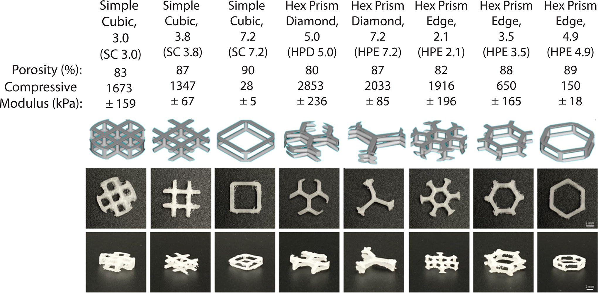
Scaffold characterization. Lattice-based scaffold made from nTopology software with macroscopic images of the fabricated NEST structures in PCL. Naming of each scaffold is from the lattice type in nTopology, and the cell size (diameter). Porosity is as calculated in nTopology. Compressive modulus values were measured via unconfined compression testing of the 10 mm diameter PCL designs. PCL, polycaprolactone.
Next, two cross-linking-based experiments were performed to determine if the presence of the NEST PCL scaffold would affect the cross-linking efficiency of the hydrogel. The first study determined if the free radicals produced during photocrosslinking could be scavenged (removed) by the PCL. 35 The photocrosslinking of GelMA happens through a free-radical polymerization reaction. The LAP photoinitiator is photo-cleaved when exposed to visible light, which generates free radicals. 36 The free radicals then diffuse through the GelMA, both while the visible light is on and once it is turned off (dark cross-linking/polymerization). The dark cross-linking will continue until (1) all the free radicals are quenched (no single free radicals left in the system) or (2) there are no more cross-linking sites (GelMA is 100% cross-linked).14,37,38
Given this, a photorheology experiment was carried out with or without a PCL coating on the stainless-steel parallel plate (Fig. 2A). The GelMA was run under oscillatory conditions for 1 min, then the visible light turned on for 1 min (short exposure) or 10 min (long exposure), and the storage modulus measured at 11 min (Fig. 2B). Given the contribution of both light and dark cross-linking, the short and long exposure was compared. 14 In the long exposure condition, there was no significant difference in the storage modulus between the PCL and no PCL groups (Fig. 2B). This confirms the presence of the PCL does not interfere with the test, for example, because of the GelMA not gripping to either plate. Instead, under the short exposure condition, there was a significantly lower storage modulus in the PCL group. This indicates the ability of PCL to scavenge the free radicals, thereby reducing the contribution of the dark cross-linking. In addition, with the visible light on for a long exposure time, any free radical scavenging by the PCL is masked by an excess number of free radicals in the system, resulting in no change in storage modulus. Full cross-linking profiles are shown in Figure 2C.
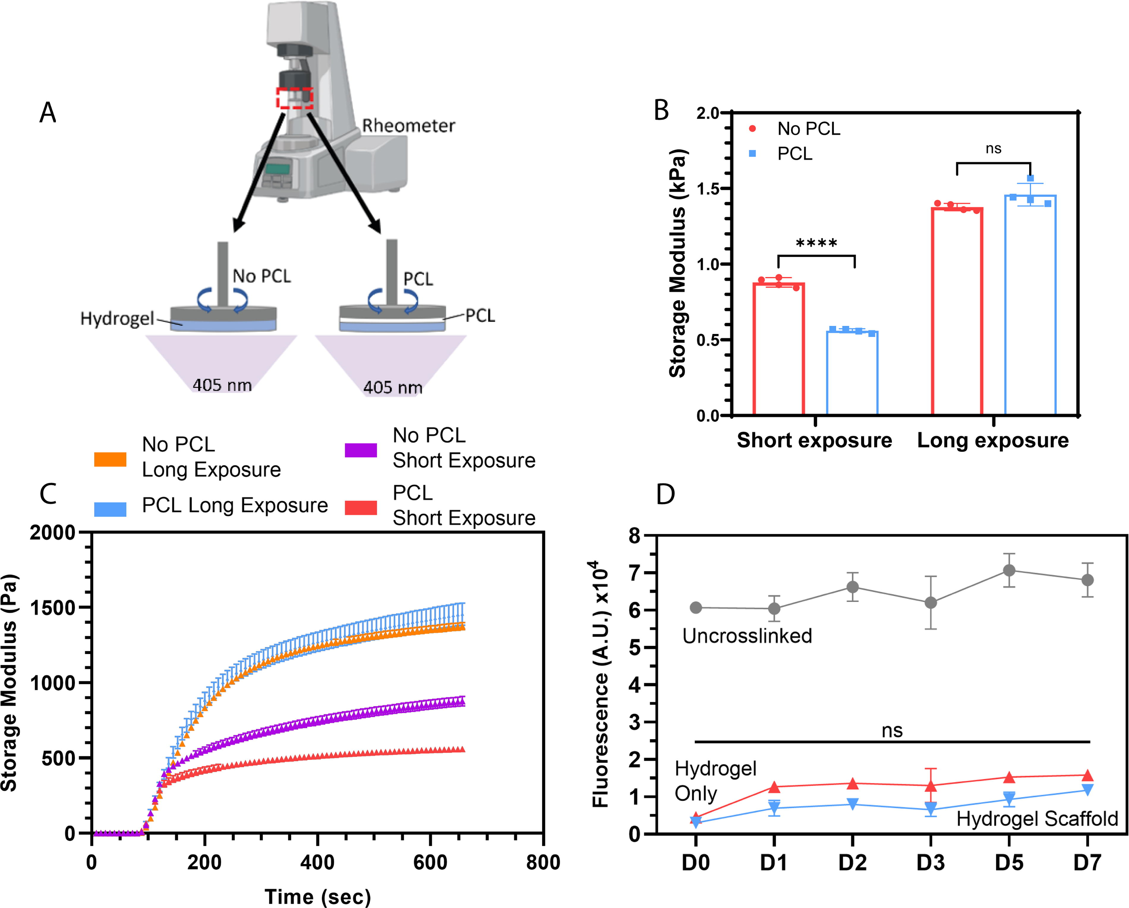
PCL-GelMA cross-linking experiments.
The second study used fluorescent FITC-GelMA to assess the bulk cross-linking stability of the hydrogel scaffold. The release of FITC from the cross-linked scaffold indicates scaffold degradation over longer terms or cross-linking efficiency in the short term. 18 Uncrosslinked FITC-GelMA served as the highest amount of fluorescence (FITC) that can be released into the supernatant from a sample. Both the hydrogel-only and hydrogel scaffold groups showed no significant change in the FITC release over the 7 days (Fig. 2D). This demonstrates the addition of the NEST PCL scaffold did not change the cross-linking efficiency.
In vitro cellular study
After the assessment of the acellular properties, the biological integration of the hADSCs laden in GelMA hydrogel with the NEST scaffold was investigated. We have previously shown the chondrogenic capability of primary hADSCs in GelMA therefore rendering the cell type a suitable source for both in vitro experimentation and in situ clinical application.18,25,27 The cellular hydrogel, with (hydrogel scaffold) or without (hydrogel only) the NEST PCL scaffold, was prepared by irradiation for 60 s, using 405 nm visible light with a light irradiance of 20 mW/cm2 before being cultured in chondrogenic differentiation media for 41 days (Fig. 3).

Workflow of the in vitro study. The hADSCs and LAP photoinitiator are mixed into the 6% GelMA before 20 µL is added to a 4 mm diameter, 2 mm height mold or to the NEST PCL scaffold before irradiation for 60 s, using 405 nm visible light with a light irradiance of 20 mW/cm2. Samples were cultured in chondrogenic media for up to 6 weeks before analysis using a variety of methods. Figure created with BioRender.com (February 21, 2024), GelMA, gelatin methacryloyl; hADSCs, human adipose-derived mesenchymal stem cells; LAP, lithium phenyl-2,4,6-trimethylbenzoylphosphinate.
Gene expression analysis
We have previously shown the chondrogenic capability of primary hADSCs in GelMA both in vitro and in situ preclinical application.18,25,27 SOX9, ACAN (aggrecan), COL1A2 (type I collagen), and COL2A1 (type II collagen) were quantified by real-time quantitative PCR (RT-qPCR) (Fig. 4). SOX9, COL1A2, and COL2A1 showed no significant difference between the groups. SOX9 is a master transcription factor and is responsible for the gene expression of COL2A1 and ACAN,39,40 and its expression was found to be similar between day 1 and day 22. COL2A1 represents 90–95% of the total collagen content in articular cartilage, but the gene expression is only activated during chondrogenesis.40,41 Importantly, COL2A1 and ACAN are expressed only at day 22, demonstrating chondrogenic differentiation of hADSC in both groups after TGFβ3/BMP6 induction. COL1A2, which represents a small portion of the remaining collagen network, is expressed by the undifferentiated hADSCs and shows an increase in expression between day 1 and day 22. COL1A2 can also be considered a marker of fibrocartilage, and therefore, it is important to evaluate the ratio of type I and type II collagen protein accumulation using immunostaining (see “Stiffness, metabolism, degradation and chondrogenic matrix composition”).

Chondrogenic gene expression analysis. The graphs represent the fold changes calculated with 2ΔΔCΤ method of SOX9, aggrecan (ACAN), type I collagen (COL1A2), and type II collagen (COL2A1) markers in RT-qPCR assay. GAPDH was used as the housekeeping gene. Graph bars represent the standard deviation between three replicates. Statistical analysis was performed using an unpaired t-test. GAPDH, glyceraldehyde 3-phosphate dehydrogenase; RT-qPCR, real-time quantitative polymerase chain reaction.
Stiffness, metabolism, degradation, and chondrogenic matrix composition
The inclusion of the NEST scaffold provides reinforcement from the instant of fabrication instead of relying on matrix production over time. This is demonstrated in vitro where the hydrogel only increases in stiffness over 41 days of culture starting at 7.37 ± 0.59 kPa reaching a stiffness of only 30.02 ± 3.47 kPa. Instead, the hydrogel scaffold had no significant difference in stiffness at 1978.63 ± 650.40 kPa on day 1 and 1504.11 ± 741.78 kPa by day 41 (p value = 0.8837) (Fig. 5A). During culture, there was no significant difference in the metabolic activity of the hADSCs in the hydrogel or hydrogel scaffold (Fig. 5B). The same FITC-GelMA release experiment was used to monitor the long-term degradation of the hydrogel. Onofrillo et al. showed the correlation between hydrogel degradation and chondrogenic differentiation. 18 Over the 41 days, there was no significant difference in FITC release between the two groups (Fig. 5C). However, a crossover point occurred at day 26 where for the remaining period the hydrogel scaffold group had a slightly higher FITC release compared with the hydrogel only. To explore how this relates to chondrogenic differentiation progression, the proteoglycan content was studied.
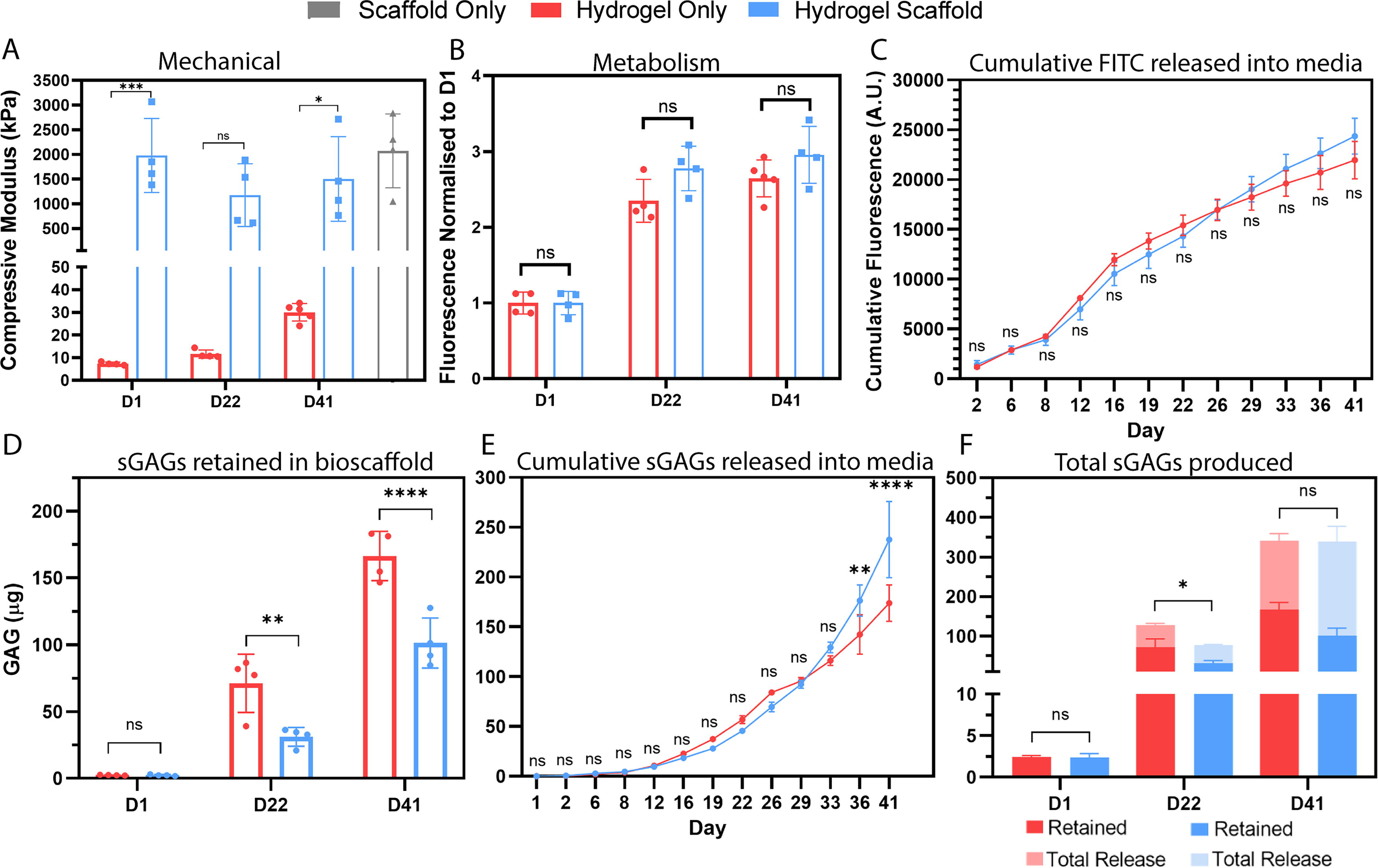
In vitro quantitative assessment: Stiffness and sGAGs.
Proteoglycans (including sGAGs) make up 10–15% of the wet weight of articular cartilage. 41 The sGAGs (chondroitin and keratin) account for over 90% of the total GAGs content at any age and health of the articular cartilage.42–44 Of the total sGAGs produced, a portion remained within the hydrogel (retained), and some were transferred into the media (released). Comparing the retained sGAGs, at the 22- and 41-day time points, there were significantly more sGAGs in the hydrogel only compared with the hydrogel scaffold group (Fig. 5C). Instead, over the first 33 days of culture, there was no significant difference in the cumulative weight of sGAGs released between the hydrogel-only and hydrogel scaffold group (Fig. 5D). However, the trend followed that as the FITC release into the media only with the crossover point between the two groups shifting from day 26 (FITC) to day 29 (sGAGs). However, when comparing the total sGAGs production (released + retained) over the long term (41 days), there was no difference in the total production (340.46 ± 13.32 µg hydrogel only and 338.92 ± 47.33 µg hydrogel scaffold) (Fig. 5E). Instead, the groups differed in their ratio between the released and retained: 51:49, respectively, hydrogel only and 70:30, respectively, hydrogel scaffold group. Considering the localized effect on photocrosslinking, the presence of the NEST PCL scaffold may facilitate this greater release. Figure 2 shows that the PCL can absorb free radicals thereby reducing the mechanical properties of the GelMA, and these localized softer regions could allow for more sGAGs and FITC to be released.
Immunohistochemistry and histology analysis
Macroscopic images of the hydrogel and hydrogel scaffolds showed an increase in opacity over culture suggesting extracellular matrix development (Fig. 6A, D, G, J, M, P). Cryosections were stained using hematoxylin (nucleus) of the hADSCs and safranin O for sGAGs. 45 On day 1, both groups feature a homogenous purple-colored staining (Fig. 6B, C, E, F), which is a background absorbance of the hematoxylin by the hydrogel. The hydrogel scaffold from day 1 shows some of the PCL pillars from the NEST structure, whereas physical dislodgement during sectioning and staining can result in empty space, most evident in the hydrogel scaffold day 22 sample. Moving from day 22 (Fig. 6H, I) to day 41, the purple hematoxylin color was largely replaced by homogenous red color in the hydrogel-only group (Fig. 6N, O). The hydrogel scaffold group also progresses to a light pink color at day 21 (Fig. 6K, L) before homogenous red staining is seen on day 42 (Fig. 6Q, R). In the hydrogel scaffold group, moving from the center toward the PCL pillars, the red staining is reduced, and some purple color can still be seen suggesting there are less or no sGAGs in these regions (Fig. 6Q). As previously mentioned, the potentially localized softer regions because of free radical absorption by the PCL could allow for more sGAGs to leave the scaffold accounting for the lesser red staining around the PCL.
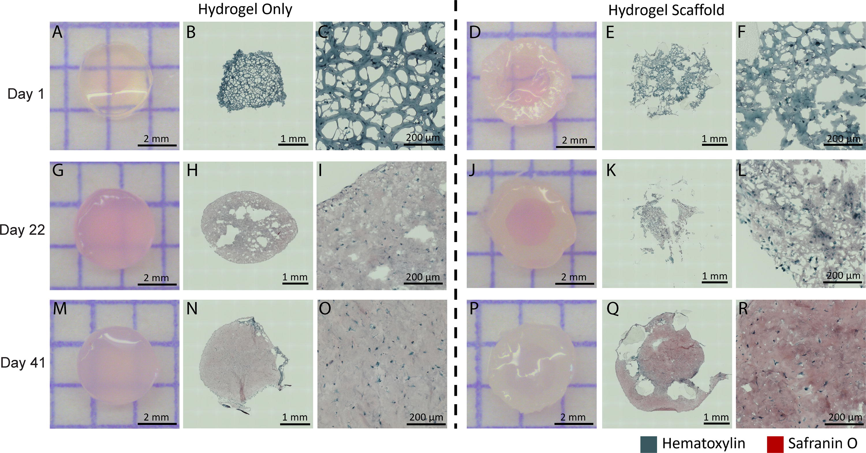
Hematoxylin and safranin O. (
The immunohistochemistry staining was used to measure the accumulation of collagens type I and type II (Fig. 7, 8). There is little type I collagen staining present in either group on day 1 (Fig. 7A–D). By day 22, some type I collagen formation was seen scattered around the entire scaffold in both groups (Fig. 7E–H). By day 41, there is a greater and more homogenous covering of type I collagen in both groups, thereby showing continued matrix production over the 41 days (Fig. 7I–L). Quantification of the staining confirmed significantly more type I collagen in the hydrogel scaffold compared with the hydrogel-only group at day 22 and day 41 (Fig. 7M). However, in both groups, the type I collagen has the strongest staining localized intracellularly and the limited extracellular levels could be an indication of low levels of overall production. 46
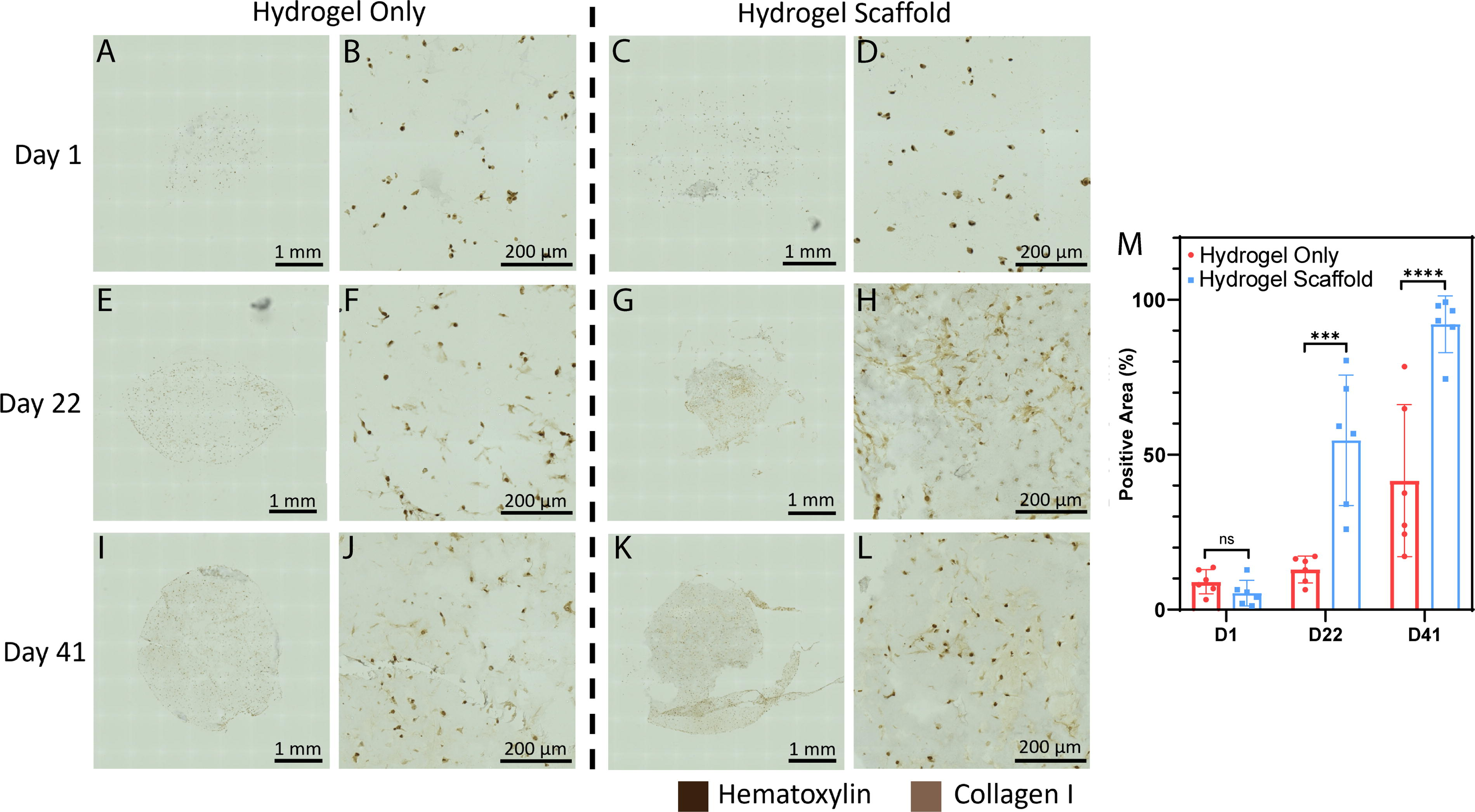
Immunohistochemistry staining.
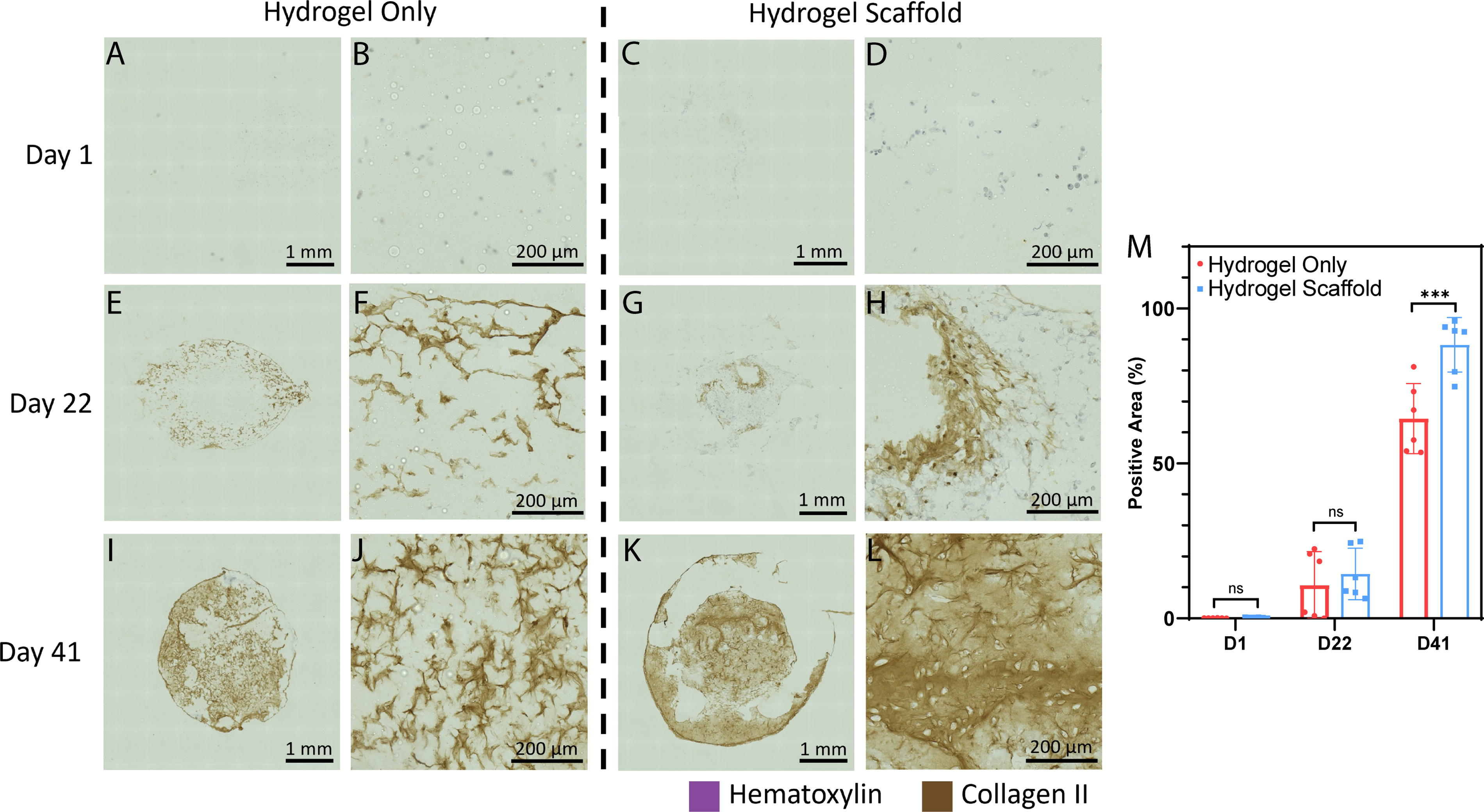
Immunohistochemistry staining.
No collagen type II was present at day 1 in either group (Fig. 8A–D). By day 22, the hydrogel-only group had type II collagen formation around the outside of the hydrogel with none in the center (Fig. 8E, F). The hydrogel scaffold group at day 22 also had type II collagen formation; however, it was densely centered around empty space or a bubble in the scaffold (Fig. 8G, H). By day 41, there was again a homogenous and deeper positive staining for type II collagen for both groups (Fig. 8I–L) with significantly more in the hydrogel scaffold group compared with the hydrogel (Fig. 8M). The collagen staining appeared denser in the hydrogel scaffold group compared to the hydrogel only. This effect could again be because of localized softer regions which may favor chondrogenic matrix accumulation. However, atomic force microscopy studies are required to evaluate the localized stiffness changes. Type II collagen is also synthesized intracellularly, but the staining shows abundant presence extracellularly, which suggests a greater overall production of type II collagen compared with type I collagen. 47 Overall, in both groups, the desired type of collagen (type II) was being produced in greater quantities than type I collagen which could instead indicate fibrocartilage. 48
Conclusions
There is still a clinical need for cartilage regeneration techniques that can develop adequate new cartilage tissue to withstand the repeated physiological loads in vivo. Here, it is shown that the soft GelMA hydrogel provides an ideal environment for hADSCs to make new matrix, highly composed of extracellular type II collagen after 41 days of culture in vitro. To withstand the physiological load requirements, lattice-based reinforcement scaffolds were made using the NEST3D printing technique. Importantly, the NEST PCL scaffold retains the same mechanical properties when combined with GelMA throughout chondrogenesis while allowing for a comparable chondrogenic process. The addition of the reinforcement NEST scaffold continues the translation of cartilage regenerative therapies toward clinical application.
Footnotes
Acknowledgments
The authors would like to acknowledge Laura Leone and the Melbourne Histology Platform at The University of Melbourne for the cryosectioning of the samples and the Biological Optical Microscopy Platform at The University of Melbourne for the use of the Zeiss Axioscan 7. The authors also thank nTopology for providing access to their software through an nTopology Educational License.
Authors’ Contributions
S.E.D.: Methodology, investigation, formal analysis, writing—original draft, and writing—review and editing. F.S.: Methodology, investigation, formal analysis, and writing—review and editing. C.O.: Conceptualization, supervision, methodology, investigation, formal analysis, and writing—review and editing. C.D.B.: Supervision, funding acquisition, and writing—review and editing. C.D.O.: Conceptualization, supervision, methodology, formal analysis, and writing—review and editing. E.P.: Conceptualization, methodology, formal analysis, funding acquisition, and writing—review and editing. S.D.: Conceptualization, supervision, methodology, investigation, and writing—review and editing. All authors have read and agreed to the published version of the article.
Disclosures Statement
No competing financial interests exist.
Funding Information
This work was supported by (1) Victorian Medical Research Acceleration Fund (2018, Round 2), (2) NHMRC-MRFF Investigator Grant (Di Bella) #1193897, and (3) Australian Technology Network of Universities Industry Doctoral Training Center (IDTC) scholarship.