




Abstract
Negative pressure therapy (NPT) has been shown to facilitate wound healing and promote hair growth in a porcine model. However, there is a paucity of research on the impact of negative pressure on hair growth in murine models. Despite the ability of nude mice to develop hair follicles, the hair they produce is often flawed towing to genetically induced keratin disorders, rendering them a pertinent animal model for assessing hair regeneration. Therefore, this study aims to investigate the effects of negative pressure on hair follicle growth in a nude mouse model. To achieve this, a customized external tissue expansion device was developed to apply negative pressure to the dorsum of nude mice. The mice were subjected to several treatment courses consisting of 15 and 30 min of continuous negative pressure at 10 mmHg, which were repeated 5 and 10 times every other day until sacrifice. Dorsal skin samples were subsequently extracted from the suction and nonsuction areas. The sections were stained with various antibodies to assess the expression of SOX-9, LHX-2, Keratin-15, β-catenin, CD31, and vascular endothelial growth factor-A, and a TUNEL assay was used to analyze cell apoptosis. The results showed that the number of hair follicles and angiogenesis were significantly higher in the suction area than in the nonsuction area in all groups. Moreover, mice that received NPT for 15 min for 10 times had a higher hair follicle density than the other three groups. Immunofluorescence staining for LHX-2 and Keratin 15 further validated the results of these findings. In conclusion, this study demonstrated that negative pressure effectively promotes hair follicle growth and angiogenesis in nude mice through SOX-9- and LHX-2-mediated follicular regeneration and β-catenin-mediated hair follicle morphogenesis.
Impact Statement
The results of this study indicate that negative pressure therapy (NPT) is effective in promoting hair growth in nude mice, as evidenced by increased hair follicle density and angiogenesis in the treated areas. Using a custom external tissue expansion device (ETED) device, 15-min NPT treatment conducted over 10 sessions demonstrated the highest follicle density. This suggest that developing a regimen for NPT may offer to create innovative treatment approaches for hair loss, ultimately benefiting individuals suffering from hair loss disorders.
Introduction
One of the common esthetic problems among men is hair loss, which affects about up to 53% of European-American men of age 40–49 years in the United States and 90% in their lifetime. 1 Meanwhile, according to a recent population study, the overall clinical prevalence of female pattern hair loss among multiracial individuals was at around 32.3%. 2 The clinical manifestation of pattern hair loss typically involves the miniaturization of hair follicles on the frontal and vertex scalp as a result of dihydrotestosterone, resulting not only to cosmetic concerns but also psychosocial and quality-of-life impairment for patients. 3 The Food and Drug Administration has previously stated that the medical treatment for androgenetic alopecia includes topical minoxidil and oral 1 mg finasteride. However, it has been noted that the therapeutic response to medication is often limited and may cause possible adverse effects, such as sexual dysfunction, hypertrichosis, and fetal defects. In some cases, the option of a hair transplant may be an effective approach to improve hair density in the affected area through the donor’s scalp. However, the donor scalp reservoir may be limited and can lead to postoperative pain and scarring.
Recent research suggests that mechanical forces can play a significant role in regulating cell proliferation and remodeling in multiple tissues, including those involved in hair growth, such as the use of fractional lasers, microneedles, skin-stretching devices, and negative pressure therapy (NPT).4–7 The idea of adopting NPT, a method that applies continuous subatmospheric pressure through a specific pump as a means of treating open and infected wounds, was first proposed by Fleischmann et al. in 1993. 8 Negative pressure wound therapy (NPWT) operates through four fundamental mechanisms to promote healing, including macroscopic tissue deformation, removal of extracellular inflammatory fluids, stabilization of the wound environment, and micro deformation. 9 The major use cases for NPWT include chronic ulcers, acute wounds, burns, and complex wounds. Emerging studies have suggested that the principles behind NPWT could be beneficial in promoting hair growth. 10 Hsiao et al. likewise demonstrated the effectiveness of applying subatmospheric pressure using an external tissue expansion device (ETED) to increase the number of hair follicles in a porcine model. 7 To the best of our knowledge, no study has been conducted to assess the impact of NPT on hair growth in a murine model. Although nude mice can develop hair follicles, the hair produced are often defective because of genetically induced keratin disorder and has been used as an animal model for evaluating hair regeneration.11,12 Therefore, the aim of this study is to investigate the effects of NPT on the proliferation of hair follicles in nude mouse model using a specially designed ETED.
Methods
Animals
Nude mice at 6 weeks of age were purchased from the National Laboratory Animal Center (Taipei, Taiwan) and maintained in a specific pathogen-free mouse room at the Animal Center of Chang Gung Memorial Hospital (Taoyuan, Taiwan). All animals and procedures for animal handling and treatment were approved by the Institutional Animal Care and Use Committee at the Chang Gung Memorial Hospital (ethical process #:2017121818). All animals were quarantined for 1 week before the experiment.
Development of ETED device and NPT treatment
The ETED device used in the present study is a custom-built transparent plastic dome-shaped suction cup (4.9 cm × 2.8 cm × 2.4 cm) equipped with a pressure monitor and a pump to ensure precise application of negative pressure. To generate a continuous and uniformly distributed negative pressure, a curved edge layout of the suction cups was specially designed to ensure that it fits the lower dorsum of nu mice. To minimize the risk of developing pressure sores, a silicone gel sheet (Smith & Nephew, Memphis, TN) was lined on the boundaries of the ETED device.
As depicted in Figure 1A, the mice were placed in the prone position, and the ETEDs were carefully positioned on the dorsal skin tissue of the mice. A negative pressure of 10 mmHg was then continuously applied in accordance with four ETED treatment courses, with Group I receiving a 15-minute treatment for 5 sessions; Group II receiving the same 15-min treatment for 10 sessions; Group III receiving a 30-min treatment for 5 sessions; and Group IV receiving a 30-min treatment for 10 sessions. A 48-h interval was maintained between each administration of the ETED, and all mice were subsequently sacrificed following their final treatment session. Consequently, all groups were sacrificed simultaneously.
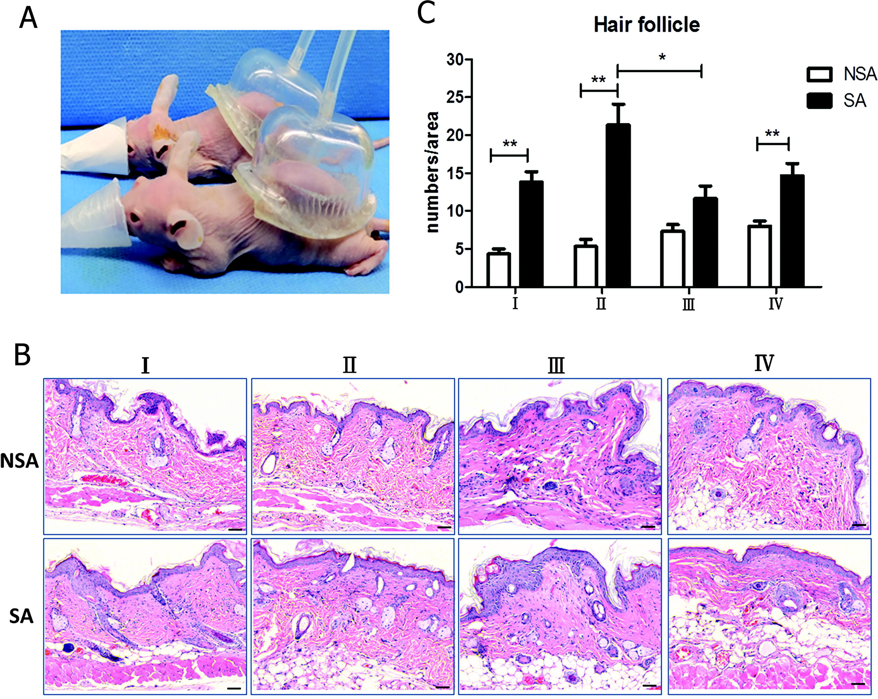
Setup of negative pressure therapy and histological change of hair follicles in nude mice after treatment.
Histological and immunohistological analysis
Quantification of hair follicles
The dorsal skin samples were harvested in two distinct regions: the suction area (SA) and the nonsuction area (NSA), which denotes normal dermal skin. The specimens were then immersed in 10% buffered formalin for 24 h, dehydrated, embedded in paraffin, and sectioned. The samples were later stained with hematoxylin and eosin (H&E), and the number of hair follicles was examined using a ZEISS Axio Scope A1 (Carl Zeiss Corporation, Oberkochen, Germany).
Hair growth expressions
To evaluate the efficacy of NPT in promoting hair growth and the expansion of hair follicles, the SA and NSA samples were stained with SRY-Box Transcription Factor 9 (SOX-9), LIM Homeobox 2 (LHX-2), Keratin-15, and β-catenin. SOX-9 plays a key role in the differentiation of the outer root sheath 9 and the formation of the hair stem cell compartment. The expression of SOX-9 in the hair follicle is critical for evaluating its ability to regenerate hair. Keratin-15 is a type I Keratin used as a marker of stem cells. SA and NSA skin sections were immunohistochemically targeted using specific antibodies. According to the standard immunofluorescence staining approach, the sections were dewaxed in xylene and rehydrated in graded alcohol. Antigen retrieval was performed by heating the sections in a citrate buffer solution at 95°C for 20 min. Antibody diluent was used to block sections with background-reducing components (Dako, Carpenteria, CA) for 1 h. After overnight incubation at 4°C with antiSOX-9 antibody (ab185966, Abcam, Cambridge, UK), antiCytokeratin 15 antibody (ab52816, Abcam) diluted 1:100, and antiLhx-2/LH2 (ab184227, Abcam) diluted 1:250, β-catenin (ab32572, Abcam) diluted 1:250 in antibody diluents and three times washing with phosphate-buffered saline with Tween 20 (PBST), the slides were incubated Goat antiRabbit IgG H&L Alexa Fluor® 488 (ab150080, Abcam) diluted 1:500 in antibody diluents for 2 h. Finally, the slides were mounted using Fluoroshield mounting medium with 4′,6-diamidino-2-phenylindole (DAPI) (ab104139, Abcam). The sections were digitalized and analyzed using the ZEISS Axio Scope A1.
Angiogenic expressions
To assess angiogenic potential following ETED treatment, we evaluated the expression of CD31 and vascular endothelial growth factor (VEGF)-A in the SA and NSA sections. The antiCD31 antibody (ab28364, Abcam) and antiVEGF-A antibody (ab52917, Abcam) were diluted 1:250 in antibody diluents and three times washing with phosphate-buffered saline with Tween20 (PBST), and the slides were incubated Goat antiRabbit IgG H&L Alexa Fluor® 594 (ab150080, Abcam) diluted 1:500 in antibody diluents for 2 h. Finally, the slides were mounted using Fluoroshield mounting medium with 4′,6-diamidino-2-phenylindole (DAPI) (ab104139, Abcam).
Terminal deoxynucleotidyl transferase assay
Cell apoptosis was quantified using a terminal deoxynucleotidyl transferase-mediated 2′-deoxyuridine, 5′-triphosphate nick end-labeling assay (ApoAlert, Mountain View, CA). Briefly, paraffin sections of SA and NSA were dewaxed in xylene and rehydrated in graded alcohol. Following the manufacturer’s instructions, the apoptotic cell nuclei were labeled with green fluorescence and visualized under a 520-nm filter.
Statistical analysis
Data are presented as mean ± standard error. Statistical analysis was performed using the GraphPad Prism software (GraphPad, San Diego, CA). Mann−Whitney tests were used to check for normal distribution. A p value of <0.05 was considered statistically significant and is noted in the figures. *, **, and *** indicate statistical significance, with p < 0.05, 0.01, and 0.001, respectively.
Results
NPT using custom ETED device promoted hair growth
The quantification of hair follicles was carried out using H&E staining, which revealed a significantly greater number of hair follicles per unit area in the SA skin as compared with the NSA skin (SA: 15.38 ± 1.17 numbers/area vs NSA: 6.25 ± 0.48 numbers/area, p < 0.0001) (Fig. 1B, C). Among the various ETED treatment groups, Group II demonstrated the highest mean number of hair follicles per unit area at 21.33 ± 2.75. This value was found to be significantly higher than that of Group III, which recorded only 11.67 ± 1.65 hair follicles per unit area (p < 0.05). Similarly, Group IV showed a mean of 14.67 ± 1.59 hair follicles per unit area (p = 0.06), whereas Group I recorded a mean of 13.83 ± 1.35 hair follicles per unit area (p < 0.05).
Analysis of SOX-9, LHX-2, Keratin-15, and β-catenin expressions in hair growth region
SOX-9 plays a vital role in hair follicle development, with continuous expression in the bulge and outer root sheath (ORS). 13 SOX-9 likewise orchestrates the formation and differentiation of cells, maintains adult hair follicle stem cells, and guides outer root sheath differentiation.13–15 The results from the immunohistochemical analysis of SOX-9 expression in the hair follicles revealed significantly elevated level of SOX-9 in SA skin (12.09 ± 0.70 numbers/area) compared with NSA skin (5.22 ± 0.70 numbers/area) (p < 0.01). Meanwhile, Group II displayed the highest SOX-9 expression (14.13 ± 1.37 numbers/area) among the treatment groups (Group I: 11.13 ± 1.20 numbers/area, p = 0.12; Group III: 12.88 ± 1.63 numbers/area, p = 0.57; and Group IV: 10.25 ± 1.14 numbers/area, p < 0.05) (Fig. 2A, Fig. S1).
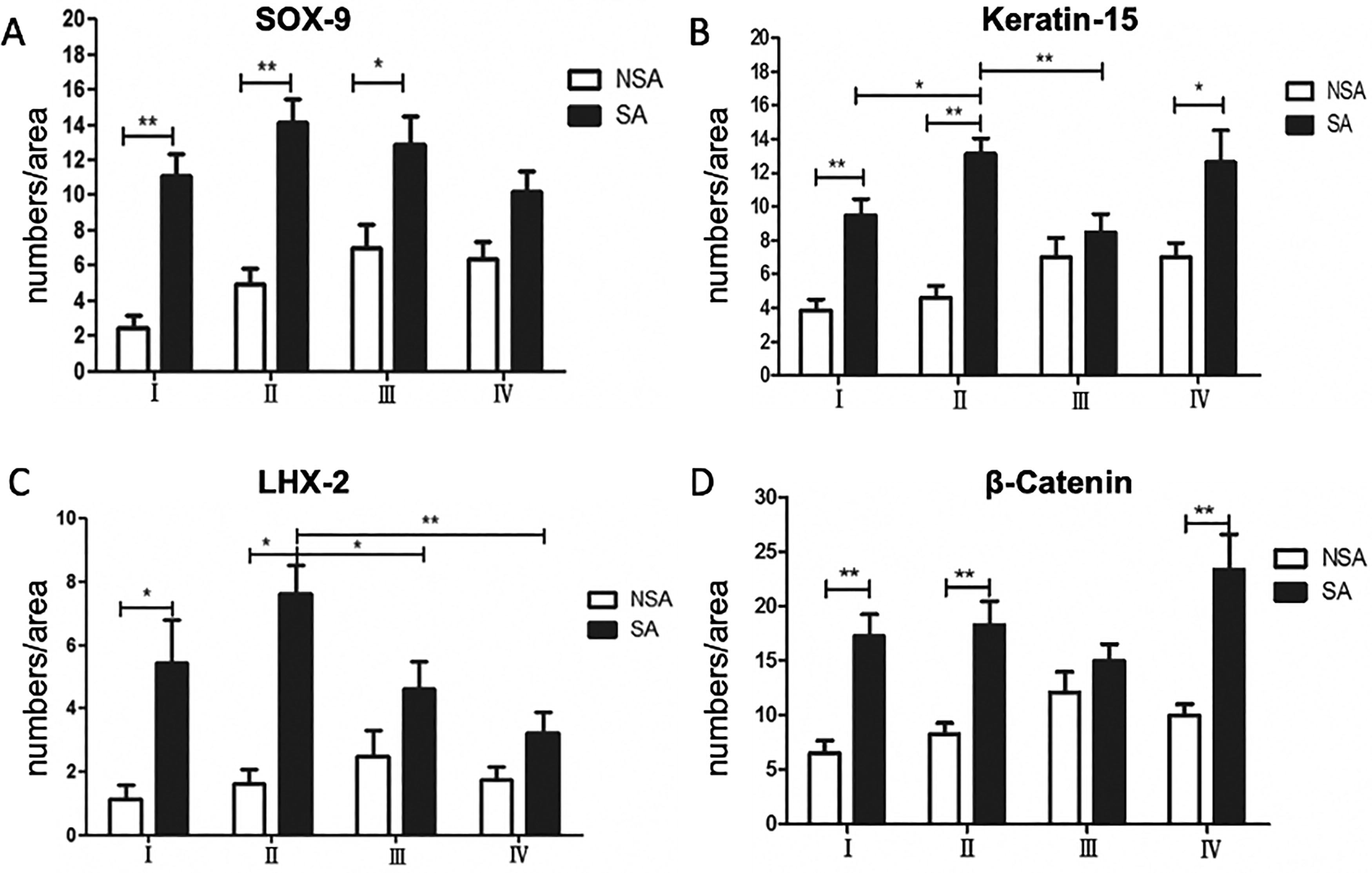
Quantification of
Keratin-15 is a type I keratin protein that is involved in maintaining the health and regenerative abilities of hair follicles stem cells and can be used to identify epidermal stem cells.16,17 In our findings, it was evident that the skin of the SA group exhibited a higher expression of Keratin-15 than the NSA group (NSA: 5.63 ± 0.48 numbers/area; SA:10.94 ± 0.70 numbers/area, p < 0.01). Furthermore, Group II (13.13 ± 0.93 numbers/area) displayed significantly higher expressions of Keratin-15 compared with the other groups like Group I (9.50 ± 0.96 numbers/area, p < 0.05) and Group III (8.50 ± 1.07 numbers/area, p < 0.05) (Fig. 2B, Fig. S2). Whereas, Group IV (12.63 ± 1.89 numbers/area, p = 0.43) showed comparable Keratin-15 expression with Group II.
In addition to hair stem cell differentiation, the expression of LHX-2, a transcription factor that regulates hair growth and is essential for anagen, was used to evaluate hair formation. 18 In summary, the level of LHX-2 expression was noticeably higher in the SA groups compared with the NSA groups. While comparing the expression of LHX-2 among different SA groups, it was observed that Group II (7.63 ± 0.89 numbers/area) showed significantly higher expression as compared with the other groups like group Group III (4.63 ± 0.86 numbers/area, p < 0.05) and Group IV (3.25 ± 0.64 numbers/area, p < 0.05) (Fig. 2C, Fig. S3). While the expression level of LHX-2 in Group I (5.43 ± 1.38 numbers/area, p = 0.19) was likewise found to be lower, this difference was not considered statistically significant.
The Wnt/β-catenin signaling pathway has been implicated in the development, regeneration, and expansion of hair follicles. 19 In order to determine whether Wnt/β-catenin promotes hair follicle regeneration, staining for β-catenin was conducted. Here, the results showed that the SA groups (18.47 ± 1.24 numbers/area) expressed significantly higher β-catenin levels than the NSA counterpart (9.22 ± 0.72 numbers/area). Still, no significant difference was found between the ETED treatment groups (Group I: 17.25 ± 2.02 numbers/area; Group II: 18.25 ± 2.23 numbers/area; Group III:15.00 ± 1.54 numbers/area; and Group IV: 23.38 ± 3.26 numbers/area) (Fig. 2D, Fig. S4).
Marked hair growth associated with increased angiogenesis expression
NPT has been shown to expedite the healing of wounds in humans by promoting angiogenesis. 20 As a result, we examined the angiogenesis marker CD31 and found that it was expressed significantly higher in all SA groups compared with its NSA group counterpart (Table 1). However, no significant difference was observed among the SA groups (Fig. 3A, Fig. S5). Our study suggests that hair growth is closely linked to angiogenesis, a process that is heavily regulated by VEGF-A. Therefore, we sought to determine whether VEGF-A expression differs in SA and NSA skin. Interestingly, the result showed that no significant difference was observed when comparing the SA and NSA in each group. However, among the SA skin groups, Group III revealed the highest expression of VEGF-A (14.38 ± 2.26 numbers/area), which was significantly greater than Group I (7.63 ± 1.05 numbers/area, p < 0.05) (Fig. 3B, Fig. S6). On the other hand, Group IV (12.63 ± 0.89 numbers/area, p = 0.6) and Group II (9.13 ± 2.07 numbers/area, p = 0.1) exhibited the second and third highest levels of VEGF-A expression, respectively, but no statistical differences were observed.
CD31 Expression in ETED Treatment Groups
ETED, external tissue expansion device; NSA, nonsuction area; SA, suction area.
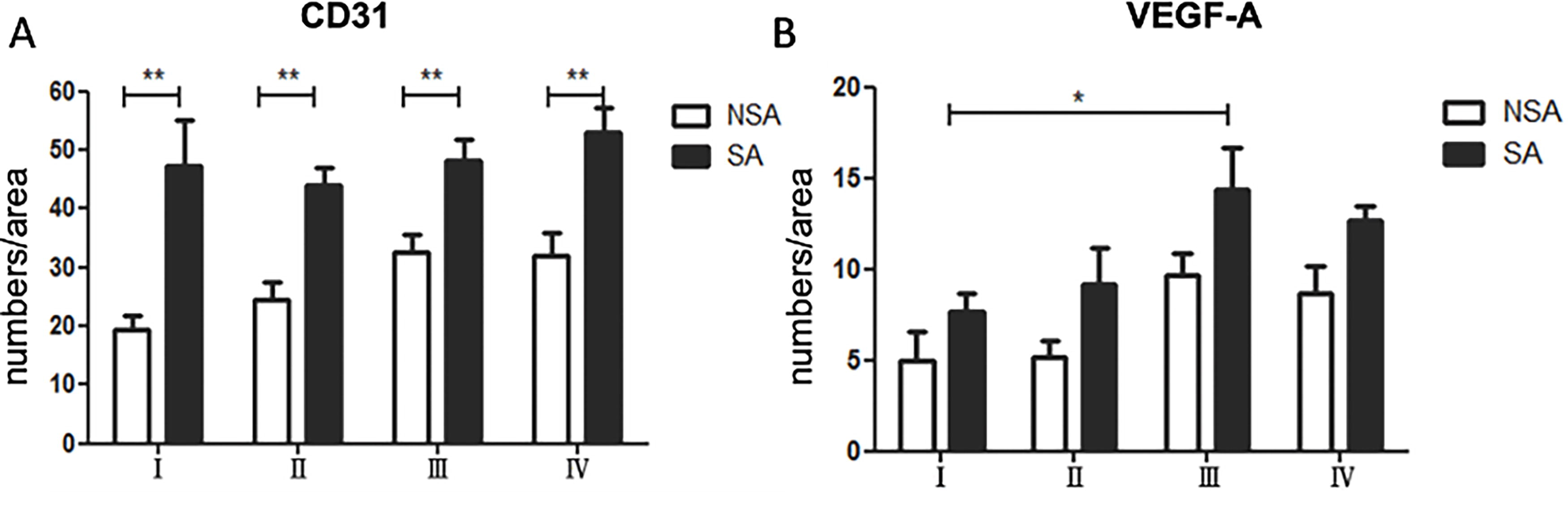
Analysis of angiogenesis expression showing increased
NPT in custom ETED device does not lead to cell death in mice
The application of negative pressure generates suction, which results in the movement of fluids through the cellular matrix, thereby creating shear and deformation forces on the cells. TUNEL assay was employed to evaluate cell death in samples of both standard abdominal and normal dermal (NSA) skin. A statistically significant increase in the number of cell deaths was observed in SA skin samples (106.48 ± 7.90 numbers/area) as compared with NSA skin samples (44.64 ± 2.47 numbers/area) (p < 0.01). Although this is the case, the microdeformations caused are a crucial aspect of vacuum-assisted therapy, as they have shown to promote cellular proliferation, angiogenesis, and granulation tissue formation.21,22 Meanwhile, no significant differences in cell death were observed among the four treatment groups (Group I: 107.80 ± 18.08 numbers/area; Group II: 100.80 ± 17.95 numbers/area; Group III: 115.30 ± 20.96 numbers/area; and Group IV: 102.20 ± 6.89 numbers/area) (Fig. 4). Therefore, our investigation revealed that neither the frequency nor the duration of exposure to NPT led to an increase in cell death.
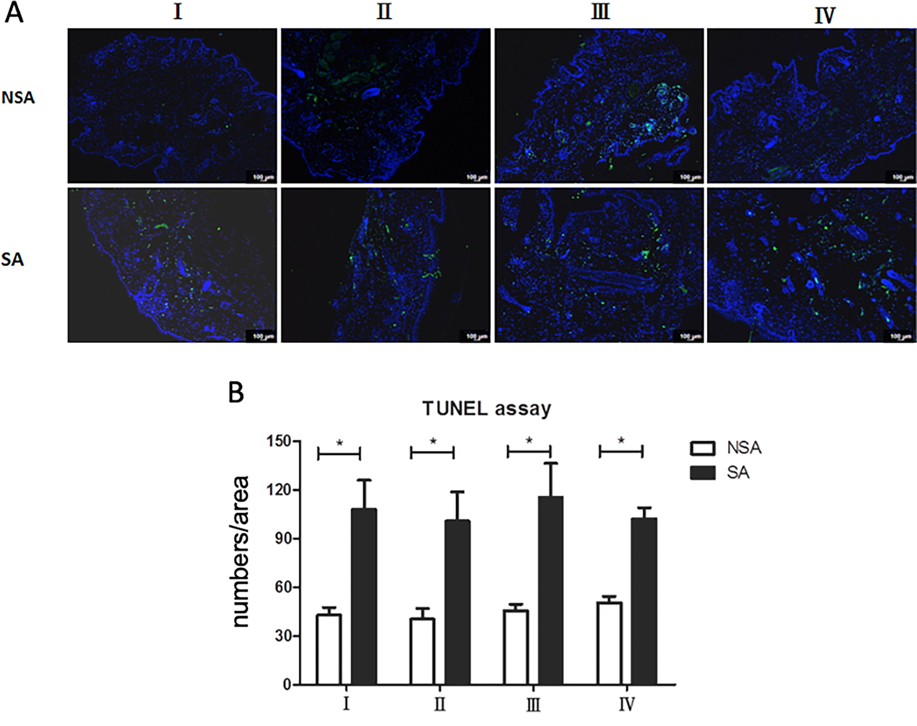
Analysis of cell apoptosis following negative pressure therapy.
Discussion
The present study demonstrated that NPT with ETED promoted hair follicle growth in nude mice. A NPT course of 15 min for 10 cycles was found to be the most effective in stimulating hair proliferation. The nude mice are characterized by athymic and hairless appearance. Hair defects in nu/nu mutants have been reported because of impaired keratinization in the hair shaft, resulting in a few short, crippled, and bent hair shafts. 23 The anagen phase of the hair cycle in these hairs was also shortened. 11 We observed an increase in the number of hair follicles visualized in the H&E stain following the NPT, which was accompanied by elevated expression of SOX-9 and LHX-2. However, no significant differences in SOX-9 and LHX-2 expression were observed between Groups I and II. Purba et al. reported that LHX-2- and SOX-9-expressing cells are dispersed throughout the ORS, including the bulge region, where the expression of SOX-9 is most pronounced in the ORS immediately beneath the bulge and LHX-2+ cells being similarly distributed between the subbulge and proximal bulb ORS. 24 The process of regrowing new hair involves stimulating stem cells in the bulge region through mediating the Sonic hedgehog (SHH) and Wnt/β-catenin signaling pathway. 25 SOX-9, a downstream of the SHH signaling pathway, has demonstrated to regulate the activin/pSMAD2 pathway and plays a pivotal role in maintaining hair follicle stem cells and guiding differentiation in the ORS of murine hair follicle. 14 On the other hand, LHX-2 is an NF-κB-controlled gene contributing to the down growth of follicle placode by activating transforming growth factor-β2 signaling. 26 In mature follicles, it plays an essential role in hair follicle stem cell growth and maintenance within the stem cell niche by regulating cell adhesion and cytoskeletal dynamics. 27 It is also necessary for anagen induction in the hair follicle cycle. 18 Therefore, the results indicated that negative pressure-induced hair follicle growth is associated with SOX-9- and LHX-2-mediated follicular regeneration.
There are several possible explanations for the increased hair follicle growth observed with NPT. The present study demonstrates that NPT promotes angiogenesis and increases VEGF expression, although the difference of VEGF levels between SA and NSA did not achieve statistical significance. This result is consistent with previous studies that NPT can increase angiogenesis-associated growth factors, including VEGF, epidermal growth factor, platelet-derived growth factor, and angiotensin-2. 28 The perifollicular vascularization increases during the anagen phase of the hair cycle and regresses during the catagen and telogen phases. 29 This phenomenon is widely recognized to be mediated by the expression of VEGF in follicular keratinocytes of the ORS, promoting hair growth and increasing hair size. 30 Hsiao et al. study has further demonstrated that negative pressure can trigger VEGF expression and angiogenesis, resulting in hair growth enhancement. 7 Second, our study also showed that the expression of the β-catenin was significantly higher in SA than in NSA. These results indicate that negative pressure could also enhance the Wnt/β-catenin signaling pathway. The Wnt/β-catenin signaling pathway is essential in initiating hair follicle stem cells for hair regeneration.31,32 Wnt-10b is upregulated in the secondary hair germ region with the increased expression of β-catenin upon anagen phase onset. 32 Another study also showed that the Wnt-3a, Wnt-5a, and β-catenin signaling pathways could stimulate hair follicle stem cell proliferation and differentiation, increasing hair follicle development. 33 Therefore, hair growth may be beneficial from the enhancement of the Wnt/β–catenin signaling pathway by negative pressure. Additionally, the negative pressure can induce hypoxia in the treated area. 34 Prostaglandin Fα is one of four bioactive prostaglandins that can be released by the endothelium of hypoxic or ischemic tissues in significant amounts and has the ability to regulate lipid homeostasis and inflammatory response. 35 Prostaglandin Fα has also been identified as a mediator that regulated the natural hair cycle and is crucial for the regulation of hair growth. It can induce stimulatory effects on the murine hair follicles and the follicular melanocytes and stimulate conversion from the telogen to the anagen phase. 36 Other studies also showed that hypoxia could increase the hair inductivity of dermal papilla cells through several molecules, including hypoxia-inducible factor-1α and nicotinamide adenine dinucleotide phosphate oxidase 4. 37
The present study revealed that a 15-min exposure to NPT administered 10 times over the course of 20 days was the most effective in promoting hair regeneration in mice. These results indicate that increasing the duration and frequency of NPT does not induce higher apoptosis in hair follicles. In contrast, Hsiao et al. demonstrated that pigs treated with suboptimal intensity of negative pressure displayed lower levels of fibroblast growth factor-1 and platelet-derived growth factor-BB, resulting in less hair follicle regeneration. 7 Therefore, it is critical to control the duration and frequency of NPT to achieve the optimal outcome of hair follicle regeneration.
NPT has shown efficacy in wound healing and various medical applications; however, certain limitations should be acknowledged. These limitations include the potential risk of complications, including pain, infection, toxic shock syndrome, or tissue damage, particularly if the therapy is not properly managed or if there are underlying medical conditions. 38 Additionally, the suitability of NPT may vary depending on the characteristics of the wound, and its effectiveness may not be universal for all types of wounds. It is crucial to consider patient-specific factors and tailor the treatment accordingly. 38 Moreover, the cost associated with NPT devices and their maintenance may pose challenges to widespread accessibility. 39 Although NPT holds promise in diverse clinical scenarios, ongoing research is essential to address these limitations and optimize its application.
Conclusion
The NPT delivered through our custom ETED has been demonstrated to be effective in promoting hair follicle growth in nude mice through the involvement of SOX-9- and LHX-2-mediated follicular regeneration, as well as angiogenesis and β-catenin-mediated hair follicle morphogenesis. Although the precise mechanisms by which the duration and frequency of NPTs influence hair follicle regeneration are yet to be fully understood, it is imperative to experiment with various treatment plans to attain the optimal outcome of hair follicle regeneration.
Footnotes
Acknowledgments
The authors would like to thank the Laboratory Animal Center of Chang Gung Memorial Hospital, Linkou, Taoyuan, Taiwan. The authors would also like to acknowledge the use of resources provided by the facilites at Taipei Medical University, New Taipei City, Taiwan.
Authors’ Contributions
C.Y.Y.: conceptualization, methodology, investigation, project administration, and writing—original draft; M.H.C.: conceptualization, supervision, funding acquisition, resources, and writing—review and editing; C.Y.C.: investigation, formal analysis, and writing—original draft; C.H.W.: methodology, investigation, formal analysis, and visualization; J.L.: writing—original draft, writing—review and editing, and visualization; W.H.: writing—review and editing; and C.H.L: conceptualization, supervision, funding acquisition, formal analysis, writing—original draft, and writing—review and editing.
Declaration of Conflict of Interest
The authors have no financial interest in any of the products, devices, or drugs mentioned in this article.
Funding Information
This project was supported by Chang Gung Memorial Hospital funding (CMRPG3L0121) and the Ministry of Science and Technology funding (MOST110-2222-E-038-001-MY3).

References
Supplementary Material
Please find the following supplemental material available below.
For Open Access articles published under a Creative Commons License, all supplemental material carries the same license as the article it is associated with.
For non-Open Access articles published, all supplemental material carries a non-exclusive license, and permission requests for re-use of supplemental material or any part of supplemental material shall be sent directly to the copyright owner as specified in the copyright notice associated with the article.
