




Abstract
Autologous bone grafts are commonly used to repair defects in skeletal tissue, however, due to their limited supply there is a clinical need for alternatives. Synthetic ceramics present a promising option but currently lack biological activity to stimulate bone regeneration. One potential approach to address this limitation is the incorporation of immunomodulatory agents. In this study, we investigate the application of microbial stimuli to stimulate bone formation. Three different microbial stimuli were incorporated in a biphasic calcium phosphate (BCP) ceramic: Bacille Calmette–Guérin (BCG), gamma-irradiated Staphylococcus aureus (γi-S. aureus), or γi-Candida albicans (γi-C. Albicans). The constructs were then implanted in both a rabbit posterolateral spinal fusion (PLF) and an intramuscular implant model for 10 weeks and compared to a nonstimulated control construct. For the PLF model, the formation of a bony bridge was evaluated by manual palpation, micro computed tomography, and histology. While complete fusion was not observed, the BCG condition was most promising with higher manual stiffness and almost twice as much bone volume in the central fusion mass compared to the control (9 ± 4.4% bone area vs. 4.6 ± 2.3%, respectively). Conversely, the γi-S. aureus or γi-C. albicans appeared to inhibit bone formation (1.4 ± 1.4% and 1.2 ± 0.6% bone area). Bone induction was not observed in any of the intramuscular implants. This study indicates that incorporating immunomodulatory agents in ceramic bone substitutes can affect bone formation, which can be positive when selected carefully. The readily available and clinically approved BCG showed promising results, which warrants further research for clinical translation.
Impact Statement
Inflammatory signals play an important role in bone repair and regeneration. Here we demonstrate the feasibility of applying microbial stimuli as a bioactive agent to promote bone regeneration in a relevant preclinical animal model.
Introduction
Bone is the second most transplanted tissue after blood. Bone grafting is a common procedure for treating defects in the skeletal system caused by trauma, tumor resection, and more. Autologous bone has the ideal properties to be used as a bone graft, however, the availability is limited and its harvesting prolongs surgical time. 1 Synthetic ceramics that mimic the inorganic components of bone are increasingly used as a substitute for bone grafts. They are well known for their osteoconductive properties, biocompatibility, and long shelf-life. 2 A recent clinical trial demonstrated that microporous biphasic calcium phosphate (BCP) ceramics had a similar success to autograft in a stand-alone instrumented posterolateral spinal fusion (PLF) study, with a fusion rate of 55% compared with 52% for autograft at 1-year follow-up. 3 Although this is promising, a 100% fusion should be the aim and therefore, the hunt for further optimization of the graft substitute continues. This may be accomplished with the next generation of calcium phosphate ceramics with submicron surface topography that was found to better induce new bone in an ectopic implant animal model. 4 For this material, its specific interaction with immune cells, such as macrophages, was hypothesized to be associated with the de novo bone formation.4–6 Modulating the interaction between a biomaterial and immune cells to stimulate bone formation has become a growing area of interest.7–9 Although short-lived, the initial inflammatory reaction toward a biomaterial can significantly impact its long-term performance and integration with the host tissue. 10
The implantation of a biomaterial initially triggers inflammation. This response determines the type of tissue that will form and at which speed. In the context of bone repair and regeneration, a well-sequenced inflammation plays a critical role in the process of osteoinduction, 11 which refers to the induction of undifferentiated progenitor cells such as mesenchymal stromal cells (MSCs) into the osteogenic lineage. 12 The inflammatory niche provides signaling molecules and chemokines that activate and attract local quiescent and distant MSCs.13–15 The concentration of relevant growth factors increases during the inflammatory stage, which normally, under homeostatic conditions are tightly regulated.16,17 Moreover, immune cells such as macrophages are among the main source of these growth factors. 18 Inflammation also supports bone repair by triggering the formation of new blood vessels, by molecules such as vascular endothelial growth factor and fibroblast growth factor. 19
On the contrary, the inflammatory response can also inhibit bone formation, for example during prolonged or chronic inflammation, as a response to an infection or to wear particles from polymeric or metal implants. 20 While proinflammatory signals can initiate the differentiation of MSCs, it is an anti-inflammatory environment that facilitates the maturation of differentiated osteoblasts. This allows cells to deposit osteoid, which eventually becomes a mineralized bone matrix. 21 Much is still unknown regarding the precise sequence and optimal range of pro- or anti-inflammatory factors required for effective new bone formation. However, former research demonstrated achieving a balanced inflammation and timing is important in this process. 14
In a previous study, we successfully applied inactivated microbe-derived agents as a proinflammatory strategy to enhance osteogenesis in a rabbit tibia model.22,23 After 8 weeks, gamma-irradiated Staphylococcus aureus (γi-S. aureus) led to significant thickening of the tibial cortex with none to minimal lytic lesions, predominantly showing a healthy bone structure. Similar findings were observed for γi-Escherichia coli and γi-Mycobacterium marinum although not as robust as with γi-S. aureus. In agreement, the application of microbial-derived agents for bone and dental implants was also reported by Shi et al. who prepared a polysaccharide fungal component, zymosan, as a coating on titanium substrates that demonstrated improvement in the osseointegration of the coated implant in a rat femur condyle defect model compared with the uncoated control. 24
To take these findings further, we explore the potential of microbe-derived stimuli to improve the osteogenesis of ceramic bone graft substitutes in a preclinical orthopedic model. Specifically, we examined the pro-osteogenic effect of the incorporation of γi-S. aureus, γi-Candida albicans (γi-C. albicans), and BCG in BCP scaffold with submicron surface topography. 25 BCG is included in the study as it is an available medicinal product containing attenuated bacterium (Mycobacterium bovis). The ceramics treated with the different microbial stimuli were studied in the well-established rabbit PLF model to assess the efficacy of bone graft substitutes.26–28 In addition, the ability to induce new bone formation was investigated in an intramuscular ectopic implant model. The findings of this study could have important implications for the development of new and improved therapies for bone regeneration and repair.
Materials and Methods
Materials
Three different microbes were tested in this study: S. aureus (Wood 46, ATCC 10832), C. albicans (ATCC 10231), and BCG. S. aureus and C. albicans were obtained from cultures of the microbiology department of Utrecht University in lysogeny broth medium at 37°C to mid-log phase, aliquoted and stored at −80°C in phosphate-buffered saline (PBS) with 40% (v/v) glycerol. Cultured microbes were killed by γi at 25 kGy (Steris AST, Ede, The Netherlands), which is the standard dose for sterilization of medical and biological products. 29 BCG (Lamepro, The Netherlands) was obtained from the pharmacy department of the University Medical Center Utrecht.
Commercially available BCP (MagnetOs©; Kuros Biosciences BV, Bilthoven, The Netherlands) in the form of granules (1–2 mm) and cylindrical discs (9 mm Ø; 6 mm height) were used as a scaffold. The BCP consisted of 65–75% tricalcium phosphate [TCP—Ca3(PO4)2] and 25–35% hydroxyapatite [HA—Ca10(PO4)6(OH)2] with a submicron needle-shaped surface topography (average crystal diameter of 0.58 ± 0.21 μm). 25 BCP scaffolds were sterilized by autoclaving at 121°C and dried at 60°C.
Whole-blood assay
The extent of the rabbit’s immune response toward different concentrations of microbial stimuli was studied in a whole-blood assay prior to the surgical intervention. Peripheral blood was collected sterilely from 5 animals in a sodium heparin-coated tube. The blood was diluted 1:5 with a culture medium containing RPMI (Gibco, 1640) 100 U/mL penicillin, and 100 U/mL streptomycin (Gibco). A 48-well plate (300 µL/well) was used for the experiment, which consisted of 270 µL of diluted blood and 30 µL of concentrated microbial stimuli in PBS. The groups tested were BCG (1 × 10³–1 × 10⁶ units/mL), γi-S. aureus (1 × 10⁴–1 × 10⁷ units/mL), and γi-C. albicans (1 × 10⁴–1 × 10⁷ units/mL). Lipopolysaccharide (0.1–100 ng/mL) was used as a positive control. The blood was stimulated for 24 h at 37°C and 5% CO2. Afterward, the plate was centrifuged at 500 ×g for 10 min at 4°C. The clear supernatant was collected and stored at −80°C until analysis. Cytokine levels of interleukin (IL)-6 and IL-1β were measured as indicators of a proinflammatory reaction using the Rabbit DuoSet® ELISA kits (R&D Systems, the Netherlands) according to the manufacturer’s protocol.
Implant preparation
Spinal fusion implants
BCP granules were sterilized by autoclaving and prepared max 1 week before surgery. In a 6-well plate, 2 cc of BCP were mixed with 2 mL of microbial stimuli (diluted in PBS), then placed on a plate shaker for 1 h at 50 rpm. The concentration of microbial stimuli tested in vivo was 2 × 10⁶ units/mL for BCG and γi-S. aureus; and 2 × 10⁷ for γi-C. albicans. Excess liquid was aspirated from the wells and the granules were air dried overnight. Subsequently, the treated BCP granules were transferred to a modified syringe, wherein the nozzle was removed, and a sterile plunger was employed to seal the opening (Fig. 1). BCP was stored at 4°C until the day of surgery. During surgery, blood plasma was used to bind the granules together. For this, 4 mL of autologous blood was collected in a citrate-coated tube and centrifuged at 2000 ×g for 10 min at room temperature. Clear plasma (1.2 mL) was mixed thoroughly with 60 μl of 0.5 M calcium chloride dihydrate (Sigma Aldrich). The syringe containing treated BCP granules was opened, supplemented with PBS (500 µL) to provide moisture, and then mixed with the plasma solution. Subsequently, the syringe was placed in an incubator (37°C) for 20–30 min until clotted. All steps were conducted under sterile conditions.
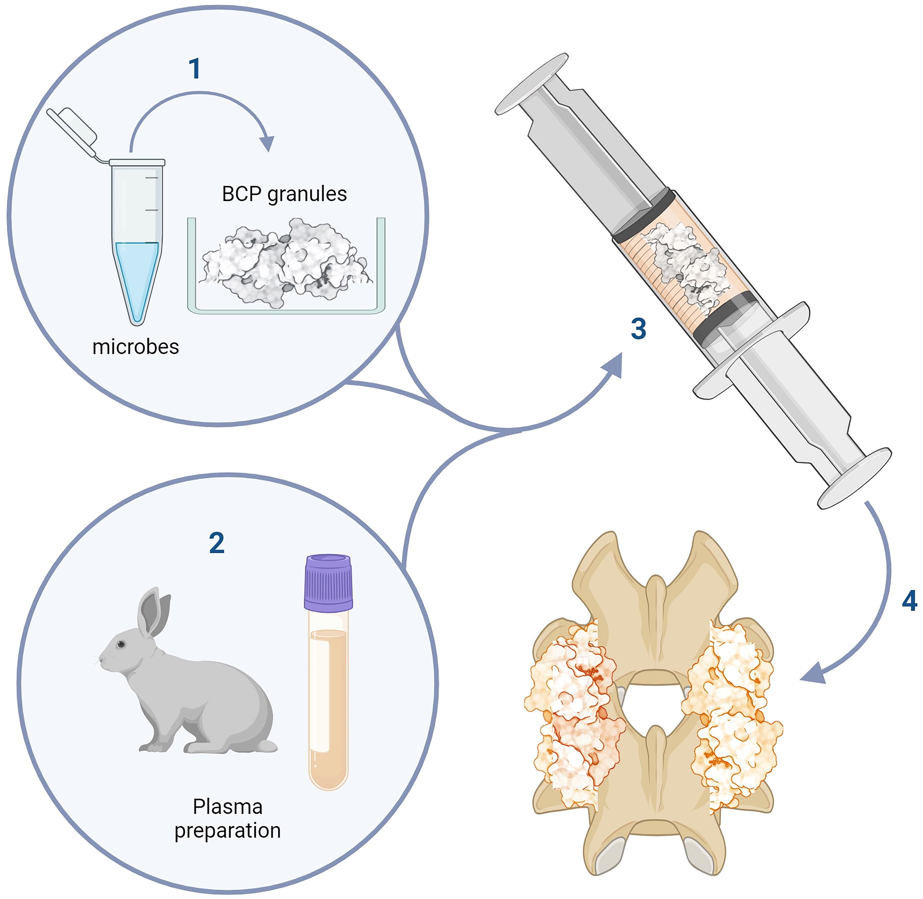
Schematic overview of the implant preparation.
Intramuscular implants
BCP discs were sterilized by autoclaving and prepared max 1 week before surgery by adding 200 µL of microbial stimuli diluted in PBS at high and low concentrations: BCG (1 × 10⁵ and 1 × 10⁶ units/mL), γi-S. aureus (1 × 10⁵ and 1 × 10⁶ units/mL), and γi-C. albicans (1 × 10⁵ and 1 × 10⁷ units/mL). Treated BCP discs were dried by air-flow overnight and stored at 4°C. All steps were conducted under sterile conditions.
Animal experiment
Twenty-four female New Zealand White rabbits (Crl:KBL, Charles River, France) were used (aged 20–25 weeks, weight 3–4 kg). All procedures were reviewed and approved by the Central Authority for Scientific Procedures on Animals (CCD, protocol number AVD1150002016445) and by the local animal ethics committee. Animals were housed in pairs at room temperature, fed standard rabbit chow, and had access to water ad libitum. The animals were acclimatized for 1 week, underwent a daily standard healthcare inspection, and were weighed weekly.
Each rabbit underwent a bilateral, single-level, noninstrumented PLF and implantation of 3 intramuscular implants in the paraspinal muscle. The animals were divided into four groups (n = 6): i) no microbial stimulus (control); ii) BCG; iii) γi-S. aureus, and iv) γi-C. albicans. Table 1 provides a description of the constructs implanted. Each animal received the same microbial stimulus for the spinal and intramuscular procedures. For the intramuscular implants, the conditions included a control (plain BCP), and a high, and low concentration of the microbial stimulus.
Overview of control and experimental groups for both types of implants
BCP, biphasic calcium phosphate; BCG, Bacille Calmette–Guérin.
Penicillin (4 × 10⁴ mg/kg s.c., Duplocilline®, Merck Animal Health, Madison, USA) was administered once as a prophylactic antibiotic. The rabbits were premedicated with buprenorphine (0.03 mg/kg s.c., Temgesic) and midazolam (0.5 mg/kg i.m.). General anesthesia was induced by etomidate (1.8 mg/kg i.v.) and maintained with sufentanyl (0.037 mg/kg) and isoflurane (2–2.5 mL/h). The rabbit was placed in a prone position. The dorsal skin was shaved and disinfected with povidone-iodine. Because the number of lumbar vertebrae is known to vary between rabbits, 30 the lumbosacral junction was used as an anatomical landmark to determine the PLF level. PLF was conducted across the third intravertebral space cranial to the lumbosacral joint, which is L4–L5 in the majority of animals. Here, a midline skin incision of 5–6 cm was made. About 2 cm bilateral from the midline, the longissimus muscles were separated bluntly until the transverse processes were exposed and cleared. Using a diamond-shaped drill (0.3 cm Ø), the paramedian surface was decorticated under constant cooling with saline until pink bedding of about 1 cm width was seen. Then, the coagulated BCP granules (about 3 cc per side) were placed overlying the transverse processes and intertransverse ligament. The wound was closed in multiple layers using 3-0 absorbable sutures (Vicryl, Ethicon).
Three intramuscular pockets were made, cranial to the PLF levels and approximately 3–4 cm apart, by a small incision of 1 cm in the fascia followed by blunt distraction. Each pocket received a BCP disc and afterward was closed with a single 3-0 absorbable suture. At the end of the surgery, a temperature monitoring chip was inserted subcutaneously. The mean operative time was ±50 min.
After surgery, the rabbits were placed in solitary housing with a heat source overnight before returning to their regular cage. The animals were handled as minimally as possible for 2 weeks by using a transport box to lift rabbits from the cage to aid fusion. 31 Buprenorphine (0.03 mg/kg s.c.; Temgesic) was administered every 12 h for 2 consecutive days as pain management. Fluorochrome labels were administered at 4, 7, and 8 weeks after surgery to detect the dynamics of mineralization, in the following order: oxytetracyline (20 mg/kg s.c.; Engemycin 10%, Mycofarm), calcein green (10 mg/kg s.c.; calcein disodium salt, 21030 Sigma-Aldrich, dissolved in 2% NaHCO3), and xylenol orange (30 mg/kg s.c.; xylenol orange tetrasodium salt, 398187 Sigma Aldrich, dissolved in 1% NaHCO3).
Body temperature was measured daily and body weight weekly to monitor for adverse systemic effects from the intervention. C reactive protein (CRP), a serum acute phase protein secreted by the liver in response to elevated cytokines, was measured prior to surgery and at weeks 3 postsurgery to identify a prolonged inflammatory response. CRP levels on weeks 1 and 2 postsurgery were not considered relevant, as they are typically elevated due to the surgical procedure itself. After 10 weeks, the rabbits were euthanized by a pentobarbital overdose. Samples were harvested and stored in 4% formaldehyde until further analysis.
Serum CRP
Blood (1 mL) was collected from the marginal ear vein of the rabbits in a serum clot activator tube (VACUETTE® CAT, 454098). Blood was left undisturbed at room temperature for 15–30 min until the blood clot was separated from the serum. Serum samples were stored at −20°C until analysis. CRP levels were measured using the Rabbit CRP (PTX1) ELISA Kit (ab157726, Abcam, USA) according to the manufacturer’s protocol.
Gross inspection and manual palpation
During the necropsy, the surgical field was examined for complications and the lumbar spine was removed. Manual palpation of the operated level was performed on the freshly obtained specimen. Flexion extension and lateral bending movements were applied to the fusion level and the level of mobility was compared to the nonintervened level. Fusion was graded as not fused, stiff (reduced movement compared with nonintervened levels), or fused (no detectable movement).
Micro computed tomography (micro-CT) imaging
The operated level was scanned in a micro-CT with a tube voltage of 90 kV and a current of 180 µA (Quantum FX, PerkinElmer, Waltham, MA). Images were taken with a voxel size of 40 µm, represented as a stack of 2D TIFF images.
Histology
The samples were embedded undecalcified for hard tissue analysis. The samples were first fixed in 4% (v/v) formaldehyde and dehydrated in an ethanol series. Subsequently, the samples were embedded in methyl methacrylate containing 20% (v/v) plastoid N (Sigma) and 2.8% (w/v) benzoyl peroxide (Luperox, Sigma), then immersed in a water bath until polymerized. Approximately 30 μm-thick sections were made with a Leica 1600 SP saw microtome (Leica, Nussloch, Germany) and stained with basic fuchsin and methylene blue for general histological assessment or left unstained for fluorochrome detection (Thunder imaging, Leica microsystem, Germany). Spinal sections were made mid-sagittal and for the intramuscular implants, transverse sections from the mid-third region of the BCP discs were used for histomorphometry.
Histomorphometry
Sections were digitized using the Leica DMi8 (Thunder) microscope equipped with a DFC9000 camera, operated by LASX software. The various tissue types (bone, bone marrow, fibrous tissue, and scaffold) were pseudo-colored using Adobe Photoshop 2020 (Adobe Systems, San Jose, USA). Area percentage was calculated for the scaffold, bone tissue, and marrow-like structures using the percentage of pixels relative to the region of interest (ROI). The ROI for the spinal fusion mass included the mid-third region of the sample, also referred to as the central region (Supplementary Fig. S1). An average of three sections were quantified for each sample.
Statistical analysis
The primary outcome of the study was bone area% by histology defined as the percentage of bone tissue formed in the ROI of the spinal fusion mass or the transverse section of the intramuscular implants. Quantified data are reported as mean ± SD. Statistical analyses were conducted using GraphPad Prism 9 software. Normality was assessed using the Shapiro–Wilk test and one-way analysis of variance (ANOVA) with Tukey’s post hoc test was used. A p-value of 0.05 was considered statistically significant.
Results
Inflammatory response towards microbial stimuli
To determine the rabbit response towards microbes, blood was stimulated ex vivo with different concentrations of the microbes. Cytokines, IL-6 and IL-1β, were measured as a general parameter for the proinflammatory response. A dose-dependent response was observed (Fig. 2). A significant response was observed from a concentration of 1 × 10⁵ units/mL for γi-S. aureus and BCG, while for γi-C. albicans a higher concentration of 1 × 10⁶ units/mL was needed. Based on these results, high and low concentrations of 1 × 10⁵ and 106 units/mL were used to prepare γi-S. aureus and BCG-treated implants, whereas 1 × 10⁵ and 107 units/mL were used for the C. albicans-treated implants.
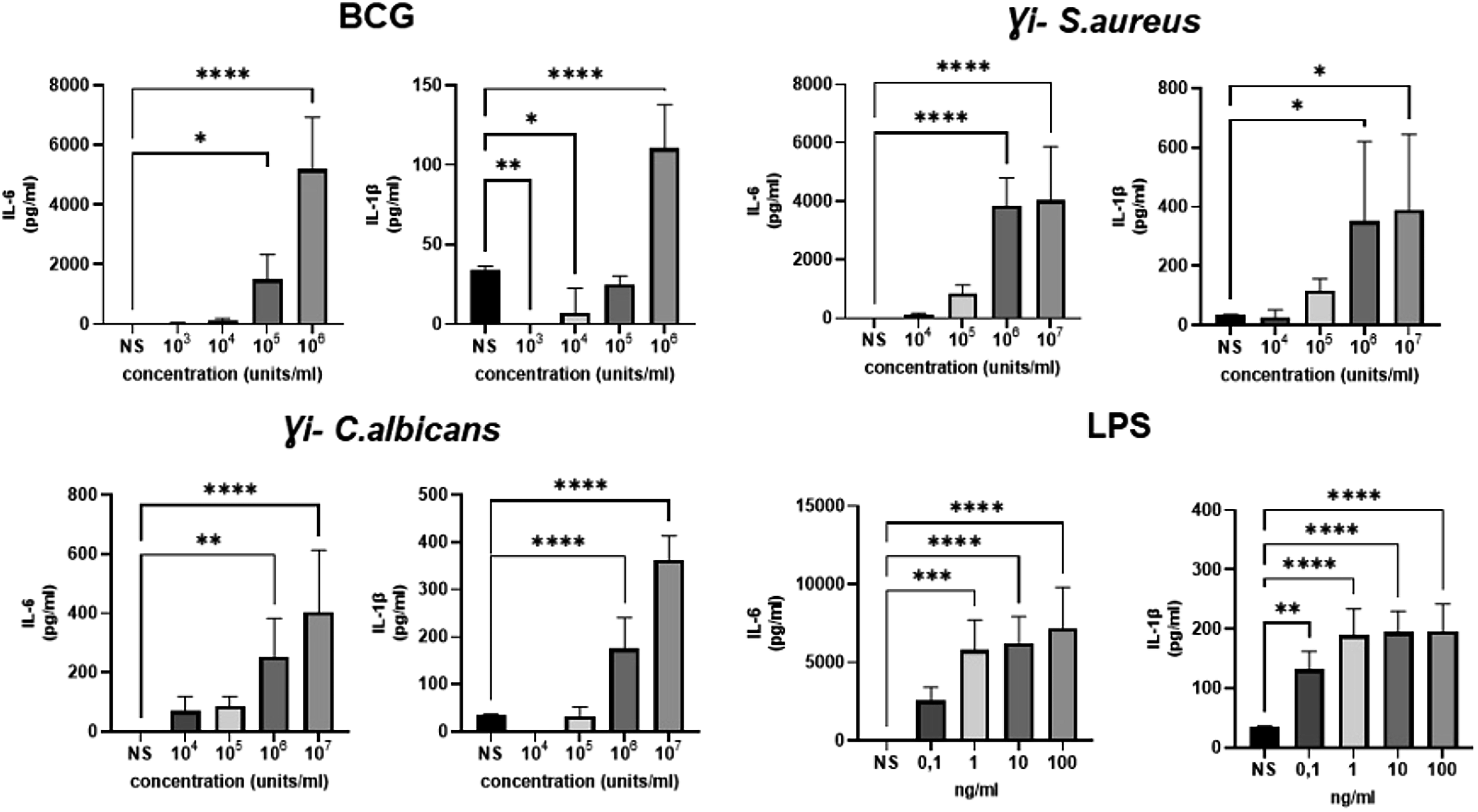
IL-6 and IL-1β levels of rabbit blood stimulated ex vivo with BCG, γi-S. aureus, and γi-C. albicans for 24 h. LPS was included as a positive control. Data are depicted as mean ± SD (n = 5), NS, nonstimulated, *p < 0.05/**p < 0.005/***p < 0.001 one-way ANOVA with Tukey’s post hoc test. BCG, Bacille Calmette–Guérin; γi-S. aureus, gamma-irradiated Staphylococcus aureus; LPS, lipopolysaccharide.
Spinal fusion mass analysis
All animals recovered well from surgery, except for one rabbit in the control group that did not recover from the anesthesia. This rabbit was euthanized and excluded from the analysis. Average temperature of the animals remained within normal limits (Supplementary Fig. S2A). All animals lost weight (7.4 ± 3.2% body weight) in the first week after surgery but gradually regained their weight and remained stable (Supplementary Fig. S2B). No prolonged systemic inflammation was observed demonstrated by comparable levels of CRP at weeks 3 post surgery with baseline (Supplementary Fig. S2C).
Gross inspection during necropsy showed granuloma tissue near the fusion mass in one animal of the γi-C. albicans group. As the fusion mass appeared normal, also by histology, we included it for analysis. One animal from the control group had intertransverse grafting on different levels and was therefore excluded from manual palpation. No other abnormalities were observed. Manual palpation indicated that all of the fusion levels were mobile, except for two out of six from the BCG group that were stiff. Micro-CT showed that remodeling of the transverse process occurred in all of the groups and new bone formed from the transverse process towards the center of the implant. In the best-performing sample of the BCG group, a clear bony bridge was observed from micro-CT (Fig. 3).
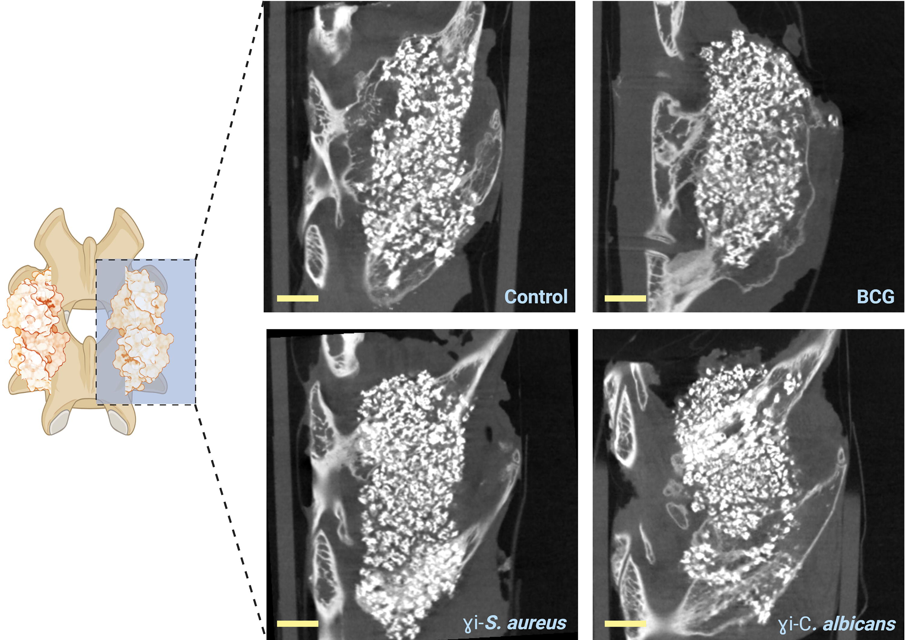
Coronal micro-CT sections (voxel size 40 μm) of the PLF implants taken after 10 weeks. Images show remodeling of the transverse process (in gray) toward BCP granules (in white) and the formation of new bone tissue (gray) surrounding the granules. Images are of the best-performing sample of each group. Scale bar = 1 cm. PLF, posterolateral spinal fusion.
From histomorphometry analysis (Fig. 4), the average bone area percentage (bone area%) was highest in the BCG group (9 ± 4.4%) compared with the control (4.6 ± 2.3%). The best-performing sample in the BCG group (12.2% bone area) showed a complete bony bridge forming between the transverse processes, not observed in any other group. While the least performing sample (2% bone area) showed active bone remodeling in the transverse processes but hardly any bone in the central fusion mass. The spinal implants of the γi-S. aureus and γi-C. albicans groups had significantly lower bone area% in the central fusion mass compared with BCG, with an average of 1.4 ± 1.4% and 1.2 ± 0.6% respectively, which was also lower than the control. General observation of all the groups depicted a well-vascularized area with islands of new bone formation seen in the central region without direct contact with the transverse processes. Fluorochrome analysis showed positive labels at week 4 (oxytetracycline), week 7 (calcein green), and week 8 (xylenol orange) throughout the fusion mass (Fig. 4D), indicating that the onset of mineralization at the central fusion mass also occurred at an early time point by 4 weeks, suggestive of induction.
New bone formation within the BCP granules shown in undecalcified sections of specimens harvested from spinal implants after 10 weeks.
Intramuscular implant analysis
No new bone formation occurred in any of the ectopic implants implanted in the paraspinal muscles for 10 weeks. The pores of the BCP were filled with well-vascularized loose connective tissue and clustering of adipocyte cells forming marrow-like tissue was frequently observed (Fig. 5). There were no signs of inflammation.
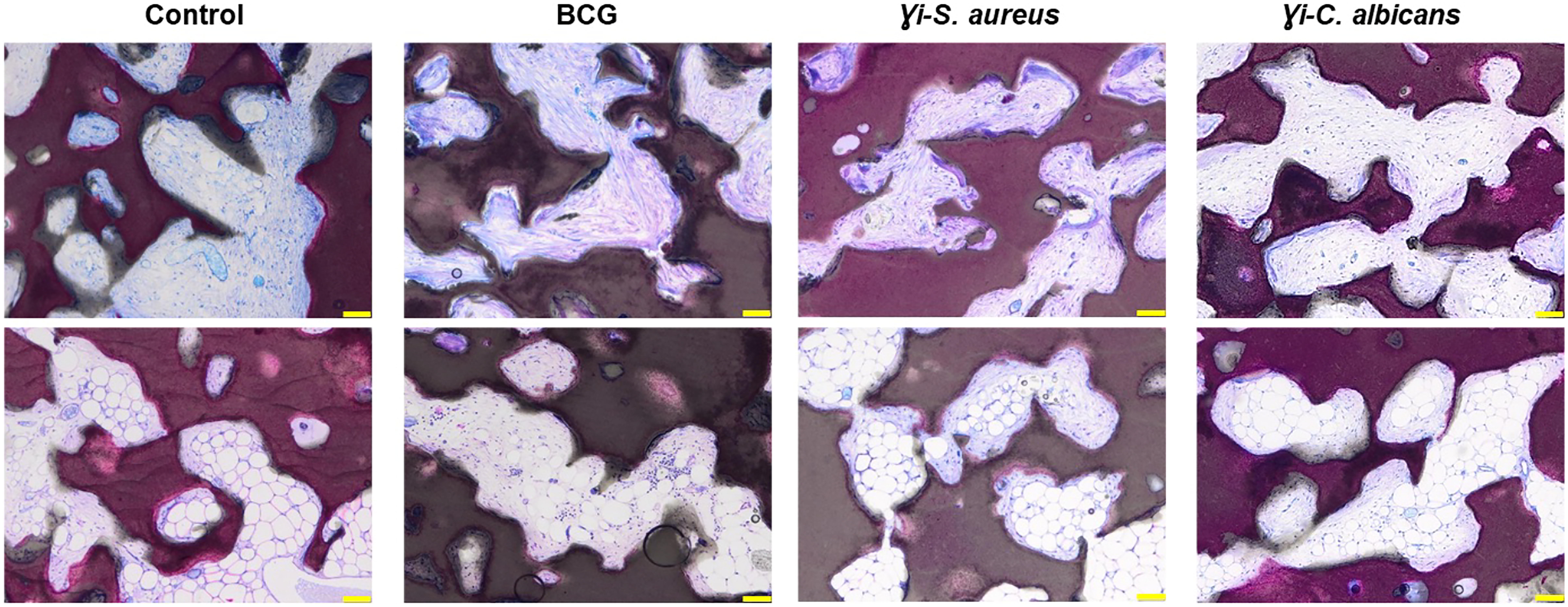
Undecalcified sections of specimens harvested from intramuscular implantation after 10 weeks. Top images show fibrous tissue filling in the pores of the BCP and the bottom images show areas in which marrow-like tissue has formed. Images are representative of each group. Scale bars = 100 µm.
Discussion
This study aimed to investigate the potential of microbial stimuli for enhancing bone formation in a rabbit PLF model. In addition, constructs were implanted intramuscularly to determine a potential osteoinductive effect. BCP with submicron needle-shaped topography was used as the scaffold, treated with either BCG, γi-S. aureus, or γi-C. albicans. The results of this study showed that immune stimuli influenced bone formation in the PLF model. Whereas γi-S. aureus and γi-C. albicans appeared to inhibit bone formation, and BCG stimulated bone formation with calcium phosphate granules.
The noninstrumented PLF model in rabbits is an established preclinical model for the evaluation of bone graft alternatives for spinal fusion.26–28 The intended primary outcome of this model is fusion, defined as a continuous bony bridge between the transverse processes. Bone formation in the central area between the transverse processes is the most challenging achievement and is a minimal requirement for fusion between two adjacent transverse processes. The formation of bone in the fusion mass follows similar principles to fracture healing, with or without the initial cartilage template. Decortication of the transverse processes exposes the trabeculae and marrow, providing a source for new vasculature and progenitor cells, 32 whereas the BCP provides a scaffold for the conduction of bone growth. In our study, only two samples in the BCG group had a rigid fusion, as was confirmed by both manual palpation and micro-CT. No fusions occurred in any of the other groups. The control group demonstrates the performance of the BCP as a stand-alone graft material in this model. Although new bone tissue was observed in the central region of the fusion mass, suggestive of induction, no bony bridges were formed. The same BCP material was tested in a previous rabbit PLF model with complete fusion achieved at 12 weeks, but only when combined with autologous bone graft (1:1 ratio) as an enhancer. 25
Compared with the control condition, γi-S. aureus or γi-C. albicans appeared to inhibit central bone formation. This may be due to inhibition of the osteoconductive process or less local bone induction. BCG however, showed more bone formation compared with the control. Altogether, these findings indicate that the inflammatory response induced by the immunomodulating compounds had an effect on local bone formation that was either inhibiting (γi-S. aureus or γi-C. albicans) or stimulative (BCG). A positive effect from BCG may be especially interesting for future investigations because of its widespread clinical use. BCG contains attenuated mycobacterium and is clinically applied as a vaccine against tuberculosis and as an immunomodulator for bladder cancer therapy. Repeated administration of BCG modifies the tumor microenvironment, making it more susceptible to T-helper and cytotoxic T-cell assaults.33,34 There is also a possibility that the effects observed are model specific. In our previous studies, γi-S. aureus (1 × 10⁹ units) led to significant thickening of bone cortex in a rabbit tibia model after 8 weeks, suggesting a pro-osteogenic effect,22,23 which was not reproduced with the γi-S. aureus in the current PLF model. The dosage, time, and location of applying the inactive bacteria also contribute to the results found. The use of ectopic bone models to evaluate induction has been implemented in many locations, such as subcutaneous, intramuscular, and in the kidney capsule. 35 Among these, the intramuscular location offers distinct advantages. Muscles are well-vascularized organs and during muscle injury and regeneration, there is a natural upregulation of bone morphogenetic protein (BMP) signaling, transforming growth factor (TGF)-β1, 36 and insulin-like growth factor (IGF)-1, 37 which are part of both bone as well as skeletal muscle regeneration. Studies looking into earlier tissue response (within 2 weeks) observed that the type of cells aggregating within the pores of the ceramics impacts long-term tissue formation within the material. 4 This emphasizes the importance of the early inflammatory response that drives the localization of different cells into the implantation site and triggers their differentiation. Literature, however, has reported a high variation in the success of the ectopic implantation model and is largely influenced by the kind of animal used and the length of the implantation process.38–41 In general, larger animals (such as goats, sheep, dogs, and baboons) and longer implantation periods have a higher likelihood of experiencing material-derived induction. It is still unclear why this phenomenon appears more frequently in larger animals than in smaller ones. A variety of physiological variations in metabolism, immune response, and muscle function are thought to be involved.38–41 For intramuscular implantation in rabbits, many studies were performed with little success.23,38,40,42,43 Only one study reported an average of 0.3% bone volume after 1 year of implantation. 38 This emphasizes that the current model with implantations for only 10 weeks is challenging. The absence of any sign of osteoinduction, even when higher concentrations of microbial stimuli were used, therefore should be interpreted within this context.
There are several limitations to this study. First, true rigid fusion was not determined by palpation, which questions the validity of the model for studying a clinically relevant endpoint. This may be the consequence of the relatively short 10-week follow-up. Fusion processes may continue beyond this period, and future investigations could explore longer follow-up durations to capture later-stage fusion events. Second is the constraint of measuring local inflammatory markers because the repository of antibodies, reagents, and resources to study the immune response in rabbits is limited. Finally, one could question the sample size of six animals in each group of the spinal fusion experiments. Literature reporting on the PLF model uses a range of 2–25 animals per group with a median sample size of 8 depending on the treatment used. 28 Future research should carefully evaluate the trade-offs between statistical power and practical constraints when designing future experiments.
The use of microbe-derived agents in grafting procedures may raise concerns on the safety of such an approach. Our preference of using killed microbes in this explorative phase of research is derived from observations that killed microbes result in a balanced mix of pro- and anti-inflammatory cytokines, that may favor bone differentiation both in vitro and in vivo.22,23,44 For future clinical translation, separate studies are conducted to investigate synthetically-derived microbial components recognized by the immune cells as pathogen-associated molecular patterns.45–47
Conclusion
This study showed promising results regarding the potential of microbial stimuli, especially BCG as an immunomodulatory adjuvant for bone regeneration. To date, there are no studies that link BCG and bone tissue regeneration and remodeling. Learning from the mechanism of action of BCG in other clinical use, we can speculate that the effects we observed on bone formation may be related to the initial inflammatory phase that affects cells of the adaptive immune system. While further research is needed, BCG presents a unique advantage as a medicinal product that is already widely used for its immunomodulatory effects in various clinical therapies.
Footnotes
Acknowledgments
Schematic illustrations in the article were created with Biorender.com.
Authors’ Contributions
N.R.R., M.C., and M.C.K. designed the experiments. N.R.R., A.D., M.C., and O.L. conducted the experiments, interpreted, and analyzed the data. N.R.R. wrote the article. M.C.K. and H.W. secured the funding and contributed to the experiment design, data interpretation, and article review. A.D. and D.G. contributed to data interpretation and article review. All authors have read and approved the final submitted article.
Ethical Approval
Animal experiments (protocol number AVD1150002016445) were reviewed and approved by the Central Authority for Scientific Procedures on Animals (Centrale Commissie Dierproeven [CCD], the Netherlands) and by the local animal ethics committee (Instantie voor Dierenwelzijn [IVD], Utrecht, the Netherlands).
Disclosure Statement
No competing financial interests exist.
Funding Information
This work is supported by PPS allowance from the Health∼Holland LSH-TKI (grant no.: LSHM18011) and the EU’s H2020 research and innovation program under Marie S. Curie Cofund RESCUE (grant agreement no. 801540).

References
Supplementary Material
Please find the following supplemental material available below.
For Open Access articles published under a Creative Commons License, all supplemental material carries the same license as the article it is associated with.
For non-Open Access articles published, all supplemental material carries a non-exclusive license, and permission requests for re-use of supplemental material or any part of supplemental material shall be sent directly to the copyright owner as specified in the copyright notice associated with the article.
