
Abstract
Primary open-angle glaucoma is a prevalent type of degenerative eye disease that results in lifelong blindness, and its critical pathogenic cause is trabecular meshwork (TM) dysfunction or decreased TM cellularity. Considering that TM develops from neural crest cells (NCCs), we investigate the potential of human embryonic stem cell (hESC)-derived NCCs transplantation for TM regeneration. We used a chemically defined method to induce the differentiation of NCCs and injected 1.0 × 10⁶ hESC-derived NCCs combined with 100 μmol/L Y-27632 into the anterior chamber of rabbit. Intraocular pressure (IOP), TM, and corneal changes of rabbits with cell transplantation were examined with TonoPEN AVIA, slit lamp microscope, dual-immunofluorescence staining, and optical coherence tomography. The hESC-derived NCCs underwent homogenous differentiation over the course of 5 days’ induction, which expressed the typical neural crest markers HNK-1, P75, SOX10, and AP-2α. NOD/SCID mice received injections of hESC-derived NCCs in the groin or axilla. There was no teratoma formation. When intracamerally injected, hESC-derived NCCs integrated into the TM tissue and expressed mature TM cell markers Aqp1, Chi3l1, and Timp3 after 7 days transplantation in rabbit eyes. The IOP and central corneal thickness basically maintained at normal levels within 2 weeks. No significant adverse effects in rabbits with hESC-derived NCC injection were observed after 5 weeks of cell transplantation. Our findings indicate that hESC-derived NCCs could integrate into the TM tissue and differentiate into mature TM cells after being injected intracamerally, showing a potential therapeutic approach to addressing TM dysfunction in the treatment of glaucoma.
Impact Statement
Glaucoma is the leading cause of irreversible blindness that poses a substantial burden on public health and the quality of life of affected individuals. We found human embryonic stem cell (hESC)-derived NCCs integrated into the trabecular meshwork (TM) tissue, and expressed mature TM cell markers Aqp1, Chi3l1, and Timp3 after intracameral injection. The results also highlighted hESC-derived NCC treatment’s safety, with no treatment-related serious adverse events during the long-term follow-up. These findings suggest that hESC-derived NCCs offer potential for new interventions for the therapy of glaucoma.
Introduction
Glaucoma is the primary contributor to permanent blindness globally. It is characterized by the apoptotic destruction of retinal ganglion cells, leading to irreversible vision loss.1,2 Increased intraocular pressure (IOP) is a significant contributing factor. 3 IOP is regulated by the equilibrium between the input and outflow of aqueous humor. 4 The primary route for the flow of aqueous humor is the trabecular outflow channel, which accounts for around 90% of the outflow. This pathway is influenced by the trabecular meshwork (TM), ciliary muscle, and the inner wall cells of Schlemm’s canal. 5 TM cells have a crucial purpose in controlling IOP and ensuring the proper functioning of the traditional aqueous outflow channel, such as phagocytosis of debris and the production of extracellular matrix (ECM).6–8 Senescence or apoptosis-induced reduction in TM cells can result in fusion and thickening of trabecular lamellae, as well as aberrant accumulation of ECM. This disruption of TM microstructure and higher stiffness resistance of TM may contribute to elevated outflow resistance.9–13
Currently, primary open-angle glaucoma (POAG) is treated by lowering IOP with medication or surgery in clinical settings. However, these treatments do not particularly target the loss of TM cells. 3 There have been reports indicating the presence of TM stem cells (TMSCs) in the insert region of the TM. These cells have the potential to compensate for reduced cell density and restore the function of the TM.14–16 Nonetheless, TMSCs are predominantly depleted or impaired in patients with POAG. 17 Several groups have attempted other stem cells for the treatment of POAG with acceptable outcomes, 18 including induced pluripotent stem cells (iPSCs)-, cranial neural crest cells (NCCs)-, and mesenchymal stromal cells-derived TM.8,16,19–21 TM originates from the neural crest during the development of vertebrates. 22 Prior research has elucidated the multilineage capacity of NCCs, including their capability to regenerate bone and peripheral nerve,23,24 as well as their potential in treating Parkinson’s disease and spinal cord injury.25,26 Therefore, it is necessary to investigate whether NCCs can be the alternative cell source for TM dysfunction.
In this study, we induced human embryonic stem cells (hESCs) into NCCs, which exhibited the typical characteristics of NCCs, including the polygonal morphology and positive markers HNK-1, P75, SOX10, and AP-2α. They also expressed neural crest-related genes P75, Sox10, Ap-2α, Ap-2β, Pitx2, and HNK-1. The findings of our study indicate that the transplantation of NCCs derived from hESCs could be a potential therapeutic approach to restore glaucomatous TM tissue function.
Materials and Methods
Animals
Six-month-old healthy New Zealand white male rabbits (Xilingjiao, Jinan, China) were kept in individual cages at a constant temperature and humidity. Six- to eight-week-old male NOD/SCID mice (Vital River Laboratory Animal Technology Co.) were used for the teratoma formation experiment. The animals were kept and treated in compliance with the guidelines set forth by the Association for Research in Vision and Ophthalmology for the utilization of animals in ophthalmic and vision research. The Ethics Committee of the Shandong Eye Institute granted approval for all animal trials.
Cell culture and differentiation
The human ES cell line H1 was provided by Zhengqin Yin. The hESCs were cultured on dishes coated with Matrigel (354277, Corning, NY, USA) and maintained in serum-free medium mTeSR1TM (STEMCELL Technologies, Vancouver, BC, Canada) in a 37°C incubator with 5% CO2; 10 μmol/L ROCK inhibitor Y-27632 (STEMCELL) was added for ESC passaging or cryopreservation resuscitation. The medium was changed every day.
The method of inducing hESCs to generate NCCs followed our previous study. 27 In brief, the hESCs were seeded on Matrigel-coated dishes and grew into 30–40% confluence after 3–4 days’ culture, then the medium was replaced with a neural crest differentiation medium consisting of Dulbecco’s modified Eagle medium/F12 (DMEM/F12, Gibco), 20% knockout serum replacement (KSR, Gibco), 2 mmol/L GlutaMAX (Gibco), 0.1 mmol/L MEM nonessential amino acids (Gibco), 0.1 mM β-mercaptoethanol (Gibco), 4 ng/mL basic fibroblast growth factor (R&D Systems, Minneapolis, MN, USA), and 1 μmol/L retinoic acid (RA, Sigma) for 5 days. The medium was changed every day.
Teratoma formation
A total of 100 μL of 5 × 10⁶ hESC-derived NCCs suspension was mixed with Matrigel (354234, Corning) in the same volume and subcutaneously injected into the groin or axilla of the NOD/SCID mice. Tumors were isolated 6–8 weeks after injection, fixed in 10% formalin, and embedded in paraffin. Sectioned tumors were stained with hematoxylin and eosin (H&E) staining.
Immunofluorescence staining
Samples, including cells and cryosections, were fixed in 4% paraformaldehyde (Biosharp, Anhui, China) for 15 min and blocked with 10% donkey serum for 1 h at room temperature. The samples were treated with primary antibodies (Table 1) overnight at 4°C and subsequently with Alexa Fluor 488- or 594-conjugated secondary antibodies (Invitrogen) for 1 h at 37°C. The nuclei were stained with 4,6-diamidino-2-phenylindole (DAPI, Beyotime Biotechnology) and imaged using fluorescence microscopy (Nikon, Japan, and Echo, USA).
List of Antibodies

Quantitative real-time reverse transcription polymerase chain reaction
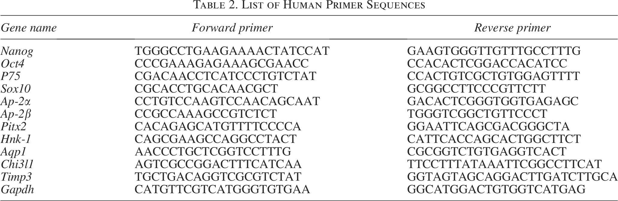
Total RNA was extracted from the differentiated NCCs and human TM tissues using the Mini BEST Universal RNA Extraction Kit (TaKaRa, Tokyo, Japan). cDNAs were synthesized using the Evo M-MLV RT Kit (TaKaRa) according to the manufacturer’s protocol. Real-time polymerase chain reaction (RT-PCR) was carried out using the SYBR Green qPCR Master Mix (Vazyme, Nanjing, China). The cycling conditions were 10 s at 95°C, followed by 40 two-step cycles (15 s at 95°C and 1 min at 60°C). The quantified data were analyzed using Sequence Detection System software (Applied Biosystems) with glyceraldehyde-3-phosphate dehydrogenase as an internal control (Table 2).
List of Human Primer Sequences
Flow cytometry
The differentiating cells derived from hESC were dissociated using Accutase (Sigma) and stained with antibody against the pluripotent markers TRA-1-60 (Biolegend) for 30 min at 4°C per manufacturer’s instructions. Premier Data Acquisition and Analysis software (Beckman Coulter, Inc., USA) was used for analysis. Data and graphs were made using the CytoFLEX flow cytometry system.
Intracameral injection of hESC-derived NCCs
The rabbits were anesthetized with intramuscular ketamine hydrochloride (40 mg/kg, Gutian Pharmaceutical Co., Fujian, China). Cell transplantation was based on previously described procedures with some improvements.20,28–30 In brief, the hESC-derived NCCs were suspended in 100 μL DMEM supplemented with 100 μmol/L Y-27632 at a density of 1.0 × 10⁶ cells, and then, injected into the rabbit’s anterior chamber (n = 9). After operation, animals treated with muscle relaxants were placed in cages and allowed to resume activity gradually. Post-transplantation treatment involved administering a 250 μL subconjunctival injection of 5 mg/mL dexamethasone sodium phosphate (Chenxin, Shandong, China) and applying tobramycin dexamethasone eye ointment (Novartis, Basel, Switzerland). Due to the immunosuppressive properties of dexamethasone, it is recommended to administer the medication once a day for a duration of 3 days after surgery. The rabbits were allocated to experimental and control groups using a random assignment method. The control group consisted of untreated rabbit eyes of the same age (n = 3).
IOP measurement
The rabbits were anesthetized topically with intramuscular ketamine hydrochloride (40 mg/kg, Gutian Pharmaceutical Co., Fujian, China) and intravenous pelltobarbitalum natricum (50 mg/kg, Sinopharm Chemical Reagent Co., Shanghai, China). An applanation tonometer (TonoPEN AVIA) was utilized to measure IOP about 5 s after promethacaine hydrochloride exposure. The average of six readings is reported by the tonometer. The investigators conducted three measurements for each data point between the hours of 9 a.m. and 11 a.m., and the order of the animals was randomized. Data from all animals were obtained in a double-blind manner.
Evaluation of cornea
The anatomical structure and central corneal thickness were detected by anterior segment optical coherence tomography (Optovue Inc., Freemont, CA, USA) built-in software. Corneal transparency was photographed using slit-lamp microscopy (Topcon, Tokyo, Japan).
Statistical analysis
GraphPad Prism 8.0 was used for statistical analysis. A comparison between two sets of data was performed with the Student’s t test, and the data are expressed as the mean ± standard deviation (SD). The two-factor analysis of variance was used to analyze the differences of IOP between the two groups at different postoperative time points. Results were considered significant at *p < 0.05, **p < 0.01, and ***p < 0.001, and specific comparisons are indicated in the respective figure legends.
Results
Differentiation of hESCs into NCCs
Human ESCs were induced into NCCs using a chemically specified approach, as previously explained. 27 The hESCs exhibited clonal morphology and were stained positive for the pluripotent markers NANOG, OCT4, SOX2, and SSEA4 (Fig. 1A, B). After 5 days of neural crest induction, the cells appeared polygonal and positive for the neural crest markers HNK-1, P75, SOX10, and AP-2α (Fig. 1C, D). In addition, transcript analysis was performed on the hESCs and differentiated cells. The expression of pluripotent genes Oct4 and Nanog decreased dramatically, whereas the neural crest markers P75, Sox10, Ap-2α, Ap-2β, Pitx2, and Hnk-1 were significantly upregulated (Fig. 1E).

Inductive differentiation of human embryonic stem cells (hESCs) into neural crest cells (NCCs).
Safety of hESCs-derived NCCs
To determine the tumorigenicity of differentiated cells, we detected the ratio of residual undifferentiated cells by flow cytometry. The FACS shows there was only 0.33% ± 0.10% cells with TRA-1-60 positive after 5 days NCCs induction (Fig. 2A). For the in vivo safety of transplanted cells, the tumorigenicity of both hESC-derived NCCs and hESCs were examined in NOD/SCID mice. There was no teratoma formed in NOD/SCID mice (0/5) with hESC-derived NCCs injection after 6 weeks, while four out of five NOD/SCID mice had palpable tumor formation after hESCs injection. The H&E staining revealed the presence of different germ layer tissues, such as mesoderm, endoderm, and ectoderm, in all the teratomas (Fig. 2B–D).
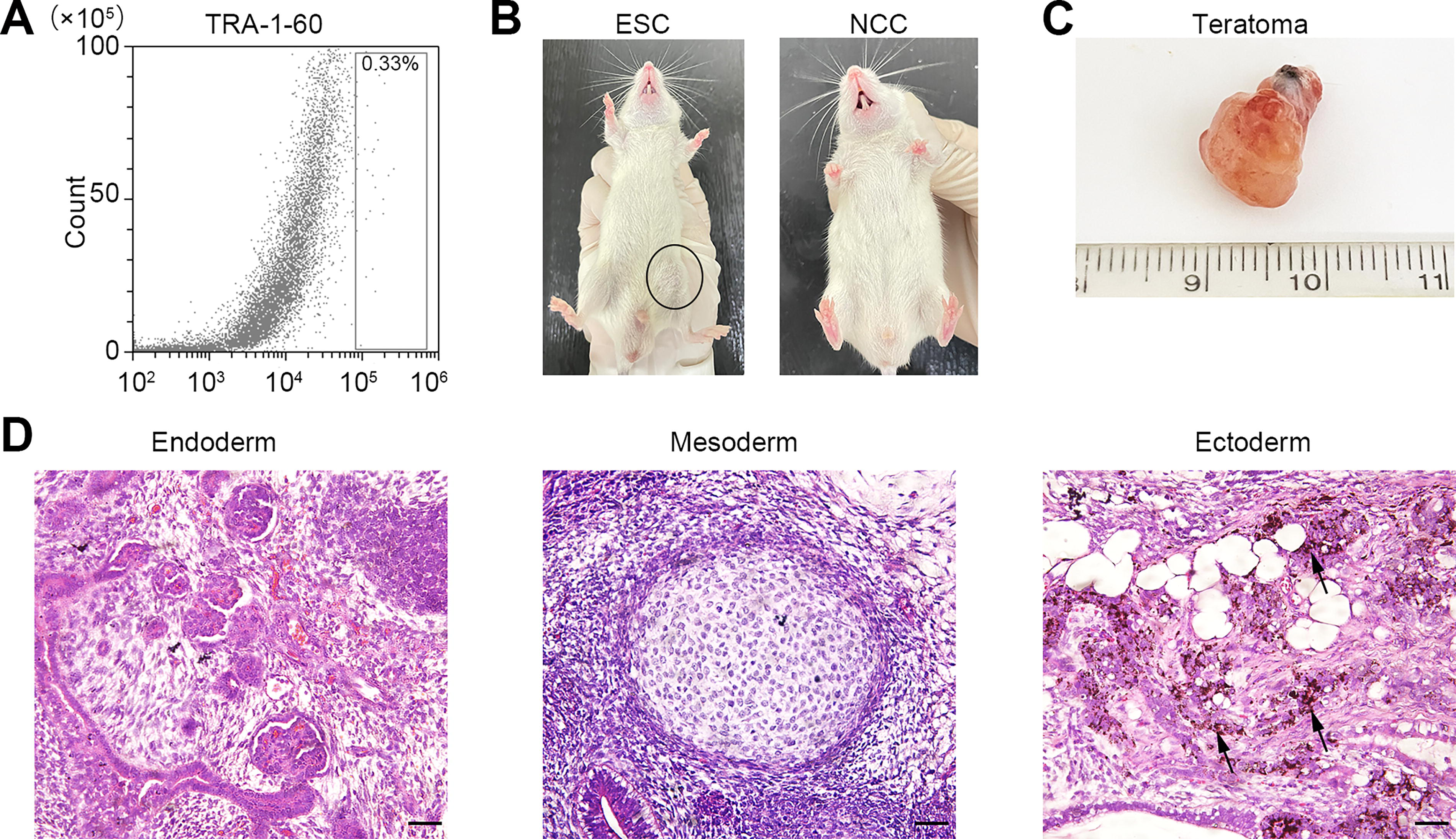
Safety of human embryonic stem cell (hESC)-derived neural crest cells (NCCs).
Injection and localization of hESC-derived NCCs into TM tissue
In order to assess the ability of hESC-derived NCCs to migrate to TM tissue, we administered the hESC-derived NCCs through intracameral injection into the anterior chamber of healthy rabbits, as illustrated in Figure 3A. To verify the structure of the anterior chamber, H&E staining of the anterior segment was performed 7 days after NCC transplantation. The study demonstrates that the integrity and physiological structures in the anterior chamber were unaffected, and there was no aberrant clustering or attachment of transplanted cells in the TM region, surface of the endothelium, or iris (Fig. 3B). In addition, we performed TM tissue immunostaining with the human-specific marker cytochrome C oxidase (HMTCO2). The HMTCO2-positive transplanted cells were well-distributed throughout all cell layers of the TM tissue (Fig. 3C).
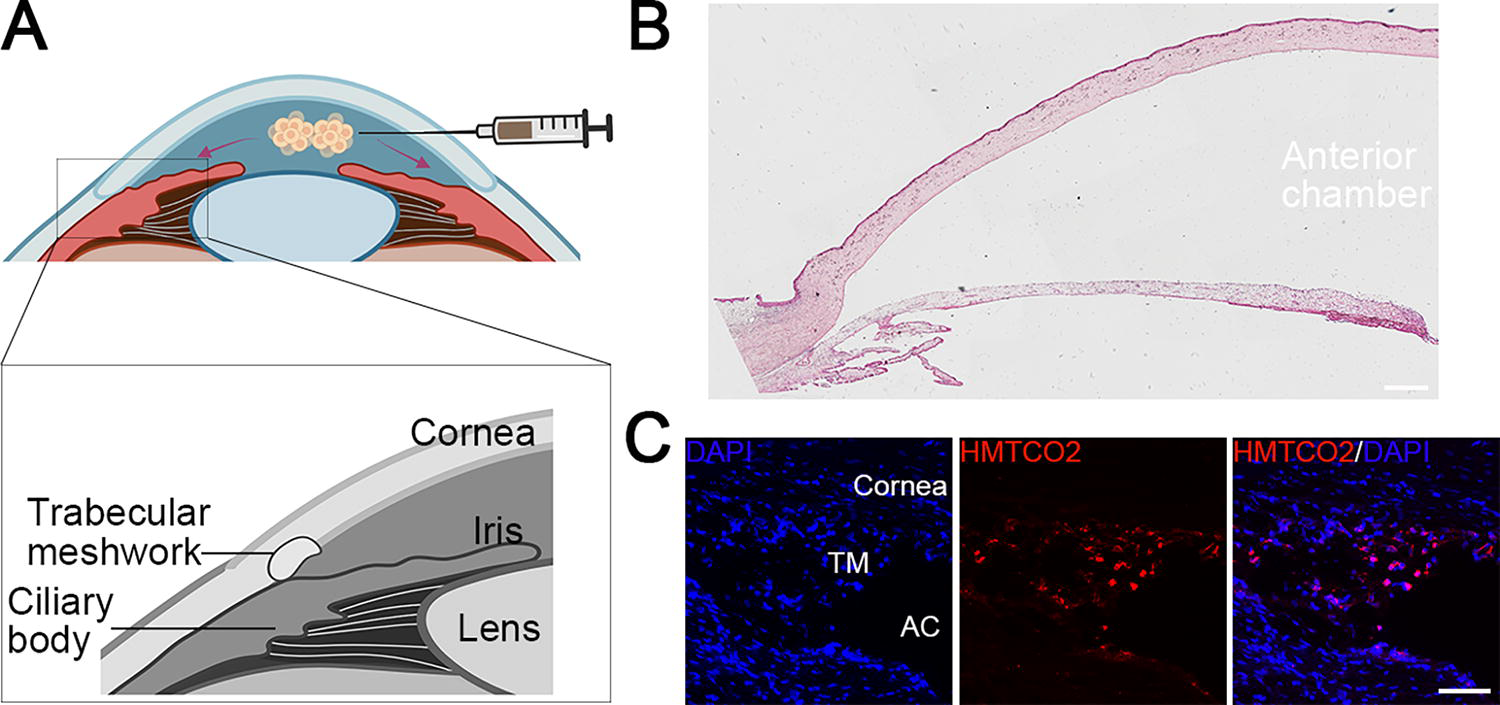
Location and integration of the transplanted human embryonic stem cell (hESC)-derived neural crest cells (NCCs).
Differentiation of hESC-derived NCCs into TM cells
To explore the fate of transplanted NCCs in vivo, we performed double immunofluorescence staining with the human MTCO2 and TM markers AQP1, CHI3L1, and TIMP3.31,32 The results showed that the transplanted HMTCO2-positive NCCs were presented in the TM region after 7 days of injection and further differentiated to TM cells with positive staining of TM markers AQP1, CHI3L1, and TIMP3, which were barely expressed preoperatively (Fig. 4A. Additional file 1: Supplementary Fig. S1). The transplanted cells survived within 14 days and continuously positively expressed the mature TM markers (Fig. 4B).

Differentiation of human embryonic stem cell (hESC)-derived neural crest cells (NCCs) into mature TM cells.
The IOP was observed for a duration of 14 days to assess the functionality of the TM, which controls aqueous humor outflow and IOP homeostasis. As shown in Figure 4C, there were no significant differences in IOP between eyes with or without hESC-derived NCCs transplantation (p > 0.05). IOP was maintained between 10 and 15 mmHg at each time point, although there was a slight fluctuation in the early phase after injection. The same tendency was observed in corneal thickness (Fig. 4D).
Long-term outcomes of hESC-derived NCC injection in rabbits
The animals were monitored for a maximum of 5 weeks to examine the long-term effects of injected hESC-derived NCCs in a living organism. Rabbit TM cryosections were stained using the human-specific MTCO2 marker, as well as the TM markers AQP1, CHI3L1, and TIMP3. As shown in Figure 5A, no positive signals of hominine cells were detected in rabbit TM with NCCs transplanted at 5 weeks, whereas the in situ TM cells expressed AQP1, CHI3L1, and TIMP3. There were no significant changes in IOP between the eyes injected with or without hESC-derived NCCs, as observed in Figure 5B. The corneas of the rabbits that received injections of hESC-derived NCCs exhibited perfect transparency and displayed a normal corneal thickness (Fig. 5C, D).

Long-term effects of human embryonic stem cell (hESC)-derived neural crest cells (NCCs) transplantation in rabbits.
Discussion
Cell therapy is a promising alternative approach for recovering TM function. 29 In this study, we generated the hESC-derived NCCs and investigated the prospect of NCCs for TM regeneration. The transplanted NCCs successfully integrated with the rabbits’ TM tissue and underwent maturation into mature TM cells within a span of 7 days. The transplanted cells exhibited complete disappearance 5 weeks post-transplantation without exerting any deleterious impact on the normal structure and function of TM tissue. These results suggested that the transplantation of hESC-derived NCCs could be a potential therapeutic strategy to restore glaucomatous TM tissue function.
The neural crest arises from the dorsal neural plate boundary during gastrulation and possesses characteristics similar to stem cells, as well as the ability to migrate.33,34 We have demonstrated that NCCs derived from hESCs have the ability to generate several cell types, including corneal endothelial cells, corneal keratocytes, mesenchymal stem cells, and peripheral neurons in vitro. 35 In this study, we found that the transplanted hESC-derived NCCs were able to migrate to the TM region. Moreover, these cells underwent further differentiation into TM cells, exhibiting the presence of mature markers within the in vivo microenvironment. Prior research has shown that several crucial attributes of NCCs, such as their ability to survive, initiating differentiation programs, and maintaining the stem-like state, are determined by their interactions with, expressions of, and alterations to various components of the ECM during the migratory phase.36–41 Furthermore, it has been suggested that environmental cues originating from adjacent tissues eventually dictate the fate of NCCs. 42 Research has shown that the nascent corneal environment affects the process of differentiation in periocular NCCs. 43 And there was an increased neuronal differentiation of transplanted enteric neural crest-derived cells after cell transplantation. 44 We assume the local signals, such as cell communications, physiological extracellular matrix, IOP, and ciliary muscle contraction, have a role in the growth and maturation of stem cells, specifically in driving the differentiation of hESC-derived NCCs in vivo.
Intracameral injection is an efficient strategy for ocular cell therapy, especially for TM and corneal endothelial-related diseases.27,29 The main concern about the transplantation of hESC-derived NCCs is ensuring safety, given the presence of many structures in the anterior chamber, such as the cornea, lens, iris, and TM. The localization and destiny of hESC-derived NCCs in vivo dictate the achievement of TM function regeneration. In this case, we found that the hESC-derived NCCs were effectively positioned in the TM region and maintained the IOP within the normal range throughout the 14-day postoperative monitoring period. Observing the IOP of the rabbits before and 1, 3, 7, and 14 days after cell transplantation revealed that while the rabbits’ IOP varied, it did not exhibit any significant difference compared with the control group. Ultimately, the IOP stabilized at a range of 10–15 mmHg. A slight IOP increase accompanied by corneal thickening was observed at the early phase of injection, which is primarily attributed to the surgical technique employed. Moreover, we determined the tumorigenicity of hESC-derived NCCs by injecting NOD/SCID mice. Different from hESC injection, there was no teratoma formation in mice with hESC-derived NCCs injection. The safety of the intracameral injection is indicated by the absence of any adverse reactions associated with the injection in the trial, as well as the lack of side effects, such as hypertension, observed in clinical trials using endothelium therapy. 45 These findings suggest that the hESC-derived NCCs are safe for restoring TM function.
To the best of our knowledge, this is the first study using hESC-derived NCCs directly for TM regeneration as opposed to differentiated mature TM cells. Therefore, we used healthy rabbits to verify the feasibility of NCCs for TM regeneration. We showed that the hESC-derived NCCs could develop and mature into TM cells in vivo; however, 5 weeks after transplantation, the cells were completely lost. We speculated that the transplanted cells were replaced by the autologous TM cells of rabbits. According to earlier research, TM-like cells generated from iPSCs or even the substances produced by TMSC increased the number of indigenous TM cells in mice.28,46,47 Furthermore, stem cell-derived exosomes have potent capabilities for reducing inflammation and restoring tissue. For instance, exosomes produced from mesenchymal stem cells offer neuroprotection to the retinal ganglion cells that are impacted.48,49 Further research is needed to discover if transplanted cells stimulate endogenous cell proliferation or secrete exosomes. Further investigation of the efficacy of hESC-derived NCCs in TM reconstruction should be carried out in pathological models like the myocilin (Y437H) transgenic mice.50,51
Cell therapy, as a promising therapeutic strategy, has made progress in multiple research fields, providing excellent support for mechanism research and strategy optimization. Our data initially demonstrated that NCCs may be a viable cell source for regaining the structure and functionality of the TM as they could be incorporated, developed into mature TM cells, and retain normal function.
Conclusions
In conclusion, we successfully differentiated hESCs into NCCs using a straightforward and efficient method. Our findings showed that the transplanted hESC-derived NCCs could generate mature TM-like cells in vivo. This makes them a novel stem cell source for restoring TM function in glaucoma. More research is needed to investigate the homing and operation of hESC-derived NCCs in a glaucoma model.
Authors’ Contributions
Z.L., X.P., and Q.Z. designed the experiments. Y.S., H.D., C.D., D.Z., Q.B., W.Z., and H.Z. performed the experiments and provided experimental support. Y.S. and H.D. wrote the article. Z.L. edited the article. All authors have read and approved the article for publication.
Footnotes
Acknowledgments
The authors would like to thank Zhengqin Yin for providing the human embryonic stem cell line H1.
Funding Information
The work was funded by the National Natural Science Foundation of China (grant number 82371058) and the Qingdao Municipal Science and Technology Bureau (grant numbers 24-1-8-smjk-16-nsh).
Data Availability
The data that support the findings of this study are available from the corresponding author upon reasonable request.
Ethics Approval
Ethics approval for the experiments was approved by the Ethics Committee of Shandong Eye Institute (Approval Number: S-2018-002). The ethics approval document entitled “Small molecule-induced generation of hES/hiPS-derived neural crest cells and the therapeutic application for eye diseases” was approved on September 27, 2017. Methods for each procedure were performed in accordance with the approved guidelines and regulations of the Ethics Committee of Shandong Eye Institute. This study did not involve human subjects.
Authors’ Disclosure Statement
The authors declare no competing interests.
Supplemental Material
References
Supplementary Material
Please find the following supplemental material available below.
For Open Access articles published under a Creative Commons License, all supplemental material carries the same license as the article it is associated with.
For non-Open Access articles published, all supplemental material carries a non-exclusive license, and permission requests for re-use of supplemental material or any part of supplemental material shall be sent directly to the copyright owner as specified in the copyright notice associated with the article.