Poster Session I
Tuesday, June 25, 2024, 6:00 PM - 7:30 PM
A2 - Developing Biofabricated Models To Recapitulate The Bone Marrow Microenvironments
D. L. Chasko, V. Thoms, S. Siegel, G. Lindberg
University of Oregon, Eugene, OR
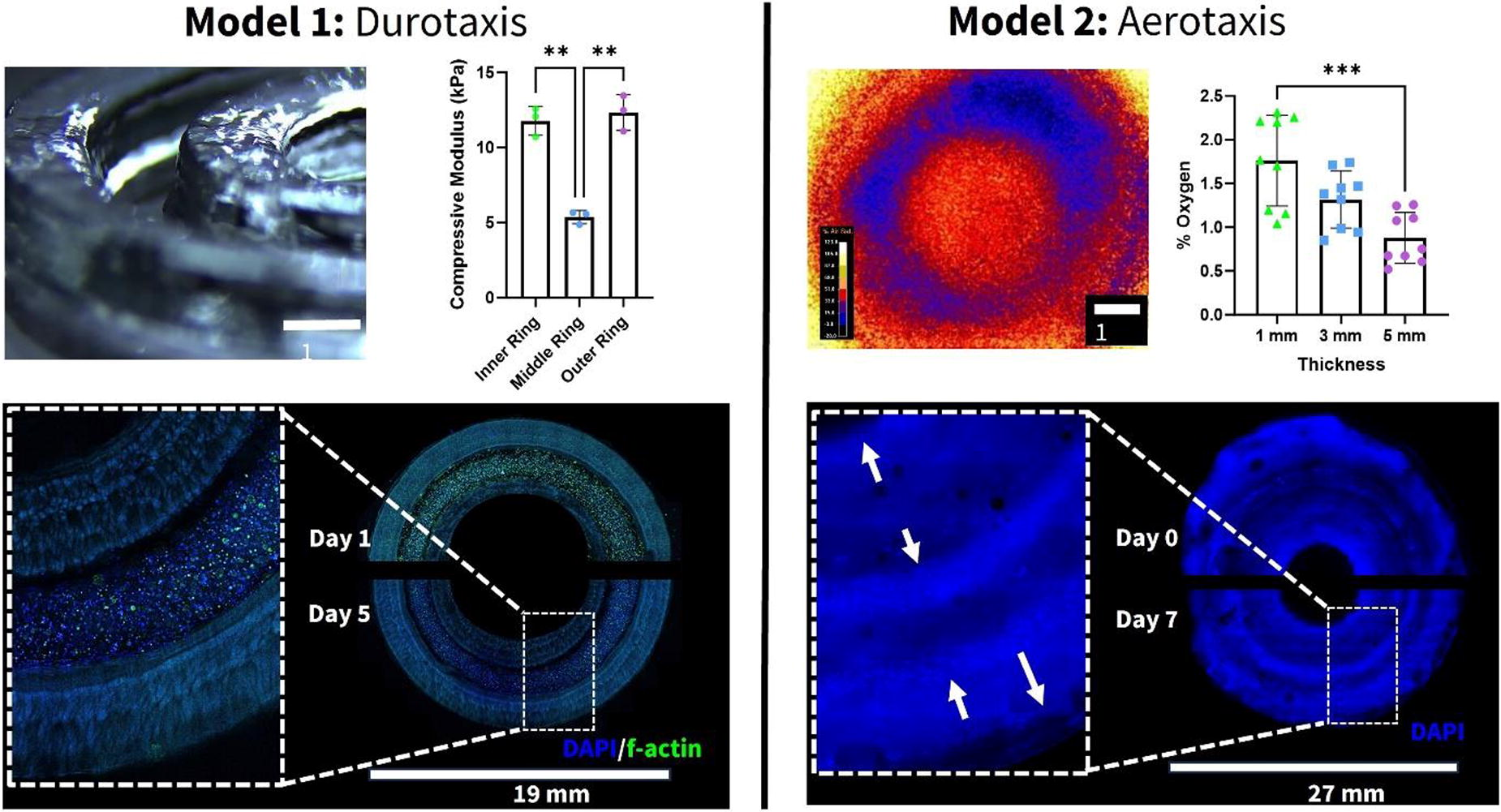
*Purpose/Objectives: In vitro models are useful tools to study bone marrow (BM) malignancies, such as leukemia or multiple myeloma. However, the complex multi-niched microenvironments that make up the BM are challenging to recapitulate in current models. These challenges include the incorporation of biophysical gradients, such as stiffness and oxygen levels, and regional changes in cell localization. This work aims to investigate the niched BM microenvironments by developing tailorable biofabricated models that probe durotaxis and aerotaxis of relevant cell populations and incorporate mechanical loading and oxygen sensing technology.
*Methodology: Allylated gelatin (GelAGE) is synthesized and used to design bioinks (7-10 wt%) that can be extruded (BioX6, 15-20°C nozzle temperature, 4°C base plate) and photopolymerized (dithiothreitol, LAP, 405 nm). Changes in polymer concentration is altered to change gel stiffness, which is evaluated using compressive loading (ElectroForce 5500). 2D oxygen sensors with VisiSens TD camera (Presens) are used to monitor real-time fluctuations in oxygen. Human bone marrow derived mesenchymal stromal cells (BMSCs) are encapsulated within various biofabricated constructs - to monitor durotaxis or aerotaxis over time. Immunohistochemistry (DAPI and f-actin) is used to monitor the location of cells over time and image analysis is used to quantify the regional location of cells throughout the construct. Within these model systems, changes in oxygen consumption and migration behavior in response to regional changes in stiffness and oxygen tension can be monitored and further evaluated.
*Results: Extrusion bioprinting facilitated the fabrication of a construct with 3 different concentric rings (representing the endosteal niche, central marrow, and perivascular niche) to monitor cellular migration in response to stiffness and oxygen gradients over time. The stiffness of GelAGE hydrogels was successfully tailored (5-160 kPa). By altering the mechanical properties of each bioprinted rings, a model system was constructed with stiff-soft-stiff regions. Cells that were seeded in the central marrow (soft) ring displayed limited durotaxis over 5 days in culture (Model 1). A second model design consisting of uniform mechanical properties but changes in ring thickness that induced linear oxygen gradients (high-low-high) was fabricated (Model 2). The bulk hydrogels induced changes in steady state oxygen levels (1-6% oxygen) in the X-Y axis with and without cells. After a culture period of 7 days, the BMSCs had migrated in the X-Y direction based on aerotaxis from the central marrow (low) ring to the endosteal and perivascular niche (high) rings. These tailorable bioinks facilitate the fabrication of multiple bone marrow models with non-linear microenvironmental factors, including variations in both stiffness and oxygen levels.
*Conclusion/Significance: Taken together, GelAGE bioinks offers the ability to fabricate 3D-constructs featuring specific regions with both varying stiffness and oxygen levels. This study represents the first report of a 3D-printed bone marrow model system with non-linear biophysical gradients that allows for a systematic in vitro study of stem cell migration. These results hold the potential to inspire more biomimetic bone marrow model designs with multi-niched biophysical gradients that influence the behavior and phenotypes of relevant cells.
A3 - 3D-printed Osteoporotic In Vitro Bone Model Under Dynamic Culture
E. Batoni1, V. Platania2, N. N. Tavernaraki2, C. De Maria1, G. Vozzi1, M. Chatzinikolaidou2
1
University of Pisa, Pisa, Italy,
2
University of Crete, Heraklion, Greece
*Purpose/Objectives: Osteoporosis is a worldwide bone disease caused by an imbalance between bone remodeling and resorption. Although animal models are still the gold standard, they are expensive, not reproducible, and reliable. Bone Tissue Engineering might be a promising alternative aiming to develop an in vitro osteoporotic bone model. The study aims to develop a three-dimensional (3D) osteoporotic in vitro bone model. Pre-osteoblastic cells were seeded on a 3D-printed osteoporotic scaffold and cultured under dynamic conditions. A physiological model was cultured under the same conditions to compare the cell response with the pathological one.
*Methodology: To mimic the architectural bone changes in osteoporosis, osteoporotic scaffolds were designed with higher porosity and lower trabeculae thickness compared to physiological ones. Scaffolds were designed in cubic shapes (4 mm x 4 mm x 3 mm) and were 3D printed with a gelatin/nano-hydroxyapatite (nano-HA) biomaterial ink as reported in a previous study. Nano-HA content was decreased in osteoporotic scaffolds to mimic osteoporotic bone's reduced mineral bone density. Scaffolds were characterized under uniaxial mechanical compression and FT-IR spectroscopy. MC3T3-E1 pre-osteoblasts were seeded onto the scaffolds and then cultured under dynamic conditions with a uniaxial cyclic compression bioreactor for 21 days. Simultaneously, cell-seeded scaffolds were statically cultured to evaluate differences in cell behaviour when exposed to mechanical stimulation. Cell viability, morphology at Scanning Electron Microscopy (SEM), and osteogenic differentiation (i.e., alkaline phosphatase (ALP) activity, collagen and calcium secretion) of the pre-osteoblastic cells were assessed at different time points.
*Results: Mechanical tests showed a significantly lower elastic modulus of osteoporotic scaffold (i.e., by 45%) compared to physiological ones due to reduced nano-HA content. FT-IR spectra reported the presence of characteristics peaks of gelatin and nano-HA in each sample. Pre-osteoblastic cells seeded onto physiological and osteoporotic scaffolds remain viable until the end of the experiment both under static and dynamic culture conditions. Even though mechanical stimulation initially decreased cell viability, this significantly surpassed that of statically cultured samples. SEM images reported good cell morphology, adherence, and spreading throughout the scaffolds. Under dynamic conditions, ALP activity increased compared to the static conditions, with the physiological samples reporting significantly higher values than those found in osteoporotic ones. Similarly, under dynamic conditions, the amount of secreted total collagen and calcium significantly surpassed the static one. Thus, mechanical stimuli enhanced the early osteogenic differentiation and the maturation towards osteoblasts. Osteoporotic samples reported a significant reduction of both osteogenic markers compared to physiological ones. Hence, under dynamic conditions, osteogenic differentiation of the pre-osteoblastic cells was enhanced, and osteogenic markers were less intense in the osteoporotic samples than in physiological ones.
*Conclusion/Significance: The work presented a first attempt at developing a 3D in vitro osteoporotic model, mimicking two parameters, the osteoporotic bone architecture/density and bone mineral content. Results demonstrated the capacity of the dynamic condition to stimulate the osteogenic differentiation in cell-laden scaffolds. Under dynamic conditions, osteoporotic samples reported lower values of osteogenic markers compared to physiological ones, which might resemble the decreased osteoblast activity in osteoporotic bone.
A4 - Developing A Scalable, Modular, Automated And Closed Process For Manufacturing Patient-specific Bone Grafts
A. Plankey
BioFabUSA | ARMI, Manchester, NH
*Purpose/Objectives: EpiBone-Craniomaxillofacial (EB-CMF) is a tissue engineered bone construct currently in clinical stage development for mandibular ramus reconstruction. Facial deformities and injuries require precise reconstruction to match anatomical shape, natural appearance, and original function. Current practices require harvesting bone from another area of the patient’s body, which has limitations and comorbidities associated with surgery and recovery. Many patients who have suffered facial bone injuries will require years of reconstructive and invasive surgery. With treatment by EB-CMF, this reconstruction process will have fewer complications, provide a custom autologous bone graft, and reduce the length of surgical procedures by eliminating the need to harvest bone from the patient. This accelerated timeline will restore the patients’ form and function while also lessening the long-term challenges associated with deforming injuries. The current production process for EB-CMF relies on the isolation and manual culture of adipose-derived, patient-specific mesenchymal stromal cells (MSC), seeding of those cells onto a decellularized bovine bone scaffold, and culture of the seeded scaffold in a custom anatomical bioreactor system, which allows it to mature into cellularized bone suitable for implantation. Due to the reliance on several manual and open processing steps, each graft is currently susceptible to operator variability, inadvertent error and contamination.
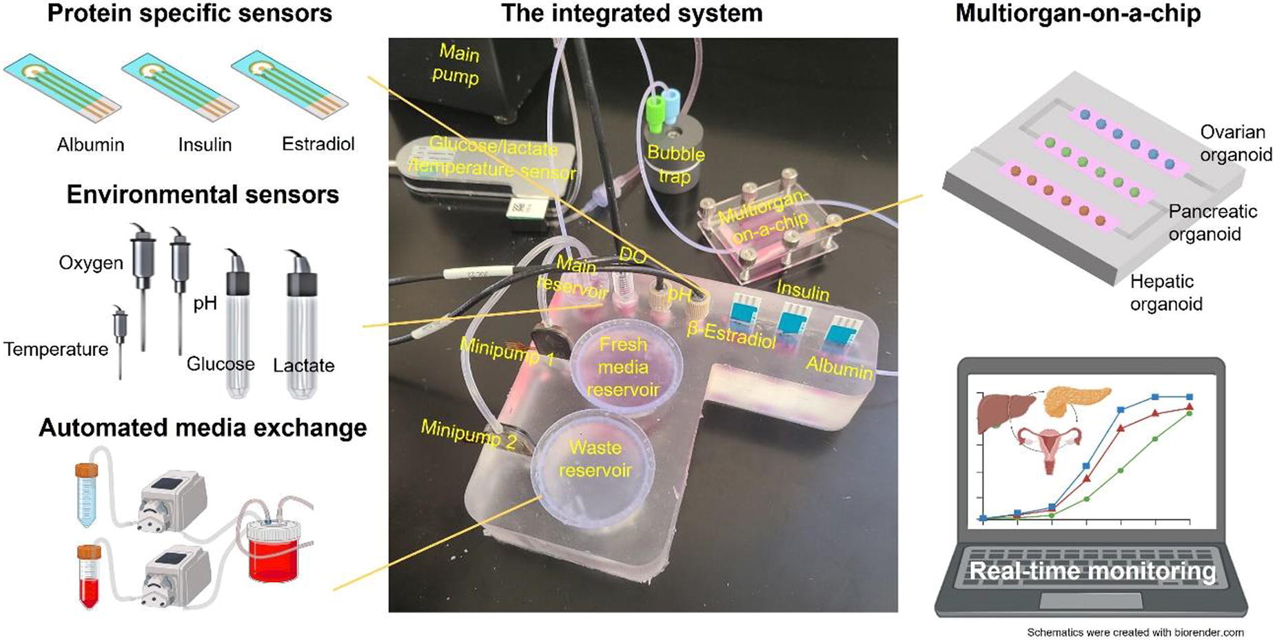
*Methodology: BioFabUSA and EpiBone are collaborating to adapt the manual EB-CMF production process to scalable, modular, automated and closed manufacturing. BioFabUSA has closed and automated the isolation of MSCs from adipose tissue by an automated Tissue Digestion system. The MSC expansion stage has also been scaled and automated using a Cell Stack Manipulator to automatically seed, harvest, and perform media changes of the various size flasks. For the maturation stage, BioFabUSA has developed a novel Centralized Fluid Management and Culture Control System (CFMCCS) to close and automate the bioreactor steps of the process. The CFMCCS is equipped with inline sensors to monitor critical process parameters and to automatically perform culture media changes for the bone grafts during maturation. These process adaptations have allowed a 10-fold increase in the number of simultaneous batches of product.
*Results: Through use of the Tissue Digester, Cell Stack Manipulator, and CFMCCS BioFabUSA has successfully created autologous bone grafts. Each stage of the expansion has met the cell specifications and the CFMCCS is currently being customized to improve ALP, DNA, and Glucose results.
*Conclusion/Significance: BioFabUSA’s technologies will enable further rapid scaling of the EB-CMF process. Automating the production line using innovative technologies like the CFMCCS improves process reproducibility and throughput while meeting quality control specifications and standards. These manufacturing solutions will support the reduction of manufacturing costs while ensuring quality product, enabling the commercial availability of an improved craniomaxillofacial treatment option. Currently, a clinical indication for the treatment of mandibular ramus reconstruction is being sought, but the EpiBone technology platform can be expanded to include other large size bone defects that will drastically improve the outcome and recovery of injuries. A high quality and efficient manufacturing process for a bone defect replacement provides improved treatment options while increasing the patients’ performance and recovery.
A5 - 3D BioprintingAnisotropic Mechanical Properties For Personalizes Osteochondral Implants
M. de Ruijter1, L. Spauwen2, A. G. Vasilopoulou3, A. Mensinga3, P. van Veenendaal4, J. Malda1
1
UMC Utrecht & Utrecht University, Department of Veterinary Sciences, Utrecht, Netherlands,
2
University of Applied Sciences Utrecht & UMC Utrecht, Utrecht, Netherlands,
3
UMC Utrecht, Utrecht, Netherlands,
4
University of Applied Sciences Utrecht, Utrecht, Netherlands
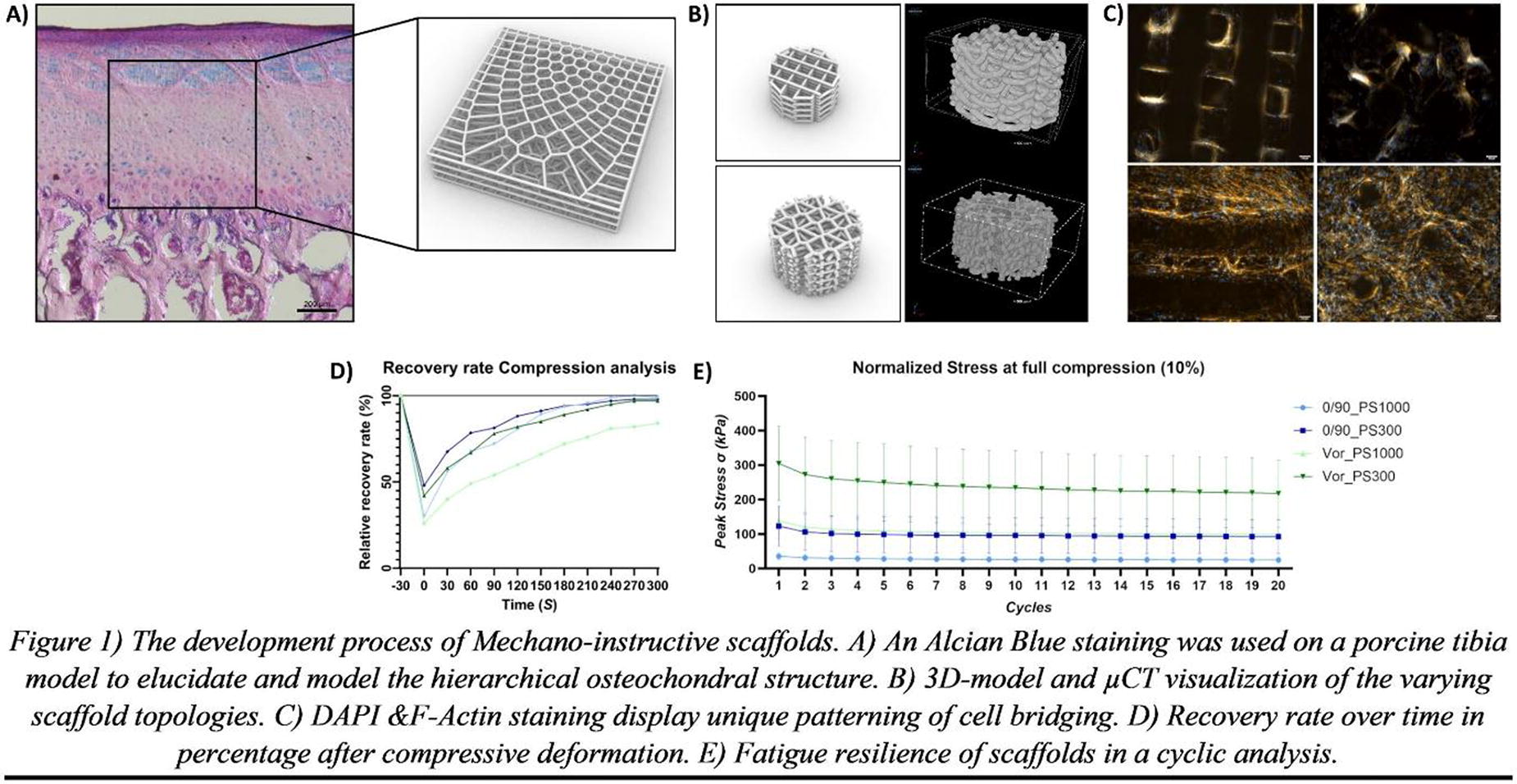
*Purpose/Objectives: Cartilage defects cause pain and immobility and are a growing global concern. As the durability of total knee replacements are limited, new treatments are needed. Biofabrication strategies show potential, osteochondral plugs (ø6 mm) with hierarchy in cells and reinforcing fibres are mechanically stable in the in vivo environment[1]. Translating this strategy to larger defects include considering patient-specific shape, size, and the anisotropic loading pattern. The aim of this study is to develop osteochondral scaffolds with anisotropic mechanical properties in patient-specific size and shape.
*Methodology: Box-shaped scaffolds (size range: 200 x 200 μm to 500 x 500 μm in 100 μm increments) from polycaprolactone were made with melt electrowriting (MEW).). Homogeneous scaffolds (equal box-spacing throughout) were compared to anisotropic scaffolds (box-spacing varied within the same scaffold). These scaffolds were then embedded in gelatine methacryloyl (gelMA) and crosslinked with Ruthenium(Ru)/ sodium persulfate(SPS). Local (ø2 mm indenter) and bulk (entire surface indentation) compression were measured. Furthermore, scaffolds were converged printed with a bone-phase (magnesium-strontium phosphate/polycaprolactone composite, extrusion-based printed), cultured in vitro for 4 weeks and analysed for interfase integrity, cartilage-like matrix production using biochemistry (Picogreen/1,9-Dimethyl-Methylene Blue assay) and histology (Safranin-O/Collagen type II staining).
*Results: Scaffolds were fabricated successfully in patient-specific shape and size with distinct and well-integrated bone- and cartilage-like zones. Anisotropy in box size affected the mechanical properties and resulted in a wide range (bulk: 0,5 MPa to 1 Mpa, local: 0,5 to 2,5 MPa) of compressive moduli throughout the scaffold. After culture, the implants showed cartilage-like matrix formation comparative to isotropic scaffold controls.
*Conclusion/Significance: Scaffolds with patient-specific shape, size and loading properties were fabricated. Manipulating the internal box-spacing enables to regulate the mechanical properties of the scaffold. This study highlights the potential of biofabrication strategies for cartilage defect treatment with living, personalized implants, with anisotropic mechanical properties.
References:
[1] de Ruijter M, Diloksumpan P, Dokter I, Brommer H, Smit IH, Levato R, van Weeren PR, Castilho M, Malda J. Orthotopic equine study confirms the pivotal importance of structural reinforcement over the pre-culture of cartilage implants. Bioeng Transl Med. 2023 Oct 19;9(1):e10614. doi: 10.1002/btm2.10614. PMID: 38193127; PMCID: PMC10771555.
A6 - Biomimicry Of Native Meniscus Tissue With Biofabrication Technologies
M. de Ruijter1, J. Korpershoek2, M. Terpstra3, P. N. Bernal3, R. J. Custers3, D. B. Saris4, L. Vonk5, R. Levato1, J. Malda1
1
UMC Utrecht & Utrecht University, Department of Veterinary Sciences, Utrecht, Netherlands,
2
UMC Utrecht & Mayo clinic, Orthopedics and Sports Medicine, Utrecht, Rochester, Netherlands,
3
UMC Utrecht, Utrecht, Netherlands,
4
UMC Utrecht & Mayo clinic, Orthopedics and Sports Medicine & University of Twente, Utrecht, Netherlands,
5
UMC Utrecht & Xintela AB, Utrecht, Netherlands
*Purpose/Objectives: Meniscus injury and meniscectomy are strongly related to the development of osteoarthritis. There is a clinical need for meniscus replacement therapies. Furthermore, in vitro models that can mimic meniscus tissue are required to test therapeutic, regenerative strategies. The meniscus has a distinctive fibre architecture and includes vascular and avascular regions. Biofabrication technologies allow high-resolution, multi-material deposition of biologically active compounds. This study aims to mimic both the crucial fibrous and the vascular/avascular components of meniscus scaffolds.
*Methodology: To mimic native fibre architecture, melt electrowriting(MEW) of medical grade polycaprolactone was used to fabricate a wedge-shaped meniscus scaffold with micrometre-scale circumferential and radial-oriented fibres [1]. To replicate the vascular and avascular regions, a pro- and anti-angiogenic bioink [2] was developed and extrusion-based bioprinted into an anatomical meniscus shaped construct with an outer pro-angiogenic and inner anti-angiogenic region. The base ink was fibrin-based. The pro-angiogenic region included type I collagen (col-1) microfibers (MFs), endothelial cells and MSCs. The anti-angiogenic region included decellularized cartilage-derived (CdECM) MFs, and meniscus progenitor cells. Scaffolds were cultured in vitro (14 days) and tested for biological and mechanical functionality.
*Results: Circumferential and radial-oriented microfibres (ø = 10μm) increased the compressive mechanical properties as compared to scaffolds without the reinforcing MEW fibres. Cell infiltration and meniscus-like matrix deposition was observed. The pro-angiogenic and anti-angiogenic bioink were successfully printed with extrusion-based bioprinting. After in vitro culture, spatial confinement of the vascular network was observed only in the outer region. ECM-derived MFs can promote and also inhibit capillary network formation.
*Conclusion/Significance: This study demonstrates the feasibility of using MEW to mimic the radial and circumferential fibre-architecture of the meniscus, and using extrusion based bioprinting to represent the vascular and avascular region. This highlights the potential of biofabrication technologies for biomimicry of meniscus tissue for in vitro models and potential regenerative implants.
References: [1] Korpershoek et al. 2021, [2] Terpstra et al. 2022.
A8 - Hybrid Layer-by-layer Machine For Creating Interactive Thin Film Coatings
A. Stokes
Newcastle University, Newcastle upon Tyne, United Kingdom
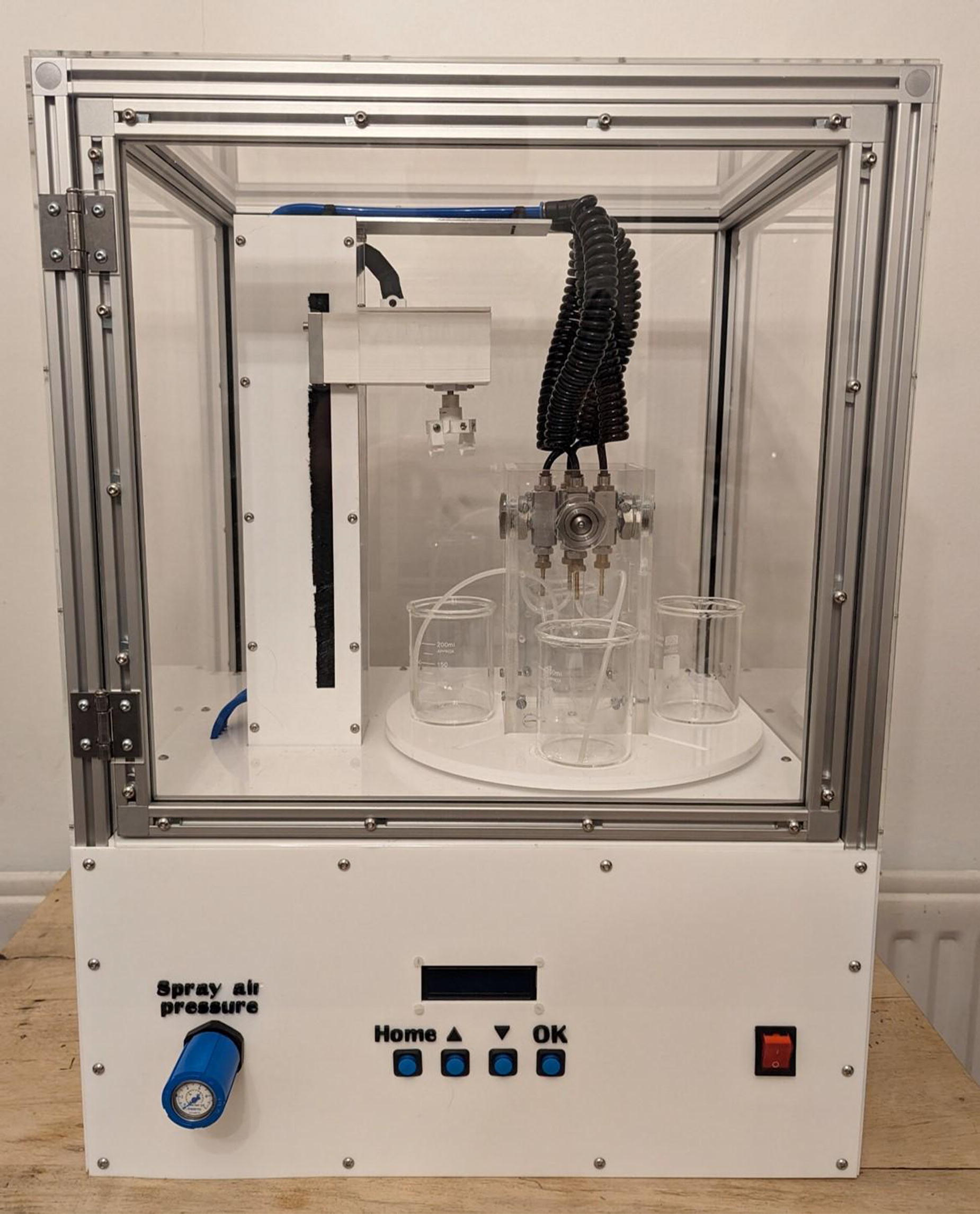
*Purpose/Objectives: In this work, the primary objective was the development and validation of a hybrid coating machine tailored for the surface functionalization of medical implants. The patented system exhibited versatility, allowing for a customizable coating process that could be applied through immersion, spray, or a combination of both. The aim was to explore potential markets for this innovative system, with a particular focus on its application in coating joint replacement implants.
*Methodology: The methodology involved utilizing the Layer-by-Layer machine for coating joint replacement implants with an antibacterial and osteoconductive surface. This was achieved by applying oppositely charged polyelectrolyte solutions layer by layer, with washing steps in between. Customization options included the incorporation of MGO or antibiotics to reduce the risk of secondary infections. The outermost layer, BMP-2, was sprayed to initiate cell repair and attachment upon implantation. Additionally, a second application of the machine was employed to fabricate multi-layered PCL-PAH-Manuka Honey membranes through dip coating. This process included 14 interspaced layers, and the membranes demonstrated potential antibacterial properties for up to 7 days in vitro.
*Results: Promising early cell studies indicated low bacterial adhesion and biofilm accumulation on the coated joint replacement implants, while also promoting osteoblast growth. The Layer-by-Layer application allowed for a range of 1 to 11 thin films, providing a means to control the release period due to weak electrostatic forces. The multi-layered PCL-PAH-Manuka Honey membranes showed efficacy in inhibiting bacterial growth, suggesting their potential use in medical applications. The rapid and replicable fabrication of these membranes further underscored the significance of the developed systems.
*Conclusion/Significance: In conclusion, the hybrid coating machine demonstrated significant potential for advancing the field of medical implant surface functionalization, particularly in the context of joint replacement implants. The Layer-by-Layer application method exhibited promising outcomes in terms of reducing bacterial adhesion, biofilm accumulation, and enhancing osteoblast growth. The fabrication of multi-layered membranes, especially those incorporating Manuka Honey, showcased substantial antibacterial properties, making them valuable for potential medical applications. Overall, these developments hold promise for improving implant success rates and addressing infection-related concerns in the medical field.
A10 - A New Strategy For Reducing The Risk Of Thrombosis On Polymeric Medical Devices
M. Crago1, R. Tan2, J. Hung3, S. G. Wise4, B. Akhavan5, M. Bilek4, S. Naficy1, F. Dehghani1
1
School of Chemical and Biomolecular Engineering, The University of Sydney,, Sydney, Australia,
2
School of Medical Science, The University of Sydney,, Sydney, Australia,
3
University of Sydney, Sydney, Australia,
4
The University of Sydney,, Sydney, Australia,
5
The University of Newcastle, Newcastle, Australia
*Purpose/Objectives: Implanted medical devices pose a threat to patient safety and functionality when adverse body reactions occur, often attributed to nonspecific protein adsorption due to suboptimal material biocompatibility. To enhance the safety and longevity of these devices, we suggest a method of incorporating stable zwitterionic grafts onto polymeric surfaces. Our hypothesis involves utilizing plasma immersion ion implantation (PIII) technologies as an effective and versatile approach to graft zwitterions onto polymeric substrates, eliminating the need for chemical initiators. This technique aims to establish enduring antifouling and antithrombogenic properties.
*Methodology: Implanted medical devices pose a threat to patient safety and functionality when adverse body reactions occur, often attributed to nonspecific protein adsorption due to suboptimal material biocompatibility. To enhance the safety and longevity of these devices, we suggest a method of incorporating stable zwitterionic grafts onto polymeric surfaces. Our hypothesis involves utilizing plasma immersion ion implantation (PIII) technologies as an effective and versatile approach to graft zwitterions onto polymeric substrates, eliminating the need for chemical initiators. This technique aims to establish enduring antifouling and antithrombogenic properties.
*Results: A range of zwitterion-grafted polymers were successfully fabricated, as evidenced by changes in elemental composition, surface morphology, and hydrophilicity, without damaging tensile or flexural mechanics. Further investigation refined the fabrication process to ensure a robust zwitterionic surface at least three months after fabrication. Subsequently, this strategy decreased fibrinogen adsorption by nearly 9-fold, and thrombosis by almost 75%, when applied to a commercial polyurethane.
*Conclusion/Significance: This study illustrates the viability of employing PIII as an effective method for grafting a polyzwitterionic layer, thereby converting the surfaces of hydrophobic polymers into nearly super hydrophilic ones, all while preserving their inherent mechanical properties. This approach streamlines the surface modification process in comparison to other methods, which often entail multiple steps and the use of excessive wet chemistry reagents, chemical linkers, or initiators. In vitro studies validate their antithrombogenic characteristics, underscoring their potential advantages in blood-contacting settings. The application of the PIII technique for surface modification with a polyzwitterion layer emerges as a promising avenue for crafting robust blood-contacting devices.
A11 - Advancing TMJ Regeneration: Crafting Multiaxial Microscaffolds With 4D Electromelting Printing, Enhancing Bone Regeneration With Nasal Chondrocytes
G. Borer1, E. Marin2, H. Xu2, X. Zhang2, E. B. Tankus1, A. Barbero3, N. Sharma1, F. M. Thieringer3, V. Basoli1
1
University of Basel, Allschiwil, Switzerland,
2
Kyoto Institute of Technology, Kyoto, Japan,
3
University of Basel, Basel, Switzerland
*Purpose/Objectives: The temporomandibular joint (TMJ) degeneration or trauma poses a treatment challenge due to its limited regenerative capacity. Nasal chondrocytes (NCs) for their high cartilage regenerative capacity and their ability to differentiate into osteogenic cells might represent a suitable cells source for the generation of implantable osteochondral to treat TMJ defects. With this final goal in mind we assess the performance of NCs upon cultured on a 4D printed polycaprolactone (PCL) microstructure scaffolds, a novel concept of scaffold with multilayer architecture acting as a spring that gives the possibility to have homogeneous cell distribution, cell viability and mechanical properties in multiaxial direction. This structure aims to influence NC stability and differentiation, capable of conducting mechanical forces post-implantation while maintaining its shape.
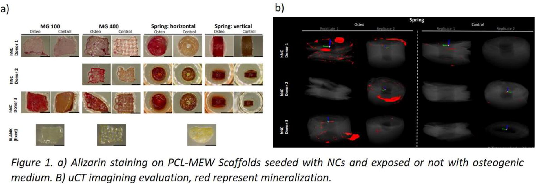
*Methodology: we isolated and cultured human nasal chondrocytes (NC) from 3 donors on medical-grade polycaprolactone printed with a melt electrowriting (MEW) 3D printer. Respectively, two architecture of PCL meshes (a lattice structure with 20 layers with interfiber distance of 250 μm; a mesh with an Auxetic structure was with 20 layers with interfiber distance of 1 mm) and a 4D-MEW spring with 280 layers with a winding angle of 60° (7.5 mm diameter, 1.2 mm height). Samples were cultured in osteogenic differentiation medium (αMEM with 0.1mM ascorbic acid and 10mM Dexamethasone) or not (Control) for 21 days. We further evaluated the viability (using life and dead assays), the expression of cartilage (SOX9, COL10A1) and bone (RUNX2, OCN, IBSP) related genes (by quantitative qPCR), extent of calcification (using Alizarin Red S staining and µ-CT imaging and conventional optic microscopy) and production of bone proteins (immunofluorescence for OCN and IBSP).
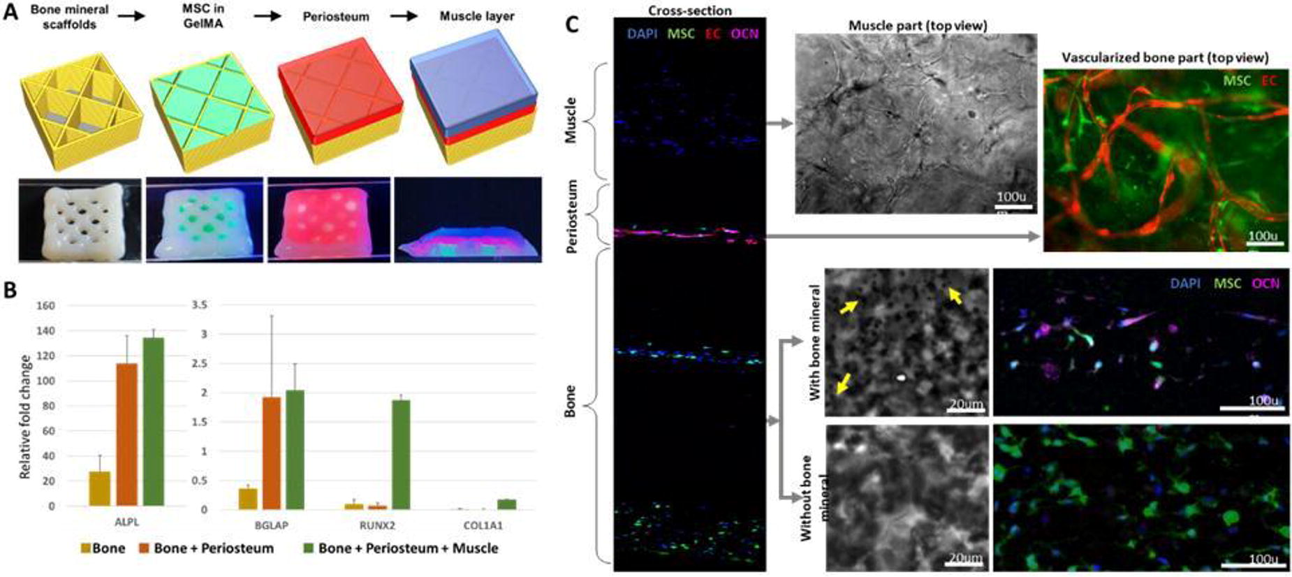
*Results: NCs exhibited prompt adhesion to medical-grade scaffolds within 24 hours, with a uniform fiber oriented distribution across the constructs after 7 days, devoid of any signs of cell mortality. After 21 days, molecular analysis of constructs revealed significantly higher expression of RUNX2, IBSP, and OCN on the osteogenically cultured samples compared to their untreated. Concurrently, a downregulation of SOX9 was observed, indicating a positive RUNX2/SOX9 ratio and signaling osteogenic differentiation.Mineral deposition analysis demonstrated increased calcification in the osteogenic group across all biological replicates, confirmed through Alizarin staining, immunohistochemistry and uCT imaging (Fig.1). These results highlight the efficacy of the 4D printed spring scaffold in promoting cell adhesion, osteogenic differentiation, and enhanced structural integrity over the culturing period.
*Conclusion/Significance: In conclusion, our findings underscore the efficacy of the 4D-MEW printed spring construct for 3D culture, exhibiting superior cell growth and mineralization characteristics. The design of 4D spring construct not only enabled multiaxial mechanical free motion but also notably elevated bone mineralization. This enhancement is likely attributed to the improved exchange of gases and nutrients facilitated by the scaffold's unique architecture. This improvement suggests the potential of 4D-MEW printed scaffold as a promising advancement in the realm of TMJ regeneration. The results provide a strong foundation for further exploration and optimization of this innovative approach, with the ultimate goal of developing clinically applicable protocols for enhanced TMJ regeneration.
A13 - Spatial And Temporal Engineering Of The Cell Niche With Wnt Coated Substrates For Tissue Engineering
M. Rotherham, H. Hajiali, A. J. El Haj
University of Birmingham, Birmingham, United Kingdom
*Purpose/Objectives: Engineering complex 3D tissues continues to be a major challenge for tissue engineering. Control of cell signalling is a critical aspect that determines cell behaviour, matrix formation and ultimately the success of implanted tissue engineered constructs. Wnt signalling plays crucial roles in a variety of biological processes, including embryogenesis, tissue patterning and stem cell maintenance in adults. The pathway is spatially and temporally regulated and dysregulation causes a wide spectrum of age related diseases. Orchestrating tissue repair by activating endogenous adult stem cells holds promise for a variety of disorders, however, the progress of exploiting Wnt signalling for regenerative medicine has been limited. Moreover, the potential risks of off-target or sustained activation of this pathway are serious and cannot be ignored. Therefore, the development of techniques for localized targeting as well as spatial and temporal regulation of Wnt signalling provides a novel therapeutic option for tissue engineering and the treatment of a range of diseases.
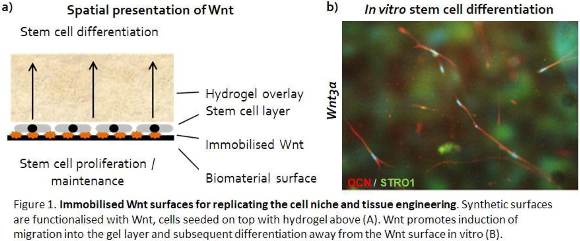
*Methodology: In these studies we covalently bound Wnt ligands onto a range of synthetic surfaces. We then tested the bioactivity of the immobilised Wnt using Wnt (TCF/LEF) reporters. We next studied how localised presentation of Wnt ligands can establish spatial gradients of differentiation of stem and progenitor cells (Fig.1a) using immunocytochemistry, histology and molecular biology. The platform was then developed to form a Wnt bandage to induce bone repair in a mouse calvarial defect model. We also examined the role that temporal stimulation of the Wnt pathway plays in the differentiation process, using Wnt agonist functionalised MNP and magnetic fields provided by a magnetic field bioreactor (MICA) to remotely actuate cell signalling. The synergistic effects of biochemical / growth factor containing differentiation media on progenitor cell fate alongside immobilised Wnt was also studied.
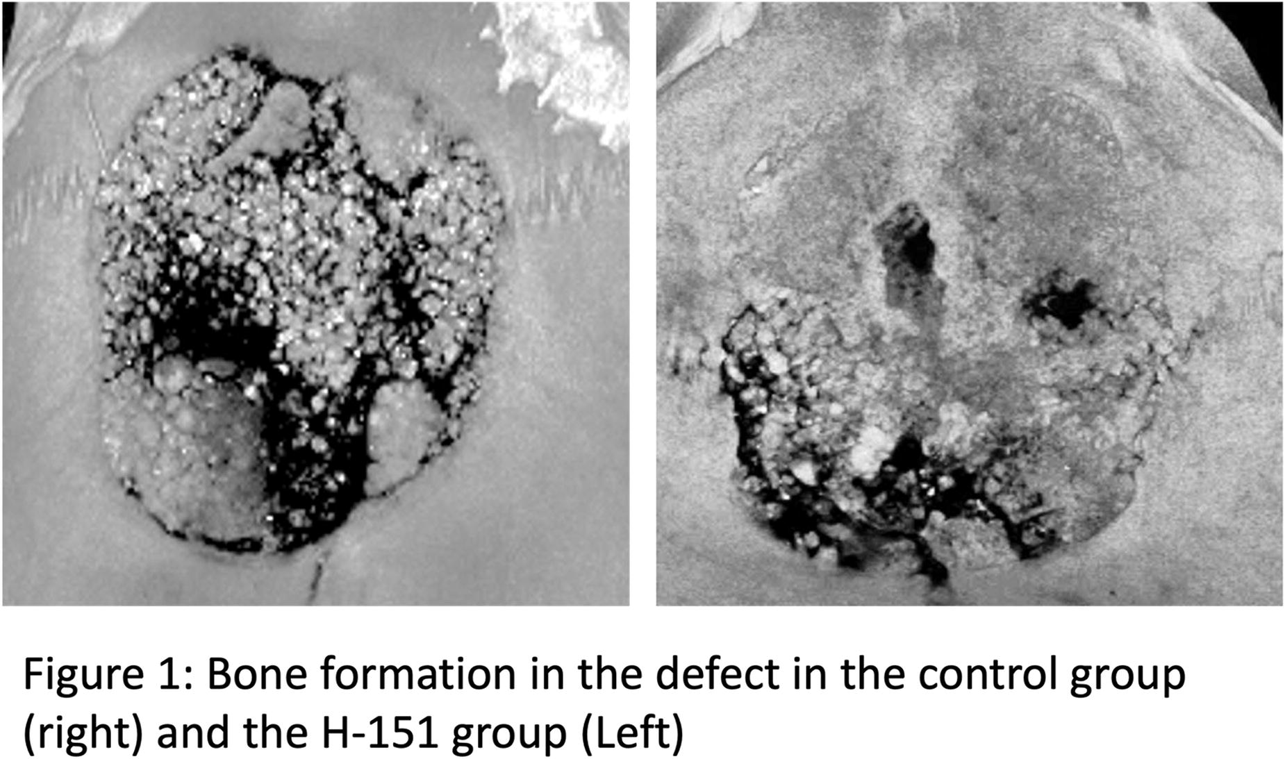
*Results: Our results showed that Wnt ligands can be successfully immobilized onto a range of surfaces in a manner that retained bioactivity. Presentation of immobilised Wnt resulted in activation of TCF/LEF responsive signalling in reporter cells, indicative of active Wnt signalling. Spatial presentation and / or temporal activation of Wnt pathways in this manner led to differentiation of progenitor cells (Fig.1b), and when applied with differentiation media, this induced a synergistic increase in differentiation markers and matrix production. Application of the immobilised Wnt bandages in vivo was able to induce new bone growth across calvarial defects after 8 weeks.
*Conclusion/Significance: Our immobilised Wnt platforms demonstrate a novel way of remotely and spatially engineering the stem cell niche to re-capitulate the signalling processes seen during development. The approach has been used to optimise cell differentiation in vitro, bone formation in vivo and works synergistically alongside biochemical factors. In the future this approach can be used alongside cell therapies in a minimally invasive manner to enhance tissue engineered constructs, develop novel bandages for tissue repair and improve the clinical outcome for patients.
A14 - 3D Bioprinting Tissue Engineered Meniscal Constructs
G. McDermott1, B. Barkatali2, M. Domingos1, S. M. Richardson1
1
The University of Manchester, Manchester, United Kingdom,
2
Salford Royal Hospital, Manchester, United Kingdom
*Purpose/Objectives: Meniscal injuries affect over 1.5 million people across Europe and the USA annually. Injury greatly reduces knee joint mobility and quality of life and frequently leads to the development of osteoarthritis. Tissue engineered strategies have emerged in response to a lack of viable treatments for meniscal pathologies. However, to date, constructs mimicking the structural and functional organisation of native tissue, whilst promoting deposition of new extracellular matrix, remains a bottleneck in meniscal repair. 3D bioprinting allows for deposition and patterning of biological materials with high spatial resolution. This project aims to develop a biomimetic 3D bioprinted meniscal substitute.
*Methodology: Meniscal tissue was characterised to effectively inform the design of biomaterials for bioprinting constructs with appropriate structural and functional properties. Histology, gene expression and mass spectrometry were performed on native tissue to investigate tissue architecture, matrix components, cell populations and protein expression regionally across the meniscus. 3D laser scanning and magnetic resonance imaging were employed to acquire the external geometrical information prior to fabrication of a 3D printed meniscus. Bioink suitability was investigated through regional meniscal cell encapsulation in blended hydrogels, with the incorporation of growth factors and assessed for their suitability through rheology, scanning electron microscopy, histology and gene expression analysis. Bioprinted 3D meniscal constructs were fabricated through zonal deposition of regionally tailored bioinks.
*Results: Meniscal tissue characterisation revealed regional variations in matrix compositions, cellular populations and protein expression. The process of imaging through to 3D printing highlighted the capability of producing a construct that accurately replicated meniscal geometries. Regional meniscal cell encapsulation into hydrogels revealed a recovery in cell phenotype, with the incorporation of growth factors into the bioink’s stimulating cellular re-differentiation and improved zonal functionality. Bioprinting of bioinks regionally enabled the fabrication of a 3D meniscal construct with regional variations in cell and matrix deposition.
*Conclusion/Significance: Meniscus biofabrication highlights the potential to print patient specific, customisable meniscal implants. Achieving zonally distinct variations in cell and matrix deposition highlights the ability to fabricate a highly complex tissue engineered construct.
A15 - Automated Biofabrication Of Biomimetic Glaucoma In Vitro Models
H. C. Lamont1, A. W. Wright1, K. Devries2, M. Jones3, I. Masood1, L. J. Hill1, S. N. Nazhat2, L. M. Grover1, A. J. El Haj1, A. D. Metcalfe1
1
University of Birmingham, Birmingham, United Kingdom,
2
McGill University, Montreal, QC, Canada,
3
Cell Guidance Systems, Cambridge, United Kingdom
*Purpose/Objectives: Primary open angle glaucoma (POAG) is currently the leading cause of irreversible blindness. POAG pathogenesis originates from the trabecular meshwork (TM) tissue, which becomes fibrotic due to trabecular meshwork cell (TMC) dysfunction. The TM exhibits a collagenous, hierarchical architecture, with TMC displaying several phenotypes in vivo. However, the development of an in vitro model that can induce in vivo-like behaviour, or as to what stimuli modulate TMC phenotypic heterogeneity, remains elusive.
This study aims to elucidate biophysical and biochemical spatial cues that induce transient TMC phenotypic states in both healthy and POAG scenarios through 3D in vitro modelling. Models were produced in an automated manner, creating a biomimetic TM environment in vitro, with tuneable collagen properties, targeted towards high-throughput testing.
*Methodology: Initially, human TMC-laden collagen hydrogels (250,000 cells/mL) were plastically compressed (PC) to modify collagen fibre density (CFD) and architecture. To induce POAG attributes, 5ng/ml of transforming growth factor-beta2 (TGF-β2 (PODS® technology)) was integrated into PC-collagen. Thereafter, TMC phenotypic differences were assessed; immunocytochemical/western blot staining on TM protein markers (elastin, α-β-crystallin, fibronectin, alpha-SMA), filamentous actin and focal adhesion (vinculin) expression. Differential gene expression through qPCR investigated mesenchymal markers; SNAIL1, TWIST1, N-cadherin, and in vivo marker, myocilin (MYOC).
Optimized parameters were further reproduced through the automated gel-aspiration-ejection method (GAE) for 96-well plate format. Differences in collagen mechanics were evaluated through nanoindentation, SEM/micro-CT imaging and CFD. TM in vitro models were assessed up to 2-weeks on metabolic capacity, western blot/immunocytochemical staining, qPCR.
*Results: Initial investigations presented modulation in TMC phenotype through PC-method. Mechanical characterization of PC-collagen presented enhanced CFD (9.06±0.58%), anisotropy (2.2-fold) and storage modulus (1307±179.37 Pa). Modulation of biophysical parameters induced; TMC cytoskeletal reorganization, nuclear deformation, focal adhesion maturation, metabolic activity (80.05±35.1% increase) characteristic TM protein expression (elastin deposition, α-β-crystallin, fibronectin), and altered mesenchymal/in vivo gene expression (SNAIL1 (12.7-fold), MYOC (4.07-fold)). Comparatively, TGF-β2 presented; cytoskeletal disarrangement, enhanced focal adhesion size, increased mesenchymal proteins expression (alpha-SMA, fibronectin and intracellular TGF-β2) and gene expression (SNAIL1 (5.53-fold), TWIST1 (5.33-fold), N-cadherin (8.07-fold)), compared to prior conditions. A profile of TMC mesenchymal characteristics by sequential modulation of biophysical and biochemical (TGF-β2) stimuli was produced.
Optimized collagen parameters bio -fabricated through the automated GAE produced similar collagen anisotropy and CFD (7.33±1.01%). After, TMC expressed relevant in vivo markers 3- days post incubation in both native and pathological models (elastin deposition, fibronectin, alpha-SMA, intracellular TGF-β2) with maintained TMC alignment and sustained, 2-week metabolic activity.
*Conclusion/Significance: A biomimetic in vitro model of the TM was optimized, producing an initial panel of positive in vivo markers that was dependent on collagen structural framework or/and prolonged TGF-β2 exposure (biophysical/biochemical spatial cues). Thus, we successfully produced a profile of transient mesenchymal states for both healthy (SNAIL1/MYOC; partial-mesenchymal) and pathological scenarios (SNAIL1/TWIST1/N-cadherin/MYOC; complete mesenchymal). Providing a novel insight into the processes that govern TMC fate and phenotypic heterogeneity in vivo. Once established, biophysical parameters were reproduced in an automated manner for future high-throughput screening.

A16
A17 - An Artificial Intestine Containing Three Types Of Cells For Transplantation Using 3D Bio-printing Technology
s. Son1,2, D. Lee1, D. Kim1,3, H. Lim1, B.-y. Lee1, S. Park1, C.-R. Jung4, J. Lee1
1
Korea Institute of Machinery & Materials, Dajeon, Korea, Republic of,
2
Chungnam National University, Daejeon, Korea, Republic of,
3
Seoul National University, Seoul, Korea, Republic of,
4
Korea Research Institute of Bioscience and Biotechnology, Dajeon, Korea, Republic of
*Purpose/Objectives: The human intestine is a dynamic and complex organ that is essential for immunological protection, waste removal and nutrient absorption. Congenital defects, atresia and stenosis in neonates and pediatric patients have a profound impact on body function. Gastrointestinal problems in particular pose a serious threat to the survival and healthy growth of infants and young children. This study investigates the need for tailor-made of artificial intestines for neonatal and pediatric patients using 3D bio-printing technology to address the challenges associated with organ transplantation.
*Methodology: The study show the ability of 3D bio-printing to produce a highly detailed small intestine with an inner diameter of 2mm, using advanced features such as multi-head printing and precise temperature control for the use of polymer and GelMA bio-inks A 6-axis controlled bio-printer (X, Y, Z1, Z2, Z3, and Rotary axis) was utilized to fabricate the artificial intestine model. The process involved the initial printing of a PCL scaffold to maintain the tubular shape. HUVECs and CCD-18Co cells were subsequently printed onto the PCL scaffold, while Caco-2 cells were seeded on the inner surface.
*Results: The fabricated bio-printed intestinal model was 2mm in internal diameter, 36mm in length and contained four layers of polymer and cell-laden bio-inks. Comprehensive characterizations, including assessments of cell viability (Live&Dead assay), cell proliferation (CCK-8 assay), Caco-2 cell differentiation (ALP&DNA assay), HUVECs differentiation (CD31 assay) and morphological analysis of microvilli using SEM, provided insight into the structural and functional integrity of the model.
*Conclusion/Significance: This study highlights the critical need for 3D bio-printing to address congenital intestinal problems in neonatal and pediatric patients. The technology demonstrated its ability to construct intricate small intestines with an internal diameter of 2 mm using multi-head printing and advanced bio-inks. These results represent a promising avenue for advancing personalized medicine and introducing innovative solutions in pediatric healthcare.
A18 - Precision Biofabrication For Meniscal Tissue Engineering: Leveraging MEW And Microvalve Bioprinting To Enable Tunable Mechanical And Cellular Gradients
F. Shields, M. Domingos, S. Richardson
University of Manchester, Manchester, United Kingdom
*Purpose/Objectives: This project addresses the critical challenge of meniscal injuries affecting over 1.5 million individuals annually in the USA and Europe, often leading to early osteoarthritis. With existing surgical treatments showing limited success in tissue regeneration, the need for innovative approaches becomes imperative.
*Methodology: Our project centres on a sophisticated biofabrication pipeline, seamlessly integrating melt-electrowriting (MEW) and microvalve bioprinting techniques to craft meniscal tissue analogues containing cellular and mechanical gradients mirroring native tissue. Optimized MEW process parameters facilitated the deposition of layers of stiff polymeric microfibres, creating microchambers that reinforced a soft hydrogel matrix. The mechanical properties of this scaffold, pivotal for mimicking native tissue, underwent rigorous evaluation through compression tests. Microvalve jetting of alginate into MEW microchambers allowed precise droplet patterning, enabling the encapsulation of human MSCs with high viability.
*Results: Our project achieved the production of aligned polymeric fibre walls emulating collagen bundles in native tissues, offering tunable fibre diameters and microchamber sizes. Fine-tuning fibre diameter and spacing resulted in scaffolds approaching physiologically relevant stiffness, potentially allowing us to tailor the mechanosensory environment for encapsulated cells. Multi-nozzle microvalve jetting attained intricate patterns of bioink droplets within polymeric microchambers, showcasing high cell viability and enabling precise patterning of cellular gradients. After 28 days in culture, the reinforced scaffolds closely emulated the compressive stiffness of native cartilaginous tissue, featuring aligned collagen deposition along the polymeric fibres and glycosaminoglycan (GAG) deposition within the hydrogel matrix.
*Conclusion/Significance: These hybrid constructs exhibited significant potential for engineering mechanically robust, cartilaginous tissue analogues, promising advancements in tissue regeneration and preclinical disease modelling.
A20 - Integrated Cryogel And Electrospun Silk Fibroin Scaffolds For Bone-tendon Interface
A. Anup, R. Millett, K. Hixon
Thayer School of Engineering, Dartmouth College, Hanover, NH
*Purpose/Objectives: Musculoskeletal injuries affect 32 million people in the United States each year, and self-repair is often inefficient in healing. Hard-to-soft tissue interfaces, e.g., bone-tendon, are impacted in 45% of musculoskeletal injuries and often requires surgical revision. There is an urgent need to develop improved models to elucidate properties that allow the interface to withstand complex mechanical loads and act as a crosstalk hub involving different cell populations. Tissue engineering can be used to develop in vitro models by combining scaffolds, cells, and bioactive factors. Combining tissue engineering fabrication methods is one way to replicate bone-tendon tissue interfaces in a single scaffold. The cryogelation fabrication technique creates spongy, durable scaffolds well-suited for bone applications, while electrospinning creates nanofibers that mimic tendon. We hypothesize that combining cryogelation and electrospinning, with silk fibroin as the base material for improved interfacial scaffold attachment, will support the development of a bone-tendon interface model.
*Methodology: Bombyx mori silk was first extracted from cocoons using a previously established protocol. Five percent silk solution in 1,1,1,3,3,3-Hexafluoroisopropanol (HFIP) was electrospun onto a metal plate, creating a fibrous tendon-like scaffold (Fig. 1B). To combine the electrospun and cryogel scaffolds, liquid silk solution (pre-cryogel) was dispensed into a plastic dish containing the electrospun layer and exposed to a 30 second sonication period. These silk cryogels are made by physically crosslinking 4.5% (w/v) silk solution using sonication followed by 24 hours of freezing at -20°C. This created porous, bone-like scaffolds (Fig. 1D). The now physically crosslinked silk and electrospun layer was placed together at -20°C for 24 hours (Fig. 1A). Finally, the combined samples were frozen at -80°C for one hour prior to overnight lyophilization before conducting imaging and experiments.
*Results: The silk cryogel had average pore areas of 49.71 µm via SEM imaging. Uniaxial compression testing of the cryogel at a strain end point of 80% resulted in a peak stress value of 29.5 kPa and an average modulus of 0.3341 kPa. Swell tests of the cryogel revealed an average swelling ratio of 1779% from the starting mass after 24 hours, with more than 60% of maximum swelling reached by one hour. Comparatively, average fiber diameter was 530 µm for the electrospun silk. Uniaxial tensile testing of the electrospun component revealed an elastic modulus ranging between 19 and 47 kPa. Ongoing work will investigate the use of additives for mineralization to improve cryogel mechanical properties and silk blends for electrospinning to further support the nanofibrous structure. Additionally, bone/tendon cell studies will be used to determine biological viability and cell differentiation potential.
*Conclusion/Significance: The outcome of this study is an integrated cryogel and electrospun scaffold using silk fibroin as the base material. By keeping the base material constant throughout the scaffold, dissimilar material contacts at the scaffold interface are reduced, improving attachment (Fig. 1C). By utilizing two fabrication methods, support of multiple zones that span the bone-tendon interface are established as an appropriate in vitro model.

A21 - Patient-specific Scaffold Fabrication Advancing Breast Implant Technology Through Tissue Engineering And Mechanical Optimization
A. Silverstein1, I. Goldfracht1, S. Kracoff-Sella2, D. Egozi2, S. Levenberg1
1
Technion, Haifa, Israel,
2
Kaplan Hospital, Rehovot, Israel
*Purpose/Objectives: Numerous women who suffer from breast cancer undergo mastectomy, a surgical procedure entailing the removal of segments of the breast. Following mastectomy, a substantial proportion of patients decide to undergo breast implantation surgery. Presently the available breast implants exhibit inherent issues such as weight, rigidity, and potential medical complications. Thus, tissue engineering has the potential to produce an improved alternative to breast implant technologies that are used today. While past studies have aspired to produce breast tissue through tissue engineering, these studies have not successfully replicated the mechanical properties inherent to native breast tissue. The objective of this research is the fabrication of a scaffold that emulates the mechanical properties of a human breast, thereby enhancing the overall patient experience and enhancing tissue proliferation.
*Methodology: A multi-step scaffold fabrication approach was employed, incorporating 3D printing, injection molding, and cell seeding techniques to produce the scaffold. The mechanical properties of the scaffold were assessed through a compression test. Subsequently, in vivo experiments were conducted by surgically implanting the scaffold into nude mice to evaluate tissue proliferation.
*Results: The results confirmed that intricate geometric configurations can generate mechanical properties resembling those of human breast tissue. Through the manipulation of geometric parameters, it is possible to optimize these mechanical properties to create a patient-specific scaffold. Furthermore, in vitro testing supported the scaffold's compatibility with adipocytes., Finally, in vivo experiments revealed enhanced tissue regeneration when utilizing a mechanically accurate scaffold.
*Conclusion/Significance: The proposed creation of a patient-specific framework guided by mechanical properties has the potential to yield superior scaffolds, not only enhancing patient comfort but also fostering the potential to improve tissue regeneration. This adaptable framework is envisioned to find applicability across diverse tissue types.
A22
A23 - Rapid Prototyping Of Silk-based Bioprinted Immunocompetent Skin Model: An Advanced Pre-clinical Platform For Skin Sensitization Assessments
B. Bhar, B. B. Mandal
Indian Institute of Technology Guwahati, Guwahati, India
*Purpose/Objectives: An essential requirement for pre-clinical evaluations of drugs and topical formulations is the availability of physiologically relevant experimental platforms, capable of replicating human skin responses. Currently, the majority of the research studies use animal models for these assessments, which has come under significant scrutiny and criticism due to both scientific and ethical concerns. Researchers have explored alternative in vitro approaches to develop biomimetic skin models. The existing collagen-based models predominantly rely on multi-step manual cell-seeding methods, resulting in low reproducibility, high costs, and limited applicability due to lack of essential components such as an immune system and blood vessels, highlighting the need for the development of more physiologically complex models. Therefore, we developed a patient-specific immunocompetent 3D bioprinted skin construct using an innovative rapid prototyping strategy to gain insights into human skin inflammatory response following the topical application of formulations.
*Methodology: A photocrosslinkable bioink was formulated by combining Bombyx mori silk fibroin methacrylate, gelatin methacrylate, and patient-specific growth factor-rich platelet releasate. Printing parameters were optimized through comprehensive evaluations of printability, physicochemical attributes, rheological properties, and tunable mechanical properties. Thereafter, microextrusion bioprinting of bilayered skin construct was conducted within a customized transwell system, with an artificial basement membrane supporting the epidermal keratinocyte layer on one side and a pre-vascularized dermal layer on the other side. The human macrophages were introduced into the model after a 14-day maturation period in the air-liquid interface (ALI) culture. Further, the matured model was evaluated through immunostaining, permeability assessments, and sensitization tests following OECD guidelines to establish its suitability for screening applications.
*Results: The formulated photopolymerizable composite bioink stands out as a programmable formulation with precise rheological properties, customized for extrusion-based printing of 3D constructs with tunable mechanical strength. The presence of photoactivated platelet releasate (PPR) significantly impacted the cellular behavior, leading to a greater presence of classically elongated fibroblasts when encapsulated within the bioink formulation as compared to the bioink without PPR. After computer-aided bioprinting of the skin construct using the bioink and 14-day ALI maturation, immunofluorescence staining of the model showed a well-defined matured epidermal layer and a dermal layer with fibroblast-secreted essential ECM component innervated with vascular endothelial cells. The biochemical estimations and gene expression analyses of the matured model validated native-like tissue maturation and epithelialization. The developed skin model effectively classified the applied skin irritants and non-irritants and even demonstrated mild irritative effects of non-classified substances emulating response of human skin through alteration in level of secreted cytokines and macrophage polarization.
*Conclusion/Significance: The work is a pioneering demonstration of the feasibility of single-step microextrusion bioprinting of patient-specific immunocompetent skin model. The fabricated model recapitulated the immune responses similar to human skin and provided an advanced platform for pre-clinical skin sensitization assessments. Therefore, the model represents a robust and versatile screening platform with substantial potential for a wide range of applications in the fields of toxicity testing, biomaterial assessment, and the study of fundamental physiological processes related to skin diseases and wound healing.
A25 - 3D Bioprinted Microphysiological In Vitro Osteochondral Model To Study Osteoarthritic Niche
S. Dey, P. Mehra, C. Jaiswal, B. Bhar, B. B. Mandal
Indian Institute of Technology Guwahati, Guwahati, India
*Purpose/Objectives: Osteoarthritis (OA) is a multifactorial degenerative disease affecting osteochondral tissue, whose molecular mechanism of initiation and progression are poorly understood. In this regard, current animal and simplified osteochondral in vitro disease models fail to mimic the human in vivo microenvironment and the pathophysiological conditions. Therefore, development of a physiologically relevant human osteochondral tissue interface model emulating the osteoarthritic niche is vital for studying the disease progression and screening potential OA therapeutics.
*Methodology: Herein, a 3D bioprinted microphysiological osteochondral complex was developed to establish an in vitro OA like disease condition. The human biomimetic osteochondral in vitro model was 3D bioprinted using photocrosslinkable silk-based bone and cartilage bioinks encapsulated with osteogenically and chondrogenically primed human adipose derived mesenchymal stem cells. Human umbilical vein endothelial cells were also incorporated in the bone bioink to recreate the stable in vivo like capillary network. Additionally, the bioprinted osteochondral constructs were matured in an in-house developed microphysiological bioreactor platform to recapitulate the osteochondral microenvironment. Following dynamic maturation, the biomimetic osteochondral model was examined through molecular and biochemical analysis to determine its suitability for induction of OA like condition. Further the matured osteochondral model was exposed to pro-inflammatory cytokines to induce early-stage OA-like pathological conditions.
*Results: The developed bioinks demonstrated shear thinning behaviour with high print fidelity, optimal permeability and tuneable rheological as well as mechanical properties. Further, the encapsulated osteogenically and chondrogenically primed cells in the dynamically matured bioprinted constructs demonstrated enhanced cellular viability, matrix deposition, distinct osteogenic and chondrogenic gene and biomarker expressions in comparison to static culture. Further, the establishment of the early-stage OA was validated through key pathological features including depletion of ECM components and upregulation of inflammatory genes and biomarker expressions.
*Conclusion/Significance: The developed microphysiological in vitro osteochondral model closely mimicked the in vivo inflammatory conditions observed in the early stage of OA. Thus, the microphysiological model can be utilized as a high-throughput platform for anti-inflammatory drug screening and for studying osteoarthritic niche.
A26 - A Hybrid 3d Additive Manufacturing Approach For Engineering Branched Vasculature
J. D. Cabral, Q. S. Thorsnes, P. R. Turner, A. A. Ali
University of Otago, Dunedin, New Zealand
*Purpose/Objectives: The aim of this work was to combine two additive manufacturing (AM) technologies used in tandem, fused deposition modeling (FDM) and melt-electrowriting (MEW), to increase the range of MEW structures possible with a focus on creating branched, hollow scaffolds for vascularization.
*Methodology: First, computer-aided design (CAD) was used to create branched mold design halves which were then used to FDM print conductive polylactic acid (cPLA) molds. Next, MEW was performed over the top of these FDM cPLA molds using polycaprolactone (PCL), an FDA approved biomaterial. After removal of the newly constructed MEW scaffolds from the FDM molds, complimentary MEW scaffold halves were heat melded together by placing the flat surfaces of each half onto a temperature-controlled platform, then pressing the heated halves together, and allowing them to cool to create branched, hollow constructs. This hybrid technique permitted direct fabrication of hollow MEW structures that would otherwise not be possible using MEW alone. The scaffolds then underwent in vitro physical and biological testing.
*Results: FDM and MEW (Fig. 1A, B) printing parameters were optimized in order to create constructs with high fidelity (Fig.1). CAD design models for MEW were constructed slightly larger than the FDM molds. Dynamic mechanical analysis showed the MEW scaffolds had an anisotropic stiffness of 1 MPa or 5 MPa, depending on the direction of applied stress. After a month of incubation, normal human dermal fibroblasts (NHDFs) were seen growing on the scaffolds via fluorescence staining and imaging, which demonstrated no deleterious effects of MEW scaffolds constructed using FDM cPLA molds.
*Conclusion/Significance: The significant potential of our hybrid additive manufacturing approach is the ability to fabricate more complex, anatomically relevant MEW scaffold constructs. Our developed methodology could be applied for a variety of tissue engineering applications, in particular, the field of vascularization.

A29 - Creating In Vitro Kidney Glomerular Microenvironment In 3d Hydrogels For Studying Chronic Kidney Diseases
B. Ghosh, K. Fenton, K. Agarwal
UiT- The Arctic University of Norway, Tromso, Norway
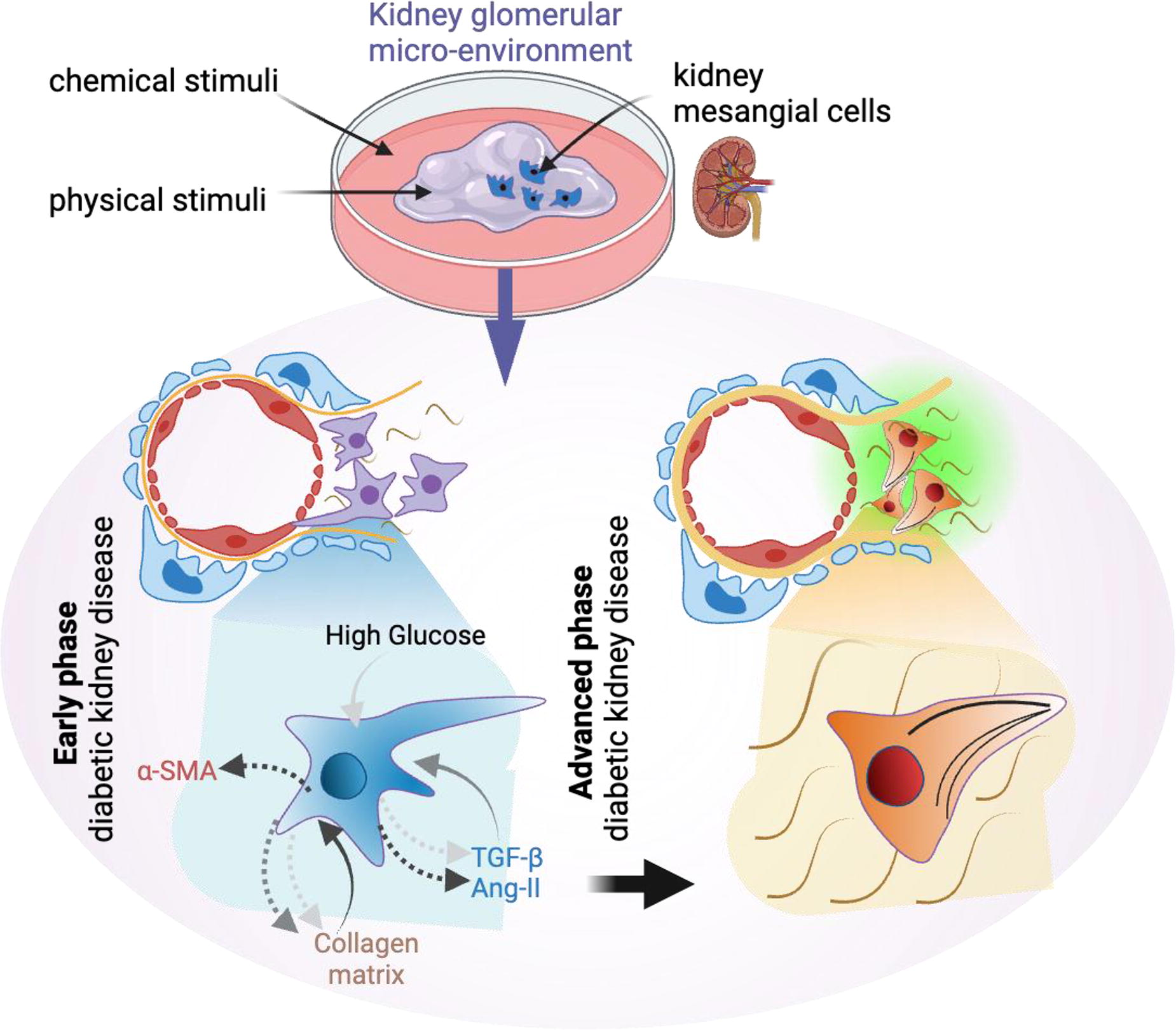
*Purpose/Objectives: The mesangium, comprised of mesangial cells and extracellular matrix, plays a vital role in maintaining glomerular structural integrity and facilitating renal filtration. Available disease models fall short in identifying cause-effect relationships in such multi-faceted and chronic diseases like diabetic kidney disease (DKD). The study primarily seeks to address the gap using a 3D hydrogel-based pathotypic model aimed at understanding such chronic diseases at defined progression stages. This study also delves into the relationship between chemical cues and matrix mechanics within the mesangial microenvironment during diabetic kidney disease (DKD).
*Methodology: The mesangial niche was emulated using photo-cured gelatin methacryloyl hydrogels, allowing us to recreate various disease stages. Disease conditions representative of healthy, short-term, and long-term DKD with fibrosis were achieved by modulating hydrogel stiffness and inflammatory cytokine TGF-beta. We evaluated the influence of matrix mechanics on mesangial cell processes, motility, and collagen IV expression, which could impact the renal basement membrane and, consequently, renal filtration efficiency.
*Results: Our findings matched the expected outcome in clinical samples. It also revealed the pivotal role of mechanical properties in shaping mesangial cell behavior. The matrix mechanical characteristics significantly influenced alpha−smooth muscle actin formation, impacting mesangial cell processes and motility. The fibrotic matrix, mimicking long-term DKD conditions, notably affected collagen IV expression, suggesting a potential thickening of the renal basement membrane and a subsequent reduction in renal filtration efficiency. A crucial revelation was the identification of a mechano-chemical dual input in late-stage DKD, contributing to an accelerated decline in glomerular function. This underscores the complexity of kidney diseases and emphasizes the need for a comprehensive understanding of both mechanical and chemical factors for effective in vitro disease models and therapeutic interventions.
*Conclusion/Significance: Our study provides valuable insights into the interplay of chemical cues and matrix mechanics within the mesangial microenvironment during DKD. By recognizing the significant influence of both factors, particularly in late-stage DKD, we contribute to a deeper understanding of glomerular function decline. This study advocates for the utility of in-vitro pathotypic models in advancing our knowledge of kidney diseases, guiding future research efforts to address existing gaps, and enhancing therapeutic strategies for late-stage kidney disease.
A30 - Three-dimensionally Printed Gelatin Methacryloyl (GelMA) Scaffolds For Culture Of Hematopoietic Stem And Progenitor Cells
N. S. Pereira, E. C. Mollocana-Lara, M. Kutrolli, E. N. McEwen, F. SaeediNejad, J. P. Quint, M. Samandari, A. Tamayol, V. Scanlon
UConn Health, Farmington, CT
*Purpose/Objectives: Intercellular crosstalk within the human bone marrow microenvironment plays a crucial role in regulating the hematopoietic response to systemic cues in vivo, however current in vitro models fall short of accurately mimicking the physiology of human bone marrow. Our research aims to develop a spatially defined in vitro human bone marrow model, incorporating a variety of cell types, cytokines, and extracellular matrices (ECMs) through 3D bioprinting. Bone marrow ECM is predominantly composed of collagen; however, collagen’s slow auto-polymerization rate limits its utility in bioprinting. Gelatin methacryloyl (GelMA), a collagen derivative, is a photocrosslinkable material that has been widely used in bioprinting to control the spatial organization of multiple cell types in different in vitro models. We hypothesize that utilizing GelMA as a bioink to print 3D multicellular structures will provide a more spatially controlled and physiologic in vitro human bone marrow model.
*Methodology: GelMA was synthesized, lyophilized, and reconstituted directly in media with cytokines to support the survival and proliferation of cells and their progeny in standard colony-forming unit (CFU) assays. Megakaryocytic-erythroid progenitors (MEPs) were isolated from CD34+ peripheral blood cells and used in the CFU assays. Mechanical properties of the GelMA were characterized through SEM, micro-indentation, and rheology. Chemical analysis was performed by NMR and Raman spectroscopy. The bio-ink was prepared using a thermal gelation process before being printed with our modified DexArm arm printer at the gelation temperatures found from rheology through a tapered 25-gauge tip.
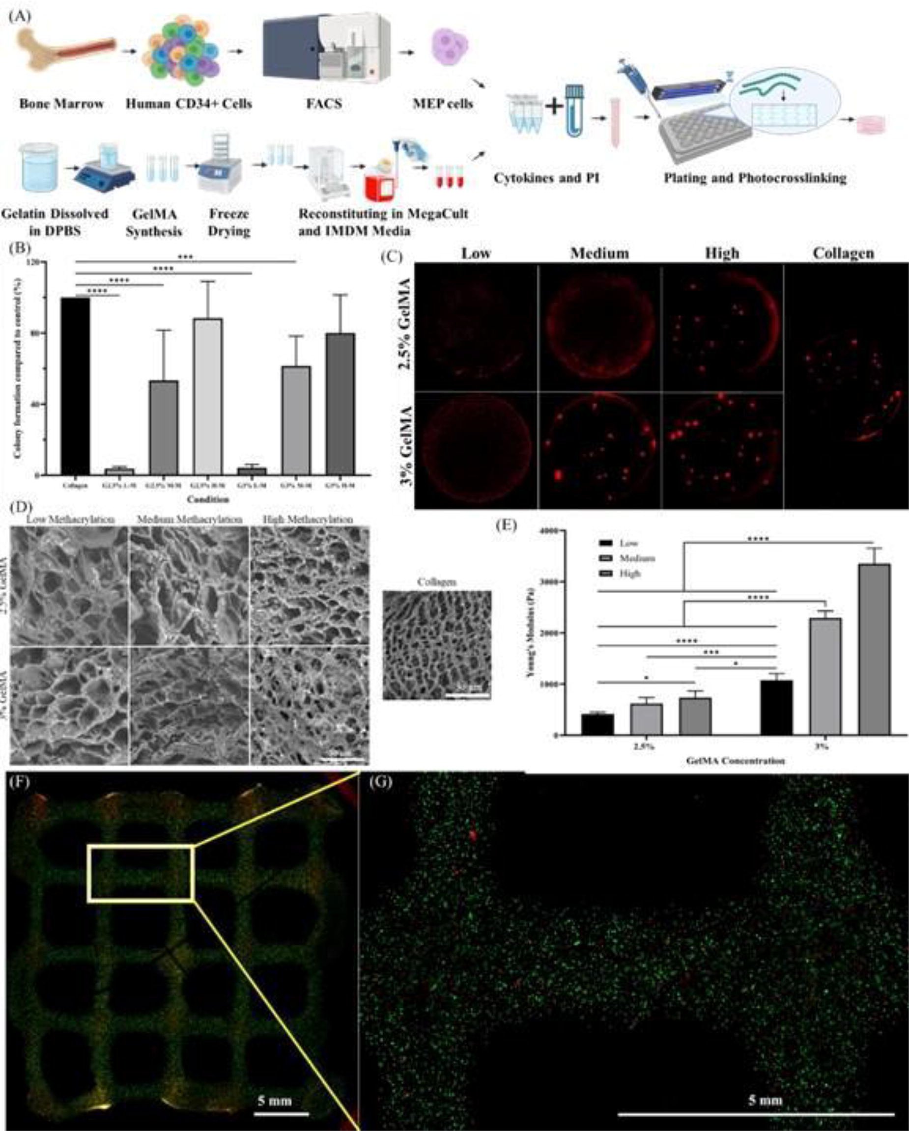
*Results: The impact of varying GelMA concentrations and photo-crosslinking durations on in vitro CFU assays of MEP was used to determine which constructs were the most favorable for the cells. We found that 10 seconds of crosslinking is insufficient to polymerize GelMA, however, 20 and 30-second UV crosslinking durations are sufficient to polymerize 3% and 2.5% GelMA, respectively. Both of these conditions successfully supported MEP colony formation, with the 2.5% GelMA concentration crosslinked for 30 seconds yielding the highest colony-forming efficiency compared to collagen-based media. Our experiments showed that GelMA with a high degree of methacrylation was more effective in supporting colony formation of both MEP and erythroid progenitor (ErP) cells than at low or medium methacrylation levels. SEM analysis shows that the GelMA high at 3% has pore sizes most similar to that of the collagen control. Furthermore, we observed high bioprintability of GelMA at the empirically optimized conditions.
*Conclusion/Significance: Importantly, we have demonstrated that several parameters of GelMA, including its concentration, crosslinking time, and degree of methacrylation, can be fine tuned to control various properties such as porosity, stiffness, cellular compatibility, and printability. These findings will inform future studies to optimize a micro-engineered in vitro model of human bone marrow. Such a model promises to significantly advance our understanding of the role of the bone marrow microenvironment in regulating the hematopoietic response to systemic cues in health and disease.
Figure 1: (A) Graphical Abstract, (B/C) Colony formation using ErP cells graph and visuals, (D) SEM of GelMA, (E) Young’s modulus, (F/G) Live/Dead assay of printed constructs with HEL cells.
A32
A33 - Improvement Of Spheroid Culture With “Pulsating Hydrogel”
S. ISHIMARU1, H. Nakamura1, M. Mukai2, S. Maruo2, N. Kojima1
1
Yokohama City University, Yokohama, Japan,
2
Yokohama National University, Yokohama, Japan
*Purpose/Objectives: In recent years, many studies have been conducted to create mini-organs (spheroids) by aggregating cells in vitro as a medical technology to restore the functions of lost organs. However, the diameter of spheroids is limited to several hundred µm due to diffusion restriction of nutrients and oxygen. If spheroids with a diameter over 1 mm (giant spheroids) can be created, they could be manipulated with tweezers in microsurgery. It is necessary for creating giant spheroids to develop a method of preventing internal cell necrosis. We have a technology to embed a hydrogel in the spheroid. If the embedded hydrogel can be pulsated, it might be possible to improve the diffusion of nutrients and oxygen. In this study, we aimed to investigate the possibility of suppressing internal cell death by using pulsating hydrogel as a pump.
*Methodology: In this study, we used the human hepatoma cell line Hep G2. “Pulsating hydrogel” was made mainly from N-isopropylacrylamide a temperature-responsive polymer. The culture medium was Dulbecco's modified Eagle's medium (DMEM) containing fetal bovine serum. Spheroids containing 500,000 cells, collagen, and a pulsating pump were created in a 3% methylcellulose medium. The volume change of the pulsating hydrogel was measured from microscope images. To pulsate the hydrogel, spheroids were incubated with a thermal cycler. Cell necrosis rate was calculated based on the amount of DNA.
*Results: The volume of the hydrogel increased by 20.6% with a temperature change from 37 °C to 30 °C. The same change rate was observed after 24 hours of pulsing with 3-minute cycle, indicating that the pulsating hydrogel was not degraded with continuous use. DNA quantity was measured after 24 hours incubation of spheroids in each condition. The results showed that the cell necrosis rate of the spheroids without the hydrogel was 41.3%. The cell necrosis rate was 33.9% in spheroids containing the hydrogel but without thermal change. Spheroid with the hydrogel pulsating exhibited 14.8% as the necrosis rate.
*Conclusion/Significance: The results indicate that pulsating hydrogel potentially suppress cell necrosis in spheroids. In the future, we will aim to establish a more stable culture method for larger spheroids by changing the shape of the pulsating hydrogel and forming channels inside of the spheroids.
A34 - Development Of A Type 1 Diabetic In Vitro And Ex Ovo Cam Disease Model Of Diabetic Wounds
M. P. Pérez González1, J. Hua2, F. Lali1, P. Sawadkar2
1
University College London, London, United Kingdom,
2
The Griffin Institute, London, United Kingdom
*Purpose/Objectives: Diabetic wounds (DW), a frequent and severe complication of diabetes mellitus (DM), exhibit distinct pathological features such as delayed cellular migration, reduced angiogenesis, and a prolonged inflammatory phase. These characteristics increase the risk of secondary infections and the potential for lower limb amputations, thereby escalating treatment costs and adversely affecting the patient's quality of life. Disease models for DW are crucial for assessing potential treatments and understanding its pathophysiology. Current in vitro disease models do not include all the aspects of DW and in vivo models do not comply with the 3R’s in research). Therefore to address this issue we have developed a 3D in vitro DW model that can recapitulate human complex pathophysiology while being sustainable are necessary for the progress of its treatments.
*Methodology: Human dermal fibroblasts (HDF) and keratinocytes (HaCaT) cultured in normoglycemic and hyperglycaemic media were used to create 3D skin-like scaffolds, with cell behaviour and morphological changes assessed. Parallel to this, Type 1 Diabetic Chicken Embryos (T1DCE) were develop using streptozotocin (STZ). Its diabetic phenotype was evaluated through immunofluorescent staining against pancreatic markers, PDX-1 and Neurogenin 3, and confirmed with the measurement of blood glucose levels. Thereafter developed normal and diabetic 3D skin-like scaffolds where placed over the chorioallantoic membrane (CAM) of healthy and induced T1DCE (CAM assay) to evaluate angiogenesis inside of the scaffolds as an effect of normo and hyperglycaemic conditions in the scaffold and in the chicken embryo.
*Results: Hyperglycaemic exposed HDF had a delayed migration when cultured in hyperglycaemic media. Both HDF and HaCaT showed an increased nuclear size in the 3D model. Immunofluorescent staining of chicken embryos treated with STZ showed an absence in fluorescence of pancreatic markers (PDX-1 and Neurogenin 3) resulting in the confirmation of the development of T1DCE. An increase in blood glucose levels was observed in T1DCE (7.6 ±0.33) when compared to control/healthy embryos (5.3±0.49). Angiogenesis was decreased in normoglycemic 3D scaffolds placed on CAM of T1DCE. This decrease was more evident in hyperglycaemic scaffolds placed on T1DCE suggesting that this effect is due to both the microenvironment of the scaffold and the systemic effects of high glucose in the chicken embryo.
*Conclusion/Significance: The combined model of 3D diabetic skin-like scaffolds and CAM assay of T1DCE has the potential to serve as a robust sustainable tool for future therapeutic testing since it replicates key aspects of a DW (in situ and systemic hyperglycaemia, delayed cell migration and decreased angiogenesis) by mimicking its in vivo environment in in vitro conditions. Because of this, our future research will focus on tailoring this model as an infection model to be treated with nanoparticles.
A35 - Optimizing Granular Hydrogel Composites For Vasculogenic Assembly
M. Hu, R. N. Kent III, F. Midekssa, G. Anyosa-Galvez, B. Baker
University of Michigan, Ann Arbor, MI
*Purpose/Objectives: Tissue vascularization remains a major challenge for the field of tissue engineering. While commonly used natural materials such as collagen or fibrin are conducive to vasculogenic assembly, these materials are not suitable for long-term tissue grafts as they are challenging to orthogonally tune physical parameters and degrade too quickly in vivo. Synthetic hydrogels are more readily modifiable to better suit tissue graft needs, but they typically slow or even inhibit vasculogenic assembly due to their nanoporous nature. Here, we explored the potential of granular hydrogel composites, a method of scaffold fabrication that incorporates microscale porosity orthogonal to hydrogel stiffness and degradability, for supporting vascular network formation.
*Methodology: 60 µm diameter dextran vinyl sulfone (DexVS) microgels were fabricated using massively parallelized microfluidic drop generators and functionalized with RGD to enable cell adhesion. DexVS microgels were suspended with human lung microvascular endothelial cells (LMVECs) (6 million/mL) in EGM2-MV or fibrinogen solution (5 mg/mL) to form microporous annealed particle (MAP) and granular hydrogel composite (GHC) gels, respectively. Samples were centrifuged at 500 G for 2 min. to pack microgels and LMVECs. Microgels were then annealed using poly(ethylene glycol) dithiol (15 mM) while fibrinogen was crosslinked with thrombin (1 U/mL). Samples were gelled at 37 ºC for 30 min., cysteine quenched for 30 min., then hydrated with EGM2-MV supplemented with fetal bovine serum (FBS; 5% v/v), VEGF (50 ng/mL), and phorbol 12-myristate 13-acetate (PMA; 25 ng/mL), with media replaced every 24 h for 3 days. Samples were assessed via nuclei staining, phalloidin staining, and immunostaining.
*Results: Our studies suggest that traditional MAP hydrogels do not support vascular network assembly. LMVECs in MAP gels adopted a monolayer-like morphology, while LMVECs in GHC gels formed cord-like structures like those seen in the fibrin positive control, indicating that GHC gels better support vasculogenic assembly as compared to MAP gels (Fig 1A, B). 3D cell morphometric analysis showed that while all gels contained similar cell densities, LMVECs in MAP gels had the largest average cell surface area, while LMVECs in GHC and fibrin gels had similar average cell surface areas (Fig. 1D). Monolayer-like morphology is typical for LMVECs interacting with 2D surfaces, indicating that LMVECs in MAP gels only interacted with microgel surfaces. In contrast, LMVECs in GHCs were encapsulated by fibrin, allowing for 3D matrix interactions and subsequent vascular assembly. We also observed lumenization within cord-like structures in the GHC gels, and podocalyxin staining confirmed proper apical-basal polarization of LMVECs making up said structures (Fig 1C, E).
*Conclusion/Significance: Taken together, these studies demonstrate the potential of GHC to support the formation of microvasculature. These findings will guide future research directions towards growing vascular networks within granular hydrogels.

A36 - 3D Bioprinted Vascular Model Via Coaxial Extrusion And Stem Cell Differentiation
S. Yogeshwaran, E. Dogan, D. Gendy, A. K. Miri
New Jersey Institute of Technology, Newark, NJ
*Purpose/Objectives: A blood vessel is organized into concentric layers with endothelial cells (ECs) organized in the core (Tunica Intima) and smooth muscle cells (SMCs) as the shell (Tunica Media). Some challenges with 3D bioprinted vascularized models have been the need for an abundant cell source and appropriate culture flow conditions. The major limitations observed in using vascular cells, such as umbilical vascular endothelial cells, are the quantity and proliferative potential of cells and their attachment to the inner wall. MSCs, as an alternate source, are among the most promising and suitable stem cell types for vascular tissue engineering. Physiological levels of shear stress have been known to promote the Endothelial differentiation of MSCs with alignment in the flow direction. Coaxial nozzle extrusion allows for the creation of a stiff wall for cell attachment. We have developed a novel 3D bioprinted double-layer vasculature model embedded in hydrogels.
*Methodology: Human bone-marrow-derived mesenchymal stem cells (hMSCs, Lonza Inc.) tagged with fluorescent particles were seeded at a high cell concentration (5-10 M cells/ml) in a two-layer tube in GelMA. The cell-laden structures were embedded in a gel construct and subjected to physiological levels of flow-induced shear stress (1-15 dyn/cm2) using appropriate cell culture media supplemented with VEGF (Lonza, 50ng/ml) for seven days. The effects of varying levels of flow-induced shear stress on MSC alignment and morphology were assessed using live/dead assay. We assessed the extent of endothelial differentiation by measuring specific biomarkers using immunostaining experiments and RT-PCR.
*Results: Collagen systems were effective in creating a lumen structure compared to other hydrogel systems, and combining the core-shell method using MSCs allowed for the creation of thick vascular linings. The MSCs were found to be more viable under flow conditions with elongation and alignment in the flow direction. Phenotype expression assessment was done for stem cell surface markers (CD29 and CD44) and endothelial markers, including platelet-endothelial cell adhesion molecule-1 (PECAM-1; CD31) and VE-cadherin (cell-cell junctions). The result showed a decrease in the stemness markers under flow, while an increase in the endothelial markers was observed after seven days of flow-induced shear stress.
*Conclusion/Significance: The optimization process involved getting a large cell-seeding density and cell adhesion in the collagen-GelMA core-shell structures. The effect of varying levels of shear stress within physiological ranges was studied. The results showed that the cells were adhesive and proliferative under flow conditions. The MSCs were aligned and elongated in the flow direction and were more viable than static conditions. The RT-PCR data for selected genes showed that the MSCs could express endothelial markers under flow conditions, with increased biomarker expression under appropriate physiological conditions. This novel method allows for the creation of vascularized structures by using MSCs as a single source for the different layers of the vessel. MSCs provide an inexpensive alternate cell source with possibilities for large-scale fabrication.
A38 - A Recombinant Collagen Derived, Thermally Non-gelling Alternative To Gelatine Methacrylate
J. Ebbecke1, D. Schlauch1, J. Meyer2, T. Fleischhammer2, L. Kloke1, A. Lavrentieva2, I. Pepelanova2
1
Cellbricks GmbH, Berlin, Germany,
2
Leibniz Universität Hannover, Hannover, Germany
*Purpose/Objectives: 3D-Bioprinting allows for the freeform fabrication of living tissue and therefore is a promising technique to be employed in regenerative medicine. For stereolithographic 3D-bioprinting, a liquid bioink is polymerised in a layer-wise process by site specific illumination to build up the desired construct. For this process, a variety of biomaterials has been modified by addition of functional groups to include the light reactive properties needed for this fabrication technique. Among these materials, one of the most widely used is gelatine methacrylate (GelMA), a modified partial hydrolysate of collagen, that supports cell attachment, is biodegradable and shows good biocompatibility and stability when used in vivo. However, as an animal derived material the use of GelMA is inevitably linked to safety concerns when used in medical applications. Furthermore, the temperature dependent gelation of this material complicates the applications in large scale stereolithographic bioprinting, since temperature induced solidification of the bioink can interfere with the printing process. To overcome this problem, a human collagen fragment resembling the collagen hydrolysate character of gelatine was recombinantly expressed in the methylotrophic yeast Koagataella phaffii. The recombinant expression does not only circumvent the safety concerns linked to animal derived materials, but additionally offers the possibility to tailor the material properties specific for the intended application. The material presented here was expressed without hydroxylation of proline, a common posttranslational modification of collagen and therefore also gelatine. As a result, the material did not form triplehelical structures as gelatine, and did not gel at ambient temperatures. However, despite the absence of triplehelical structures and hydroxiproline, the material did still support cell attachment and spreading. After functionalisation with methacryloyl groups, the material underwent rapid crosslinking and hydrogel formation following light exposure in presence of a photoinitiator and reached a stiffness comparable to GelMA. Under high strains, the material furthermore showed strain hardening behaviour comparable to GelMA based hydrogels.
*Methodology: x
*Results: x
*Conclusion/Significance: Concluding, a novel recombinant biomaterial that did not show temperature dependent gelation under bioprinting conditions but still exhibited desired mechanical properties as well as biological activity was developed. This material is a promising candidate to be used in future medical bioprinting applications to avoid risks linked to animal or human derived material.
A39
A43
A44
A45 - Studying The Role Of The ECM In The Development Of Different Colorectal Cancer Subtypes
F. J. Grundland Freile1,2, S. Campinoti2, L. Mikheeva1, B. Almeida2, C. Luni3, L. Urbani2, M. Holland1
1
Medical and Molecular Genetics, King’s College London, London, UNITED KINGDOM,
2
Roger Williams Institute of Hepatology, London, UNITED KINGDOM,
3
Dipartimento di Ingegneria Civile, Chimica, Ambientale e dei Materiali, University of Bologna, Bologna, ITALY
*Purpose/Objectives: Colorectal cancer (CRC) is the 2nd most common cause of cancer-related deaths worldwide. Survival rates have improved modestly despite recent advancements in pharmacological treatments. CRC can be classified into four phenotypes called consensus molecular subtypes (CMS 1-4). The CMS4 is associated with drug resistance, increased metastasis, lower survival, and extensive remodelling of the extracellular matrix (ECM) compared to the other subtypes. Changes in the matrisome are a defining feature in solid cancers and have been strongly implicated with disease progression and immune evasion. Yet, no study has focused of how changes in ECM composition in CMS4 CRC are potentially implicated in its increase malignancy.This project aims to investigate the role of the ECM in the aggressiveness of CMS4 CRC tumours using multi-omics analysis of bioengineered cancer models. We wish to identify ECM components that influence cancer cell gene expression and behaviour, with the potential to discover new therapeutic targets in the tumour microenvironment.
*Methodology: Using a publicly available data set of CRC cell line gene expression, we classified 55 CRC cell lines into their CMS subtype, choosing 4 CMS4 cell lines for our subsequent experiments (SW480, SW837, SW1417 & CACO-2). Additionally, we performed bulk-RNA sequencing on n=67 patient-derived CRC tissue samples and, applying a classifier, we obtained the patient respective CMS classification. We then performed mass spectroscopy on our CMS classified CRC samples to characterise the CMS4 matrisome composition using proteomic analysis. Based on the results obtained, we compared the growth of the CMS4 cells cultured as spheroids (3D culture without ECM) to cells grown in 3D in ECM-scaffolds. Cultures were characterised for their gene expression in comparison with 2D-cultured cells and the transcriptomic profile of the CRC patient-derived tissues.
*Results: From our proteomic analysis of different CMS CRC’s, collagens and other ECM proteins were enriched in the CMS4 tissue samples compared to the other CMS. The matrisome profile identified will be used to enrich ECM-scaffolds for CMS4-mimicking 3D cultures of CRC cells. The 3D ECM-scaffold (collagen-based) culture of our CMS4 cell lines demonstrated different cell invasive properties, such as increased expression of ECM-remodelling enzyme TG2, epithelial to mesenchymal transition marker (vimentin), and cell proliferation (ki67) compared to scaffold-free spheroid cultures.
*Conclusion/Significance: Overall, we have established a clinically relevant model of CMS4 CRC using subtype-specific cell lines and ECM-scaffolds to investigate the role of the ECM in driving aggressive phenotypes. The project we are currently studying how patient-derived colon organoids and CRC cell line interact with decellularised human CMS4 CRC tissue, to understand how the complex composition of the matrisome affects the cell behaviour for future functional and clinical investigations.
A46 - Evaluating Role Of Fibroblasts In Chemotherapy And Immunotherapy
S. Chakraborty, A. Skardal
The Ohio State University, Columbus, OH
*Purpose/Objectives: Fibroblasts are the most abundant stromal component in the tumor microenvironment and are crucial in tumorigenesis. However, these can be tumor-promoting or tumor-suppressing depending on the extent of cancer progression. The purpose of this piece is to study how fibroblasts behave in presence and absence of chemotherapeutic drugs and immune cells.
*Methodology: Cisplatin, 5-Fluorouracil and Regorafenib, in concentrations of 1, 10 and 100 uM were added to mildly metastatic Caco-2 (colorectal cancer) organoids, constructed using methcrylated collagen type-I and thiolated hyaluronic acid-based hydrogel, with and without human normal lung The organoids are photopolymerized in place with a short UV pulse. The drugs were removed the day after, and ATP -based viability assays as well as LIVE/DEAD imaging were done 1, 4 and 7 days after drug removal. In a separate set of studies, fibroblasts were added to A-375 (highly metastatic melanoma) organoids constructed using the same hydrogel and treated with 1uM small molecule kinase inhibitor drugs Alisertib (ALS) and Abemaciclib (ABE) for five days. NK-92 (natural killer) cells were added to the system on day 5 and similar imaging and viability assays as mentioned before were performed on day 6.
*Results: Greater viability was observed in Caco-2-HNLF organoids. However, in case of A-375-HNLF organoids, viability was greater in samples without drug. Also, IL-6 and IL-8 (fibroblast interaction in presence of immune cells) and SDF1-α (confirmatory action of fibroblasts) were present in greater quantities for drug treated samples.
*Conclusion/Significance: These initial studies suggest that fibroblast behavior is heavily affected by drug treatment, extent of metastasis and presence of immune components.
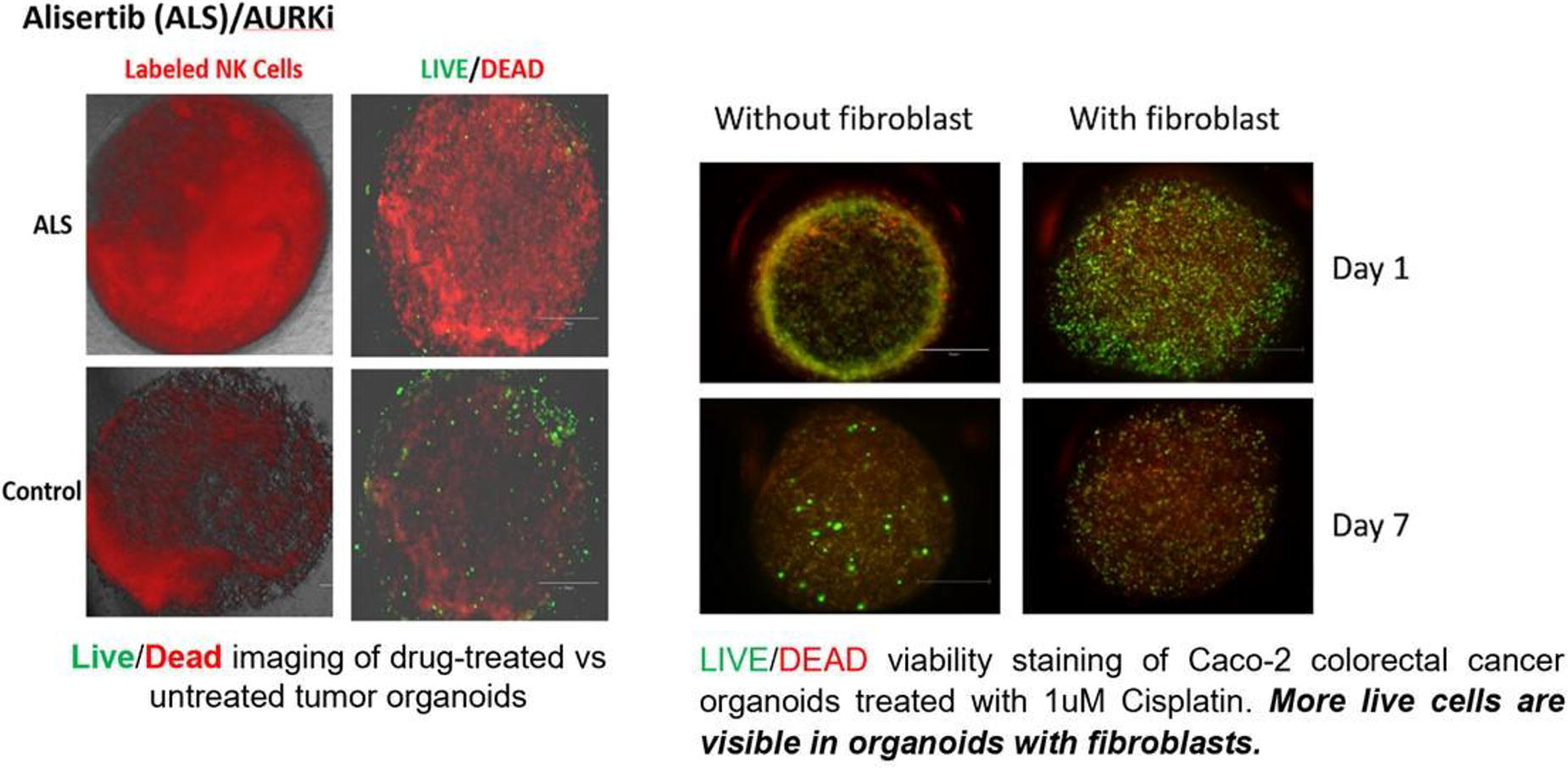
A47 - Development Skin Organoids As A Universal Platform For Skin Physiology, Injury, And Disease Modeling
G. Nomdedeu-Sancho, A. Gorkun, N. Mahajan, N. P. Edenhoffer, D. J. Gironda, A. E. Derby, A. Atala, S. Soker
Wake Forest University, Winston-Salem, NC
*Purpose/Objectives: Current in vitro human skin tissue equivalents are restricted by the inclusion of a limited number of skin cell types, failing to reproduce the cellular and structural complexity of the human skin. We overcame this hurdle by generating six cell-type self-organizing human skin organoids (hSOs) that recapitulate the microanatomy and function of the human skin. These organoids serve as a reliable model for skin development, physiology, chemical and radiation damage, and cancer.
*Methodology: The mixture of six key skin cell types in non-adhesive conditions led to the formation of hSOs, which were cultured for 21 days. Retinol and Dextran-FITC (4kDa) were used to test skin physiology and barrier integrity, respectively. hSOs were exposed to UVB (150mj/cm2) for radiation modeling, and chemical damage was examined by incubating hSOs with 1% Triton solution, Isopropanol, Hexyl Salicylate, and 5% KOH. To model skin cancer, SK-MEL-28 melanoma cells were included in the hSOs, and melanoma dermal invasion was examined through embedding the melanoma organoids (mSOs) in a dermis-like fibroblast-laden collagen gel.
*Results: hSOs maintained their skin-like layered microstructure for the 21 days of culture, including a cornified epidermal layer and a dermal-hypodermal core, and reproduced skin-like functions, such as the formation of an epidermal barrier, vasculogenesis, and active melanogenesis with a growing pigment accumulation. Moreover, they demonstrated a relevant physiological response to retinol by converting it to retinoic acid. UVB irradiation resulted in ER stress and apoptosis, and chemical irritant treatment produced a reaction relative to each compound’s irritation index. Melanoma cells within the hSOs proliferated and invaded the superficial layers of the epidermis, mimicking Pagetoid spread. When mSO were embedded in a dermis-like gel, melanoma cells invaded the gel and interacted with the surrounding fibrous gel, reproducing melanoma dermal invasion and allowing for the study of tumor-stroma interactions.
*Conclusion/Significance: We created a novel multicellular hSO model that recapitulates skin architecture and function in a high-throughput manner. This in vitro model could be used as a platform for dermatopathology and skin cancer research, expanding our knowledge of skin pathology, injury, and disease. Incorporating patient-derived melanoma cells in the mSOs would also improve personalized melanoma treatment.
A48 - Development Of Patient-derived Breast Organoids Capture Disease Heterogeneity: A Promising Tool For Personalized Medicine And Drug Development
N. Wajih, W. Jeong, M. Howard-McNatt, E. Levine, S. Lee, K. Votanopoulos, S. Soker
Wake Forest University School of Medicine, Winston-Salem, NC
*Purpose/Objectives: Breast cancer is one of the most frequently diagnosed cancers in women and is associated with a high mortality rate. More than 20 subtypes of breast cancer have been identified. Advancements in patient-derived organoid technology have made it possible to preserve cellular, structural, and tissue microenvironments that mimic the tissue in vivo.
*Methodology: We developed patient-derived breast organoids from normal and cancer biopsies to recapitulate the structure of breast tissue. Breast biopsies were enzymatically digested to obtain single-cell suspensions. Patient-derived organoids (PTOs) were fabricated by encapsulating unsorted breast cell population in a Collagen: Heprasil hydrogel. Normal and cancer PTOs were challenged with chemotherapeutic drugs to assess their viability. Additionally, unsorted breast cells were bio-printed on an alginate matrix in 96 well plates for high-throughput drug cytotoxicity analysis.
*Results: We successfully developed breast cancer PTOs in 13 individual patients and eight matched normal breast PTOs. Histological analyses of the breast PTO showed breast tissue characteristics with well-defined acini on H&E staining. Immunofluorescence staining showed the expression of breast cell-specific biomarkers, including Herceptin receptor 2 (HER2), progesterone receptor (PR), and Estrogen receptor (ER). Zona occludin 1 and 2 and keratin 19 expression in luminal cells and expression of Keratin 14 and P63 in basal cells suggested correct polarization in the organoids. On a pooled basis, paclitaxel inhibited PTOs viability by 26.6% (p<0.0051, n=13). Doxorubicin inhibited PTO viability by 64% (p<0.0001, n=13), cyclophosphamide inhibited viability by 39.5% (p<0.003, n=5), and the combination of paclitaxel and doxorubicin further inhibited cell viability by 80.25 % (p<0.0001, n=13) compared to the untreated control. A similar PTO viability inhibition pattern was observed in normal breast PTOs. Moreover, the histology of bio-printed breast PTOs mimics breast tissue characteristics. Printed cancer PTOs also responded differentially to chemotherapy drugs.
*Conclusion/Significance: Patient-derived normal and breast cancer PTOs and bio-printed PTOs recapitulated the histological features of breast tissue in culture and responded to chemotherapy. Moreover, high-throughput bio-printing of PTOs will accelerate the drug discovery and personalized medicine platform. Additionally, the use of normal breast PTOs could provide insight into the protection of normal cells from chemotherapy drugs, potentially improving therapeutic outcomes in breast cancer patients.
A50 - Effect Of Mechanical Micro-Environment On Growth Of Multilayered Cell Spheroid Of Cervical Cancer And Vascular Endothelial Cells
G. Koizumi, R. Akiyama, M. Miyamoto, S. Miyata
Keio University, Yokohama, Japan
*Purpose/Objectives: Growth and invasion of cancer tissue are closely related to a mechanical micro-environment. The mechanical environment, as well as the biochemical environment, affect the process of cancer cell migration, invasion, and metastasis. We hypothesized that the compressive stress caused by the exclusion of surrounding tissue could affect the proliferation and invasion process of tumor cells. In this study, we proposed an in vitro tumor model using multilayered spheroid culture to simulate the mechanical microenvironment and investigate the relationship between invasion process and mechanical environment.
*Methodology: Multilayered cell spheroids were constructed by cervical cancer cells and human vascular endothelial cells to simulate the earliest stage of tumor development. Single-layered spheroids formed by cancer cells were also generated to evaluate the interaction between cancer and endothelial cells. The spheroids were embedded and cultured in agarose gels with different concentrations to vary the stiffness of sorrounding gels.
*Results: The multilayered spheroids embedded in gels at higher concentrations showed peculiar growth after 4-5 day of culture whereas the single-layered spheroids showed after 8-9 day of culture. In addition, the multilayered spheroids showed the higher tissue permeability than that of single-layered spheroids. It was considered that the higher tissue permeability improved the exchange of nutrition and gas to promote cell proliferation and invasion.
*Conclusion/Significance: In conclusion, the mechanical microenvironment and the interaction of endothelial cells affected the growth and invasion of tumor cell spheroids.
A53 - Collagen-based Scaffolds As Primary And Bone Metastatic Models For Breast And Prostate Cancer
E. Sainsbury, L. Costard, N. Kamal, H. Ramanayake, C. M. Curtin
Royal College of Surgeons in Ireland, Dublin, Ireland
*Purpose/Objectives: Cancer cell culture in two dimensions (2D) has been employed to investigate intricate tumorigenic mechanisms, yet it lacks the necessary structural microenvironment for meaningful cell-cell and cell-matrix interactions. The adoption of three-dimensional (3D) cancer cell culture addresses these shortcomings, allowing cells to exhibit phenotypes and respond to stimuli in a manner more akin to in vivo biological systems. Additionally, gene therapy has emerged as a promising cancer treatment, leveraging the localized release of genes for a defined duration. Despite this potential, the effective delivery of genes to impede tumour growth remains a significant obstacle in its clinical application. This study aims to develop and characterise of 3D in vitro culture collagen-based scaffold models for cancer, specifically simulating the extra-cellular matrix (ECM) of primary breast and prostate cancer tumours by investigating the effect of hyaluronic acid and chondroitin sulfate, as well as secondary cancer bone metastasis using nanohydroxyapatite. The objective is to utilize these advanced models to assess the effectiveness of nanoparticle-mediated gene delivery, establishing them as competent anti-cancer platforms.
*Methodology: Triple-negative breast cancer (BC) cells (MDA-MB-231 and MDA-MB-436) and PC3 and LnCAP prostate cancer (PC) cell lines were grown on a range of collagen-hyaluronic acid and collagen chondroitin sulfate scaffolds to model breast and prostate primary tumours respectively and on collagen-nanohydroxyapatite (nHA) scaffolds to model bone metastasis. BC and PC cells on scaffolds were visualized using SEM. Cell growth on primary cancer scaffold models was assessed over 14 days and gene expression comparison was performed by qPCR to validate the models. microRNA (miRNA-146a-5p) and siRNA (siVEGF) delivery was performed using Lipofectamine RNAiMAX to inhibit BC cell growth in 2D and 3D. Uptake was confirmed by confocal imaging and functionality was confirmed by quantitative rtPCR. DNA and metabolic assays were performed to evaluate effects on proliferation and metabolic activity after nucleic acid delivery. The bone metastasis model enabled assessment of co-cultures of osteoblasts and cancer cells and analysis of the effect of cancer cells on osteoblasts in terms of growth, calcium deposition and cell migration.
*Results: All collagen-based scaffolds supported BC and PC cell growth. Significant changes were identified in gene expression comparing BC cells cultured on the two different scaffolds and in 2D. Nucleic acids were successfully delivered and significant inhibition of cell growth and vessel formation was measured. Furthermore, differences in the effects of miR-146a and VEGF manipulation in 2D compared to the two different scaffolds were demonstrated. Co-cultures in the bone metastasis model showed inhibition of osteoblast growth and calcium deposition and is under further investigation.
*Conclusion/Significance: This research focuses on characterizing collagen-based scaffolds designed to accurately replicate the ECM of primary BC and PC tumours, including bone metastasis. The study showcases the uptake and effectiveness of nucleic acid delivery to breast cancer cells cultured on these scaffolds, with observed correlations to in vivo effects documented in the literature. The findings underscore the appropriateness of these scaffolds for modelling the microenvironments of breast and prostate cancer tumours, providing a platform to assess the efficacy of gene therapy.
A56 - Development Of High-throughput In Vitro Breast Cancer Model: The Effect Of Basement Membrane Proteins On Caner Aggressiveness
W. Jeong1, D. Kim1, N. Wajih1, H.-W. Kang2, S. Soker1, S. Lee1
1
Wake Forest Institute for Regenerative Medicine, Wake Forest University School of Medicine, Winston Salem, NC,
2
Ulsan National Institute of Science and Technology, Ulsan, Korea, Republic of
*Purpose/Objectives: Triple-negative breast cancer (TNBC), a particularly aggressive subtype, exhibits distal metastasis in approximately 46% of cases. Hence, precise first-line chemotherapy treatment is crucial to mitigate invasive and metastatic tendencies. Patient-derived breast cancer organoids (PDBOs) have been extensively investigated for personalized drug screening, as they maintain patient-specific characteristics related to chemo-sensitivity and resistance. However, the process of core-needle biopsy and subsequent organoid culture leads to the loss of tumor-matrix interactions, impacting cancer aggressiveness as observed in the patient's origin. Therefore, there is a pressing need to develop an in vitro breast cancer model that accurately reflects varying levels of aggressiveness for personalized drug response assessments.
*Methodology: In this study, we established an in vitro breast cancer model by incorporating TNBC spheroids with the basement membrane (BM) proteins to mimic different degrees of cancer aggressiveness. The embedding-printing method enabled the co-localization of cancer spheroids with the BM proteins into an alginate-based matrix. To optimize the embedding-printing of TNBC spheroids, gelatin in the bioinks was adjusted for viscosity, and the circularity of printed spheroids was calculated. To evaluate cancer aggressiveness, MDA-MB-231 cell lines were printed by the range of BM concentration: 0, 10, 25, and 50%. The spheroids were printed into 96-well plates for high throughput screening of chemotherapy and migration assay. At last, patient-derived organoids were applied in the high throughput screening platform. The formation of the organoids was confirmed by immunostaining, and patient-specific response in chemotherapy was tested to predict patient treatment.
*Results: Consequently, the embedding-printing performed self-assembly of cancer spheroids in 3 days as calcium-containing cell bioink selectively crosslinked alginate-based embedding matrix. Optimization of viscosity in bioinks by gelatin improved printability in circularity (0.9) and stability with the lowest coefficient variation. The BM proteins enhanced nuclear polymorphism and the accumulation of extracellular matrix (ECM) in the 3D-printed breast cancer spheroids, as evidenced by histological analysis. Spheroids with 25% BM matrix exhibited a 1.21-fold higher proliferation rate, 1.37-fold improved drug resistance, and a 1.48-fold higher Ki-67 positive nuclear ratio compared to spheroids without the BM proteins. Patient-derived TNBC organoids were then 3D printed into a 96-well plate for high-throughput screening. Remarkably, the spheroids had positive expression in luminal cytokeratins, tight junction, and ECM markers. The spheroids with BM proteins demonstrated patient-similar drug response in drug sensitivity.
*Conclusion/Significance: We employed the embedding-printing platform technique to construct a 96-well plate-based in vitro cancer model incorporating BM proteins. This method allowed to modulate cancer aggressiveness, facilitating personalized drug response assessments. Our approach revealed the intricate interplay between the tumor microenvironment and patient responses, thereby contributing to the progression of precision medicine. In future studies, our strategy will concentrate on achieving spatial control over stromal tissue and cancer spheroids within well plates. This emphasis aims to enhance the physiological relevance between the cancer platform and patient responses. The utilization of this 3D bioprinting technique in fabricating in vitro cancer models positions it as a valuable high-throughput screening system applicable to various tissue-derived cancers.

A57
A58 - Investigating Cell Driven Fibrotic Remodelling Using 3D Renal In Vitro Models
A. R. Upadhyay1, A. Micalet1, D. Bakkalci1, E. Moeendarbary1, M. Butler2, M. Bergin2, U. Cheema1, M. Loizidou1
1
University College London, London, United Kingdom,
2
UCB Pharma, Slough, United Kingdom
*Purpose/Objectives: The dynamic biophysical microenvironment of the renal extracellular matrix is integral in regulating healthy organ function. Matrix remodelling is an iterative process, which can guide cell behaviour and pathological fibrosis occurs due to aberrant tissue repair. Fibrosis, characterised by the desmoplastic ECM deposition and tissue stiffening, is prominent in chronic kidney disease and renal carcinomas (Figure 1). We engineered biomimetic 3D constructs to recapitulate the matrix composition and mechanical fidelity of native renal tissues; investigating cell-matrix interactions in disease progression.
*Methodology: Collagen I [2 mg/ml] and laminin [50 µg/ml] constructs were engineered with renal stromal cells or cancer cells, at 2.5 ×10^5 seeding density. ECM protein density was increased by plastic compression of collagen hydrogels, for a final collagen density of 6% v/v. Cell types include cancer cell lines ACHN and 786-O, as well as diseased renal fibroblasts (TK188) and patient derived cells from chronic kidney disease.Mechanical properties were analysed using the Kinexus Pro+ Rheometer (Netzsch). Immersion-well geometry was used, testing at 1Hz frequency and 37°C. Storage modulus, phase angle, LVE region and stress-strain rate curves were analysed. Gene expression of 24 markers defining the healthy/diseased kidney matrix was determined using qPCR.
*Results: By day 21, 786-O cells remodelled and softened the collagen matrix from 544.8 Pa ± 46.8 to 355.8 Pa ± 29.9. Matrix degradation was confirmed by collagen-I gene downregulation and MMP7 and vimentin gene upregulation. ACHN cells remodelled and stiffened the dense matrix from 490.9 Pa ± 68.6 to 1246.3 Pa ± 140.6: with significant gene upregulation of collagen I, collagen IV and LOX.
The diseased fibroblasts soften the dense matrix over time (1.6 kPa ± 134.9 to 759 Pa ± 99) with upregulation of α-SMA and MMP9. However, co-cultures of 786-O and fibroblasts initially stiffen the collagen dense matrix to 2 kPa. On the other hand, fibrotic patient cells stiffened the collagen matrix over time (471.97 Pa ± 25.3 to 997.33 Pa ± 46.9). This is reinforced by enhanced gene expression of collagen IV, Twist and TG2.
*Conclusion/Significance: Renal cancer cells remodel their biophysical environment, altering the construct’s material properties. In our models, collagen density and stromal complexity influence cell behaviour, causing alterations in biomechanics and gene expression. These models generate physiologically relevant stiffness, which can be used to investigate the interactions between key cell types and the physical environment. Drugs targeting MMPs and TG2 will be used to determine if we can disrupt the fibrotic remodelling.

A59 - Designing A New 3D Osteosarcoma Scaffold-based Model With Enriched Cancer Stem Cells: Advancing Into Tumour Engineering
G. Bassi1,2, A. Rossi1,3, M. Saqawa1,3, E. Campodoni1, M. Sandri1, P. Sarogni4, V. Voliani5,6, S. Panseri7, M. Montesi1
1
National Research Council of Italy, Faenza, Italy,
2
Department of Neuroscience, Imaging and Clinical Science, University of Studies “G. D’Annunzio”,, Chieti, Italy,
3
Department of Chemical, Biological, Pharmaceutical and Environmental Sciences, University of Studies of Messina, Messina, Italy,
4
Center for Nanotechnology Innovation@NEST, Istituto Italiano Di Tecnologia, Pisa, Italy,
5
Center for Nanotechnology Innovation@NEST, Istituto Italiano Di Tecnologia, Pisa, Italy,
6
Department of Pharmacy, School of Medical and Pharmaceutical Sciences, University of Genoa, Genova, Italy,
7
National Research Council, Faenza, Italy
*Purpose/Objectives: Osteosarcoma (OS) remains a formidable challenge to treat despite the relentless efforts of medical clinicians and scientists. The malignancy of OS is due to several factors including the presence of a small percentage (0.05-1%) of Cancer Stem Cells (CSCs) endowed with self-renewal and pluripotency capabilities. CSCs reside within a complex tumour microenvironment (TME), niche, which is responsible for facilitating intricate crosstalk between CSCs and extracellular matrix, tumour and non-tumour cells. Conventional two-dimensional (2D) in vitro systems fail to test potential effective therapies for such a complex disease. Consequently, the scientific community is actively working to engineer three-dimensional (3D) in vitro models that more faithfully replicate OS, providing reliable platforms for oncological research and preclinical screenings. In this study, we designed, developed, and investigated 3D OS models composed of enriched CSCs and a hybrid hydroxyapatite-based scaffold (MgHA/CoII).
*Methodology: The optimization of the 3D OS models occurred in two stages: (1) serial generations of enriched CSCs as sarcospheres, and (2) the implementation of the niche within the scaffold via two injection-based approaches (Open and Close injection) for seeding the selected sarcospheres generation (G1) in a confined niche-like system. A comprehensive characterization in vitro and in vivo using Chorioallantoic Membranes (CAM) models, evaluating spheroidal phenotype, gene and protein expression, and tumorigenic properties was performed.
*Results: The 3D models demonstrated significant upregulation of stem and niche markers compared to scaffold-free sarcospheres, with a significant increase in the expression of OCT-4, SOX-2, and HIF-1α observed in the close injection approach. Immunohistochemistry analysis corroborated the gene expression data, and in vivo proof of concept experiments confirmed the tumorigenicity of the 3D OS models in CAM model.
*Conclusion/Significance: “Tumor Engineering in Osteosarcoma” represents a dynamic and promising field with immense potential to advance our understanding of tumour biology, enhance prognostic capabilities, and facilitate the discovery of effective therapeutic interventions.
A60 - Assessing The Effects Of Inhibitors Of Collagen Remodeling In An Ex-vivo Tumor Organoid Model
N. Edenhoffer, L. Agasthya Reddy, G. Nomdedeu Sancho, K. Votanopoulos, S. Soker
Wake Forest Graduate School of Arts and Sciences, Winston-Salem, NC
*Purpose/Objectives: Fibrosis alters the composition and mechanical properties of the extracellular matrix (ECM), facilitating tumor progression and metastasis. This complex remodeling process is regulated by a balance between collagen synthesis and degradation, mediated by key enzymes such as lysyl oxidase (LOX) and matrix metalloproteinases (MMPs) that represent potential therapeutic targets for the treatment of fibrotic tumors with potential to metastasize. In this study, we generated tumor organoids and assessed the impact of LOX and MMP inhibitors on fibrosis and ECM remodeling, providing insights on tumor-TME interactions and physical changes that may contribute to metastasis.
*Methodology: Tumor organoids were generated by mixing liver fibroblasts (LX-2) with collagen and culturing them with or without colorectal cancer spheroids (HCT-116). These were treated with LOX or MMP inhibitors (BAPN and marimastat, respectively) every 48 hours for 6 days. Organoids were harvested on days 1 and 6 for fibrosis analysis, through measurements of organoid contraction and collagen fiber properties.
*Results: Organoids formed with LX-2s alone increasingly remodeled the surrounding collagen, characterized by overall contraction of the organoid, a high degree of fiber bundling, and reduced fiber width. The presence of spheroids increased the rate of contraction and bundling marginally, but resulted in reduced levels of contraction compared to LX-2 only organoids regardless of inhibitor treatments. BAPN and marimastat were effective at reducing the effects of remodeling overall. Marimastat was more effective in preventing contraction of the organoids, especially in combination with spheroids, while BAPN was more effective in preventing collagen bundling. Of note, marimastat and spheroids together resulted in a similar prevention of bundling as BAPN groups, but without spheroids this effect was lost.
*Conclusion/Significance: Our tumor organoids modeled dynamic aspects of the TME which allowed us to determine the impact of LOX and MMP inhibition in ECM remodeling and the potential of using these inhibitors in the treatment of fibrotic tumors prone to metastasize. These findings highlight the complexity of enzyme interactions in ECM remodeling and points toward the need for targeted therapeutic strategies that account for the heterogeneity of cellular and extracellular components within the tumor microenvironment. By further modulating our model with diverse cancer cell and fibroblast types, including patient-derived cells, we expect to enhance our understanding in the effect of ECM dynamics in tumor progression, ultimately paving the way for more personalized and effective cancer treatments.
A61 - Metastasis-on-a-chip Modeling For Neuroendocrine Neoplasms
S. R. Anderson1, K. G. Nairon1, A. Nigam2, T. Khanal1, P. H. Dedhia1,2, M. D. Ringel1,1, A. Skardal1,2,1
1
The Ohio State University, Columbus, OH,
2
The Ohio State University and Arthur G. James Comprehensive Cancer Center, Columbus, OH
*Purpose/Objectives: The endocrine system is a key regulator of the biological processing that occurs within the human body as it involves the production and release of hormones that impact cell growth and development in several areas of the body. Dysfunction of the endocrine system—overproduction of hormones such as thyroid stimulating hormone (TSH) and steroids (e.g., aldosterone, cortisol, and androgens) is a notable hallmark of endocrine tumors and impacts tumor progression. Current models of endocrine tumors of both the thyroid and adrenal glands include 2D in-vitro systems and mouse systems that fail to accurately recapitulate cell-cell interactions and the immune tumor microenvironment. Previously we have developed 3D in-vitro tail vein inspired metastasis-on-a-chip (MOC) models that have proven to exhibit metastatic cell proliferation and variable treatment response with respect to both differentiated thyroid cancer (DTC) and adrenocortical carcinoma (ACC). Both cancers exhibit metastatic dormancy resulting in late detection and rapid progression upon treatment—emphasizing the need for a microphysically accurate neuroendocrine modeling system for biomarker detection and immune pathway investigation.
*Methodology: Updated MOCs that include primary tumor sites for metastasis kinetics measurement have been successfully used with both colorectal and DTC cell lines (FTC236 and hTh74). Validation of these models include selective engraftment favoring MOCs with primary tumors vs MOCs that have primary sites comprised of control cells (without genetic knock-down). Continued application of this MOC with ACC cell lines such as H295R will allow for further investigation of endocrine neoplasms and the metastatic cascade as well as immune environment changes. Metastasis kinetics have also been measured via confocal microscopy showing differences between control and DTC MOCs, and for ACC MOCs. Immune regulation via RCAN 1.4 pathway in DTC and via IGF2 and Wnt pathways in ACC are being queried.
*Results: We have previously showed that RCAN 1.4 loss increases NFE2L3 and IL-8 expression in 2D model systems and RCAN 1.4 loss facilitates T cell exhaustion in mouse models. In summary, we have investigated metastasis kinetics and immune regulation in MOC systems for DTC and ACC. In DTC, RCAN 1.4 loss significantly enhances metastasis kinetics. Confirmation of the role of IL-8 with the RCAN 1.4 loss was measured via ELISA, suggesting it is a key player in the metastatic cascade of DTC. Studies are ongoing in our ACC MOC, in which inhibition of IGF2 and Wnt separately or in combination modulate metastasis kinetics as well.
*Conclusion/Significance: Successful investigation and immune studies using both ACC and DTC MOCs could prove useful in developing a tumor modeling system that functions effectively for more/all endocrine neoplasms.
A62 - Bone-on-a-chip Models For Assessing Prostate Cancer Cell Extravasation
A. Mykuliak1,2, H. Wickramaarachchige1,2,3, A. Eskelinen4,1, K. Kuismanen2, A. Gebraad1,2,3, S. Miettinen1,2
1
Tampere University, Tampere, Finland,
2
Tampere University Hospital, Tampere, Finland,
3
University of Eastern Finland, Kuopio, Finland,
4
Coxa Hospital for Joint Replacement, Tampere, Finland
*Purpose/Objectives: Metastases are responsible for more than 90% of cancer-related deaths with bone being the predominant site of metastases in breast and prostate cancer. Metastasis is a multi-step process in which cancer cells first break away from the primary tumor, circulate in the blood stream and then exit the circulation to enter a secondary tissue. This last step involves the transmigration of the cancer cells through the vascular wall; a process known as extravasation. The tissue-specific microenvironment of the secondary organ (e.g., resident stromal cells and secreted chemokines) determines the extravasation sites and plays a significant role in metastasis progression. It is currently unknown what makes bone a preferred site of metastasis. The aim of the current study was to develop in vitro models of bone using human stem and progenitor cells and investigate the role of bone-specific microenvironment in the extravasation of prostate cancer cells.
*Methodology: We have emulated the extravasation process in vitro using two approaches: In the first approach a blood vessel is modelled by a monolayer of human umbilical vein endothelial cells (HUVECs) on the fibrin hydrogel interface in AIM biotech microfluidic chips. PC3 prostate cancer cells labelled with green fluorescent protein were introduced to the vessel channel. After 24 h, we assessed the number of extravasated PC3 cells that had crossed the endothelial monolayer as a percentage of the total number of PC3 cells in the chips. In an alternative approach a bone-specific microvasculature was formed inside the gel channel of the AIM biotech chips using a combination of human bone marrow mesenchymal stem/stromal cells (BMSCs) and HUVECs in fibrin. Next, PC3 cells were perfused through the microvascular network to study extravasation. A bone-specific microenvironment is made by incorporating bone cells (osteoblasts and osteoclasts) into our extravasation models.
*Results: A blood vessel was modelled by continuous endothelial monolayer formed at the media-hydrogel interface of a microfluidic chip. Formation of an intact monolayer was confirmed by Dextran permeability assay and immunofluorescent staining for VE-cadherin. We have differentiated bone-forming osteoblasts from BMSCs and embedded them in a fibrin gel. The cell showed transitional morphology with small cell body and long processes forming initial interconnections and positive staining for osteoblast marker collagen type I. We have differentiated bone-resorbing osteoclasts and macrophages from human blood monocytes and added them to the medium channel. Preliminary results showed that presence of osteoclasts increased the PC3 extravasation through the endothelial monolayer.
Perfusable bone-specific microvascular networks were formed within the microfluidic chip. PC3 prostate cancer cells were shown to extravasate through the microvascular network as early as 6 h. The cells adhered to the endothelium, extended protrusions, and eventually, invaded into the extravascular space to complete the process of extravasation.
*Conclusion/Significance: We will combine both osteoclasts and osteoblasts into our models to fully represent the bone microenvironment. We will use the models to study the molecular mechanisms that make bone a preferred site of metastasis. Our models could also be used to study anti-metastatic drugs targeting cancer cell extravasation.
A65
A66 - Hydrogel Beads As Artificial Erythrocytes For Ex-vivo Organ Perfusion: Rheology, Mechanical Properties And Hemocompatibility
G. Hentschel, T. Bode, S. M. Leal Marin, C. M. Winkler, M. Müller, B. Glasmacher
Leibniz University Hannover, 30823, Germany
*Purpose/Objectives: Genetically modified xenotransplants show promise in addressing organ shortages. To prepare organs for transplantation and assess compatibility, perfusion with human blood is vital. However, due to the limited availability of blood reserves, alternative solutions to conventional perfusion methods must be developed. To address this, artificial erythrocytes made from hydrogels and functionalised with human haemoglobin (Hb) have been developed. In addition, iron oxide nanoparticles (IONP) were integrated for sorting purposes. The aim is to replicate the natural erythrocytes in terms of shape, size, rheological and mechanical properties, as well as oxygen-binding capacities, to produce a consistent and reliable artificial blood substitute. The study evaluates rheological and mechanical behaviour of three types of artificial erythrocytes.
*Methodology: Poly-sodiumarcylate-co-arcylamide P(SA-Am) hydrogel microparticles (beads) were produced using microfluidic systems. Three types of beads were compared: (I) Pure P(SA-Am) beads, (II) IONP-integrated beads, and (III) Hb-functionalized P(SA-Am)/IONP-beads. To integrate IONP, nanoparticles were dispersed at a concentration of 10mg/ml into the monomer solution and sonicated for 3 min. To functionalize P(SA-Am)/IONP beads, they were incubated in a 2mg/mL Hb solution, cross-linked with 5vol.% glutaraldehyde, and subsequently incubated in another Hb solution. Rheological analysis was performed using plate-plate rheometry at shear rates ranging from 5 to 2000 1/s. Mechanical behavior was assessed using AFM with a 50nN load force. Hemocompatibility was investigated by conducting platelet deposition studies and determining hemolysis ratio.
*Results: The average diameter for all bead types was 350±23µm without significant differences between types. The bead surface did not change due to nanoparticle integration. Rheometric experiments confirmed shear thinning behavior for all types at moderate to high shear rates. Integration of IONP affected the viscosity and sedimentation behavior of the artificial erythrocytes, whereas additional Hb functionalization had no significant impact. Mechanical testing revealed an elastic modulus comparable to that of human erythrocytes for non-functionalized beads, whereas bead types with integrated IONP showed an increase in elastic modulus of about 200%. Platelet deposition experiments revealed limited morphological changes of the platelets over the investigated time period (60 min). No significant differences between bead types were detectable. Unfunctionalized beads induced 5,3±1,4% of contacting erythrocytes to hemolyze. Incorporation of IONP increased the hemolysis to 8,5±2,34%. Functionalization with Hb decreased the hemolysis ratio on the bead surface to 3,2±0,7%.
*Conclusion/Significance: The study shows that all bead types have consistent diameters. Ongoing experimental studies are investigating fabrication protocols to decrease particle diameter. Nanoparticles for sorting purposes were successfully integrated without affecting the bead surface. Rheometric experiments demonstrate shear thinning behavior for all bead types at moderate to high shear rates, providing promising initial results for mimicking physiological flow. Mechanical testing shows that non-functionalized beads exhibit a comparable elastic modulus to human erythrocytes. Further investigations are necessary to reduce the influence of IONP on the mechanical response. Platelet disposition and hemolysis studies demonstrate good short-term hemocompatibility, long-term experiments still need to be conducted. These findings provide valuable insights into the performance and biocompatibility of artificial erythrocytes for potential applications in ex-vivo organ perfusion.
A67 - Novel Perfusion System Mimicking Interstitial Flow Velocities For Large 3d Tissue Culture
E. Reid1, S. Brown1, A. Dzankovic1, A. Dijkstra2, T. Haines1, N. Hibino1
1
University of Chicago, Chicago, IL,
2
Radboud University, Arnhem, Netherlands
*Purpose/Objectives: While many studies using microfluidics devices show that tissues cultured in perfusion conditions have many positive effects on the molecular transport and extracellular matrix development of tissues grown in vitro, few studies attempt to develop perfusion bioreactors and systems that can maintain 3D tissue constructs at a scale similar to interstitial flow in vivo. While perfusing tissues with high flow velocities increases distribution of nutrients and removal of waste and applies more shear stress induced mechanical stimulation which is useful for developing vasculature, it cannot determine how tissues will maintain their viabilities when grafted into the native tissue’s environment. A tissue culturing system with flow velocities similar to those found in vivo is necessary to develop and test the viabilities of prototypical tissue grafts for clinical use. To address this problem, this study develops and verifies the utility of a novel perfusion system designed to maintain tissue patches up to 1cm3 in flow velocities below 10um/s, similar to interstitial flow.
*Methodology: We designed and determined the approximate flow velocities through the tissue samples of 5 perfusion system configurations. Perfusion system configurations 1-3 consisted of a fluid reservoir, roller pump, and bubble trap, connected in series by medical grade silicon tubing with one of our three custom bioreactor prototypes designed to house a 1cm3 tissue sample. Configurations 4 and 5 divided the flow into a parallel channel just before the bioreactor. The experimental set up’s conditions emulated those of the closed perfusion system by submerging the tubing from the bioreactor outlet in a partially filled graduated cylinder and vacuuming the accumulating fluid into separate storage containers. In order to determine the flow velocities around the tissue sample, we used PBS to measure each configuration’s volumetric flow rate at 0.1, 0.2, 0.3, 0.5, and 1 RPM (n=3) and divided by the bioreactor’s cross-sectional area.
*Results: All perfusion systems showed volumetric flow rates that are linearly proportional to their RPM setting as predicted by roller pump induced flow properties. Out of the five perfusion system configurations, 1 and 5 achieved minimum flow velocities below 10 um/s;at the roller pump’s lowest setting (0.1 RPM) which is required to accurately model interstitial flow. The single channel configurations 1-3 produced minimum flow velocities of 8.8, 24.0, and 21.7 um/s while the parallel channel configurations 4 and 5;produced minimum flow velocities of 10.2 and 9.5um/s respectively.
*Conclusion/Significance: Out of the;5 perfusion system configurations we crated,;configurations 1 and 5 successfully achieved minimum flow velocities below our desired threshold. Achieving flow velocities <10um/s justifies the utility of our perfusion system for effective large tissue construct culture and vascularization experimentation. Tissues cultured in these conditions will reap the benefits of shear stress induced mechanical stimulation and improved oxygen and biomolecular transport, while being cultured in an environment that mimics the conditions of the native tissues it will be grafted into.

A69 - Matrix Biophysical Cues Influence Cardiac Reprogramming
A. J. McCoy, A. J. Putnam
University of Michigan, Ann Arbor, MI
*Purpose/Objectives: Heart disease is the leading cause of death in the United States. Myocardial infarctions (MI) occur when blood flow to the heart is blocked, resulting in the death of cardiomyocytes (CM) and a reduction in pumping capacity. Because adult CMs have a very low proliferative potential, tissue engineering (TE) strategies, including the reprogramming of resident cardiac fibroblasts into induced CM (iCM), have been explored as an auspicious solution to fully restore post-MI heart function.
Established cardiac reprogramming protocols typically involve cells cultured on tissue-culture plastic or Matrigel substrates, but such protocols suffer from low reprogramming efficiencies and poor iCM maturation. Previous research has shown reprogramming efficiency and iCM maturation are enhanced in vivo and in vitro in naturally derived, cardiac-like 3D hydrogels, suggesting that the microenvironment provides instruction cues. However, the effects of specific 3D ECM cues on reprogramming efficiencies remain poorly understood.
Poly(ethylene glycol) (PEG) hydrogels are a useful tool for probing the effects of the microenvironment on cells in 3D in part because their inert backbone can be functionalized with peptides and proteins and their biophysical properties can be readily tuned. We hypothesize that 3D PEG hydrogels designed to mimic the cardiac microenvironment will improve cardiac reprogramming efficiency and iCM maturation and elucidate the influence of specific ECM cues on cardiac reprogramming.
*Methodology: Mouse embryonic fibroblasts (MEF) were reprogrammed to iCM following an established protocol utilizing temporal expression of the Yamanaka Factors (“OSKM”) via a tetOn promoter. Briefly, MEFs were transduced with lentiviruses containing tetO-FUW-OSKM and FUW-M2rtTA in the presence of polybrene. MEFs were then encapsulated in 3, 5, or 10 wt.% 8-arm PEG-norbornene hydrogels (1 mM RGD, 1 mM LAP, 90% of available norbornenes post-RGD addition crosslinked with VPMS) or plated on 1:50 2D Matrigel. During the first nine days of culture, MEFs were dedifferentiated through expression of OSKM and a medium conducive to reprogramming. After dedifferentiating, cells were given a chemically defined medium containing bone morphogenetic protein 4 (BMP4), which drives them to an iCM phenotype. Cells were analyzed for beating colonies before being fixed and immunostained with antibodies for alpha-actinin and cardiac troponin I (cTnI).
*Results: MEF were successfully reprogrammed into iCM in 3D PEG hydrogels. The reprogrammed cells spontaneously beat and stained positive for alpha-actinin and cTnI, two CM markers (Fig. 1A). To improve low reprogramming efficiencies, we optimized cell density, transducing virus concentrations, and polybrene concentrations, and observed a marked increase in Oct4 positive colonies after 6 and 9 days of culture (Fig. 1B+C). Our results suggest that softer and more degradable matrices support greater numbers of beating and non-beating colonies after 21 days, indicative of enhanced reprogramming efficiencies.
*Conclusion/Significance: Utilizing cell-adhesive and protease-susceptible PEG hydrogels, we have identified specific biophysical properties that enhance cardiac reprogramming and iCM maturation in vitro. Future work will include probing different integrin binding domains and differences in crosslinking density. These biophysical cues may have broader applicability to cardiac tissue engineering and iPSC generation, which also utilizes OSKM expression to drive cellular phenotype.
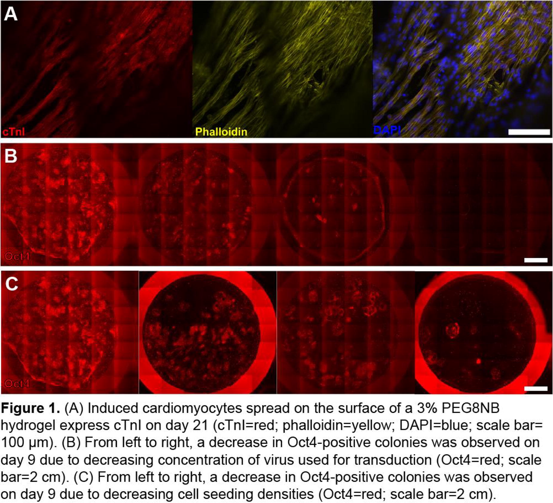
A70 - A Biomimetic Leaf Model To Investigate Endothelialization Of Engineered Constructs
T. Enderli1, E. Scarpignato1, Z. Lorton1, P. Gatenholm2, C. A. Bashur1,1
1
Florida Institute of Technology, Melbourne, FL,
2
CellHEAL AS, Sandvika, Norway
*Purpose/Objectives: Graft failure due to inadequate vascularization is a primary challenge for tissue engineering. One potential strategy involves 3D bioprinting with template channels for vascular networks, but more research is needed on emerging bioink materials and cellularization strategies. Sodium alginate and nanocellulose bioinks have promise for 3D bioprinted constructs. In this study, we are developing a new model system to investigate vascularization strategies. We are casting alginate/nanocellulose hydrogels in a biomimetic 3D printed leaf-structure mold, with a central channel and side channels branching off in a pinnate geometry, to serve as a vascular perfusion system. This approach is advantageous to single channel methods since the multiple channels subject endothelial cells (ECs) to different levels of shear stress simultaneously. This study develops a biomimetic leaf-inspired model and uses analytic and experimental methods to evaluate the effect of shear stress within complex channels on endothelialization.
*Methodology: Different nanocellulose (Ocean Tunicell) /alginate (NovaMatrix) ratios were used, and the cast samples were crosslinked with 100mM CaCl2. Mechanical properties were characterized. Leaf-inspired channels were treated with 0.05mM sodium periodate and laminin to promote cell adhesion, and then samples were sealed in custom 3D-printed perfusion chambers and stored in 5.8mM calcium chloride to equilibrate to physiological conditions. A needle was attached to the leaf mold central opening to prevent backflow and perfuse the channels. Next, the channels were seeded with human umbilical vein ECs (HUVECs) at 3x10⁶ cells/mL. After cell attachment, further media perfusion was provided with peristaltic pumps. A Comsol® finite element model was used to characterize the flow rates and shear stresses at individual locations within the samples. Cell attachment and viability is also characterized at specific locations using confocal microscopy. Significance was determined with one-way ANOVA and Tukey.
*Results: Compression testing and rheology indicated that the 80:20 ratio of nanocellulose/alginate had ideal mechanical properties with shear thinning properties, toughness, and the highest modulus (n=6), and was used for further studies. The perfusion chamber design keeps samples submerged in CaCl2-supplemented media to prevent desiccation and degradation, and excess media from all of the channels is siphoned into a collection chamber for recirculation. This allowed for either continuous or intermittent perfusion of channels. Another advantage of the perfusion chamber design is that samples can be imaged without removal from the chamber which allow non-destructive time studies on the same sample. Imaging of channels after incubation showed good cell adhesion within the channels but not on control surfaces, indicating that surface modification is necessary. Ongoing work includes characterizing HUVEC attachment and viability after 1, 4, and 7 days of culture and correlating to shear stress at that location.
*Conclusion/Significance: Nanocellulose/alginate inks provide constructs that are robust and easy to handle unlike many other bioinks. When this material is formed into construct with biomimetic leaf-inspired channels and perfused, this offers an efficient way to test and optimize different parameters (e.g., shear stress) for their impact on tissue vascularization. Overall, this system both saves material and shows a direct comparative effect of different shear stress levels on ECs.
A71 - Investigating Endothelial Damage During Ischemic Stroke Treatment Using In-vitro And Finite-Element Modeling
Z. W. Lorton, A. Regina Martinez Dehesa, C. A. Bashur,1, V. Chivukula
Florida Institute of Technology, Melbourne, FL
*Purpose/Objectives: Large vessel occlusion during acute ischemic strokes requires mechanical thrombectomy (MT) procedures using stent retrievers (SR), where the clot becomes entrapped within the stent struts, allowing the withdrawal of both the stent and the clot. However, complete clot removal typically takes several attempts, which can amplify device-related endothelial cell injury (EI). EI can decrease the chance of successful MT reperfusion and potentially induce complications such as vasospasm. This project aims to characterize the extent of EI during MT using an in vitro vascular platform and a finite-element model (FEM). Impact of SR diameter and detailed characterization of SR mechanical forces are assessed and correlated with human umbilical vein endothelial cell (HUVEC) response.
*Methodology: HUVECs were seeded on fibronectin (4 µg/cm2) coated true-scale PDMS arterial phantoms with 2.5mm luminal diameter. Cells were cultured for four days to improve luminal cell coverage. Test groups (n=6) either underwent a simulated MT using two different-sized clinical SRs (Trevo® XP PROVUE 3 × 20mm and 6 × 25mm) or no MT (control). Afterwards, the phantoms were stained for fluorescence (DAPI and phalloidin) and imaged with SEM to evaluate cell viability and function. The radial force applied by the SRs and the uniaxial shear stress during MT were characterized. Finally, a FEM characterizing the force distribution over time is being developed for both SR sizes.
*Results: Fluorescent and SEM images showed disruption in the functional HUVEC layer and significant decreases in cell confluency and area coverage after SR testing. SEM images also suggested that SRs can physically damage the PDMS surface. Further, there was noticeable variability in cell coverage along the path of the stent. Surprisingly, there were no significant differences in cell coverage between small and large SRs. The large and small SRs applied 220±12.3 and 16.7±5.78 mN forces, respectively, on a testing system with a 2.5mm gap. Shear forces applied when removing the SRs were also significantly higher for the large (87.8mN) compared to the small (13.7mN) SRs. Interestingly, there were large temporal variations in forces over SR removal, especially for the large SR, suggesting that struts may stick in the lumen at times before progressing forward. This may help to explain the high variability in cells in areas that were damaged. FEM results also indicate heterogeneous spatial and temporal shear stress distribution over the luminal area, with regions in direct contact with the SR demonstrating peak stresses.
*Conclusion/Significance: An in vitro model was successfully used to identify EI caused by both SR devices. While differences in the extent of damage with SR size were unclear, the large SR applied more radial and shear force on the arterial phantom wall. It is possible that a shear force threshold for cell damage has been reached for both sized SRs, preventing differences from being detected. Better understanding the details and risks of EI after MT has the potential to improve the long-term success of MT surgeries. Finally, further experimental and FEM investigation is needed into how SRs move along a vessel wall both with and without blood clots.
A72 - Evaluation Of Oxygen-binding Capacity And Magnetic Extraction Of Hydrogel-based Artificial Erythrocytes
T. Bode, G. Hentschel, S. Leal Marin, C. Winkler, M. Müller, B. Glasmacher
Leibniz University Hannover, Hannover, Germany
*Purpose/Objectives: Genetically modified xenotransplants may offer a solution to the shortage of donor organs. To prepare the organs for transplantation and assess compatibility, perfusion with human blood is required. However, due to the shortage of blood reserves, it is crucial to develop alternatives. To address this issue, human hemoglobin (Hb) functionalized artificial erythrocytes made from hydrogels were developed, aiming to replicate red blood cell in terms of shape, size, rheological and mechanical properties as well as their oxygen-binding capacities. This study evaluates the artificial erythrocytes' oxygen-binding capacity and their possible extraction from a vascular system using magnetic nanoparticles guided by magnetic fields.
*Methodology: Poly-sodium acrylate-co-acrylamide (P(SA-Am)) hydrogel beads were functionalized with Hb by incubating them with a 2 mg/mL Hb solution for 4 hours on a shaker plate, followed by cross-linking using a 5% glutaraldehyde solution. To further enhance functionalization, the beads were incubated with a 2 mg/mL Hb solution for 1 hour. After washing the beads three times with PBS, they were incubated in PBS for 7 days. Subsequent experiments were conducted after washing the particles with PBS again. Iron oxide nanoparticles (IONP) were mixed with the hydrogel before bead formation to encapsulate them. Microscopy was used to measure the average bead diameter. (Cryo-)SEM was used to evaluate changes in surface morphology and topography. The surface coating composition was analyzed using EDX and FTIR spectroscopy. Oxygen binding was qualitatively assessed through chemiluminescence using a Luminol assay.
*Results: The measured beads' average diameter was 2473±48 µm (non-functionalized) and 2689±32 µm (IONP-functionalized). Functionalization with Hb caused a color change from clear to a red-brown tint, while the addition of IONP resulted in a dark brown color. FTIR measurements of the Hb-functionalized beads, compared to non-functionalized beads and pure Hb, showed the presence of Hb on the beads' surface. IONP spectra were observed in the spectra of IONP-functionalized beads. SEM measurements revealed dark spots in the IONP-beads, which EDX confirmed to be IONP. Cryo-SEM, in combination with EDX, showed iron deposits on the surface of the Hb-functionalized beads. The surface morphology of both functionalized beads did not show significant changes compared to non-functionalized beads. Hb functionalization resulted in chemiluminescence upon addition of luminol. The intensity decreased after storage in PBS for several days. The IONP-beads could be guided out of a mock vessel using a magnet.
*Conclusion/Significance: The study shows that hydrogel beads have been successfully functionalized with human Hb, making them potential candidates for artificial erythrocytes. The observed changes in color and chemiluminescence suggest that the artificial erythrocytes contain Hb and may have the capacity to bind oxygen like natural red blood cells. However, a quantitative evaluation of the oxygen-carrying capacity and release kinetics is still necessary. The decrease in chemiluminescence intensity over time raises concerns about the stability and long-term performance of the artificial erythrocytes. Further work will address their long-term behavior, potential degradation, and stability under various physiological conditions. It is also necessary to investigate their interactions with biological systems, including possible immunogenic responses and toxicity concerns.
A73 - Development Of An Animal Component-free, Chemically Defined Human Umbilical Vein Endothelial Cell Culture Media And Adaptation Method
L. Brunmaier, T. Walker
South Dakota School of Mines & Technology, Rapid City, SD
*Purpose/Objectives: The development of defined cell culture media presents a host of advantages including, reduction of disease transmission, facilitating translational research, and providing fine tuned bioassays. Fetal bovine serum (FBS) has a nearly universal use in cell culture, but presents significant drawbacks such as, batch-to-batch variability, risk of disease transmission and ethical concerns. As interest continues to grow around the development of biomimetic in vitro assays and translational biomedical research, so does the need for full identification of cell media components and stimulating factors. Here we present our animal free (AF) media formulations and our human umbilical vein endothelial cell (HUVEC) adaptation procedures. Our goal in this study is to identify the simplest media formulation capable of sustaining viable HUVEC growth while finding the most efficient route to adapting the cells to serum free conditions in an effort to preserve numerous fully serum free passages for experimentation.
*Methodology: Testing parameters include two different adaptation methods used to introduce HUVECs to AF media. Direct adaptation exposes the cells to 100% AF media after passaging, where gradual adaptation slowly introduces the AF media in defined proportions over time. At low FBS concentrations, cell attachment can become compromised. To combat this issue, we also tested our various attachment protein coatings in the cell culture vessel.
*Results: We found that cells growing in high AF media percentages, using attachment proteins of human origin to coat the surface of the culture vessel sustained cell attachment and growth comparable to our control HUVECs growing in standard low serum media. We obtained fully serum free, animal free conditions with one media formulation, and narrowed our surface combinations coatings to a single protein coating.
*Conclusion/Significance: This work outlines the methodologies and reasoning for implementing specific steps during the adaptation process that can be used for HUVECs and other human cell lines, and can motivate quantitative, tunable bioassays to accelerate biomedical research and translation.
A76 - Engineering Conductive Vascular Grafts With S-PEDOT For Next Generation Vascular Prostheses
T. K. Brown, R. Daso, J. Tropp, C. L. Chao, C. Dang, B. Jiang, J. Rivnay
Northwestern University, Chicago, IL
*Purpose/Objectives: Cardiovascular diseases are a leading cause of mortality and morbidity worldwide, often necessitating the replacement of damaged blood vessels with prosthetic vascular grafts. Conventional vascular grafts suffer from limitations such as poor long-term patency and the inability to provide real-time information about graft performance. This project aims to develop a novel generation of vascular grafts that are not only biocompatible but also electrically conductive. These bioelectronic vascular grafts will pave the way for smart vascular prostheses with built-in bio-sensing capabilities, addressing the shortcomings of current vascular graft technologies.
*Methodology: Highly soluble, self-doped conductive polymer sulfonated poly(3,4-ethylenedioxythiophene) (S-PEDOT) was synthesized via oxidative polymerization. Rat aortas were decellularized and subsequently incubated in varying concentrations of S-PEDOT or S-EDOT at 37°C. After incubation or polymerization, all aortas underwent a 24-hour leaching step at room temperature to remove unbound material. Conductivity of the modified aortas were measured via 4-point probe. To evaluate in vitro biocompatibility of S-PEDOT incorporation, human vascular endothelial cells (HUVECs) and aortic smooth muscle cells (HAoSMCs) were cultured on collagen hydrogels incubated with S-PEDOT. Morphological assessment with phase contrast microscopy and resazurin viability assays were performed after 48 hours. Thrombogenicity was assessed with a whole blood coagulation assay. To assess biocompatibility in vivo, S-PEDOT-incorporated aorta sections were implanted subcutaneously in Sprague-Dawley rats for two weeks. Masson’s trichrome and immunofluorescence staining for macrophages (CD68+ cells) were performed to assess inflammation. Additional immunofluorescence staining for vascular and immune cells is currently underway.
*Results: The final molecular structure of S-PEDOT demonstrates a sulfonate group for water solubility (blue dashed box) and a methyl group for improved stability (green solid box) (A). Modified grafts demonstrated thorough monomer and polymer penetration as seen by the change in color through (dashed circle) and along the length of (solid box) the vascular wall (B). Conductivity was increased by an order of magnitude between aortas incubated in 10- and 100-mM S-EDOT before polymerization (C). Vascular cells demonstrated some rounding (D), but overall maintained viability on S-PEDOT-modified collagen over 48 hours (E). Collagen gels with S-PEDOT performed similarly to unmodified collagen gels, both demonstrating higher thrombogenicity than tissue culture plastic, as expected (F). No evidence of infection, seroma, or delayed wound healing was observed in subcutaneous implantation studies, though nuclear infiltration was noted to be qualitatively elevated (G).
*Conclusion/Significance: Conductive vascular grafts can be generated by coating decellularized aortas with S-PEDOT via in situ polymerization or incubation in previously polymerized S-PEDOT. Aortas incubated in 100 mM S-EDOT and polymerized demonstrate improved conductivity. S-PEDOT-modified extracellular matrix materials demonstrate hemocompatibility and biocompatibility both in vitro and in vivo.

A77 - Insulin Treatment Prevents Deterioration Of PCL Graft In A Diabetic Rat Model
K. Oyama1, Y. Kikuchi1, T. Yoshida2, D. Naruse3, S. Kunioka1, H. Kamiya1
1
Asahikawa Medical University, Asahikawa, JAPAN,
2
Life Materials Development Section, Toyama Industrial Technology Research and Development Center, Human Life Technology Research Institute, Nanto, JAPAN,
3
Business Development and Quality Control Department, Iaazaj Holdings Co., Ltd, Nanto, JAPAN.
*Purpose/Objectives: An artificial vascular graft with small diameter less than 4 mm is in high demand, however, it is not clinically available for revascularization treatment yet. Biodegradable nanofiber such as poly-caprolactone (PCL) is one of the most studied materials in this study area, and PCL based-artificial vascular graft (PCL graft) has shown promising results in “healthy” animal model. Although patients undergo revascularization therapy usually have some complications such as diabetes mellitus, studies to test the efficacy of PCL graft in “pathological condition” are limited. Therefore, in this study, we aimed to characterize the performance of PCL graft under diabetic conditions.
*Methodology: PCL grafts with 1 mm inner diameter were created using an electrospinning method and added hydrophilicity by coating with polyvinyl alcohol (PMID 36035951). PCL grafts were implanted into abdominal aorta of Wister rats and tested their performance. Diabetic rat model (DM rats) was created by administration of streptozotocin (STZ) via tail vain injection. To evaluate the effect of controlling pathologic status on PCL grafts, an insulin-treated DM rat model (tDM rats) was created by daily administration of neutral protamine Hagedorn insulin on DM rats. Immunohistological staining was performed to evaluate the intima regeneration.
*Results: In healthy rats, PCL grafts showed 100 % (7/7 rats) and 62.5 % (5/8 rats) patency rate at 2 and 6 weeks after implantation, respectively. CD31 positive and alpha-smooth muscle (αSM) positive intima layers on inner lumen of PCL grafts were observed in all the patent cases with 14.7% and 100% of intima coverage rate at 2 and 6 weeks, respectively. In contrast, DM rats (blood sugar levels over 300 mg/ml) showed significant decrease in patency rate of PCL grafts at 2 weeks (0%, 0/8 rats), and thrombus formation and aneurysm were observed in explanted PCL graft from MD rats. tDM rats showed the controlled blood sugar level less than 300 mg/ml using insulin and 100% (5/5 rats) patency rate of PCL graft at 2 weeks. In addition, CD31/ αSM positive intima was engrafted on inner lumen of PCL grafts, and the intima coverage rate observed in tDM rats was comparable with the one observed in healthy rats at 2 weeks.
*Conclusion/Significance: In this study, we demonstrated that DM deteriorated the performance of PCL graft and that controlling blood sugar level using insulin rescued the deterioration of PCL graft performance. Our results indicated that pathological condition could alter the performance of nanofiber based-vascular grafts and highlighted that importance of using an animal model that reflects disease conditions in clinical patients.
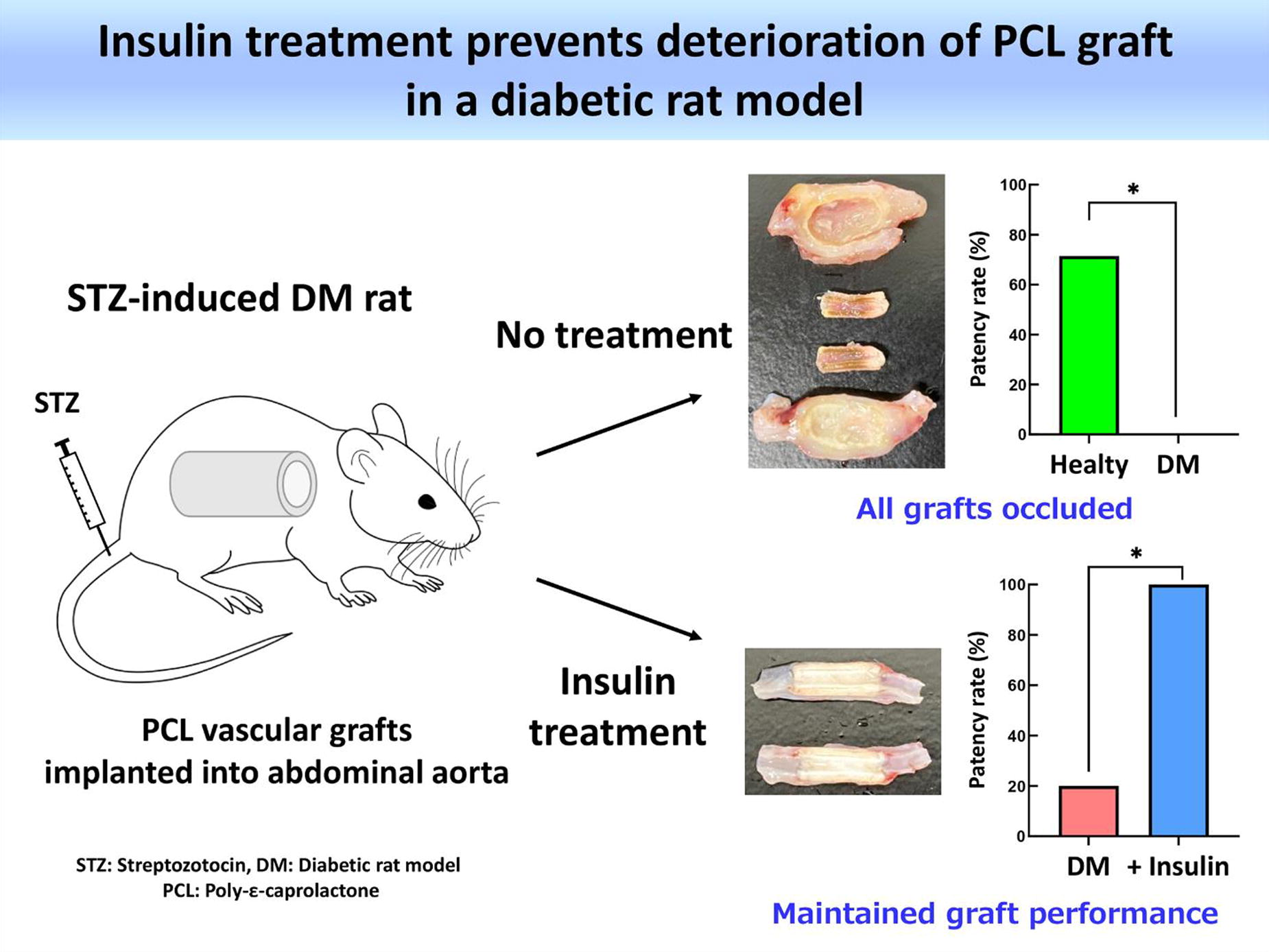
A79 - Electrospinnable And Photo-curable Polyester Elastomers To Engineer Bioresorbable Grafts For In Situ Remodeling In A Rat Abdominal Aorta Model
X. Ding, J. Goldman, W. He, B. Lee
Michigan Technological University, HOUGHTON, MI
*Purpose/Objectives: Occlusive artery diseases are the leading cause of death worldwide. Bioresorbable graft has been proposed to replace the occluded arteries for in situ regeneration. A resorbable graft is a porous conduit made from biocompatible and biodegradable polymers. Upon implantation orthotopically, host cells will infiltrate and grow in the porous conduit to transform it into a living conduit as the polymer is gradually degraded and absorbed by the host. To design an efficiently remodelable graft, we hypothesize that the grafts need to be designed to simultaneously possess key features of robust elasticity, suitable mechanical properties, and appropriate hydrophilicity and degradation rate. To this end, we have designed a pair of functional polyesters that are electrospinnable and photocurable to engineer such a resorbable graft for studies in a rat abdominal aorta interposition model.
*Methodology: We designed a pair of alkyne- and thiol-functionalized polyesters that were electrospun and UV-crosslinked to fabricate elastic fibrous conduits. The weight ratios of the prepolymer pair and electrospinning conditions were used to control the graft parameters, such as pore size, porosity, mechanical properties, and degradation rate. The resorbable graft was implanted in a rat abdominal aorta interposition model for a preliminary study.
*Results: The thiol-bearing polyester is condensed from 1,8-octanediol, sebacic acid, and thiomalic acid to yield poly(1,8-octanediol-co-sebacate-co-thiomalate) with 50 mol.% thiomalate (PT). The alkyne-functionalized block copolyester is condensed from glycerol, alkyne-functionalized serinol, sebacic acid, and PCL block to yield poly(ε-caprolactone)-b-poly(glycerol-co-sebacate-co-alkyne-serinol) with 50 mol% PCL block and 20 mol% alkyne pendants (PA). These monomers have been proven to be safe for in vivo use, and thus ensure our designed graft is biodegradable and bioresorbable. Different from prior designs, our PA and PT prepolymer mixture is electrospinnable and photocurable to fabricate elastic fibrous conduits bearing free thiol groups for further functionalization (Fig. 1A-C). The pristine PAPT graft with the current parameters was physically absorbed with heparin and implanted in a rat abdominal aorta interposition model over 3 months. No abnormal signs were observed across the experimental time. The graft shows a limited remodeling with a clot formation and a slow endothelialization in this preliminary study (Fig. 1D). This is mainly because of small pore size on the luminal side, relatively low porosity and slow polymer degradation, and inappropriate hydrophilicity. This preliminary study gives us a direction to improve the graft parameters for more detailed studies.
*Conclusion/Significance: We intend to design a fully remodelable and resorbable graft for arterial regeneration. The graft parameters such as pore size, porosity, permeability, hydrophilicity, wall thickness, mechanical properties, and degradation rate need further improvements by adjusting the prepolymer ratios and concentrations, and electrospinning conditions. Our PAPT graft bears free thiol groups for further functionalization through thiol-ene click chemistry. We will covalently attach heparin to the graft to improve anti-coagulation performance. Both the heparin-immobilized and heparin-absorbed grafts with improved parameters will be systematically evaluated in the same rat models in the next step.

A80 - Enhanced Immunomodulation In Electrospun PEUU-gelatin Tissue-engineered Vascular Grafts Through Optimized Structural Design
M. A. Rodriguez-Soto1, A. Riveros1, I. C. Orjuela-Garzón1, I. M. Fernández-Calderón1, C. F. Rodríguez1, N. Suarez Vargas1, C. Ostos2, C. Muñoz Camargo1, J. C. Cruz1, S. Kim3, A. D’amore3, W. Wagner3, J. C. Briceño1
1
Universidad de los Andes, Bogotá, Colombia,
2
Universidad de Antioquia, Medellín, Colombia,
3
University of Pittsburgh, Pittsburgh, PA
*Purpose/Objectives: The advent of tissue-engineered vascular grafts (TEVGs) represents a breakthrough in regenerative medicine, with their success contingent upon directing cell behavior to encourage endothelialization and stem cell differentiation. A critical phase involves initial inflammation to stimulate angiogenesis, followed by the suppression of chronic inflammation, which can precipitate graft failure. We present a novel TEVG with a multilayer configuration that integrates a physicochemical gradient, variable porosity, and interconnectedness to foster an immunomodulatory milieu conducive to tissue regeneration.
*Methodology: Our innovative four-layer TEVG is crafted via electrospinning polyester-urethane-urea (PEUU) with gelatin B. The luminal layer is enhanced with peptides that promote anticoagulation and endothelial maturation. The external layers are composed of varying ratios of PEUU to gelatin B (75:25, 85:15, 95:5), establishing a porosity gradient as evidenced by Masson's trichrome staining. Mechanical integrity is assessed through tensile strength tests, with degradation kinetics appraised via weight loss and computational modeling. In vitro macrophage cultures, and macrophage-endothelial cell cocultures, delineate the immunomodulatory efficacy of the TEVG. Immunofluorescent imaging, cytokine profiling, and M1/M2 gene expression analyses elucidate the immunological landscape.
*Results: The TEVG features a 3 mm diameter, 1.5 mm wall thickness, and 11 cm length, with a strategic porosity variation. Masson's trichrome stain illustrates a luminal layer of tightly woven fibers (0.6 ± 0.1 μm diameter), transitioning to looser, thicker fibers (1.3 ± 0.1 μm diameter) in the outer layers. Mechanical properties align with native porcine carotid arteries, suggesting biocompatibility. Degradation studies project a lifespan of 1500 days. Early macrophage responses indicate an M1-predominant hybrid phenotype, with a subsequent shift towards an M2-dominant profile, conducive to angiogenesis and tissue repair. Cocultures show improved endothelial adhesion and macrophage polarization towards M2 phenotypes, compared to controls.
*Conclusion/Significance: The TEVG's innovative design promotes endothelialization and favorable macrophage polarization, mirroring the mechanical properties of native arteries and potentially reducing immunogenic complications. This strategy exemplifies a significant stride in the advancement of TEVGs for regenerative medicine applications.

A81 - Multigraft-Bac: An Antimicrobial Tissue-engineered Vascular Graft For Prophylactic Infection Control
M. A. Rodriguez-Soto1, M. Ayala-Velásquez1, I. C. Orjuela-Garzón1, C. A. Polanía2, C. A. Velandia2, J. C. Cruz1, C. Muñoz Camargo1, J. G. Barrera2, J. Camacho-Mackenzie2, W. Wagner3, J. C. Briceño4
1
Universidad de los Andes, Bogotá, Colombia,
2
Fundación Cardio Infantil Instituto de Cardiología, Bogotá, Colombia,
3
University of Pittsburgh, Pittsburgh, PA,
4
Universidad de los Andes, Fundación Cardio Infantil Instituto de Cardiología, Bogotá, Colombia
*Purpose/Objectives: Vascular graft infections are a critical challenge in implant surgeries, significantly contributing to high morbidity (45%) and mortality (30%). The predicament is exacerbated by the delayed diagnosis, resilient biofilms, and the prevalence of antibiotic-resistant bacterial strains. Our study introduces Multigraft-Bac, a novel polymeric tissue-engineered vascular graft (TEVG) imbued with intrinsic antimicrobial properties to preclude such infections effectively.
*Methodology: Utilizing electrospinning, we fabricated a biphasic TEVG from polyester-urethane-urea (PEUU). The luminal fibers were modified with anticoagulants, endothelial adhesion peptides, and a broad-spectrum antimicrobial peptide. Graphene Oxide (GO) nanoplatforms laced with silver nanoparticles were embedded within the adventitial layer to enhance antimicrobial efficacy. Fourier-transform infrared spectroscopy (FTIR) and thermogravimetric analysis (TGA) validated functionalization and thermal stability. Biocompatibility was assessed through MTT and hemolysis assays. Platelet response was evaluated via aggregation, activation (LDH assay), and clotting time tests. Antibacterial activity against S. aureus and E. coli was gauged using disc diffusion and leachate toxicity assays, with SEM employed for bacterial membrane damage assessment. PEUU without modifications served as a control.
*Results: The fabricated graft, measuring 3 mm in diameter and 1 mm in wall thickness, displayed heterogenous porosity. In vitro evaluations confirmed the successful functionalization of the graft's bioactive components and even distribution of GO nanoplatforms. The graft demonstrated over 80% cell viability, favorable cellular adhesion, and anticoagulant properties that reduced platelet aggregation and clot formation. Remarkably, the graft manifested potent bactericidal and bacteriostatic actions against S. aureus and E. coli, with SEM revealing discernible bacterial membrane disruption. Control samples of unmodified PEUU did not exhibit these antimicrobial properties.
*Conclusion/Significance: This study successfully integrates multiple defensive mechanisms within a TEVG, offering substantial antimicrobial activity—a vital attribute for vascular grafts intended for high-risk patient scenarios, including limb reconstruction post-trauma or treatment of mycotic aneurysms. Future investigations will extend to in vivo assessments, utilizing a porcine carotid interposition model to validate the graft's clinical applicability.

A82 - In Vivo Assessment Of A Native-Like Three Layers Small-Diameter Vascular Graft With A Biostructural Gradient Design Approach
M. Barbuto1,2,3, T. Fujii1, K. Kohyama1, L. Modica De Mohac3, A. Adamo3, S. Kim1, G. Hussey1, S. F. Badylak1, G. Ghersi2, W. R. Wagner1,4, A. D’Amore1,3
1
McGowan Institute for Regenerative Medicine, University of Pittsburgh, Pittsburgh, PA,
2
University of Palermo, Palermo, Italy,
3
Ri.MED Foundation, Palermo, Italy,
4
University of Pittsburgh, Pittsburgh, PA
*Purpose/Objectives: Arteries are composed of three distinct tunicae: intima, media, and adventitia. These tunicae differ not only in cytological composition but also in mechanical and physiological functions. Current clinical design approaches for small-diameter synthetic vascular grafts (VG) generally do not recapitulate native blood vessels’ structural heterogenicity. In particular, one of the most common mechanisms of failure in commercially available VG is intimal hyperplasia, a structural thickening that generally leads to decreased lumen patency and thrombus formation. This complication can be attributed to the absence of an adequate structure that allows the development of a functional endothelium. It can be speculated that, to be successful, a synthetic VG should mimic the complexity of native vessels’ architecture to prevent mechanical mismatch between the native tissue and the graft and improve the hemodynamic conditions. This study introduces a novel three layers (3L)VG with an architecture based on a biostructural gradient that includes hierarchical porosity and bioactive components. We hypothesized that this structure may prevent hyperplasia and mechanical mismatch, allowing de novo tissue formation while progressively morphing into a vessel surrogate.
*Methodology: To fabricate the 3LVG, Poly(carbonate urethane) urea was processed via Thermally Induced Phase Separation (TIPS) and Electrospinning (ES). The combination of these fabrication techniques created layers with different pore size ranges: ES 6-23 µm and TIPS 30-100 µm. To assess the biostructural gradient paradigm, ES and TIPS layers were used as tunica intima or adventitia to assemble two 3LVG configurations: Intima-derived Cell Recruitment (ICR) and Adventitia-derived Cell Recruitment (ACR). In both configurations, the tunica media was composed of small intestinal submucosa derived Extracellular Matrix (ECM) gel, as a bioactive layer. In the 3LVG configurations, the tunicae intima, media, and adventitia were structured as follows: ICR TIPS-ECM-ES and ACR ES-ECM-TIPS. Each configuration was implanted in a rat model for 4 weeks (ICR n=10 and ACR n=10) and 8 weeks (ICR n=5 and ACR n=5). The patency of the 3LVG was evaluated by ultrasound and CT scan just before animal sacrifice. Histological characterization evaluated cell infiltration and tissue remodeling.
*Results: There was one death in the ICR group and two in the ACR group during surgery. At 4 weeks, the ICR group showed one occlusion case, potentially due to a surgical procedure. The ICR group also showed one partial stenosis on the proximal side. The ACR group had all grafts patent. At 8 weeks, the ICR group had one occlusion of unknown cause; conversely, the ACR group had all implants patent and functional. Histological analysis confirmed that the ES layer, with its smaller pore size, limited cell infiltration, while the TIPS allowed cell infiltration. The ECM layer appeared to facilitate cell infiltration and growth. Collagen deposition was observed in the intima layer for both configurations.
*Conclusion/Significance: These results suggest that a structural gradient with larger porosity in the tunica adventitia and smaller in the tunica intima could be effective in dictating cell proliferation and tissue elaboration that could prevent tunica intima overgrowth (100% patency rate at 4 weeks for the ACR).
A83 - Rational Protein Design Identifies PDGF- And VEGF-Specific Protein Binders For Tunable Affinity-Controlled Protein Delivery
J. E. Svendsen, C. Asnes, S. C. Oh, K. Fear, M. Ford, P. Hosseinzadeh, M. H. Hettiaratchi
University of Oregon, Eugene, OR
*Purpose/Objectives: Computational design enables the discovery of novel protein binders with precise control over binding interfaces for finely tunable protein-protein interactions as compared to standard cell sorting or directed evolution approaches. The objective of this research was to apply computational protein design to tune the affinity strengths of protein binders for vascular endothelial growth factor (VEGF) and platelet-derived growth factor (PDGF) and use these binders to control VEGF and PDGF delivery from hydrogels. To achieve this objective, we integrated a combination of in silico modelling, site-directed mutagenesis (SDM), and Rosetta-based rational design to create small protein domains called affibodies with variable affinities for VEGF and PDGF.
*Methodology: Alphafold2 was used to generate structures for known affibodies displaying moderate affinity for PDGF or high affinity for VEGF (dissociation constants, KD,PDGF = 855±285nM; KD,VEGF = 39.0±8.8nM). Computational docking softwares ZDOCK and HDOCK were used to model binding interfaces between affibodies and growth factors. The most likely bound interfaces were selected through Rosetta scoring metrics and cross-referenced with PDGF and VEGF receptor-bound structures. For VEGF affibody SDM, three target d-aspartic acids were mutated to alanine to disrupt binding interface charge-charge interactions: D28A, D32A, and D36A (Fig.1A). Mutant VEGF affibody sequences were cloned into EBY100 yeast and incubated with 10-10,000nM of VEGF to determine dissociation constants (Fig.1B). For PDGF, Rosetta design scripts were used to introduce mutations for 18 affibody residues at the binding interface (Fig.1C). Rosetta scoring metrics were used to select stable interfaces, and bound structures were visually inspected. Candidate mutants were screened by Rosetta docking to determine approximate target specificity and interaction stability. VEGF and PDGF affibodies were cloned into pet28B+ plasmids and transformed into E. coli. Soluble affibodies were purified by metal affinity and size exclusion chromatography, and their purity and structure were verified. Dissociation constants of PDGF-specific affibodies were determined by biolayer interferometry. Affibodies were conjugated into 5%(w/v) polyethylene glycol-maleimide (4-arm PEG-mal, 20KDa) hydrogels and crosslinked with dithiothreitol (DTT). Hydrogels were loaded with growth factors at a 500:1 affibody:growth factor ratio. Protein release at 37 °C over 7 days was quantified using enzyme-linked immunosorbent assays.
*Results: Three unique affibodies that were generated by applying alanine scanning to a high-affinity VEGF-specific affibody retained their specificity for VEGF and displayed affinities spanning two orders of magnitude (KD = 109-4176nM) (Fig.1B). Three unique PDGF-specific affibodies were designed by Rosetta from a starting moderate-affinity affibody and displayed affinities spanning two orders of magnitude (KD = 1.7-58.3nM) (Fig.1D). When affibodies were conjugated to hydrogels, high-affinity, rationally-designed PDGF affibodies decreased PDGF release compared to the original moderate-affinity affibody (Fig.1E).
*Conclusion/Significance: We have demonstrated the ability to finely tune VEGF and PDGF binder affinity while retaining target recognition by using SDM and Rosetta design approaches. PDGF-specific affibodies controlled PDGF delivery from hydrogels, with affibody affinity inversely related to protein release. In the future, we will investigate the potency of affibodies as modulators of VEGF and PDGF bioavailability. Our work demonstrates the utility of computational protein design in engineering affinity-based systems for sustained delivery of therapeutic proteins.

A84
A85 - 3d Tissue Engineered Model For Endothelial To Mesenchymal Transition (endomt)
I. Jain, N. F. Huang
Stanford University, Palo Alto, CA
*Purpose/Objectives: Endothelial-mesenchymal transition (EndoMT) is a biological process in which vascular endothelial cells (ECs) acquire a mesenchymal identity that give rise to lineages like fibroblasts and smooth muscle-like cells. Pathologically, EndoMT is involved in the progression of numerous cardiovascular diseases such as atherosclerosisClick or tap here to enter text., in which ECs give rise to smooth muscle-like cells within the plaque. However, there is essentially a dearth of knowledge of how the biochemical and biomechanical cues from the extracellular matrix (ECM) milieu directly influence the progression of EndoMT. The direct effects of biomechanical properties on EndoMT are difficult to study in vivo, owing to complex microenvironmental factors of soft tissues. In contrast, in vitro platforms allow the study of biomechanical and associated mechanosignaling pathways in a well-controlled environment.
To address the limited knowledge of ECM effects on EndoMT, we propose to develop a 3D tissue engineered model with independently tunable biochemical and biomechanical cues, including stiffness, stress relaxation rate and ECM composition. The main
research objective
is to encapsulate primary human aortic endothelial cells into tunable viscoelastic 3D alginate hydrogels and study EndoMT progression in 3D.
*Methodology: Alginate hydrogels were prepared using Ultrapure alginate (Novamatrix) of 280 kDa and 70kDa will at 2% w/v final concentration and crosslinked using calcium sulfate and different concentrations. The gels were made for 4 different biomechanical formulations: Soft and fast relaxing (19.52mM Ca+; 70kDa Alginate), Soft and slow relaxing (13.01mM Ca+; 280kDa alginate), Stiff and fast relaxing (25.7mM Ca+; 70kDa) and Stiff and slow relaxing (18mM Ca+, 280kDa). Human primary coronary artery endothelial cells (HCAECs, Lonza) were cultured to form spheroids using Aggrewell 800 plates. HCAECs spheroids were then co-encapuslated with fibroblasts in the alginate hydrogels for 7 days. EndoMT was induced by using media with no supplements such as VEGF and bFGF (that inhibit EndoMT) and using TGFb treatment (20ng/mL).
*Results: We could successfully fabricate the 4 alginate hydrogels with different mechanical and viscoelastic properties. HCAEC spheroids survived well in the 3D alginate hydrogel system and actively interacted with the fibroblasts in the hydrogels. Additionally, we observed viscoelasticity dependent migration of ECs from the spheroid into the alginate hydrogel and vessel like tube formation in the 3D model. Lastly, we quantified higher SM22 expression (EndoMT) in ECs in slow relaxing hydrogels, pointing towards viscoelasticity dependent progression of EndoMT.
*Conclusion/Significance: These results underscore the importance of mechanical factors such viscoelasticity in addition to stiffness in progression of diseases like atherosclerosis. Future work involves performing transcriptomics analysis to uncover the mechanism behind viscoelasticity dependent EndoMT progression.
A86 - Predictive Oxygen Transport In A Synthetic Embryonated Avian Eggshell: A Computational Model
C. Conci1, L. Cherubin1, M. Laganà2, M. T. Raimondi1
1
Politecnico di Milano, Milano, Italy,
2
Gemma Prototipi, Longone al Segrino, Italy
*Purpose/Objectives: Aiming to accelerate the discovery and preclinical testing of new biological drugs, particularly for those targeting the micro circulation (e.g. in diabetes, retinal disease, kidney disease, virology, etc.), the dramatic Sars-Cov-2 pandemic infection brought to light the urgent need for experimental models. In the same way, the vascular network is crucial to the development of antiangiogenic, antibody- and small-molecule-based cancer treatments. The chicken embryo appears to be the ideal model for doing quick and flexible research on how the functioning vascular network responds to injected stimuli. In order to do this, we created a computer model of an embryo at various stages of development in order to find a synthetic material that can replace the eggshell and yet provide physiological oxygenation.
*Methodology: In order to depict a live chicken embryo during the eighth day of development and describe its own protective shell, we ran computational simulations (COMSOL). In order to simulate the egg, we created a prolate ellipsoid mesh with 195k tetrahedral pieces and radii of 1.4 and 1.7 cm. We considered the eggshell to be a porous material with a thickness of 300µm, a porosity of less than 2%, and a tortuosity parameter of one. In contrast, we considered a thin film made up of layers of the inner and outer shell, each with a total thickness of 65µm. We hypothesized that the embryo was located in the center of the egg, with an albumen layer of 18 cm3 in the bottom section and an air sac above with a volume of 3 cm3. We assumed a consuming surface of about 41 cm2 for the chorioallantoic membrane, which is the respiratory organ of the embryo. The peak oxygen consumption, or Vmax, was calculated to be 5.14x10⁻⁶ mol/m2s, and the kinetics were guided by the Michaelis-Menten equation. Next, we considered several PDMS thicknesses for a membrane replacement material: 100 (fabrication technique limit), 365 (physiological value), and 800 µm (thickness limit).
*Results: From the outside to the inner limits of the egg, the concentration of oxygen dropped less than 1% as the thickness of the eggshell increased. The mean oxygen content in the air sac was found to be 8.152mol/m3. When analyzing the thinner and thicker PDMS layers, respectively, simulations on PDMS revealed a drop in air sac concentration ranging from 20% to 98% with regard to physiological values.
*Conclusion/Significance: We developed a mathematical model that can forecast the oxygen concentration levels in a chicken embryo on its eighth day of development. With a mean error of less than 3%, our model corresponds with experimental results reported in the literature. We are presently doing measurements to confirm boundary condition specified in model, to calibrate the model and to extend its validity up to the 12th day of embryonic development. Additionally, we are putting our first PDMS-based synthetic eggshell alternative through experimental testing. Acknowledgements European Research Council (ERC, project BEACONSANDEGG, G.A. 101053122)
A89 - A Bimetallic-organic-framework Loaded With Annexin A5 For The Treatment Of Inflammatory Bone Resorption
Z. Jia, w. zhang
Shanghai Ninth people’s hospital, Shanghai, China
*Purpose/Objectives: The microenvironment surrounding bone tissue is crucial for maintaining bone homeostasis, especially in inflammatory diseases such as osteomyelitis, periodontitis, rheumatoid arthritis (RA), and purulent arthritis. Annexin A5, a potential protein drug for treating inflammatory bone-related diseases, has shown promising effects. However, due to its susceptibility to clearance, a sustained release system is required. In this study, an AnxA5@MOF nanodrug system was designed, incorporating a Ca-Sr-MOF core and Annexin A5 with sustained release capabilities under low pH conditions.
*Methodology: In the initial phase, Ca-Sr-MOF was synthesized using a solvothermal method, and its SEM, XRD, and BET characteristics were analyzed. The sustained release properties were also assessed. For evaluating the therapeutic impact on inflammatory bone resorption, we employed an LPS-induced cranial inflammation-induced bone resorption mouse model. Micro-CT was utilized for structural observations, and cranial sections underwent staining for structure (HE; Masson), macrophage-related markers (CD206/INOS), osteoclast-related markers (TRAP), and osteoblast-related markers (ALP). Additionally, mRNA analysis was conducted on bone tissues. In vitro experiments focused on testing the regulatory effects of AnxA5@MOF on macrophages, ROS clearance in the microenvironment, as well as osteoclast and osteoblast differentiation.
*Results: SEM results revealed the self-assembly of Ca-Sr-MOF into nanoflower-like structures with an average diameter of 46.783 micrometers. Elemental mapping exhibited uniform distribution of C, N, O, Ca, and Sr, with a doping ratio of Ca to Sr at 2:1. XRD analysis confirmed the successful synthesis of Ca-Sr-MOF with a well-defined crystal structure. This material demonstrated reversible binding and pH-responsive characteristics, acting as a barrier to maintain protein structure and bioactivity, and facilitating the successful loading of AnxA5 into Ca-Sr-MOF for in vivo applications. In the LPS-induced cranial bone resorption model, AnxA5 was injected every two days, while Ca-Sr-MOF solution and AnxA5@MOF were injected every seven days. The integrity of the cranial bone was observed on days 7 and 21 through Micro-CT imaging and 3D reconstruction. Compared to healthy rats, the model group exhibited significant bone loss. Quantitative analysis indicated that the AnxA5@MOF group surpassed the Ca-Sr-MOF and AnxA5 alone groups in terms of new bone volume percentage (BV/TV), trabecular thickness (Tb.Th), trabecular number (Tb.N), and bone mineral density (BMD). These results suggest that AnxA5@MOF exhibits effective therapeutic effects on LPS-induced cranial bone resorption. Consistent with in vivo experiments, in vitro studies demonstrated that AnxA5, Ca-Sr-MOF solution, and AnxA5@MOF all displayed robust antioxidant capabilities, with AnxA5@MOF exhibiting superior effects compared to the individual use of AnxA5 or Ca-Sr-MOF. AnxA5@MOF demonstrated significant inhibitory effects on M1 macrophages, contributing to the attenuation of their pro-inflammatory activity. Simultaneously, for M2 macrophages, AnxA5@MOF exhibited a promoting effect, guiding them toward a more anti-inflammatory phenotype. Regarding osteoclasts, AnxA5@MOF exhibited notable inhibitory effects. Meanwhile, the promoting effect on osteoblasts indicated that AnxA5@MOF may exert a positive influence on bone formation.
*Conclusion/Significance: In summary, this study provide robust support for the potential therapeutic applications of AnxA5@MOF in regulating macrophages and maintaining the balance of the bone microenvironment.
A90 - Photo-controlled Microrna-21 Delivery For Osteogenesis In Palatal Expansion
B. Wang
Shandong University, Jinan, China
*Purpose/Objectives: Rapid maxillary expansion (RME) is a common therapy for maxillary transverse deficiency, while relapses after RME usually occur because of insufficient osteogenesis in the expanded suture. MicroRNA-21 (miR-21) was reported as an important post-transcriptional modulator to promote osteogenesis. Thus, local delivery of exogenous miR-21 might be a therapeutic strategy to enhance osteogenesis during RME. However, proper protection of miR-21 to precisely exert effects in the target cells and avoiding potential side effects is challenging.
*Methodology: To address this, an advanced delivery system based on a photo-controlled nanosystem was constructed. Specifically, photo-controlled-microRNA-21 (PC-miR-21) was first synthesized. Upconversion nanoparticles (UCNPs) modified with polyetherimide (PEI), i.e. UCNPs@PEI, were constructed as the light transducer and the delivery carrier. PC-miR-21 was loaded onto the UCNPs@PEI by electrostatic attraction, resulting in the UCNPs@PEI@PC-miR-21 nanocomplexes. These nanocomplexes have good aqueous dispersibility and biocompatibility. Furthermore, the UCNPs@PEI could protect PC-miR-21 from biodegradation and deliver PC-miR-21 to the cytoplasm. Afterwards, PC-miR-21 can convert to active miR-21 under the near-infrared (NIR) light applied to the mid-palatal suture. Finally, the efficacy of the UCNPs@PEI@PC-miR-21 was verified in a cell stretch experiment and a miR-21 knockout mice with RME study.
*Results: The results suggested that with NIR light irradiation, UCNPs@PEI@PC-miR-21 upregulated the expression of osteogenic key markers: ALP, RUNX2, and COLIA1 in the levels of both genes and proteins in vitro. Besides, the in vivo results further corroborated that UCNPs@PEI@PC-miR-21 photo-controllably upregulated osteogenic markers of ALP, RUNX2, and osteoprotegerin. Meanwhile, it induced fewer osteoclasts formation by TRAP staining.
*Conclusion/Significance: In conclusion, UCNPs@PEI@PC-miR-21 nanoparticles with NIR light could facilitate the remote and precise delivery of exogenous miR-21 to the mid-palatal suture to promote bone formation during RME. This work represents a cutting-edge approach of gene therapy to promote osteogenesis in the mid-palatal suture during RME and provides a frontier scientific basis for the later clinical treatment.
A91 - Physical Stability And Antibacterial Properties Of Tannic Acid-based Supramolecular Coating
M. Hosseini, L. Moghaddam, L. Barner, D. W Hutmacher, F. M Savi
Queensland University of Technology, Brisbane, Australia
*Purpose/Objectives: Infection and microbial colonisation on the scaffold (implant or medical device) surface present a clinical challenge that often necessitates antibiotic therapy or, in a significant number of clinical cases, implant removal. Hence, efficient antibiotic-free coating strategies have become increasingly attractive, especially to combat infections and minimise the risk of antibiotic resistance. Tannic acid (TA) is a natural polyphenol with intrinsic antibacterial properties. The supramolecular structure of TA allows the design and manufacture of antibacterial coatings with a nano topography and morphology; however, to our knowledge, there is no report on the development and in vitro characterisation of antibacterial coatings based on these structures in medical implants. Medical-grade polycaprolactone (mPCL) has been translated from bench to bedside in the field of scaffold-guided tissue regeneration. In this context, we propose an organic solvent-free strategy for the formation of poly(tannic acid) (PTA) with supramolecular nanostructures and its physical adsorption on the surface of 3D-printed mPCL scaffolds.
*Methodology: The supramolecular structure of TA was investigated by UV-visible spectroscopy, near-infrared microscopy, Fourier transform infrared spectroscopy (FTIR), and X-ray diffraction. Dynamic light scattering and transmission electron microscopy determined the size and morphology. The immobilisation of PTA onto the mPCL surface was studied using FTIR, X-ray photoelectron spectroscopy, and scanning electron microscopy (SEM). The polymerisation of TA was tuned by pH to achieve a uniformly coated surface with a long-term physical stability. In vitro antibacterial performance was evaluated against Staphylococcus aureus, most often responsible for implant-related infections.
*Results: By adjusting the pH of the TA solution, TA molecules can develop into spherical-shaped PTA aggregates with an average size of <100 nm mainly through the synergy of hydrogen bonding and π-π stacking interactions. The size and morphology of PTA aggregates were also affected by varying pH. The polymerised TA demonstrated higher stability and antibacterial effects than TA while retaining an amorphous pattern. When PTA nanostructures were coated on mPCL scaffolds, the SEM images showed the same morphological characteristics. The stability studies indicated the high physical stability of the PTA coating in different biological media, including phosphate buffer saline (pHs 7.4 and 5.4) and cell culture media (DMEM, and DMEM/FBS). Also, the coated scaffolds were capable of inhibiting the growth S. aureus.
*Conclusion/Significance: Our results demonstrate enhanced antibacterial potency and improved bioavailability, highlighting the capability of polymerised TA to fulfill therapeutic applications. These findings aim to further develop and characterise the PTA supramolecular antibacterial coating for implant surfaces, both in vitro and in vivo.
A93
A94 - Instructing Endothelial Cells Against Cartilage Degeneration Through Microencapsulated Bevacizumab In 3D Printed Collagen
A. Abbadessa1, P. Nuñez Bernal2, G. Buttitta1, A. Ronca3, U. D’Amora3, C. Zihlmann4, N. Stiefel4, L. Ambrosio3, J. Malda2, R. Levato2, J. Crecente-Campo1, M. Alonso1
1
University of Santiago de Compostela, Santiago de Compostela, Spain,
2
University Medical Center Utrecht, Utrecht, Netherlands,
3
Institute of Polymers, Composites and Biomaterials, National Research Council (IPCB-CNR), Naples, Italy,
4
Geistlich Pharma AG, Wolhusen, Switzerland
*Purpose/Objectives: Pathological angiogenesis (PA) induced by endothelial cells is a crucial attribute of several chronic diseases. In the case of osteoarthritis, PA mediated by the vascular endothelial growth factor (VEGF) contributes to cartilage degeneration and implant rejection. In line with this, the anti-VEGF bevacizumab (BVZ) has been shown to support cartilage regeneration. Nevertheless, previous studies harnessed this effect only by BVZ injections which lack BVZ controlled delivery, or by incorporating BVZ in molded scaffolds which are unable to mimic biological complex shapes. Herein, we innovate by integrating 3D printing with drug delivery, to obtain cell-instructive constructs with biomimetic complex shapes and controllable BVZ release. The presented work pursues this integration by 3D printing a medical-grade collagen functionalized with BVZ-releasing poly(lactic-co-glycolic acid) (PLGA) microparticles.
*Methodology: BVZ-loaded PLGA microparticles were prepared according to the water-in-oil-in-water (w/o/w) double emulsion method, by using different PLGA/BVZ ratios and surfactants with different hydrophilic/lipophilic balance (HLB). Entrapped and released BVZ was quantified by Bradford and enzyme-linked immunosorbent assay (ELISA). BVZ microparticles were embedded in a medical grade collagen ink and 3D printed using an extrusion 3D printer. The shape fidelity of complex and hallow 3D printed constructs was evaluated, and the microparticles distribution throughout the scaffold was visualized by Scanning Electron Microscopy (SEM). The anti-angiogenic activity of released BVZ was tested in a 3D model of human umbilical vein endothelial cells (HUVECs) and bone marrow-derived mesenchymal stromal cells (hbMSCs), which elicits HUVECs-driven capillary network formation.
*Results: The encapsulation and release of BVZ were tuned as a function of the varying formulation parameters. High EE (83%) was obtained when using a low-HLB surfactant, i.e., Span 60 due to its good ability of stabilizing w/o emulsions. Good EE was also obtained when using high-HLB Pluronic, due to its ability to prevent protein premature leakage by distributing itself at the o/w interface. Particles prepared using Pluronics exhibited a sustained release of BVZ over 21 days, whereas those prepared using Span 60 featured a high burst release of 25% followed by a plateau. Furthermore, an increase of the PLGA/BVZ ratio caused an increase in the EE up to 93% and a slower release. These results allowed the selection of the two most promising formulations. 3D printing of the microparticle-loaded collagen ink resulted in the fabrication of shape-stable meniscus, nasal and auricular structures with high shape fidelity and homogeneously distributed microparticles (Figure 1a). Moreover, a bizonal meniscus mimicking the native vascular and avascular regions was successfully printed. Released BVZ reduced the vascularization by HUVECs, as evidenced by the significant reduction in the average vessel area, length and number of junctions (2-fold decrease) as compared to negative controls, without adversely affecting cellular metabolic activity (Figure 1b-c).
*Conclusion/Significance: We successfully combined BVZ microparticles with a 3D printed collagen to generate endothelial cell-instructive materials with biomimetic complex shapes to tackle PA during cartilage degeneration. This strategy leverages the spatial control over the biomaterial offered by 3D printing and the temporal control over the released protein provided by the drug delivery system.
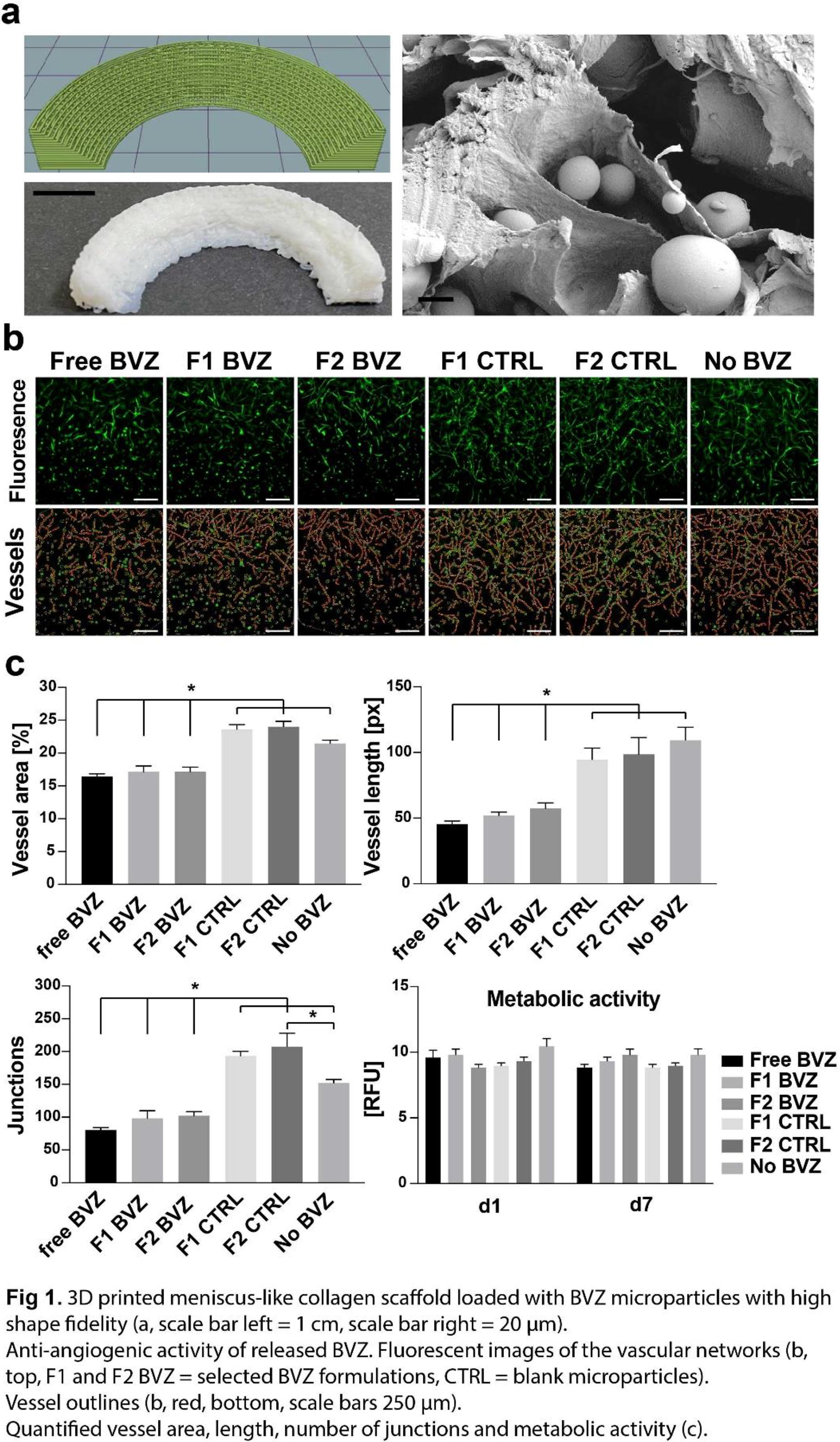
A95 - Nano-in-micro Technologies Enabling Controlled Local Release Of Therapeutics
X. Bai1, E. Spessot2, D. Maniglio2, S. Butterworth1, A. Tirella2
1
University of Manchester, Manchester, United Kingdom,
2
University of Trento, Trento, Italy
*Purpose/Objectives: Several generations of nanoparticles have been designed to better control drug release and targeting modalities to a specific site. However, drug-loaded nanoparticles are known to have a long circulation time and often causing side effects to healthy cells. Encapsulation of drug-loaded nanoparticles into a hydrogel is promising to ensure localization, increase retention time at the target site, hence control release of therapeutic agents in space and time.
*Methodology: Based on our previous work describing polymeric nanoparticles for the sustained release of a small molecule (hydroxyl-FK866), we explored the interaction of such PLGA-based nanoparticles with modified alginate hydrogels as 3D-printable biomaterials for encapsulating therapeutic molecules.Nanoparticles were coated with chitosan and their confinement within alginate hydrogels was evaluated using atomic force microscopy (AFM) measurements. Further, rheological characterisation and shape fidelity evaluations were performed to assess the printability of alginate-based drug delivery microsystems. Finally, efficacy for controlled release of hydroxyl-FK866 was evaluated on macrophages and cancer cells, THP-1 and MDA-MB-231.
*Results: The coating of hydroxyl-FK866-PLGA nanoparticles with chitosan was proven effective strategy for confinement in hydrogels formulated with a blend of oxidized alginate and unmodified alginate. Further ionic crosslinking was used to match mechanical properties of soft human tissues and ensure stability of hydrogels in physiological conditions. We observed a slight reduction in mechanical properties (approx. 5%) and degradation (approx. 10% weight loss) after 5 days (complete cell culture medium, 37°C, 5% CO2).Composite alginate hydrogels loaded with nanoparticles were 3D printed as micro technologies and purposely designed to: 1) match the properties of soft tissues, hence reducing any inflammatory effects after implant and 2) load a known concentration of hydroxyl-FK866-PLGA nanoparticles. Efficacy of 3D-printed systems was evaluated in vitro showing increased cytotoxicity of approx. 20% in cells treated with 3D-printed composite hydrogels vs free hydroxyl-FK866-PLGA nanoparticles.
*Conclusion/Significance: Alginate-based biomaterial inks were formulated and loaded with multifunctional nanoparticles, exploiting their use in precision medicine using 3D printing technology. 3D-printed composite alginate hydrogelsencapsulating nanoparticles are emerging technologies for personalized medicine, enabling precise controlover spatial and temporal release of therapeutic agents. Moreover, the proposed manufacturing strategy for injectable biomaterials for the controlled delivery of small therapeutics ensures increased efficacy at the target site, as well as have direct impact on reducing discomfort to patients paving the way towards new delivery strategies for personalized medicine.
A96 - Photo-Carbon Monoxide Releasing Molecules For Antibacterial Properties In Vascular Grafts
B. Caner, M. Hammad, A. Elgattar, Y. Liao, C. A. Bashur
Florida Institute of Technology, Melbourne, FL
*Purpose/Objectives: A better hemodialysis access graft is needed for individuals with kidney failure. In addition to graft stenosis, vascular access-related infections are a significant cause of illness and death. This is further complicated by rising antibiotic resistance bacteria. Thus, alternative hemodialysis grafts are needed. We have previously shown that carbon monoxide (CO) can promote endothelial cell (EC) proliferation, and it may also be able to provide antibacterial properties at the same dose. Thus, this study investigates CO release and antibacterial effects from spincoated scaffolds loaded with CO-releasing molecules (CORMs) and activated with visible light.
*Methodology: The photo-CORM DK4 was prepared with a 5-step synthesis, and then mixed with ester-terminated polycaprolactone (PCL) for spincoating. Films were prepared at 0, 5, and 10% w/w (CORM/PCL) using hexafluoro-2-propanol and then characterized. The K-12 MG1655 strain of E. coli was utilized for 6 and 8 hours in both planktonic and biofilm growth experiments. CO release profiles were assessed using 470 nm blue light. The impact of different concentrations of CO was evaluated through SEM, live-dead, and colony forming unit (cfu) assays, Coculture is being conducted to observe the effects of CO on both bacteria and GFP+ ECs. Mammalian cell viability in a bacterial- mammalian coculture is assessed after blue light activation. Two-Way ANOVA and Tukey multiple comparisons were performed.
*Results: Increasing concentrations of DK4 in spincoated films led to systematically higher contact angles (n=3), with 68° and 82° for pure PCL and 10% DK4/PCL, respectively. The films were approximately 20µm thick with some variability between samples. The topography of the coverslips showed peaks and valleys for bacteria to interact with due to individual PCL crystals. Interestingly, the 10% DK4 conditions were also more light stable than 5% and resulted in more gradual and extended release of CO. Based upon SEM, high number of bacteria and the highest degree of biofilm maturity after 6 and 8 h were found on PCL only controls (n=3). While some bacteria still attached to the 5% and 10% non-activated DK4 conditions, less biofilm maturity was observed. After activation and CO release, 10% DK4 had negligible bacteria on the surface. However, activation of 5% DK4 did not show as clear of a reduction in the bacteria. In preliminary results, 10% DK4 also appeared to show benefits with other assays. This included an apparent decrease in colonies from cfu after activation and the highest dead/live ratio when in planktonic form. We are currently performing co-culture studies to better understand the impact of this dose.
*Conclusion/Significance: These thin, spincoated samples were able to release enough CO to provide antibacterial properties at DK4 concentrations which were non-toxic in our previous EC studies. The highest loading, 10% DK4, appeared to provide a unique release profile and clearer anti-bacterial benefits. We hypothesize that a more delayed release is advantageous because CO is a gas that can rapidly diffuse out of culture. However, hydrophobicity and topography differences between spincoated samples may also impact biofilm formation and anti-bacterial properties, and need further investigation in thicker, 3D grafts.
A97 - Microfluidic Based Nanoprecipitation Of Polymeric Nanoparticles For Targeted Immunomodulation Of Beta-cell Allografts
J.-P. A. Pham, M. M. Coronel
University of Michigan, Ann Arbor, MI
*Purpose/Objectives: Using biomaterials to manipulate signaling through immune checkpoint receptors and suppress alloreactive immune cells has recently shown promise in the context of allogeneic islet transplantation, in which co-transplanted ∼200 µm diameter microgel particles displaying programmed-death ligand 1 (PD-L1) reprogrammed local immune cells at the transplantation site toward tolerogenic phenotypes. In this study, we investigated whether PD-L1-conjugated nanoparticles (PD-L1 NPs) could also facilitate immunomodulation for future use cases involving minimally invasive administration.
*Methodology: Custom microfluidic chips were fabricated using an accessible and facile technique in which 3D-printed negative molds were used to embed features into polydimethylsiloxane (PDMS). Hydrodynamic flow focusing (HFF) was used to facilitate nanoprecipitation of PLGA-PEG-Biotin polymer in an aqueous antisolvent solution. The rapid diffusive mixing results in spontaneous assembly of NPs on the order of microseconds, with diameters determined by solvent and antisolvent flow rates. An optimized synthesis protocol was obtained for large-scale production of NPs, capable of processing ∼5 mg of polymer per hour. The collected suspension of PLGA-PEG-Biotin NPs were reacted with SA-PD-L1, a chimeric protein of streptavidin (SA) and PD-L1. The effect of PD-L1 presentation on macrophage polarization was evaluated in vitro through co-incubation with murine bone marrow-derived macrophages (BMDMs).
*Results: Nanoparticle tracking analysis (NTA) was used to determine the average particle diameter and polydispersity index (PDI). Implementation of an HFF platform for nanoprecipitation led to NPs with average diameter of 107 nm and a polydispersity index (PDI) of 0.1. HFF platform also proved amenable to high-throughput production, with 25E9 particles generated from 6 mg of polymer over a timespan of 1.5 hrs. Particles exhibited stable average diameters, with a 15% increase in diameter over a 7-day period when stored in the concentrate form at 4°C. Additionally, NTA confirmed that NP concentration was stable over the course of a week. Conjugation efficiency of chimeric protein to the surface of NPs was ∼70% post-fabrication as confirmed by staining of PD-L1 NPs with an anti-streptavidin dye and flow cytometry analysis, in which the number of positively and negatively stained PD-L1 NPs were quantified with fluorescent counting nanobeads. SA-PD-L1 attachment was validated by performing a western blot on the filtered PD-L1 NP reaction mix, which showed that minimal free SA-PD-L1 below the filter molecular weight cutoff remained unconjugated. Lastly, PD-L1 NPs were found to exhibit no significant effects at a 7000:1 ratio on BMDM viability, polarization state based on F4/80, CD86, and CD163 marker expression for M0, M1, and M2 states respectively, PD-L1 expression, or PD-1 expression after 24 hrs. of exposure.
*Conclusion/Significance: These studies indicate that this fabrication pipeline is a highly efficient and reproducible means of generating biodegradable NPs capable of robust surface functionalization with immunomodulatory proteins. Additionally, outcomes of co-culture with BMDMs provided strong evidence that NPs do not elicit macrophage activation in vitro. In total, these findings form the foundation for future applications of PD-L1 NPs as a minimally invasive means of improving islet transplantation outcomes without chronic systemic immunosuppression.
A102 - Controlled Delivery Of Rhbmp-2 In Engineered Charged Collagen For Improved Bone Quality
L. D. Sweeney, G. A. Calderon, V. L. Ricci, A. S. Mejevdiwala, S. L. Bennett
Integra Lifesciences, Princeton, NJ
*Purpose/Objectives: Collagen is an attractive scaffold for drug delivery due to its favorable biocompatibility possessing inherent cell attachment sites. However, current engineered collagen systems offer little control over attachment and release of macromolecules or alter the biocompatibility of the collagen through crosslinking. This work strives to develop a simple, robust system for drug delivery through independent alteration of charge density and degradation time of collagen. Since macromolecules possess different isoelectric points, we can exploit the charge density to preferentially sequester and release macromolecules in the resulting collagen scaffold. This work focuses on the binding and delivery of a positively charged macromolecule (rhBMP-2) to demonstrate the efficacy of the system.
*Methodology: Rigorous material characterization of crosslinking confirmed our capacity to independently control net charge and collagen degradation. After, we investigated binding and release profiles via UPLC and the potential therapeutic impact by determining the corresponding osteogenic and adipogenic potential to varying rhBMP-2 concentrations with human mesenchymal stromal cells (hMSCs) seeded on collagen. Osteogenic capacity was evaluated by Alizarin Red staining (ARS), Alkaline Phosphatase (ALP) activity, and energy dispersive X-ray spectroscopy (EDS). Off-target effects were determined through Oil Red O staining and leptin ELISA for adipogenesis.
*Results: After confirming the osteoinductive nature of rhBMP-2 in 2D monolayers with ARS and ALP activity, we seeded hMSCs on our engineered collagen scaffolds to determine resulting changes in osteoinduction. We found that negatively charged collagen promotes a rhBMP-2 sensitive dose response and significantly greater ALP activity than other groups at the higher doses, suggesting that the negative charge sequesters bioactive rhBMP-2 to a greater extent than other collagen foams tested (control collagen sponge positive, and neutral). We confirmed that resulting mineralization on the charged collagen is correlated to calcium deposition through EDS elemental imaging on sample surfaces. Toward an alternative differentiation pathway, we recapitulated adipogenic differentiation of hMSCs simply by delivering high rhBMP-2 concentration in osteogenic media. We found increasing rhBMP-2 concentration from 100 ng/mL by ten-fold increments up to 1 µg/mL triggers an increasing presence of Oil Red O-positive lipid droplets. Further, our leptin ELISA results bore similar rhBMP-2 dose dependency with increased leptin secretion across several engineered collagen scaffolds (collagen sponge, neutral, and negative). However, the negatively charged collagen decreased leptin secretion at higher doses of rhBMP-2.
*Conclusion/Significance: Altogether, we show we can impact mineralization via our charged collagen platform by its interaction with rhBMP-2. Moreover, we show that high levels of rhBMP-2 can elicit adipogenesis, but that our negatively charged collagen platform may mitigate such lipid deposition while improving the osteogenic differentiation of hMSCs Beyond use with BMP-2, we have gathered data to support the use of the platform with additional macromolecules of interest (EGF and PDGF-bb). These data have demonstrated that positively charged collagen preferentially sequesters the negatively charged EGF while the negatively charged collagen preferentially sequesters the positively charged PDGF-bb. We surmise that this charged collagen platform could be exploited as a tool for several drug delivery applications relying simply on the innate isoelectric properties of a macromolecule of interest.
A104 - Optimizing Localized Delivery Of Biological Cargo Via Gelatin Nanoparticles For Tissue Injury
C. Tobo1, J. Tadiwala1, A. Jain2, P. Jellis1, J. Kornbluth2, K. Garg1
1
Saint Louis University, Saint Louis, MO,
2
Saint Louis University School of Medicine, Saint Louis, MO
*Purpose/Objectives: Mesenchymal stem cells (MSCs) support tissue formation, blood vessel growth, release growth factors, and suppress inflammation. Extracellular vesicles (EVs) are secreted by mammalian cells for intercellular communication. They contain bioactive molecules such as lipids, proteins, and mRNAs unique to the cell of origin and recapitulate MSC’s broad therapeutic effects. As an acellular byproduct, EVs can readily circulate through organs, elicit a minimal immune response, avoid phagocytosis, and stimulate cellular responses. Administration of EVs in vivo results in rapid clearance from blood circulation. This work's goal is to augment the therapeutic effect of EVs by prolonging their retention at the injury site. To achieve this goal, we focus on gelatin, a widely studied delivery vehicle for small biomolecules with many biological advantages. We prepare and utilize gelatin nanoparticles (GNPs) with different surface charges to electrostatically bind to treatments to improve delivery and prevent rapid clearance.
*Methodology: Gelatin nanoparticles were prepared using a desolvation method and crosslinked using carbodiimide chemistry. Type A or B gelatin in acidic or alkaline conditions was subjected to desolvation to obtain GNPs of different surface charges. The GNPs were analyzed by a dynamic light scattering Malven Zetasizer to determine size and zeta potential. GNP cytotoxicity was assessed by a lactate dehydrogenase assay following incubation with MSCs. EVs were isolated from MSCs using the ExoQuick-Ultra method and analyzed for size using nanoparticle tracking analysis (NTA). Acidic GNPs were mixed with EVs in different ratios, and zeta potential of the individual and combined solutions was measured.
*Results: GNPs derived from type A gelatin under acidic pH conditions showed an average size of 81.84 nm and a zeta potential of 12.76 mV. GNPs derived from type B alkaline under alkaline pH conditions showed an average size of 41.42 nm and a zeta potential of -13.80 mV. [KG1] Both type A and B GNPs were not cytotoxic to MSCs. MSC-EVs were 181.3 nm in size with a zeta potential of -14.6 mV. The combined solution of GNP: EV (400:1) resulted in a complete inversion of the zeta potential to a positive value of 10.33 mV. This result suggests complete coating of EVs by GNPs.
*Conclusion/Significance: We have synthesized positively and negatively charged GNPs that are non-cytotoxic and can be electrostatically linked to oppositely charged biomolecules. Ongoing studies are investigating effects of GNP:EV conjugates on macrophages. Future studies will determine the efficacy of GNP-conjugated therapies in modulating a muscle tissue injury site in a rodent model.

A106 - Biomaterial-based Concepts For The Delivery Of Bacteriophages
C. Vater, R. F. Richter, K. Schütz, H. Bretschneider, L. Schlicht, M. Stiehler, K.-P. Günther, A. Lode
University Hospital Carl Gustav Carus and Medical Faculty of Technische Universität Dresden, Dresden, Germany
*Purpose/Objectives: Bacterial infections are a severe problem and can lead to devastating complications with high mortality rates. Thereby biofilm formation on implants and antibiotic resistances denote major challenges. Since only few new antibiotics have been developed, bacteriophages (phages) - viruses that infect specific bacteria without infecting mammalian or plant cells - could be a promising alternative or addition to antibiotic therapy. However, little is known so far about optimal dosages, application routes and application frequency. In this study we investigated the release of Escherichia coli (E. coli) and Staphylococcus capitis (S. capitis) specific phages from different biomaterials as well as their antibacterial efficacies.
*Methodology: In a first experiment mesoporous bioactive glass (MBG) was loaded with 2 different E. coli specific phages and subsequently incubated for up to 44 days at 37 °C in phage buffer. At defined time points supernatants were taken and analyzed regarding active phages using the bacterial growth curve and spot plaque assay method. In a second experiment the biomaterials Lyostypt® (bovine collagen sponge), beta-tricalciumphosphate (β-TCP) and TISSEEL (fibrin tissue sealant), that are all approved for clinical application, as well as mineralized collagen (MCM) were loaded with either E. coli or S. capitis specific phages. After incubation in human plasma like medium for up to 28 days, bacterial growth curve and spot plaque assay analysis were carried out to determine active phages. Additionally, phage containing supernatants were tested regarding their efficacy against biofilms.
*Results: Using MBG, active phages were released for up to 44 days as verified by at least 40 % decreased bacterial growth and visible plaques. Lyostypt®, β-TCP and TISSEEL released active E. coli and S. capitis specific phages for up to 28 days that decreased bacterial growth by also at least 40 %. Applying MCM, bacterial growth reduction was detected for E. coli specific phages for up to 28 days but for S. capitis specific phages for only 3 days. For all biomaterials tested the number of visible plaques was decreasing with increasing incubation time and depending on the kind of phages (highest numbers for E. coli specific phages). Biofilms formed by E. coli were not affected by supernatants from any of the biomaterials. In contrast, biofilms formed by S. capitis showed signs of dissolution when treated with supernatants from all biomaterials. This finding was depending on the time of incubation and consequently the concentration of released phages with the most harmful effect induced by supernatants from TISSEEL.
*Conclusion/Significance: Phages active against E. coli and S. capitis can be released in a sustained manner from MBG, MCM and the already clinically used biomaterials Lyostypt®, β-TCP and TISSEEL making them promising phage delivery systems to support conventional antibiotics therapy. However, further experiments are needed to determine the optimal type of phages and dosages and to evaluate possible synergistic effects when combined with antibiotics as well as to develop strategies for the treatment of biofilms.
A108 - The Role Of Physical Effects In Material Induced Bone Formation
S. Oghazian, S. Lau, E. Harvey, N. Makhoul, J. Barralet
McGill University, Montreal, QC, Canada
*Purpose/Objectives: Reconstruction of cranial defects presents challenges due to the intricate local environment and unique skull features. Achieving successful reconstruction is challenging due to the proximity of the brain and the dynamic nature of the dura matter. Although autografts are the gold standard, they have issues such as resorption and limited availability that limit utility. Synthetic materials show promise but fall short in promoting rapid cranial repair. Cell-based tissue engineering must overcome regulatory and safety concerns surrounding stem cell isolation and potential unintended side effects. This study aims to establish a controlled environment, utilizing physical stimuli to promote targeted bone formation. We posit that these bony and hematopoietic tissues could effectively substitute current methods, addressing associated limitations in cranial defect repair.
*Methodology: We conducted this pre-clinical study using male Wistar rat models after approval from McGill University Animal Care Committee (UACC, #7660). Glass and silicone open-ended tubes were introduced subcutaneously and subperiosteally in the skull region. To insert the tubes beneath the skin, a horizontal incision on the nuchal skin provided access, and the tube was positioned over the cranium. A vertical incision over the midline of the animal's head was made to place the tube directly over the bone, and a periosteal flap was secured over the implant. All tubes had identical dimensions (6 mm height, 7 mm inner diameter, and 9 mm outer diameter). Following the euthanasia 8 weeks after implantation, gross examination, micro-CT analysis, and histological evaluation were conducted on all specimens.
*Results: Macroscopic examination of silicone tubes showed a central cord of tissue with an average height of 3.15 mm and diameter of 0.28 mm; cords in the glass group were shorter and thicker (average height: 0.25 mm, diameter: 1.45 mm). Dermal tissue drawn into glass tubes was significantly greater compared to the indiscernible skin tissue inside silicone tubes, although basal tissue volume did not show a significant difference (p-value > 0.5). Micro-CT evaluation indicated no bony structures in silicone tubes, while osseous features were present in all glass specimens. The average volume of bone was 1.63 mm³ in subcutaneous implants and 11.8 mm³ in subperiosteal implants. In subcutaneous glass tubes, bony structures did not contact the calvarium, while in the subperiosteal group, they attached near the walls, resembling cranial bone with a similar cancellous and cortical structure. Histological examination highlighted vascularization in all samples’ cords. In the silicone group, basal tissues comprised connective tissue and a distribution of inflammatory cells, with no evidence of bone formation. Microscopic examination of basal tissue in glass tubes confirmed the presence of the osseous tissue.
*Conclusion/Significance: Our study suggests that physical stimuli inducing bone formation can be applied via hard and inert glass tubes. Soft silicone tubes displayed cord formation but lacked bony structures, while glass tubes exhibited osseous features. These findings emphasize the potential of physical stimuli for targeted cranial defect repair. Further research is crucial for optimizing this approach for application in critical-sized defects, either through direct defect coverage or as a viable source for autograft harvest.
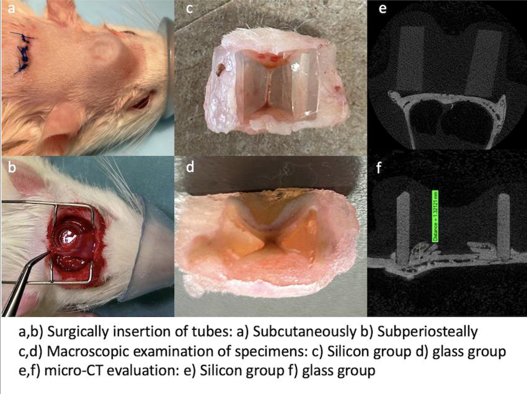
A109 - Development Of A Three-dimensional, Fibrin-based Oral Mucosa Tissue Equivalent For Characterization Of Oral Pathogens
L. Karchalla, C. Gaviria, A. Atala, P. McNutt
Wake Forest Institute of Regenerative Medicine, Winston Salem, NC
*Purpose/Objectives: The oral cavity is second only to the gastrointestinal tract in the prevalence of archaea, protozoa, bacteria, fungi, and viruses. These microorganisms predominantly maintain a commensal or non-pathogenic status under normative physiological conditions. Productive infection of the oral cavity by opportunistic and pathogenic microbes under conditions of pathological stress can result in severe morbidities, including cancer, abscess, and sepsis. Microbial pathogenesis of the oral cavity is predominantly studied using simple two-dimensional cell models and animal models that may inadequately capture the complexity and unique properties of the human oral cavity. To develop a more authentic representation of oral disease, we developed a three-dimensional oral mucosa tissue equivalent (OMTE) constructed from human primary cells.
*Methodology: Oral fibroblasts and HUVEC cells were embedded in a fibrin-based gel followed by apical seeding of oral keratinocytes. Seven days after the addition of oral keratinocytes, OMTEs were airlifted to allow differentiation of the epithelial layer. At 7-28 days after genesis, OMTEs were fixed for histological analysis and immunohistochemistry using antibodies against phenotypes, including CK4, CK5, CK13, Vimentin, and CD31. The formation of an epithelial barrier was measured by trans-epithelial electrical resistance (TEER) and lucifer yellow penetration.
*Results: Preliminary characterization of OMTEs confirms the functional and molecular maturation of the OMTEs. OMTEs exhibited functional integrity of the epithelial barrier by increasing TEER values and reduced Lucifer yellow penetration. Histology and immunohistochemistry corroborated functional maturation and demonstrated the appropriate cellular distribution and expression of buccal cell types. The epithelium was multilayered and pseudostratified, consistent with primary buccal tissue. Fibroblasts are well distributed throughout the fibrin gel and exhibited structures similar to in vivo histological morphologies. Within seven days after plating, endothelial cells assembled into a robust, ramified neocapillary network, involving formation of thin-walled vessels with large luminal areas that remained intact for at least 21 days.
*Conclusion/Significance: Functional, molecular, and histological data indicate that three-dimensional, fibrin-based OMTEs replicate the critical elements of the anatomy and functions of native oral mucosa. We are currently testing the suitability of OMTEs as a platform to characterize host responses to orally targeted pathogens, beginning with oral viruses. Collectively, these data suggest OMTEs are a novel platform to study the interactions of microorganisms and the oral mucosa.
A113 - Effects Of Different Concentrations Of Bioceramic In Alginate On Bioinks For Bone Tissue Regeneration
R. P. Silveira1, N. Maurmann1, R. L. Siqueira2, L. M. Notargiacomo1, C. A. Ferreira1, P. Pranke1,3
1
Universidade Federal do Rio Grande do Sul (UFRGS), Porto Alegre, Brazil,
2
Pesquisador Independente, Itabira, Brazil,
3
Instituto de Pesquisa com Células-tronco (IPCT), Porto Alegre, Brazil
*Purpose/Objectives: Alginate forms three-dimensional hydrogel used as bioinks in tissue engineering. Bioceramic-infused bioinks provide enhanced bioactivity and structural support. This study examined the effect of varying bioceramic concentrations in alginate hydrogel on stem cell viability and osteogenic differentiation capacity.
*Methodology: A 1% w/v solution of sodium alginate mixed with bioceramic (BC) at concentrations of 0, 1, 5, or 10% w/v and cross-linked with 50 mM calcium chloride was tested. The hydrogels were characterized by scanning electron microscopy (SEM), thermogravimetric analysis and cone-plate rheometry. For the in vitro study, mesenchymal stem cells at a density of 200,000 cells per well were mixed with 100 µL of the hydrogel. After 7 days, cell viability was assessed using the MTT assay. Osteogenic differentiation was analyzed after 28 days by Alizarin Red staining. Data were expressed by mean ± standard deviation.
*Results: SEM showed hydrogel-porous structures with thin walls, and the increased BC produced more mineralized material. In the rheological analysis, increases of up to 5% in the BC concentration resulted in a decrease of viscosity in comparison to the 0% BC sample, with small variations between the different additions of BC. The samples with 1% and 5% bioceramic, therefore, remained within the same range, slightly lower than that of the 0% sample, while the 10% bioceramic sample exhibited higher viscosity than the others. The samples displayed pseudoplastic behavior, meaning their viscosities decreased as the shear rate increased. In the thermogravimetric analysis, all the samples exhibited a wide first thermal event between 25 and 150°C, mainly related to water loss and possible low molecular weight compounds. Alginate without the bioceramic sample showing the highest mass loss (97.5%) and the 10% bioceramic sample displaying the lowest mass loss (83.6%). There was also a second thermal event of lower intensity between 225 and 300°C, due to degradation of the alginate polymer chain. The samples showed increasing residue according to the higher levels of BC added. The bioceramic significantly increased cell viability and osteogenic differentiation in a dose-dependent manner (P≤0.05). The absorbance values in the MTT were 0.06±0.01, 0.07±0.00, 0.08±0.01 and 0.09±0.01, respectively for alginate with 0, 1, 5, and 10% BC. Alizarin Red absorbance was 0.2±0.1 and 0.3±0.1, respectively for the 2D culture of stem cells undifferentiated and differentiated without biomaterials and in alginate 2.0±1.3 without BC, 2.3±0.6 for 1%, 3.5±0.2 for 5% and 4.0±0.2 for 10% BC.
*Conclusion/Significance: The composite of bioceramic in alginate provided a 3D microenvironment that serves as a platform to replicate the structure and function of organs and tissue in the human body. Specifically, the gel formulation with 1% alginate and 10% bioceramic demonstrated the most favorable outcomes in vitro tests with the cells, making it a potential bioink for 3D bioprinting for bone tissue engineering. Combining alginate and bioceramic shows promise for effective growth support and stimulation of osteogenic differentiation for bone tissue engineering. Financial support: Office of Naval Research Global (ONRG Award N62909-21-1-2026), CNPq, FINEP, INCT-Regenera and Stem Cell Research Institute.
A115 - Effects Of Dentine Conditioning On Growth Factor Release And Dental Pulp Stem Cell Response
Q. Zeng, R. Moses, M. Evans, A. J. Sloan
University of Melbourne, Melbourne, Australia
*Purpose/Objectives: Liberation of the sequestrated bioactive molecules from dentine, by action of the applied dental materials, has been proposed as an important mechanism in inducing a dentinogenic response in teeth with viable pulps. Growth factors within the dentine matrix play a key role in inducing proliferation and differentiation of dental pulp stem cells (DPSCs) to induce dentine repair and regeneration. Routine dental materials including polyacrylic acid, phosphoric acid and ethylenediaminetetraacetic acid (EDTA) have been shown to induce liberation of these growth factors. The aim of this study was to investigate the effects of these agents on TGF-B1, VEGF and BMP2 release and duration of application to induce optimum release.
*Methodology: Human dentine slices were conditioned using 10% EDTA, 10% polyacrylic acid, 37% phosphoric acid and 10% citric acid for 30s, 1 min, 2.5min or 5min. The conditioning agents polyacrylic acid, phosphoric acid and EDTA were each applied using microbrushes to simulate clinical practice and application, with conditioned slices incubated for 72h. Conditioned media was collected post treatment and analysed using enzyme-linked immunosorbent assay (ELISA) for TGF-B1, VEGF and BMP2 release. Conditioned media was also applied to dental pulp stem cells (DPSC)s, with gene expression of key cellular and molecular markers analysed using qPCR to evalute effects on cell viability and differentiation along with qualitative microscopy and cytometry.
*Results: Upregulated TGF-B1 and VEGF release was observed following EDTA, polyacrylic acid, phosphoric acid and citric acid conditioning, compared to untreated control. However, increased BMP2 release was only observed following citric acid. Increased release of TGF-B1 was highly significant following EDTA (all time-points) and phosphoric acid (1min and 2.5min) conditioning . Significant upregulation of VEGF release was observed following 30s EDTA conditioning, and polyacrylic acid for all time periods. Increased conditioning time resulted in reduced growth factor release. DPSCs treated with conditioned media from shorter treatment time points demonstrated viability in comparison to those exposed to conditioned media from longer treatment timepoints.
*Conclusion/Significance: Conditioning of dentine surfaces using EDTA, polyacrylic acid and phosphoric acid appreead to result in upregulation of TGF-B1 and VEGF release from the tissue. Data suggests that conditioning of dentine surfaces under 2.5min in duration appeared to be sufficient to induce significant growth factor release, which is also more achievable clinically, and maintain DPSC viability. This has longer term implications in the repair of pulp tissue and long term viability following clinical treatment and the and the development of regenerative endodontic approaches and vital pulp therapy.
A117 - A Novel Intraoperative Infection Detecting Liquid For Medication-related Osteonecrosis Of The Jaw
S. Arimoto1,2, M. Kubo2, K. Shigenaga2, A. Yoneda2, D. Takeda2, T. Inagaki1, M. Shigeoka2, T. Hasegawa2, A. Hokugo1, M. Akashi2
1
David Geffen School of Medicine at UCLA, Los Angeles, CA,
2
Kobe University Graduate School of Medicine, Kobe, Japan
*Purpose/Objectives: Antiresorptive medications, such as bisphosphonates and denosumab, are effective for the management of cancer, osteoporosis, and multiple myeloma. However, medication-related osteonecrosis of the jaw (MRONJ) is a serious and common complication in patients receiving these drugs who undergo dental extractions. Several recent reports recommend surgical treatment, guidelines for determining the surgical margins of MRONJ have not yet been developed. Therefore, this study aimed to investigate the efficacy of various widely available and inexpensive dyes for detecting infected bone areas and develop a novel intraoperative infection-detecting liquid for MRONJ.
*Methodology: Nine-week-old female Sprague-Dawley (SD) rats received intravenous (IV) injections of zoledronic acid hydrate (ZOL) in phosphate-buffered saline weekly for 10 weeks. IV injections were administered into the tail vein. The upper left first and second molars were extracted 1 week after the first dose of ZOL. A 2-mm-diameter round drill was used to expand the extraction sockets and create a bone defect of size 3 × 5 × 3 mm, and mechanical stimulation using a dental chisel was performed weekly to prevent gingival cover. Osteonecrosis was defined as persistent bone exposure for 8 weeks after tooth extraction. This protocol was confirmed to create a stable rat model of MRONJ with exposed and necrotic bone. First, we performed a preliminary experiment using five dyes in a rat model of MRONJ. Eighteen female SD rats were divided into the undyed (n = 3), acid red (n = 3), Brilliant Blue (n = 3), phloxine (n = 3), methylene blue (n = 3), and Rose Bengal (n = 3) groups. Based on visual inspection and histological findings, Brilliant Blue and phloxine were selected for further evaluation. Next, twenty-eight female SD rats were divided into the control (saline) (n = 7), undyed (n = 7), Brilliant Blue (n = 7), and phloxine (n = 7) groups. All rats received ZOL or saline IV weekly for 18 weeks. Each dye was applied to experimentally induced MRONJ. The stained area was completely removed, and the bone defect was closed with sutures. Eight weeks after surgery, soft-tissue evaluation and histological and microcomputed tomography analyses were performed.
*Results: The area of oral mucosal swelling was significantly smaller in the phloxine group than in the undyed and Brilliant Blue groups (P <0.05). The remaining osteonecrosis area in the phloxine group was significantly smaller than that in the undyed group (p<0.05).
*Conclusion/Significance: We performed a preliminary experiment using five dyes in a rat model of MRONJ, and based on the results, we evaluated the treatment effects of Brilliant Blue and phloxine. The results indicate that phloxine can be used to differentiate necrotic bone from normal bone during surgery. Further studies are required to improve the intraoperative infection-detecting liquid. We hope that our findings will contribute as the first step toward complete excision of necrotic bone during surgical treatment of MRONJ.
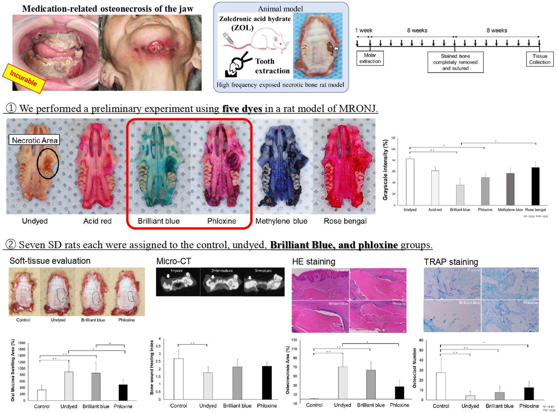
A118
A119 - Intracellular Calcium Dynamics In Response To Low-Intensity Ultrasound Stimulation
K. P. Grassie1, F. Wang1, H. Anderson1, B. D. Huey1, Y. M. Khan2,1
1
University of Connecticut, Storrs, CT,
2
UConn Health, Farmington, CT
*Purpose/Objectives: Mechanical forces play a key role in regulating mesenchymal stem cell fate, bone maintenance, and fracture repair. Low-intensity ultrasound-derived acoustic radiation force (ARF) has become a promising tool in tissue engineering for stimulating cells with such forces in order to enhance osteogenic activity. In the collective effort to understand the mechanisms by which cells respond to ARF, intracellular calcium ion concentration ([Ca2+]i) changes have been observed as an immediate response to acoustic forces. [Ca2+]i dynamics are well-known to be triggered by mechanical stimuli and activate pathways leading to osteogenic differentiation and bone formation. However, it has not been determined how ARF stimulates Ca2+ responses and if the response depends on stimulus intensity. Here, we have performed in vitro live-cell fluorescence imaging of [Ca2+]i within osteogenic progenitor cells exposed to ARF with two different intensities that correspond to clinically relevant ranges used for bone repair to see if ARF intensity can be used to modulate Ca2+ response, and perhaps subsequent cell behavior.
*Methodology: MC3T3 preosteoblasts cultured on coverslip-bottomed dishes were incubated in Fluo-4 calcium indicator dye using a Fluo-4 AM Kit (ThermoFisher) with a Hoechst nuclear counterstain. Imaging medium was then added to each dish before placement on the stage of an epifluorescence microscope (Leica) with an ultrasound transducer immersed into the medium 1-2 mm above the cells. Imaging was performed continuously with the following timeline: 1-minute baseline, 5-second burst of ultrasound (0, 30, 300 mW/cm2), and 15-minute recovery. Ionomycin was then immediately added to elicit maximal Ca2+ responses and imaged for 3 more minutes. Image sequences were analyzed with MATLAB codes that calculated mean Ca2+ fluorescence per cell for all cells in view (100-200 per field), visualized with color-mapped log-scale histograms of fluorescence intensity vs. time. Mean and standard deviation of these traces for the entire population of imaged cells were also plotted.
*Results: Figure 1 shows representative fluorescence images of the cells and average Ca2+ fluorescence traces for all cells in view. Figure 1A-F show fluorescence response just before (A-C) and 4 minutes after ARF (D-F). Histograms reveal that, beyond spontaneous Ca2+ responses without ultrasound present, stimulation with just 5 seconds of ultrasound triggers noticeable increases in cytoplasmic Ca2+ in the majority of cells. Moreover, peak [Ca2+]i responses occur later and are more pronounced with higher intensity, suggesting that the Ca2+ response is not binary but rather can be controlled by stimulus intensity. However, in both conditions, [Ca2+]i responses relax to comparable elevated levels several minutes later but are still active given the response to ionomycin. This suggests that [Ca2+]i dynamics are influenced by acoustic intensity and that elevated ultrasound-induced [Ca2+]i may take several minutes to recover.
*Conclusion/Significance: These findings suggest that Ca2+ signals, which are known to be upstream of important osteogenic pathways, can be modulated by the intensity of ARF. Future studies will evaluate if this dependence on input extends to the temporal/pulsation pattern of ARF. The ability to manipulate [Ca2+]i via ARF would allow for the design of optimized stimulation regimens that maximize osteogenic activity.
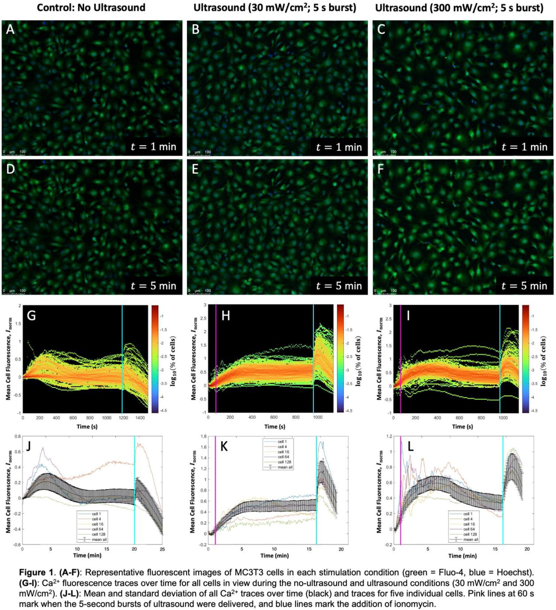
A121 - CO-loaded, Ultrasound-sensitive Microbubble Delivery For Reducing Blood Brain Barrier Permeability
R. Jourdain1, S. Changizi1, A. Han2, V. Chivukula1, C. A. Bashur1,1
1
Florida Institute of Technology, Melbourne, FL,
2
Virginia Tech, Blacksburg, VA
*Purpose/Objectives: Vascular cognitive impairment and dementia (VCID) is caused when blood components leak through the blood brain barrier (BBB), and the BBB becomes more permeable with age and disease. Most VCID treatments temporarily disrupt the BBB to deliver drugs to treat existing cognitive symptoms, but we are developing a unique strategy to repair the BBB and prevent further damage by delivering carbon monoxide (CO) from ultrasound-sensitive microbubbles (MBs). Previously, we computationally determined that the amount of free, MB-released CO should be able to positively impact the ECs. However, not all MBs rupture with low-frequency ultrasound, and endocytosis of CO-containing MBs could also deliver CO to the ECs. This study uses both experimental and computational analyses to determine the unruptured and endocytosed MBs that could reach the BBB.
*Methodology: Perfluoropentane-based MBs were prepared through a nanoprecipitation method (1.30±0.40 µm) and loaded with CO. A gelatin-based phantom with a 3 mm diameter embedded straw was used to experimentally determine the MB rupture percentage and the speed that they move with ultrasound. GFP+ human umbilical ECs (HUVECs) were cultured and imaged with time-lapse, confocal microscopy to investigate the MB endocytosis rate. One-way ANOVA and Tukey comparisons were used. These particles were modeled using computational fluid dynamics (CFD) to investigate the amount of MB that reach the ECs at the BBB arterial wall after ultrasound activation in the internal carotid artery (ICA). The CFD model considers both hemodynamics with pulsatile blood flow and ultrasound pressure-induced movement of the MBs (100 MBs released over 8 s). The impact of gravity, buoyancy, and different percentage of MB rupture are considered. Plane-wave application of 3, 7, and 18 MHz ultrasound were also investigated.
*Results: Experimental results using an 18 MHz transducer showed that MBs moved in response to application of ultrasound. Preliminary data showed average and maximum speeds of 10 mm/s and 100 mm/s, respectively, which is only slightly less than the 67 mm/s average MB speed due to blood flow alone in our CFD simulations. Further, differences in applied ultrasound parameters impacted the experimental rupture percentage. The normalized number of ruptured microbubbles/image systematically increased with intensity and was significantly higher for 100% intensity. Computational simulations also showed MB movement with application of ultrasound. With increasing wave frequency for simulated 3-18 MHz transducers, the MBs moved faster within the application region. Our ongoing CFD studies are combining MB movement from ultrasound and blood flow to determine the number of available MBs to be endocytosed by ECs at the BBB. Our preliminary experimental studies show particles with diameters between 0.82-6.3 µm accumulating within ECs but not the larger agglomerates.
*Conclusion/Significance: This study notes important considerations for this different application of ultrasound to deliver CO-loaded MB to promote EC growth and BBB healing. The results show that clinical ultrasound machines with different frequencies and intensities will impact the rate and amount of CO that can be delivered. Further, this study links traditional ultrasound analysis with hemodynamics and MB movement, and shows how this is important to consider.
A122 - Alignment Of Primary Cardiomyocytes Using Ultrasonic Standing Waves
J. F. Murphy,1, X. Chen, X. Yu, X. Li, Y. Huang
University of Cambridge, Cambridge, United Kingdom
*Purpose/Objectives: Cardiovascular disease is the leading cause of death worldwide; the development of cardiovascular therapies is hindered by a lack of suitable in vitro microphysiological models for the human heart. Isolated cardiomyocytes undergo acute remodeling and dedifferentiation while stem cell-derived cardiomyocytes fail to fully reach a mature phenotype, having distinctly different metabolic pathways, reduced calcium signaling, rounded morphology, and reduced contractility. To overcome these limitations, which often prevent the translatability of the results seen in these models, platforms need to better recapitulate the in vivo microenvironment. One key component missing from current cell culture platforms is the rigid hierarchical architecture seen in the myocardium; the random distribution and orientation of cardiomyocytes seen in many platforms prevent physiological force and signal propagation through adjacent cells.
*Methodology: This research uses ultrasonic standing waves to reform the highly aligned native tissue architecture seen in the myocardium. A pair of parallel piezoelectric transducers were stimulated using sinusoidal alternating voltage at their resonant frequencies. Isolated murine cardiomyocytes were added to the generated pressure field and allowed to settle. The effects of different substrate conditions on patterning were subsequently investigated, including both adherent (laminin-coated) and non-adherent substrates.
*Results: Acoustic patterning was able to provide planar migration into 2D lines evenly spaced throughout the culture platform at the pressure nodes; additionally, due to the non-circular shape of cardiomyocytes, the acoustic waves provided a torque that was minimized when cardiomyocytes were oriented with their major axis parallel to the pressure nodes. These effects combined to form tightly packed lines of cardiomyocytes that were aligned along the same axis. Non-adherent substrates such as uncoated glass provided the most uniform patterning as they allowed migration and rotation even after contact with the surface while adherent substrates only allowed patterning during the settling phase because if contact was made before fully reaching a node the adhesive force would prevent further migration.
*Conclusion/Significance: To our knowledge, this research is the first to demonstrate that ultrasonic standing waves can be used to control both the orientation and alignment of non-spherical cell types, expanding the applications of ultrasound cell patterning technology to include a rotational alignment component in addition to the typical planar spatial patterning. This is applied to guide the reformation of anisotropic tissue-like architectures seen in the myocardium that have been difficult to recapitulate in vitro to date.

A123 - Ultrasound As A Non-destructive Sensor For Bioreactor Monitoring: Preliminary Results With Spinach Scaffolds
R. Thyden, M. Serpe, C. Lee, R. Povinelli, L. Perreault, G. Gaudette, K. He, B. Ranger
Boston College, Chestnut Hill, MA
*Purpose/Objectives: Cellular agriculture, or the biomanufacturing of animal meat products, offers a promising alternative to conventional meat production by minimizing the use of animals, shortening the supply chain, and transforming human interactions with nutrient sources. One of the most common strategies for cultured meat production involves attaching and growing anchorage dependent animal cells to an edible scaffold. By leveraging plant properties, we are using plants as edible scaffolds for animal cell growth, taking advantage of the biocompatibility of plant extracellular matrix (ECM). This approach will ultimately allow for low-cost, scalable production of muscle tissue for various types of meat, poultry, and fish. To optimize the production of bio-manufactured cell-based meats using plant scaffolds, we are exploring bioreactor designs that will be used to proliferate, perfuse and stimulate the cells on plant scaffolds. Currently, the conventional way to monitor cell populations on individual scaffolds and ensure quality control (QC) is destructive and not conducive for scale-up. Therefore, there is a pressing need for non-destructive and low-cost sensors for QC in bioreactor designs.
*Methodology: Ultrasound imaging is relatively low-cost imaging modality that is commonly used in a wide variety of fields, ranging from healthcare to non-destructive material testing, to visualize and characterize a plethora of substances. Our objective is to develop ultrasound imaging methods for non-destructive evaluation of cell populated scaffolds that can ultimately be utilized for QC in bioreactors. As shown in Figure 1A, we envision that ultrasound may be used at various stages of a bioreactive process: verifying decellularization, cell adhesion, and differentiation.
*Results: A Clarius L15 ultrasound probe was mounted, using a clamp, above a small water basin (Figure 1B). The basin contains a sample of the spinach leaf scaffold. We collected image data at different time points during the decellularization and re-cellularization process. Figure 1C shows an image of a decellularized spinach leaf scaffold containing cell proliferation wells. As shown, the proliferation wells are distinctly visible in the images. All resulting images were analyzed using WEKA, a semi-automatic image segmentation software available through ImageJ to calculate mean pixel intensities of the decellularized spinach leaf scaffold and proliferation wells (Figure 1D). Preliminary calculations demonstrated significant differences between mean pixel values of the decellularized scaffold and the proliferation wells.
*Conclusion/Significance: Preliminary analysis of mean pixel intensity when comparing proliferation sites and decellularized sites on spinach leaf scaffolds demonstrate that ultrasound imaging is a feasible technology for monitoring the presence of animal cells on scaffolds. When incorporated into a bioprocess production system, ultrasound sensors may allow for scalable QC measures such as real-time, non-destructive, reproducible, and multi-parameter analysis.
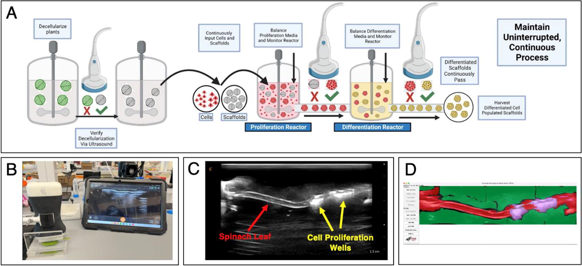
A125 - A Minisaturised Imaging Window for Directing and Visualizing Foreign Body Reactions In Vivo
C. Conci1, L. Sironi2, D. Panzeri2, E. Jacchetti1, D. Inverso2, R. Martinez-Vazquez3, G. Cerullo3, R. Osellame3, M. Collini2, G. Chirico2, M. T. Raimondi1
1
Politecnico di Milano, Milano, Italy,
2
University of Milano Bicocca, Milano, Italy,
3
Istituto di Fotonica e Nanotecnologie IFN-CNR, Milano, Italy
*Purpose/Objectives: Standardized evaluation of the fibrotic response to biomaterial implantation relies exclusively on ex vivo histological methods, which need a large number of animals to be sacrificed. Instead, intravital microscopy enables more detailed microscopy visualizations and fewer laboratory animals; nevertheless, currently available imaging techniques associated with this operation are highly invasive and unethical. To advance the field, we built a miniature imaging window, the Microatlas, incorporating a micro scaffold able for controlling tissue regeneration in vivo. In order to facilitate tissue regeneration and time-lapse imaging at the interface with an implanted biomaterial, the Microatlas can be subcutaneously implanted in a living creature. Here, we measured the foreign-body reaction in the same tissue district at different times during implantation using label-free two-photon microscopy within the Microatlas implanted in vivo and repopulated by the host cells and blood vessels.
*Methodology: We produced the micro scaffolds, size 500x500x 100μm3, using two-photon polymerization of a polymer known as SZ2080 on glass coverslips with a thickness of 12 mm. In live chick embryos, we conducted the chorioallantoic membrane (CAM) experiment 3, 4, and 7 days following Microatlas implantation (implanted at the embyonic incubation day 7). In vivo inspection of the imaging window was performed using two-photon label-free (TPEF) and confocal microscopy. Second harmonic generation (SHG) imaging of collagen I was performed. Ex vivo assessments were carried out by sacrificing embryos. The CAM was either paraffin-sectioned, stained with hematoxylin and eosin, or labeled with DRAQ5 and observed by confocal microscopy.
*Results: Confocal and two-photon analyses at the implantation sites in chick embryos revealed tissue development inside the scaffolds and neo micro vascularization, along with the existence of a capillary density six times higher than in the control tissue (unimplanted). SHG revealed the presence of a collagen-I layer that was preferentially orientated and had a density similar to control areas. Cell density could be measured using confocal microscopy and label free TPEF, which revealed a two-fold higher infiltration rate than in control tissues (unimplanted). Numerous infiltrating cell types, such as granulocytes, fibroblasts, and endothelial cells, were denoised, segmented and then quantified thanks to deep learning based algorithms.
*Conclusion/Significance: The Microatlas was able to control and to host foreign body reaction to the micro scaffolds, in terms of cell repopulation, collagen synthesis and capillary development. Our results open up the possibility to corroborate or to replace traditional histopathology with label-free linear and non-linear microscopy, in compliance with the European ISO10993. In fact, our method will make it possible to closely track the dynamic, in vivo, and minimally invasive tissue response to implanted biomaterials. At the present stage, we plan to broaden the scope of data that may be collected by incorporating insights from fluorescence lifetime imaging and multiple virtual stains. Acknowledgements EU, FET-OPEN project IN2SIGHT, G.A.964481; ERC ,project BEACONSANDEGG, G.A.101053122.
A127
A129 - Pattern Recognition Receptor Agonist Loaded Hydrogels Decreases Scarring In A Rat Rotator Cuff Injury Model
S. E. Winston, D. Von Stade, C. Plaisance, R. Impastato, L. Chow, L. Pezzanite, S. Dow, K. McGilvray
Colorado State University, Fort Collins, CO
*Purpose/Objectives: Rotator cuff (RC) repair surgeries are failing at 20-94% rates. Histological analyses of repaired tendons show a lack of a regenerated transition region (enthesis) and prominent scarring, leading to a weaker bone-tendon junction. While scarring is a multimodal process, one of the largest drivers in these processes is hypothesized to be the cytokines produced by the local and infiltrating immune cells responding to the wound. The correct balancing of inflammatory and anti-inflammatory signaling to matrix-producing cells (tenocytes) is crucial for correct tendon regeneration, but this ratio is still unknown. This has brought significant interest in modulating the immune response as an avenue to improve tendon healing. Pattern Recognition Receptor Agonists (PRRAs) are a class of molecules that have been used for cancer treatment and vaccine adjuvants. These agonists are associated with activating the innate immune response through receptor classes such as Toll-like (TLR) and nod-like receptors (NLR). We aim to investigate the effects of NLR-3 and TLR-3 agonist-loaded hydrogels and determine if there are differential effects on an injured tendon healing in a rat model through histopathological means.
*Methodology: Sprague Dawley Rats (N=32) were injured bilaterally through a partial RC release model and immediately received injections of a methylcellulose/hyaluronic acid (1.5wt%/0.5wt%) blend hydrogel with NLR3 agonist Aluminum Hydroxide (Invivogen) (ALOH) or TLR3 agonist Poly(I:C) (Invivogen) at concentrations of 50 ng/mL. In addition, select rats also received a melt electrowritten polycaprolactone (PCL) mesh. Rats were euthanized at 1-, 4-, and 12-weeks post-injury. Shoulders were embedded in paraffin, 5 µm sections were cut, and stained with Hematoxylin and Eosin (H&E). Slides were blindly scored on a 5pt 0-4 according to ISO 10993-06, as well as signs for tendon-specific markers such as tenocyte activity, tendon vascularity, and collagen disorganization.
*Results: After 4 weeks, the ALOH group showed lower levels of fibrosis around the tendon (Figure 1a), lower levels of tendon vascularity (Figure 1b), and lower levels of collagen disorganization (Figure 1c) compared to the injured control. The ALOH+mesh group also had lower levels of collagen disorganization, indicating that the PCL effected collagen formation and local reactivity. The Gel+Poly(I:C) group only deomstrated lower levels of tendon vascularity.
*Conclusion/Significance: The ALOH groups had different responses compared to the control as well as the Poly(I:C) groups, indicating that activating the immune system through NLR and TLR pathways has a differential response on the healing cascade of a tendon wound. ALOH groups had signs of increased healing in the surgical site and in the tendon body after 4 weeks as collagen disorganization and tendon vascularity are signs of tendon degeneration. Additionally, similarities in the scoring of the ALOH groups with and without mesh indicate that the ALOH is directing the immune response rather than the PCL mesh, showing potential as an immunomodulatory augment to other synthetic implants, which are known to have advantageous mechanical properties but less than desirable immune responses.
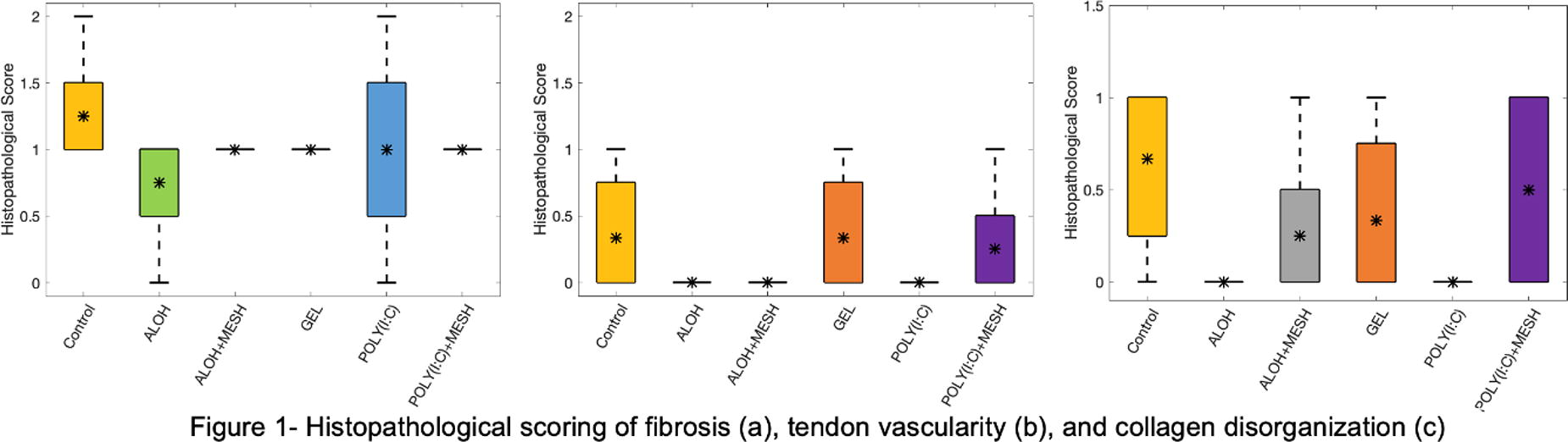
A130 - Dysregulated Inflammatory Response And Poor Fracture Healing In Polytrauma
A. M. Saiz1, M. Rahmati1, R. Gresham1, T. Baldini1, J. Burgan1, M. Lee1, B. Osipov1, B. Christiansen1, T. Khassawna2, F. Wieland3, Z. Sabouri2, A. Lopes3, C. Blanchet3, K. Leach1
1
UC Davis, Sacramento, CA,
2
Justus-Liebig University Giessen, Giessen, Germany,
3
Institute of Metallic Biomaterials, Geesthacht, Germany
*Purpose/Objectives: Bone healing is primarily concerned with treating isolated fractures in orthopedic patients. However, fractures in over 30% of polytrauma patients with multiple injuries exhibit impaired healing. There are significant gaps in our understanding of how fractures heal in this polytrauma environment. We aimed to characterize the temporal local and systemic immune responses to polytraumatic injuries in a polytrauma murine model using four experimental groups including healthy, chest trauma, isolated fracture and polytrauma groups.
*Methodology: To study the local and systemic immune responses and cytokine expression after injury, we collected serum, bone marrow from uninjured limb, femur, and lungs at 0 h, 6 h, 12 h, 24 h, 72 h & 3W following injury. Immune cell distribution from isolated bone marrow, femur, or lung tissue were assessed with a BD Fortessa 18-color flow cytometer. Femur tissues were collected after 3 weeks to study fracture healing using micro computed tomography, histological staining, immunohistochemistry, torsion test and small angle X-ray scattering.
*Results: Our flow cytometry results indicated significantly higher expression of innate immune cells in the polytrauma group compared to other groups locally and systemically. In contrast, the expression of B and T cells was dramatically suppressed in the polytrauma group at 6h following injury and remained low throughout the 3 weeks’ timeline of the study. The decreased expression of B and T cells demonstrated an exhaustion of the adaptive immune system response that could be a contributor to fracture nonunion (Figure 1A).
*Conclusion/Significance: Our data confirms the early, dysregulated inflammatory state in polytrauma correlated with impaired fracture healing and the formation of a poor and dysregulated callus in this polytrauma group (Figure 1B&C). Taken together, this study elucidates the role of persistent immune and inflammatory dysregulation that leads to poor fracture healing.

A131 - Trpv4 Inhibition Dampens Inflammation And Enhances Osteogenesis: A Potential Novel Therapeutic To Promote Fracture Repair
S. Petrousek, G. Soares Kronemberger, S. O’Rourke, L. Shanley, A. Dunne, D. Kelly, D. Hoey
Trinity College Dublin, Dublin, Ireland
*Purpose/Objectives: Fracture healing is a complex, highly regulated process with consecutive and linked phases of inflammation, repair, and remodelling. The initial inflammatory phase, mediated in part by infiltrating macrophages, is critical to long term healing and can be dramatically affected by environmental factors such as excessive mechanical loading in unstable fractures or augmented levels of inflammatory cytokines in disease, leading to delayed/non-healing of fractures. Recent studies have demonstrated the potential role for calcium channels and signalling in inflammatory pathologies. In particular, TRPV4, a known mechanosensitive Ca2+ channel, has been linked to both respiratory and cardiac inflammatory pathologies, and is being therapeutically targeted using the TRPV4 antagonist GSK2798745 (GSK) in current clinical trials. In this study, we investigated the potency of this TRPV4 antagonist to modulate macrophage phenotype and function, in addition to osteogenesis and angiogenesis in the context of bone healing.
*Methodology: Human THP-1 monocytic cells were differentiated into macrophages with 100nM PMA at 1 × 10⁶ cells/ml in complete RPMI medium for 3 days. To evaluate the potency of GSK, PMA-differentiated macrophages were first pre-treated for 2 hours with 10nM, 100nM, and 1μM of GSK to inhibit TRPV4 activity, followed by a 24-hour differentiation into pro-inflammatory M1 phenotype with LPS (100ng/mL) and IFN-ϒ (20ng/mL). mRNA and cytokine levels were analysed by real-time PCR and ELISA, and macrophage conditioned media was collected. The osteogenic and angiogenic potency of the GSK-treated macrophage secretome and GSK drug alone was first assessed by performing a 14-day osteogenesis assay and quantifying proliferation, intracellular ALP and calcium deposition, as well as a 10-hour tube formation assay and quantifying the number of segments.
*Results: First, we analysed macrophage phenotype following GSK treatment and confirmed downregulation of pro-inflammatory M1 markers such as CD80 and IL-1β on a gene expression level. Similarly, a 40% decrease in pro-inflammatory IL-6 and TNF-α secretome was observed with the higher 1μM dosage demonstrating how TRPV4 inhibition can modulate macrophage phenotype. Next, we performed functional assays to assess the effect of the GSK-treated M1 macrophage secretome and the GSK drug alone on osteogenesis and angiogenesis. While GSK treatment induced lower levels of inflammation in a pro-inflammatory macrophage, we observed no impact of the M1-GSK treated secretome on mesenchymal stem cell (MSC) proliferation, intracellular ALP and calcium deposition, as well as on the number of segments, suggesting limited impact on subsequent osteogenesis and angiogenesis. However, we demonstrated a positive effect of the GSK drug alone on osteogenesis with a 20-fold increase in MSC proliferation, 2-fold increase in intracellular ALP and enhanced calcium deposition as shown qualitatively by Alizarin Red staining. No impact of the GSK drug was observed on angiogenesis.
*Conclusion/Significance: Our findings indicate that modulation of the immune environment can be achieved targeting TRPV4. TRPV4 inhibition via GSK2798745 was shown to inhibit M1 polarisation and to promote regenerative properties in MSCs with enhanced bone deposition. This study indicates a potential role for TRPV4 in macrophage behaviour and highlights potential targets to therapeutically modulate the immune response, establishing a regenerative environment to bolster bone repair.
A132 - Immunomodulation Of Glial Cell Activation With Local Sustained Delivery Of Pituitary Adenylate Cyclase-activating Polypeptide (PACAP) Results In Improved Functional Recovery In Stroke-injured Mice
E. Ho1, D. X. Li1, D. Akbar1, R. Siu1, C. M. Morshead1, D. Chatenet2, M. S. Shoichet1
1
University of Toronto, Toronto, ON, Canada,
2
INRS, Laval, QC, Canada
*Purpose/Objectives: Ischemic stroke is one of the leading causes of death and permanent neurological disability worldwide. There remain no treatment options that restore brain function after stroke, underlining the need for regenerative approaches. Immunomodulating the stroke microenvironment towards a pro-reparative phenotype offers the potential for neural tissue repair. Following ischemic stroke, resident glial cells (astrocytes and microglia) become activated to create an extremely hostile microenvironment that diminishes cell survival and exacerbates brain damage; yet these cells play dual roles that also contribute to tissue regeneration for improved neural repair. Pituitary adenylate cyclase-activating polypeptide (PACAP) is a potent factor that modulates the activation state of microglia towards a pro-reparative phenotype. However, it is unstable in circulation and exhibits detrimental side effects when systemically delivered, emphasizing the need for local sustained delivery strategies. Here, we developed a hydrogel-nanoparticle composite for the minimally invasive, local delivery of PACAP to the brain and tested this strategy in endothelin-1 (Et-1) stroke-injured mice.
*Methodology: To achieve sustained release, positively charged PACAP was electrostatically adsorbed to the negative surface of poly(lactic-co-glycolic acid) (PLGA) nanoparticles incorporated in a hyaluronan-methylcellulose (HAMC) hydrogel; as the PLGA degrades, the negative surface charge is neutralized and PACAP is released. This strategy significantly reduces protein denaturation and increases loading as compared to conventional PLGA encapsulation methods. The HAMC hydrogel is biocompatible, shear thinning to enable injection through a fine-gauge needle, and is inverse thermal crosslinking which allows for in-situ thermally controlled gelation at physiological temperatures. The hydrogel-nanoparticle composite was applied epicortically onto the cortex at the cortical stroke lesion site. Stroke was induced in mice by injecting Et-1, a vasoconstrictor that causes local constriction of the surrounding blood vessels followed by reperfusion, thereby mimicking human stroke.
*Results: We demonstrated that sustained delivery of PACAP improved the physical strength and functional forelimb recovery of injured mice 28 days after stroke. Importantly, the treatment decreased the number of apoptotic cells in the stroke microenvironment in the first week post-stroke and decreased infarct volume 1-month post-stroke. PACAP stimulation also attenuated reactive astrogliosis and microglia activation and resulted in an increase of Iba1+Arg1+ pro-reparative microglia and a decrease of Iba1+CD86+ pro-inflammatory microglia. This phenotypic shift in microglia states may account, at least partially, for the functional improvements post-stroke, and paves the way for new therapeutic strategies targeting the immune response for stroke recovery.
*Conclusion/Significance: Our data substantiate the role of PACAP in neural repair through neuroprotection and anti-inflammatory effects. This study provides insights into the timeframe of immunomodulatory interventions for stroke treatment where prolonged PACAP stimulation for 10 days can drive microglia in the stroke microenvironment to promote repair. This system could also be broadly applicable to other neurological disorders affected by microglial dysfunction.
A134 - An Infusible Extracellular Matrix Material Improves Survival In An Animal Model Of Multi-organ Dysfunction Syndrome Via Modulation Of Inflammation Regardless Of Age
M. L. Karkanitsa, J. Mesfin, M. Nguyen, A. Chen, M. Hepokoski, K. L. Christman
University of California San Diego, La Jolla, CA
*Purpose/Objectives: Mortality in septic shock patients is elevated with age, particularly due to organ damage triggered by excess systemic inflammation. While most deaths caused by septic shock are triggered by inflammation-induced multi-organ failure, there are no immunomodulatory therapeutics used routinely in this patient population. As a result, there is a need for an immunomodulatory therapeutic that can be systemically administered to promote survival in severe septic patients that functions regardless of age. To address this need, an infusible extracellular matrix biomaterial was evaluated in a non-aged and aged animal model of severe septic shock.
*Methodology: 9 week or 78-week C57 Bl/6 mice were intravenously administered iECM or saline (9 week: n = 47 (iECM), n = 65 (saline); 78-week: n = 14 (iECM), n = 15 (saline)) after performing a dual dose injection of lipopolysaccharide and survival was monitored for 20 hours post treatment. Log-Rank survival analysis was performed to determine significance in survival with material treatment. Animals were sacrificed and blood, lung, and kidney tissues were harvested and processed into a single cell suspension. Samples were pooled (n=2) into two separate samples for a total of 4 animals per treatment per age group. 20,000 cells per sample were loaded onto a 10x Chromium Next GEM 3’ HT chip and sequenced by NovaSeq S4 technology. Data was analyzed using Seurat for R.
*Results: Survival analysis showed significant improvements in survival with iECM treatment in 9-week mice, with similar trends in 78-week animals (Fig 1A). Cell types within lung and kidney tissue were successfully identified, with neutrophils being the largest cell type identified within lung tissue and proximal tubule cells being the largest cell population from kidney tissue with iECM treatment. Within lung tissue, neutrophil activation was altered in aged mice, with decreased expression of Camp, Asprv1, and Mgp in 78-week mice regardless of treatment (Fig. 1B) Interestingly, iECM treatment successfully altered neutrophil activation in 9-week mice as shown by decreased expression of Mgp, Cd177, and Tnf (Fig 1B). UMAP analysis of proximal tubule cells in kidney showed novel clusters of proinflammatory cells in 9-week saline-treated animals (Fig 1C), as confirmed by pathway enrichment analysis (Fig 1D) showing elevated TNF and JAK-STAT signaling within this cell population that was absent with iECM treatment.
*Conclusion/Significance: While age alters the immune drivers of inflammation in an animal model of MODS, iECM material promotes survival and retains its immunomodulatory and pro-regenerative properties. Single cell RNA sequencing of lung and kidney tissues identify tissue-specific drivers of inflammation and how they are altered with age as well as with iECM treatment. ScRNAseq analysis of blood is ongoing.

A135 - Attenuating The Alloimmune Response In Transplantation Through Co-implantation Of Autologous, Donor Antigen-primed Lymph Node Stromal Cells
C. M. Li, C. R. Mulligan, L. N. Teles, G. C. Gonzalez, O. C. Umland, A. A. Tomei
University of Miami, Miami, FL
*Purpose/Objectives: Patients receiving allogeneic tissue transplants require lifelong immunosuppression, reducing quality of life and applicability of transplantation procedures. We aim to reduce or eliminate the need for immunosuppression following allogeneic tissue transplantation by co-transplanting fibroblastic reticular cells (FRCs), a lymph node stromal cell subtype. FRCs are unconventional antigen-presenting cells that express self-antigens and MHC class I and II, enabling them to promote the regulation of antigen-specific T cells. Additionally, FRCs have limited expression of co-stimulatory CD80 and CD86 and upregulate programmed death-ligand 1 and 2 following interferon γ (IFNγ) treatment. Altogether, these mechanisms can contribute to restraining autoreactive T cells and can possibly be extended to prevent alloreactive T cell activation in transplantation. We hypothesize that autologous FRCs (i) pre-primed with donor cell lysate, (ii) pre-conditioned with IFNγ to upregulate antigen-presenting machinery, and (iii) co-transplanted with allogeneic donor tissue will reprogram the recipient’s alloimmune response toward inactivation/anergy through the above mechanism(s).
*Methodology: In all experiments, recipient-matched C57BL/6 FRCs were pre-conditioned with 10ng/mL IFNγ for 72h. To demonstrate FRC uptake of allogeneic donor cell-derived antigens, FRCs were pre-primed with NIT1 (non-obese diabetic background) donor cell lysate overnight. Cell lysate was generated by water bath sonication of NIT1 cells pre-labeled with CellTrace (overnight). No, low (100 IEQ), or high (1000 IEQ) doses of NIT1 cell lysate were administered to FRCs. To demonstrate FRC presentation of antigen after uptake, FRCs were treated with 50µM OVA257-264 (SIINFEKL) peptide overnight, and presentation in MHC I was detected with a SIINFEKL-H2Kb antibody (Biolegend). Dendritic cells (DC) and macrophages were used as control and derived from age-matched C57BL/6 mice. Three biologically distinct cell lines/preparations were tested. Readouts were obtained by flow cytometry and analyzed by one- or two-way ANOVA with Tukey’s multiple comparisons testing.
*Results: Following pre-treatment with NIT1 lysate, FRC viability was not affected while DC and macrophage viability decreased 10-20% with high lysate dose. Across all cell types, uptake of fluorescently labeled NIT1 lysate was dose-dependent, and IFNγ pre-treatment enhanced lysate uptake only in DCs and macrophages at low dose. At high dose, a greater proportion of FRCs took up NIT1 lysate (∼96%) compared to DCs (∼81%) and macrophages (∼70%). Additionally, analysis of mean fluorescence intensity indicates that FRCs have ∼2.4- and 1.4-fold higher antigen uptake compared to DCs and macrophages, respectively. Following pre-treatment with OVA peptide, FRC viability was not affected while DC and macrophage viability decreased ∼15% and 5%, respectively, due only to IFNγ treatment. IFNγ potentiates OVA peptide uptake and presentation, with >99% positive cells in all cell types. Analysis of median fluorescence intensity indicates that FRCs have ∼2-fold higher OVA peptide presentation compared to DCs.
*Conclusion/Significance: FRCs have antigen uptake and presentation capacities equal to or greater than that of DCs and macrophages, predictive of effective interaction with antigen-specific T cells. Recipient FRCs are capable of uptaking allogeneic donor cell antigen and could provide immunomodulation of recipient T cells alloreactive to donor tissue. Follow-up studies will confirm FRC-alloreactive T cell interaction and suppression in vitro and in vivo transplantation models.
A136 - Investigating The Immunomodulatory Effects Of Manuka Honey And Its Components To Prevent Neutrophil-Mediated Fibrosis
E. N. Main, J. C. Huang, G. L. Bowlin
University of Memphis, Memphis, TN
*Purpose/Objectives: Neutrophils use reactive oxygen species (ROS) activity and a specialized form of cell death named NETosis to kill and ensnare invading pathogens. While these neutrophil behaviors are critical in preventing infection, a dysregulated response can be deleterious and lead to tissue damage and fibrosis at host-biomaterial interfaces. Manuka honey has demonstrated potent antibacterial properties and recently, anti-inflammatory potential. It was hypothesized that applying potentially therapeutic compounds found in abundance within Manuka honey to pro-inflammatory neutrophils will reduce intracellular ROS activity and prevent NETosis. Additionally, it was investigated whether these compounds act more effectively as isolates or synergistically within whole Manuka honey. Thus, a combination of components, as well as concentrations of whole Manuka honey were also assayed. Pinobanksin, pinocembrin, and chrysin are the most abundant flavonoids and methyl syringate is the most abundant phenolic component in Manuka honey. Concentrations between 1 nM and 1 mM of each flavonoid, 10μM and 2mM of methyl syringate, 0.1% v/v and 10% v/v Manuka honey, and combinations of both 1 nM-1 mM of each flavonoid plus 10μM-2mM of methyl syringate were assayed for reductions in NETosis and intracellular ROS activity.
*Methodology: DCFH-dyed primary neutrophils (for ROS) and unstained cells (for NETosis) were isolated from donor blood (following IRB protocol) and seeded at 100,000 cells/well in 150 µL HBSS (without calcium and magnesium), 0.2% autologous serum, and 10mM HEPES. Manuka honey components or whole Manuka honey concentrations were added, and intracellular ROS and NETosis were stimulated with 100 nM PMA. Intracellular ROS activity was determined via spectrophotometer at an Ex/Em of 485/535 at 3 and 6 hours. For NETosis, cells were stained with 0.25 µM Sytox orange at terminal time points to stain extracellular NET DNA measured at 3 and 6 hours via spectrophotometer (Ex/Em 540/580). Results were expressed as relative ROS activity or NET formation (percentage) compared to positive control (stimulated cells) with standard deviation (n=5). Data were pooled, tested for normality, and statistically compared to positive control using Wilcoxon rank sum tests.
*Results: Briefly, when compared to positive control levels, individual flavonoids showed small effect sizes, and surprisingly, higher concentrations of flavonoids, especially in combination, stimulated ROS activity by up to 105%. Whole Manuka honey reduced NET levels by up to 91% in step-wise increasing pattern across all concentrations, but only reduced ROS activity by 36% in a narrow concentration range. Methyl syringate, however, reduced NET levels by up to 68% and ROS activity by 66% within a broad therapeutic window of 10-1700 μM. Additionally, despite increasing ROS activity, flavonoid combinations including methyl syringate reduced NET levels, but in a similar pattern and magnitude to just methyl syringate.
*Conclusion/Significance: The data from this study demonstrate that methyl syringate and whole Manuka honey are potent modulators of NET formation. In comparison with whole Manuka honey, methyl syringate is much more effective at lowering intracellular ROS activity. The results of the methyl syringate study versus other Manuka honey components indicates that methyl syringate drives the anti-inflammatory capabilities of Manuka honey demonstrated in previous studies.
A137 - Hypermotility Rescues Paralysis-inhibited Embryonic Tendon Development In A Stage-dependent Manner
I. Vien1, A. Jana1, C.-L. Wu2, C. K. Kuo1,3
1
University of Maryland, College Park, MD,
2
University of Rochester, Rochester, NY,
3
University of Maryland School of Medicine, Baltimore, MD
*Purpose/Objectives: Reduced fetal movements are associated with tendon-related birth defects (e.g., clubfoot). Studies in the chick embryo have shown that kicking frequency regulates calcaneal tendon mechanical properties during development. During later development, day (D)17-D19, paralysis inhibited embryonic tendon mechanical property development, whereas hypermotility enhanced mechanical properties. This work investigates how kicking frequency affects younger embryo tendons and whether increased kicking frequency rescues paralysis-inhibited mechanical development. We hypothesize that paralysis inhibits mechanical development of the chick calcaneal tendon as early as D13, and that hypermotility rescues mechanical development after paralysis.
*Methodology: White Leghorn chick embryos were treated with either pancuronium bromide (PB) to induce flaccid paralysis, 4-aminopyridine (4-AP) to induce hypermotility, or saline for natural motility. Calcaneal tendons were harvested for tensile testing to evaluate mechanical properties or analyzed using bulk RNAseq for differentially expressed genes (DEGs). Conditions included: i) 48h paralysis at different stages (D13-D15, D15-D17, D17-D19), and ii) paralysis for different durations from D13-D15 (48h) or D13-D17 (96h), followed by 48h of saline or 4-AP to induce natural or hypermotile movement, respectively. We used One-way ANOVA with Tukey’s multiple comparison test to analyze pairwise statistical differences within 3 or more groups. DEGs were determined using DESeq2. A minimum of N=3 (biological replicates) was analyzed for each treatment group.
*Results: Paralysis during D13-D15, D15-D17, and D17-D19 each resulted in significant reductions in tendon elastic modulus and ultimate stress, but not peak strain, compared to control (Fig. 1A). Paralysis effects were stage-dependent, with the greatest differences in mechanical properties after D13-D15 paralysis. Following 48h paralysis, both natural motility and hypermotility increased modulus and ultimate stress compared to continuous paralysis, although only hypermotility rescued mechanical properties to saline control levels (Fig. 1B). Following 96h paralysis, neither rescue condition improved mechanical properties compared to continuous paralysis (Fig. 1C). Interestingly, hypermotility rescue involved fewer up- and down-regulated DEGs than natural motility rescue, relative to continuous paralysis and saline control (Fig. 1D). Furthermore, 96h paralysis led to 6-fold more up- and down-regulated DEGs than 48h paralysis, relative to saline control.
*Conclusion/Significance: Paralysis led to inferior tendon mechanical properties in a stage-dependent manner, with the greatest effects during D13-D15, suggesting mechanics play more significant roles in earlier stages of tendon development. Hypermotility and natural motility rescued mechanical properties fully and partially, respectively, but only after the shorter paralysis. In previous work, paralysis reduced lysyl oxidase activity levels, suggesting collagen crosslinking may be involved in mechanoregulation of tendon development. However, additional mechanisms may also be involved given the numerous differences in DEGs between natural motility and hypermotility rescue of paralysis-inhibited mechanical properties, as compared to continuous paralysis and saline control. Differences in DEG profiles also suggest natural motility and hypermotility rescues after paralysis may involve divergent molecular mechanisms. These exciting results show that embryo movement frequency directly impacts tendon mechanical properties during development and lay the foundation for mechanistic studies to inform the development of therapeutic solutions for tendon-related birth defects.
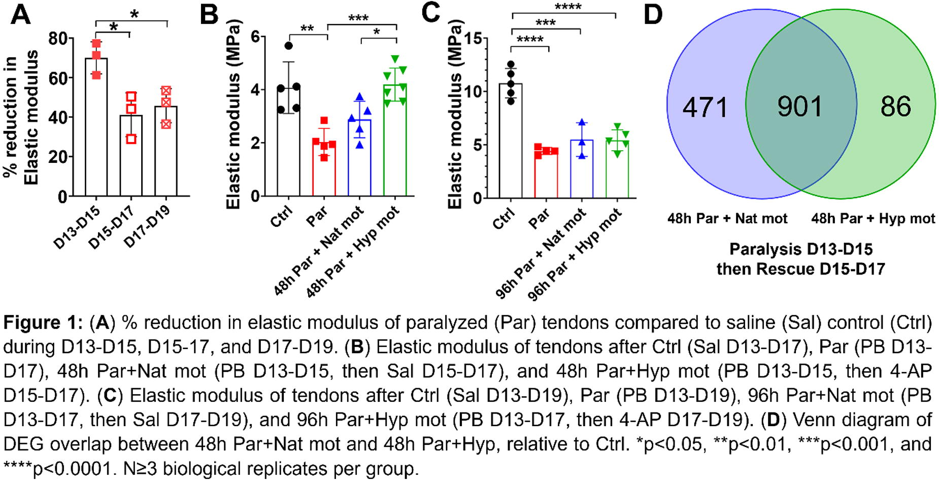
A138 - Can Macrophage Polarization Be An Early Indicator Of Outcome After ACL Reconstruction?
J. Spierings1, L. Geomini1, M. Buenen1, M. van der Steen2,3, R. P. Janssen2,1,4, K. Ito1, J. Foolen1
1
Eindhoven University of Technology, Eindhoven, Netherlands,
2
Maxima Medical Centre Eindhoven/Veldhoven, Eindhoven, Netherlands,
3
Catharina Hospital Eindhoven, Eindhoven, Netherlands,
4
Fontys University of Applied Sciences, Eindhoven, Netherlands
*Purpose/Objectives: Anterior cruciate ligament reconstruction (ACLR) with tendon autografts can display an adverse in vivo remodeling response. Macrophages play an important role, particularly during early graft healing. Macrophages can be divided into two main types: the pro-inflammatory M1 and the pro-remodeling M2 macrophages. We propose that cytokines and DNA remnants, released by apoptotic and necrotic cells in the graft, steer macrophages more towards an M1 phenotype. We also propose that this is an adverse response, which can be prevented by graft decellularization before implantation. Our previous work displayed patient-to-patient differences in cytokine secretion from leftover graft tissue from ACLR surgery, which may be an indication of why some grafts rupture. This study aims to investigate the effect of factors released by (leftover) graft tissue on macrophage polarization and correlate this to patient demographics.
*Methodology: Remnant tissue, leftover from surgery, of semitendinosus and/or gracilis tendons was collected after ACLR (METC N16.148 & N21.063). Tendons were processed immediately after retrieval (fresh, F), frozen at -20°C (fresh-frozen, FF), or decellularized (D). Next, tendons were cultured in a custom-made set-up under a preload of 15N for 7 days. To study influences of the factors released by cells, macrophages, differentiated from the human monocytic cell line THP-1, were exposed to the conditioned medium. After 24 hours, macrophage RNA was isolated, and real-time qPCR was performed to examine macrophage polarization.
*Results: Our results showed that gene expression of the pro-inflammatory marker TNF-α by THP-1 cells was significantly higher after being exposed to conditioned medium from decellularized grafts when compared to fresh (p < 0.05) and fresh-frozen (p < 0.01) grafts. Besides, the decellularized group showed a significantly lower gene expression in pro-remodeling marker TGF-β1 than the fresh-frozen group (p < 0.01). Gene expression of both IL-1β and CD163 were not significantly affected by any treatment. Next to that, preliminary results showed a potential correlation between patients' age and TNF-α gene expression by macrophages exposed to conditioned medium from fresh grafts, as younger patients tended to show higher TNF-α expression.
*Conclusion/Significance: Contrary to our hypothesis, gene expression of TNF-α and TGF-β1 showed that decellularized tendon tissue promotes M1 polarization of THP-1 cells. This may indicate that decellularization-induced damage-associated molecular patterns (DAMPs), e.g. collagen damage, overshadow the potential effect of dying and/or remnant cells in the tendon graft on the inflammatory response by macrophages. Therefore, we are currently working on a revised decellularization protocol, which is believed to induce fewer DAMPs. On the other hand, a trend towards a higher TNF-α expression by macrophages exposed to tendons from younger patients (< 23 years old) was found. This is interesting as, clinically, younger patients show a higher graft-rupture rate, and it is currently not fully understood why. These results reveal that pro-inflammatory polarization of macrophages may contribute to the risk of graft rupture. In the future, our data will be correlated with self-reported post-operative outcomes to improve our understanding of patient-specific recovery after ACLR and/or contribute to finding a biomarker that can help surgeons towards personalized rehabilitation.

A139 - Macrophages-derived Extracellular Vesicles As Potential Mediators Of Tendon Repair
S. Bagur-Cardona1, A. L. Graca2, R. M. Domingues3, M. E. Gomes3, M. Gomez-Florit1
1
Health Research Institute of the Balearic Islands, Palma, Spain,
2
University of Minho, Guimaraes, Portugal,
3
University of Minho, Barco - GMR, Portugal
*Purpose/Objectives: Tendinopathy is a chronic condition that causes persistent pain and disability and substantially affects the quality of life of patients. Currently, treatment strategies focus on symptomatology and do not address the underlying disease, frequently involving inflammation and fibrosis. Macrophages play a critical role in the immunological responses by promoting or inhibiting tissue inflammation and repair. Classically, macrophages can differentiate into either pro-inflammatory (M1) or pro-reparative (M2) phenotypes in response to various stimuli. Therefore, this study aimed to study how extracellular vesicles (EVs) derived from polarized macrophages can affect the inflammatory response of tendon cells in order to develop therapeutic approaches for tendon regeneration.
*Methodology: Human primary monocytes were isolated from buffy coats and differentiated to macrophages by adherence to plastic surfaces. Cells were then stimulated with interferon-γ or interleukins -4 and -13 (IL-4, IL-13), to induce macrophages polarization into M1, M2, and hybrid M1/M2 phenotypes. Subsequently, the EVs were isolated from the culture medium by size exclusion chromatography. The physical and biochemical characteristics of EVs were analyzed. The impact of these nanovesicles on the inflammation and injury scenarios of human tendon-derived cells (hTDCs), which had previously been stimulated with interleukin-1 beta (IL-1ß) to mimic an inflammatory scenario, was assessed by gene expression analysis, western blot and confocal microscopy.
*Results: We were able to isolate three different nanovesicles populations from polarized macrophages, showing the typical shape, size and surface markers of EVs. M1, M2, and M1/M2 macrophages-derived EVs showed distinct proteomic profiles. Notably, most of the detected pro-inflammatory cytokines and chemokines had higher expression levels in M1-derived EVs and were mostly absent in M2-derived EVs. Hence, by acting as a biological cue, we observed that M2 macrophage-derived EVs increased the expression of the tendon-related marker tenomodulin (TNMD) and tended to reduce the presence of pro-inflammatory markers in IL-1ß-treated hTDCs.
*Conclusion/Significance: Overall, these results show that EVs derived from polarized macrophages might be a potential tool to modulate the immune system responses becoming a valuable asset in the tendon repair and regeneration fields worthy to be further explored.
A140 - Exosome Cell Origin Affects In Vitro Markers Of Tendon Repair In Human Macrophages And Tenocytes
D. von Stade1, M. Meyers1, J. Johnson1, T. T. Schlegel2, A. Romeo3, D. Regan1, K. McGilvray1
1
Colorado State University, Fort Collins, CO,
2
American Orthopedic Partners, Denver, CO,
3
Shoulder Elbow Sports Medicine, Chicago, IL
*Purpose/Objectives: The unique biology and mechanical properties of tendon lead to a poor response to surgical repair and intervention, such as rotator cuff re-tear rates of greater than 70% after surgical repair in at risk populations, driving interest in adjunct therapies needed to improve this response. Mesenchymal Stromal Cells (MSCs) are often investigated as part of treatment regimens due to the widespread therapeutic effects, i.e. reducing inflammation and improving mechanical resilience of tendons. These benefits have been attributed to their cell signaling capacity and similar effects have been demonstrated in the application of isolated MSC-derived exosomes. As much of this work has been driven by the extrapolation of work done with MSCs and their exosome isolates in other tissues, it is important to know if this extrapolation is appropriate, or if this application of exosomes to the tendon microenvironment can be readily improved upon, by learning from native, differentiated cell exosomes’ effect on biomarkers of tendon healing.
*Methodology: In this study, human peripheral blood derived monocytes were differentiated into macrophages and directly educated with exosomes sourced from adipose-derived MSCs (MSCdEV), from tenocytes (TdEV) or a negative control (phosphate buffered saline, PBS). All exosomes were purified from primary cell enriched media using size exclusion chromatography. Concentrations were standardized and particles characterized using nanoparticle tracking analysis and protein marker verification. Target cell uptake was confirmed using lipophilic fluorescent labeled exosomes with confocal Z-stacked imaging, compared to exosome free label controls (figure, tenocyte uptake of TdEVs). Macrophage cytokine expression was analyzed using a multiplexed immunoassay of 24-hour cell enriched, serum-free media. Scratch wound assays were performed by manual scratching of a cell monolayer, washing, and treating cells prior to automated well imaging every 2hr and automated image analysis.
*Results: Macrophages educated with TdEVs drove decreased secretion of pro-inflammatory IL-8/CXCL8, IP-10/CXCL10 and MCP-1/CCL2, while MSCdEVs increased the excretion of these pro-inflammatory markers. When this enriched media was applied to tenocyte monoculture scratch-wound analyses, 24hr wound closure rates were fastest when TdEV-educated macrophage-enriched media was applied as compared to MSCdEV- or PBS-educated macrophage-enriched media, or a serum-enriched media positive control.
*Conclusion/Significance: This preliminary work demonstrates an in vitro differential effect of exosomes by cell source on key factors in tendon healing, i.e. primary human macrophage signaling and tenocyte mobilization. Of greatest interest is this indirect effect of the tenocyte-derived exosomes on simulated wound closure via macrophage signaling. As macrophages play a dynamic role in the acute and chronic stages of tendon repair and regeneration, the full picture of the effects of these shifts in macrophage response requires further investigation in animal models. Information gleaned from investigating tenocyte-derived exosomes could help guide the design of effective adjunct therapies for tendon regeneration and repair.

A141 - Localized Delivery Of A Hedgehog Agonist From Electrospun Scaffolds Enhances Tendon-to-bone Integration
R. Madi, J. Marcelin, T. Kamalitdinov, X. Jiang, S. Assi, D.-H. Kim, R. Mauck, A. Kuntz, N. Dyment
University of Pennsylvania, Philadelphia, PA
*Purpose/Objectives: Purpose/Objective: Electrospun scaffolds, infused with varying concentrations of SAG, a small-molecule agonist of the Hedgehog (Hh) signaling pathway, were integrated alongside a tendon graft in a metaphyseal transverse tibial tunnel (TTT) in mice. The objective of this study was to improve tendon-to-bone integration via localized delivery of SAG. This was achieved by incorporating SAG into electrospun scaffolds, evaluating the properties of these scaffolds post-incorporation, and assessing the in vivo impact of SAG release on Gli-1 expression and tendon-to-bone integration within a bilateral TTT model.
*Methodology: Methodology: All animals and procedures were IACUC-approved. Scaffold Fabrication: Polycaprolactone (PCL) solution (35 wt%) was prepared in DMF/THF with the Hh agonist at different concentrations (0, 0.001, 0.01, 0.1 mg/ml). The solution was spun from an 18G needle at 15kV onto a rotating mandrel (8 m/s, 2.5 ml/hr). SEM: Fiber alignment and diameter (100 fibers per sample) were measured from images acquired on an FEI Quanta 600 ESEM (Fig. 1). Tunnel Integration Study: TTT surgeries were performed on 24 mice. Graft tail tendons were harvested from the mouse, and a sterile scaffold was added to the graft bundle. A TTT was drilled in the metaphysis using a 27G needle (Fig. 1E). The graft with the scaffold was passed through the tunnel (Fig. 1E’). The limbs were harvested on day 14. Mineralized Cryohistology and Regional qPCR: Limbs were fixed, embedded, and sectioned undecalcified with cryofilm. The medullary canal was microdissected from tissue sections for qPCR. Adjacent sections were stained with Alizarin Complexone (AC) to visualize deposited minerals fluorescently on a Zeiss Axio Scan.Z1. AC area was normalized to the area of the tendon graft and scaffold and quantified. Statistical Analysis: Fiber diameter and orientation and in vivo qPCR and AC area were compared via one-way ANOVA (p<0.05). Outliers were identified and removed using the ROUT method.
*Results: Results: SAG incorporation into electrospun scaffolds did not affect fiber orientation or diameter. SAG infusion resulted in only minor changes in fiber dispersion and average fiber diameter in SAG scaffolds compared to blank scaffolds (Fig. 1 A-D). SAG release in vivo stimulated Hh signaling. SAG release yielded a dose-dependent effect on Gli1 expression, with the 0.1 mg/ml SAG group having ∼3-fold higher expression (p=0.009, Fig. 1G). Localized SAG release increased mineralized fibrocartilage (MFC) formation in the tendon graft and mineral deposition in the scaffold. To monitor the tunnel integration process, we measured the deposited mineral area in the tendon graft and scaffold (red in Fig. 1H). We found that SAG concentration impacted AC staining in a dose-dependent manner, with the 0.1mg/ml group having higher MFC formation in the tendon graft (p=0.044, Fig. 1I) and higher mineral deposition in the scaffold region of the 0.01 mg/ml (p=0.048) and 0.1 mg/ml (p=0.047) groups (Fig. 1J).
*Conclusion/Significance: Conclusion/Clinical Significance: This study demonstrates that localized delivery of Hh agonists is a viable and promising therapeutic strategy for enhancing tendon-to-bone integration while minimizing side effects from systemic delivery.

A142
A143 - Biofabricated 3D Microenvironment with nano-carrierspromotes tenogenic events in healthy and pathologicalhuman Tendon Progenitor Stem Cells
G. Della Porta1,2, G. Cortella1, E. Lamparelli1, M. Ciardulli1, J. Lovecchio3,4, E. Giordano4, N. Maffulli5
1
University of Salerno, Baronissi (SA) Italy, Italy,
2
University of Salerno, Fisciano, Italy,
3
Reykjavík University, Reykjavík, Italy,
4
University of Bologna, Cesena, Italy,
5
Sant’Andrea Hospital, Rome, Italy
*Purpose/Objectives: Tendon stem/progenitor cells (TSPCs) were recently identified as tendon-specific adult stemcells that play a key role in tendon homeostasis, repair or regeneration once tissue is injured. Biomoleculessuch as transforming factor b 1 (TGFb1) or growth differentiation factor 5 (GDF-5) have been reported to beessential for stem/progenitor cell recruitment into degenerative tendons and promote tenogenesis. The studydescribes an advanced biomimetic collagen matrix, bioprinted with human tendon stem/progenitor cells(hTSPCs) and functionalized with transforming factors loaded carriers to promote tenogenic events in bothhealthy and pathological cells along 21 day of dynamic in vitro culture.
*Methodology: Peptides loaded nano-carriers were formulated by microfluidic technology. Healthy andtendinophatic tendon samples were obtained from patients undergoing surgery after traumas (Review Boardprot./SCCE n. 151 on 29.10.2020). hTSPCs were collected and characterized by flow cytometry. Thebioprinter used was a ROKIT INVIVO 4D2, with a printing velocity of 5 mm/s, noozle size of 22G and infill of25%. The bioink was collagen (Advanced BioMatrix, USA) loaded with 10 mg/mL of bio-hybrid nano-carriers.Biofabricated scaffold shape was cylindrical (5x2 mm) and cultured under perfusion (flow rate: 1 mL/min).Cell tenogenic and inflammatory events were monitored using Live/Dead staining, qRT-PCR, andimmunofluorescence assays, Wester blotting and ELISA assay.
*Results: hTSPCs and transforming factors loaded nano-carriers were successfully integrated into thePhotoCol® matrix: Live/Dead staining indicated over 85% viability within the 3D printed constructs.
Immunostaining and qRT-PCR assays of healthy TPSCs confirmed significant up-regulation of tendon-related markers: collagen type 1, scleraxis, and tenascin related genes, respectively, after 14 days of dynamic culture. The data suggested also that the 3D microenvironment promoted pathological TSPCs, in acomparable manner to healthy TSPCs, supporting simultaneously cell survival and proliferation.
Interestingly, pathological hTSPCs showed different morphological features and an impaired balance of pro-inflammatory and anti-inflammatory cytokines, when compared to healthy hTSPCs.
*Conclusion/Significance: The biofabricated 3D microenvironment allowed to collect data on tenogenic events for bothhealthy and tendinopathic stem cells, as well as the study of the activity of novel tendon-specifictherapeutical tools for tendon nanomedicine. The work was partially funded within the project “PerspectivesFor Future Innovation In Tendon Repair (P4FIT), an European joint Doctorate Programme.
A144 - Advanced Imaging Modalities Of EvokeTM Demineralized Bone Fibers For Potential High-Throughput Osteoinductive Screening
D. C. Dashti1, C. Chen1, M. Hinson1, C. J. Ronholdt2, N. T. Remlinger1
1
Pinnacle Transplant Technologies LLC, Phoenix, AZ,
2
New Vision BioLOGIC INC., Dallas, TX
*Purpose/Objectives: Imaging bone tissue engineered constructs via scanning electron microscopy (SEM) and histology are powerful modalities to characterize osteoinductive (OI) potential. Evoke, a bone repair tissue product of Pinnacle Transplant Technologies (PTT), is a demineralized bone fiber (DBF) comprised of long demineralized fibers created from donated human cadaveric cortical bone. Evoke possesses prominent levels of osteoinductive growth factors and has demonstrated favorable new bone formation via a standard in vivo rodent implantation model. SEM captures the biomechanical framework of Evoke fibers, which creates cellular migration conduits to facilitate enhanced OI. SEM also confirms the ideal porosity and surface topography for an ECM fiber network architecture to encourage intrinsic native osteoblasts to attach, migrate, and deposit new bone throughout the ECM fiber scaffold. Histopathology analysis through hematoxylin and eosin (H&E) images can confirm OI potential in vivo for which Evoke DBF produces new bone, new cartilage, and new bone marrow (scored via Edwards et al. scale; ref. Methodology #2). The analyses of both SEM and H&E histopathology can lead to sophisticated high-throughput screening via digitized automated whole slide imaging of bone tissue engineered constructs like that of Evoke to confirm OI potential.
*Methodology: All Evoke DBF test articles (TAs) underwent PTT’s proprietary tissue processing methods. 1)
Scanning Electron Microscopy (SEM) Imaging
: Evoke DBF TAs were obtained from a minimum of three (3) donors for SEM imaging. The samples were prepped and imaged using standard operating procedures (Nanofiber Solutions, Dublin, OH). 2)
Osteoinductive (OI) Pathology Imaging Analysis
: Evoke DBF TAs were obtained from twelve (12) donors for testing in a 28-day in vivo athymic nude rat model following the ASTM F2529-13 and Edwards et al. (1998) study. The in vivo study involved implanting the TAs into a muscle pouch in each rear limb. After 28 days, extracted TAs were prepared for H&E and histopathology was assessed via the Edwards semi-quantitative scoring scale and digitally replicated by automated whole slide imaging.
*Results: 1) Figure 1A illustrates a SEM image of Evoke for elongated interwoven fibers that are crucial for establishing cellular migration highways and pore sizes of 100 μm+ which are essential for cellular integration into the Evoke scaffold. 2) H&E analysis in Figure 1B reveals the implantation of Evoke DBF in an athymic nude rat model results in new bone, new cartilage, and new bone marrow formation.
*Conclusion/Significance: The imaging results of both SEM and H&E histology demonstrate that Evoke DBF has OI potential. Automated whole slide imaging could warrant expedited OI assessments of other DBF or unique demineralized bone matrix (DBM) products. Future methods of automated imaging could support high-throughput OI assessment for bone tissue engineering products/applications.

A145
A146 - Pearlbone™, A Bioactive Bone Substitute Converted By Nacre From Western Australia
R. Ruan, B. Shiroud Heidari, M. Zheng
laboratories of Orthopaedic Research, The University of Western Australia, Perth, AUSTRALIA.
*Purpose/Objectives: Bone substitutes are the most promising solution to address the shortage of bone grafts for dental and orthopaedics conditions. However, being restrained by slow resorption rate and poor osseointegration, bone substitutes result in less satisfactory clinical outcomes than using autograft and allograft. Nacre, as known as the mother-of-pearl, has been highlighted as a potentially suitable resource for bone implant material for decades that are yet to be translated to a clinically usable bone substitute. In this study, we are aiming to convert nacre, an overproduced byproduct from Western Australian pearling industry to a bone substitute, which meets the clinical standards. We hypothesise that the incorporation of nacre could regulate the biodegradation and increase the bioactivity of the composite, hence promotes the bone regeneration.
*Methodology: The bone substitute material was prepared through a patented method (WO/2018/218305), which comprises the two steps of self-setting chemical reactions that can be occurred in low temperature and ambient pressure. Based on the method, a series of products in different form, such as granules and mouldable putty, that trademarked as PearlBone™ can be produced. The products were characterized by XRD, SEM and mechanical testing. And the in vitro bioactivity was tested through SEM, XRD, FTIR and mechanical testing by immersing the material into the stimulated body fluid for different time points. Then, we seeded the MC3T3-E1 cells on the material, the cytocompatibility and osteostimulative properties were examined through MTS method, confocal microscopy and the expression of osteogenic markers. In the in vivo studies, the biodegradation was tested by implanting the PearlBone™ pallets into rabbit’s back fascia, and the bone healing efficacy was examined by implanting PearlBone™ particles and putty to rabbit distal femoral critical bone defects, then analysed through Micro-CT and histology.
*Results: The results show the bone substitute material has unreacted nacre particles tightly integrated into the composite containing multiple biocompatible and osteoconductive calcium orthophosphates. The compression strength of the composite achieved 10.12 MPa that is superior to human trabecular bone. The in vitro bioactivity tests showed the formation of apatite with the progressive nano-porous flake-like crystals on the surface of the composite over 28 days. Furthermore, the composite showed the capability to host the attachment and proliferation of osteoblast cells and to promote their osteogenic differentiation. The confocal microscopy also evidenced that the expressions of RUNX2 and OPN are significantly high expression on day 1 and day 7, respectively. PearlBone™ material demonstrated a more efficient in vivo degradation rate without the compromise of physical structure. Finally, PearlBone™ particles demonstrated enhanced bone healing efficacy and biocompatibility in the healing of rabbit critical bone defect model compared to the conventional bone substitute (Hydroxyapatite). And PearlBone™ putty showed equivalence in early-stage bone healing when compared to the commercial bone putty products (MagnetOs™ and MasterGraft ®).
*Conclusion/Significance: PearlBone™ prepared through the self-setting reactions possess advantages including faster biodegradation and osteostimulative property, offering a promising bone substitute for bone regeneration. This study provides a method that convert nacre into a bone substitute material, which meets the clinical standards.
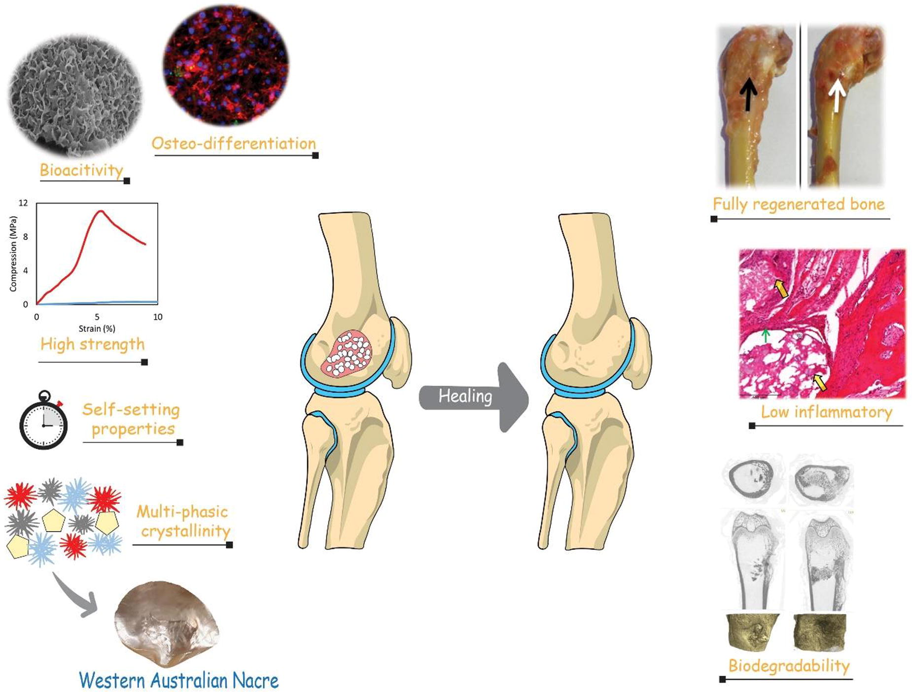
A147 - Osteogenic Differentiation Of Induced Pluripotent Stem Cells For Musculoskeletal Regenerative Medicine
F. Qu1,2,3, O. Oyebamiji2,3, F. Guilak2,3
1
University of Washington, Seattle, WA,
2
Washington University, St. Louis, MO,
3
Shriners Hospitals for Children - St. Louis, St. Louis, MO
*Purpose/Objectives: Humans have limited regenerative potential of musculoskeletal tissues following limb loss. To this end, successful attempts to regrow missing limbs could significantly improve the prognosis for amputees. The murine digit has been used to study mammalian limb regeneration, where stem/progenitor cells regrow the digit tip after distal amputation, but the mechanisms controlling skeletal elongation and patterning remain unclear. We hypothesize that the expression of a limb-like developmental program in osteoprogenitors is required for proper bone morphogenesis after digit amputation. To address this question in vitro, we sought to develop a procedure to derive osteoblasts from murine induced pluripotent stem cells (iPSCs).
*Methodology: Murine iPSCs were differentiated toward a mesenchymal state using a high-density micromass to produce pre-differentiated iPSCs (PDiPSCs). PDiPSCs in monolayer were exposed to the following media conditions: growth (Control; DMEM-HG base with basic fibroblast growth factor), osteogenic (Osteo; α-MEM base with l-ascorbic acid, dexamethasone, β-glycerophosphate), or osteogenic with supplements (Osteo+; Osteo with insulin, transferrin, and sodium selenite, nonessential amino acids, 2-mercaptoethanol). As a positive control, MC3T3 murine pre-osteoblasts were cultured in growth (Control; α-MEM base) and Osteo media. At 0, 14, 21, and 28 days, cultures were stained with Alizarin Red (AR) and Oil Red O (ORO) to assess calcium deposition and lipid accumulation. To evaluate osteogenic (Runx2, Sp7, Bglap) and adipogenic (Adipoq) gene expression, qPCR was performed at 0 and 21 days, with all samples compared to their respective Control group at day 0 (n=3-4/group/time). To improve osteogenesis, PDiPSCs were cultured for up to 28 days in Osteo+ media with 0, 10, and 50 ng/mL of recombinant bone morphogenetic protein-2 (BMP-2) and assessed by AR and ORO staining (n=3/group/time). Significance was assessed by 1- or 2-way ANOVA with Tukey’s post-hoc test (p<0.05).
*Results: Murine PDiPSCs were successfully differentiated toward the osteoblast lineage. Both PDiPSCs and MC3T3 pre-osteoblasts produced mineralized cultures when exposed to osteogenic media (Osteo) as measured by Alizarin Red staining. While supplements to the Osteo media (Osteo+) enhanced PDiPSC osteogenesis, Alizarin Red staining remained lower than MC3T3s in Osteo media. Both Control and Osteo+ media increased PDiPSC adipogenesis, as evident by Oil Red O staining. Runx2, Sp7, and Adipoq were upregulated in PDiPSCs, but not MC3T3s, at 21 days compared to day 0, with Sp7 significantly increased for PDiPSCs cultured in Osteo+ media compared to all other groups. Bglap was upregulated relative to day 0 for all groups, with PDiPSCs exhibiting the greatest fold change when cultured in Osteo+ media. BMP-2 supplementation improved PDiPSC osteogenesis in a dose- and time-dependent manner, where 50 ng/mL of BMP-2 was the most effective at stimulating mineralization and reducing lipid accumulation at early time points.
*Conclusion/Significance: These data indicate that PDiPSCs may be driven toward osteogenesis and/or adipogenesis depending on the soluble cues present, representing a novel in vitro platform to probe the osteogenic differentiation of stem/progenitor cells. While standard osteogenic media components stimulated osteogenesis in a small portion of PDiPSCs, additional BMP-2 supplementation was required to induce osteogenic differentiation at a level comparable to MC3T3 pre-osteoblasts.

A149 - Two Dimensional Boron Nanosheets As An Osteogenic, Anti-microbial And Mechanically Reinforcing Additive For Bone Tissue Engineering Scaffolds
J. Maughan1, F. J. O’Brien2, J. N. Coleman1
1
School of Physics, Trinity College Dublin, Dublin, IRELAND,
2
Department of Anatomy and Regenerative Medicine, Royal College of Surgeons in Ireland, Dublin, IRELAND.
*Purpose/Objectives: Boron is an essential trace element in the body, with beneficial effects on bone health ranging from anti-inflammatory behavior to promoting bone and blood vessel formation. We propose that boron, when combined with porous collagen scaffolds designed specifically for bone repair, might encourage regrowth of bone across fracture defects. 2D nanomaterials offer properties that cannot be achieved with bulk materials, due to their unique morphology and high surface area, and we thus hypothesized that 2D boron nanosheets combined with our collagen scaffolds would potentially exhibit enhanced properties such as antimicrobial activity and increased mechanical strength.
*Methodology: Boron is non-layered, therefore a liquid phase exfoliation approach was developed to synthesize the desired morphology of thin nanosheets using a crystalline boron precursor. Boron-containing collagen (BColl) scaffolds with a range of boron loadings (0 vol%, 0.5 vol%, 1 vol%, 2.5 vol% and 5 vol%) were produced by liquid-phase homogenization followed by lyophilization (Fig 1H).
*Results: The 2D morphology of the nanosheets was confirmed by atomic force microscopy, scanning electron microscopy and transmission electron microscopy (Fig 1A, 1B, 1C). Boron was then shown to significantly increase Young’s modulus in the BColl films (Fig 1I), improving the capacity of BColl scaffolds to act as stiff substrates for mechanoresponsive bone cells in bone repair. Once physical characterization of the boron was complete, the biocompatibility of the material was established. Osteoblasts from a cell line (MC3T3) and subsequently osteogenic-differentiated mesenchymal stem cells from rat femora (rMSCs) were grown in the presence of BColl, in film and 3D scaffold configurations. Metabolic activity and DNA content analysis showed robust cellular health and proliferation at all concentrations, in films and scaffolds, and with both cell types, such as in Figure 1D, which shows robust proliferation of rMSCs in BColl scaffolds over 21 days. To demonstrate the osteogenic potential of the BColl scaffolds, long-term (28 day) scaffold culture yielded increased mineralization for both osteoblasts (Fig 1E) and rMSCs (Fig 1F), indicated by a significant increase in calcium deposition for all BColl samples compared to collagen controls. This data was supported by confocal imaging of rMSC morphology, with cells exhibiting widespread coverage and excellent infiltration of BColl scaffolds (Fig 1G). Finally, E. coli bacteria grown on the surface of BColl films showed significantly lower viability compared to collagen-only samples (Fig 1J), demonstrating its potential for treating and preventing bone infections after scaffold implantation.
*Conclusion/Significance: In summary, this study demonstrates that boron nanosheets can be used as an effective nanocomposite additive to enhance the potential of scaffolds for bone tissue regeneration. Their novel 2D structure and intrinsic material composition promote osteogenesis, maintain high biocompatibility, offer mechanical reinforcement of weak collagen scaffolds, and demonstrate anti-microbial behavior. Rather than being intrinsic properties of boron, the anti-microbial and reinforcing effects of the material are hypothesized to arise due to its unique 2D morphology, setting BColl apart from scaffolds that require multiple components to achieve the same effects. This makes BColl a promising multifunctional biomaterial with significant clinical potential for bone repair applications.

A151 - Decellularization-based Modification Strategy For Bioactive Xenografts Promoting Tendon Repair
H. Jin, L. Wang, J. Jiang
Shanghai Sixth People’s Hospital Affiliated to Shanghai Jiao Tong University School of Medicine, Shanghai, China
*Purpose/Objectives: Xenografts have emerged as a promising option for severe tendon defects treatment. However, despite undergoing decellularization, concerns still remain regarding the immunogenicity of xenografts. Because certain components within the extracellular matrix also possess immunogenicity.We propose a novel strategy of post-decellularization modification aimed at preserving the endogenous capacity of cells on collagen synthesis to mask antigenic epitopes in extracellular matrix. To verify the feasibility of this strategy, a human-derived rosiglitazone-loaded decellularized extracellular matrix (R-dECM) is developed.
*Methodology: Cryopreservation at deep low temperatures and supercritical CO2 extraction are used for tendon decellularization. dECM loaded with rosiglitazone is prepared using an immersion method. HPLC is used to characterize the release profile of rosiglitazone. CCK-8, live/dead staining, and scanning electron microscopy are used to evaluate the biocompatibility of R-dECM. In vitro evaluations, including qPCR, immunofluorescence, flow cytometry, Western blot, and RNA sequencing, are conducted to assess the effects of R-dECM on M1 and M2 macrophages as well as tendon stem cells. An SD rat patellar tendon defect model is used to evaluate the in vivo therapeutic effects of R-dECM.
*Results: R-dECM can release rosiglitazone for over 7 days in vitro. By suppressing M1 macrophage polarization, R-dECM protects the migration and collagen synthesis abilities of tendon-derived stem cells (TDSCs), while also stabilizing the phenotype of M2 macrophages in vitro. RNA sequencing reveals R-dECM can mitigate the detrimental crosstalk between TDSCs and inflammatory cells. When applied to a rat patellar tendon defect model, R-dECM effectively inhibits early inflammation, preventing chronic inflammation. Its duration of function far exceeds the release time of rosiglitazone, implying the establishment of immune evasion, confirming the effectiveness of our proposed strategy. And R-dECM demonstrates superior tendon repair outcomes compared to dECM.
*Conclusion/Significance: R-dECM is a bioactive xenograft that exhibits weaker immune rejection and superior tendon repair effects compared to dECM.
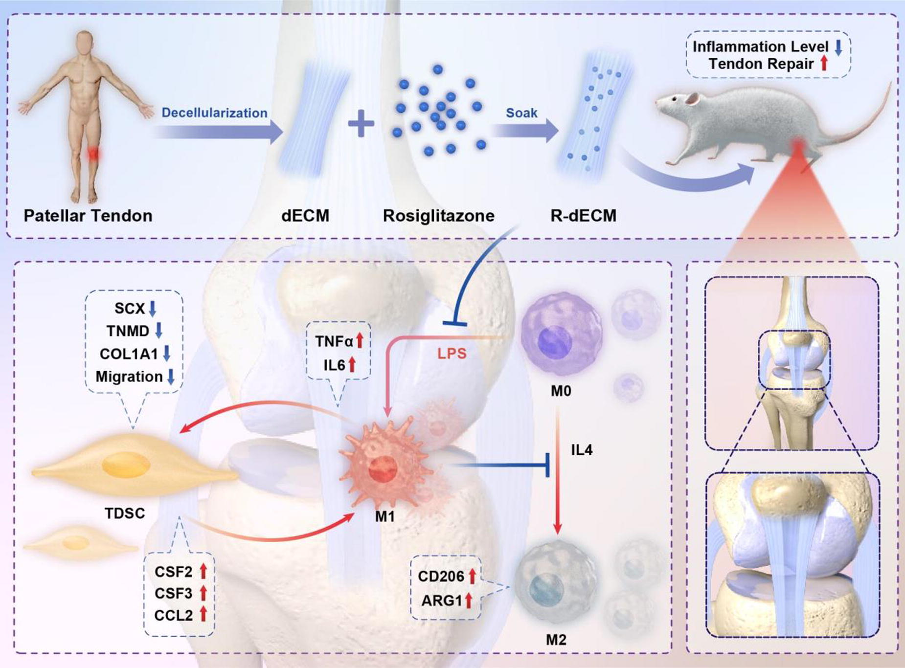
A152 - Remote Upregulation Of Active Yap-1 In Human Adipose Stem Cells Embedded In Magnetic Tendon-like Collagen/elastin Matrix
A. L. Wright1, E. Okur1, T. Broomhall1, K. Dhillon1, S. M. Mithieux2, A. S. Weiss2, M. Snow3, A. J. El Haj1
1
University of Birmingham, Birmingham, United Kingdom,
2
University of Sydney, Sydney, Australia,
3
Royal Orthopaedic Hospital NHS Trust, Birmingham, United Kingdom
*Purpose/Objectives: Research into musculoskeletal disorders, including tendinopathies, relies on 3D models that have failed to translate to preclinical data. Development of more physiological engineered constructs which recapitulate facets of native tendon and its pathology would provide further advancement in identifying new therapeutics. We have further developed previously reported magnetic tendon-mimetics by creating complex environments which include extracellular matrix components combined with mechanical cues. By adding elastin into our collagen I-based constructs, we can more closely mimic the tendon environment. We further demonstrate the ability to remotely control tenocyte-associated mechanotransduction in 3D hydrogel seeded with human adipose-derived stem cells (hASCs), isolated from donor tissue.
*Methodology: Characterisation of Patellar-fat pad isolated cells was achieved through immunofluorescent staining. hASC-associated markers CD34 and CD90, and tenocyte-associated markers tenomodulin (TNMD) and scleraxis (SCXA) were investigated. Cells from three donor samples (1, 2, and 3) were analysed.
3 mg/mL collagen I hydrogels with and without tropoelastin dosing were quantified for elastin through a colorimetric assay (Fastin Assay Kit, Biocolor). Material characterisation was evaluated through optical coherence tomography (OCT) imaging and MechAscan analysis.
Collagen-tropoelastin constructs were magnetised with anti-collagen I magnetic nanoparticles. 5-days post hASC seeding. Magnetic constructs were exposed to a 20-minute cyclical gradient of a magnetic field. Immunofluorescent staining and confocal microscopy allowed visualisation of hASC active yes-associated protein-1 (YAP-1) expression. Image analysis (CellProfiler) was employed to quantify and compare integrated density of active YAP-1 expression.
Relevant ethical approval for this work has been obtained (Ref:17/EM/0030).
*Results: Immunofluorescent staining of isolated hASCs from donors 1, 2 and 3 confirmed their stemness through positive expression of CD90 and CD34. It also revealed inter-donor variability in tenogenicity of hASCs. Donor 1 and 2 populations expressed considerably less tenomodulin (TNMD-ve hASCs) than donor 3 populations (TNMD+ve hASCs).
We demonstrate that 3 mg/mL collagen I hydrogels dosed with 200 ug/mL tropoelastin have a significantly higher concentration of elastin than controls.
Experimental characterisation of the constructs demonstrated the spatial variation in ECM properties which included the predominant native tendon matrix proteins/derivatives, collagen I and elastin. The addition of tropoelastin to the matrix was also found to alter the mechanical properties of hydrogels, as determined through OCT imaging and MechAscan analysis.
Immunofluorescent staining and image analysis of active YAP-1 expression in collagen I/elastin embedded hASCs revealed a significant increase in YAP-1 activation in magnetically stimulated constructs populated with TNMD-ve hASCs (Donor1/2), compared to non-stimulated controls (n=3). No significance difference in TNMD+ve hASC (Donor 3) YAP-1 activation could be determined (n=3), with comparable positive active-YAP-1 expression in both non-stimulated and stimulated constructs.
*Conclusion/Significance: We have developed a tendon-like collagen/elastin matrix model using patient-derived human adipose stem cells (hASCs). This model allows the application of mechanical forces through remote application of biomagnetic fields. Stretch application to the constructs results in upregulation of tenocyte-associated mechanotransduction in TNMD-ve hASCs. Currently, we are scaling this approach into an organ-on-a-chip model to demonstrate the potential for remote control systems in application of mechanical forces using these devices.
A153 - Differentiating Human Bone Marrow And Adipose-derived Stem Cells Towards Ligamentogenic Lineage Using Physiological Oxygen Tensions For Tissue Engineering Applications
T. Klaylat, P. Mounsef, P. Martineau, D. Rosenzweig, R. Gawri
McGill University, Montreal, QC, Canada
*Purpose/Objectives: Ligament injuries are common musculoskeletal injuries. Due to biomechanics and poor blood supply, ligament tissue does not heal and regenerate properly after injury. Ligament reconstruction surgery using auto/allografts suffers from high failure, complication, and revision rates. Fabricating implantable cell-laden bioengineered ligament grafts is one potential alternative solution, but it still faces the challenge of non-integration at the graft site. Nevertheless, the optimal source of cells and culture conditions for bioengineering ligament grafts remain challenging. Stem cells from various sources, bone marrow (hBM-MSCs) and adipose-derived (hAD-MSCs), have been used for ligament graft bioengineering with limited success. Ligament tissue and the articular joint space have lower oxygen tensions than arterial and venous levels, and it is critical for optimal tissue physiology and bioengineered graft integration as the grafts grown in standard non-physiological tissue conditions face “adverse conditions” upon implantation. In this study, we hypothesized that culturing hBM-MSCs and hAD-MSCs under optimized near physiological oxygen tensions would enhance their differentiation into the ligamentogenic lineage for use in TERM applications for ligament tissues.
*Methodology: hBM-MSCs and hAD-MSCs (RoosterBio) were cultured for 10 days at 37°C in DMEM supplemented with 10% Fetal Bovine Serum, 1% Antibiotic/Antimycotic, 5ng/mL TGF-β, and 1ng/mL bFGF under three different oxygen tensions: atmospheric and standard cell culture (20% O2), intravenous (5% O2), and intraarticular (2% O2) oxygen tensions. Cell-seeded culture plates were placed in a standard incubator with 5% CO2 at 37°C for atmospheric oxygen tension. For intravenous and intraarticular oxygen tensions, hypoxia incubator chambers (StemCell Technologies) were sealed, and flushed with 5% O2 and 2% O2 gas premixes respectively and placed inside an incubator at 37°C with media changes every 3 days, after which the chambers were resealed and reflushed. After 10 days, cell proliferation and viability were assessed, AlamarBlue assay was performed to evaluate the metabolic activity, and qPCR was performed to evaluate gene expression of ligament/tendon markers.
*Results: Our data show that 5% oxygen tension does not affect the viability of hBM-MSCs and hAD-MSCs, whereas 2% oxygen tension slightly decreases the viability of hBM-MSCs. Growth rate analyses reveal that lower oxygen tensions do not affect cell growth and population doubling. AlamarBlue assay readings indicate a significant increase in the metabolic activity of hBM-MSCs under 2% and 5% oxygen tensions and hAD-MSCs under 5% oxygen tension. Finally, qPCR results indicate higher levels of collagen type-1, tenascin C, and scleraxis expression in hBM-MSCs under 2% oxygen tension. Gene expression analysis for the same markers in hAD-MSCs was inconclusive, and no statistically significant difference was observed.
*Conclusion/Significance: These findings suggest that hypoxic culture conditions enhance the ligamentization potential of hBM-MSCs cultured in culture media with TGF- β and bFGF. Optimizing culture conditions for differentiating hBM-MSCs and hAD-MSCs into the ligamentogenic lineage would allow us to use these cells in bioengineered ligament grafts for ligament reconstruction. Moreover, priming and maturing these cell-seeded bioengineered grafts under near-physiological conditions potentially enhances their integration at the intraarticular graft site, thus lowering revision rates and enhancing the patient’s quality of life.
A155 - Regeneration Of The Patellar Tendon Using Scaffold-free 3D Constructs Consisted Of Adipose Tissue-derived Mesenchymal Stem Cells In Rabbits
S. Kashimoto, D. Murata, H. Yoshizato, T. Nonaka, A. Nakamura, M. Mawatari, K. Nakayama
Faculty of Medicine, Saga University, Saga City, Japan
*Purpose/Objectives: Patellar tendon-bone autografts are a commonly used surgical technique for anterior cruciate ligament reconstruction. However, the tendon at the donor site does not heal through a regenerative process, but rather through the formation of a fibrotic scar, which reduces tissue quality and mechanical properties. Currently, donor site healing is an important clinical issue. The objective of this study was to regenerate tendons by autologous implantation of scaffold-free three-dimensional constructs into patellar tendon defects in rabbits.
*Methodology: Adipose tissue-derived mesenchymal stromal cells (ADSCs) were isolated from interscapular adipose of Japanese white rabbits (male, 25-35 weeks old, n=15) and expanded. Some of the collected cells were induced into osteogenic, chondrogenic, and adipogenic lineages in differentiation assay, and the others were cultured to produce cell aggregates (diameter 550-600 μm). Cylindrical cell constructs (inner diameter 2.5 mm, height 3.0 mm, thickness 0.8mm) were then formed using a Kenzan method with a bio 3D printer. The mechanical properties of the constructs were measured using a tensile tester. After the constructs were cut into bands (8.0 mm long, 3.0 mm wide, 0.8mm thick), equal-sized defects were created in the center of the patellar tendons of both hind limbs, and then the constructs were implanted into the defect on the right hind limbs and sutured with 7-0 nylon thread (left hind limbs were untreated as controls). Autopsies were conducted at 6 weeks (n=5), 12 weeks (n=5), and 24 weeks (n=5) postoperatively and histological evaluation was performed using a histological scoring system (total score of 15 points).
*Results: The ADSCs were positive for Alizarin red in osteogenic, Alcian blue in chondrogenic, and Oil Red O in adipogenic induction, respectively. The average breaking strength of the constructs were 637.3 ± 17.1 mN/mm2. The scores of the implanted group at 6, 12, and 24 weeks after surgery were 6.8 ± 1.6, 7.0 ± 1.7, and 9.4 ± 0.9, respectively. On the other hand, , the scores at the same time points were 4.8 ± 1.4, 5.81 ± 1.1, and 9.6 ± 1.1 in the control group, respectively. All numeric data are reported as means ± standard error. The scores were compared between the implanted and control groups at the same time points and between the time points in the implanted or control groups with t-test and one-way ANOVA test. P < 0.05 was considered statistically significant.There was no significant difference between the implanted and control groups at 6, 12, and 24 weeks postoperatively. A significant difference was only observed in scores between 6 and 24 weeks in the control group (p=0.01).
*Conclusion/Significance: It was supposed that we had successfully created band-shaped scaffold-free 3D constructs from rabbit ADSCs and caused early-stage tendon healing of the implanted group compared to that of the control group. Our future plans include increasing the number of surgeries and measuring the mechanical strength of patellar tendons after the surgeries. And also, we are planning to track the implanted cells by labeling and then research on the differentiation of implanted cells into tenocytes in more detail.
A157 - Cross-talk Of Rho/ROCK And TGF-β3 Signaling In Tenogenic Differentiation
S. Niebert1, M. Melzer2, G. Scheiner-Bobis2, F. Ullm3, T. Pompe3, J. Burk1
1
University of Veterinary Medicine, Vienna, Austria,
2
Justus-Liebig-University, Giessen, Germany,
3
Leipzig University, Leipzig, Germany
*Purpose/Objectives: Tendons are frequently subject to injuries due to mechanical overload, particularly but not exclusively in athletes. Tendons are prone to scar formation and fibrosis during healing because of their hypocellularity and hypovascularity, making them very susceptible to re-injury. Therefore, regenerative therapy approaches need to address this unfavorable outcome of the healing process. Mesenchymal stromal cells (MSC) are studied as an agent in regenerative therapies for a wide range of conditions, including chronic tendon lesions. Currently, MSC are believed to act primarily via immunomodulation. Nevertheless, they can also influence the production and remodeling of tendon tissue by tenogenic differentiation and formation of tendon matrix. MSC do not only have effects on other cells, they also are affected themselves by the environment they are applied to. These environmental stimuli include the secretome of endogenous cells as well as components of the extracellular matrix (ECM). Tenogenic differentiation is impacted by transforming growth factor (TGF)-β3 signaling via the Smad2/3 pathway on the one hand, and by factors like ECM components and mechanical load signaling via the Rho/ROCK pathway on the other hand. Consequently, the potential interaction between these pathways is of utmost interest in the context of tendon healing. Smad 2/3 proteins are feasible as the potential link between the two pathways. They possess several phosphorylation sites in the carboxy-terminal region as well as in the so-called linker region. Upon TGF-β binding the carboxy-terminal region becomes phosphorylated which leads to activation and tenogenic differentiation. The phosphorylation sites in the linker region are important because of their regulatory effects on the stability, activity, and nuclear transport of the Smad proteins.
*Methodology: Therefore, we aimed to elucidate if and how the TGF-β3 and the Rho/ROCK signaling pathways interact regarding tenogenic differentiation. In order to do so, we cultured human adipose-derived MSC either as monolayer or on collagen I matrices and treated them with a ROCK inhibitor (Y-27632) or TGF-β3, alone or in combination. Subsequently, MSC were analyzed at several time points for the expression of tenogenic markers and components of the signaling pathways like Smad 2/3 by quantitative RT-PCR and the phosphorylation of Smad2/3 in the carboxy-terminal region as well as in the linker region by Western blot.
*Results: As expected, cultivation on collagen matrix promoted tenogenic differentiation as well as treatment with TGF-β3 did. Moreover, both cultivation on collagen matrix and ROCK inhibition supported the TGF-β3 effect. ROCK inhibition also increased the expression of ECM proteins, especially in MSC cultured on a collagen matrix. The carboxy-terminal phosphorylation of Smad proteins induced by TGF-β3 was consistent in all donors and unchanged by ROCK inhibition or collagen, whereas the phosphorylation of the regulatory linker sites was affected by both treatments.
*Conclusion/Significance: In summary, our findings support the hypothesis that the phosphorylation status of the linker of Smad 2/3 plays an important role in the integration of signals by ECM and soluble growth factors in tenogenic differentiation of human MSC.
A158 - Continuous Expansion Of Human Cruciate Ligament Cells For Ligament Tissue Engineering
T. Klaylat1, P. Martineau1, C. Gao1, J. Fradette2, S. Nazhat1, D. Rosenzweig1, R. Gawri1
1
McGill University, Montreal, QC, Canada,
2
Laval University, Laval, QC, Canada
*Purpose/Objectives: Anterior cruciate ligament (ACL) ruptures are prevalent musculoskeletal injuries. Due to biomechanics and poor blood supply, ligament tissues fail to heal properly after injury. The current standard of care, ACL reconstruction surgery (ACL-R), is associated with high failure, complications, and revision rates, thus creating an unmet clinical need for alternative solutions. Tissue engineering regenerative medicine (TERM) for ligament tissues is an emerging field. Cell-seeded bioengineered constructs are superior to cell-free ones. Nevertheless, uncovering the optimal cell source for ligament TERM applications remains challenging. Stem cells from diversified sources have been examined with limited success. Using primary ligament cells for ligament TERM applications has dual limitations of limited cell numbers and loss of phenotype (dedifferentiation) on cell passaging. In the current study, we investigate cell number expansion using a biaxial strain device (cellarator) that provides a continuously expanding cell culture surface, thereby expanding primary ligament cell populations without the need for passaging and the resulting dedifferentiation.
*Methodology: Primary human ligament cells were isolated by enzymatic digestion from resected surgical samples (per the IRB-approved protocols and informed consent). Cellarator silicone inserts were chemically activated and coated with collagen type-1. Petri dishes of sizes 35mm, 60mm, and 100mm coated with silicone elastomer and treated similarly to the silicone inserts served as control. Isolated cells were seeded on the cellarator device with a 10-day continuous stretching protocol. Coated 35mm Petri dishes were seeded at the same cell density, and cells were trypsinized and passaged onto 60mm and 100mm dishes. At the experimental endpoint, the continuously expanded cells were in passage P1, whereas the cells on the surface area matched Petri dish were in P3. Cells were imaged at each passaging time point to assess cell alignment and morphology. qPCR was performed to determine the gene expression of ligament markers collagen type-I, III, and VI, tenascin C, decorin, α-SMA, and scleraxis. Metabolic activity was assessed using AlamarBlue assay.
*Results: Cell morphology data shows that continuously expanded ACL cells showed a more spindle-like morphology and parallel arrangement than their serially passaged counterparts. Similarly, continuously expanded primary ACL cells maintained their growth rate, while serially passaged ACL fibroblasts showed an altered growth rate. In addition, continuously expanded primary ACL cells displayed a higher metabolic activity compared to their passaged counterparts. Our gene expression analysis results show a declining trend in the transcriptional expression of ligament-specific markers collagen type-I, III, VI, scleraxis, decorin, and tenascin C from P0 to P3. However, continuously expanded ACL fibroblasts had a significantly higher gene expression (3 to 5-fold changes) of collagen type-III and VI, scleraxis, and tenascin C compared to their passaged counterparts (at P3).
*Conclusion/Significance: Our results demonstrate that continuous expansion is a unique technique to expand primary human ACL fibroblast populations for ligament TERM applications while maintaining their primary phenotype. These expanded populations of primary ACL cells can be seeded on bioengineered ligament scaffolds to be used in ACL-R, thus enhancing the surgical outcomes and quality of life of populations at risk of ACL rupture, such as athletes.
A160 - Assessing The Effect Of Cyclical Strain On HBMMSCs Differentiation On 3D-co-printed Scaffolds For Ligament Replacement
A. Dussault1, T. Klaylat1, G. Goel1, I. Villemure2, R. Gawri1, D. Rosenzweig1
1
McGill University, Montreal, QC, Canada,
2
Polytechnique Montréal, Montreal, QC, Canada
*Purpose/Objectives: The anterior cruciate ligament (ACL) is one of the major ligaments in the knee, connecting the femur to the tibia and stabilizing the joint. The ACL is notably at risk in sports involving sudden changes in direction, cutting and jumping. ACL reconstruction using an autograft or an allograft is the standard treatment for ACL injuries. However, those grafts are suboptimal as they are associated with risks of infection, pain, stiffness, instability, donor site morbidity (autograft) and immune reactions (allograft). Most importantly, the reduced mechanical properties of those grafts result in patients often being unable to perform their sport at pre-injury level, which can result in a new injury. One way of resolving this issue is through tissue engineering, using cell-seeded 3D-printed scaffolds.This project aims to develop a stem cell-seeded 3D-co-printed scaffold that could act as a ligament replacement. The first objective is to design a scaffold, evaluate its mechanical properties and assess material-derived stem cell differentiation towards a tenogenic (tendon-like) lineage. The second objective is to evaluate the impact of cell stretching on tenogenic differentiation.
*Methodology: The scaffolds are co-printed with a softer material (Layfomm60, a mix of TPE (thermoplastic elastomer) +PVA (polyvinyl alcohol)) in the middle to drive cell differentiation towards a tendon/ligament phenotype, and a more rigid one (PLA) on the outer parts, to boost mechanical properties. The scaffolds are seeded with stretched (bi-axial, f=0.001 Hz, 10% or 15% stretch for 8h followed by a 16h pause, twice for a total of 48h) and non-stretched human bone marrow MSCs (hbmMSCs) to assess the effect of “priming” cells. Mechanical properties are assessed through tensile testing. Cell viability and organization are evaluated through live/dead, metabolic activity through alamar blue. Cell differentiation is assessed through qPCR, using tenascin-C, scleraxis and collagens I & III as tendon/ligament markers.
*Results: Previous work from our laboratory has shown that Layfomm and Layfelt both drive tenogenic differentiation, whilst PLA drives osteogenic differentiation. Preliminary results show that co-printing may have a synergistic effect on cell behavior, as adipose-derived stem cells organize and spread differently when seeded on co-printed scaffolds than when seeded on single material scaffolds. Results suggests that mechanical stimulation upregulates collagen III (1.7x to 3.7x for 10% stretch, 2x to 3.3x for 15%, with respect to static culture) and tenascin-C (1.9x to 8.3x for 10%, 1.2x to 8.6x for 15%), while downregulating collagen I.
*Conclusion/Significance: As grafts used in ACL surgeries fail to restore activity to pre-injury levels, we have yet to find an optimal replacement. The development of biocompatible ACL scaffolds could help reduce the financial and health burden of patients with ligament/tendon injuries. Indeed, the less invasive tactic of using a 3D-printed scaffold instead of an autograft may reduce the duration of the surgery (i.e., less incisions need to be made) and eliminate donor site morbidity. It also has the potential to reduce post-surgery pain, healing time and risk of infection.
A161 - Paracrine Effects Of Macrophage Phenotype On Tendon Tissue Remodelling
A. Mansoor, H. Brouwer, K. Ito, C. Bouten, A. Smits, J. Foolen
Technical University of Eindhoven, Eindhoven, Netherlands
*Purpose/Objectives: Tendinopathy is characterized by tendon tissue degeneration and the transformation of the aligned anisotropic extracellular matrix (ECM) towards a disorganized isotropic ECM. Simultaneously, spindle-shaped tenocytes change into stellate tenocytes. Restoration of the healthy tissue organization is an important challenge to restore its function. Macrophages are thought to be one of the key regulators during the remodelling phase, and their polarization into a spectrum of phenotypes is hypothesized to play a vital role in both tissue remodelling and fibrosis. Their function is influenced by environmental cues such as cytokines and topographies. The interplay between different macrophage phenotypes and tenocytes, and how this influences tendon remodelling remains largely unknown. To get a better understanding of the processes involved, we aim to elucidate the effect of paracrine signalling of distinct macrophage phenotypes on tendon-like tissue remodelling.
*Methodology: 3D constrained microtissue platforms were used in-vitro to create aligned tendon-like tissues consisting of fibroblasts in a collagen type I gel. The microtissues were cultured in conditioned media (CM) produced from macrophages in three different biochemically induced polarization states, (IFN-γ+LPS stimulated M1, IL-4+IL-13 stimulated M2a, and IL-10 stimulated M2c) and in control RPMI medium. Microtissue waistcoat contraction, gene expression and cellular orientation were analysed to get more insight into the remodelling behaviour. The polarisation state of the macrophages and the contents of CM were analysed using multiplex-ELISA.
*Results: ELISA confirmed the polarisation of the macrophages into the three different phenotypes. Control medium resulted in a higher waistcoat contraction in comparison to the CM groups, while the contraction between the CM groups were comparable. The alignment and elongation of the cells appeared to be the strongest in the control groups as compared to the CM groups. Differences in gene expression between CM groups were comparable. The concentration of the secreted factors in all the CMs were decreased after 48 hours.
*Conclusion/Significance: Macrophage-secreted factors were found to have a minimal influence on remodelling in terms of contraction, orientation, and remodelling marker genes. The minimal differences seen can be attributed to the CM. CM is produced by exposing M0 macrophages to stimulating factors (SF) for 24 hours, after which medium is replaced with fresh medium without SF. This was done to not allow the SF to have an effect on tissue remodelling. However, this had a strong side effect, namely that the concentration the secreted factors in the conditioned medium was very low at 48 hours, i.e., much lower compared to 24 hours. Nevertheless, the CM still had an effect, since a difference in waistcoat contraction and morphology was seen between the control and CM groups. As the next step, the detected secreted factors from the CM will be added to tissue cultures at concentrations that are representative for the 24h to study the effect at higher concentrations without the influence of the SF. Further unravelling of the interplay between macrophages and tenocytes is crucial for steering functional tendon healing.

A162 - Comparative Analysis Of Tenogenic Gene Expression In Adipose-derived Stem Cells In Response To Various Biochemical Stimuli
J. YOO, H. Kim, S. Lee, J. Nam, J.-W. Park
Seoul National University College of Medicine, Seoul, Korea, Republic of
*Purpose/Objectives: Tissue engineering is emerging as a promising strategy for tendon repair. However, tendon tissue engineering is hampered by difficulties in obtaining appropriate seed cells, in determining the best condition for proliferation and differentiation, and in selecting optimal cell carriers for the degenerated tendon. Although tenocytes have been effectively utilized for this purpose, harvesting these cells from autologous tendons is difficult due to the limited cell number and donor site morbidity. In comparison, adipose-derived stem cells (ADSCs) are preferable seed cells that can be easily harvested with minor donor site injury. ADSCs have already been applied for tendon regeneration, demonstrating that these cells may be practical seed cells for tendon tissue engineering. As mentioned, however, establishing an efficient strategy to differentiate the ADSCs is still challenging. In this study, we investigated the tenogenic gene expression of ADSCs within various tenogenic differentiation media, such as growth/differentiation factor 5 (GDF5), bone morphogenetic protein 12 (BMP12), transforming growth factor beta-1 (TGFβ-1), and transforming growth factor beta-3 (TGFβ-3).
*Methodology: Human ADSCs were achieved from the detached fat tissue during total hip arthroplasty after informed consent and approval of our institutional review board. Total RNA was isolated from cultured cells after 1, 7, 14, and 21 days of cell seeding using the RNeasy Mini Kit (Qiagen, Germany). Expression of tenocyte phenotype was completed with five sets of primers for Type I and III collagen, scleraxis (SCX), tenascin C (TNC), and tenomodulin (TNMD). qPCR was carried out using SYBR Green PCR master mix (Biotool, USA) on an Applied Biosystems ViiA 7 Real-Time PCR System. All PCRs were performed in triplicates. The reference gene glyceraldehyde phosphate dehydrogenase (GAPDH) was used to normalize gene expression, and relative fold changes were calculated using the 2−ΔΔCt method. Statistical analysis was performed by one-way repeated measures ANOVA test in gene expression between the control and treated groups.
*Results: On adhesion assay, the initial cell seeding efficiency was comparable in all media. On proliferation assay, cells proliferated, and the cell number increased significantly in all media, with the highest fold in control media. To assess the effects of each biochemical stimulus on the tenogenic differentiation potential of ADSC, cells were treated, and gene expression was measured by qPCR. In qPCR, all five groups demonstrated substantial expression of COL1A1 and COL3A1. Compared to the control group, the cells treated with TGFβ-1 and TGFβ-3 showed increased expression of SCX and TNC in 7, 14, and 21 days, while the ADSCs treated with GDF5 and BMP12 showed a non-specific increase in SCX and a decrease in TNC, a tendon-related gene.
*Conclusion/Significance: Our study suggested an optimal tenogenic inducer to differentiate the adipose-derived stem cells to tenocytes using various biochemical stimuli and timing for implantation, which can be used as a tissue-engineered approach for the regeneration of tendinopathy patients.

A163 - Isolation And Characterization Of Canine Tendon Derived Stem Cells
S. KIM1,2, B.-J. Kang1,2
1
Department of Veterinary Clinical Science, College of Veterinary Medicine and Research Institute for Veterinary Science, Seoul National University, Seoul, Korea, Republic of,
2
BK21 FOUR Future Veterinary Medicine Leading Education and Research Center, Seoul National University, Seoul, Korea, Republic of
*Purpose/Objectives: With the increase companion dogs’ activities such as agility training, fetch, running, it leads to rise associated injuries such as bone fracture, abrasion, or tendon rupture. In Particularly, tendons, which enhance stability by connecting bone and muscle, have limitations in recovering to normal function. After tendon injury, such as tendinopathies, tendon rupture, tendonitis, healing abilities of tendons are limited because musculoskeletal soft tissues, like tendons, are primarily composed of extracellular matrix (ECM) with small amounts of cell populations. For these reasons, healing is delayed, and scar tissue formation is accelerated. To overcome these limitations, providing adequate stem cells to the tendon tissue is also undertaken and, in this field, tendon derived stem cells have emerged as a promising source of regenerative medicine materials. But there are no studies about characterizing tendon derived stem cells in dogs. Here, we firstly isolate and characterize the stem cells from dog’s tendon.
*Methodology: To isolate tendons derived stem cells (TDSCs), digesting normal calcaneal tendon tissues derived from dog with collagenase I, cells were extracted and rejuvenated. For characterizing TDSCs, cell morphologies were checked daily in all passages to establish morphological features. Tendon-specific markers, stemness markers and pluripotent markers are confirmed by qRT-PCR, immunocytochemistry, and flowcytometry. Tri-lineage differentiation capabilities verified through qRT-PCR and staining calcium deposits (alizarin red stain), lipid droplets (oil red O stain) and GAGs (alcian blue stain).
*Results: TDSCs exhibit a spindle-shaped morphology like mesenchymal stem cells but have a comparatively more rounded shape. They kept their morphology and proliferation rate throughout 7 passages; the survival rates are higher than 97% and growth curves of 3rd, 5th, 7th exhibited similar patterns. Alizarin red (staining calcium deposits), oil red O (staining lipid droplets) and alcian blue (staining GAG) show the capability of tri-lineage differentiation of the TDSCs and it had been also verified through the upregulated factors in qRT-PCR. These cells manifest tendons specific markers (scleraxis, tenacin C, tenomodulin, mohawk) and also express the surface and intranuclear stemness markers (CD90, CD44, SOX2).
*Conclusion/Significance: This research is the first report identifying tendon derived stem cells from dog calcaneal tendon tissues. As characterizing tendon derived stem cells, it will provide more appropriate in vitro model of normal tendon derived stem cells. This study offers both a theoretical foundation and experimental support for the potential utilization of TDSCs in treating tendon injuries. *Acknowledgements: This work was supported by the National Research Foundation of Korea (NRF) grant funded by the Korea Government (MSIT) (No. 2023R1A2C1003001).
A164
A165 - Tailoring The Degradability Of Bioactive Glass To Suit Various Application Sites And Ages
A. GHARBI1,2,3, W. Cheikhrouhou-Koubaa2, N. Derbel3, N. Ashammakhi1,4
1
Institute for Quantitative Health Science and Engineering (IQ), Michigan State University, East Lansing, MI 48824, MI,
2
Digital Research Center of Sfax, Centre de Recherche en Numérique de Sfax (CRNS), Technopole of Sfax, BP 275, 3021 Sfax, SFAX, Tunisia,
3
National Engineering School of Sfax (ENIS), University of Sfax, 3038 Sfax, SFAX, Tunisia,
4
Department of Biomedical Engineering, Colleges of Engineering and Human Medicine, Michigan State University, East Lansing, MI 48824, MI
*Purpose/Objectives: Bioactive glass (BaG) is an appealing material due to its ability to enhance bone formation, making it attractive for use in dentistry and bone surgery. The controllability of BaG implant dissolution has become an important objective in addressing variations in healing rates seen in different regions of the skeleton and different age groups. To achieve this, modification of the composition (fluorine and boron content) of the BaG was explored.
*Methodology: To investigate the fluorine (CaF2) and boron (B2O3) incorporation influence in the BaG network and to validate the fabrication protocol, a reference composition of BaG was adopted. CaF2 and B2O3 were introduced to the quaternary BaG system SiO2-Na2O-CaO-P2O5 in concentrations ranging from 5 to 20 wt.% to fabricate fluorine bioactive glass (BaG-F) and boron bioactive glass (BaG-B). Either CaF2 or B2O3 was mixed with sodium metasilicate pentahydrate (Na2SiO3 5H2O), calcium metasilicate (CaSiO3), sodium metaphosphate (Na3P3O9), and calcium carbonate (CaCO3), and heated the mixture at 1300oC. The molten vitreous material was then tempered in preheated molds and annealed for 4 h at a temperature near that of its glass transition temperature (Tg). Degradation was investigated by immersing BaG samples in simulated body fluid (SBF). X-ray powder diffraction (XRD) technique was used to identify the obtained biomaterials phase. Infrared spectroscopy (FTIR) and differential thermal analysis (DTA) were applied to prove fluorine or boron insertion in the BaG structure. The bioactivity of produced BaGs was checked using an inductively coupled plasma-optical emission spectrometer (ICP-OES). Scanning electron microscopy (SEM) and energy dispersive x-ray spectroscopy (EDS) techniques were employed to assess the biomaterial resorbability.
*Results: The fabrication of BaG with a high fluorine content of up to 20 wt.%, which was previously unattainable was successfully achieved in this study using the melting method. Additionally, the structural stability of produced constructs was attained by increasing the fluorine content, leading to a slowed degradation rate in SBF. It was also found that by increasing boron concentration up to 20 wt.% in BaG-B, crystallization rate and the quality of the newly formed hydroxyapatite (HAp) layer on the surface of BaG-B was improved. The presence of boron also enhanced the degradation of BaGs in SBF. Previous attempts by other groups faced challenges, including the fabrication technique employed, resulting in limited success.
*Conclusion/Significance: It is possible to slow down BaG degradation rate by increasing its fluorine content and accelerate its degradation by increasing its boron content. Produced BaG with high boron content can be suitable for use in children and the upper limb, while that with high fluorine content can be applied for treating older patients and in the lower limb. Our next step involves evaluating the performance of these materials in vivo.
A166 - 3D Printing Of Polycaprolactone Scaffold With Multiscale Porosity Via Incorporation Of Sacrificial Sugar Particles
M. Kutrolli, N. S. Pereira, V. Scanlon, M. Samandari, A. Tamayol
University of Connecticut, Storrs, CT
*Purpose/Objectives: Bone tissue engineering has drawn significant attention and various biomaterials have been tested. Polymers such as polycaprolactone (PCL) offer excellent biocompatibility, reasonable mechanical properties, and biodegradability. However, PCL scaffolds suffer a critical drawback: a lack of micro/mesoporosity, affecting cell attachment, tissue integration, and mineralization. It also results in a slow degradation rate. While 3D-printing has addressed the issue of macroporosity through CAD-guided fabrication, PCL scaffolds still exhibit poor smaller-scale porosity. To overcome this, we generated composites of PCL, hydroxyapatite (HA), and powdered sucrose (PS). The latter serves as a sacrificial material to generate porous particles after sucrose dissolution. Additionally, we have incorporated dexamethasone (DEX) to boost the PCL osteogenic properties. The resulting scaffolds maintain controlled macroporosity from the lattice print structure, but also develop micro/mesoporosity within PCL fibers when exposed to aqueous environments.
*Methodology: The study involved mixing PS into solvent-dissolved PCL in different weight rations of PS to PCL (70:30, 50:50, and 30:70 wt%). The resulting composite was used for 3D printing of scaffolds at room temperature. Printability was optimized by adjusting pressure, speed, and layer height through filament collapse and fusion test. Enzymatic degradation, porogen leaching, and DEX release profiles were characterized. Physical properties were assessed using wettability, SEM, and micro-CT to quantify the porosity (percentage, pore size, and interconnectivity). Raman spectroscopy was used to verify the absence of sugar after leaching. Mechanical characteristics were evaluated via compression testing before and after porogen leaching. Bone marrow stromal cells (BMSCs) behavior in the printed scaffolds was studied by assessing viability, metabolic activity, osteo-differentiation, and mineralization.
*Results: The scaffolds with a 70% sugar concentration exhibited superior printability and reached a highest porosity of 80%, but performed poorly during mechanical testing. A 50% PS concentration demonstrated a 70% porosity, with an average pore size of 25 µm, favoring cell attachment. No trace of sucrose was found in Raman after leaching the sugar for 8 hours. Water contact angle results show the improved hydrophilicity as the sugar concentration increased, making the scaffolds more conductive to cell adhesion. Behavior of bone marrow stromal cells (BMSCs) showed positive viability and proliferation results with an increasing trend of mineralization and osteo-differentiation as the sucrose concentration increased. The addition of HA and DEX also promoted mineralization and osteo-differentiation in the cultures.
*Conclusion/Significance: The integration of PS as porogen at a concentration of 50%wt within PCL scaffolds presents a promising approach to address the poor cell attachment and tissue integration issues of PCL in bone tissue engineering. The method allows for the fabrication of scaffolds with tunable porosity and mechanical properties, suitable for various applications. The addition of HA and DEX further enhanced the scaffolds. Future studies will apply the scaffolds in an in-vivo model to thoroughly investigate their performance.
Figure 1: (A) PS leaching as a function of soaking time. (B) Enzymatic Degradation. (C) Printability showing filament collapse and fusion test. (D) Water contact angle. (E) SEM images showing porosity. (F) Raman of the different concentrations.

A167 - Beta-tricalcium Phosphate (beta-TCP) And Polylactic Co-glycolic Acid (PLGA) Composite Scaffolds For Bone Tissue Engineering
P. S. Bossini1, P. Brassolatti2, L. A. Lopes1, C. A. Castro2, A. L. Andrade3, G. Pereira2, J. V. Silva2, L. R. Avó2, F. F. Anibal2, A. C. Duarte2
1
NUPEN - Institute of Research and Education in the Health Area, São Carlos-SP, Brazil,
2
Federal University of São Carlos (UFSCar), São Carlos-SP, Brazil,
3
University Brazil (UB), São Paulo-SP, Brazil
*Purpose/Objectives: Critical bone defects remain a significant challenge in the field of regenerative medicine. Despite advancements in treatment methodologies, many outcomes in tissue regeneration continue to be unsatisfactory. Consequently, the exploration of bone substitutes made from various synthetic materials, such as beta-tricalcium phosphate (beta-TCP) and polylactic co-glycolic acid (PLGA), has been explored in recent years. The primary objective of this study was to assess the effectiveness of scaffolds composed of beta-TCP/PLGA as temporary bone substitutes in treating critical bone defects induced in the calvaria of rats.
*Methodology: For this purpose, 48 Wistar rats (12 weeks old, weighing approximately 300 g) were selected and divided into two distinct groups: control group and biomaterial group. The critical bone defect measuring 8 mm-diameter was surgically induced in the medial region of the skullcap. The biomaterial composed of beta-TCP/PLGA (VitaGraft - DMC Equipment Import and Export-Co. Ltda, São Carlos/SP, Brazil) was implanted in scaffold form and, subsequently, each group of animals was euthanized at intervals of 15, 30 and 60 days post-surgery (8 animals for each time point). A Two-Way Analysis of Variance (ANOVA) with a subsequent Tukey's post-hoc test was employed, and the data were analyzed using Graph Pad PRISM 5.0, with the significance level of 5% (p≤0.05).
*Results: The histopathological analysis indicated a more favorable repair process in the groups treated with the biomaterial. This was evidenced by an enhanced deposition of granulation tissue and significant formation of new bone tissue, observed both in the periphery and in the center of the lesion, contrasting with the findings of the control group. Morphometric analysis confirmed these findings, revealing a statistically significant enhancement in the area of newly formed bone tissue in the beta-TCP/PLGA group compared to the control group. Additionally, it was possible to observe greater immunoexpression of collagen type I (Col-I) in the groups with biomaterial implants at all experimental times evaluated, which is consistent with the greater amount of mineralized bone matrix assessed by morphometry. The results of receptor activator of nuclear factor kappa-Β ligand (Rank-L) immunoexpression were higher in the periods of 30 and 60 days, indicating an increase of the degradation of the biomaterial and the remodeling activity of the bone.
*Conclusion/Significance: Therefore, it can be concluded that beta-TCP/PLGA played a beneficial role in facilitating the essential cellular activities and responses at the site of the lesion and enhanced the bone neoformation, presenting itself as a promising alternative in the treatment of critical bone defects.
A168 - Nitric Oxide-releasing Biomimetic Scaffold For Regeneration Of Osteoporotic Bone Through Regulation Of Homeostasis
D. Han, J.-K. Lee
CHA university, Seongnam-si, Korea, Republic of
*Purpose/Objectives: Compared to healthy individuals, patients with osteoporosis experience a higher risk of bone fracture and an extended healing period. And the regeneration of critical-sized bone fractures in osteoporosis involves various challenges in clinical trial applications. Therefore, it is necessary to develop an ideal scaffold that can effectively regenerate fractures in osteoporotic patients. Osteoporotic bone regeneration is a challenging process which involves the occurrence of sophisticated interactions. Although various polymeric scaffolds have been proposed for bone repair, research on osteoporotic bone regeneration remains practically limited. In particular, achieving satisfactory bone regeneration with osteoporotic drugs including bisphosphonates is challenging. We demonstrated that the BPM-ZAB could serve as a promising biodegradable bone implant for effective bone healing in rats with osteoporosis.
*Methodology: The poly(lactic-co-glycolic acid) (PLGA)/magnesium hydroxide (MH)/bovine bone-derived extracellular matrix (ECM) scaffolds (biomimetic PLGA/MH/ECM scaffold, BPM) were prepared using the freeze-drying method with micro-ice particles as a porogen. ZAB was formed by bonding the carboxylic group of citric acid on the zinc oxide (ZO, Z) surface with the amine group of alendronate (ALN, A) using the EDC/NHS reaction and then bonding the positively charged BMP2 (B) with the negatively charged ZA. A combination of the ZO/ALN/BMP2 nanoparticles (ZAB) was prepared to induce the sustained release of NO, ALN, and BMP2, respectively. In this study, we prepared a ZAB-immobilized bioinspired PLGA/MH/ECM scaffold (BPM-ZAB) using supersaturated-calcium solution.
*Results: Nanoparticles containing bioactive agents of ZO, ALN, and BMP2 are incorporated to the biomimetic scaffold to impart multifunctionality such as anti-inflammation, angiogenesis, anti-osteoclastogenesis, and bone regeneration. Especially, nitric oxide (NO) generated from ZO stimulated the activity of cGMP and protein kinase G; in addition, ZO downregulates the RANKL/osteoprotegerin ratio by suppressing the Wnt/
β
;-catenin signaling pathway. The new bone were formed much better in the osteoporotic rat model than in the normal model through the regulation of bone homeostasis via the scaffold.
*Conclusion/Significance: We have successfully prepared a novel nitric oxide-releasing bioinspired scaffold with regulation of homeostasis to achieve effective bone regeneration in the osteoporotic rat femoral defect model. The main goal of implantable biomaterial design is to prepare an ideal scaffold that is easily applicable and has various effects. Consequently, we achieved our aim by showing the potential of BPM-ZAB to be applied for osteoporotic bone defects through antibacterial effect, NO production ability, osteoclast inhibition capacity, and osteogenic effects. Taken together, this bioactive agents-laden bioinspired scaffold is preferable for bone regeneration exquisitely compared to the previous approach. It can potentially improve stem cell therapy, especially in patients with osteoporosis.
A172 - Enhancing Osteogenesis: Synergistic Impact Of Nanohydroxyapatite And Microrna-21.
A. Ng, R. Subramaniam, J. Law, E. Alias, L. Shanmuganantha, U. Vijaykumar
Universiti Kebangsaan Malaysia, Cheras, Malaysia
*Purpose/Objectives: Bone fractures experiencing delayed healing, a condition known as non-union, often require additional medical interventions to address complications. Current medical interventions, still exhibit deficiencies such as being bioinert, lacking osteointegration, and leading to implant failure in the long term. Recognizing the critical nature of bone regeneration, this study delves into the combined impact of microRNA-21 (miR-21) and nanohydroxyapatite (nHA) on osteogenesis. The investigation specifically targets the expression of osteoblastic genes, including Runt-related transcription factor-2 (RUNX2), osteocalcin (OCN), osteopontin (OPN), and osteoprotegerin (OPG), in human mesenchymal stromal cells (hMSCs).
*Methodology: The initial phase centered on characterizing and validating hMSCs, nHA, and miR-21. hMSCs were obtained from Wharton’s Jelly of the umbilical cord in patients who underwent a Cesarean procedure, while miR-21 and nHA were acquired in fully prepared forms from respective suppliers. Characterization of hMSCs involved assessing their tri-lineage differentiation capacity through staining and measuring surface marker expression via flow cytometry. The size and morphology of nHA were analyzed using dynamic light scattering (DLS) and field emission scanning electron microscopy (FESEM). MiR-21 was validated through confocal imaging, and its incorporation onto nHA was confirmed and quantified using confocal imaging and flow cytometry, respectively.In the second phase, miR-21 was incorporated into nHA (nHA-miR-21) solely through the adsorption method, validated using flow cytometry and confocal imaging. For the third phase, five groups were established (control: untreated, nHA, miR-21, nHA-miR-21, and the antagomir group) to assess the role of nHA-miR-21. hMSCs were osteoinduced and treated with nHA incorporated with miR-21 (nHA-miR-21). A dose-response curve and cell viability percentage were generated for all five groups treated differently using the Presto Blue assay. The expression of bone marker proteins and gene regulations was assessed through the Alkaline Phosphatase Kit, Immunocytochemical Staining, Western blotting, and polymerase chain reaction
*Results: The characterization of hMSCs by flow cytometry and tri-lineage differentiation met all the criteria required by ISCT. The nHA exhibited a size range of 40nm to 200nm with a spherical morphology. Confocal imaging demonstrated the expression of FAM-tagged miR-21 through green fluorescence. Flow cytometry analysis clearly defined the incorporation of nHA-miR-21 by comparing it with raw nHA (showing low-density particles), raw miR-21 (undetected due to its micro-scale range), and nHA-miR-21 incorporated (with higher particle density and fluorescence emission). Confocal imaging captured the uptake of nHA-miR-21 by cells, compared to raw nHA with no fluorescence emission and miR-21 not taken up by cells. The 3D image clearly showed the uptake of miRNA by nHA and its coating. The growth kinetics, cell viability, and mineralization activities of hMSCs consistently increased daily after exposure to nHA-miR-21 compared to other groups. This demonstrates that exogenous miR-21 upregulates bone-related gene expressions and osteogenic protein production without inducing toxicity in hMSCs.
*Conclusion/Significance: The combination of miR-21 with nHA led to a significant increase in ALP and RUNX2, crucial bone-forming proteins, suggesting a potential synergistic effect on osteogenesis. This research provides valuable insights into a novel approach for promoting bone healing and regeneration.

A173 - Ccl2 Enhances Macrophage Migration And Osteogenesis In Rabbits More Than Mice.
I. Shinohara1, S. K. Chow1, M. Toya1, M. Murayama1, Q. Gao1, Y. Susuki1, M. L. Lee1, B. Young1, M. Tsubosaka1, S. B. Goodman2
1
Stanford University, Palo Alto, CA,
2
Stanford University, Mountain View, CA
*Purpose/Objectives: Local cell therapy has recently gained attention as a treatment strategy for fractures with bone loss and refractory joint disease. Mesenchymal stromal cells (MSCs) are not only involved in bone formation, but also induce macrophage (mϕ) migration during bone regeneration via cross-talk between MSCs and mϕ. C-C motif chemokine ligand 2 (CCL2) is an inflammatory chemokine and is associated with migration of mϕ and MSCs during inflammation. In our previous study, overexpression of CCL2 by genetic modification in mice enhanced macrophage migration and bone formation. Rabbits are reported to secrete lower levels of CCL2, and the effect of CCL2 on bone formation may be different from that of mice. In this study, MSCs from mice and rabbits were used to compare the effects of overexpressing CCL2 on bone formation in different species.
*Methodology: Bone marrow MSCs from 10-12 week-old mice and mature New Zealand White rabbits were used for the following experiments. Genetically modified CCL2 overexpressing MSCs were produced using murine and rabbit CCL2-secreting pCDH-CMV-CCL2-copGFP-expressing lentiviral vectors, respectively. The experiments used cells from mice and rabbits and included: (1) unaltered MSCs (2) temporary stimulation with recombinant CCL2 protein (recCCL2) using a single addition of 10 ng/ml in 24 h, and (3) MSCs overexpressing CCL2 by genetic modification (CCL2+MSCs). Cell proliferation assays were performed to evaluate the cytotoxicity of recombinant CCL2 and CCL2+MSCs. Scratch test cell migration assays were performed in indirect co-culture with mϕ to evaluate the function of CCL2+MSCs. Osteogenesis was assessed in monocultures and co-cultures with mϕ using alkaline phosphatase (ALP) and alizarin staining as osteogenic differentiation assays. Statistical analysis was performed using the Mann-Whitney U test or the Kruskal-Wallis test. P values less than 0.05 were considered significant.
*Results: There were no significant differences in cell proliferation among the groups for either mice or rabbits. Regarding the cell migration assay, both mice and rabbits showed a significant decrease in distance and area after 24 h in the recCCL2 and the CCL2+MSCs group compared to MSCs alone. No significant difference in osteogenesis was observed among the groups in monoculture. In murine co-cultures with mϕ, the percentage of ALP staining was not significantly different among the groups, and the alizarin red positive area was significantly greater in the group temporarily stimulated with CCL2 protein. Conversely, in rabbits, the area of staining for ALP and alizarin red was significantly greater in the recCCL2 group as well as the CCL2+MSCs group (See figure below).
*Conclusion/Significance: We examined the effects of CCL2 on mϕ migration and bone differentiation using recCCL2 and genetically modified CCL2+ MSCs from mice and rabbits. CCL2+MSCs did not enhance markers of osteoblast expression in mice; however, CCL2+MSCs enhanced osteoblastic differentiation and calcified matrix formation in rabbits. It is likely that osteoblastic differentiation was enhanced in rabbits more than mice due to the low basal secretion of CCL2 in rabbits. Local cell therapy using immunomodulation may provide a putative strategy to enhance bone healing.
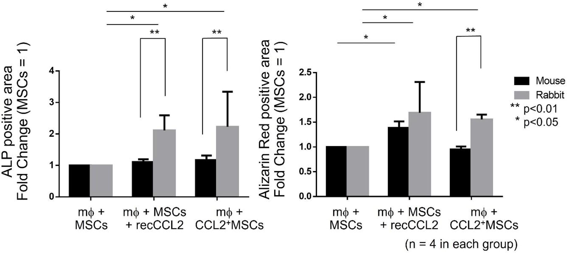
A174 - In Vitro And In Vivo Evaluation Of A Tissue-engineered Construct For Critical Lesionbonerepair
A. L. Lara Bertrand, W. Cárdenas, B. Camacho, I. Silva Cote
Instituto Distrital de Ciencia, Biotecnología e Innovación en Salud - IDCBIS, Bogotá, Colombia
*Purpose/Objectives: According to the WHO, 1.71 million people suffer from musculoskeletal disorders, the leading cause of disability worldwide. 25% of these disorders are fractures, 21% are caused by disease, 10% are amputations, and other types of trauma cause 44%. Bone injuries or disorders can be painful, limit mobility and require long recovery periods, and existing treatments are often painful. They may require more than 2 surgical interventions, and most do not promote bone formation, resulting in a poor quality of life for the patient.Tissue engineering research has succeeded in developing products using different technologies, biomaterials and human Wharton's jelly mesenchymal stromal cells (hWJ-MSC) to promote bone regeneration. Therefore, we have developed a medical product focused on bone repair that incorporates PLA (polylactic acid) as the base material for the 3D structure and a PLA/HAP (hydroxyapatite) electrospun membrane coating, in addition to incorporating hWJ-MSC to form a bone construct. We evaluated in vitro the biocompatibility of the scaffold with Wharton's jelly-derived mesenchymal stromal cells, the release of bone repair-related factors, the evaluation of mechanical strength, and its efficacy in repairing critical bone loss in lagomorph biomodels.
*Methodology: To evaluate the in vivo repair potential of the construct, we used a lagomorphic model. The construct, the cell-free coated 3D scaffold and crushed bone (positive control) were implanted into a 2 cm femoral bone defect. Follow-up was performed for six months, including observations every three days, radiographs at 1, 3, and 6 months, and histologic analysis by hematoxylin and eosin, Masson's trichrome at the end of the sixth month.
*Results: Mechanical tests showed that the strength of the scaffold is similar to that of trabecular bone tissue. Both the construct and the coated 3D scaffold engrafted well into the lesions; the biomodels supported the leg without discomfort after surgery, and the wound showed no inflammation to the naked eye. Radiographic and histologic analysis of the specimens showed the formation of new bone in both the construct and the 3D-coated scaffold, new cartilage, immature bone tissue, endothelial cells, and bone marrow. Notably, no inflammatory cells were observed, demonstrating the biocompatibility of the construct.
*Conclusion/Significance: Based on these data, we demonstrate that both the bone construct and the coated 3D scaffold promote the repair of critical bone lesions. These results demonstrate the potential of the tissue-engineered product developed in this study as a novel treatment strategy for significant bone loss.
A176 - Direct Current Electrical Stimulation Enhances Cell Viability And Osteogenic Differentiation Of Mesenchymal Stem Cells In 2D And 3D Conditions
E. R. Briggs1,2, S. M. Richardson1, K. A. Mace1, J. J. Blaker1, S. H. Carmell1
1
The University of Manchester, Manchester, United Kingdom,
2
Henry Royce Institute, Manchester, United Kingdom
*Purpose/Objectives: Large bone defects present a clinical challenge due to impaired or delayed healing. Bone tissue engineering (TE) approaches attempt to overcome the limitations of native healing by providing an external source of osteoprogenitor cells and architectural support through 3D scaffolds. Electrical stimulation (ES), the application of an external electrical field, has been shown to enhance cell proliferation, direct cell migration and enhance the differentiation potential of mesenchymal stem cells (MSC) in 2D conditions. However, its direct application to cell-laden 3D scaffolds is yet to be studied. Our study aimed to enhance cell proliferation and osteogenic differentiation of human MSC (hMSC) seeded in 3D scaffolds by applying a controlled electrical regime directly to constructs, in order to streamline and optimise the fabrication process of cell-laden scaffolds for bone TE.
*Methodology: A direct current (DC) ES regime of 100 mV/mm for 1 hour/day was delivered to primary hMSC seeded in 2D monolayers or into 3D porous sponge scaffolds. After 7 days of ES, cell viability was assessed using a Live/Dead fluorescence imaging assay, metabolic activity using an Alamar Blue assay, and dsDNA quantification using a Quant-iT dsDNA PicoGreen assay. Osteogenic differentiation was assessed after 7, 14, 21 and 28 days of ES using RT-qPCR to analyse gene expression of specific osteogenic markers (Runx2, Coll1a1, and BSP), and histochemical staining to assess mineralization (Alizarin Red stain). Stimulated samples were compared to non-stimulated controls in order to determine the effects of applying DC ES to hMSC in 2D and 3D.
*Results: Results showed ES significantly enhanced hMSC number and metabolic activity in 2D and 3D conditions after 7-days of ES. Fluorescence Live/Dead imaging of stimulated hMSC in 2D and 3D conditions showed no observable difference in cell morphology and alignment compared to non-stimulated controls. In 2D and 3D conditions, although ES downregulated the expression of the differentiation marker Runx2, ES significantly upregulated the expression of BSP, a gene marker for bone matrix mineralisation. In 2D stimulated samples, the peak expression of BSP occurred at an earlier time-point (day 21) compared to non-stimulated controls (day 28). In 3D stimulated samples, the peak expression of BSP occurred at an earlier time-point (day 14) compared to non-stimulated controls (day 21). Histochemical staining of 2D and 3D samples showed increasing mineralisation up to day 28, indicated by positive Alizarin Red staining for calcium, with observable increased deposition in stimulated samples after 21 days of stimulation.
*Conclusion/Significance: Herein, we have demonstrated that in line with previous studies investigating the effects of DC ES in 2D conditions, a regime of 100 mV/mm for 1 hour/daily enhances cell proliferation and osteogenic differentiation, specifically mineralisation, when applied directly to hMSC seeded in 3D scaffolds. Therefore, it may be recommended that a controlled DC ES regime can be applied directly to 3D scaffolds to enhance future bone TE approaches.
A177 - Successful Osteoclast Incorporation In An In Vitro 3D Model Of Endochondral Ossification: Adding A New Level Of Complexity
A. Garmendia Urdalleta, J. Witte-Bouma, N. Kops, A. Lolli, E. Farrell
Erasmus MC, Rotterdam, Netherlands
*Purpose/Objectives: The lack of complex in vitro bone models limits the investigation of processes involved in bone formation and repair, as well as bone-related diseases such as bone cancer metastasis. Modelling bone formation via the process of endochondral ossification (EO) in vitro is currently limited to the recapitulation of the initial stages of the process, namely induction of chondrogenesis and mineralisation of the newly formed matrix. However, these models lack the crucial cartilage remodelling process, which is mainly mediated by chondroclasts/osteoclasts. The absence of these multinucleated cells was shown to cause disturbances on the EO process but their introduction into in vitro 3D bone models has thus far proven challenging. In this work we added a new level of complexity to the current in vitro EO models through the introduction of human monocyte-derived osteoclasts into an in vitro cartilage template actively undergoing mineralisation. With this model we aimed to recapitulate osteoclastogenesis within a transient cartilage template in an in vitro setting.
*Methodology: Chondrogenic and mineralised pellets were formed using paediatric human marrow stromal cells (hMSCs). They were cultured for 21 days in the presence of transforming growth factor beta-3 (TGF-β3) (10 ng/ml) (chondrogenic) or TGF-β3 and β-glycerophosphate (10 mM) (mineralised). Pellets were co-cultured with CD14+ human monocytes in a consensus medium (αMEM 5% FCSi, ascorbic acid (25 µg/ml), RANKL (30 ng/ml) and M-CSF(25 ng/ml)) indirectly (transwell setting) or directly up to 14 days. In situ osteoclastogenesis and invasion of pellets were investigated by osteoclast counting, tartrate-resistant acid phosphatase (TRAP) staining, cathepsin K (CTSK) immunostaining and gene expression analysis (DC-STAMP, OC-STAMP, CTSK, TRAP and ATP6V0D2).
*Results: Indirect co-culture of CD14+ human monocytes and pellets leads to successful osteoclast formation for both chondrogenic and mineralised pellets, with no clear differences between groups according to TRAP staining, osteoclast counting and expression of osteoclastogenic markers. Upon direct contact, monocytes effectively attached to the surface of the pellets and differentiated into mature osteoclasts in situ (Figure 1). This was evidenced by positive TRAP and CTSK staining and expression of osteoclastogenic markers. Slight differences in the number and size of osteoclasts formed in direct contact with pellets were observed, with smaller and fewer osteoclasts present on the surface of mineralised pellets as compared to chondrogenic pellets. Osteoclastogenesis was not limited to the surface of the pellets, signs of osteoclast invasion of the inner cartilage matrix were also observed in this model, suggesting a possible active resorption of the matrix. Invasion was also slightly different between pellet conditions, with osteoclast presence limited to the superficial layers of mineralised pellets while it reached deeper layers in chondrogenic pellets.
*Conclusion/Significance: This study demonstrated that osteoclasts can successfully form on in vitro chondrogenic and mineralised hMSC pellets. Signs of invasion and possible active resorption of the pellets was also observed. Adding further complexity to this model will provide us with a tool to increase our understanding of the bone formation process as well as bone diseases in a (highly) controlled in vitro environment.
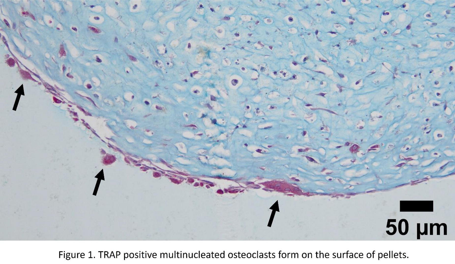
A178
A180 - Production Of Antimicrobial Peptide Conjugated Dual Effecting Putty Grafts For The Treatment Of Bone Infections And Induced Osteogenic Differentiation
E. Bilgiç, G. Pulat, O. Karaman
Izmir Katip Celebi University, Izmir, Turkey
*Purpose/Objectives: Bone grafts that are developed using principles of tissue engineering are commonly employed in the treatment of bone deficiencies. Injectable bone grafts, putty grafts, are administered locally to relevant surgical area through a specialized syringe representing a preferred approach for addressing bone defects characterized by irregular geometric structures. Although current treatments for bone deficiencies yield relatively satisfactory results, bone infections remain a significant problem, leading to chronic conditions that hinder bone healing. In the management of bone infections, conventional broad-spectrum antibiotics are commonly utilized. Nevertheless, its usage is disadvantageous due to development of resistance in bacteria and osteoblast toxicity. Therefore, antimicrobial peptides (AMPs) are promising agents for infection treatments due to their broad-spectrum antibacterial activity, controllable biocompatibility, and limited resistance generation. KR-12, a cationic AMP, is the smallest active portion of human cathelicidin LL-37 peptide with strong antibacterial activity. Furthermore, KR-12 does not have a toxic effect on mesenchymal stem cells (MSCs), on the contrary, it increases osteogenic differentiation. Herein, the ultimate aim is to develop Putty grafts, functionalized with KR-12, which have a dual effect, supporting osteogenic differentiation and showing antimicrobial activity, which can provide successful repair of bone defects.
*Methodology: KR-12 was synthesized on 4-Methylbenzhydrylamine (MBHA) resin. To obtain maximum hydroxyl groups on β-TCP, cold atmospheric plasma (CAP) was performed. KR-12 was conjugated to β-TCP by silanization with APTES in anhydrous hexane followed by succinimidyl 3-maleimidopropionate in DMF and 1 mM peptide in DMF and conjugation was confirmed with FT-IR. Putty graft was prepared by mixing AMP conjugated β-TCP with a 3% solution of hydroxypromyl methyl cellulose. Characterization of Putty/KR-12 was performed using FT-IR and SEM. MTT analysis and live/dead staining were performed on MSCs to examine the effect of the Putty/KR-12 on cell viability. Calcium and ALP quantification assays on days 7, 14, 21, and 28 were conducted to evaluate the effect of Putty/KR-12 on osteogenic differentiation of MSCs.
*Results: The elongation of the peaks at 3600-3000 cm-1 in the FT-IR spectrum showed that the -OH groups of β-TCP were successfully increased by CAP. Successful conjugation was confirmed with changes at 1460-1240 cm-1 in FT-IR spectrum. According to the SEM images, peptide conjugation did not cause change on the Putty graft. Developed bone grafts did not have an undesirable effect on MSC viability. Furthermore, a significant increase in DNA content and ALP amounts was observed in Putty/KR-12 compared to Putty. For further studies, antimicrobial tests of Putty and Putty/KR-12 will be performed on both planktonic and biofilm forms of MRSA and MDR P. Aueriginosa. DNA quantification assay, immunofluorescence staining, q-PCR analysis for collagen type I, osteopontin and osteocalcin genes on days 7, 14, 21, and 28 will be conducted.
*Conclusion/Significance: β-TCP and AMP conjugation for the first-time is considered a major advance in the development of pioneering antimicrobial bone grafts. The outcomes of this study would help to enhance the effectiveness of conventionally used bone grafts by functionalizing with AMPs that is able to induce dual effect as supporting osteogenic differentiation and showing antimicrobial activity.
A181 - The Addition Of Inorganic Ions To Three Dimensional Printed Composite Scaffolds Improves Bone Regeneration Potential
M. Nikody1, J. Li1, D. Koper1, E. Rosado Balmayor2, P. Habibovic1, L. Moroni1
1
Maastricht University, Maastricht, Netherlands,
2
RWTH Aachen University Hospital, Aachen, Germany
*Purpose/Objectives: Bone possesses intrinsic regenerative capacities, however, defects exceeding a certain size do not heal spontaneously. Such defects caused by congenital disorders, trauma, or tumour resection are known as critical-sized bone defects. Treatment of such defects requires surgical intervention. Transplantation of autologous tissue remains the gold standard treatment favoured by clinicians. However, autologous bone grafting is associated with donor-site morbidity and limited accessibility of the graft tissue. This contributes to an increasing need for an alternative solution in the form of off-the-shelf, synthetic, biodegradable graft substitute materials for the treatment of critical-sized bone defects. Recent advances in the field of biomaterials development focus on improving the osteogenic and angiogenic properties. One of the approaches to improve the regenerative properties of biomaterials, without changing their synthetic character, is the addition of inorganic ions with known osteogenic and angiogenic properties, for instance, zinc, copper, or cobalt.
*Methodology: In this study, bioactive composites with a high ceramic content were additively manufactured by 3D fibre deposition. Composites consisted of a combination of poly(ethyleneoxide terephthalate)/poly(butylene terephthalate) (1000PEOT70PBT30, PolyActive, PA) and 50% beta-tricalcium phosphate (β-TCP) with the addition of zinc and copper or zinc and cobalt in the form of a coating of the β-TCP particles. β-TCP particles were immersed in a zinc and copper or zinc and cobalt solution with a concentration of 15 or 45 mM for 7 days. Osteogenic and angiogenic properties were assessed in vitro employing a direct culture with clinically relevant human mesenchymal stromal cells (hMSCs) and indirect culture of human umbilical vein endothelial cells (HUVECs). Furthermore, scaffolds were implanted intramuscularly in a mini-pig model.
*Results: The composition of scaffolds influenced the osteogenic and angiogenic properties in vitro. In particular, scaffolds containing 15 mM of zinc and cobalt positively influenced the early expression of RUNX-2, COL-1, OCN, OPN, and VEGF-A. In a tubule-formation assay, HUVECs formed vascular networks under all tested conditions without statistically significant differences among quantified parameters. Ingrowth of connective tissue into all scaffold pores was observed after 12 weeks of intramuscular implantation in mini-pigs. No clear bone formation was observed at the chosen time point. However, early signs of osteogenic differentiation were observed in all composites. Furthermore, histology confirmed blood vessel infiltration into all samples. Interestingly, vessels with particularly small diameters were observed in the presence of zinc and cobalt, while larger vessels were present in samples with the addition of zinc and copper. Moreover, the presence of multinucleated, osteoclast-like cells was observed in all scaffolds.
*Conclusion/Significance: The addition of zinc and copper as well as zinc and cobalt to bioactive composites with a high ceramic content resulted in improved osteogenic, and angiogenic properties of the composite scaffolds. In particular, formulation with zinc and cobalt seemed to have the strongest effect. Further analysis will focus on the assessment of the immunomodulatory effect of added inorganic ions on hMSCs. In addition, quantification of specific histology staining of the in vivo samples will be performed to unravel potential differences between scaffold compositions.
A183 - Sculpting Scaffold-Free Cartilage Templates Through 3D Bioprinting Of Sacrificial Alginate Bioinks
A. Dimopoulos, K. Ioannidis, H. Svitiva, C. Androulidakis, I. Papantoniou
KU Leuven, Leuven, Belgium
*Purpose/Objectives: In recent years we have witnessed the rise of endochondral ossification (ECO) as a robust paradigm for bone tissue engineering solutions. This cartilage to bone tissue transition is encountered during bone development and fracture healing. ECO Tissue engineered solutions have been provided through high-density hydrogel-based strategies as well as organoid bioassemblies engineered cartilage templates exhibiting positive in vivo outcomes. However, these implants are mostly formed through self-assembly processes and hence large-scale and anatomically complex cartilaginous implants with geometric precision cannot be produced. Moreover, the presence of foreign materials such as bioinks is well known to trigger immune response from the host. Therefore, we developed a high cell-density bioprinting strategy, using sacrificial alginate bioinks resulting in functional scaffold-free cartilaginous tissues able to rapidly form large and pure ossicles upon implantation.
*Methodology: We developed a chemically modified partially crosslinked alginate bioink (2% - 3% w/v) that could be printed in one extrusion step maintaining its structural form, giving us the advantage to 3D bioprint high cell densities (3-23 Million cells/ml). This bioink enabled the bioprinting of cartilaginous implants that could maintain structural precision and enable complex shapes to be produced. Upon chondrogenic differentiation of hPDCs within the bioink, condensed into clusters which gradually grew while abundant extracellular matrix was produced throughout the entire volume of the engineered tissues. We demonstrated that the alginate bioink used could be selectively dissolved at various timepoints resulting in structurally competent constructs. Finally, constructs were implanted ectopically to evaluate their bone formation capacity for 4 and 8 weeks.
*Results: Cells exhibited high viability over all time points, while gene markers indicating a prehypertrophic/hypetrophic phenotype were found to be upregulated over time (SOX9, COL10A1, IHH and others). Secreted ECM was assessed by evaluating the following markers for staining (Safranin-O, Collagen type II, Alcian blue) while we also carried out confocal microscopy to observe the abundance of organized collagen within the implants scaffold. In addition, fluorescent antibody staining for confocal microscopy analysis, were carried out for hypertrophic markers (COL2A1, IHH, MMP13 and others) for the hypertrophic (and bone forming) cartilage implant. Mechanical characterization experiments were implemented to measure the gradual increase in young modulus over the timepoints, related to ECM secretion (60-80kPa). Significant differences were observed between the 3D Bioprinted and scaffold-free implants upon even from the 4th week. Bone formation capacity and bone maturation were significantly improved for scaffold free implants while pure ossicles exhibiting cortical and trabecular bone structure and a fully developed bone marrow cavity were observed.
*Conclusion/Significance: In conclusion, our novel bioprinting process using partial crosslinked sacrificial alginate bioinks allows for the creation of highly cellular density constructs with structural integrity. The resulting scaffold-free implants demonstrated enhanced bone regeneration capacity as compared to alginate implants, as ossicles where forme already by week. These findings pave the way for producing large anatomical 3D bioprinted scaffold-free cartilage implants to address clinical limitations in long nonunion bone defects.

A184 - Anisotropic Porous Hydroxyapatite/Gelatin Scaffolds For Trabecular Structure Reconstruction During Arthrodesis
A. Pavlov, B. Gadomski, J. Johnson
Colorado State University, Fort Collins, CO
*Purpose/Objectives: Osteoarthritis is one of the most common foot conditions within the United States with more than three million cases reported annually. This condition is most frequently reported in the first metatarsophalangeal (MTP) joint and leads to patient pain, and irregularities within the toe orientation. This leads to loss of mobility and severe discomfort during walking if left unchecked. For patients unresponsive to non-invasive treatments for this condition, the standard of care is arthrodesis. First MTP arthrodesis is a joint fusion approach that entails removal of both articular cartilage surfaces with preparation of the bony surfaces into appropriate opposing geometries. To maintain anatomical symmetry and unaltered gait, there exists a need for graft material capable of replacing the lost material and fusing to the bone without significantly lengthening the surgical or recovery process. While collagen/hydroxyapatite blends have been proven to generate scaffolds, gelatin has emerged as a potential alternative, offering the same ability to create structures of controlled porosity within hydroxyapatite at lower cost and without fearing degradation.
*Methodology: Commercial hydroxyapatite powder was mixed with a 10% W/V solution of microbiology grade gelatin in decadal concentrations ranging from 30:70 to 70:30. This slurry was then cast into silicon molds and directionally frozen at -20°C before being lyophilized. This created an anisotropic porous network within the hydroxyapatite matrix. These were then analyzed using micro computed tomography to quantify pore size, bone volume fraction and density. Afterwards the samples were crosslinked with a 1% W/V glutaraldehyde solution at 2-hour intervals, ranging from 2 to 16 hours with a 24-hour batch being used as a control. Unconfined compression testing was performed at a rate of 1mm/min up to 60% strain. Ultimate compressive stress and young’s modulus for the 8 different crosslinking times groups were calculated. Following this a one-way ANOVA with Tukey HSD post hoc test was performed using alpha = 0.05.
*Results: MicroCT analysis of samples revealed a linear relationship between Gel:HAp ratio and density and pore size. In contrast, these data illustrate the logarithmic relationship between Gel:HAp ratio and bone volume fraction and porosity. The 60:40 Gel:HAp samples exhibited porosity values of 0.62 most closely replicating porosity noted in human trabecular bone; as such, this group was chosen to proceed to mechanical testing/assessment of crosslinking on mechanical properties. Mechanical testing of samples revealed a non-monotonic relationship between crosslinking time and ultimate compressive strength, with a maximum at 8 hours. Specifically, the 8-hour group exhibited a 2-fold increase in ultimate compressive strength as compared to the 2-hour time (p=0.0038).
*Conclusion/Significance: Current research delineates a substantial advancement in the synthesis of a Gel:HAp graft material that closely emulates the porosity and mechanical attributes of human trabecular bone. By regulating the Gel:HAp ratio and optimizing crosslinking time, we have successfully engineered a material with potential applicability as a bone surrogate in arthrodesis procedures. This innovation presents a significant step towards enhancing osteoconductivity and mechanical stability in joint fusion procedures, which may ultimately contribute to improved surgical outcomes and patient quality of life.
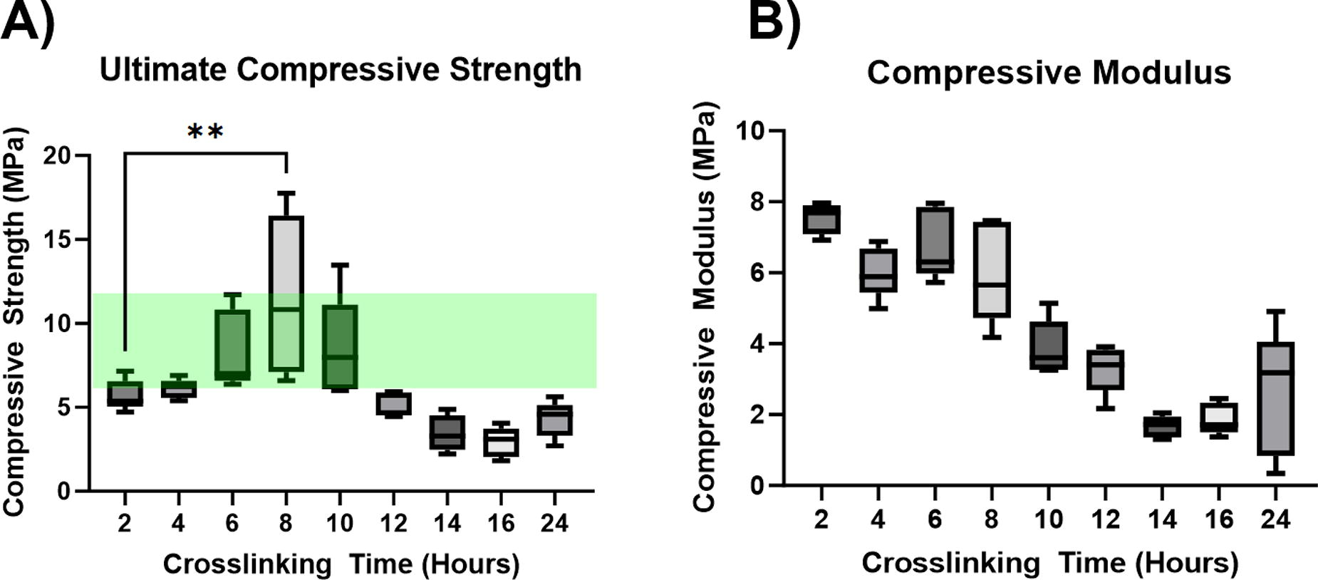
A186 - Facilitated Fracture Repair Via Noninvasive Localized Cold Therapy
M. Zakaria1, J. Allard2, J. Garcia1, J. Matta1, Y. Honjol1, D. Schupbach1, F. Mwale3, M. Grant3, E. Harvey1, G. Merle2
1
Montreal General Hospital, Montreal, QC, Canada,
2
Polytechnique Montréal, Montreal, QC, Canada,
3
Lady Davies Institute Jewish General Hospital, Montreal, QC, Canada
*Purpose/Objectives: The ability of fractured bone to regenerate and undergo repair is often compromised. Successful fracture healing involves an inflammatory cascade leading to bone repair. Cold treatment is commonly used to prevent inflammation after musculoskeletal injuries and earlier work on using cold stimuli for critical cortical defect healing has shown to enhance bone growth. However, no study has looked at its effect or mechanism on fractures.Here, we hypothesized that enhanced bone healing demonstrated using cold therapy in cortical defects will be replicated in a clinically relevant fracture model and cold therapy stimulates angiogenesis leading to improved vascularity.
*Methodology: Bilateral femoral fractures using retrograde nailing were formed in C3H strain mice. Initially, a guidewire was inserted followed by the usage of a homemade 3-point impact device to produce a closed midshaft femur fracture. A 24-gauge stainless steel needle tip was then implanted over the guide wire to stabilize the fracture and the guide wire was removed.Experimental legs were immersed in a cold-water bath reaching an internal bone temperature of 19 degrees Celsius for 15 minutes daily. Core-body and control leg temperatures were maintained with a heating pad. Femurs were harvested at days 7, 14, 28 and underwent micro-CT analysis. Staining of ALP, TRAP, and VEGF followed.
*Results: Day 28 timepoint analysis revealed daily exposure of fractured femurs to cold therapy increased bone volume/tissue volume (BV/TV) by 15% when compared to untreated controls (p-value<0.001). Simultaneously, a 1.42% increase in channel volume/tissue volume within cold-treated femora illustrates the prevalence of an enhanced vascular network (p-value=0.05). Biomarkers ALP and TRAP demonstrated expressions consistent with physiological bone remodeling within cold-treated femora at day 28, with staining revealing a significant 5.3% reduction in ALP (p-value=0.028) detection without significant alteration of TRAP expression (p-value=0.44). Bony callus formation is visibly enhanced at the 14-day timepoint from daily exposure of fractured femurs to cold therapy, reflecting a 6.61% increase in BV/TV when compared to untreated controls (p-value<0.001).
*Conclusion/Significance: Findings confirm that localized cold treatment accelerates bone growth in a clinically relevant closed femoral fracture model. A strong increase in BV/TV coupled with decreased ALP staining of osteoblasts without significantly altered TRAP staining of osteoclasts indicates osteoclastic-driven bone remodeling is attained faster within cold-treated femora. Simultaneously, improved vascularity indicates enhanced angiogenesis. Currently, potential fluctuations in genetic expression, vasomotor tone, pH, and hemoglobin oxygen saturation are being investigated to uncover the mechanistic propensity of therapeutic hypothermia.
A187 - Scaffold-guided Bone Tissue Engineering Using The Reamer-irrigator-aspirator (RIA) Bone Harvest System For Critical Sized Bone Defect Reconstruction
F. Medeiros Savi1,2,3, C. E. Dlaska1, J. Henkel1,4, D. S. Sparks1, F. Russell1, S. Saifzadeh1,5, M. A. Schuetz1, D. W. Hutmacher1,2,3
1
Queensland University of Technology, Brisbane, Australia,
2
Centre in Regenerative Medicine, Brisbane, Australia,
3
Max Planck Queensland Center for the Materials Science of Extracellular Matrices, Brisbane, Australia,
4
Lutheran Hospital Goettingen-Weende, Goettingen, Germany,
5
Medical Engineering Research Facility, Brisbane, Australia
*Purpose/Objectives: The incidence and burden of bone fractures is a global public health issue. In 2019, Australia ranked among the top three countries with 1.6 million reported fractures that exhibited impaired healing, requiring clinical intervention. Fractures of the tibia were among the prevalent anatomical sites afflicted, with non-union rates of 14%. This not only prolongs the treatment duration but also extends the overall years of impairment for those affected, resulting in significant healthcare costs. The current gold-standard treatment relies on autologous bone grafts (ABG), mainly harvested from the iliac crest (ICBG), and from the intramedullary canal of long bones via the Reamer-Irrigator-Aspirator (RIA) System. Viably comparable to the conventional ICBG treatment the latter stands out for its ability to collect substantial volumes of intramedullary bone graft with associated osteogenic properties. However, both approaches face constrains including limited availability, post-operative complications, and donor site morbidities. These inherent challenges have spurred the search for treatment solutions rooted in tissue engineering. Scaffold guided bone tissue engineering (SGBTE) has emerged as a promising alternative to fulfill the escalating demand for bone graft material among orthopaedic surgeries. A significant focus of our research has been on harnessing the potential of medical grade polycaprolactone (mPCL) and tricalcium phosphate (TCP), which has shown remarkable regenerative capabilities in treating critical-sized bone defects of both 3cm and 6cm. Utilizing our well-established sheep animal model and the unique benefits of mPCL-TCP scaffolds in retaining and compartmentalizing the RIA material within the scaffold matrix, this study aimed at exploring the feasibility of using a reduced volume of RIA ABG for SGBTE for the treatment of a 6cm critical-sized bone defect.
*Methodology: Using fused deposition modeling, cylindrical mPCL-TCP scaffolds with central fenestrations were created (outer diameter: 20mm, inner diameter: 8mm, length: 60mm). Scaffold features comprised 70% porosity, fully interconnected pores (300µm filament diameter, 1200µm separation), forming a 0/90° lay-down pattern. A 6cm defect was created in 22 sheep tibiae. The defects were treated with either RIA alone, mPCL-TCP scaffold with RIA-ABG, or ICBG alone. The study spanned a 3- and 12-month time point interval. Bone formation and mechanical properties were evaluated through x-rays, Micro-computed tomography (µCT), biomechanical testing, scanning electron microscopy (SEM), backscattered electron microscopy (BSE), second harmonic generation (SHG), immunohistochemical and histological analyses.
*Results: Corroborating X - ray and µCT evaluations, the immunohistochemical and histological results showed limited bone formation extending from the host bone sites at 3-months' time point for the RIA alone group. Robust bone formation with complete bridging of the 6cm defect was achieved at 12-months for the mPCL-TCP scaffold combined with RIA-ABG. SEM evaluation revealed presence of osteocytes in direct contact with the mPCL-TCP struts, viable osteocytes within ABG fragments, as well as high coverage of the ABG fragment’s surface with newly formed bone.
*Conclusion/Significance: The multimodal histological and immunohistochemical outcomes indicate that even after 12-months of implantation, ABG fragments continue to demonstrate osteoconductive properties. Furthermore, when integrated with mPCL-TCP scaffolds, they not only retained their structural integrity but also prevented resorption, thereby enhancing the bone formation.
A188
A190 - Development Of Bioactive Instructive Hydrogels For Bone Tissue Engineering
J. Idaszek, M. Stefanska, R. Owda, S. Caglar, M. Volpi, E. Choinska
Warsaw University of Technology, Warsaw, Poland
*Purpose/Objectives: Decellularized extracellular matrix (dECM) is obtained from native tissues using physical, chemical and/or enzymatic decellularization protocols. These protocols have tremendous effect on decellularization efficacy (i.e., residual DNA content), preservation of native macromolecules or growth factors and, therefore, affect cell-material interactions. So far, bone tissue was decellularized using enzyme trypsin. However, this protocol can lead to disruption of dECM proteins, depletion of glycosaminoglycans (GAGs), and thus reduces its bioactivity. Therefore, the objective of this study was to investigate the effect of different demineralization and decellularization parameters on bioactivity of bone-derived dECM.
*Methodology: Porcine femurs were demineralized using 0.6 M HCl at room temperature (“RT”) or 4°C (“C”) for 48h, and decellularized using a chemical method (“D”, 1% Triton X-100) at 4°C for 72h or enzymatically (“E”, 0.25% trypsin) at 37°C for 24h, giving in total three types of dECMs: D_RT, D_C, E_C. Type I collagen was used as control. The biochemical composition was determined using DNA, total collagen and DMMB (GAGs content) assays. The 2% dECM sols were prepared by digesting the dECM with pepsin (10:1 ratio) for 72 h at RT. Following the neutralization, the sols were mixed with 1% alginate at 1:1 ratio, combined with bone marrow-derived human mesenchymal stem cells (hMSCs, 107/ml), and transferred into ion eluting moulds (1% agarose, 30 mM SrCl2). The hMSCs-laden hydrogels were cultured in proliferation (α-MEM, 1% PS, 10% FBS, 1 ng/ml of FGF2), osteogenic (α-MEM, 10% FBS, 1% PS, 10 mM β-glycerophosphate , 50 μg/ml ascorbic acid-2-phosphate, 10 nM 1,25-dihydroxy-vitamin D3, 10 ng/ml of dexamethasone) and chondrogenic (DMEM/F12, 1% PS, 10 ng/ml TGF-β3, 100 nM dexamethasone, ITS supplement, 1.25 mg/ml BSA, 100 μM ascorbic acid-2-phosphate, 1 mM sodium pyruvate) media for up to 28 days. Viability was evaluated by live/dead staining and MTS assay; ALP activity was determined using para-nitrophenyl phosphate as a substrate and normalized to DNA content.
*Results: The decellularization yielded the lowest concentration of DNA (12.7±1 ng/ 1 mg of dECM) only when it was performed using Triton X-100 and the demineralization was carried out at 4 °C (D_RT). Utilization of the detergent led to approximately 80% higher retention of GAGs than the trypsin treatment. The investigated demineralization and decellularization conditions did not affect the viability of hMSCs, however, they had a strong effect on the ALP activity in the absence of osteogenic and chondrogenic factors. The detergent-treated D_RT induced higher ALP activity than the enzymatically-derived E_C (96±7 vs 14±4 nmol/μg DNA at day 28 in proliferation medium, respectively), which was similar to the ALP activity in osteogenic (79±45 vs 50±6 nmol/μg DNA for D_RT and E_C, respectively) and chondrogenic (88±20 vs 69±7 nmol/μg DNA for D_RT and E_C, respectively) media at day 28. Interestingly, the highest level of GAGs was measured when hMSCs were cultured in dECM-based hydrogels in proliferation and chondrogenic media.
*Conclusion/Significance: Decellularization of bone using nonionic detergent Triton X-100 yields the highest efficacy and preserves the bioactivity of the dECM, as demonstrated by increased ALP activity even in the absence of differentiation-inducing factors.
A191 - Mechanistic Propensities Of Cryotherapy In A Bone Injury Site
M. Zakaria1, Y. Honjol1, D. Schupbach1, P. Azizi1, M. Grant2, F. Mwale2, E. Harvey1, G. Merle3
1
Montreal General Hospital, Montreal, QC, Canada,
2
Lady Davies Institute Jewish General Hospital, Montreal, QC, Canada,
3
Polytechnique Montréal, Montreal, QC, Canada
*Purpose/Objectives: Applying a cold stimulus to bone has been shown to increase the healing response. However, the mechanisms upon which acute cold stimulates bone-forming cells are paradoxical and not well understood. It has been previously established that when the vascular network in and around bone is compromised, hypoxia occurs near the fracture site, leading to the activation of a key mechanism in fracture healing to restore blood flow. Furthermore, acute cold exposure has also been shown to trigger upregulation of RNA-binding motif protein 3 (RBM3) and Peroxisome proliferator-activated receptor gamma coactivator 1-alpha (PGC-1α) both of which have essential roles in osteogenesis. The objective of this study is to elucidate the mechanism by which cold therapy affects bone formation in-vivo at the injury site. Here we hypothesize that localized application of cold temperature will lead to elevated induction of hypoxia in the bone injury site indirectly in conjunction with increased detection of RBM3 and PGC-1a which is responsible for upregulation of osteoblast differentiation.
*Methodology: A bilateral cortical bone defect model was applied to thirteen male C3H strain mice aged 2-3 months. Following the formation of the defect, the region was flushed with phosphate-buffered saline (PBS) to remove any remaining bone fragments. Hypoxyprobe was diluted in saline to reach a final concentration of 2.45 mg/mL in accordance with manufacturer guidelines. 1.5mL of the solution was intraperitoneally injected 7 days post-operation into the mice. Five minutes after the time of injection, the experimental hindlimb of the mouse was exposed to an ice-water bath for fifteen minutes. An internal temperature of 19 degrees Celsius within the mouse hindleg was measured in previous studies done at our lab using the same methodology. Detection involved a secondary antibody sequence with a fluorescein isothiocyanate (FITC)-conjugated system. FITC conjugated Anti-RBM3 Antibody for RBM3 and Anti-PGC-1α Antibody for PGC-1a were used. Adducts formed between pimonidazole and hypoxic cells were detected by incorporating anti-pimonidazole antibody. Secondary antibody horse radish peroxidase-conjugated rabbit anti-FITC was then used. 3,3′-diaminobenzidine (DAB) staining was then utilized to visualize the areas of interest through immunoperoxidase staining.
*Results: A 5.6% increase (p-value<0.01) compared to untreated controls in the number of hypoxic cells within and around the cortical bone defect in the hindlimbs of mice following exposure to an acute cold stimulus demonstrates detectable localized hypoxia induction through the application of cold exposure. Increased detection of PGC-1a (p-value<0.005) and RBM3 (p-value<0.008) were prevalent in newly forming bone cells within cortical defects following cold exposure signifying elevated presence of proteins involved within the osteoblastogenesis pathway.
*Conclusion/Significance: The results illustrate the development of a detectable hypoxic environment that provides a conjunctive avenue to explore certain regenerative pathways dependent upon hypoxic conditions within the early stages of bone repair. PGC-1a and RBM3’s increased presence within a bone injury site such as a cortical defect may reflect their importance in the bone repair process in conjunction with the mechanistic propensities of cold therapy.
A194 - Investigating The Role Of Dental Pulp Stem Cells In Polylactic Glycolic Acid (plga) Nerve Conduit In Peripheral Nerve Repair: A Study In Rat Model
S. Abdullah1, N. Mohamed Haflah2, M. Abdul Razak2, S. Y. Thow2, A. S. Naicker1, O. S. Htwe1, Y. Lokanathan2, M. H. Ng2
1
Universiti Kebangsaan Malaysia, Kuala Lumpur, Malaysia,
2
Universiti Kebangsaan Malaysia, Cheras, Malaysia
*Purpose/Objectives: Traumatic peripheral nerve injuries may cause debilitating with a reported incidence of 2.6%. As an alternative to autograft, nerve conduits are vital in bridging a nerve gap and guiding axonal regeneration and are currently commercially available. Human dental pulp stem cells (DPSCs) are ectoderm-derived stem cells that possess mesenchymal stromal cell and neural cell properties. A major benefit of DPSCs is that they can be isolated in a simple and autologous fashion during routine dental procedures without ethical concerns. The objective of this study is to elucidate the role of DPSC seeded on nerve conduits in neurologic recovery of peripheral nerve injury.
*Methodology: To obtain aligned PLGA nanofibres electrospinning method was used.Electrospinning setting including using a 1ml syringe (diameter 4.6909mm) with a blunted 21G needle loaded with PLGA solvent under the following parameters: flow rate of 0.10 ml/hour, voltage of 18-20 kV, needle tip to mandrel distance of 34.5cm and mandrel rotation speed of 1400 rpm(maximum). A collagen mat was placed on a 25 cm x 8 cm aluminum foil and anchored with tape and the aluminum foil was placed on the mandrel. Electrospinning was done for 2 hours to produce a thin membrane of aligned nanofibres at the rotating mandrel. Dental pulp tissue was harvested and underwent cell culturing and neurogenic induction of MSC. It was then seeded onto aligned polylactic glycolic acid (PLGA) nanofibres nerve conduits.The in vivo phase involved 18 adult Sprague Dawley rats, divided into 3 groups which were conduit with stem cells (Group A), conduit without stem cells (Group B), and control group without conduit (Group C). A 10 mm sciatic nerve defect was created and grafted with a nerve conduit for groups Aand B. Electrophysiological studies and footprint analysis were performed at 2, 4, 8, and 12 weeks after surgery. Data from each group of rats was collected and analysed with adjusted Epsilon method with Greenhouse Geisser variation.
*Results: For electrophysiological studies, at week 12 the mean amplitude for group A and group B was 13.9mV, 6.0mV, 0mV respectively. The difference was statistically significant . Similarly, there was significant difference in mean conduction velocity measuring 72.50 m/s, 58.50 m/s and 0 m/s for groups A,B, and C, respectively. For the footprint analysis, groups A and group B had longer stride lengths and wider stride widths compared to group C, but was found to be not statistically significant. The animals in groups B and C had significantly higher incidences of ulceration and autonomy, a sign associated with neuropathic pain compared to group A.
*Conclusion/Significance: Conclusion: This study suggests that the presence of DPSCs in nerve conduits can improve neurologic recovery in PNI with segmental defects and prevent or alleviate neuropathic pain. Further studies are required to elucidate the role of DPSCs in nerve conduction improvement and pain relief mechanisms.
A196 - Development Of Polyacrylamide Hydrogel As A Novel Cell Sheet Fabrication Platform Exhibiting Physiological Stiffness
V. Irawan
Technical University of Munich, Garching, Germany
*Purpose/Objectives: Cell sheet is a tissue-engineering product consisting of monolayer cells embedded in its matrices, offering the advantage of preserving cellular viability. Thus, cell sheet has been used to effectively deliver therapeutic cells. Regardless of cell types, cell sheet is fabricated on highly stiff substrates (stiffness modulus: 106 kPa) that goes beyond the physiological value (10-1-103 kPa). The excessive stiffness has been reported to impair cell activities. This implies that the current fabrication platforms inhibit cell sheet from achieving its optimum therapeutic potential. To this end, we conceive a new cell sheet fabrication platform with physiological stiffness by using polyacrylamide (PAc) hydrogel (10-1-102 kPa). PAc surface is coupled with biotin, which bind the streptavidin (Kb=1015). Next, low-binding biotin is bound to the streptavidin (Kb=107) to support the cell attachment, which in turn can be removed by elution with excess biotin to induce cell detachment.
*Methodology: The PAc hydrogel was prepared by adding acrylic acid to the standard acrylamide monomers. Biotin-NH2 was coupled to the acrylic acid of PAc via carbodiimide chemistry. The success of biotin coupling was evaluated by incubation with fluorescent streptavidin-Alexa. Desthiobiotin (DSB) was selected as low-binding biotins. To check the elution efficacy, fluorescent DSB-ATTO and DSB-RGD were bounded to streptavidin-modified PAc for the study of biotin elution and for the cell culture. HS-27 fibroblastic cells were seeded on the substrate in complete medium. Elution in cell culture was done in 5 mM biotin in serum free medium.
*Results: PAc with stiffness 45 kPa was used for this study. Upon incubation with the fluorescent streptavidin (Fig. 1A), the unmodified PAc and biotin-coupled PAc showed the absence and presence of fluorescent signal, respectively. In Fig. 1B, immersion in PBS for 15 minutes do not decrease the fluorescent signal of DSB. Elution with free biotin (5 mM) in phosphate buffer, rapidly diminished the fluorescent within 2 minutes. The eluted DSB was present in the supernatant as confirmed by plate analysis. The cellular F-actin were organized into fibres for cells attached on the BT/SAv/DSB-RGD-modified PAc and cover slip control. In contrast, the F-actin were diffused for the unmodified PAc. The cell area and circularity for cells attached on unmodified PAc, BT/SAv/DSB-RGD-modified PAc, and coverslips are 1,380 µm2, 0.48; 1,774 µm2, 0.39; and 3,445 µm2, 0.35. This result indicates that BT/SAv/DSB-RGD modification enhance cell attachment and allow the cell to sense the hydrogel stiffness. The attached cells can be grown into the monolayer structure by prolonging culture time (Fig. 1D). Upon incubation in excess biotin (5 mM) for 15 minutes, 53% of cell regions started to detach and roll. More cells detached at longer incubation time, i.e., at 60 minutes (83%). However, detachment begins on the middle instead of edge part, resulting in the patchy cell sheet.
*Conclusion/Significance: The polyacrylamide has been successfully modified with bioin-streptavidin interactions which facilitates the cell attachment, monolayer formation, and the subsequent detachment. Further studies are undertaken to improve the quality of detached cells, to check the presence of matrices, and to use substrate with lower stiffness.
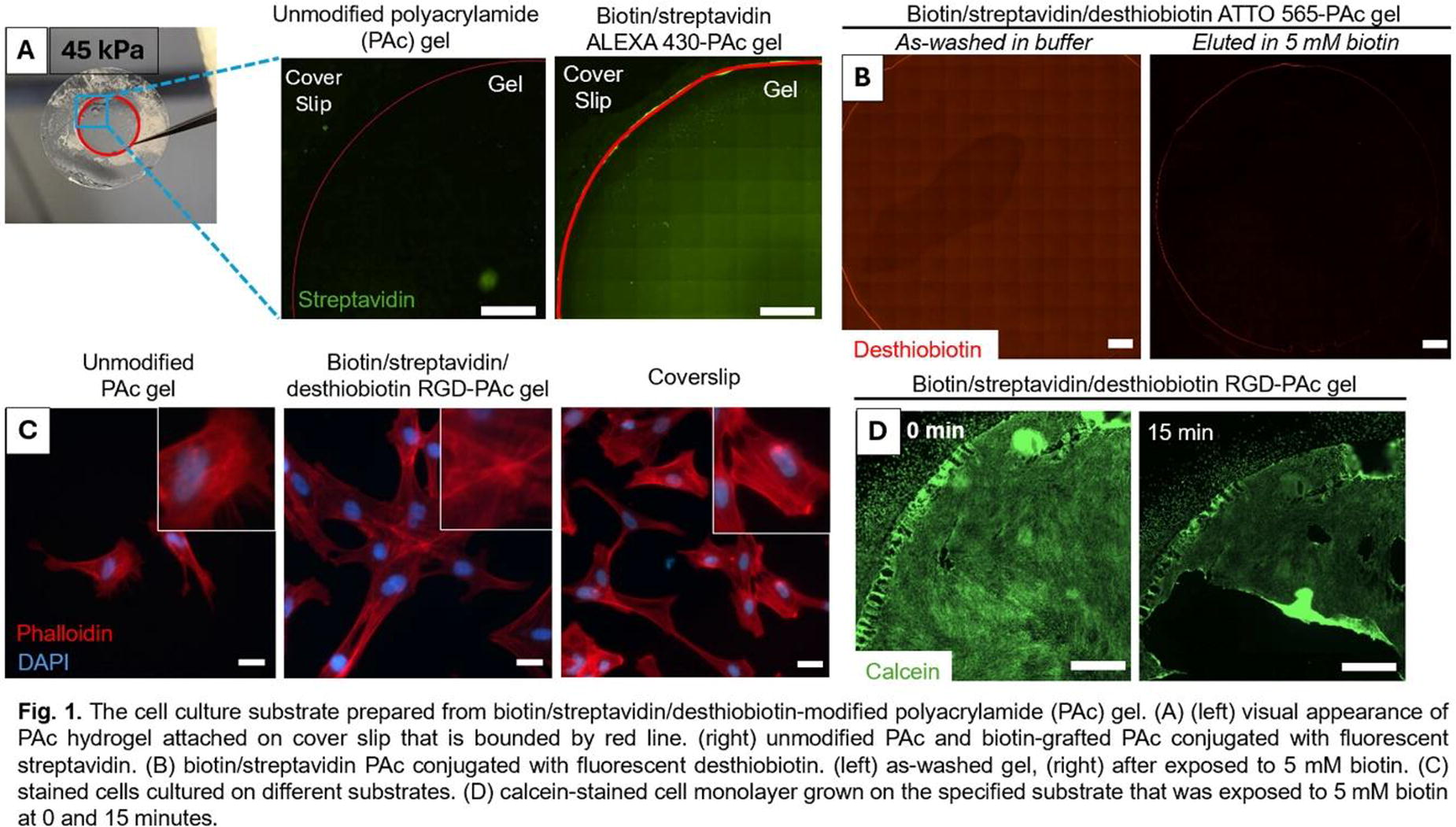
A197 - In Vivo Testing Of Human Induced Pluripotent Stem Cell-Derived Schwann Cells And Their Precursors For Nerve Tissue Engineering
E. A. Atkinson, R. Powell, O. Guillemot-Legris, P. O. Smith, J. B. Phillips
UCL, London, United Kingdom
*Purpose/Objectives: Nerve autografts remain the clinical gold-standard intervention for gap repair following a peripheral nerve injury, providing aligned Schwann cells that support and guide neuronal regeneration. However, limitations include donor site morbidity, limited donor tissue availability and inadequate recovery. Tissue engineering has potential to overcome such limitations by providing a construct of biomaterials and therapeutic cells, that can bridge the nerve gap. Human induced pluripotent stem cells (hiPSCs) are derived from a clinical-grade human neural stem cell line (CTX0E03) and therefore are promising for use in translational tissue engineering. This work aims to assess the survival and host response of hiPSC-derived Schwann cells and Schwann cell precursors in a collagen nerve repair construct, following implantation into a rodent nerve gap model.
*Methodology: hiPSCs were successfully differentiated into Schwann cells and Schwann cell precursors and subsequently incorporated into collagen hydrogels using gel aspiration and ejection, a technique that simultaneously aligns and stabilises hydrated hydrogels. Cellular and acellular constructs wrapped in a silicone tube were implanted in a 10 mm gap in the rat sciatic nerve. Animals were culled after two weeks and tissue was harvested to assess the survival and phenotype of the implanted cells and regenerative interaction between the host and graft tissue. Alu-qPCR, a technique used to quantify the number of human Schwann cells and Schwann cell precursors remaining within each distal half of the EngNT constructs after two weeks by specifically amplifying human DNA, where one million cells were implanted in each cellular condition. Furthermore, the number of axons, CD31 positive and S100 positive cells were confirmed with immunohistochemistry.
*Results: At this time point, 1% cell survival was observed from both cellular constructs. This suggests there may not be an advantage in terms of survival in differentiating hiPSCs to Schwann cells rather than to Schwann Cell Precursors before implantation. Histological analysis confirmed more axons were present in both types of cellular graft compared with the acellular graft, with similar numbers of CD31 or S100-positive cells detected in both cellular groups.
*Conclusion/Significance: In conclusion, cellular constructs supported more regenerative cell infiltration, however, there was no observable difference at this time point between the use of differentiated Schwann cells versus Schwann cell precursors.
A198 - An Experimental Computational Approach To Improve Peripheral Nerve Repair Strategies
M. Berg, O. Guillemot-Legris, J. B. Phillips, R. J. Shipley
University College London, London, United Kingdom
*Purpose/Objectives: Peripheral nerve injuries can be debilitating, affecting millions of people worldwide every year. The gold-standard treatment for repairing gaps in nerve tissue, the autograft, is associated with donor site morbidity and limited functional recovery. Engineered Neural Tissue (EngNT) is developed at the UCL Centre for Nerve Engineering to address these issues. Cylindrical constructs made of stabilized collagen hydrogel populated with therapeutic cells are tailored to support nerve regeneration, e.g., by secreting vascular endothelial growth factor (VEGF) to promote revascularization. Optimizing the spatial distribution (or seeding) of therapeutic cells in EngNT to maximize therapeutic cell survival or/and VEGF secretion is complex, especially using in vivo experiments in isolation, given their cost and the number of seeding patterns available.
Mathematical models can be used to explore in silico the impact of different cell-seeding strategies and select the best performing patterns to put forward for in vivo testing. We propose the first experimental-computational approach (see figure) that enables the creation of robust mathematical models predicting the performance of cell-seeding strategies used in nerve tissue engineering.
*Methodology: We first devise a model describing cell-solute interactions inside EngNT using a mechanistic approach, i.e., using a system of continuous, coupled, non-linear, diffusion-reaction equations. We inform such a model, using dedicated in vitro experiments where viable cell density, oxygen, glucose, and VEGF levels are measured in EngNT, for different cell types with high therapeutic potential (rat differentiated adipose-derived stem cells, human neural stem cells and rat Schwann cells). We then integrate measures into the model using Bayesian inferences, which allows us to take into account experimental uncertainty.We then simulate cell-solute interactions in EngNT during the first day following transplantation into the nerve gap and screen a wide range of cell-seeding patterns that could be manufactured. In particular we quantify, for each pattern, the cell survival and VEGF secretion.
*Results: Simulations indicate that the performance of a given cell-seeding strategy is mainly controlled by the average initial cell density in the tissue rather than the details of the pattern shape. In particular, we identify that seeding cells in densities close to the crowding threshold values beyond which VEGF secretion and cell proliferation are hindered is associated with larger VEGF secretion and cell survival. We further find that for such cell densities rectangular patterns concentrating cells close to the extremity of the construct, i.e. close to the nerve stump, promote therapeutic cell survival and VEGF secretion, while seeding cells preferentially close to the center of the construct has opposite effects. To validate these two in silico predictions, we manufacture the associated patterns and directly compared the simulated VEGF secretion values after a day against both in vitro and in vivo experiments in rats. : In both cases we find good agreement between experiments and simulations.
*Conclusion/Significance: This proof-of-concept study demonstrates, for the first time in the context of peripheral nerve repair, that an experimental-computational approach can be used to optimize the design of engineered tissue. This unlocks the opportunity to accelerate the development of novel nerve repair strategies.

A199 - Targeting Peroxisome Proliferator-activated Receptor Gamma Receptor (ppar-γ) As A Therapeutic Target To Enhance Neurite Outgrowth In A 3d Co-culture Model
A. A. Alhamdi1,2, J. B. Phillips1,2, M. L. Rayner1,2
1
Department of Pharmacology, UCL School of Pharmacy, London, United Kingdom,
2
UCL Centre for Nerve Engineering, London, United Kingdom
*Purpose/Objectives: Peripheral nerve injury (PNI) is a common neurological disorder which may result in life-long pain and disabilities. Nerve damage can be repaired surgically, but even in the best-case scenario regeneration is often slow and incomplete (∼1mm/day). This slow rate of regeneration results in inadequate innervation of the target organ, leading to atrophy and functional impairment. Currently, no pharmacological therapy is given in the clinic to promote nerve regeneration following nerve injury. Thus, a robust drug treatment has the potential to accelerate nerve regeneration and re-innervate the end organ sooner, increasing functional recovery. Recent findings using in vitro and in vivo models suggest that peroxisome proliferator-activated receptor gamma (PPAR-γ) could be a potential therapeutic target for the development of drugs to treat peripheral nerve damage. Non-steroidal anti-inflammatory drugs (NSAIDs), a drug class with anti-inflammatory and analgesic properties, including ibuprofen, sulindac sulfide, indomethacin and diclofenac, have been found to activate PPAR-γ. In this study, neurite outgrowth, a universal method to evaluate regeneration in vitro, was assessed following treatment with a panel of PPAR-γ agonists on SH-SY5Y cells.
*Methodology: The effect of the PPAR-γ agonists on neurite outgrowth was analysed using a 3D co-culture model consisting of SH-SY5Y human neuroblastoma cells seeded on top of a collagen gel embedded with SCL 4.1/F7 Schwann cells. The 3D co-cultures were subjected to drug treatments for 7 days, followed by immunofluorescence and microscopy to analyse neurite length. The effect of the agonists was also studied on SH-SY5Y human neuroblastoma cells in isolation. Cells were also treated with GW9662, an irreversible PPAR-γ antagonist or a combination treatment of the optimal dose of PPAR-γ agonists and antagonist.
*Results: The findings reveal a significant increase in neurite outgrowth with PPAR-γ agonist treatment. Among the tested PPAR-γ agonists, ibuprofen showed the greatest effect on neurite outgrowth, increasing neurite length by approximately 2.2-fold, followed by sulindac sulfide 1.9-fold and diclofenac 1.4-fold. Interestingly, naproxen, a NSAID suggested to lack PPAR-γ affinity, had no impact on neurite outgrowth in SH-SY5Y cells. Additionally, it was observed that this beneficial effect on neurite outgrowth was diminished when GW9662, an irreversible PPAR-γ antagonist, was combined with an optimal dose of ibuprofen and sulindac sulfide.
*Conclusion/Significance: In summary, these findings suggest that targeting PPAR-γ significantly increases neurite outgrowth in human neuronal cells, thus suggesting that it could serve as a therapeutic target for improving the rate of regeneration in nerves.
A200 - Enhanced Peripheral Nerve Regeneration With Bioengineered Amnioticand Umbilical Wraps: A Comparative Study
D. Katana, Y. Surucu, K. G. Marra
University of Pittsburgh, Pittsburgh, PA
*Purpose/Objectives: Peripheral nerve injuries (PNIs) pose significant challenges due to their profound impact on sensation and motor function. Standard treatments often fall short in preventing long-term deficits, highlighting the need for innovative approaches. This study explores the efficacy of bioengineered nerve wraps derived from human amniotic and umbilical tissues in supporting sensory and functional recovery post-sciatic nerve transection in rats.
*Methodology: In this study, the Lewis rat model was employed to evaluate the regenerative potential of bioengineered wraps on sciatic nerve recovery. The control group underwent a standard sciatic nerve transection followed by surgical repair, while treatment groups received additional support from wraps derived from amniotic or umbilical tissues. Sensory recovery was monitored by evaluating cold nociception with acetone evaporation, mechanical sensitivity with von Frey filaments, and pressure sensitivity using a rat pincher test across a period of 12 weeks. Additionally, motor function was assessed through gait analysis, measuring the Sciatic Function Index (SFI) at the 12-week endpoint. At the end of the study, animals were sacrificed, and gastrocnemius muscle samples were collected from both the injured and healthy limb sides for weight and volume assessments.
*Results: The amniotic wrap mitigated the loss of pressure sensation, maintaining baseline levels throughout the study, while the umbilical wrap's recovery pattern exhibited initial sensation preservation, followed by a conspicuous increase in sensitivity, indicating a transient hyper-reactive phase, before returning to baseline functionality. Cold sensation recovery in the amniotic wrap demonstrated initial improvement by week 3, with progression towards normalization, while the umbilical wrap showed similar trends by week 5, exhibiting recovery within the timeframe observed for the control group. In punctate sensation, the umbilical group achieved normalization by week 4, with the amniotic group demonstrating a decrease in sensitivity before normalizing at week 6, suggesting an accelerated sensory recovery compared to the control group's week 8 normalization. Motor function analysis, while showing no significant difference in SFI scores among groups, indicated that the amniotic wrap group presented higher toe spread values and lower SFI scores, suggesting a trend towards functional improvement. Significant differences in muscle volume and weight post-injury were observed across all groups, indicating substantial muscle atrophy caused by the injury. However, the amniotic membrane group exhibited a more robust statistical outcome, indicating a higher level of statistical significance when compared to the control group.
*Conclusion/Significance: The study demonstrates that amniotic and umbilical wraps effectively expedite sensory recovery in PNI without impeding motor function recovery. The observed trends in the amniotic wrap group towards improved motor function alongside the expedited sensory recovery, underscore the potential of these treatments. The study affirms the value of continuing research to optimize the therapeutic strategies for PNI, with bioengineered wraps showing promise in enhancing the sensory and motor recovery process.
A201 - Controlled Local Release Of Aspirin From PLGA Nanofibers For The Treatment Of Peripheral Nerve Injury
J. Lan, H. Gregory, M. L. Rayner
University College London, LONDON, United Kingdom
*Purpose/Objectives: Aspirin is a common nonsteroidal anti-inflammatory drug (NSAID) that has been explored in recent studies for its neuroprotective effects in central nervous system disorders. However, there has been little investigation into the role of aspirin in the peripheral nervous system. Currently there are no therapies routinely prescribed following a peripheral nerve injury, despite drug therapies demonstrating potential in speeding up the rate of regeneration. Therefore, the aim of this study was to investigate the effect of aspirin on peripheral nerve regeneration and the development of a biomaterial embedded with aspirin to be used as a local drug delivery device.
*Methodology: The promotional effects of aspirin and aspirin-loaded poly (lactic-co-glycolic acid) (PLGA) nanofibers on neuronal outgrowth were tested using an in vitro model of NG108-15 cells. The aspirin-loaded nanofibers were characterized using scanning electron microscopy, X-ray diffraction and Fourier transform infrared spectroscopy. In addition, an UV spectrophotometer was used to analyze and plot the drug release profiles of the PLGA nanofibers
*Results: Aspirin and aspirin-loaded PLGA nanofibers promote neuronal outgrowth in NG108-15 cells compared to non-drug-treated controls. The results of the material characterization showed that nanofibers were amorphous and in vitro drug release from aspirin-loaded PLGA nanofibers was rapid in the first 4 h with ∼ 75% of the drug released within 7 days.
*Conclusion/Significance: This study identified aspirin as a potential novel drug therapy for the treatment of peripheral nerve injuries, and the use of biomaterials for local delivery of aspirin promotes neuronal outgrowth and provides a basis for the future development of drug-loaded biomaterials suitable for clinical translation.

A202 - Impact Of Pre-vascularization In A Tissue-engineered Nerve Conduit To Repair Peripheral Nerve Injuries In A Rabbit Model
O. Hayouni1, A. Thibodeau1, T. Galbraith2, F. Berthod1, H. T. Khuong1
1
Faculty of medecine, Laval university, Québéc, QC, Canada,
2
Centre Multidisciplinaire de développement du génie tissulaire (CMDGT), Québéc, QC, Canada
*Purpose/Objectives: The vascular system plays a crucial role in sustaining the function of peripheral nerves by providing them with oxygen and nutrients. This vasculature is also important in supporting nerve regeneration. In fact, it was found that blood vessels precede Schwann cell migration as well as axonal extension, suggesting the important link between neurite growth and vascular growth. The gold standard for the repair of peripheral nerve lesions is to perform an autograft, which require to harvest a healthy sensory nerve from the patient causing significant morbidity. To prevent such drawback, various types of artificial conduits were developed. However, one of the major challenges in the development of a larger and longer nerve conduit for peripheral nerve repair is the slow neovascularization process of the graft after transplantation. Our study aims to create a living tissue-engineered nerve tube with an in vitro pre-established capillary network. Such a nerve tube by connecting to the host's vasculature should be able to quickly establish adequate blood flow in the graft. In addition, pre-formed capillaries might serve as guiding ramp for axons. This rapid graft vascularization should thereby accelerate axonal migration.
*Methodology: The nerve conduit is made of human cells in the form of a rolled living fibroblast sheet which is seeded with endothelial cells to stimulate the development of a network of capillary-like structures. This internal tube is then inserted into a hollow external tube, with a rigid structure and good mechanical properties acting as an epineurium. Nerve conduits were transplanted for 6 weeks into immunosuppressed New Zealand rabbits to bridge a 4 cm sciatic nerve gap. Indirect immunofluorescence and histological coloration were done to assess if the pre-established human capillary network was maintained 6 weeks after transplantation, and if it was successfully anastomosed with the rabbit vascular network.
*Results: After the successful implantation of nerve tubes in rabbits, their internal structure was quickly remodeled. Presence of red blood cells in the pre-established human capillary network showed the effectiveness of the vascular anastomosis. The creation of a pre-vascularized nerve tube has the potential to significantly enhance the speed of blood flow reconnection within the graft. This vascularization is a crucial factor in facilitating axonal migration over extended distances by providing adequate oxygenation and nutrition of the microenvironment to support tissue regeneration.
*Conclusion/Significance: Our approach to developing a pre-vascularized living nerve conduit that provides a favorable microenvironment for axonal extension and accelerate graft vascularization could be a novel and promising strategy. It could enable the repair of large caliber nerves over long distances (> 3 cm) by designing nerve conduits specifically adapted to each patient.
A204 - In Vitro Evaluation Of Gelatine-based Hydrogels As Potential Fillers For Corneal Wounds
C. Romo-Valera1, E. Appel2,2,2, J. Arluzea1,3, N. Andollo1,3
1
University of the Basque Country UPV/EHU, Leioa, Spain,
2
Stanford University, Palo Alto, CA,
3
Biobizkaia Health Research Institute, Barakaldo, Spain
*Purpose/Objectives: Corneal epithelial defects are common injuries in ophthalmology. They typically heal with standard eye drops and antibiotics, but in some cases, they can evolve affecting the corneal stroma or even perforating the eye. This can result in visual impairment and compromise the structural integrity of the eyeball. In such cases, non-invasive eye drops that aim to restore the normal structure and function of the epithelium are usually preferred. More severe cases may require surgical interventions such as tissue grafts or stem cell transplants. The present study focuses on the in vitro evaluation of the physicochemical properties, biocompatibility, and release kinetics of four gelatin-based hydrogels that could temporarily fill corneal defects and progressively release molecules that enhance healing without requiring surgical intervention.
*Methodology: The study investigated four photocrosslinkable gelatin-based hydrogels composed of gelatine and riboflavin phosphate (RFP). One hydrogel contained only gelatine and RBP, while the others were combined with 2.5% dextran (D), 0.4% hyaluronic acid (HA), and 1% methyl cellulose (MC), respectively. RFP was chosen as the photoinitiating agent due to its natural origin, non-toxicity, and extensive clinical use.
*Results: Rheological measurements showed that all hydrogels were injectable, had a gel-like and shelf-healing behaviour, and could be deposited in the corneal wound. The incorporation of components resulted in a higher degree of swelling and expansion, especially in the case of HA, which could be attributed to its high water-binding capacity. The gelatine-RFP hydrogel was found to meet the light transmission requirements of the cornea based on transmittance and transparency measurements. However, the introduction of any of the three components resulted in a decrease in both parameters. Additionally, the release kinetics of three different compounds, representing high (150 kDa), medium (66 kDa), and low (0.44 kDa) molecular weight molecules, were compared over a two-week period. The hydrogel incorporating D significantly slowed down the release of compounds of different sizes. The hydrogels incorporating HA and MC exhibited comparable performance, with a decrease in the release kinetics of high and medium molecular weight compounds compared to the gelatine-RFP hydrogel. Considering cell biocompatibility, viability values of almost 99% were demonstrated for both human corneal epithelial cells (HCE) and 3T3 fibroblasts through live/dead assays and cell cycle analysis. Besides, the cytocompatibility of the gelatine-RFP based hydrogel was further confirmed by cultivating primary human corneal stromal cells within it.
*Conclusion/Significance: These results point to the functionalised gelatine-RFP-based hydrogel as a potential treatment for corneal injuries and support further ex vivo and in vivo tests to confirm its ability to accelerate the repair and regeneration of corneal defects.
A205 - Extraocular Muscle Matrix Promotes Myoblast Adhesion, Proliferation And Directional Fusion
C. Philips1, L. Terrie1, A. Weisrock1,2, P. Lecomte-Grosbras2, I. Mombaerts3,4, L. Thorrez1
1
KU Leuven, Kortrijk, Belgium,
2
Centrale Lille, Lille, France,
3
University Hospital Leuven, Leuven, Belgium,
4
KU Leuven, Leuven, Belgium
*Purpose/Objectives: The extraocular muscles (EOMs) are a unique type of skeletal muscle, responsible for ocular motility and opening of the upper eyelid. They can be affected by distinct diseases, such as Graves’ orbitopathy, myasthenia gravis, chronic progressive external ophthalmoplegia or by palsy from the third, fourth or sixth nerve. As a result, patients experience double vision from restrictive or paralytic strabismus. Strabismus surgery is directed to reposition the muscle and, in specific cases, with the use of the bovine pericardium membrane Tutopatch® as a spacer to lengthen the muscle tendon. However, these procedures do not imply restoration of the muscle function. Therefore, the aim of our study was to develop a muscle-derived alternative through decellularization of human EOMs. In this way, the extracellular matrix (ECM) of EOMs is obtained, which contains tissue-specific molecules and growth factors to support and guide muscle regeneration.
*Methodology: Four different EOMs, the medial, lateral, superior and inferior rectus muscle, of three human cadaveric donors were isolated and decellularized with 1% sodium dodecyl sulfate (SDS) for 72 hours followed by incubation in 40 000 U/l DNase I for 3 hours at 37°C. The efficiency of decellularization was analyzed through histology, DNA quantification and gel electrophoresis. Furthermore, the remaining ECM was evaluated through biochemical analysis of glycosaminoglycans and collagen, histology and biomechanical analysis. From the medial and inferior rectus muscles, cryosections of 20 µm were cut to further evaluate cellular interaction with myoblasts. A live/dead assay and an alamar blue assay were performed to assess viability and metabolic activity, respectively. Moreover, fusion of myoblasts was evaluated through a tropomyosin staining.
*Results: Decellularization with 1% SDS and DNase I resulted in a drastically reduced DNA content, without any visible DNA fragments on histology or gel electrophoresis. Furthermore, the ECM was well preserved, in particular the collagen content. However, the decellularized EOMs were found to be less stiff than their native counterparts. With regard to cytocompatibility, myoblasts were found to adhere well to the EOM cryosections and had an increasing metabolic activity over the 4-day follow-up period. At the end of the 7-day culture period, the myoblasts had fused into myotubes which were arranged in a clearly aligned pattern, in contrast to control cultures on plastic which had a random organization.
*Conclusion/Significance: Taken together, our results demonstrate that it is possible to create an acellular matrix from human cadaveric EOMs, which supports adhesion, proliferation and fusion of myoblasts. This ECM-based scaffold could potentially be used as an alternative to Tutopatch® for EOM tendon elongation in patients with restrictive or paralytic strabismus. However, further follow-up studies will have to elucidate if muscle regeneration can be supported in vivo.
A206 - Therapeutic Potential Of A Combinatorial Matrix Bound Nanovesicle/fluvastatin Treatment In A Porcine Optic Nerve Crush Injury Model
K. Montanari1, M. Dewey2, T. Kuwajima3, G. S. Hussey4, G. Bramblett5, R. M. Sappington6, F. Zor7, Y. Kulahci7, V. S. Gorantla7, S. F. Badylak4
1
University of Pittsburgh/UPMC, Pittsburgh, PA,
2
University of California Santa Barbara, Santa Barbara, CA,
3
University of Pittsburgh School of Medicine, Pittsburgh, PA,
4
University of Pittsburgh, Pittsburgh, PA,
5
Clinical Rationale LLC, Austin, TX,
6
Wake Forest School of Medicine, Winston-Salem, NC,
7
Wake Forest School of Medicine, Wiston-Salem, NC
*Purpose/Objectives: The default healing response to Central Nervous System (CNS) injury includes a pro-inflammatory immune response and secondary tissue destruction. After ocular trauma, secondary trauma due to inflammation and a default healing response that forms scar tissue are major factors contributing to permanent vision loss. Inflammation and scarring remain serious clinical problems with devastating downstream consequences. A therapeutic platform that can be administered soon after ocular trauma to redirect the default healing response toward one that limits inflammation and promotes functional tissue remodeling over scarring does not presently exist.Previous work has identified both Fluvastatin, an FDA-approved drug, and matrix bound nanovesicles (MBV), a unique class of extracellular vesicle embedded within FDA-approved bioscaffolds, as promoters of neuroprotection and axon regeneration. The objective of the present study was to evaluate a combinatorial MBV/Fluvastatin therapy that could be delivered intravitreally to rapidly interrupt primary or cyclical pro-inflammatory signaling cascades, mitigate retinal ganglion (RGC) cell axon degeneration and death, and preserve visual function in a porcine optic nerve crush model.
*Methodology: Adult Yorkshire-Duroc-Landrace mix pigs were subjected to an optic nerve crush in one eye followed by three intravitreal injections of MBV + Fluvastatin or saline at 1 hour, 2 days, and 5 days post-injury. On day 14, visual function was assessed by ERG/VEP. The animals were subsequently sacrificed, and the brain, optic nerve, and retinas were dissected, histologically processed, and stained for GAP43 (a marker for axonal regeneration) and βIII-tubulin (indicative of neuroprotection), respectively.
*Results: Gross examination of explants showed a significant reduction in muscle necrosis and fibrotic tissue in animals treated with MBV + Fluvastatin compared to the saline group. Moreover, in saline-treated groups, GAP43 staining showed a distinct crush site denoted by an abrupt decrease in staining intensity. In contrast, the MBV + Fluvastatin group showed positive staining for GAP43 extending the full length of the optic nerve compared with the control saline group (8.95% vs 1.71%, respectively).
*Conclusion/Significance: In a clinically relevant porcine optic nerve injury model, a combinatorial therapy of MBV + Fluvastatin preserved the tissue architecture around the eye and optic nerve and showed a higher percentage of axonal regeneration and protection compared to controls. These encouraging data show the promising therapeutic potential of MBV + Fluvastatin treatment for optic nerve injury. Although several other experimental strategies have shown success in promoting RGC regeneration after injury, these multi-factorial approaches often include gene therapies of which downstream effects are unclear. In contrast, the MBV/Fluvastatin therapy is derived from FDA-approved materials, thereby reducing clinical translation hurdles, and is designed to be an “off-the-shelf” early intervention therapy to stabilize ocular injuries, to reduce secondary trauma, and to promote functional recovery.
A207 - 3d Encapsulation Of Kidney Organoids Influences The Differentiation Outcome
S. Fagiolino, F. Perin, A. Rak-Rasewska, H. Kearney, L. Moroni, C. Mota
Maastricht University, Maastricht, Netherlands
*Purpose/Objectives: Organoids hold great promise as novel in vitro models in the field of precision medicine, disease modelling, and drug testing (Corrò et al., 2020). Despite being considered miniaturized organs, organoids are still anatomically and functionally different from the native organ, lacking specialised and mature cell types and appropriate extracellular matrix (ECM) clues. The status quo is that these organoids mimic an early developmental stage and embryonic-like organ, which is not suitable for the aforementioned applications. To overcome this, understanding the native ECM properties and the cellular composition of the envisioned organs is pivotal to better support organoid development similar to the in vivo microenvironment. Therefore, we hypothesize that encapsulating the developing organoids in an ECM-like environment would further support their differentiation and maturation.
*Methodology: In this study, we tested the effect of ECM stiffness in directing the tubulogenesis of encapsulated kidney organoids. For this, we functionalysed alginate with norbornene (DS +/- 25%) to create an UV crosslinkable material and tailored the stiffness of the hydrogels using PEG. We selected three stiffnesses to test defined as soft, representing an embryonic like kidney, intermediate, representing the stiffness of the adult healthy kidney, and stiff, representing a pathological-like environment (Geuens et al., 2021; Handorf et al., 2015).
*Results: Results of organoids encapsulated in these conditions showed the presence of tubular structures expressing different gene patterns depending on the hydrogels condition selected. The organoids encapsulated in the soft environment at the end of the differentiation expressed the highest level of off-target markers, highlighting that tubule-specific cells are still immature and not fully differentiated. Contrarily, organoids grown in gels with a more physiological stiffness resulted in a significative stiffness-dependent increase in the expression of kidney-specific genes, which was associated with a marked decrease in the off-target expression. Furthermore, by analysing the putative pathological effect of the stiffest microenvironment, we found that organoids expressed pro-fibrotic and kidney-specific injury markers. Interestingly, these fibrotic organoids were responsive to anti-fibrotic drugs such as Pirfenidone, which sensitively lowered the expression of collagens in the treated organoids.
*Conclusion/Significance: Overall, these findings support the hypothesis that ECM microenvironment stiffness can influence organoid differentiation towards a more physiological or pathological phenotype. Therefore, using hydrogels with tunable and tailored properties can advance the use of organoids as in vitro models.
A209 - A Developmentally-inspired Hypoxic Condition Promotes Kidney Organoids Differentiation
H. Lim1, D. Kim2, H. Yoon1, J. Kang1, D. Kim2, T.-E. Park1
1
Ulsan National Institute of Science & Technology, Ulsan, Korea, Republic of,
2
Pohang University of Science and Technology, Pohang-si, Korea, Republic of
*Purpose/Objectives: The development of the human kidney is a complex and cooperative process through interaction of ureteric bud (UB) and metanephric mesenchyme (MM), which come from anterior intermediate mesoderm and posterior intermediate mesoderm, respectively. Recently, kidney organoid models derived from human induced pluripotent stem cells (hPSCs) have successfully recapitulated the structures and functions of the human kidney, and they have allowed insights to study the human kidney development and diseases. However, a notable limitation of current hPSC-derived kidney organoids is their lack of a comprehensive collecting duct network, primarily due to the inability to simultaneously induce UB and MM differentiation, which significantly restricts their applicability in a range of research studies.
*Methodology: Hypoxic microenvironment regulates various signals during the development of the human embryo, and hypoxia-inducible factor-1α (HIF1-α) is a transcription factor responsible for regulating a variety of target genes. In particular, HIF1-α is expressed in the human fetal kidney progenitors, implying its role in the formation of the UB and collecting ducts. Leveraging this insight, we developed a methodology to promote kidney organogenesis in vitro by controlling oxygen tension during the differentiation of kidney organoids.
*Results: By mimicking the oxygen tension found during kidney development, we have successfully induced co-differentiation of UB and MM in a single culture. This approach promoted branching morphogenesis and nephron induction, leading to advanced kidney organogenesis in vitro. The upregulation of HIF1-α influenced WNT signaling patterns, enhancing the interaction between MM and UB, which is crucial for kidney development. The organoids produced through this method feature a network of UB-originated collecting ducts linked with MM-originated nephron structures, mirroring the complexity found in human kidneys. These organoids display a higher degree of maturity, as shown by the increased presence of kidney function markers and a more kidney tissue like micro-anatomy of tubules and collecting ducts. Through single-cell and bulk RNA sequencing, we also demonstrated a significant progression in organoid maturation. The hypoxia-enhanced kidney organoids effectively recapitulated the pathophysiology of polycystic kidney diesease (PKD). In response to forskolin, a cyst inducing chemical, they exhibited a robust induction of cysts within entire tubules including distal tubules and collecting ducts. Additionally, these hypoxia-enhanced kidney organoids were more responsive to cyst inhibitors, underscoring the utility of our model in evaluating the efficacy of PKD medications.
*Conclusion/Significance: The hypoxia-enhanced kidney organoids faithfully recapitulate the human renal physiology, featuring complete and matured renal structures and functions. Given that previous reports that primarily induced cysts only in proximal tubules, our observations of cyst formation in distal tubules and collecting ducts highlight the potential of our kidney organoids. Our hypoxia-enhanced kidney organoids model demonstrates promise as an in vitro model for studying kidney branching morphogenesis, disease modeling, and drug screening.
A211
A213 - Cryopreservation Of Early-stage Human Brain Organoids In Matrigel On A Pillar Plate For Organoid-based Assays
M. Zolfaghar1, P. Acharya1, P. Joshi2, M.-Y. Lee1
1
University of North Texas, Denton, TX,
2
Bioprinting Laboratories Inc., Dallas, TX
*Purpose/Objectives: Cryopreservation of mammalian cells and small spheroids in cryovials is commonly used for the prolonged storage of the cells at extremely low temperatures in a -80°C freezer or a liquid nitrogen dewar. Recently, there have been advancements made in cryopreservation of progenitor cells and small cancer organoids, which reduces the time necessary for stem cell differentiation and enhances reproducibility of organoid generation. Nonetheless, cryopreservation of human organoids is challenging in general due to diffusion limitation of cryoprotectant agents (CPAs) into the core of organoids. To address this issue, we have developed a miniature cryopreservation protocol of early-stage human brain organoids (HBOs) on a pillar plate platform to streamline organoid-based assays.
*Methodology: To demonstrate cryopreservation of human organoids on the pillar plate, early-stage HBOs were generated by differentiating iPSCs into embryoid bodies (EBs) and neuroectoderms (NEs) in an ultralow attachment (ULA) 384-well plate, which were transferred to the pillar plate with Matrigel. The pillar plate with an array of early-stage HBOs encapsulated in Matrigel was immersed in four different CPAs in a deep well plate, including PSC cryopreservation kit, CryoStor CS10, 3dGRO, and 10% DMSO, frozen in a -80°C freezer overnight, and then stored in a liquid nitrogen dewar. The viability of HBOs on the pillar plate after thawing and rinsing was measured with CellTiter-Glo 3D cell viability assay kit. The effect of CPAs, organoid size, and CPA exposure time on cell viability after thawing were investigated. In addition, further differentiation of early-stage HBOs on the pillar plate was evaluated with RT-qPCR and immunofluorescence staining.
*Results: The PSC cryopreservation kit was found to be the most effective and the least toxic for cryopreservation of early-stage HBOs on the pillar plate. In addition, smaller HBOs showed higher cell viability after cryopreservation and thawing as compared to the large organoids. Furthermore, 80 minutes of incubation with the PSC cryopreservation kit at room temperature was necessary for diffusion of the CPA into the core of HBOs in 400 - 600 µm diameter. The cryopreserved early-stage HBOs were successfully differentiated into mature HBOs for 30 days with a similar gene expression pattern compared to their non-cryopreserved counterparts.
*Conclusion/Significance: The cryopreserved early-stage HBOs on the pillar plate maintained high viability after thawing and successfully differentiated into mature HBOs. The in-situ cryopreservation of HBOs on the pillar plate could be applicable to other small organoids and streamline organoid-based assays since cryopreservation, thawing, culturing, staining, rinsing, and imaging of organoids can be performed in a single system with minimal damage to the 3D structure of organoids during the processes.

A217 - In Vitro Intestinal Model Composed Of Epithelial Cell Layer, Encapsulated Enterobacteria, And Seesaw Stage
M. Nishino, N. Ito, T. Usui, J. Fukuda
Yokohama National University, Hodogaya-ku, Yokohama, Kanagawa, Japan
*Purpose/Objectives: Intestine is the main organ where most of the orally administered drugs are absorbed into the body. In vitro intestinal model would therefore provide important insights for drug development. Although enterobacteria play critical roles in most drug metabolisms and absorption, it is still challenging to co-culture intestinal epithelial cells and enterobacteria due to the quite large difference in growth rates. The purpose of this study is to develop an intestinal model composed of microencapsulated enterobacteria, an epithelium cell layer, and a seesaw stage for shaking culture.
*Methodology: Droplets of a sodium alginate solution containing bacteria were dropped into calcium chloride solution using a centrifugal force, resulting in the formation of a few thousands of bacteria-encapsulated hydrogel beads, 250 μm in diameter. Then, the surfaces of the beads were sterilized by a short time exposure to 50 % ethanol solution. Human Caco-2 intestinal epithelial cells were seeded and cultured on the 6 well inserts for 1 day for adhesion. Then, the beads were placed on the epithelial cell layer and cultured for 6 days on the seesaw stage at 60 rpm.
*Results: E. coli and Lactobacillus were encapsulated into the hydrogel beads respectively, and their survival after the surface sterilization were revealed by the X-Gal reaction. After the E. coli beads were placed on epithelial cell layer, its barrier function was maintained for at least 6 days of culture while it was rapidly lost after adding E. coli suspension without encapsulation. Interestingly, the E. coli bead loading and the seesaw stage culture induced villi-like structures with approximately 80 µm in height, but not with the Lactobacillus beads. Furthermore, the E. coli bead loading and the seesaw stage culture upregulated expression of drug metabolizing genes, CYP3A4 and P-gp.
*Conclusion/Significance: These results suggest that this system provides intestinal model better mimicking in vivo environment. Our next step is to study interactions between epithelial cells and bacteria.
A218 - Establishment Of A Thyroid Microtissue Model Based On Co-culture Of Thyroid Microvasculature With Thyroid Follicular Organoids
A. Rama Varma, V. Sundaresan, K. Sadtler, P. Fathi
National Institute of Biomedical Imaging and Bioengineering, Bethesda, MD
*Purpose/Objectives: Organ-on-chip models consist of microfluidic devices in which tissues can be grown, and which allow researchers to control various conditions in order to mimic the physiological environment that the tissue would be subject to within the body. This may include the application of mechanical forces, such as shear stress and flow conditions, as well as the recapitulation of the microenvironment in order to achieve a model with high fidelity. Such devices are becoming increasingly popular among researchers aiming to model various organs due to their improved accuracy in reflecting the in vivo phenotype, as well as their utility in drug testing and the modeling of diseased states. Despite recent growth in the field, there remain many organs which have yet to be thoroughly explored using organ-on-chip models. The thyroid gland is one such organ. The human thyroid is a butterfly-shaped organ that is responsible for the production of thyroxine and triiodothyronine, which are hormones involved in the regulation of processes ranging from organ function to digestion. Disorders of the thyroid, such as autoimmune diseases and cancers, are complex conditions that can interrupt many of these processes. An organ-on-a-chip model of the thyroid would provide an opportunity to study the thyroid and its function with greater accuracy and may provide a bridge to the development of personalized models. We are developing a vascularized organ-on-a-chip model in order to model thyroid autoimmune diseases and cancer.
*Methodology: We have successfully demonstrated the formation of thyroid microvasculature by culturing primary human thyroid microvascular endothelial cells (HTMECs) on collagen-based gels. Furthermore, using a similar protocol, we have induced the formation of thyroid follicle-like organoids from immortalized human thyroid epithelial cells (huThyrECs).
*Results: Preliminary experiments involving co-culture of HTMECs and huThyrECs suggest that gel-based co-culture will result in the formation of vascularized networks, and further experiments are underway to confirm these observations. Data on comparisons between co-cultures on different collagen-based gels will be presented. The results thus far demonstrate the potential for the creation of a vascularized tissue model with heterogeneous co-culture of thyroid endothelial and follicular cells.
*Conclusion/Significance: In further experiments, a vascularized tissue model will be established by co-culture of thyroid endothelial cells with follicular organoids within microfluidic devices. We will then subject the chip to shear stress and dynamic flow conditions in order to analyze the effects of these forces on cell morphology and behavior. This device will have potential applications in studying healthy and diseased thyroid tissue, as well as testing drugs.
A220
A221
A223 - Regulation Of Vascular Density And Diameter Of Three-dimensionally Aligned Microvasculature Using Surface Acoustic Wave
Y. Choi, Y. Lee, B. Kang, H. Lee
Yonsei University, Seoul, Korea, Republic of
*Purpose/Objectives: Blood vessels play a vital role in the maturation of tissues and organs by supplying oxygen and nutrients to tissues. An inadequate vascular network would lead to hypoxia and hinder the proliferation of the tissue. As the oxygen diffusion distance in hydrogel is reported to be approximately 400 µm, the adequate supply of oxygen and nutrients should be considered when cultivating and engineering a large and thick three-dimensional tissue. Also, the vasculatures in vivo have different vascular densities and arrangements based on the function, location, aging or even diseased condition of the tissues. Therefore, to mimic the microphysiological environment of the biological tissue in vivo, it is essential to not only embed microvascular structure within, or vascularize, the engineered tissue in vitro, but also regulate the vascular features such as vascular density, diameter, and directionality.
*Methodology: In this study, we aimed to create microvascular tissues with varying vascular features using standing surface acoustic waves (SSAW). A piezoelectric lithium niobate (LiNbO3) substance generates surface acoustic wave as a response to the electrical signal from interdigital transducer electrodes. The acoustic waves from both sides create the three-dimensional acoustic potential wells in a fibrin gel-laden glass chamber where the HUVEC (Human Umbilical Vein Endothelial Cells) can be collected and rapidly aligned three-dimensionally within 10 seconds. We regulated the vascular density by modifying the frequencies, thereby tuning the alignment spacing of blood vessels in the range of 70 ∼ 210 µm. The vascular diameter was adjusted by changing the cell seeding concentration in the construct ranged from 5 × 10⁶ cells/mL to 2 × 10⁷ cells/mL.
*Results: After three days of cultivation, a 400µm-thick mature vascular tissue with the enclosed lumen and the diameter between 8.6-33.6 µm were constructed. The morphological aspects of vasculature were examined by quantitative analysis of vascular density, vascular diameter, and directionality. The vascular density, indicated by vascular coverage percentage, decreased with a lower cell density. The vascular diameter increased with greater alignment gap and decreased with a lower cell seeding density. Moreover, the narrower alignment spacing resulted in isotropic-like vascular networks. The functional maturation of the vascular tissue was examined by perfusion of micro-sized fluorescent beads.
*Conclusion/Significance: In conclusion, we can engineer the overall structure of the microvasculature and quantify the impact of vascular density and diameter on the three-dimensional vascularized tissue maturation in vitro. The microvasculature with tunable intervascular distances, diameters, or directionalities will provide a better platform to recapitulate the micropathophysiological conditions of various tissues in vivo. Also, regulation of cell positions and vascular features will be beneficial in quantitatively investigating the effects of drugs on the vascular structures of various tissues in diseased conditions.

A225 - Bioengineering Donut-like Human Brain Organoid On A Microfluidic Plate
c. tao, z. wen, c. pu
wuhan university, wuhan, China
*Purpose/Objectives: Human brain organoids (hBOs) are regarded as a high-fidelity 3D brain model and increasingly used in neurobiology and neurological studies. Current standard method to generate hBOs demands complex procedures among several devices and is inaccessible for in situ observation of cerebral corticogenesis of the same single organoid, which hampers phenotypic studies of neurodevelopment and neuropathogenesis. Furthermore, the high heterogeneity and low reproducibility between batches of the conventional hBOs pose significant challenges in drug screening and neurotoxicity testing. Additionally, the conventional hBOs suffer from emergence of core necrosis as their sizes increase to 3 mm or above.
*Methodology: Herein, we combined principles from developmental biology and tissue engineering to generate donut-like human brain organoids (DhBOs) on a single microfluidic plate. This microfluidic plate allows static and dynamic culture of 36 DhBOs on a 12-well plate with each well containing three brain organoids. Furthermore, we performed Zika virus (ZIKV) infection to construct DhBO model with neuropathological symptoms of microcephaly.
*Results: Compared to conventional hBOs, the microfluidic plate generated DhBOs with improved homogeneity in organoid size, total RNA and protein quantities, and the transcriptional levels of brain-specific genes. Additionally, the unique design of the embedded microfluidic construct allowed in situ observation of cerebral corticogenesis and significant reduction of core necrosis. Comparative analysis revealed that DhBOs exhibited enhanced neurogenesis, increased wrinkling index, and improved hindbrain specification compared to hBOs. Furthermore, we performed Zika virus infection of DhBOs and observed inhibited morphogenesis and reduced growth in the DBOs, which were consistent with neuropathological symptoms of Zika virus induced microcephaly. Additionally, we found that Zika virus infection led to depletion of neural stem cells and induced neuroinflammation. Single-organoid based proteomic analysis further confirmed the aforementioned neuropathology.
*Conclusion/Significance: Overall, we anticipate that this bioengineered DhBO platform offers a valuable tool for investigating cerebral corticogenesis and studying phenotypic changes associated with neurological diseases.
A226 - Enhancing Intracellular Delivery Of Mrna Therapeutics Using Biomechanics Principles
J. Chen1, A. Patel1, M. Mir1, M. R. Hudock2, M. R. Pinezich2, M. Bacchetta3, G. Vunjak-Novakovic2, J. Kim1
1
Stevens Institute of Technology, Hoboken, NJ,
2
Columbia University, New York, NY,
3
Vanderbilt University Medical Center, Nashville, TN
*Purpose/Objectives: Recent advancements in messenger RNA (mRNA) and lipid nanoparticle (LNP) technologies have opened up new possibilities for treating human diseases. However, a significant hurdle persists: the inefficient delivery of mRNA molecules into the cytoplasm of target cells, where the synthesis of therapeutic proteins is supposed to happen. Studies show that a substantial portion of the mRNA delivered using LNPs (nearly 98%) ends up trapped and eliminated through the endo-lysosomal pathway. In response to this challenge, we present a paradigm-shifting approach that harnesses fundamental biomechanical principles to maximize intracellular mRNA delivery.
*Methodology: We formulated ionizable DLin-MC3-DMA (MC3) LNPs incorporated with GFP-encoding mRNA strands and fluorescent tracers (Fig. 1A). The LNPs were introduced into in vitro-cultured fallopian tube non-ciliated cells (FNE) or lung carcinoma epithelial cells (A549). Subsequently, the cells were exposed to predefined oscillation forces to promote the fusion of endosome membrane with the trapped LNPs that can facilitate release of payload into the cytoplasm (Fig. 1B). Downstream effects of the mechanical stimulation were assessed by monitoring tracer intensity, GFP expression, lysome distribution, metabolic activity, and cell viability.
*Results: The results revealed a significant increase in the intracellular expression of delivered payloads through LNPs in cells subjected to the oscillation treatment compared to untreated cells (Fig. 1C). For example, vibration-treated cells emitted robust fluorescence signals generated by tracer molecules, such as calcein. Untreated cells showed significant co-localization of lysosomes and the tracers. Notably, stronger GFP signals were detected in the treated cells, indicating that mechanical stimulation played a crucial role in enhancing intracellular mRNA delivery and subsequent target protein synthesis. TEM analysis suggested that the fusion of endosomes with LNPs could be responsible for the increased payload delivery (Fig. 1D). Cellular analysis suggested no adverse effects of the mechanical stimulation on cell functions and viability (Fig. 1E).
*Conclusion/Significance: Our research highlights the promise of the biomechanically facilitated mRNA delivery method, offering precise control over the expression of therapeutic proteins. This breakthrough has the potential to expand the treatment possibilities for a broad spectrum of diseases, including those that have historically proven difficult to address.

A227 - Single-mode Electromagnetic Resonance (SMER) System For The Fast And Uniform Rewarming Of Cryopreserved Tissues
Z. Shu1, S. Ren2, D. Gao3
1
University of Washington Tacoma, Tacoma, WA,
2
Seattle University, Seattle, WA,
3
University of Washington, Seattle, WA
*Purpose/Objectives: Cryopreservation of cells, tissues, and even organs, has been playing an essential role in various biomedical research and applications, including cellular therapy, tissue engineering, regenerative medicine, and organ transplantation. Unfortunately, lethal damage can happen to the cryopreserved samples during the preservation process. Complexity increases significantly when the cryopreserved sample is scaled up from cells to tissues or organs, or the sample volume is increased from several mL to hundreds of mL. In addition to the requirement of an optimal cooling process, an ultrafast and uniform rewarming process is also critical in order to avoid fatal devitrification/ice-recrystallization and thermal stress-induced fractures for the successful cryopreservation of tissues and organs. Extensive studies have been conducted on the cooling process in cryopreservation; however, fast and uniform rewarming for large-volume samples remains a challenge. Our team has endeavored to develop a single-mode electromagnetic resonance (SMER) system for the rewarming of cryopreserved complex or large-volume samples.
*Methodology: An SMER rewarming system consists of the EM power subsystem, control subsystem, protection subsystem, monitoring subsystem, and cavity subsystem. A dynamic feedback control mechanism was developed to achieve real-time monitoring of the drifting resonance frequency and adjustment of the feeding EM frequency accordingly to maintain the resonant state inside the EM heating cavity. To evaluate the performance of the SMER system, rabbit jugular veins were vitrified in 25mL solutions and then rewarmed. For comparison, three different rewarming methods were applied: (1) uniform but slow rewarming by natural air convection, (2) relatively fast but nonuniform rewarming in the water bath, and (3) fast and uniform rewarming by the SMER system. To further enhance the heating uniformity, superparamagnetic nanoparticles with low concentrations were also added to the samples in the SMER rewarming. After rewarming, tissue viability was assessed by alamarBlue staining. The vein functionality was evaluated by measuring the contraction and relaxation response to agonist (Histamine) and antagonist (Sodium Nitroprusside) stimulation.
*Results: Recorded temperature profiles and thermal gradients showed that the SMER system achieved a significantly higher rewarming rate (326.9±7.5 °C/min, compared to 4.5±0.1 °C/min in air convection rewarming and 46.5±4.9 °C/min in water bath rewarming, P<0.05) and heating uniformity. Viability of veins warmed by the SMER system was 86.5±5.6%, with no statistical difference (P=0.3675) from the fresh samples, while significantly higher than the water bath (26.7±7.8%) and air warming methods (32.1±11.0%). Fresh tissues and those rewarmed by the SMER system indicated initial contractile and relaxation response to the agonist and antagonist at a lower concentration, while tissues warmed by water bath and air convection showed 5 times lower reactions than SMER and represented the loss of biological functionalities after cryopreservation.
*Conclusion/Significance: Our SMER rewarming system can provide an ultrafast and uniform heating process for the rewarming of cryopreserved samples with large volumes. Combining with the efforts on the optimization of vitrification solutions, we can holistically address the challenges in vitrification, i.e., achieving both efficient vitrification and ultrafast/uniform rewarming, decreasing the toxicity, and avoiding the “thermal runaway” nonuniform heating phenomenon in electromagnetic heating.
A228 - Long-term Hypothermic Storage Preserves Angiogenic Potential Of Prevascularized Spongy-like Hydrogels
S. Freitas-Ribeiro1,2, H. Moreira1,2, L. P. da Silva1,2, J. Noro1,2, B. Sampaio-Marques3,2, P. Ludovico3,2, M. Jarnalo4,5, R. Horta4,5, A. P. Marques1,2, R. L. Reis1,2, R. P. Pirraco1,2
1
3B's Research Group, I3Bs – Research Institute on Biomaterials, Biodegradables and Biomimetics, University of Minho, Headquarters of the European Institute of Excellence on Tissue Engineering and Regenerative Medicine, Guimarães, Portugal,
2
ICVS/3B’s–PT Government Associate Laboratory, Braga/Guimarães, Portugal,
3
Life and Health Sciences Research Institute (ICVS), School of Medicine, University of Minho, Braga, Portugal,
4
Department of Plastic and Reconstructive Surgery, and Burn Unity, Centro Hospitalar de São João, Porto, Portugal,
5
Faculty of Medicine - University of Porto, Porto, Portugal
*Purpose/Objectives: The persistent shortage of organs and tissues for transplantation presents a substantial challenge to global healthcare systems. Recent progress in Tissue Engineering (TE) has given rise to innovative strategies designed to address this problem. In the specific case of strategies for vascularized tissues, prevascularization is considered a critical feature since it is essential to ensure the survival and integration of TE products after implantation. Concurrently, the need to develop protocols that preserve the key biological properties of TE products during transportation from the fabrication site to the bedside has been recognized. Existing preservation methods such as cryogeny, exhibit limitations that hinder the clinical implementation of TE products. Short-term preservation methods, such as hypothermia, have been proposed as potential alternatives that avoid the drawbacks of cryogenic methods. Being prevascularization a key property of TE products, hypothermic preservation must be capable of preserving prevascular networks in TE products. The objective of this work was to assess the efficiency of a well-known commercial hypothermic preservation solution, Hypothermosol (HTS), on the preservation of capillary-like structures within prevascularized TE constructs.
*Methodology: A 3D prevascularized construct was generated by leveraging the intrinsic angiogenic capabilities of the stromal vascular fraction (SVF) of human adipose tissue and the angiogenesis-supporting conditions provided by previously described. GGDVS-RGD spongy-like hydrogels. SVF was seeded on the hydrogels and cultured for 14 days in the absence of angiogenic growth factors until spontaneous formation of a capillary network. After preservation at 4ºC using either the preservation gold standard HTS or α-MEM as preservation media, the extension, complexity and cellular integrity of the created prevascular network was evaluated. In vivo functionality of the prevascularized 3D contructs was then assessed by examining blood vessel recruitment and integration with the host tissues after implantation in a chick chorioallantoic membrane (CAM) model.
*Results: HTS preserved the complexity and interconnectivity of the established prevascular network to levels similar to those observed for fresh, unpreserved constructs. In constrast, with α-MEM, a clear reduction in prevascular structures, along with elevated levels of apoptosis and autophagy at the cellular level were observed. In vivo assessment using CAM model revealed that HTS preservation, unlike α-MEM, maintained the angiogenic potential of constructs, recruiting a similar number of blood vessels from the host and exhibiting similar integration with host tissue when compared to fresh, unpreserved constructs. This was further confirmed by the increased number of implanted human cells incorporated into newly formed blood vessels in the HTS group compared with the α-MEM group.
*Conclusion/Significance: Overall, this research highlights the importance of evaluating the efficiency of preservation solution like HTS to effectively preserve the prevascularization of TE constructs. HTS was shown to mitigate hypothermia-induced cell damage and preserve the angiogenic potential of prevascular networks, offering a valid solution for TE products transportation and storage. Acknowledgements: ERC grant CapBed (805411).
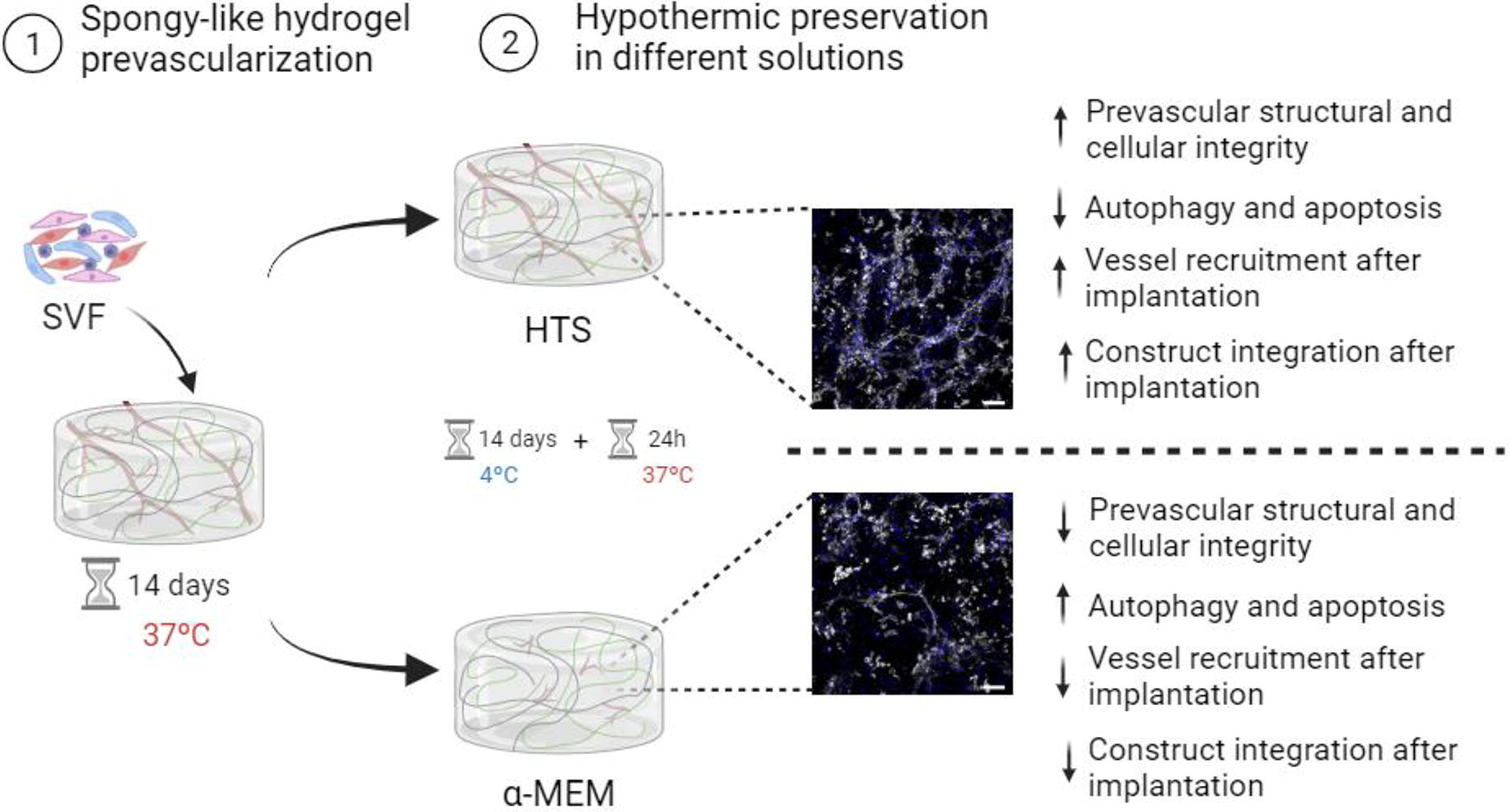
A229 - Ex Vivo Perfusion Of The Mouse Uterus
Y. Ping, J. Castillo Prado, I. Rogers
University of Toronto, Toronto, ON, Canada
*Purpose/Objectives: The biggest barrier to studying key early developmental events is the inaccessible nature of mammalian embryos. Existing imaging methods provide only snapshots and require uterus removal, which can potentially impede growth. Culturing of embryos ex-utero has proven to be difficult and exclude important fetal-placenta-uterus interactions. To overcome these limitations, this project aims to develop an ex vivo organ perfusion (EVOP) system to culture and maintain the mouse uterus for an extended period of time. To our knowledge, there is no established ex vivo perfusion methodology for the mouse uterus. This system would provide easier access to the embryos for imaging and real-time longitudinal data collection while maintaining a physiologic environment for growth. This system also offers the potential for studying blastoid implantations, model uterine diseases and provide a platform to study reperfusion injuries for uterus transplantation.
*Methodology: For this, the uterus is isolated, the vasculature is cannulated with a catheter and connected to a bioreactor previously developed by the Rogers Lab to culture mouse kidney and pancreas. We tested different cannulation points and for optimal perfusion. Hormones, various culture medium, and nutrient supplements are being tested to determine the ideal culture medium for mouse uterus maintenance and growth. We assess the performance of the ex vivo cultured uterus by analyzing metabolic activity through clinical chemistry of the culture media and histological staining. Hematoxylin and eosin (H&E), Terminal deoxynucleotidyl transferase (TdT) dUTP Nick-End Labeling (TUNEL) assay, and immunohistochemistry (IHC) staining with Cleaved-Caspase 3 are used to evaluate cell death, determine organ viability, and assess culture media compatibility.
*Results: Results show that using the infrarenal aorta as a cannulation point leads to successful perfusion of the uterine horn and ovaries, and cells are viable and the organ is metabolic active after a 24-hour culture period.
*Conclusion/Significance: The uterus EVOP culture will enable observation of development in real-time and help us understand fundamental questions of developmental biology. This system could also be utilized to investigate various reproductive diseases such as preterm labor, infertility, and uterine cancer, as well as reperfusion injuries in transplantation. The system is a valuable tool for preclinical studies in uterus transplantation, offering a cost-effective alternative to larger animal models currently used like sheep. The uterus EVOP system is a promising tool for accelerating advancements in our understanding of both developmental biology and reproductive health.
A231 - Innovative Normothermic Ex-vivo Perfusion System: An Affordable System For Pig Organs Without External Oxygen Tanks And Red Blood Cells
C. Lui1,2, J. Castillo-Prado1,2, S. Ganesh3, F. Calderon-Novoa3, T.-P. Chu3, M. Kawamura3, M. Selzner3,1, I. Rogers1,2
1
University of Toronto, Toronto, ON, Canada,
2
Lunenfeld Tanenbaum Research Institute, Toronto, ON, Canada,
3
University Health Network, Toronto, ON, Canada
*Purpose/Objectives: In our laboratory, the primary objective is to engineer a transplantable human organ utilizing a patient's cells and decellularized pig organs as the scaffold. A crucial component of this endeavour is normothermic ex-vivo perfusion (NEVP) organ culture, which is essential for the growth and maintenance of the new organ. In the landscape of ex-vivo (NEVP) machines for pig organs, the predominant reliance on external oxygen tanks and red blood cells for oxygenation, coupled with the high cost of both the machinery and single-use accessories, poses a formidable challenge to the widespread adoption of ex-vivo (NEVP) studies. In response, we are establishing an affordable ex-vivo (NEVP) perfusion system for pig organs using a native pig kidney as a model, building upon an existing mouse bioreactor. To enhance accessibility and cost-effectiveness, the system incorporates reusable accessories and eliminates the dependence on external oxygen sources and red blood cells.
*Methodology: Redesign a pig NEVP system to enhance its usability and stability, medium flow, media supplementation, oxygenation, and contamination prevention for pig kidneys.
*Results: The bioreactor was redesigned to optimize medium flow, incorporating flow sensors to determine the maximum and consistent flow rate to the kidney. To counter vasoconstriction, a vasodilator was introduced as a supplement. Identification and resolution of a potential contamination source within the system, specifically related to surrounding tissue from the kidney, mitigated contamination risks. A pressure sensor was incorporated for the establishment of a physiologically similar flow rate. Oxygen sensors were utilized to monitor oxygen consumption during ex-vivo organ perfusion. Experiments were conducted to assess the impact of oxygenator silicone tubing length and arrangement on oxygenation. The effectiveness of integrating oxygen carriers, such as perfluorocarbon (PFC), is currently under investigation. Additionally, hepes was introduced for media supplementation to stabilize pH. Our system includes albumin and creatinine for kidney health analysis, particularly for assessing filtration function. Subsequent phases of the project will focus on determining the optimal osmolarity of the medium, replacing creatinine with inulin, and incorporating kidneys from live pig donors.
*Conclusion/Significance: Notably, successful maintenance of a native pig kidney using the ex-vivo perfusion system was achieved for 24 hours.
A232
A235
A241 - Tetraterix, A Thermoresponsive Hydrogel Platform Technology For Tissue Regeneration And Delivery Of Bioactive Compounds
F. Oveissi1,2, S. Maleknia1, J. Almazi3, D. Calder4, Y. Wang5, J. Maitz5, D. Traini3,6, P. Young3,7, H. Xin Ong3,6, F. Dehghani2, A. Fathi1,2
1
Tetratherix Pty Ltd, Sydney, Australia,
2
School of Chemical and Biomolecular Engineering, The University of Sydney, Sydney, Australia,
3
Respiratory Technology Group, Woolcock Institute of Medical Research, Sydney, Australia,
4
Faculty of Health and Medical Sciences, School of Biomedical Sciences,University of Western Australia, Perth, Australia,
5
Burns and Reconstructive Surgery Research Group, ANZAC Research Institute, Concord, Australia,
6
Macquarie Medical School, Faculty of Medicine, Health and Human Sciences, Macquarie University, Sydney, Australia,
7
Department of Marketing, Macquarie Business School, Macquarie University, Sydney, Australia
*Purpose/Objectives: Despite the advances in hydrogel-based technologies in tissue engineering and regenerative medicines, only a limited number of those technologies have been translated to the market and clinical settings. This presentation will discuss the critical pillars for a successful ‘translatable’ biomaterial and highlight the significance of ‘platform technology’. Most importantly, the recent pre-clinical and clinical results of TetratherixTM platform technology for (i) engineering soft and hard tissues and (ii) delivery of bioactive compounds (growth factors, mRNA, polypeptides, and proteins) will be presented.
*Methodology: The capabilities of TetratherixTM hydrogel for engineering soft and hard tissues were assessed through a series of small and large animal models, e.g., mice subcutaneous, sheep osteotomy, ex-vivo bovine cadaveric subchondral defect model, and rats and porcine skin grafting. Also, histopathology, acute toxicity and weight of various organs were studied to investigate the toxicity and inflammatory response of the hydrogel. Subsequently, in a pilot clinical trial (n=10), the safety and regenerative potential of the technology was examined.The capabilities of TetratherixTM hydrogel to encapsulate and effectively deliver multiple therapeutics were evaluated. In a wide range of preclinical in vitro and in vivo animal studies, the technology was used for sustained release of a range of compounds, including ciprofloxacin HCl, tumour necrosis factor-alpha (TNF-α), transforming growth factor beta 1 (TGF-β1), and recombinant human bone morphogenetic protein 2 (BMP2).
*Results: The product elicited a negligible local inflammatory response and was fully resorbed into non-toxic components with minimal impact on internal animal organs in vivo. Pre-clinical animal studies confirmed that the engineered hydrogel supports the regeneration of both soft and hard tissues by providing a temporary matrix to support host cell ingrowth and neovascularization. In a pilot clinical trial, the engineered hydrogel was successfully administered to a socket site post tooth extraction and formed an adhesive hydrogel that stabilized blood clots and supported soft and hard tissue regeneration.Within the application of drug delivery, the TetratherixTM hydrogel system did not induce cytotoxic or inflammatory effects in vitro. Loading of a model antibiotic drug and multiple cytokines of varying molecular weights and surface charges did not impede the flowability and thermo-responsive characteristics of the hydrogel system. In vitro and in vivo studies confirmed that the hydrogel system and its gelation process were benign and did not detrimentally impact the biological activity of the loaded compounds. Further, the vehicle system provided a relatively high encapsulation efficiency (e.g., ∼99% for TGF-β1 and BMP2), mitigated the burst release, thus provided a sustained release profile for a wide range of therapeutics.
*Conclusion/Significance: Our findings collectively show that TetratherixTM hydrogel possesses all critical characteristics of a platform technology for engineering both hard and soft tissues. Overall, the results demonstrated the potential of TetratherixTM hydrogel as a universal carrier for sustained delivery of therapeutics.
A242 - Optimization Of A Way Of Enhancing Elastin Like Recombinamers Production With CRISPR-mediated Transcriptional Activation Technology
V. Chaskovska, P. Rodríguez-Alonso, D. Venegas-Bustos, M. Alonso-Rodrigo, J. C. Rodríguez-Cabello
University of Valladolid, Valladolid, Spain
*Purpose/Objectives: Literature has described that CRISPR-mediated transcriptional activation technology (CRISPRa) can boost gene expression in bacterial cells. For that, three plasmids are typically introduced into the bacterial cell: one carrying the gene of interest, another encoding the catalytically inactive Cas9 protein (dCas9) fused to the transcription activator, and a third plasmid bearing the guide RNA (gRNA), which is designed to be complementary to a specific site on the gene of interest-carrying plasmid to locate transcription activator close to promoter of target gene. This work aimed to improve elastin-like recombinamers (ELRs) production by utilizing CRISPRa. ELRs are known for their unique thermoresponsive properties and biocompatibility. They hold potential in biomedical applications ranging from drug delivery to tissue engineering. However, the industrial scale is hampered by low yield and inefficiencies in traditional expression systems.
*Methodology: To be able to use CRISPRa for our aim, we should have modified the abovementioned technology. As a reporter, a gene encoding ELR was used, as a transcriptional activator - T4 phage anti-sigma factor called AsiA, as gRNA - RNA, specific to the plasmid, encoding the ELR gene. We were able to co-transform these three plasmids into E. coli ( BLR) and get efficient transformation, but no difference in elastin-like recombinamers expression was observed compared to the established method without CRISPR-mediated transcriptional activation technology. Therefore, we changed the promoter on a plasmid with a gene encoding ELR from T7 to J23110 and the strain of bacteria -from BLR (E. coli) to MG1655 (E. coli). The difference in ELR production was evaluated compared to the control, and the gene expression was quantified by quantitative PCR (qPCR).
*Results: The outcomes of our approach showed the potential to use CRISPRa to produce ELRs. No changes in the integrity of polymers were detected compared to ELRs produced using an established method. Nevertheless, further system modification should be done to improve the yield of ELRs.
*Conclusion/Significance: The successful harnessing of CRISPRa for ELR production represents a significant leap in synthetic biology and biomaterial science, offering prospects for academic research and industrial biotechnology.
A243
A259 - Bioadhesives For Cartilage Repair And Regeneration
S. Paul1, K. Schrobback1, P. Tran1, C. Meinert2, J. Davern1, A. Weekes1, U. Nedunchezhiyan1, T. Klein1
1
Queensland University of Technology, Kelvin Grove, Australia,
2
Gelomics, Kelvin Grove, Australia
*Purpose/Objectives: Cartilage is frequently damaged and has poor capacity for repair. Development of easy to use and robust treatments for damaged cartilage could reduce the burden of both acute injuries and osteoarthritis. Hydrogels are promising materials for repair and regeneration of damaged cartilage, with potential for injectability and similarities to the cartilage extracellular matrix. However, hydrogels still have shortcomings including poor mechanical properties and suboptimal integration with surrounding cartilage tissue. To help overcome these limitations, this work aims to develop hydrogels that are injectable, cytocompatible, mechanically robust, and highly adhesive to cartilage.
*Methodology: Gelatin methacryloyl (GelMA) and glycol chitosan (GC) blends were generated and photo-crosslinked with visible light and photoinitiators (LAP and Ru/SPS) to form hydrogels. Bovine chondrocytes were mixed into GelMA-GC hydrogel solutions, which were injected and crosslinked within a cartilage ring and cultured in static and dynamic mechanical loading conditions. Hydrogel mechanical properties were evaluated using compression, indentation and adhesion tests, and durability was evaluated in an ex vivo bovine knee joint model.
*Results: Ru/SPS-crosslinked hydrogels exhibited higher compressive and tensile moduli, and most prominently higher adhesive strength with cartilage, than LAP-crosslinked hydrogels, and remained intact through joint manipulation ex vivo. Chondrocytes remained viable in the hydrogel system and showed tissue regeneration potential in long-term static and dynamic culture, with adhesive strength (push-out tests) increasing from 38 kPa to 52-60 kPa over four weeks of culture.
*Conclusion/Significance: GelMA-GC hydrogels demonstrated superior adhesive strength compared to existing bioadhesives and were compatible with chondrocytes in culture. These results provide further motivation for future research on these bioadhesive hydrogels for cartilage repair and regeneration.
A260 - Quaternized Chitosan/Graphene Oxide/Collagen Scaffolds Loaded With Huangbai Liniment With Efficient Hemostatic And Antibacterial Properties And Anti-inflammatory Properties For Infected Wound Healing
K. Hu
Wuhan Third Hospital, Wuhan, China
*Purpose/Objectives: Infectious wound has caused substantial economic losses and considerable threats to global health. The traditional treatment for bacterial infections lay on antibiotics. However, the overuse of antibiotics has led to multidrug-resistant (MDR) bacteria, developing alternative therapeutics to eliminate bacterial infections without inducing MDR has become an emergent goal. Huangbai Liniment (Compound Phellodendron Liquid, CPL) consists of forsythia, honeysuckle, and dandelion. It has the functions of clearing away heat and detoxification and reduces swelling and aging. It is used for diseases such as wound infection, wound healing, dermatitis and eczema, bedsores, ulcerative colitis, and diabetic ulcers. Therefore we develop a sponge scaffold loaded with CPL , which may have good mechanical and antibacterial properties and anti-inflammatory properties that can accelerate the repair of infected wounds.
*Methodology: We used a one-step mixture of collagen, quaternary chitosan, graphene oxide solution and huangbai liniment to prepare a QCS-GO-Col sponge porous scaffold loaded with CPL by cross-linking and lyophilization. Systemic in vitro and in vivo experiments were conducted to evaluate the physiochemical, antibacterial, anti-inflammatory and hemostatic properties and to determine the effect of the scaffold on cell viability, cell proliferation, and promote angiogenesis and its ability to repair the infected wounds.
*Results: We first prepared a QCS-GO-Col scaffold loaded with CPL with a porous and interconnected structure under the observation of scannng electron microscope. The FT-IR, XRD, and Raman spectrum experiments suggested the successful cross-linking and preparation of the QCS-GO-Col scaffold loaded with CPL. The fabricated scaffold loaded with CPL showed good mechanical, antibacterial (Staphylococcus and E. coli), and hemostatic properties in vitro. Furthermore, no significant biotoxicity was observed when cultured with fibroblasts. Finally, the designed QCS-GO-Col scaffold loaded with CPL was demonstrated to have superior antibacterial efficacy in vivo and accelerate the repair of infected wounds. Histological staining suggested that the QCS-GO-Col scaffold loaded with CPL promoted wound repair by inhibiting inflammation and promoting angiogenesis.
*Conclusion/Significance: In this study, we successfully prepared a QCS-GO-Col scaffold loaded with CPL, which has good mechanical, antibacterial, and hemostatic properties, and accelerated the repair of infection wounds by destroying bacterial structure, inhibiting inflammation, and promoting angiogenesis.
A261 - Extracellular Matrix Guided Differentiation Of Human Stem Cells In The Decellularized Mouse Kidney Vasculature
A. Bhadwal1,2, J. Castillo-Prado1,2, Y. Niu1,2, I. Rogers1,2,3
1
University of Toronto, Toronto, ON, Canada,
2
Lunenfeld-Tanenbaum Research Institute, Toronto, ON, Canada,
3
University Health Network, Toronto, ON, Canada
*Purpose/Objectives: Chronic kidney disease affects over 10% of the global population, leading to a high demand for bioartificial kidneys that can be faithful research models. It has been demonstrated that the biological pathways directing differentiation during organogenesis are maintained in the adult ECM and can direct differentiation during organ regeneration. This project focuses on the recellularization of a decellularized mouse kidney’s vasculature with human progenitor cells to study extracellular matrix (ECM) driven cell differentiation. This project can provide insight on renal vascular development as well as provide a model for studying specialized interactions in isolation such as that between endothelial cells, podocytes, and the ECM.
*Methodology: For decellularization mouse kidneys are perfused with a low concentration of sodium dodecyl sulphate (SDS)—a detergent, previously demonstrated to effectively clear native cells while preserving the ECM’s composition as well as microarchitecture. SDS solubilizes cell membranes leaving behind an intact ECM scaffold of the whole organ. The decellularized kidneys are treated with a kidney protein solution which has been demonstrated to allow for the rebinding of growth factors and proteins on the ECM that may be lost during decellularization. Recellularization is achieved through the perfusion of human embryonic stem cell derived mesoderm cells via the renal artery. The recellularized scaffolds are cultured in a bioreactor previously developed in the Rogers lab designed specifically for mouse kidneys. The tissue is analyzed using hematoxylin and eosin staining to gauge cell coverage and health while immunohistochemistry and qPCR are used to detect for endothelial markers. Single cell RNA sequencing is used to identify the variety of endothelial cell populations generated by the ECM.
*Results: SDS perfusion for 24hrs at 0.12mL/min resulted in the best balance of glycosaminoglycan (GAG) retention and cellular clearance out the of the various timepoints (8, 24, 48, 72hr) and flow rates (0.12, 0.24, 0.45mL/min) tested. GAGs are structures on the ECM which sequester organ and site-specific growth factors guiding cellular differentiation, thus their retention is vital. Preliminary recellularization data indicates cellular adhesion primarily within the cortical microvasculature. Analysis on the stages of cellular differentiation is ongoing.
*Conclusion/Significance: Whole mouse kidney decellularization has been optimized to maximize GAG retention and cellular clearance. Recellularization experiments demonstrate cellular adhesion within the cortical microvasculature. Future directions include further analysis into the status of cellular differentiation, as well as introducing podocytes during recellularization to study the complex endothelial, ECM, and podocyte crosstalk in the kidney. Additionally, further work into reepithelialization along with revascularisation holds promise for generating mini humanized kidneys which can be used as a platform for disease modelling and drug testing.
A262 - Development Of Small Intestine Graft With Mesentery By Decellularisation, As A Potential Treatment For The Intestinal Failure.
M. P. Pérez González1, J. Hua1, F. Lali1, S. Gabe2, P. Sawadkar1
1
The Griffin Institute, London, United Kingdom,
2
St Mark’s Hospital, London, United Kingdom
*Purpose/Objectives: The small intestine plays a crucial role in the digestive system, aiding in the digestion and absorption of vital nutrients and water. When SI is unable to perform its function, it is considered an Intestinal Failure (IF). Diseases such as Short Bowel Syndrome (SBS), a pathology that causes a reduction of the SI length and chronic malabsorption, account for 64.3% of IF cases and have a prevalence of 3-4 per million. Additionally, inflammatory bowel disease and intestinal tumours can cause IF. Current IF treatments focus on improving nutrition through oral, enteral, or parental nutrition (PN), the preservation of SI to maximize its remaining functions, or its replacement through intestinal transplantation. Intestinal transplantation (IT) is considered when patients show no response to medical treatment, are on lifelong PN and experience complication, but IT relies on the availability of donors and conveys the risk of immune rejection. For this reason, it is important to generate alternative treatments through tissue engineering that can be tailored to each patient and that don’t depend on donors.
*Methodology: Specimens of porcine small intestine, each with an average mass of 2.2 grams and a length of 3 centimeters, inclusive of the mesenteric tissue, were harvested for decellularization studies. These specimens were subjected to a tri-modal decellularization regimen, encompassing physical (induced osmotic shock), chemical (application of detergents), and enzymatic (exposure to proteases and nucleases) processes, all under continuous agitation. The duration of exposure to these decellularization agents was standardized at four days. Subsequent to the treatment, a comprehensive evaluation of the decellularization protocol's efficacy was undertaken, utilizing a suite of analytical techniques such as histological examinations, immunofluorescence staining, DNA quantification, and analysis via agarose gel electrophoresis.
*Results: Histological assessment showed preservation of the tissue’s microarchitecture, with preservation of crypts and absence of nuclear staining. Immunofluorescent staining with DAPI showed absence of nuclei in both intestinal and mesentery samples. DNA quantification using Nanodrop resulted in a DNA concentration of 12.8 ng/mg in mesentery and 26.67 ng/mg in intestinal samples which is below the required DNA value to be considered decellularised (<50 ng of DNA per mg of dry tissue). Agarose gel electrophoresis confirmed the absence of genetic material.
*Conclusion/Significance: According to our results, decellularisation using osmotic shock coupled with detergents and followed by enzymes can successfully remove nuclear content with the preservation of the macro and microarchitecture of SI and mesentery. This protocol can potentially be used and tailored to suit bigger samples of small intestine while preserving the mesentery, and serve as a source for future tissue-engineered solution for small intestine transplantation.
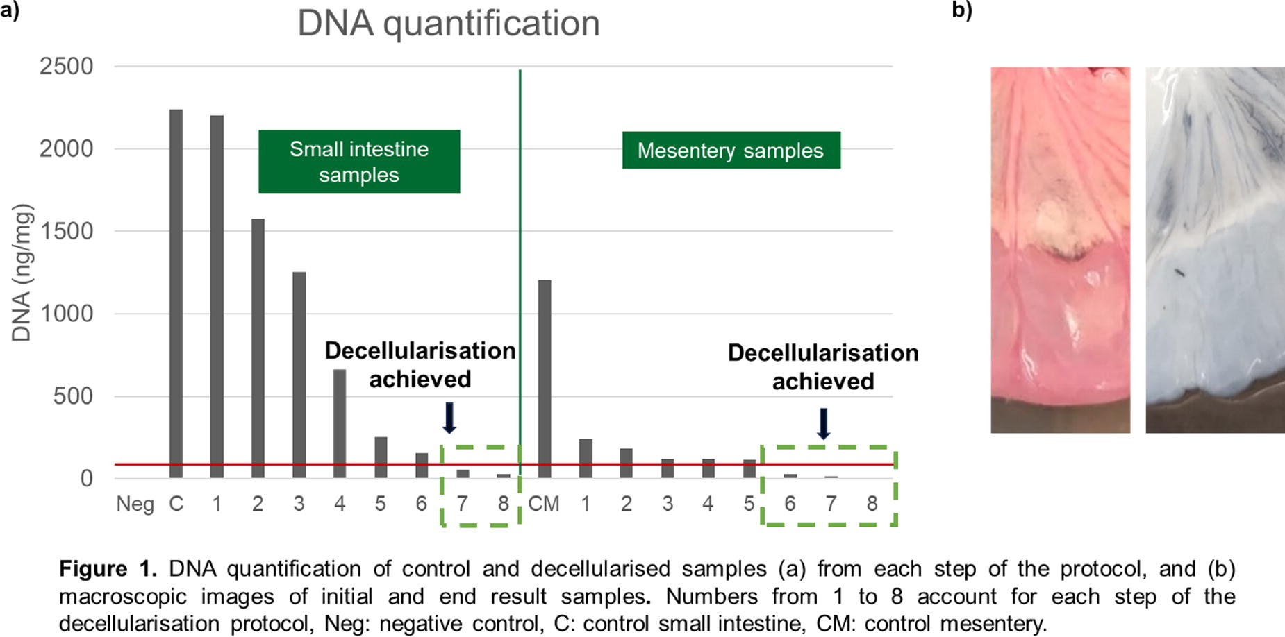
A263 - Development Of In Vitro Novel 3d Dynamic Intestinal Gastric System And Its Application In Evaluating H. Pylori Treatment
C.-Y. Kao, Y.-C. Huang
National Taiwan University of Science and Technology, Taipei, Taiwan
*Purpose/Objectives: Colorectal cancer is the second most common cause of cancer death in the United States. There is a great need to study the mechanism for colorectal cancer metastasis and develop novel strategies to treat colorectal cancer patients. Studies have shown that bacterial pathogens, diseases, and improper diet may cause an imbalance of intestinal microbiota, which will lead to an increased risk of colorectal cancer. On the contrary, a healthy diet with some probiotics can improve the intestinal microbiota and reduce the risk of colorectal cancer. Research and products of probiotics have increased dramatically in these few years. The use of probiotics was evaluated in the recent guidelines for the management of colorectal cancer metastasis; however, several questions remain, including the effectiveness of specific probiotic strains, dosages, and duration of adjuvant probiotic therapy, and more data are needed. Although the effectiveness of probiotics can be tested through animal studies, there are still species differences between animal and human microbiota. Animal ethics issues also need to be considered. The use of in vitro dynamic intestinal models to investigate the survival of probiotic strains in the gastrointestinal system has been reported. These in vitro dynamic gastric-intestinal systems mimic the human bulk physiological conditions such as pH, intake, and emptying in the intestine. However, the microphysiological condition at the cell level is omitted in these in vitro dynamic intestinal systems. This research aims to develop a novel in vitro dynamic gastric-intestinal system that mimics both bulk and microenvironment in the stomach and intestine.
*Methodology: An in vitro dynamic gastric simulation system equipped with an H. pylori 3D printed carrier system was used to evaluate the inhibitory effect of various probiotics on H. pylori. A novel in vitro dynamic intestinal epithelial cell culture system consisting of a dynamic Transwell cell culture system and a glass bioreactor. Caco-2 cells have been successfully cultured into a differentiated monolayer with specific values for transepithelial electrical resistance (TEER), phenol red permeability (Papp), and differentiation characteristics enzyme activity (ALP activity, SUC activity).
*Results: The results indicate that in the dynamic stomach simulation system, adding 1000 mg/L of amoxicillin sodium achieves the best inhibition of H. pylori, with an inhibition rate of 93.16%. Further experiments combining probiotic Lactobacillus reuteri with amoxicillin sodium showed the best inhibitory effect at 1000 mg/L amoxicillin sodium + 7ml OD1.0 L. reuteri, reaching an inhibition rate of 99.85%. In the static intestinal simulation system, Amoxicillin sodium and H. pylori decrease cell viability by 5.2% and 7.7%, respectively, indicating potential damage to cells upon entering the intestinal environment. Additionally, L. reuteri does not cause harm to cells in the simulation system.
*Conclusion/Significance: We have successfully built a novel in vitro dynamic gastric-intestinal system that mimics both bulk and microenvironment in the stomach and intestine. In the future, it is also possible to prepare organoids from the patient's biopsy and test them in these two dynamic intestinal epithelial cell culture systems to develop personalized medicines for colorectal cancer patients and achieve the goal of precision medicine.
A264 - Human Placenta Derived Bioinks For Enhanced Extracellular Matrix Mimicry
A. Arslan1, J. Hackethal2, A. Dobos1, J. Van Hoorick1, A. Chmielewska3, S. Wolbank3, M. Lunzer4, M. Narzt5, A. Teuschl5, H. Redl3
1
BIO INX, Ghent, Belgium,
2
THT Biomaterials, Wien, Austria,
3
LBI Trauma, Wien, Austria,
4
Upnano GmbH, Wien, Austria,
5
Morphomed, Wien, Austria
*Purpose/Objectives: Biofabrication holds great promise in various applications including tissue engineering and personalized medicine. However, a critical bottleneck lies in the limitation of applied bioinks. Currently, most bioinks rely on hydrogels, often incorporating only one or two (bio)polymers, thus oversimplifying the composition of the natural extracellular matrix (ECM). To address this issue, researchers are exploring more sophisticated matrices to better replicate the complexity of the ECM. For instance, Matrigel, a basement-membrane matrix derived from mouse sarcoma, has been used in cell culture applications for more than four decades due to its close resemblance to natural ECM. However, it is associated with several drawbacks such as poor processability via 3D printing, non-human derivation, overabundance of growth factors and poor mechanical integrity. In this research, we present an alternative approach by developing bioinks derived from human placenta and formulating them for extrusion-based bioprinting and multiphoton lithography.
*Methodology: Human placenta substrate (HPS, THT Biomaterials) was chemically and/or physically modified to obtain formulations printable via extrusion-based and multiphoton lithography-based printers. The flow properties and curing kinetics of the bioinks were evaluated via rheology whereas their biological properties were evaluated via cell encapsulation assays using human foreskin fibroblasts (HFFs).
*Results: Bioinks developed for extrusion-based bioprinters demonstrated excellent printability, facilitated by a straightforward physical crosslinking step without the requirement for light or crosslinking agents. Proteins in HPS were successfully modified with photo-crosslinkable groups with the aim to enable processing through multiphoton lithography. HFFs encapsulated within the bioinks exhibited high cell viability throughout a 28-day culture period.
*Conclusion/Significance: This innovative strategy not only eliminates the use of animal-derived products but also ensures a more accurate representation of the diverse composition of ECM. The resulting bioinks demonstrate significant potential for biofabrication applications, providing a more robust and clinically translatable platform for tissue engineering and organ modeling.
A265 - ANIMAL FREE RECOMBINANT HUMAN SERUM ALBUMIN FOR OPTIMIZING IMMUNE AND PLURIPOTENT STEM CELL MEDIUM
B. Perrault, X. Lu, Y. Wang, M. Andersen, J. Guinn, N. Larson, G. Herr
Bio-Techne, Minneapolis, MN
*Purpose/Objectives: Serum or serum derived proteins such as albumin have long been a critical cell media component for achieving high levels of cell expansion and function. While multiple media claim to be animal free, particularly for immune cells, they still require serum supplementation to achieve clinically relevant levels of cell growth. Animal derived serum products pose significant safety concerns including bacterial and viral contamination, immunogenicity, and lot-to-lot variability. To overcome these obstacles, we evaluated an animal-free recombinant human serum albumin with defined fatty acid profiles with commercially available and in-house animal-free medium for T-cell, NK-cells, and induced pluripotent stem cells (iPSC).
*Methodology: For each cell type, we assessed key metrics including expansion, phenotype, and major functions after culturing in media with or without albumin supplementation. Peripheral blood mononuclear cells (PBMC) derived NK and T-cells were expanded for two weeks in flasks and G-Rex culture vessels. NK cells were activated using feeder cells or NKp46 antibody and cultured with IL-2, IL-18, IL-21, and IL-15. Albumin increased CD56+/CD3- NK cell growth by over 300-fold without compromising their ability to recognize and kill K562 cells as assessed via Incucyte. T-cells activated using CD3/CD28 antibodies and cultured with IL-7 and IL-15. Albumin supplementation increased CD4+ and CD8+ T-cells expansion by more than 30 folds compared to without.
*Results: Interestingly, we observed a significantly high percentage of T stem memory cells using the fully defined animal free formulation with albumin. Human iPSCs were cultured for at least 5 passages in medium with or without albumin. iPSC with albumin showed over 120% increased expansion with high stemness marker expression including Oct3 and Nanog and typical iPSC colony morphology. Additionally, these iPSC retained pluripotency as they were able to differentiate into all three primary germ layers.
*Conclusion/Significance: These results affirm the effectiveness of recombinant animal free albumin to increase expansion of multiple key therapeutic cell types without compromising function, improve reproducibility, enhance safety, and potentially streamline regulatory approval for regenerative medicine development.
A266 - Characterization Of The Molecular Composition And In Vitro Regenerative Capacity Of Platelet-based Bioproducts
A. Acebes-Huerta1, P. Martínez-Botía2, G. Carbajo-Argüelles3, J. Fernández-Fuertes4, M. Muñoz-Turrillas5, A. Ojea-Pérez6, A. López-Vázquez4, J. A. Eble7, L. Gutiérrez8
1
Platelet Biotechnologies S.L (PlaBiTe), Oviedo, Spain,
2
Instituto de Investigación Sanitaria del Principado de Asturias (ISPA), Oviedo, Spain,
3
Platelet Biotechnologies S.L (PlaBiTe) and University of Oviedo, Oviedo, Spain,
4
Hospital Universitario Central de Asturias, Oviedo, Spain,
5
Centro Regional de Transfusión de Toledo-Guadalajara, Toledo, Spain,
6
Centro Comunitario de Sangre y Tejidos de Asturias, Oviedo, Spain,
7
Institute of Physiological Chemistry and Pathobiochemistry (University of Münster), Münster, Germany,
8
University of Oviedo and Instituto de Investigación Sanitaria del Principado de Asturias (ISPA), Oviedo, Spain
*Purpose/Objectives: The use and demand of platelet-based bioproducts in regenerative medicine is steadily increasing. Platelet-rich plasma (PRP) is the most popular platelet-based bioproduct and its therapeutic application constitutes a relatively new approach in several clinical fields. However, the therapeutic outcome of PRP remains highly controversial, as most studies fail to characterize the product properly, which is also not helped by the fact that there is no consensus on PRP preparation, post-production processing and application methods. Adding to this, in most territories, PRP use is only allowed in an autologous manner.Our objective was to characterize PRP and related subfractions at the molecular and functional level, to be able to develop novel platelet-derived bioproducts that could overcome the current limitations of PRP-based therapies.
*Methodology: Sample preparation. We used PRP produced in a closed system using the equipment of a certified blood bank (Centro Comunitario de Sangre y Tejidos de Asturias), from independent healthy donors (interrupted blood donations, i.e. discard material). PRP related subfractions were obtained by differential centrifugation: Plasma-free Platelet Lysate (PFP-L) and Platelet Poor Plasma (PPP). The method to obtain Platelet Secretomes (PLT-S) is protected by international patent application (PCT/IB2022/057936). The agonists used to induce platelet cargo release were: TRAP6, convulxin, aggretin A, phorbol 12-myristate 13-acetate or collagen. Molecular and functional characterization. 37 molecules were measured in bioproducts and related subfractions by using multiplex technology (Human Procartaplex; Invitrogen). The regenerative potential of bioproducts was evaluated in vitro by scratch wound closure assays using fibroblast cell cultures. Cell-free gaps were monitored for 48 hours using a time-lapse microscopy Zeiss AxioObserver Z1 microscope (Carl Zeiss, Germany) and the photographs were analyzed with ImageJ.
*Results: The molecular and functional capacity of PRP and related subfractions revealed that the bioproducts that exert the most potent regenerative effect are those containing platelet-derived cargo. Therefore, we developed a method to obtain Platelet Secretomes (PLT-S), whose composition consists exclusively in the platelet granule cargo, highly enriched in growth factors and bioactive molecules, and subsequently free of plasma and unwanted cellular/platelet components. The comparison of the molecular profile between bioproducts containing platelet-derived cargo (PRP, PFP-L and PLT-S) revealed, as anticipated, that the PLT-S was the most enriched in growth factors and bioactive molecules. Interestingly, the potential to induce cell proliferation and/or migration shown by PLT-S was superior to that exerted by the rest of PRP-based bioproducts, which correlates with the enrichment of the bioproduct in platelet-derived cargo.
*Conclusion/Significance: A better understanding of the nature and the active components of platelet-based bioproducts is crucial to optimize and standardize production methods as to improve the clinical benefit of the therapy. Given its characteristics, PLT-S represents a promising option to explore novel applications in regenerative medicine and other clinical fields: it would allow allogeneic use and the production method could be optimized to minimize batch-to-batch variation, or to incorporate post-processing steps to allow nanoencapsulation (targeted delivery) or storage with off-the-shelf purposes.
A267
A268 - Effects Of The Soluble Fraction Of Extracellular Matrix On The Esophageal Cancer Cell Phenotype
D. J. Romero1,2, S. O. El-Mossier1,2, E. N. Halton1, L. Zhang1,2, G. S. Hussey1,2, S. F. Badylak1,2
1
University of Pittsburgh, Pittsburgh, PA,
2
McGowan Institute for Regenerative Medicine, Pittsburgh, PA
*Purpose/Objectives: The incidence of Barret’s esophagus (BE) and esophageal adenocarcinoma (EAC) is increasing at an alarming rate, and esophageal cancer is one of the world’s leading causes of cancer death. The current standard of care for EAC is esophagectomy. This procedure is associated with high morbidity, post-surgical complications, and decreased quality of life. There is a clear and unmet need for novel and more effective therapeutic strategies to repair damaged or diseased esophageal tissue. Biologic scaffolds, composed of extracellular matrix (ECM), have been developed as surgical mesh materials, powders, and hydrogels, and are approved for a wide range of clinical applications, including esophageal reconstruction. However, the biological mechanisms by which functional tissue restoration is mediated remain poorly understood. Our group has demonstrated that ECM bioscaffolds, particularly the degradation of ECM bioscaffolds, can influence metaplastic and neoplastic tissue, leading to the remodeling of the mucosa from a diseased to a healthy state in rodent and canine models. We hypothesized that the mitigation of the esophageal neoplastic cell phenotype is mediated by signaling molecules within the ECM. Therefore, we evaluated the in vitro effects of a saline-soluble fraction of ECM on esophageal cancer cells.
*Methodology: The porcine urinary bladder was decellularized to obtain ECM, which was then lyophilized, powdered, tumbled in saline, centrifuged, filtered, and concentrated 200-fold by volume to produce the saline-soluble fraction of ECM (UBM-SF).
*Results: We found that UBM-SF decreases colony formation properties and enhances the mesenchymal-epithelial transition (MET) of esophageal cancer cells. The soft agar colony formation assay revealed a significant decrease in both the number and percentage area of colonies following treatment with UBM-SF. Interestingly, no associated changes were observed in cell proliferation; however, morphologic alterations were noted, indicating a decrease in cellular malignancy. Additionally, expression and localization of epithelial and mesenchymal markers showed primarily a progressive enrichment of E-cadherin at the plasma membrane, suggesting a partial recovery of the normal epithelial phenotype. In this context, treatment with UBM-SF also modified the dynamics of collective cell migration, as evidenced by a significant decrease in the percentage of gap closure over an 18-hour period through time-lapse imaging.
*Conclusion/Significance: These findings highlight the effect of the soluble components derived from ECM upon esophageal cancer cells, demonstrating their potential as a modulator of the MET-EMT to mitigate the neoplastic cell phenotype. Importantly, the acquisition of an epithelial state has been found to have a profound effect on the ability of cancer cells to form colonies. By directly inhibiting the EMT process, migration, and potentially metastasis, UBM-SF emerges as a promising therapeutic agent for preserving and restoring the esophageal epithelial phenotype.
A272 - Mending The Eardrum: Decellularized Cartilage For Tympanoplasty
N. Charoensombut
Otolaryngology, Children's Hospital of Philadelphia, Philadelphia, PA.
*Purpose/Objectives: Pediatric myringotomy, a procedure where drainage tubes are placed to equalize pressure and remove fluid from the middle ear, is the most common childhood surgery with over 1 million per year. In most cases, the tubes extrude, and tympanic membranes (TM) heal naturally; however, in approximately 100,000 children/year, the TM does not heal, resulting in persistent perforation. These children then require eardrum repair (tympanoplasty) to restore an intact TM membrane which is essential for optimal hearing, as well as to prevent further complications such as infection and drainage (otorrhea), enlargement of the perforation, and development of irreversible scarring (tympanosclerosis). During tympanoplasty, an autologous graft is used to repair the perforation; cartilage harvested from the ear is often used as a repair material with acceptable healing rate and good stiffness to prevent retraction of the repaired TM. However, harvesting grafts from the donor site increases surgical time and morbidity, and when revision procedures are required, there can be insufficient autologous graft material especially for smaller children. In fact, pediatric tympanoplasty still suffers a 20% failure rate due to insufficient graft stiffness, adherence, and integration. Therefore, there exists a major opportunity to develop an alternative grafting option for pediatric tympanoplasty, to circumvent the major challenges associated with autologous grafts.
*Methodology: To overcome these challenges, it would be ideal for the surgeons to have an off-the-shelf material which is more practical for eardrum surgery, adheres well to the perforated tympanic membrane, has good stiffness and mechanical stability to not be dislodged, and rapidly and effectively integrates with the surrounding tissue promoting the healing of the eardrum. We bioengineered MENiscus Decellularized (MEND) for mending the perforated eardrum. Our unique approach is the first of its kind and exploits the selective removal of elastin fibers and blood vessels exclusively present in the fibro-elastic cartilage of the allogeneic meniscus. These native structures are harnessed to form microchannels that promote effective recellularization after enzymatic decellularization. This approach circumvents the limitations of using decellularized articular cartilage, which does not possess elastic fibers that can be removed to create channels.
*Results: Notably, decellularized fibrocartilage matches the fibrous nature of the TM, promoting repair and provides mechanical strength similar to autologous cartilage currently used in the clinic. In our first study, we used a rat model of TM perforation comparing outcomes of repair with MEND, autologous ear cartilage, autologous ear fascia (another clinical option used by surgeons), and untreated perforation. The results showed that, MEND and cartilage both rapidly closed the perforation, but cartilage did not remodel into the tissue. MEND and fascia restored the native TM structure, but fascia showed poor mechanical properties and was easily dislodged, similar to what happens in clinic.
*Conclusion/Significance: Overall, MEND had the mechanics of cartilage and the integration capacity of fascia and was the fastest at closing the perforation, indicating its potential superiority to the standard of care.
A275
A276 - Chondrocyte-derived Decellularized Matrices Support The Expansion And Redifferentiation Of Superficial Zone Chondrocytes
T. J. Manzoni1, A. Ho1, A. Sheldrake2, T. J. Block3, J. Parreno1
1
University of Delaware, Newark, DE,
2
StemBioSys, San Antonio, TX,
3
GoldiloxBio, San Antonio, TX
*Purpose/Objectives: Articular cartilage serves as a crucial tissue for joint function, providing a lubricating surface for frictionless movement and load distribution. These properties are attained by zone-specific molecular expression in native articular cartilage. The superficial zone chondrocytes (SZC) produce the lubricating molecule, Proteoglycan-4, which is critical in providing a frictionless environment for the movement of one cartilage surface over another. Current methods of bioengineering cartilage may be limited in their ability to produce a robust superficial zone.
Articular cartilage has a limited ability to self-repair and when damaged degeneration is initiated, ultimately leading to Osteoarthritis. Bioengineered cartilage holds promise in repairing small cartilage defects, preventing osteoarthritis progression. Monolayer expanded (passaged) chondrocytes, harvested from full-thickness cartilage, are an FDA approved cell source for cartilage repair. However, our previous work has determined that cell expansion limits the number of SZC after passaging due to poor attachment and slow proliferation on polystyrene dishes. Therefore, it is necessary to optimize expansion methods to obtain an adequate number of SZC for bioengineering purposes. Furthermore, expansion results in cell dedifferentiation that includes reduced expression of cartilage matrix and Proteglycan-4. Previous studies showed that culturing full-thickness chondrocytes on a chondrocyte derived-decellularized extracellular matrix (CD-ECM: CELLvo ™ ChondroMatrix, StemBioSys, San Antonio, TX, USA) can improve proliferation and reduce dedifferentiation. Here, we hypothesize: CD-ECM will promote attachment and proliferation of SZC. Passaged SZC derived from CD-ECM culture will be less dedifferentiated and have a superior capacity to form cartilage tissues that express PRG4.
*Methodology: Isolated primary bovine SZC were seeded on either polystyrene or CD-ECM. Cell attachment, expansion rate, and gene expression during passaging was characterized. After two passages, passaged SZC derived from either polystyrene or CD-ECM were seeded in scaffold-free three-dimensional culture and maintained in redifferentiation media. Tissue thickness was measured, and immunohistochemistry was used to determine matrix composition of the formed tissues.
*Results: When seeded on CD-ECM, primary SZC had greater cell attachment (Figure 1A). As compared to polystyrene, SZC on CD-ECM culture flasks reached 70-90% confluency earlier from P0 to P1 (13 vs. 8 days) and from P1-P2 (7 days vs. 9 days). By passaged 2, mRNA levels for SRY-box transcription factor 9, Aggrecan, and Clusterin were elevated in cells passaged on CD-ECM compared to polystyrene. There were no differences in mRNA levels of collagen-II and proteoglycan-4 between polystyrene and CD-ECM at P2. However, culturing on CD-ECM repressed mRNA levels for dedifferentiation markers Collagen-1, Tenascin-C, Alpha-smooth muscle actin (Figure 1B). After redifferentiation, both formed tissues expressing Collagen-II, Aggrecan, and Proteoglycan-4 with limited expression of Collagen-I, tissues derived by cells passaged on CD-ECM were thicker.
*Conclusion/Significance: Overall, the use of a CD-ECM improves SZC expansion through increased primary cell attachment, decreased time to reach confluency, and the limiting of differentiation. In addition, SZC passaged on CD-ECM can be redifferentiated to form bioengineered tissue that express Proteoglycan-4. The use of CD-ECM for passaging SZC may help in achieving an adequate cell number of cells for bioengineering purposes.

A277 - DECELLULARIZED PORCINE UTERINE TUBE DERIVED EXTRACELLULAR MATRIX AS NOVEL A BIOMATERIAL FOR IN VITRO TUBAL MICROENVIRONMENT RECONSTRUCTION
G. D. Almeida1,2, R. S. da Silva1, M. S. Gibin3, V. d. Gonzaga3, H. dos Santos3, F. Sato3, M. Baesso3, L. Hernandes3, C. R. Long2, L. Relly2, A. O. Carreira1,4
1
Faculty of Veterinary Medicine and Animal Science, University of São Paulo, São Paulo, Brazil,
2
Texas A&M University, College Station, TX,
3
State University of Maringá, Maringá, Brazil,
4
Center of Human and Natural Sciences, Federal University of ABC, Santo André, Brazil
*Purpose/Objectives: Uterine tube microenvironment is able to modulate several pre-implantational cellular processes during embryonic development. Even with advances of in vitro reproductive technologies (IRT), implantational failures are still frequent and, most of them, due to unclear mechanisms. Evidences demonstrate that the uterine tube is responsible for several molecular stimulation that establish the early maternal-embryo communication, which affects the entire gestational process. One of the main challenges to study the pre-implantational period, mainly in humans, is the availability of samples, which impairs a deep understanding of molecular mechanisms involved in this period. In this context, the development of models that mimic tubal morphophysiology may help to elucidate molecular mechanisms related to gestational losses and to improve the performance of IRT technologies. The application of biological matrices to reconstruct artificial tissue microenvironments has demonstrated a great potential in bioengineering field, and, more recently, in the reproduction area. Therefore, this work aimed to develop and characterize biomaterials derived from porcine uterine tube segments to be used in the reconstruction of a biomimetic tubal microenvironment.
*Methodology: For that, porcine uterine tubes (n=10) were segmented in three portions (infundibulum, ampulla and isthmus) and decellularized using solutions of 0.1% SDS and 0.5% Triton X-100. The decellularization efficiency were evaluated using DAPI staining for nuclei detection and total genomic DNA quantification. Regarding the ECM components, a morphological characterization was performed based on histological, immunohistochemical (type I and III collagen, laminin, elastin and fibronectin) and ultrastructural analyses. The quantification was performed by ImageJ software. An ECM physic-chemical analysis was performed by FTIR and Raman spectroscopy also to evaluate the level of preservation after the decellularization. Then, an in vitro cytocompatibility assay was performed using endothelial progenitor cells derived from canine yolk sac (YS) and human epithelial cell line HEK293. Cell-biomaterial interaction was evaluated through scanning electronic microscopy (SEM) and cell viability by resazurin assay.
*Results: Regarding the results, DAPI staining revealed absence of nuclei in all three generated scaffolds and the DNA quantification showed a decrease of 97.6% in the infundibulum, 97.8% in the ampulla and 94.7% in the isthmus compared to the native tissue, which attested the decellularization efficiency. Morphoquantitative histological revealed that the general ECM components remained preserved after the decellularization process. The immunohistochemical data also attested that the main structural and glycoadhesive ECM proteins were preserved as well. In addition, SEM analysis demonstrated that the tissue 3D structure remained intact. Spectroscopic analyses showed no differences between native and decellularized samples in the main bands related to collagen, proteoglycans, glycosaminoglycans and elastin. The in vitro assays attested that the scaffolds were cytocompatible, once they allowed integration between the ECM and cells and high viability rates.
*Conclusion/Significance: In conclusion, these data demonstrate that the generated biomaterials derived from decellularized tubal regions are well structured, with the ECM composition preserved and cytocompatible. This infers that they can be used as suitable biomaterials for cell culture and work as a substrate for tubal microenvironment reconstruction, which may be applied in 3D in vitro models and in assisted reproduction biotechnologies.

A279 - Decellularized Liver Matrix For Tissue Engineering
E. v. Hengel, L. Reniers, J. Willemse, L. van der Laan, J. de Jonge, M. Verstegen
Erasmus MC, Rotterdam, Netherlands
*Purpose/Objectives: The Impact of Sterilization on Decellularized Livers: Assessing Microbiome, Matrix Proteins, and Biocompatibility E.V.A. van Hengel, L. Reniers, J. Willemse, L.J.W. van der Laan, J. de Jonge & M.M.A. Verstegen
Department of Surgery, Erasmus MC Transplant Institute, University Medical Center Rotterdam Liver tissue engineering holds great promise for addressing the growing demand for transplantable organs. Decellularized human liver matrices serve as potential scaffolds for tissue engineering, due to their unique architecture and extracellular matrix (ECM) proteins. However, the removal of microorganisms and potential pathogens to meet quality criteria for advancing these scaffolds toward clinical applications is of importance to ensure safety and compliance with medical device regulations. Sterilization procedures can, however, negatively impact the quality of the ECM. This study compares five techniques on their sterilization efficacy and their effects the extracellular matrix and biocompatibility.
*Methodology: Human livers (N=3), deemed unsuitable for transplantation, were decellularized by perfusion with 4% Triton-X-100 and 1% ammonia solution. The resulting ECM was sterilized using Gamma radiation (1000 Gy), UV radiation (254 nm), exposure to Supercritical Carbon Dioxide (sCCO2), Peracetic acid (0.5%), or Antibiotic-Antimycotic (10%) treatment. The microbial burden was determined using standardized microbiological methods. The effect on the extracellular matrix composition and biomechanical properties, including collagen content, fiber orientation, stiffness, and glycosaminoglycan content was assessed. Particularly, crosslinking or denaturation of proteins was studied using two-photon microscopy, Picrosirius Red (PSR), H&E, and Collagen IV staining. Furthermore, the biocompatibility of the sterilized matrices was assessed by analyzing cell viability after recellularization with intrahepatic cholangiocyte organoids (ICOs) (N=3) and HepG2 cells.
*Results: Exposure to Gamma radiation, sCCO2, and 10% Antibiotic-Antimycotic were effective in removing micro-organisms from decellularized liver scaffolds, while exposure to UV radiation and Peracetic acid were not found sufficient. Micrococcaceae, S. Maltophilia, Bacillus species, and fungi were detected in these scaffolds. Visualization of the overall structure of collagen fibers with two-photon microscopy and protein staining showed alterations in the protein matrix and modifications in fiber-to-fiber orientation of collagens after exposure to UV and Gamma radiation. Recellularization with ICOs and HepG2 cells resulted in viable and proliferating cells in all scaffolds, confirming that the sterilization methods did not affect the biocompatibility of the decellularized scaffolds.
*Conclusion/Significance: Exposure to UV radiation and Peracetic acid did not result in the complete removal of micro-organisms. Sterilization by Gamma radiation and UV radiation resulted in alterations in matrix protein composition and fiber orientation. The biocompatibility of the scaffolds was not altered after sterilization. Going forward, we suggest sCCO2 or Antibiotic-Antimycotic treatment as it limits damage to the matrix proteins. This study demonstrates the importance of sterilization of decellularized liver matrices for tissue engineering purposes and potential clinical applications.
A281 - DECELLULARIZATION OF BOVINE SPINAL CORD MENINGES VIA SUPERCRITICAL CO2 AND EVALUATING THE EXTRACELLULAR MATRIX PERFORMANCE FOR NEURAL TISSUE ENGINEERING APPLICATIONS
T. Kurt1, E. Ozudogru1, U. Cengiz1, B. Derkus2, Y. E. Arslan1
1
Canakkale Onsekiz Mart University, Canakkale, Turkey,
2
Ankara University, Ankara, Turkey
*Purpose/Objectives: One of the main goals of modern tissue engineering is the invention of novel decellularization methods that reduce the immunogenicity and risk of rejection of the obtained acellular tissue. Current decellularization methods generally use chemical detergents that can damage tissue structure and cannot be well purified from the tissue, thus causing toxicity. To upgrade decellularization results while reducing contamination and time costs, utilizing natural, environmentally friendly, and non-toxic supercritical CO2 (scCO2) has recently emerged as a promising alternative decellularization technique to harsh detergents. Herein, we offer a fast, effective, and detergent-free decellularization technique for bovine spinal cord meninges.
*Methodology: Bovine spinal cord meninges were decellularized by a combination of scCO2 (at three different pressure values) and enzyme (DNase/RNase) methods. Then, dsDNA content analysis, glycoseminoglycans (GAGs) assay, hydroxyproline (HYP) content, and hematoxylin and eosin (H&E) stain, which are traditional analyses for decellularization, were performed. Additionally, tissues were digested with pepsin enzyme and incubated at neutral pH and temperature to achieve 3D hydrogel. A compression test was conducted in a micromechanical testing device to ascertain the hydrogels' strength and elastic modulus. Finally, cellular adhesion, survival, proliferation, and morphology were confirmed with glioblastoma cells via XTT and Live/Dead assays.
*Results: The tissue was successfully decellularized, which was confirmed by the aforementioned studies. The scCO2 method reduced the decellularization period from 1 week to only 3 days. The DNA content analysis of decellularized tissues was found to be below 50ng/mg dry tissue in all three methods. Besides, there seemed to be no significant differences in GAGs and HYP contents between native and decellularized tissues. In the H&E studies, there seemed to be no nuclei in decellularized samples. This result was also compatible with dsDNA content analysis. The mechanical strength of the hydrogel obtained by decellularizing with detergent was significantly lower than that of the hydrogels decellularized with scCO2. Finally, XTT and Live/Dead assays showed that the cyto-viability of the glioblastoma cells on scCO2-decellularized matrices more than the control group.
*Conclusion/Significance: The results clearly demonstrate that the combination of scCO2/enzymatic methods is highly effective in decellularization of tissues. Furthermore, the findings suggest that these detergent-free techniques may replace the conventional detergent-based decellularization methods. We believe that the use of scCO2 in the decellularization of human and/or animal tissues is important, and the technique fits green and sustainable agreements. This work was supported by the Health Institutes of Türkiye (TUSEB) (Project ID: 2022-B-03-24533).
Keywords: Supercritical CO2, decellularization, spinal cord meninges, neural tissue engineering
A282 - 3D HYDROGELS FROM DETERGENT-FREE DECELLULARIZED SPINAL CORD MENINGES REINFORCED WITH HYDROPHILIC SILK FIBROIN FOR REGENERATIVE MEDICINE APPLICATIONS
T. Kurt, Y. E. Arslan
Canakkale Onsekiz Mart University, Canakkale, Turkey
*Purpose/Objectives: Decellularization involves removing cell components from the extracellular matrix (ECM) using different methods. The use of detergents is the most common of such techniques. The drawbacks of using detergents include a degraded matrix, prolonged processing, and unwanted residuals. Furthermore, removing cellular components during harsh decellularization often results in softer tissues with reduced mechanical stability compared to natural tissues. In the current study, we have developed a detergent-free sonication method for the decellularization of bovine spinal cord meninges. The ECM was reinforced with hydrophilic silk fibroin (hSF) fragments to increase the mechanical properties of 3D hybrid hydrogels.
*Methodology: Bovine spinal cord meninges (SCM) were subjected to a series of decellularization protocols, including 125 watts of ultrasonic power and nuclease treatment. Decellularized SCM (dSCM) were digested with an acidic pepsin solution to obtain a pre-gel. After decellularization, DNA content analysis was performed using Qubit™ 4 Fluorometer to determine dsDNA residues. The hydroxyproline (HYP) and glycosaminoglycans (GAGs) content in native SCM (nSCM) and dSCM were determined using spectrophotometric techniques. On the other hand, hSF from B. mori silk was degummed in boiling sodium carbonate solution and then dried. The dried silks were dissolved in CaCl2:H2O:EtOH. This aqueous solution was dialyzed and lyophilized. Lyophilized hSF was then added to the pre-gels of dECM at different ratios. Horseradish peroxidase and H2O2 were added to initiate the crosslinking and generate mechanically stable 3D hybrid hydrogels. A micromechanical testing device with a 10N load cell was used to evaluate the mechanical characteristics of these hybrid hydrogels, and their rheological characteristics were determined using a DHR-2 rheometer in the 0.1-100 Hz range. The angiogenic properties of 3D hybrid hydrogels were investigated using the In Ovo chorioallantoic membrane (CAM) analysis.
*Results: The DNA content analysis revealed that dsDNA in dSCM was <50 ng/mg dry weight. Furthermore, HYP and GAGs contents showed no significant difference between nSCM and dSCM, as determined by one-way ANOVA analysis. Then, mechanical test results indicated that the mechanical strength of the hydrogels with hSF increased compared to the dECM hydrogel alone. Based on the step-strain rheological characterization, it was observed that the hydrogels exhibited recovery following deformation. Finally, In Ovo CAM assay showed that there were more blood vessels at the time point of explantation compared to the initial time point.
*Conclusion/Significance: The results of related analysis showed that the proposed decellularization method was satisfactory compared to conventional ones because no damage seemed to the matrix structure while removing cellular components. The findings from micromechanical testing and rheology analysis demonstrate that incorporating SF into dECM enhances the mechanical robustness and flexibility of the resulting hydrogel. The In Ovo Cam analysis shows that the hybrid hydrogel produced is biocompatible and has an angiogenesis-inducing effect. In conclusion, we believe that the 3D hybrid hydrogel from dSCM/hSF has the potential to be used in regenerative medicine applications. The financial support of the Canakkale Onsekiz Mart University Scientific Research Projects Coordination Unit is acknowledged (Project ID: FYL-2023-4519).
Keywords: Sonication assistant-decellularization, spinal cord meninges, silk fibroin, enzymatic crosslinking.
A283 - Scalable And High-throughput In Vitro Vibratory Platform For Vocal Fold Tissue Engineering Applications
A. Biehl1, R. Colmon1, A. Timofeeva1, A. M. Gracioso Martins1, K. Peters1, G. Dion2, D. Freytes1
1
North Carolina State University, Raleigh, NC,
2
University of Cincinnati, Cincinnati, OH
*Purpose/Objectives: Pathologies leading to dysphonia, such as vocal fold (VF) cysts, paralysis, and scarring/fibrosis, are estimated to affect millions worldwide. The development, maintenance, and regeneration of VFs following injury are heavily influenced by phonation-induced mechanical stimulation. Thus, various in vitro systems, speakers, actuators, and airflow-based bioreactors have been developed to closely mimic the mechanical environment of the VFs. These bioreactors incorporate relevant biomaterials, biochemical cues, and cells to enable faster, more controlled, and affordable characterization of potential VF therapies. Even so, there needs to be standardized parameters for oscillatory regimes, mechanical forces, experimental units, and biomaterials used in the system. This study aimed to design, fabricate, and characterize a high-throughput, easy-to-construct, and affordable platform that simulates the VF microenvironment in vitro.
*Methodology: The platform described in this study comprises a commercially available bottomless 24-well plate fitted with a flexible membrane atop a custom-designed waveguide equipped with a set of piezoelectric speakers for micron-scale vibrations. Frequency and amplitude precision were evaluated using Laser Doppler Vibrometry (LDV) by independently measuring the displacement of the flexible membrane and the piezoelectric speaker during various oscillatory routines. Human Vocal Fibroblasts (HVOX) and Human bone marrow-derived mesenchymal stem cells (hMSCs) were cultured on top of the flexible membrane and vibrated for 1 and 2 hours each at 100% volume at 100Hz prior to pro-fibrotic and pro-inflammatory gene expression being analyzed via quantitative real-time polymerase chain reaction.
*Results: Our vibrational platform produced a maximum volumetric strain on the surface of the Tegaderm membrane of up to about 0.25%, according to our LDV measurements. Gene expression indicated significantly lower MMP1 expression in HVOX at high displacement ranges and in hMSCs at low displacement ranges. Lower ACTA2 expression was detected compared to the static control in HVOX.
*Conclusion/Significance: Our team has created a unique vibratory platform that can generate frequencies within the range of human phonation. What sets our platform apart is that the wells experience varying displacement magnitudes. This feature allows for greater repeatability of these displacements and could prove to be advantageous for future studies focused on understanding cellular responses to the phonatory regime in different anatomical positions of the vocal fold, such as medially or laterally, as well as specific cell types within the different layers of vocal tissue.
A285 - A Deep Learning Model To Predictsuitable Water Contents For Sustainability Decm-based Corneal Biofabrication
X. Wang, A. A. Awad, Z. M. Ali, M. G. Shibru, S. Luo, K. Liao, A. Al Shehhi, V. Chan, P. R. Corridon
Khalifa University, Abu Dhabi, United Arab Emirates
*Purpose/Objectives: The global scarcity of donor corneas cannot fulfill the rising need for transplantation; therefore, creating high-throughput alternatives is imperative. Decellularized corneas have been investigated as an option due to their structural and functional capacities. A key aspect of the decellularized extracellular matrix (dECM) is its mechanical performance, which relies on the retention of glycosaminoglycans post-decellularization, influencing the water content of the corneal analogs and, ultimately, their mechanical performance. In this project, we first created various corneal substitutes with five types of water content. We then conducted intraocular compression, tensile, and stress-relaxation studies to examine the impact of water content on mechanical performance. Finally, we applied a deep learning model to precisely predict the mechanical properties of scaffolds under any given water content as we aim to devise a large-scale ophthalmic tissue bioengineering platform.
*Methodology: Using a sustainable approach, we obtained roughly 500 ovine cornea samples from our main slaughterhouse partner. The samples were then sectioned to a consistent size. We created acellular scaffolds using various combinations of surfactants (1% and 4%) and decellularization periods (2 days and 4 days), i.e., 1%2days, 1%4days, 4%2days, and 4%4days, which would be compared with native corneas. All the samples were freeze-dried and divided into five subsequent conditions to vary water content (2%w/w, 38%w/w, 78%w/w, 150%w/w, and 200%w/w) and subjected to multiaxial intraocular compression, tensile, and stress-relaxation studies. Before mechanical testing, each sample was coated with a consistent layer of mineral oil to prevent evaporation during experimentation. A multilayer perceptron (MLP) algorithm, based on the collected data, was employed to establish the predictive model based on 78%w/w native physiological content.
*Results: The intraocular compression characteristics of scaffolds with a water content of 150%w/w and 200%w/w were similar to those of native corneas (78%w/w in sheep and humans). Within the acceptable displacement range (0-1.5mm), the maximal intraocular pressures endured by the corneal scaffolds were higher than those in humans (10-21 mmHg), and ranged from 15-40 mmHg. The Young’s modulus of human corneas falls in the range of 0.17-0.40 MPa. By comparison, the 1%4D group, with a water content of 78%, via vertical and horizontal tests, achieved a Young’s modulus of 0.5 MPa.
*Conclusion/Significance: Using sustainable materials in tissue engineering and regenerative medicine approaches, we devised an artificial intelligence model that defines the mechanical features of corneal analogs under different water content. Stress-relaxation tests were conducted to explore the viscoelastic properties of the decellularized alternatives. Currently, there are no reported data on this feature of the cornea. However, our studies concluded that scaffolds with physiological water content that were examined in the vertical plane showed trends similar to those of native corneas. The presentation of stress plateaus indicated that the scaffolds possess appreciable viscoelasticity, and by applying the MLP model, we can predict the water-dependent mechanical performance with an error margin of less than 0.0001. This algorithm can help identify the most suitable water content for dECM-derived kerato-prostheses that can be used as substrates for corneal biofabrication.
A286 - Bioprinting Cellularized Constructs Using A Cardiac Tissue Hydrogel And A Low-cost Perfusion Decellularization System
D. C. Peña-Cordova, L. N. Pérez-Tulich, J. Valdivia-Silva
UTEC, Lima, Peru
*Purpose/Objectives: Cutting-edge technologies like tissue engineering and 3D bioprinting can address medical needs in regenerative medicine effectively, such as the creation, reconstruction, and transplantation of human tissues and organs. In Peru, research has yet to be conducted in these areas, despite being ranked as the penultimate country in South America for organ donation rates in 2022. According to its Health Minister, the transplant waiting list reached more than 6,000 patients, and 732 transplants were performed, which represents only 12% of Peruvians who had the opportunity to participate in this type of procedure. Decellularization of organs and tissues combined with 3D bioprinting provides a rapid and robust approach to assembling functional tissues in vitro. Therefore, the following study proposes the creation of an affordable bioink for 3D bioprinting applications derived from decellularized cardiac tissue.
*Methodology: We design and manufacture a low-cost perfusion decellularization system, that requires the assembly of pressure and temperature indicators, a decellularization chamber, a pump driver, and reagent reservoirs. Subsequently, we standardize the decellularization procedures for the pig heart to obtain the decellularized tissue required for the gel. We prepared formulations of hydrogels and different proportions of crosslinking agents; thus, the best formulation was chosen. We then combine the hydrogel with in vitro differentiated cultured cells to create the bioink. Finally, physical-chemical (SEM) analysis, mechanical rheology, and cell viability tests (DAPI) were used to evaluate the mechanical and biological properties of this biomaterial.
*Results: Our decellularization system and methods successfully remove cellular content from pig hearts within the established range of residual DNA values (<50 ng dsDNA per mg ECM dry weight). The H&E-stained tissue section reported the lack of visible nuclear material and checked the objective of adequate decellularization. The hydrogel of 40 mg/mL was the best formulation concerning mechanical and biological properties compared to the others. SEM results showed the preservation of the three-dimensional structure of the ECM, and an increase in collagen fibers as a result of the crosslinking mechanism with riboflavin. The rheological tests of the dECM hydrogel were determined before and after inducing gelation, increasing the temperature from 10°C to 37°C. Before gelation, the viscosity of the 40 mg/ml dECM formulation at 1 Pa is 437.10 ± 0.010 Pa.s. Hydrogel at 37°C exhibited a preference for shear thinning behavior and retained its shape and form, making it an ideal material for cell-printed constructs. The cytocompatibility of the bioink was high, and the cell viability in the printed construct was >85% in all study groups (control and formulations).
*Conclusion/Significance: In conclusion, we managed to design and implement a low-cost decellularization system by perfusion compared to those that exist on the market in order to obtain an adequate decellularized extracellular matrix from a pig heart. The dECM hydrogel obtained was used to print by extrusion 3D model structures with high biocompatibility and mechanical profiles that can resemble the in vivo environment of specific tissues, for example, bone tissue.

A287 - Development Of Decellularized/devitalized Extracellular Matrix Through The Use Of Engineered Cartilaginous Microtissues
J. Bai1,2, H. Svitina1,2, I. Papantoniou1,2
1
KU Leuven, Leuven, Belgium,
2
Prometheus Division of Skeletal Tissue Engineering, Leuven, Belgium
*Purpose/Objectives: Bone fractures are among the most common injuries and a total of 178 million cases were reported worldwide yearly. Among them, critical-sized long bone defects represent a considerable challenge and very often result non-unions. Cartilaginous microtissue based implants, derived from human-periosteum-derived cells (hPDCs), have been demonstrated to be promising for repairing bone defects. In addition they have been shown to recapitulate early phases of endochondral ossification (EO) pathway providing a developmental blueprint that can be translatable to in vitro processes. Nevertheless, the clinical application of living products is constrained by challenges in preservation, transportation and allogenic immune responses. Decellularization and devitalization techniques offer a viable solution by enabling the removal of cells while retaining the inherent structural, biochemical, and biomechanical cues of the extracellular matrix (ECM). The cartilaginous microtissues were able to secret a substantial amount of reorganized ECM, demonstrating the potency of their contribution to EO. However, it is still unknown how the microtissue ECM can be decellularized or devitalized while maintaining functional characteristics. In this study, we have explored the decellularization and devitalization of these cartilaginous microtissues with different processing methods and compared them with each other based on morphology, biochemistry, and chondrogenic differentiation promotion ability. Our final goal is to develop a functional protocol for decellularization of our cartilaginous microtissues in preparation for further treatment of critical-sized long bone defects.
*Methodology: After cell expansion, hPDCs were seeded in microwell platform and cultured as microtissues in chondrogenic medium to reach the cartilage intermediate differentiation stage for 14 days. Subsequently, these microtissues were utilized as source material for various protocols, employing a combination of physical, chemical, and enzymatic techniques, including lyophilization, freeze-thawing, hypotonic solution, Triton X-100, and hydrochloric acid. Then the decellularized or devitalized ECMs (dECMs) obtained were compared to untreated microtissues. The characterization and verification involved bright-field and SEM microscopy for morphological characterization and residual DNA measurement, sulphated glycosaminoglycans analysis and proteomics analysis for biochemical composition evaluation. Furthermore, the dECMs were recellularized to facilitate gene expression analysis, enabling a comparison of their potential in promoting chondrogenic differentiation.
*Results: We found that microtissue architecture was well preserved by decellularization or devitalization and their differences in morphology based on macro and micro levels. It was observed that the dECMs underwent a certain degree of shrinkage subsequent to treatment. However, no statistically significant difference was observed when compared to the microtissues. Due to the depletion of excessive ECM, the decellularization groups demonstrated obvious DAPI staining differences and efficient DNA elimination rate exceeding 99%. To further elucidate the differentiation of the recellularized dECMs, gene expression of chondrogenic and hypertrophic markers were analysed. According to qRT-PCR results, the lyophilization group showed notable chondrogenic and pre-hypertrophic differentiation potential compared to other groups.
*Conclusion/Significance: Our study affirms the maintenance of cartilaginous microtissue structure post-decellularization or devitalization. Decellularized groups effectively removed DNA but experienced some loss of excessive ECM. Notably, the lyophilization-based devitalization group preserved a substantial portion of ECM and demonstrated remarkable potential for chondrogenic and hypertrophic differentiation, crucial for addressing long bone defects.

A288 - Developing an innovative bioprosthetic heart valve utilising decellularised pericardium
J. Roe, A. Desai, L. M. Jennings, H. Berry
University of Leeds, Leeds, United Kingdom
*Purpose/Objectives: Calcific aortic stenosis (AS) is the progressive narrowing of the aortic valve due to degenerativecalcification over time, leading to an increasingly severe reduction of blood flow and subsequent riskof heart failure. Current bioprosthetic aortic valve replacements (BAVR) also calcify, and suffer fromlong-term durability issues, which are often attributed to the glutaraldehyde crosslinking andresidual immunogenicity of the biomaterial. This study aimed to evaluate a biomaterial derived fromdecellularised pericardium coupled with an innovative PEGylation method for construction of BAVRthat are resistant to calcification.
*Methodology: Patches (25 cm 2 ) of bovine pericardium were decellularised using low-concentration sodium dodecylsulphate, and histological analysis was performed to confirm decellularisation. Samples weresubsequently treated with polyethylene glycol (PEG) N-Hydroxysuccinimide (NHS) at increasingmolar ratios (PEG:Lysine residues) of 1:1, 1:10, 1:1000, glutaraldehyde concentrations (0.1 % & 0.6%) or 1-ethyl-3-(3- dimethylaminopropyl) carbodiimide (EDC, 30 mM).
*Results: The degree of crosslinking was determined using the colorimetric TNBS (2,4,6-trinitrobenzenesulfonic acid) assay, differential scanning calorimetry (DSC), and resistance tocollagenase digestion. The hydration degree and water activity of bound water was analysed.Surface topography and collagen D-banding was assessed using scanning electron microscopy (SEM)and atomic force microscopy.
*Conclusion/Significance: Successful decellularisation was determined through the absence of nuclear material in histologicallyprocessed samples. Resistance of decellularised samples to both thermal and enzymatic degradationincreased with GA and EDC treatment, whilst PEGylated samples were comparable to untreatedsamples. A reduction of available amines was observed across each chemically treated group usingthe TNBS assay.Visual analysis of surface features using SEM revealed a complex and interwoven basementmembrane, with a cross-sectional view that reflected the extracellular matrix structure observed inthe histologically processed samples. Preliminary AFM studies provided information on collagen D-band periodicity for further investigation.This study explored the use of PEG-NHS as an innovative chemical treatment to producepericardium-derived biomaterials that may resist in vivo calcification and prolong the life of BAVRimplants. An array of biological and material property tests to investigate these novel anti-foulingmolecules, and their effect on decellularised pericardium has been developed. High magnificationanalysis techniques such as AFM and SEM provide further data that may support the selectionprocess for future BAVR biomaterials. Data presented here forms the basis for future investigationsinto the development of the next generation of transcatheter heart valves using decellularised tissuecoupled with PEGylation.
A289
A290 - Optimising Decellularization Protocol For Ovine Temporomandibular Joint Discs
D. Trindade1,2,3, C. R. C. Calado4, A. C. Maurício2,5,6, N. Alves1,7, C. Moura1,3,8
1
Center for Rapid and Sustainable Product Development, Polytechnic of Leiria, Marinha Grande, Portugal,
2
Veterinary Clinics Department, Abel Salazar Biomedical Sciences Institute (ICBAS), University of Porto (UP), Porto, Portugal,
3
Polytechnic Institute of Coimbra, Applied Research Institute, Coimbra, Portugal,
4
CIMOSM—Centro de Investigação em Modelação e Optimização de Sistemas Multifuncionais, ISEL—Instituto Superior de Engenharia de Lisboa, Instituto Politécnico de Lisboa, Lisboa, Portugal,
5
Animal Science Studies Centre (CECA), Agroenvironment, Technologies and Sciences Institute (ICETA), University of Porto, Porto, Portugal,
6
Associate Laboratory for Animal and Veterinary Science (AL4AnimalS), Lisboa, Portugal,
7
Associate Laboratory for Advanced Production and Intelligent Systems (ARISE), Porto, Portugal,
8
Research Centre for Natural Resources Environment and Society (CERNAS), Polytechnic Institute of Coimbra, Coimbra, Portugal
*Purpose/Objectives: The fibrocartilaginous disc of the temporomandibular joint (TMJ) is one of the main contributors to TMJ dysfunction due to its avascular and shock-absorbing properties. It is susceptible to several challenging conditions as perforation, thinning, or displacement, that ultimately leads to degenerative outcomes, as osteoarthritis and osteoarthrosis. The clinical manifestations can have a significant impact on individuals' quality of life and conventional treatments, as intra-articular injections, do not satisfy the regenerative potential. In advanced cases, total removal of the TMJ disc can restore correct movement and limit pain but it leads to bone remodelling. Biological supports used to replace/regenerate tissues have been developed, and the use of decellularized animal tissues shows great promise, as a tissue-specific composition and architecture is obtained, while the immunogenic components are removed. Although this technique has been investigated, it is still little explored for the TMJ disc, so the optimal decellularization method is yet to be established.
*Methodology: To this end, various decellularization protocols were applied to ovine TMJ discs and compared in terms of composition and mechanical properties. The methodologies tested included solvents (ethanol/acetone), detergents (sodium dodecyl sulphate (SDS) and Triton X-100) in concentrations of 0.1%, 0.5%, and 1% (w/v)) and an enzymatic approach with accutase. Furthermore, a physical freeze-thaw technique was implemented (-20ºC followed by 37ºC), in which 1 cycle was combined with all the detergent percentages and 3 cycles with the lowest percentage, since it gave better results. The effectiveness of the cellular removal was assessed through DNA quantification by PicoGreenTM. To assess the preservation of the extracellular matrix (ECM), sulphated glycosaminoglycans (sGAGs) and soluble collagen quantifications were carried out by the dimethylmethylene blue assay and Sirius red method, respectively. Characterisation was complemented by mechanical compression tests and scanning electron microscopy (SEM).
*Results: Across all protocols, cell content was eliminated, except for the ethanol/acetone and accutase treatments (decrease of only 15%). The highest percentage of the detergents consistently led to a pronounced decrease in cell content. However, it also exerted the most significant impact on the ECM components, particularly in the SDS samples. When 1 freeze-thaw cycle was performed, the SDS samples obtained better results compared to the Triton-X samples, both in terms of cell removal and ECM maintenance. These alterations were further validated through mechanical testing, where both SDS and Triton-X at the higher percentage resulted in a stiffer disc. SEM confirmed that the integrity of the collagen fibres ultrastructure was preserved. Interestingly, the optimal result was achieved by combining 3 cycles of freeze-thaw with 0.1% Triton-X, as the cellular content decreased from 163.8 ng/mg to 48.65 ng/mg dry weight and soluble collagen and sGAGs were preserved at 80% and 48%, respectively. Moreover, the Young’s modulus of the disc remained unaltered at 2.34 ± 0.09 MPa.
*Conclusion/Significance: These preliminary results contribute to the understanding of optimal decellularization protocol for TMJ discs by serving as a base for the use of this material in tissue engineering strategies. Furthermore, sterilisation methods should also be evaluated to guarantee the safety/integrity of this material.
A291
A292 - Extraction Of The Extracellular Matrix Without The Use Of Detergent. Analyzing The Differences Between Chemical And Mechanical (physical) Methods For Obtaining Decellularized Extracellular Matrix (DECM) While Maintaining The Original Protein Structure
M. Wesołowski, K. Woźniak, M. Klak, P. Wrochna, M. Wszoła
Polbionica Ltd., Warsaw, Poland
*Purpose/Objectives: Regenerative medicine faces a major challenge in obtaining organs for transplantation. A method that allows for alternative help to patients is the 3D bioprinting of bionic organs and bionic tissues. So far, natural and synthetic materials have been developed that serve as natural scaffolds used in bioprinting. In medicine, natural biomaterials are preferred over chemically synthesized ones. The former is characterized by excellent biocompatibility and biodegradability. Among them is the decellularized extracellular matrix (dECM). The analysis of previously used tissue decellularization methods allowed us to discover that the type of method directly affects the residual components of dECM. The existing types of decellularization describe biological, chemical and physical methods. The aim of the study was to demonstrate that effective decellularization of soft organs is possible without the use of toxic detergents.
*Methodology: The biological material consisted of pig pancreases. Material was ground and used for different types of decellularization. These were experiments using chemical methods (Triton X-100, sodium deoxycholate (SDC), PBSx1 solution, isopropanol solutions), biological methods (lipase digestion) and physical methods (mechanical fragmentation (HC), freezing and thawing (FTH), Supercritical Extraction CO2 (SCCO2). Prior to SCCO2, the material was freeze-dried. In cases where the material was rinsed in solutions, freeze-drying was one of the final stages of the experiment. Then the material was ground using a cryogenic grinder. For each sample, the content of: dsDNA, fat (Soxhlet method), glycosaminoglycans (GAGs) and collagen was determined.
*Results: Compared to the native pancreas with a dsDNA concentration of 638.53±45.17ng/mg, decellularization by the SCCO2 resulted in decrease of dsDNA content to 332.06±6.34ng/mg. The dsDNA concentration in dECM obtained through other decellularization methods was significantly lower (<5ng/mg). The fat content in the native pancreas was 17.96±0.96% and decreased to <4% using Triton X-100 solution. Using individual methods, it was 4.38±0.16% after HC, 8.65±0.88% after rinsing in SDC and 13.06±0.84% after SCCO2. After using other decellularization methods, no reduced fat was observed. The collagen content was as follows: for dECM after rinsing in Triton X-100 was >900.00µg/mg, rinsing in PBSx1 561.58±12.17µg/mg, lipase digestion 505.12±2.56µg/mg, isopropanol rinsing 458.42±41.90µg/mg, SDC rinsing 384.59±31.67µg/mg and the result of FTH was 184.00±8.25µg/mg. The other methods resulted in lower collagen content but still satisfactory. Respectively HC 70.38±0.44µg/mg, SCCO2 51.64±0.55µg/mg. Determination of GAGs content indicates the highest level of this component in pancreas decellularized by SCCO2 8.74±0.05µg/mg and HC 4.90±0.01µg/mg, other methods resulted in very low amount of GAGs (<1µg/mg).
*Conclusion/Significance: The best decellularization method turned out to be the detergent-free method using physical methods (HC). The low content of dsDNA and fat as well as the concentration of GAGs without the use of chemicals makes the dECM obtained by this method a major step in modern tissue engineering. All tested chemical and biological methods showed low dsDNA content, which indicated effective removal of cellular elements. Although the low level of GAGs indicates the washing out of dECM components with lower molecular weight. This reduces its biological properties.
A293 - Use Of Naturally Derived Chemical Crosslinkers To Enhance Mechanics Of Bone-ECM Hydrogels.
J. N. Jones, L. J. White
University of Nottingham, Nottingham, United Kingdom
*Purpose/Objectives: Decellularised extracellular matrix (ECM) hydrogels have been used as coatings for cell culture, delivery vehicles for cells and more recently bioinks. Whilst ECM hydrogels are injectable soft materials, inducing crosslinks with the collagen network provides an avenue to stiffen the ECM polymer matrix and increase mechanical strength. This work explored the effects of commonly used glutaraldehyde (GA) in comparison to naturally derived genipin (GP) and a second naturally derived crosslinker (NX) on bone derived ECM hydrogels and the resultant modulation of gel properties.
*Methodology: Bovine tibia were demineralised using 0.5 M hydrochloric acid and delipidated (chloroform:methanol) prior to trypsin based decellularisation. The resultant bone ECM (bECM) was solubilised through pepsin-based digestion and neutralised to yield bECM hydrogels at an 8 mg/mL concentration. ECM hydrogels were immediately immersed in solutions of GA, GP or NX at concentrations of 1, 10 and 50 mM for a total of 2 hours, followed by multiple PBS washes. Assessment of crosslinked hydrogels was performed using rheological analysis and enzymatic degradation. In vitro toxicity testing was conducted using sample-conditioned media and direct contact with cross-linked hydrogels and assessed via PrestoBlue® and CyQuant LDH cytotoxicity assays.
*Results: Exposure to all crosslinkers conferred significant increases in complex shear modulus (G*) Relative to non-crosslinked (NC) gels, exposure to 50 mM NX (NX50) resulted in an 85-fold increase in G*, followed by NX10 with a 39-fold increase, GA50 with a 10-fold increase, GA10 with an 8-fold increase, GP50 with a 4-fold increase and GP10 with a 2-fold increase. However, these increases correlated to significant decreases in the linear viscoelastic range of the hydrogels, inferring increased stiffening of the construct and a reduced ability to cope with higher shear strains. Significant decreases in swelling ratio were also observed in crosslinked hydrogels, indicating increased stability of the hydrogel structure against hydrostatic pressures following crosslinking. Despite increases in strength and stability following GP exposure, no increased resistance to degradation was observed, with both GP gels degrading over the same time period as NC gels. However, use of GA resulted in increased resistance, with GA50 gels resisting complete degradation over the 21-day test period. Use of NX resulted in little visual change in the crosslinked gels following collagenase treatment, with free protein quantification confirming markedly lower levels of release compared to all other gels. Addition of sample-conditioned media to SH-SY5Y neuroblastoma cells demonstrated no significant cytotoxicity for either GA or GP reagents, however significant reductions in cell metabolism were observed for NX crosslinked samples. However, upon direct contact with the sample, no significant metabolic challenge or cytotoxicity was observed for any reagent or concentration.
*Conclusion/Significance: Naturally derived reagents such as GP and NX are viable alternatives in the mechanical modulation of bECM hydrogels. Each reagent had different impacts on hydrogel mechanical properties and resistance to degradation. Crosslinking by different reagents at different concentrations provides a route to modify bECM hydrogels mechanical and physical characteristics.

A295 - Cell-derived Extracellular Matrix - Characterization, Application And Modification Of A Promising Biomaterial
S. Nellinger1, P. J. Kluger2
1
Reutlingen Research Institute, Reutlingen, Germany,
2
Reutlingen University, Reutlingen, Germany
*Purpose/Objectives: Aim of this study was to comprehensively characterize and compare two types of extracellular matrix (ECM) - human adipose tissue-derived ECM (dECM) and cell-derived ECM (cdECM). Specifically, we aimed to investigate their macromolecular composition, structural characteristics, biological purity, and immunogenic potential. Additionally, we sought to explore the potential applications of cdECM in tissue engineering and regenerative medicine.
*Methodology: Our methodology involved a systematic analysis of dECM from human adipose tissue and cdECM from human adipose-derived stem cells (ASCs) at both the stem cell stage and the adipogenic differentiated stage. The examination encompassed various aspects, including macromolecular composition, structural characteristics, biological purity assessments, and an analysis of immunogenic potential. Additionally, we examined the effect of cdECM on the spontaneous formation of prevascular-like structures by microvascular endothelial cells. To enhance the versatility of cdECM, we utilized metabolic oligosaccharide engineering for specific modifications without compromising the native ECM structure. This involved the integration of azido groups, addressable by bioorthogonal copper-catalyzed azide-alkyne cycloaddition (CuAAC), and dienophiles (terminal alkenes, cyclopropene), addressable by an inverse-electron-demand Diels-Alder (IEDDA) reaction.
*Results: Our findings revealed crucial insights into the differences and similarities between dECM and cdECM. The systematic characterization allowed us to understand the macromolecular composition, structural nuances, biological purity, and immunogenic potential of both ECM types. We found significant differences in collgen and sGAG content and topographical features.
Notably, our investigations demonstrated that cdECM, especially when derived from adipose stem cells, played a significant role in enhancing the adipogenic differentiation of adipose derived stem cells and the spontaneous formation of perivascular-like structures by microvascular endothelial cells.
*Conclusion/Significance: In conclusion, the study underscores the promising potential of cdECM as a biomaterial with diverse applications in tissue engineering. The combination of systematic characterization, comparison with dECM, and bioorthogonal modifications highlights the versatility and efficacy of cell-derived ECM. These findings contribute valuable insights to the field of regenerative medicine, suggesting that cdECM is a promising candidate for various applications due to its tailor-made nature (stem cells) and reproducible manufacturing processes. The ability to modify cdECM without compromising its native structure further enhances its adaptability for specific biomedical purposes. However, the possible applications are not limited to biomedicine. The cDECM could also be an interesting material in the rapidly growing field of cultured meat.
A296 - Comparative Analysis Of Liver Tissue Decellularization Protocols To Preserve High Levels Of Collagen
P. Mirosław, W. Serafin, R. Piwowar, A. Romanik-Chruścielewska, M. Klak, M. Wszoła
Foundation of Research and Science Development, Warsaw, Poland
*Purpose/Objectives: Liver diseases cause approximately 2 million deaths annually worldwide. Liver bioengineering is a promising strategy that could potentially find applications in liver tissue regeneration or recreation of a functional liver substitute. Artificial organoids can be used in the drug discovery or toxicology research which would reduce the use of model animals. Recent developments in this field enable the use of decellularized organs as scaffolds that allow the preservation of the native structure of the extracellular matrix (ECM), which, after being populated by cells, are used as a biomaterial to develop a 3D tissue model. Detergents, such as sodium dodecyl sulphate (SDS) and Triton-X100 are the most applied chemical agents for liver decellularizing procedures.However, due to their high toxicity, new, reagents are being looking for to produce a high-quality extracellular matrix.The aim of this study was to demonstrate the feasibility of a different detergent-enzymatic decellularization protocols to generate an acellular natural matrix from porcine liver.
*Methodology: Thirteen different decellularization protocols were performed. Liver tissues were flooded with selected detergent solution: SDS (Sigma Aldrich), SD (sodium deoxycholate) (Sigma Aldrich), Triton X-100 (Sigma Aldrich) or Tergitol 15.S.9 (Sigma Aldrich) with the addition of ammonium hydroxide. Some of them were pre-incubated in a trypsin-EDTA solution. DNase I was used to eliminate the scattered DNA fragments. Tissue purification was performed with a neutral buffer - phosphate-buffered saline (PBS). The decellularization or wash solution was changed 1-3 times per day. Decellularized material was prepared for lyophilization. The obtained lyophilizates were milled using a cryogenic mill. DNA from powdered ECM was isolated using Qiamp DNA Mini Kit (Qiagen).To separate and identify the size of the DNA fragments in ECM, electrophoresis was performed.Soxhlet extraction was used for yield of lipids. The sulfated glycosaminoglycan (Biocolor) and collagen (Sigma-Aldrich) content was quantified. Quantification of low ethoxylated non-ionic surfactants like Triton X-100 and Tergitol was conducted spectrophotometrically by the potassium salt of tetrabromophthalein ethyl ester, while SDS by colorimetric assessment of detergent-methylene blue complex.
*Results: DNA concentrations of powdered matrix obtained from each variant were significantly lower than in native tissue. Variants using: Tergitol proved to be the most effective in removing residual DNA. Double-stranded DNA decreased in decellularized tissues more than 99% compared to native livers,no DNA fragments were identified. Studies have also indicated that enzymatic digestion allows for increased efficiency in eliminating fat. An approximately 10-fold enrichment of the final material in collagen compared to the native tissue was observed. GAG content was reduced. Generally, the loss of GAG is related to the detergent-enzymatic process of decellularization. The concentration of non-ionic detergents in the final products was low, but residual SDS content was detected in the samples decellularized with this agent.
*Conclusion/Significance: It has been shown that the use of Tergitol in liver decellularization is reasonable. The method using Tergitol in the liver decellularization process allows to reach DNA content to the expected level, reduction of total fat content and preservation of collagen. It also does not require the use of toxic SDS.
A297
A298
A299 - Synergistic Effects Of Composite Alginate-Gelatin Biopolymeric-Bioactive Glass Scaffold On The In Vitro Osteogenic Potential Of Human Mesenchymal Stem Cells Studied Using Non-invasive Reporters
T. Av1, M. Arango Ospina2, J. F. Welter1, R. Detsch3, A. Boccaccini4, R. A. Somoza1
1
Case Western Reserve university, Cleveland, OH,
2
University of Erlangen-Nuremberg, Erlangen,, Germany,
3
Case Western Reserve university, Erlangen, Germany,
4
University of Erlangen-Nuremberg, Erlangen, Germany
*Purpose/Objectives: More than 2.2 million bone grafts are performed yearly. More than half of these are for spinal arthrodeses, with other general orthopaedic and maxillofacial applications making up the rest. The bone graft market was estimated at USD 4.46 Billion in 2022 and is projected to reach 6.69 Billion by 2030. Donor material for grafting is limited and bone graft substitutes have the advantage over autografts and allografts of unlimited supply, easy sterilization and storage. The progression of the cellular phenotype during the fabrication of engineered bone constructs determines the resulting tissue structure and function, hence its clinical usefulness. To avoid implantation of suboptimal tissues, assessment of molecular predictors of success and failure with the acquisition of desirable features (related to differentiation events) during fabrication becomes essential. The ability of hMSCs to differentiate into bone has been documented. Here we explore the use of different compositions of bioactive glass (modified to release Zn++ or Cu++ ions) as a scaffold component that can stimulate the growth of new bone tissue using engineered MSCs that report bone differentiation events in real-time.
*Methodology: In this study, we fabricated composite porous tissue engineering scaffolds including gelatin (1%)-alginate (2.5%) and different formulations of borosilicate bioactive glass nanoparticles (of base composition in wt%: 37.5 SiO2, 22.6 CaO, 5.9 Na2O, 4.0 P2O5, 12.0 K2O, 5.5 MgO, 12.5 B2O3) containing either Zn or Cu. Gelatin was used to enhance cell attachment. The scaffolds were crosslinked with calcium chloride and lyophilized. Physico-chemical characterization studies were performed. In vitro studies were conducted using hMSCs engineered to express luciferase under the control of RUNX-2 and Osteocalcin promoters. The osteogenic differentiation with and without bioactive glass scaffolds was assessed and quantified based on the activation of the reporters. Cell viability was evaluated using the WST-8 assay.
*Results: Our results showed that incorporating bioactive glass into the scaffolds did not affect cell viability when concentration was below 1% (m/v) and improved the degradation rates. The functionalized scaffolds also improved the cells’ osteogenic differentiation ability based on activation of the RUNX2 and osteocalcin reporters. In particular, the presence of six different formulations of bioactive glass designed to release Zn and Cu ions improved the maintenance of the osteogenic phenotype (based on the sustain activity of the RUNX2 and Osteocalcin reporters) compared to scaffolds without bioactive glass, where reporter activity decreased overtime. A more significant effect on the activation of both reporters was observed with the bioglass formulations doped with Cu. The reporter activities increased with the 2Cu and 3Cu formulations.
*Conclusion/Significance: Cells grown on porous alginate-gelatin scaffolds functionalized with Cu and Zn ion-dispersing bioactive glass showed enhanced and sustained osteogenic activity based on the activation of Runx2 and Osteocalcin reporters. These novel scaffold formulations have significant applications as a bone graft substitute and/or for tissue engineering applications. The use of non-invasive molecular reporters demonstrated the utility of these sensors to monitor critical tissue engineering outcomes and will be useful to improve the manufacturing of novel scaffolds for tissue engineering applications.
A300 - Composite Natural-based Hydrogels Engineered To Model Tissue-specific Microenvironments.
R. Kay1, A. Tirella2, O. Tsigkou1, K. Williams1
1
The University of Manchester, Manchester, United Kingdom,
2
The University of Trento, Trento, Italy
*Purpose/Objectives: The extracellular matrix (ECM) is a three-dimensional structure that provides mechanical and biochemical support, directing the phenotypes of surrounding cells, and is highly involved in biological mechanisms like disease development and progression. It is known that variation of key features of the ECM, such as viscoelasticity, density, and composition play an important role in directing cell behaviour, however the exact mechanism behind this remains unknown. The scarce knowledge is possibly due to the limited availability of biomaterials able to precisely model multiple ECM features in vitro. Viscoelasticity of ECM has been widely identified as a mechanical property of interest involved in cell-extracellular matrix interactions. For example, this interaction is frequently deregulated in the tumour microenvironment during cancer progression. Developing biomaterials that independently tune viscoelasticity and composition in vitro is key to further investigating their effects.
*Methodology: Alginate-based hydrogels have been selected to ensure a high level of control over the mechanical properties of the resulting materials. Oxidised alginate (OA) a highly versatile, natural-based polysaccharide, allowing the conjugation of PEG and specific peptides for control over both mechanical properties and ECM-mimicking components respectively. Composite hydrogels of pristine alginate, OA, gelatin, and PEG, with calcium chloride as a crosslinker, successfully create tuneable hydrogels, capable of mimicking the mechanical properties of soft human tissues and tumours. By varying the degree of oxidation of OA, molecular weight of PEG, and concentration of ionic crosslinkers, a range of target mechanical properties can be mapped, independently of the biochemical properties. This enables facile testing of the effect of viscoelasticity on the cell-substrate interaction in vitro. The materials have been investigated through rheology and compression. To directly compare viscoelasticity between samples, a standardised method of testing and analysis has been developed. A computer model uses raw data from mechanical tests (from hydrogels and human tissue samples) and calculates values for the elastic, viscous, and viscoelastic elements of the sample. As cell-substrate interactions take place over short time periods (∼1s), the amount of energy released immediately after removal of the applied stress is used to quantify viscoelasticity.
*Results: For the hydrogel samples, between 0.01% and 0.5% strain energy is released within the first 0.5s in either shear or compression, in line with the values from human tissues. The lower the energy released, the more viscous the material. Increasing the molecular weight of PEG increases viscosity, while increasing calcium crosslinking increases elasticity. The order of magnitude difference between the values demonstrates that the mechanical environment experienced by the cells varies, impacting the mechanical cues and therefore the cellular response.
*Conclusion/Significance: Composite alginate hydrogels are highly versatile substrates for in vitro models, capable of mimicking a wide range of tissue-specific microenvironments and enabling enhanced understanding of the role viscoelasticity in cellular behaviour and disease progression. By varying the composition of the hydrogel, the viscoelasticity of the resulting substrate is altered within a platform with consistent chemical and biological properties. The next step is to investigate the effect of this change on cell behaviour, such as proliferation, migration, and adhesion.

A302 - Elucidating The Role Of The Piezo1 Pathway For Tuning Immune Response In Wound Healing
L. Shang1, C. Kang1, P. Scumpia2, D. Di Carlo1
1
Bioengineering, UCLA, LA, CA,
2
Dermatology, UCLA, LA, CA.
*Purpose/Objectives: In the past few years, microporous annealing particle (MAP) gels have become more prevalent due to their tuneable properties and minimally invasive delivery. Previous applications of MAP have shown tremendous potential by stimulating the immune system and imparting an increased regenerative, rather than fibrotic, healing of skin wounds, with the presence of neogenic hair follicles and sebaceous glands. Through increasing the stiffness of the MAP gel, we found that there is an enhancement in the magnitude, but not polarization of adaptive immune responses to MAP scaffolds. However, we require a better understanding of the mechanosensing pathways that can affect adaptive immune responses to MAP hydrogel, such as Piezo 1, Piezo 2, and Yap/Taz/Hippo pathways, to further tune immune responses from MAP scaffolds.
*Methodology: MAP scaffolds of different stiffness were created by changing the weight percent of PEG with 5% vs 7.5% with a resultant increase in storage modulus by 4-fold. A subcutaneous implant model was used to evaluate the immune response to the hydrogel. To evaluate the role of the Piezo1 pathway, we used LysMCrePiezo1fl/fl mice which knock out the Piezo1 pathway in myeloid cells. Cells were isolated from the implants and analysis was formed through flow cytometry and scRNA sequencing.
*Results: Utilizing subcutaneous implants of MAP gels into wild-type mice, we found increased recruitment of total immune cells at Day 7 of the 7.5% MAP when compared to 5% MAP implants. Through flow cytometry, we found that the majority of cells (60-70%) recruited to both MAP conditions were macrophages. However, since the 7.5% MAP hydrogels resulted in a higher cell density, we see a significant difference between the conditions in macrophage, monocyte, and eosinophil density by Day 7. Similar to previous work, enhancing MAP stiffness did not change the polarization of macrophages, and dendritic cells, nor changes in lymphocyte populations such as CD4+ helper T cells, CD8+ cytotoxic T cells, or B cells. This was further supported through our scRNA sequencing results which indicate that the cell populations observed between the different stiffness MAP gels were similar. There, however, is a difference in expression levels of pro-inflammatory and pro-regenerative markers. To elucidate whether the Piezo1 mechanosensing pathway participates in immune cell recruitment to stiffer MAP hydrogel scaffolds, we performed implants in LysMCrePiezo1fl/fl. Importantly, 7.5% MAP scaffolds implanted into LysMCrePiezo1fl/fl mice did not increase in recruited myeloid cells when compared to 5% MAP scaffolds.
*Conclusion/Significance: These studies highlight the role of material stiffness in immune responses to hydrogel scaffolds suggest the role of Piezo1 in innate immune cells in regulating stiffness-dependent immune responses and suggest that tuning mechanosensing pathways could affect skin regeneration in response to hydrogel scaffolds.
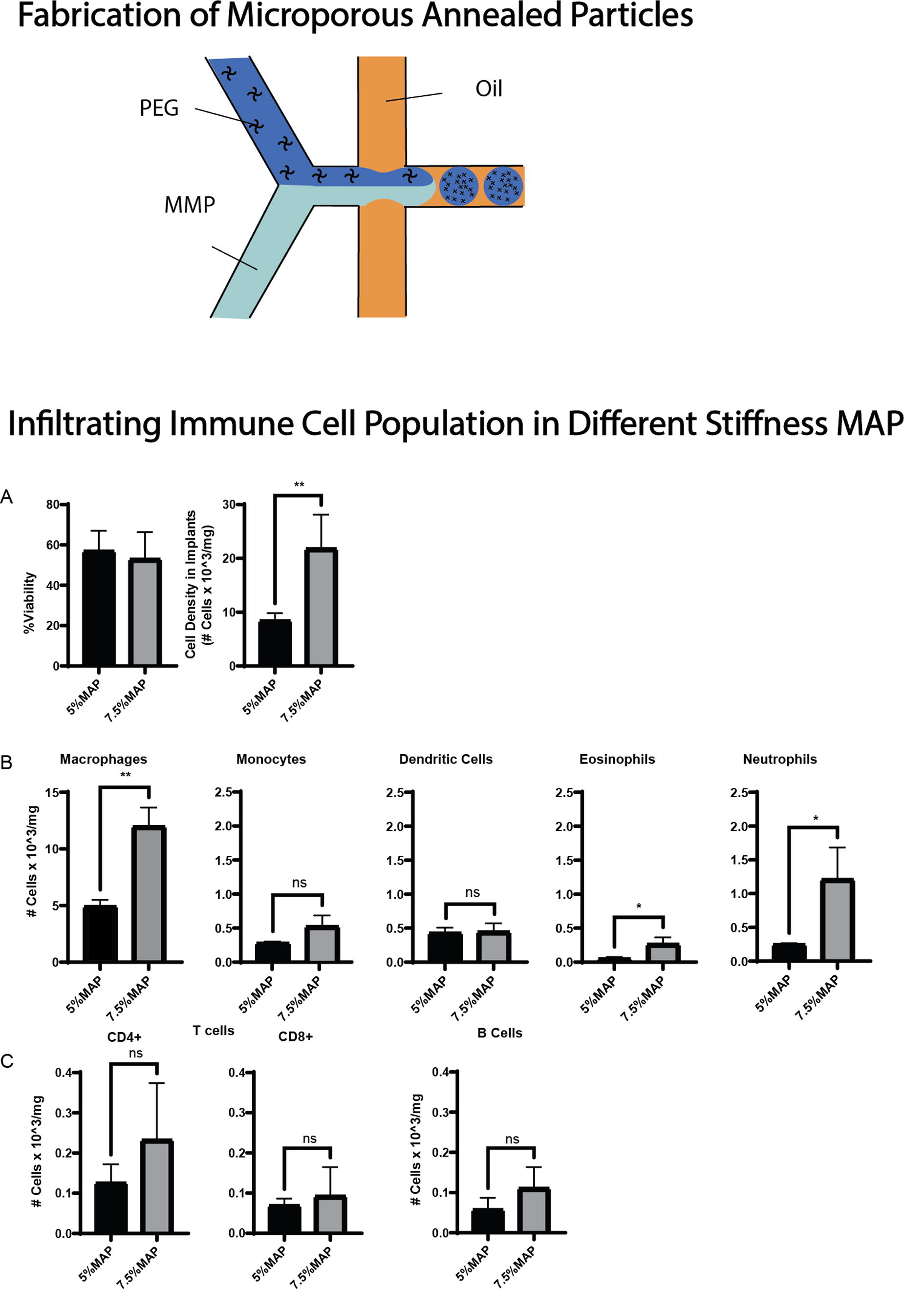
A303 - The Effects Of Keratin-coated Titanium On Osteoblast Function And Bone Regeneration.
E. Ranjit, S. Hamlet, T. Shelper, R. M.Love
Griffith University, Gold Coast, Australia
*Purpose/Objectives: Surface modification of titanium by coating with natural bioactive polymers has been suggested as a means to improve bone healing in dental implant applications. Wool-derived keratin, due to its demonstrated ability to promote bone formation, has been suggested as such a potential bioactive material. As limited knowledge is available on the biological effects of keratin on osteoblasts, the aim of this study was to assess the effects of two novel methodologies used to coat titanium surfaces with keratin, on osteoblast function in vitro and bone healing in vivo.
*Methodology: Titanium was silanized with APTES and crosslinked with glutaraldehyde to facilitate covalent binding with keratin. Two keratin-coated titanium surfaces were fabricated via solvent casting (Ti-KC) and functionalization (Ti-KF) respectively. The attachment, proliferation, viability and cellular morphology of MG-63 osteoblast cells cultured on these modified surfaces were determined by semi-automated quantitative microscopy. Osteogenic gene and protein expression were quantified in vitro, and the impact of keratin-modified surfaces on bone healing was assessed in vivo using an intraosseous calvaria defect model in nude rats over three weeks.
*Results: MG-63 proliferation over 14 days on the Ti-KF surface was the same as on uncoated titanium. On the Ti-KC surface, proliferation initially lower on days 1 and 7 was similar to control levels by day 14. No significant differences in cell viability were seen. Elongated spindle-shaped cells were seen on functionalized and untreated titanium on day 1, whereas oval-shaped cells were noted on the coated surface. Over time in culture, however, MG-63 cells on all surfaces became spindle-shaped with no statistical difference in morphological measurements. Osteogenic gene expression on the modified surfaces was higher compared to uncoated titanium at all times. Keratin coating significantly enhanced osteogenic protein expression i.e., OPN, OCN, COL-1 and BMP in vitro. Histological analysis of recovered calvaria specimens showed defects covered with keratin-coated titanium had a higher percentage of linear new bone and new bone area, two weeks after implantation compared to that in defects covered with titanium alone. After three weeks, new bone formation was similar in all samples.
*Conclusion/Significance: The surface topography and chemistry of biomaterials is known to influence a wide range of biological responses such as cell migration, adhesion, proliferation, and differentiation in adherent cells. This study showed that covalently bound keratin on titanium surfaces did not have any lasting negative impacts on MG-63 cell attachment, morphology or subsequent proliferation. The keratin-coated surfaces stimulated osteogenic protein expression in adherent MG-63 osteoblasts. Furthermore, preclinical studies in rodents showed keratin stimulated earlier intraosseous calvaria bone healing, suggesting keratin may have the potential to promote more rapid and earlier bone healing in implant applications.
A304
A305 - Nanoencapsulation Of Human Intestinal Organoids Using Silk Protein Ionomers As A Platform For Cell Transplantation Therapy
B. Wang
Tufts University, Medford, MA
*Purpose/Objectives: Current challenges with cell transplantation include achieving long-term cell survival, minimizing immunogenicity, and ensuring the functional integration of transplanted cells into the native host tissue. Nanoencapsulation of mammalian cells using silk ionomers provides a temporary protective shield that enhances cell survival in challenging environments, such as those after transplantation. The coatings are also tunable in terms of duration and surface properties.
*Methodology: While previous silk nanocoating studies have primarily focused on the use of immortalized or individual cells for encapsulation methods, the present research explores the nanoencapsulation of human intestinal organoids - multicellular aggregates - using chemically modified silk fibroin-based layer-by-layer (LbL) electrostatic deposition technique. The coating on the surface of organoids was characterized by confocal microscopy and scanning electron microscopy (SEM). Cell viability post-coating was assessed using Live/Dead stain while continued cell proliferation was studied via AlamarBlue assays post-encapsulation.
*Results: The application of silk ionomers on the organoids was successfully achieved using both 3 and 5 bilayer coatings with no change seen in cell viability. Further, no significant impact on cell proliferation was seen on organoid growth when compared with non-coated control organoids highlighting the biocompatibility of the coating.
*Conclusion/Significance: The successful nanoencapsulation of organoids demonstrates the feasibility of encapsulating larger, multicellular structures without compromising cell viability or adversely impacting the cell functions. These findings serve as a platform for various biomedical applications, particularly in the field of transplantation therapy involving larger cell aggregates. Future directions of this project involve the investigation of the effect of coating on cell fates by post-encapsulation differentiation of the intestinal organoids. Functional testing of the protective layer to assess its resistance to physiologically relevant factors such as enzymatic degradation and inflammatory cytokines will be reported. Finally, modulation of the coating post-encapsulation with an aqueous polyethylene glycol (PEG) solution and its impact on mechanical properties of the coating via atomic force microscopy (AFM) will also be reported.
A306 - Fabrication Of Oxygen Releasing Scaffold For The Construction Of Thick 3D Tissue
D. Tomioka, M. Matsusaki
Osaka Univeristy, Osaka, Japan
*Purpose/Objectives: In tissue engineering field, in vitro/in vivo construction of thick three-dimensional (3D) tissues is still a major challenge because limited diffusion of oxygen inside these tissues causes cell necrosis. To address the oxygen shortage in 3D tissues, oxygen releasing materials have attracted much attention in recent years. Calcium peroxide (CaO2) is one of the most common oxygen sources for these materials since it generates oxygen, hydrogen peroxide and calcium hydroxide by the reaction with water. However, the initial burst release of oxygen from CaO2 is issue due to the rapid reaction with water. Herein, we have achieved sustained oxygen release by suppressing the reaction between CaO2 and water through the formation of hydroxyapatite (HAp) on the surface of CaO2 using phosphate buffer (PB) (Figure a). It is known that calcium hydroxide reacts with phosphoric acid to provide HAp. Thus, the calcium hydroxide derived from CaO2 is expected to reacted with phosphoric acid in PB to form HAp on CaO2. Since HAp served as a diffusion barrier, HAp-CaO2 showed sustained oxygen release compared to untreated CaO2. Furthermore, oxygen releasable gelatin hydrogels enzymatically crosslinked by transglutaminase were fabricated using HAp-CaO2 and catalase which decomposes toxic hydrogen peroxide into oxygen (Figure b). In this study, oxygen releasing hydrogels were applied to cell culture and construction of thick 3D tissues.
*Methodology: HAp coating was achieved by simple immersion of CaO2 microparticles in PB. For the preparation of oxygen releasing hydrogels, gelatin, transglutaminase and catalase were mixed with untreated-CaO2 or HAp-CaO2. Normal human dermal fibroblasts (NHDF) were cultured with 3 days preincubated oxygen releasing hydrogels for 7 days under hypoxia condition. 3D tissue was fabricated using L929 fibroblast cells in the presence of oxygen release hydrogel.
*Results: Scanning electron microscopy, energy dispersive X-ray spectroscopy and X-ray diffraction confirmed HAp coating on CaO2 by PB treatment. The hydrogel with untreated-CaO2 showed burst release of oxygen and oxygen release was stopped at day 3. Therefore, preincubated hydrogel including untreated CaO2 didn’t improved cell proliferation so much under hypoxia condition. On the other hand, the hydrogel with HAp-CaO2 showed sustained oxygen release over 10 days. As a result, sustained oxygen release from hydrogel including HAp-CaO2 improved cell proliferation under hypoxia condition even after 3 days preincubation. 3D tissue was fabricated in the presence of the oxygen releasing hydrogel. In the absence of the hydrogel, the hematoxylin-eosin (HE) staining image showed many gaps in the 3D tissue, suggesting cell death due to oxygen shortage. On the other hand, in the presence of the oxygen releasing hydrogel including HAp-CaO2, the HE staining image showed a thick 3D tissue with high cell density, suggesting that oxygen supply from the hydrogels improved cell viability in the thick 3D tissue (Figure c).
*Conclusion/Significance: This study introduces a new strategy for sustained oxygen release from CaO2 that can be applied to a broad range of biomaterials. We propose that HAp-CaO2 and these oxygen releasing hydrogels show great potential for solving the oxygen shortage problem in regenerative medicine and tissue engineering fields.
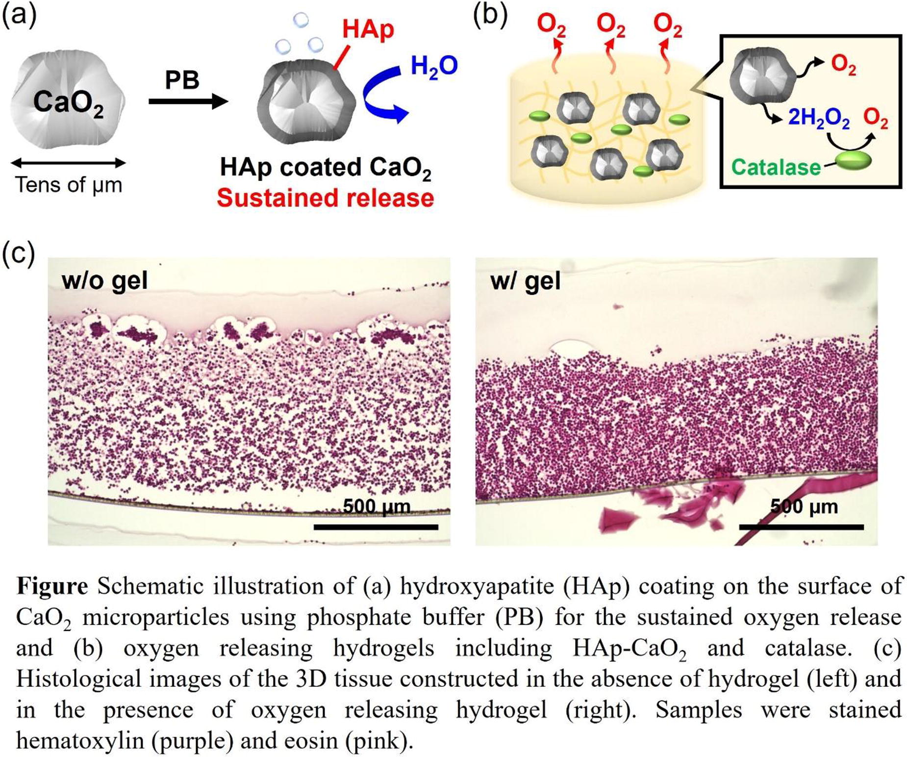
A311 - Immunogenicity Of Different Hydrogels Used For Cell Culture, Tissue Engineering, And Bioprinting
S. Karaman, M. ElGendi, J. Teo
New York University Abu Dhabi, Abu Dhabi, United Arab Emirates
*Purpose/Objectives: Researchers are now using hydrogels for 3D cell culture, as opposed to 2D platforms, because they are more promising in mimicking the extracellular matrix around cells. Hydrogels are 3D polymer networks, that retain water, allowing the support of cells in terms of mechanics, cell adhesion and protein sequestration. Various natural and synthetic hydrogels are being used for cell culture, tissue engineering, and bioprinting. However, there are many aspects to consider when using them, including the origin of the gels, cytotoxicity, pore size, stiffness, degradability, etc. These variables all affect how cells interact and grow, and whether the tissue in question is modeled as precisely as possible for the cells to behave as they should. While many options exist, little is known about the immune reaction to these materials. If these hydrogels are to be used for implantation in the body, or as platforms for testing materials or drugs to be later used in the body, then we should understand the effect of gels on the activation of the immune response. Typically, these materials are tested for cell viability and a certain percentage of live cells is accepted. However, this means that the immune cells, the main defense of the body, are not taken into consideration.
*Methodology: We characterized, and tested a wide range of materials that were formulated into hydrogels, including methacrylated collagen, dextran, alginate, and silk. Certain variables such as crosslinking agent and time, and gel concentration were varied. Then, we characterized each hydrogel by imaging using a 3D confocal microscope, and measuring pore size and fibril size using image analysis software. By directly embedding monocytes into these gels we tested their viability as well as their immune response by studying their specific cell markers and cytokine secretion profile using flow cytometry. We also assessed the phagocytic activity of differentiated macrophages and dendritic cells. This is to ensure that not only do the immune cells survive but that there is no immune response triggered by these materials.
*Results: Our results highlighted the importance of using materials with appropriate stiffness and pore size to recreate the extracellular matrix of specific cells without the activation of an immune response. By characterizing the mechanical aspects of each hydrogel, we also highlighted the use of different combinations of crosslinking times and gel concentrations, for different applications of the hydrogels. We selected the 3 most compatible hydrogels for cell culture and bioprinting based on having the lowest immunogenicity and highest compatibility with different cells: monocytes, macrophages, and dendritic cells.
*Conclusion/Significance: Overall, we find that certain materials are more biocompatible for tissue engineering and cell culture. We show that these materials have a minimal effect on the immune response and highlight 3 materials that can be used further for in vivo implantation due to their low immune response and the ability to mitigate foreign body responses (FBR). These materials can also be used in 3D bioprinting, and for cell culture of therapeutic cells that will be later transferred with minimal risk of rejection.
A313
A315 - Bioconjugation Of Pre-oxidized Nanocellulose Scaffolds Promotes Cell Adhesion And Preserves Structural Integrity In Vitro
L. Kölby1, K. Oskarsdotter1, P. Apelgren1, S. Li2, S. Simonsson2, P. Gatenholm1, K. Säljö1
1
Institute of Clinical Sciences, Gothenburg, Sweden,
2
Institute of Biomedicine, Gothenburg, Sweden
*Purpose/Objectives: biocompatibility, high water content and tuneable chemical properties. The nanocellulose-alginate gel can be dimensionally stabilized by post-printing exposure to divalent cations. The resulting printed construct is stable both in vitro and in vivo, and elicits a low immunogenic response and foreign body reaction. However, due to its inert nature, nanocellulose does not enable cell adhesion, limiting its capability of promoting vascularization. Sodium periodate (NaIO4) oxidation of cellulose increases cellulose aldehyde content, enabling bioconjugation with molecules containing amine groups. In a previous study, we demonstrated how bioconjugation with laminin after topical application of NaIO4 to a nanocellulose-alginate bioink scaffold significantly improved cell adhesion in vitro compared to untreated controls. Unfortunately, topical application of NaIO4 caused severe scaffold disintegration and loss of dimensional stability. A possible solution to address the loss of structural integrity could be pre-oxidation of the nanocellulose followed by covalent binding of laminin prior to mixing with alginate, thus preserving the gelation capability of the alginate.
*Methodology: We compared cell adhesion after bioconjugation with laminin to scaffolds of either pre-oxidized nanocellulose (NCOX) or of regular nanocellulose (NC). Overall cell viability, surface coverage and structural integrity were evaluated. Circular scaffolds () were 3D printed, crosslinked, treated with human laminin and seeded with adipose-derived endothelial progenitor cells (CD34+CD45-) sorted from human lipoaspirate using fluorescence activated cell sorting (FACS). Scaffolds without laminin were used as negative controls, and scaffolds treated with NaIO4 according to previously published protocol were used as positive control. The cells were cultured on the scaffolds for up to10 days and sampled at days 1, 4, 7 and 10.
*Results: The NCOX scaffolds treated with laminin showed high cell density- and high cell viability compared to both positive and negative controls. In addition, the scaffolds exhibited excellent preservation of structural integrity. NCOX scaffolds not only comparable but exceeded the surface coverage on topically oxidized scaffolds.
*Conclusion/Significance: From these preliminary data, we conclude that laminin-conjugation of pre-oxidized nanocellulose is an efficient and non-destructive method for improving cell adhesion with preserved structural integrity of 3D printed scaffolds. Therefore, this method increases the application potential of nanocellulose-alginate bioink for large-scale tissue engineering.
A318 - Electrospinning Technique For Coating 3d Grafts And Yarns In Regenerative Engineering
A. A. Abedini, N. Saveh shemshaki, L. S. Nair, C. T. Laurencin
University of Connecticut, Storrs, CT
*Purpose/Objectives: Electrospinning, a versatile technique used for making nanofiber-based matrices, has been used in the regenerative engineering of musculoskeletal tissues. However, challenges exist in making mechanically robust 3-dimensional porous grafts.
*Methodology: In this study, we employ a novel electrospinning approach to coat Poly (L -lactic) acid (PLLA) based yarns and 3-D braided grafts with nanofibers made of PLLA or gelatine. Tensile strength and microstructural changes in the samples pre- and post-coating were evaluated. Additionally, C2C12 myoblasts were cultured on both coated and uncoated samples to assess cell adhesion.
*Results: Mechanical properties and porosity of the coated samples were not adversely affected by coating. On the other hand, introducing nanofibers could enhance cell adhesion in coated samples.
*Conclusion/Significance: This novel method could help scientists maintain the high mechanical strength and porosity of bare grafts while enhancing cell adhesion and growth using nanofiber coating. This approach has the potential to produce grafts for tissue regeneration with enhanced effectiveness.
A319 - Validation Of Decellularized Cortex And Medulla Extracellular Matrix For Creating An In Vitro Ovary
Y. Franko, E. Ribes Martinez, M. de Almeida Monteiro Melo Ferraz
Ludwig-Maximilians University of Munich, Munich, Germany
*Purpose/Objectives: Research in mammalian fertility decline and wildlife extinction has spurred the development of Assisted Reproductive Techniques. Growing early stage follicles in a lab setting holds promise for improving disease models, toxicity tests, and fertility preservation methods. While successful live offspring birth from cultured mouse follicles exists, in large mammals, the results are still limited. Complexity of follicle growth requires replicating in vivo biomechanics in artificial scaffolds, presenting a significant challenge. Our goal is to create an “ovary-on-a-chip” using decellularized extracellular matrix (dECM) from both the cortex and medulla to mimic native tissue properties, ensuring ideal conditions for long-term follicle survival in co-culture.
*Methodology: Bovine ovarian cortex and medulla hydrogels were prepared using sodium deoxycholate (SDC). The hydrogels were then evaluated for DNA (n=4), RNA (n=4) and detergent (n=10) content to prevent potential toxicity. LC-MS/MS (Ultimate 3000 nano-LC system, coupled to a Q Exactive HF-X mass spectrometer) of dECM was performed (n=5). Further nanoindentation (n=3), and porosity (n=4) analysis were performed to characterise the biomechanical and biological properties of the hydrogels (10mg/mL). Biocompatibility and stability were tested by culturing stromal cells in hydrogels and assessing their viability (n=3). Bovine pre-antral follicles were isolated and cultured in cortex ECM, and follicle entrapment and viability were evaluated at day 5 (n=2). Data analyses were carried out in R, using a generalised linear model.
*Results: Isolated ECM showed complete DNA and RNA removal for both cortex and medulla. SDC content was undetectable. Among the ECM-related proteins, collagens were more abundant in the cortex than in the medulla (86 vs 78%), while medulla had higher proteoglycans content (14 vs 8%), with similar content values for other ECM proteins. Laminin, a vital ECM component, was also detected, among other constituents. Porosity of cortex and medulla hydrogels was similar to native tissue (p>0.05). A mean reduction of 37 and 90% of hydrogel area on days 7 and 28, respectively, was observed, demonstrating that the stromal cells promote ECM degradation. The addition of 0.5% alginate, a natural polysaccharide that's not found in the mammalian ECM, reduced this loss to 22 and 43%, and 80% of stromal cells remained alive for the duration of culture. Both medulla and cortex hydrogels had reduced Young’s modulus when compared to the native tissue (0.86 vs 3.8 and 0.89 vs 5.7 kPa, respectively; p<0.05). Nevertheless, both hydrogels presented similar elasticity and visco-elasticity properties to the native tissues. Preliminary trials indicated non-mixing of cortex and medulla stromal cells within their respective ECM, during co-culture. Attempts to entrap follicles faced challenges, with substantial follicle loss either during crosslinking or media change. Among found follicles, 56 and 58% were dead in 2D and within hydrogel, respectively.
*Conclusion/Significance: The use of decellularized ECM stands as a promising strategy in the development of a more biomimetic ovary-on-a-chip platform. Further experiments will be carried out to ensure necessary biomechanical conditions are present in the ECM hydrogels and later co-culture experiments will be performed to evaluate the viability and growth of early follicles.
A320 - Vaginal Extracellular Matrix Derived Hydrogel Modulates Vaginal Myofibroblast Differentiation In A Stiffness-dependent Manner
M. Therriault, M. Egnot, B. Brown, P. Moalli
University of Pittsburgh, Pittsburgh, PA
*Purpose/Objectives: Pelvic organ prolapse (POP) is a disease in which the organs descend from their normal anatomical position in the pelvis. ∼13% of women seek surgical POP repair by age 80. Surgeons balance recurrence risks in native tissue-based surgeries with the complications associated with mesh-augmented repairs, most commonly pain, and mesh exposure. Our studies in large animal models suggest that vaginal fibroblasts are key mediators of mesh complications. We hypothesize that mechanical cues in the extracellular matrix (ECM) direct the fibroblast toward quiescence versus activation. Under heightened mechanical stress, activated fibroblasts transform into myofibroblasts, developing smooth muscle actin stress fibers and depositing collagen, causing tissue contraction and fibrosis, a likely mechanism for pain. Conversely, fibroblasts protected from essential physiologic stresses degrade matrix and undergo apoptosis, resulting in tissue loss, proposed mechanism for exposure. We created a hydrogel using decellularized porcine vaginal extracellular matrix (vECM) to study fibroblast interactions at a localized level in a biomimetic model. Our goal was to investigate vaginal fibroblast behavior in vECM hydrogels with varying stiffness, achieved by modifying vECM density. Our objective in developing this 3D bioassay was to 1) define the fibroblast response to vECM of varying mechanical stresses to define the transition from physiologic to pathologic, and 2) to develop new biomaterials that maintain fibroblast quiescence.
*Methodology: Porcine vaginas were decellularized using a sodium deoxycolate detergent method. The lyophilized tissue was digested in a 1000U/mL Pepsin and 0.01M HCl solution for 48 hours. Following lyophilization of the digest, samples were reconstituted at a desired protein concentration (5, 10, 20, and 25 mg/mL) and neutralized using 0.1N NaOH. vECM hydrogels were imbued with suspensions of human vaginal fibroblasts collected from pre-menopausal women with normal pelvic organ support. Gel mechanical properties were measured by dynamic rheometry. Apoptotic cells were measured using a Tunel assay. Immunolabeling αSMA and vimentin characterized fibroblast phenotypic changes.
*Results: Decellularized vaginal tissue maintained structural proteins and mechanical properties. The vECM gel storage modulus increased with concentration (85 Pa for 5 mg/mL, 224 Pa for 10 mg/mL). Human vaginal fibroblasts seeded at 250,000 cells/gel in 5-25 mg/mL gels demonstrated greater manipulation ability in 5 and 10 mg/mL gels compared to 20-25 mg/mL gels (Figure 1A). Time-course images of 10mg/mL and 20mg/mL gels over 14 days showed enhanced cell contraction in 10mg/mL gels (Figure IC). At day 0, no colocalization of myofibroblast or apoptosis markers was observed. By day 7, cells attached to the matrix, expressing myofibroblast markers in stiffer vECM hydrogels (vimentin plus αSMA), indicating differentiation and activation of the cells in stiffer vECM hydrogels (Figure 1B).
*Conclusion/Significance: The results indicate the successful fabrication of a tunable in vitro system to test vaginal fibroblast behavior by modifying the concentration of protein to modulate mechanical properties using a relevant homologous tissue source. The results demonstrate the ability to modify vaginal fibroblast behavior by tuning mechanical cues in the environment. Further work seeks to ascertain more information about the activation state through cellular outputs and the regulatory processes activated by mechanical cues.

A321 - Unraveling The Matrix: Decoding Vagina Fibroblast Cell Responses To Substrate Stiffnesses
T. Obisesan1, S. Kottapalli1, Y.-l. Wang2, S. Abramowitch1, P. Moalli3
1
University of Pittsburgh, Pittsburgh, PA,
2
Carnegie Mellon University, Pittsburgh, PA,
3
University of Pittsburgh Medical Centre, Pittsburgh, PA
*Purpose/Objectives: Pelvic organ prolapse (POP) is a common gynecologic disorder in women with a tremendous physical and psychological burden. Owing to high failure rates (∼70% at 5 years) when using a woman's own tissue, surgeons augment surgical repairs with polypropylene mesh, a polymer with a stiffness that is orders of magnitude stiffer than native vagina. Mesh implants cause complications including exposure through the vaginal epithelium and pain likely due to stiffness mismatches between mesh and vagina. Pain can persist in up to 50% of patients after mesh removal. Fibroblasts play an important role in wound healing and can transition to myofibroblasts (FMT) when triggered by increased tissue (matrix) stiffness. Though myofibroblasts provide mechanical support for wounds via the acquisition of α-smooth muscle actin (αSMA) fibers, their persistence is always pathologic causing tissue contraction and fibrosis. We hypothesized that tuning the stiffness of polyacrylamide gel to match that of a native premenopausal vagina will result in fibroblast quiescence while stiffening beyond physiologic levels will trigger myofibroblast formation.
*Methodology: Polyacrylamide gels (PAGs) were manufactured with progressively increased stiffness (5, 10, 20, 40 KPa) by varyingthe crosslinking density of acrylamide and bis-acrylamide. The gel was polymerized between activated glass coverslips using UV light exposure and securely integrated into a bio chamber. PAGs were functionalized using a catalyzed gelatin-azide solution with the catalysts: copper sulfate (CuSO4), tris(3hydroxypropyltriazolylmethyl) amine (THPTA), and sodium ascorbate applied over the surface of the gel in the absence of light. This created a covalent bond to enhance fibroblast attachment to the PAGs via fibroblast integrin receptors. 75,000 cells from a primary fibroblast culture established from a premenopausal woman without POP were introduced to gels. After 3, 5, 7, and 10 days of incubation, FMT was quantified using immunofluorescence markers: αSMA (myofibroblast) and vimentin (fibroblast). Open-source image analysis software QuPath-0.4.3(Queen’s University, Belfast, Northern Ireland) was used for a semi-quantitative analysis.
*Results: Increasing the stiffness of PAGs accelerated FMT which occurred at earlier incubation periods in the higher stiffness gels (20kPa and 40kPa: figure 1a-b). At day 3, quiescent fibroblasts were observed in higher amounts on the low stiffness PAGs (5kPa and 10kPa: figure 2a-b). In lower stiffness gels, 40% -70% fewer fibroblasts had completed or were in transition to becoming myofibroblasts compared to higher stiffnesses PAGs which had 80-98% myofibroblasts on day 3 (Figure 1a-b). All vaginal fibroblasts eventually transitioned to myofibroblasts by day 5 independent of PAG stiffness. Cells with fragmented DAPI nuclear stains were counted as dead.
*Conclusion/Significance: Vaginal fibroblasts are highly sensitive to substrate stiffness with increased stiffness accelerating fibroblasts to myofibroblasts transition. A functionalized PAG provides a unique model to investigate fibroblast mechanoresponsive behavior. Further investigation into fibroblast survival and quiescence as a response to stiffness is warranted. More detailed analysis of the fibroblast response is ongoing including the expression of matrix components, TGF-β, apoptotic (DNA fragmentation), and quiescence (bromo-uridine) markers. Understanding fibroblast behavior and maintaining quiescence is critical for developing novel softer materials for POP surgery to the benefit of millions of affected women worldwide.

A323 - The Effect Of H-emscs-macrophages Co-seeded Pcl-ha Electrospun Patch On Repairing Endometrial Injury
X. Zhang1,2, J. An1, T. Ma2,3, W. Wang4, C. Simmons2, J. Santerre2
1
Binzhou medical university, Yantai, China,
2
University of Toronto, Toronto, ON, Canada,
3
University of Hongkong, Hongkong, China,
4
Yuhuangding Hospital, Yantai, China
*Purpose/Objectives: Intrauterine adhesion (IUA) refers to the damage of the basal layer within the endometrium due to miscarriage, infection, or medical injury, which can lead to partial or complete obstruction of the uterine cavity. The main clinical symptoms of IUA include decreased menstrual volume, amenorrhea, infertility, repeated abortion, and periodic lower abdominal pain and currently IUA has approximately a 32% prevalence in women worldwide. Human endometrial mesenchymal stem cells (H-EMSCs) can inhibit endometrial fibrosis and promote the repair of endometrial tissue injury. However, the effect of direct injection of stem cells into the damaged endometrium is limited, due to the low survival rate and the short cell retention time. A potential strategy for pro-H-EMSCs survival is to seed the cells onto functional biomaterial-based electrospun membranes, to create an endometrial patch, increasing the survival rate of H-EMSCs and prolonging the contact time between H-EMSCs and the damaged endometrium for better endometrial tissue repairing outcomes. In addition, it is acknowledged that macrophages are important immune cells that participate in acute and chronic inflammation and they are also critical for maintaining the homeostasis and modulating the repair and remodelling processes of the endometrium. Macrophages can be polarized to the M1 (more pro-inflammatory) or M2 (more anti-inflammatory or wound-healing) phenotypes when stimulated by the different types of endometrial tissue micro-environmental cues (i.e., growth factors, cytokines, mechanical stress/strain). Polycaprolactone (PCL)-hyaluronic acid (HA) electrospun membranes were found to support H-EMSCs adhesion, proliferation and mesenchymal stem cell marker expression according to our group’s previous studies. However, the interactions of H-EMSCs and macrophages on PCL-HA electrospun fibres and if the H-EMSCs-macrophages co-seeded PCL-HA electrospun patch can enhance endometrial injury repair still remain unknown.
*Methodology: This study isolated H-EMSCs from human endometrial tissue and co-cultured H-EMSCs and macrophages on PCL-HA electrospun membranes to create an endometrial patch. A rat endometrial damage model was created mimicking the physical damage caused by curettage and the H-EMSCs/macrophages co-cultured PCL-HA patch was implanted into the rat uterus to evaluate its effects on repairing endometrial injury.
*Results: It was found that the H-EMSCs/macrophages co-seeded PCL-HA patch caused a significant increase in the thickness of the endometrium and the number of the endometrial glands in the rat model vs. the no seeding (PCL-HA material alone) and H-EMSCs mono-culture. Additionally, it was found that the co-cultured PCL-HA patch enhanced CD31 and VEGF expression at the gene and protein levels in the explanted endometrial tissue vs. the mono-cultured or no cell seeding patches. Moreover, the co-cultured patch also reduced collagen production (by Masson’s staining) and fibrotic marker expression (α-SMA) of the rat endometrial tissue. At the same time, in the PCL-HA electrospun fibre-based co-culture system, the macrophages showed a gradual decrease of CD80/CD86 expression, but a gradual increase of CD163 /CD206 expression, suggesting that the co-cultured patch supported a shift of M1 to M2 macrophages after being implanted in the rat endometrial damage model.
*Conclusion/Significance: This study provides significant insights into applying PCL-HA biomaterial-based co-culture systems for endometrial tissue repair and the treatment of IUA.
A324 - Fibroblast Growth Factor-2 (FGF-2) Promotes Melanocyte Proliferation In Epithelial Cell Cultures
K. Ferland, B. Magne, D. Larouche, L. Germain
Université Laval, Québec, QC, CANADA
*Purpose/Objectives: Severe burn patients in lack of donor sites for the harvesting of autografts can be treated with self-assembled skin substitutes (SASSs) produced in our laboratory from a small patient’s skin biopsy. However, the pigmentation is not fully restored after SASS grafting, resulting in pigmentation defects in treated patients. These pigmentation defects are probably caused by the dilution of melanocytes in epithelial cell cultures that are used to produce the epidermal part of the SASS. Melanocytes are responsible for skin pigmentation by producing melanin, a chromophore that absorbs ultraviolet light. Therefore, patients lacking pigmentation after SASS grafting could be more at risk of developing skin cancer. A preclinical study has already demonstrated that a supplementation of SASS with melanocytes improves pigmentation after grating onto athymic mice. However, the technique used is time-consuming and the culture medium promoting melanocyte proliferation contains carcinogenic factors such as phorbol esters. Therefore, this option is not compatible with a clinical application. As an alternative, we hypothesized that the supplementation of the SASS culture medium with a melanogenic factor, such as Fibroblast Growth Factor-2 (FGF-2), could increase melanocyte growth and improve the pigmentation of SASS. The objective of this research is to evaluate the effects of FGF-2 on melanocyte proliferation in epithelial cell cultured in monolayer and in the SASS model in vitro and in vivo after grafting onto athymic mice.
*Methodology: We first evaluated the role of FGF-2 in a dose dependent manner on melanocyte proliferation. We then assessed whether skin keratinocyte secrete FGF-2 or not. After determining the optimal dose of FGF-2, we calculated melanocyte proliferation in epithelial cell cultures. We then produced SASS by tissue engineering. Sheets were produced with dermal fibroblasts before adding epithelial cells that were cultured with FGF-2. SASS were analyzed in vitro and grafted onto athymic mice for 42 days to follow pigmentation in vivo after grafting. The proportion of melanocytes was evaluated in SASS using flow cytometry and immunofluorescence assays using TYRP1 as melanocyte marker. Pigmentation of SASS was evaluated with melanin dosage by spectrophotometry and histological staining.
*Results: Our results suggested that FGF-2 promotes melanocyte proliferation without stimulating keratinocyte proliferation at an optimal dose of 0.2nM. Keratinocytes secreted very low amount of FGF-2 (0.003nM) under tested conditions. Our results also suggested that at the optimal dose of 0.2nM, FGF-2 increased the proportion of melanocytes in monolayer cultures of epithelial cells. Our results also showed that FGF-2 increased the number of melanocytes in SASS in vitro but had no visible effects on pigmentation. FGF-2 supplementation had no deleterious effects on keratinocyte growth or differentiation. FGF-2 treated SASS have been grafted onto athymic mice and pigmentation will be analyzed in vivo.
*Conclusion/Significance: Our results suggest that FGF-2 stimulates melanocyte proliferation in monocultures, epithelial cell cultured in monolayers and in SASS without affecting epidermal cell growth or differentiation. Future experiments will aim to determine whether combination of several melanogenic factors would induce melanin production in vitro. Our ultimate goal is to produce homogeneously pigmented SASS that will benefit to patients.
A325 - Polyphosphate Releasing Biocompatible Hydrogel Nanoparticle Emulsions Disperse P. Aeruginosa Biofilms For Topical Wound Healing
S. Motamed, R. Negru, J. Zhou, F. Teymour, G. Papavasiliou
Illinois Institute of Technology, Chicago, IL
*Purpose/Objectives: Over 30 million individuals in the U.S. and over 400 million globally have diabetes with an estimated 15% developing debilitating chronic wound infections the care of which costs up to $9 billion annually in the U.S. The majority of pathogens in diabetic wounds include antibiotic-resistant biofilms which are challenging eliminate with standard wound care regimens including debridement, topical wound dressings, antibiotics, and systemic therapeutics. Most topically-applied therapeutics have been optimized against planktonic cells with non-approved to specifically target biofilms. We aim to develop topical ointments sustaining the release of therapeutic compounds to eradicate biofilms and promote healing of diabetic wounds. To achieve this, we synthesized and characterized Biocompatible hydrogel Nanoparticle Emulsions (BCNE) loaded with the anti-virulence agent, sodium hexametaphosphate (PPi), and tested the efficacy of en masse addition of PPi-BCNE on P. aeruginosa in-vitro biofilm dispersal.
*Methodology: BCNE was synthesized using inverse-phase miniemulsion polymerization. The aqueous phase consisted of V50 as the initiator, N-vinyl pyrrolidone (NVP) comonomer, PPi or NaCl (44 mM), and PEG diacrylate (575Da) as the crosslinker. The organic phase was comprised of soybean oil and PGPR emulsifier (FDA-recognized as GRAS). The aqueous phase was added dropwise to the oil phase followed by mechanical and ultrasonic homogenization, nitrogen bubbling, and polymerized in an incubator shaker for 2 hours at 70rpm and 60°C. Nanoparticle diameter and distribution for blank and PPi-BCNE were quantified using a Nanosizer (LM10, Malvern) equipped with nanoparticle tracking analysis software (n=3). P. aeruginosa biofilms were grown on tryptone yeast (TY) plates, incubated overnight at 37 °C. An inoculation loop of plated bacteria was cultured overnight at 37 C in 3mL of TY media. Liquid cultures were diluted 1:20 in TY media, transferred into 96-well plates, and incubated at 37°C. P. aeruginosa biofilms treated with 60 and 6 mM free PPi, blank-BCNE, PPi-BCNE, Soybean Oil, and TY (control group) for 24 and 48 hrs followed by 0.1% crystal violet staining for 15 minutes and 90% ethanol for 5 minutes (n=6 for each treatment) for measuring OD540. Biofilm dispersal was quantified for the same groups with confocal microscopy. Treatments were added for 24 hrs to performed biofilms, removed, and stained with a live/dead BacLight kit in 0.85% NaCl solution (3:1000) for 15 mins followed by rinsing with a 0.85% NaCl solution. Biofilm thickness and biomass were quantified using COMSTAT (n=6 for each treatment). PPi release kinetics from BCNE were quantified using a surrogate scaffold method (n=3).
*Results: Blank and PPi-BCNE exhibit uniform particle size distribution and mean nanoparticle diameters of 104.7±16.2nm and 156.9±18.4nm, respectively. In-vitro findings revealed significant dispersal of biofilms following PPi-BCNE treatment in terms of OD540, biofilm biomass, and thickness compared to the no-treatment control. An over 99% reduction in biofilm average thickness and biomass occurred following PPi-BCNE treatment compared to treatment with free PPi.
*Conclusion/Significance: En masse treatment of PPi-BCNE on P. aeruginosa biofilms resulted in their effective dispersal. Current efforts focus on designing BCNE ointments co-delivering angiogenic peptides and PPi to stimulate neovascularization and biofilm dispersal in diabetic wounds confounded with biofilm.
A326 - Topical Synthetic Melanin For Skin Regeneration
D. Biyashev1, N. K. Paul2, U. V. Onay1, S. T. Evans1, Z. E. Siwicka2, M. Demczuk1, N. C. Gianneschi2, K. Q. Lu1
1
Northwestern University, Chicago, IL,
2
Northwestern University, Evanston, IL
*Purpose/Objectives: Severe acute injury of the skin can lead to massive induction of reactive oxygen species (ROS), inflammation and cell death, and impaired and prolonged tissue healing. These exaggerated responses may lead to excessive scar formation. In the skin, endogenous melanin is known to be an efficient scavenger of ROS playing a key role following exposure to damaging UV radiation.
*Methodology: We recently demonstrated in models of severe blistering skin injury resulting from exposure to high dose UV (100mj/cm2) or toxic alkylating compounds (nitrogen mustard) that intervention with engineered topical synthetic melanin particles (SMPs) significantly decreases skin swelling, reduces time to eschar detachment, and increases rate of wound closure compared to vehicle controls.
*Results: Consistent with these observations, biomarker studies using immune arrays demonstrate significant downregulation of apoptotic and inflammatory signaling pathways confirmed with by TUNEL staining in skin tissue sections. Additional studies demonstrate suppression of ERK1/2 phosphorylation and decreased expression of matrix metalloproteinase-9 (Mmp9). Furthermore, topical SMPs increases skin superoxide dismutase (SOD) and pretreatment of animals with pharmacologic inhibitors of SOD abrogated the benefits of topical SMPs in vivo. Immune cell analysis of the skin ex vivo reveals that SMPs increased infiltration of ‘inflammation-resolving’ immune cells including TGFβ+/IL-10+ monocytes and macrophages. To optimize interaction with the skin, SMP surface chemistry were modified to increase hydrophobicity characteristics for better attachment with the stratum corneum. While both hydrophilic and hydrophobic SMPs significantly dampened edematous skin responses compared to control assessed by bi-fold skin thickness (p<0.01 for both particles; n=6-9 animals per group), hydrophobic-modified SMPs demonstrated a greater reduction in wound depth by 22% (p=0.02) vs. 12% reduction with unmodified SMPs by histological analysis. The hydrophobic SMPs treated group demonstrated significant downregulation of Cxcl1 compared to control in models of chemical exposure (38% decrease; p=0.026; n=5-9 mice) and high dose UV exposure (43% compared, n=3-5). Cxcl1 is a potent chemotactic cytokine for innate immune cells that serve to amplify inflammation in the skin. Protein expression analysis using an inflammatory protein array showed differential expression of 15 proteins by hydrophobic-modified SMPs compared to the unmodified SMPs (p<0.05; cut-off 1.5-fold). These proteins are associated with the pathways including regulation of receptor signaling pathway via stat, regulation of inflammatory response, and positive regulation of phosphorylation. These studies are complimented by in vitro human skin explant studies which demonstrate that SMP intervention significantly decreases sub-epidermal blister formation and reduces necrotic keratinocytes caused by chemical injury.
*Conclusion/Significance: Therefore, SMPs are promising and effective candidates as topical therapies for accelerated wound healing. SMPs with greater skin contact lead to significant dampening of inflammatory responses resulting dramatic mitigation of wound depth. These findings support SMPs as an important novel class of topical compounds for skin regeneration.
A327 - Bioactive Chitosan-gelatin Hydrogel For Treating Diabetic Foot Ulcer
V. Ganesan, S. Sen
Indian Institute of Technology Bombay, Mumbai, India
*Purpose/Objectives: Dermal wound healing is impaired in diabetic patients. While there are multiple factors that have been identified till date to cause this, our recent work show that fibroblasts in diabetic patient have thicker layer of glycocalyx. We showed that disintegrating the glycocalyx resulted in faster migration and higher contractility of dermal fibroblasts. Our aim in this work is to develop a bioactive hydrogel for treating diabetic wound healing by targeting the fibroblast glycocalyx.
*Methodology: Here we prepare a chitosan-gelatin based hydrogel that is crosslinked via UV irradiation. We entrap the gel with Neuraminidase and silver nanoparticle to improve wound healing and exhibit antibacterial property respectively. The in-vitro studies are carried out using 3T3 fibroblast and human dermal fibroblasts isolated from both healthy and diabetic patients. We use male Wister rats to evaluate our formulations.
*Results: The combination of chitosan and gelatin increased the young's modulus the hydrogel and also reduced the rate of degradation. The incorporation of silver nanoparticles in the gels gave them anti-bacterial activity when tested against S. Aureus and E. Coli bacteria strains. Fibroblast cells were successfully isolated from healthy and diabetic human skin samples. These cells were stained for vinculin and alpha SMA as markers of fibroblasts and used for all in-vitro experiments. The incorporation of Neuraminidase in the gels resulted in smaller focal adhesions and faster migration of the cells. The cells treated with Neuraminidase had higher contractility which was quantified by staining for pmcl. Scratch assay was done to evaluate wound healing efficacy of the gels.
*Conclusion/Significance: The chitosan-gelatin hybrid gels performed better than the Gelatin gels. The presence of Neuraminidase further improved wound healing rate of the diabetic fibroblasts. Our bioactive gel could be used as a feasible strategy to treat diabetic wounds.
A329 - Development Of Scaffolds From Poly(lactic Acid-co-glycolic Acid), Gelatin And Polyethylene Oxide For Use In Skin Injuries
T. F. Elautério1, N. Maurmann1, D. Czarnabay1, S. B. Araujo1, C. A. Ferreira1, P. Pranke1,2
1
Universidade Federal do Rio Grande do Sul (UFRGS), Porto Alegre, Brazil,
2
Instituto de Pesquisa com Células-tronco (IPCT), Porto Alegre, Brazil
*Purpose/Objectives: Skin injuries have an impact on thousands of individuals annually, representing a significant public health challenge. In tissue engineering, the production of scaffolds plays a pivotal role as these three-dimensional structures provide a supportive framework for cell attachment, proliferation, and tissue regeneration. The aim of this work was to standardize polymeric solutions with poly lactic-co-glycolic acid (PLGA), polyethylene oxide (PEO) and gelatin with the intention of producing scaffolds through electrospinning for the treatment of skin wounds.
*Methodology: Scaffolds were produced by electrospinning containing PLGA and POE, with and without gelatin. Solutions were prepared at a concentration of 34% PLGA, 20% gelatin, and 4% PEO for the solution with gelatin and 44% PLGA and 4% PEO for the gelatin-free solution. In the gelatin solution, a voltage of 21 kV was applied, with a relative humidity of 15%, a temperature of 33°C, and a flow rate of 0.1 mL/h. For the solution without gelatin, the voltage was 19 kV, with a relative humidity of 25%, 30°C, and 1 mL/h. After electrospinning, the resulting biomaterials were cross-linked with N-(3-dimethyl aminopropyl)-N' and N-hydroxysuccinimide. The scaffold analysis included morphological characterization by scanning electron microscopy (SEM), contact angle with water, Fourier Transform Infrared Spectroscopy (FTIR) and thermogravimetric analysis (TGA). Cell viability was assessed by the 3-(4,5-dimethylthiazol-2-yl)-2,5-diphenyltetrazolium bromide (MTT) assay with stem cells, using cells seeded on culture plates as a live cell control, and Triton-treated cells as a dead control.
*Results: Scanning electron microscopy revealed randomly spun fibers, without beads with diameters of 0.8±0.2 and 0.9±0.2μm respectively in the scaffolds of PLGA/PEO and PLGA/PEO/gelatin. FTIR tests revealed characteristic bands of PLGA/PEO in all the tested biomaterials, as well as gelatin in biomaterials containing this component, indicating the maintenance of the properties of the polymers after solubilization and electrospinning. TGA analysis highlighted that the presence of PLGA/POE increased the thermal stability of gelatin. The investigation of the contact angle showed that both materials are hydrophilic, the biomaterial with gelatin presenting an angle of 62±13°, with was smaller in comparison with the biomaterial without gelatin, which registered 86±18°, closer to the hydrophobic range. The results of the MTT assay indicated no significant difference in cell viability between the biomaterials and the control, suggesting that the scaffolds did not impair cell survival. The absorbance values after 1 day of stem cell cultivation were 0.438±0.049, 0.505±0.074, 0.477±0.116 and 0.011±0.004 in the control, electrospun without gelatin, with gelatin and Triton, respectively.
*Conclusion/Significance: The biomaterials developed from PLGA/PEO/gelatin demonstrated greater thermal stability, preserved the characteristics of the PLGA, PEO and gelatin, presented a reduced contact angle, and presented fibers of approximately 1 μm in diameter. Furthermore, the stem cells cultivated in the materials maintained high viability. The use of gelatin in the scaffold shows promise for supporting stem cells and can be used in skin tissue engineering. Financial support was provided by the Office of Naval Research Global (ONRG Award N62909-21-1-2026), the National Council for Technological and Scientific Development (CNPq), FINEP, INCT-Regenera and the Stem Cell Research Institute (IPCT).
A330 - Rejuvenation Of Human Adult Dermal Papilla Cells By Partial Reprogramming For Hair Regenerative Medicine
A. Nanmo1, A. Suzuki1, T. Kageyama1,2, J. Fukuda1,2
1
Yokohama National University, Yokohama, Japan,
2
Kanagawa Institute of Industrial Science and Technology (KISTEC), Ebina, Japan
*Purpose/Objectives: Hair loss is not a life-threatening disease, but the patients experience a decline in quality of life. Current treatments for hair loss include medications and hair transplantation, which do not always provide satisfactory results. Therefore, hair regenerative medicine has emerged as a promising therapy for the treatment of hair loss. Dermal papilla cells (DPCs) play a key role in regulating hair growth and hair regeneration in the hair cycle, and transplantation of DPCs showed successful hair regeneration in animal models. However, the hair regenerative ability of DPCs of adult origin is largely lower than that of fetal or neonatal origin. It is ethically and practically difficult to obtain a large number of fetal or neonatal cells for hair loss treatment. In this study, we propose a partial reprogramming of adult DPCs using short-term expression of Yamanaka factors (OCT4, KLF4, c-MYC, SOX2: OKMS) for rejuvenation and improvement of hair inductive ability.
*Methodology: Two types of genes, tetO-tdTomato-hOKMS and M2rtTA, which binds to the tetracycline response element promoter of tetO-tdTomato-hOKMS in response to doxycycline and activates gene transcription (tdTomato and hOKMS), were introduced into cultured human DPCs using lentiviruses. Reprogramming was initiated 24 hours after lentiviral transduction by adding doxycycline (2 µg/ml) to the DPC growth medium. On day 2 of reprogramming, DPCs were sorted for viable tdTomato-positive cells by flow cytometry and then cultured on gelatin-coated plates. On day 7 of reprogramming, DPCs were replated onto a layer of inactivated murine embryonic fibroblast (MEF) feeder cells, and on day 8 of reprogramming, the medium was switched to human embryonic stem cell medium. On day 12 of reprogramming, expressions of hair-inductive marker (endogenous SOX2, VCAN)/human fetal DPC marker (KLF3, EBF1, GLI1) genes and human OCT4 protein were determined by RT-PCR and immunostaining, respectively.
*Results: Immunostaining of OCT4 was performed on day 12 to verify that the vector was well designed for this approach. Since we designed the vector to contain the promoters for tdTomato and OSKM on tetO-tdTomato-hOKMS, respectively, there is a possibility that only tdTomato or OSKM will be expressed. The results showed that tdTomato-positive DPCs locally co-expressed human OCT4 in the nucleus, while tdTomato-negative DPCs did not express it. Thus, we verified that the introduced vectors functioned well in DPCs. In addition, gene expression of hair-inductive and human fetal DPC markers was upregulated in tdTomato-positive DPCs compared to tdTomato-negative DPCs on day 12. Although further experiments, including analysis of typical aging markers and animal assays, are required, partial reprogramming may induce a fetal-like state and enhance the regenerative potential of DPCs.
*Conclusion/Significance: This study showed the potential of reprogramming DPCs for hair regenerative medicine. If human hair regeneration can be achieved by transplanting rejuvenated DPCs, this approach may address cellular senescence during culture and aging in individuals.
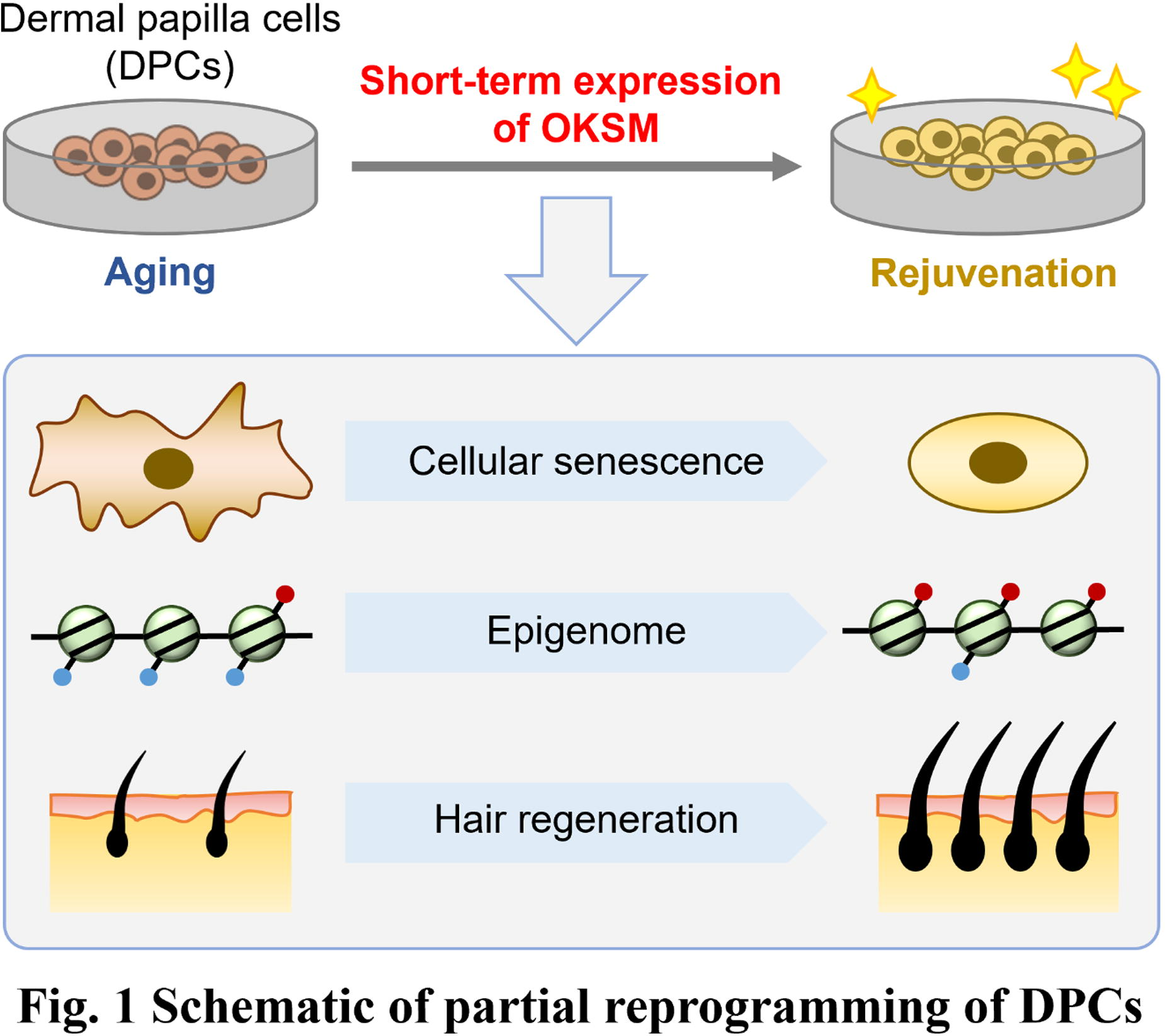
A331 - Advanced Skin Organoid Models: Integrating Hipsc Andadvanced Skin Organoid Models: Integrating Hipsc And Organ-on-a-chip Technology For Infection & Disease Research
A. Reigl1,2, N. Köder3,1, L. Hauf2, P. Loskill3, M. Engstler2, F. Groeber-Becker1, D. Groneberg1
1
Fraunhofer ISC, Wuerzburg, Germany,
2
University Wuerzburg, Wuerzburg, Germany,
3
Eberhard Karls Universität Tübingen, Tübingen, Germany
*Purpose/Objectives: Human skin, a complex organ comprising the epidermis, dermis and hypodermis, exhibits a sophisticated arrangement of different cell types and structures. Reproducing the complex vascular architecture of human skin in vitro remains a major challenge in tissue engineering and regenerative medicine. Skin organoids (SO) based on human induced pluripotent stem cells (hiPSCs) mimicking the skin are remarkable in their ability to recapitulate the intricate architecture of the human skin, including skinlayers and the formation of hair-like structures.
*Methodology: Advancing this area of research, our efforts have led to the cultivation of skin organoids that exhibit cystic structures with an inside-out orientation, closely resembling certain aspects of native skin morphology. To enhance their practical utility, we integrated these organoids into an Organ-on-a-Chip (OoC) system, creating a suitable in vitro testing environment that supports air-liquid interphase.Collaborating with Loskill's laboratory, we have successfully transitioned these organoids to a perfusable platform, marking a significant advancement in dynamic perfusion which facilitates physiologically relevant nutrient exchange and waste removal within the organoid structure.
*Results: Transitioning from the structural to the functional applications, classic organoids have been utilized to study infectious diseases such as human sleeping sickness, caused by the African parasite Trypanosoma brucei, and to model cancer, providing a comprehensive simulation of complex skin tissues.In a unique experiment, skin organoids were exposed to tsetse flies infected with Trypanosoma brucei on day 140. We observed selective infiltration of the parasite, which expressed the fluorophore tdTomato, into the dermal layer of the organoids, while the epidermal layer remained largely unaffected. The skin layers were identified using markers for different cell types: keratinocytes (CK5), dermal cells (vimentin), adipocytes (Nile red) and complex hair peg formation (indicated by the expression of CK5 and CK17). Seven days after infection, the parasites had spread throughout the organoid and accumulated in specific areas of the skin layer.
*Conclusion/Significance: In conclusion, the integration of various techniques, including hiPSC technology and innovative culture methods like OoC platforms, has led to more advanced and physiologically relevant skin models. These sophisticated skin organoids not only provide insights into basic skin biology, but also hold great promise for drug testing, personalized medicine and the development of novel therapeutic interventions for skin-related diseases.
A332 - Isolation And Cultivation Of Sweat Glands To Integrate Them Into Tissue-Engineered Human Skin Substitute
H. De Koninck1,2, C. Martel1,2, D. Larouche1,2, L. Germain1,2
1
Université Laval, Québec, QC, Canada,
2
Centre de recherche en organogénèse expérimentale de l'Université Laval/LOEX, Québec, QC, Canada
*Purpose/Objectives: Severe burns are among the worst types of traumas, and they are very difficult to treat. Thus LOEX has developed an autologous tissue-engineered skin substitute (TES) that can cover a patient's entire body from a single small skin biopsy. However, this model contains no skin appendages. Sweat glands are skin structures that perform several essential functions, including sweat secretion, waste excretion and thermoregulation. However, they have a limited capacity to regenerate after severe damage caused by deep burns. To date, there is no effective treatment available to restore sweat gland function to patients who have irreversibly lost them. My project aims to isolate, culture, and characterize sweat gland cells (SGCs) in order to integrate them into the TES model to regenerate functional sweat glands.
*Methodology: SGCs were isolated from human donor skin biopsies. The skin was cut and then digested with enzymes. The sweat glands were isolated under phase contrast microscope and digested once more to obtain SGCs. They were then cultured either on feeder layers or as spheroids, using keratinocyte culture medium. Finally, SGCs were characterized by immunofluorescence to examine their expression profile of markers identified in the skin (Keratins 18 and 19), some of which are specific to sweat glands (carcinoembryonic antigen, aquaporin 5).
*Results: Our preliminary results showed that the most effective enzymes for isolation of sweat glands were thermolysin, collagenase and DNase, since they increased the quantity of SGCs recovered and cultured. The additional trypsin digestion of the glands also proved a faster method to attain confluency than explant culture. Furthermore, characterization by immunofluorescence showed that monolayer culture affected the differentiation status of SGCs, shifting their marker expression profile to one more akin to keratinocytes. Interestingly, SGCs cultured as spheroids continued to express sweat gland markers, as long as they were not cultured in monolayers prior to spheroid formation. Additionally, supplementation of the culture medium with ectodysplasin-A, an important ligand for sweat gland development, did not maintain gland marker expression in SGCs cultured in monolayer, nor reestablish it in spheroids after monolayer culture. This indicates that the 3D organization is most important for SGCs to retain their gland-like character.
*Conclusion/Significance: We conclude that our current monolayer culture strategy is not optimal for growing SGCs. Experiments are underway to develop a more appropriate culture model that will help preserve the glandular character of SGCs and allow for significant proliferation (unlike spheroids). In parallel, SGCs integration in TES is also being tested to witness the impact of a more physiological 3D environment on SGCs differentiation. Ultimately, the optimization of isolation and culture techniques will facilitate the integration of SGCs into our reconstructed human tissue-engineered skin substitutes.
A333 - Injectable Biostimulators Promote Collagen Stimulation In Vitro
G. M. Machado1, L. Rago2, N. Maurmann3, P. Pranke3, C. Rizzatti-Barbosa4, M. Camara Brew1
1
Universidade Luterana do Brasil (ULBRA), Canoas, Brazil,
2
Leandro Rago Institute, Niterói, Brazil,
3
Universidade Federal do Rio Grande do Sul (UFRGS), Porto Alegre, Brazil,
4
Centro Universitário Ingá (UNINGÁ), Curitiba, Brazil
*Purpose/Objectives: Injectable biostimulators are becoming increasingly popular as biomaterials designed to stimulate the production of new collagen fibers, which are a crucial component of healthy skin and connective tissue. This study evaluates the cellular response and properties of these emerging biostimulators, driven by the growing demand for minimally invasive procedures. The work investigates the effects of various biostimulator treatments on fibroblast viability and collagen biosynthesis in vitro. Additionally, the physical and biochemical properties of the materials were characterized.
*Methodology: The biomaterials underwent characterization through optical microscopy and zeta potential analysis. The fibroblast MRC5 were exposed to biostimulators including Rennova® Elleva (containing poly-l-lactic acid - PLLA), Rennova® Diamond (hydroxyapatite), Ellansé®, Sculptra® (PLLA), and Radiesse® (hydroxyapatite) at concentrations of 0.5, 1, or 1.5mg/ml of the active ingredient. Cell viability assays (MTT) and quantification of collagen synthesis by Sirius Red staining were performed after 2 and 10 days of treatment, respectively. The results were presented as mean ± standard deviation.
*Results: The study found that Rennova®Diamond and Radiesse® biostimulants have a similar morphology with spherical, symmetrical and uniform particles. Ellansé® also contains very similar particles. Rennova®Elleva, on the other hand, had an irregular shape similar to asymmetrical “snowflakes” of varying sizes. Sculptra® had an irregular and asymmetrical shape with varying sizes, but with rounded angles. Rennova®Eleva and Sculptra® showed stability in zeta potential. The hydroxyapatite particles had a regular shape, similar to polycaprolactone, while PLLA had an irregular shape. In the Ellansé® group, there was a decrease in cell viability at all concentrations and with Rennova®Diamond at 1.0 and 1.5mg/ml (p<0.05). At 1.5mg/ml, the mean ± standard deviation of cell viability was 0.212±0.030 for the control (medium containing serum); 0.186±0.007 for Radiesse®; 0.177±0.017 for Rennova® Diamond; 0.182±0.012 for Ellansé®; 0.186±0.006 for the control (medium containing injectable water); 0.160±0.013 for Rennova®Elleva; and 0.157±0.012 for Sculptra®. Fibroblasts treated with the biostimulants Radiesse®, Rennova®Eleva and Sculptra® showed a significant increase in collagen quantification compared to the control group. The mean ± standard deviation values of collagen biosynthesis at 1.5mg/ml were as follows: control (serum in culture medium) 0.294±0.025, Radiesse® 0.392±0.049, Rennova®Diamond 0.334±0.071, Ellansé® 0.307±0.017, control (injectable water in culture medium) 0.274±0.019, Rennova®Elleva 0.417±0.082, and Sculptra® 0.451±0.072.
*Conclusion/Significance: None of the brands or doses exhibited cytotoxicity, as the treatments did not cause a decrease in cell viability greater than 30%. In addition, there was a significant increase in collagen quantification in the Radiesse®, Rennova®Elleva and Sculptra® groups compared to the control and there were no differences in collagen biosynthesis between hydroxyapatite brands or between PLLA brands. This study contributes to the understanding of the mode of action of biostimulants in vitro. Financial support: Coordination for the Improvement of Higher Education Personnel Foundation (CAPES), National Council for Technological and Scientific Development (CNPq), Financier of Studies and Projects (FINEP), National Institute of Science and Technology for Regenerative Medicine (INCT-Regenera) and Stem Cell Research Institute (IPCT).
A334 - Handheld Bioprinter: Direct Deposition Of Serum-free Bioink On Wounds For Epidermis Formation In Skin Bioprinting
W. Wong, R. Chai, C. Beh
Agency for Science, Technology and Research (A*STAR), Singapore, Singapore
*Purpose/Objectives: Current treatments for burn patients who experience substantial skin damage typically involve wound reconstruction using meshed skin autografts. However, donor site availability and morbidity remain a challenge for obtaining these autografts. The results can also be aesthetically unpleasing as it involves cutting rows of staggered slits onto the skin for greater coverage. In recent years, in situ bioprinting has emerged as an attractive alternative by promoting healing using patient cells. However, existing bioprinting equipment designs have large footprints that make deployment in the operating theater challenging. While handheld designs have been proposed, these have often been bulky and unwieldy. Consequently, neither approach has gained much traction with clinicians despite promising pre-clinical results. This paper discusses our work in designing a handheld bioprinter for the operating theatre, capable of applying bioink to body contours, such as the axillary region. Using a specially formulated serum-free mixture of collagen and cells, we can directly deposit a contiguous layer of keratinocytes onto burn wounds. The bioink rapidly gels after application, providing greater control over the deposition process.
*Methodology: The handheld bioink dispenser is designed with consideration for ease of use and constraints of the operating theater. Collagen and cells are housed in separate cartridges within the dispenser, which is actuated using a single pneumatic line. Phase change material is used to chill the bioink, increasing the pot life as the components pass through the mixer. The collagen-cell mixture is then dispensed through a rollerball applicator, which enables omnidirectional deposition of the bioink. In vitro experiments were performed by one-step deposition of the collagen-based bioink containing human primary keratinocytes onto a dermal template to form a collagen-Human Skin Equivalent (c-HSE). Histological studies and immunofluorescence staining were performed to detect the presence of a stratified epidermis through the expression of various cell proliferation and differentiation markers.
*Results: The fabricated prototype can be held and operated with one hand, and the bioink is dispensed using either a foot pedal or finger trigger. Using this prototype, we were able to cover a 100 cm2 area within 10 seconds. Cells deposited using this approach maintained viability at > 90%. Histological studies of c-HSE sections from in vitro experiments showed successful generation of an epidermis with stratified layers following deposition and maturation. Formation of a mature epidermis was further confirmed by immunofluorescence staining of markers of different epidermis layers. Lastly, basement membrane protein collagen IV was present at the dermal-epidermal junction, indicating that a robust epidermis has been generated.
*Conclusion/Significance: We have designed a handheld bioink dispenser for skin bioprinting, allowing for uniform deposition over body contours. Additionally, we have developed a serum-free bioink that enabled keratinocytes to proliferate and differentiate into a mature epidermis through a one-step deposition process. The resulting epidermis recapitulates native skin characteristics, as determined by histology and immunofluorescence. Although in vivo studies are still ongoing, preliminary results indicate that our approach can indeed promote formation of healthy epidermis in the animals.
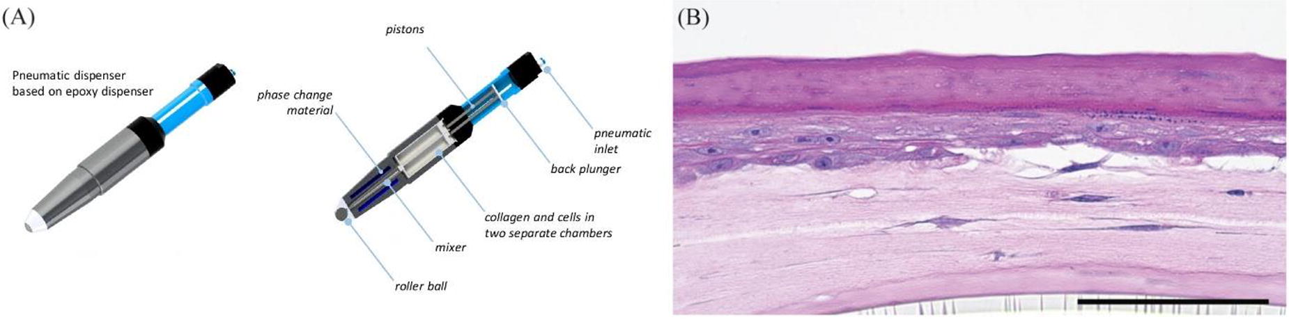
A336 - A Novel Crosslinking Approach For Biomanufacturing Of A Collagen Hydrogel Dermal Template For Third-degree Burn
W. Chan1, B. Le1, H. Ezhilarasu1, P. Murugan1, W. Woo1, A. Anantharajan1, G. Soo2, W. Kerk2, B. Lok2, W. May3, D. Choudhury1
1
Bioprocessing Technology Institute, A*STAR, Singapore, Singapore,
2
Singapore Institute of Manufacturing Technology (SIMTech), A*STAR, Singapore, Singapore,
3
A*STAR, Singapore, Singapore
*Purpose/Objectives: The prevalence of burn injuries worldwide poses substantial medical challenges, particularly with third-degree burns that require skin grafts for wound closure and skin regeneration. Existing products commonly rely on porous collagen sponge scaffolds to replace the dermal layer. Notably, collagen hydrogels, with unique biocompatibility and mechanical properties, have been underexplored in burn wound treatment. Moreover, collagen hydrogels offer more consistent and efficient production without the need for freeze-drying. Our aim is to fill this research gap by assessing and refining a collagen hydrogel dermal template, investigating its potential to enhance the healing in burn wounds.
*Methodology: A UV-crosslinkable methacrylated bovine collagen type I material (Col-MA) was employed in the fabrication of a collagen hydrogel. The degree of functionalization (DOF), indicating the efficiency of collagen methacrylation, was assessed using an indirect method measuring the ratio of free amine group concentration before and after functionalization, assuming the amine group as the primary methacrylate target. To enhance hydrogel stiffness, a novel crosslinking step utilizing an aldehyde solution was applied, enabling fine-tuning of stiffness and degradation profiles. We developed an in-house process for scaling up Col-MA production and fabricating large-size hydrogel scaffolds with high throughput and consistency. In vitro characterization of collagen hydrogels included mechanical testing, enzyme digestion assays, and cell assays. Subsequently, hydrogels (d=8mm) were subcutaneously implanted in a rat model for 14-21 days to assess biocompatibility, integration, and degradation profiles in vivo, with Integra collagen sponge used as a control.
*Results: We achieved approximately 10% DOF for Col-MA. Using UV crosslinking alone, the 1% Col-MA hydrogel exhibited a stiffness of around 2 kPa. By employing a combination of UV and chemical crosslinking (UV/CC), we increased the stiffness to 10-40 kPa, depending on the concentration and duration of CC. In our in vitro assay with a human dermal fibroblast cell line (WS-1) cultured on the hydrogel scaffolds, there was a consistent increase in metabolism (assessed via presto blue assay) over a 14-day period. Notably, WS-1 cells exhibited the ability to penetrate the hydrogel matrix at increasing depths as the scaffold's stiffness decreased. Our ongoing animal experiment will provide insights into the in vivo compatibility of Col-MA hydrogel, including integration into the skin and cellular penetration.
*Conclusion/Significance: Our collagen hydrogel template presents a promising approach for developing skin grafts in burn wound treatment. The research received support from the Additive Manufacturing for Biological Material (AMBM) program (Grant no A18A8b0059, A*STAR, Singapore). Figure shows various Col-MA hydrogel template (A) 2.5x2.5cm, (B) 96-well format, (C) 1-cm disc, (D) 1.5-cm disc with pattern, (E) collagen hydrogel coating (F) sterility test result.

A337
A338 - Development And Safety Assessment Of Acellular Dermal Matrix Using Genetically Modified Porcine Skin Containing Genes Suppressing Immune Rejection.
D. Yoon1, N. Ganbayar1, J. Hur1,1, D. Kym1,1, K. Choi2, J. Shim2
1
Hangang Sacred Heart Hospital, Hallym University Medical Center, Seoul, Korea, Republic of,
2
Optipharm, Inc., Cheongju-si, Korea, Republic of
*Purpose/Objectives: Acellular dermal matrix (ADM) is widely recognized as a medical device extensively used for regenerating skin damaged by burns or other physical injuries. Currently, there are various types of ADM in clinical use, primarily crafted using the skin of bovine and porcine, with some commercially available options utilizing North Atlantic cod fish skin which is naturally rich in omega-3 fatty acids. In this study, we aimed to enhance the performance of existing ADM by developing it from the skin of genetically modified pigs (transgenic pigs) with suppressed immune rejection responses for organ transplantation. The transgenic pigs used included double knock-out (DKO:CMAH, B4galNT2), triple knock-out (TKO:CMAH, B4galNT2, iGb3s), and quadruple knock-out (QKO: GGTA1, CMAH, B4galNT2, iGb3s). This ADM was developed using the skin from each transgenic pig, and its safety was assessed through in vitro tests and animal trials.
*Methodology: In this experiment, we conducted cell experiments using human-derived keratinocytes and fibroblasts to compare the effects through submerged culture and air-liquid culture. As a result, this ADM derived from transgenic pigs exhibited remarkably similar performance to conventional products. Additionally, to assess the safety of the ADM, experiments were conducted by transplanting 1x1 cm² ADM under the skin of SD Rats and by performing skin grafts after removing the skin.
*Results: In all experiments, no signs of inflammatory reactions were observed, and it was confirmed that there were no issues with skin regeneration compared to existing products. Therefore, this ADM was confirmed to have no safety concerns.
*Conclusion/Significance: Subsequent experiments will evaluate whether immune rejection occurs when this ADM is transplanted multiple times at intervals in primate transplantation trials, and additional evaluations of efficacy are planned.
A339 - Tailored Solutions For Chronic Wounds Healing: Medicated Bi-layered Patches For Personalized Care
S. Bernardoni1, E. Campodoni1, M. Saqawa1, F. Bonvicini2, G. Gentilomi2, A. Grimaldi3, M. Montesi1, M. Sandri1
1
ISSMC - Institute of Science, Technology and Sustainability for Ceramics, Faenza, Italy,
2
Department of Pharmacy and Biotechnology, Alma Mater Studiorum - University of Bologna, Bologna, Italy,
3
Department of Biotechnology and Life Science, University of Insubria, Varese, Italy
*Purpose/Objectives: Chronic wounds pose a growing challenge to global healthcare due to factors like ageing population and increasing incidence of conditions such as diabetes. Given the complexity of these lesions, traditional approaches often fail to address the hurdles associated with chronic wound treatment, particularly in the presence of a concomitant infection, necessitating innovative solutions. In response to this pressing need, biomaterials and tissue engineering have emerged as promising tools for tackling chronic wound management. By exploiting biomaterials' unique attributes- biocompatibility, biodegradability, bioactivity— and combining fabrication techniques, innovative dressings are developed and fine-tuned for active participation in the healing process. This research aims to develop biomimetic and bi-layered hybrid patches designed to address the dual challenges of sustaining targeted antimicrobial activity and assisting tissue regeneration. We propose an innovative design mimicking the natural structure of the skin, featuring an outer compact layer replicating the epidermis for protection against external contaminants, and an inner, porous layer, resembling dermal tissue, exhibiting high biocompatibility, bioresorbability, and the ability to absorb wound exudates, providing regenerative stimuli.
*Methodology: Chitosan and gelatine were selected for their antimicrobial properties and structural affinity with connective tissues. A biohybrid matrix was developed by inducing the growth of biomimetic hydroxyapatite nanoparticles doped with bioactive magnesium ions (MgHA) onto a gelatine-based polymeric phase. This process emulated a nature-inspired biomineralization technique, resulting in a mineral phase that closely mimics biological apatite. These biohybrid matrixes were assembled into mono and multi-layer, to obtain 3D patches with highly tuned chemical and structural features, through different fabrication techniques (freeze-drying and solvent casting) and cross-linking by physical methods. The doping of HA with bioactive metal ions, such as Mg2+, enables their delivery in situ at the local basic pH caused by the infectious status. Additionally, to boost their antimicrobial potential, the inner layer can be medicated in situ with Vancomycin or other tailored antimicrobial agents to address the patient's need. The selected drugs can be adsorbed on the matrix in an effective way before the dressing application and be locally released on the infection site along with the bioactive ions.
*Results: Comprehensive chemical characterization confirms the incorporation of low crystalline, ion-doped HA within the patches, while precise control over chemical stability, degradation kinetics, and drug release patterns is achieved through cross-linking utilizing physical methodologies. The patches exhibit excellent physical properties, including exudate absorption, moisture permeability, and biodegradability, meeting all the criteria to be employed as a wound dressing. Ongoing biological studies will determine the patch's cytocompatibility, assess antibacterial potency against reference strains, and examine wound closure and tissue regeneration efficacy in vivo using medicinal leeches as animal model.
*Conclusion/Significance: We presented a novel biomimetic and bi-layered hybrid patch for the care of chronic wounds. The integration of antimicrobial properties, regenerative capacity, and precise control over physiochemical characteristics demonstrates the potential of this approach in addressing chronic wound management, offering a personalized solution. Ongoing biological studies will further validate the efficacy and safety of these patches, paving the way for potential advancements in the field of wound care.
A340
A341 - Culture Systems Matter: Comparing The Longevity, Regenerative Capabilities, And Secretome Production Of Mesenchymal Stem Cells In Different Systems
J. Hodge1, A. Medina-Lopez2, A. Mellott2
1
University of Kansas Medical Center, Kansas City, KS,
2
Ronawk, Inc., Overland Park, KS
*Purpose/Objectives: The inherent regenerative and adaptive nature of Mesenchymal Stem Cell (MSCs) is critical to the ultimate success of future MSC-derived therapies. Notably, the compositional plasticity of the MSC-Secretome has demonstrated the capacity to shift in a stimulus-dependent manner. However, MSCs lose their adaptive and secretory properties as their intrinsic “stem-like” phenotype degenerates within current culture systems. Thus, there remains a need to improve the efficacy of long-term culture systems necessary to reproducibly achieve robust clinical products and circumvent the current bottleneck of developing translatable MSC-derived therapies. This research aimed to develop a modular tissue-mimetic culture system for MSC expansion and evaluate the role of the MSC phenotype overtime on the efficacy of secreted biologics.
*Methodology: Adipose-derived Mesenchymal Stem Cells (ASCs) were cultured in 2D, Spheroid, Matrigel, or Bio-Block® culture systems for a period of up to 1 or 4 weeks (P2 or P6 time-equivalent for 3D relative to 2D). Bio-Blocks are 3D-printed tissue-mimetic hydrogel systems with a unique microarchitectural design. ASCs were extracted from these systems and evaluated for viability, proliferation, and senescent activity, in addition to trilineage differentiation, MSC marker expression, and “stemness”. ASC conditioned medium (CM) was collected throughout, saved and analyzed. Both the soluble protein secretion and extracellular vesicle (EV) production were quantified and further investigated via multiple proteomic assays. The antioxidant/oxidant and proteinaceous content was evaluated within the cell lysate, intra-vesicular compartment, and secreted soluble fraction. Lastly, ASC-EV regenerative quality/potency was assessed via standardized dosing of EVs to endothelial cells (EC) to evaluate key functional changes.
*Results: ASCs expanded within Bio-Blocks exhibited enhanced/retained expression of “stem-like” phenotypic markers through 4 weeks of culture, in addition to improved expression of trilineage markers RUNX2, PPARG, and SOX9. This resulted in more efficient trilineage differentiation potential upon media challenge towards osteogenic, adipogenic, and chondrogenic lineages (Fig. 1A). Additionally, expansion of ASCs within the Bio-Block system resulted in greater than a 4x decrease in expression of apoptotic markers, such as BAX (Fig. 1B), and a significant decrease in expression of senescent markers, p16 and p53 (Fig. 1C). Similarly, culture within Spheroids and Matrigel resulted in a >2-fold increase in caspase-3/8/9 activity, relative to Bio-Blocks. When assessing for subsequent effects on ASC secretory activity, ASCs exhibited similar baseline levels at the “P2-equivalent” timepoint for each system. However, after 4 weeks in culture, a significant decline in secretion of soluble protein and EVs was exhibited in all groups, except for the Bio-Block system, which demonstrated a significant increase in protein and EV production (Fig. 1D). Notably, ASCs in Bio-Blocks significantly increased their relative proportion of EV-to-Protein secretion, suggesting a shift in secretory dynamics with enhanced EV production. Lastly, when dosing ECs with ASC-EVs from each system, ASC-EVs modulated several key pathways, including CDH5, CCND1, and HIF1A. However, ECs exhibited enhanced migratory and proliferative activity after treatment with EVs from Bio-Block culture, relative to other systems.
*Conclusion/Significance: Together, these data compare popular 3D culture modalities and highlight the improved culturing capabilities of the Bio-Block system to generate biological therapeutics with greater efficacy.
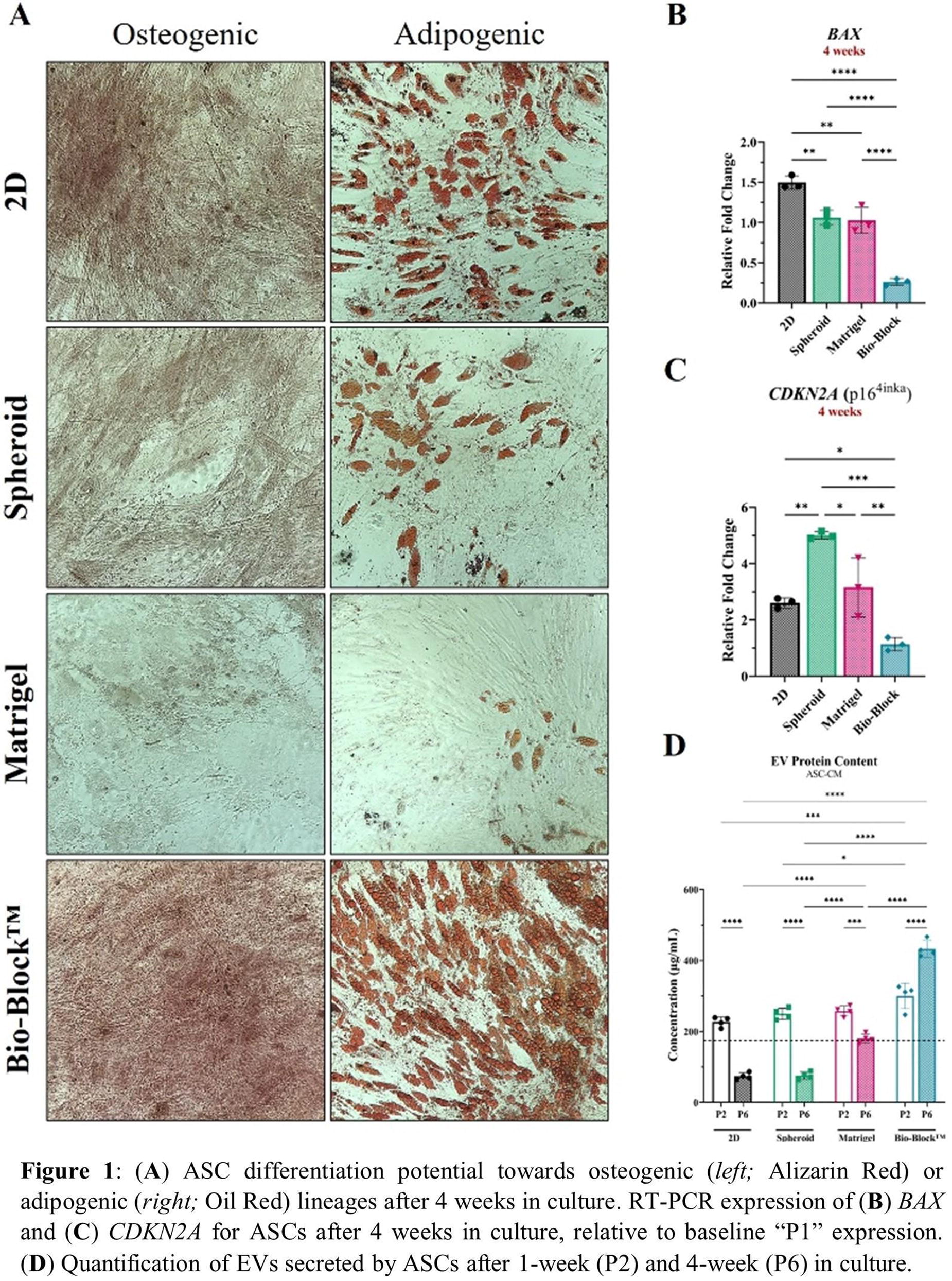
A342 - A Computational Approach In Developing A Novel Mesenchymal Stem Cells Based Therapeutic Cocktail Against Rheumatoid Arthritis
M. Tariq, N. Farid, D. Advani, N. Kohli
Khalifa University of Science and Technology, Abu Dhabi, United Arab Emirates
*Purpose/Objectives: Rheumatoid Arthritis (RA) is an autoimmune chronic inflammatory disease characterized by bones and cartilage damage with a Global prevalence of around 1%. Mesenchymal Stem Cells (MSCs) have been thoroughly investigated for RA, however, their clinical use is limited due to issues related to cell engraftment. It is now well established that it is not the cells themselves but the secreted factors of MSCs (termed the secretome), that are involved in tissue healing and regenerative response. Therefore, this study aimed to develop a novel therapeutic cocktail based on the proteins secreted by adipose tissue derived MSCs (ADMSCs) and their interactions with molecular signatures of RA.
*Methodology: We used Scopus and Pubmed to search the following keywords ("secretome" OR “exosome” OR “extracellular vesicle”) AND ("mesenchymal stem" OR “stromal cells”) AND ("Proteomic"). Only studies reporting proteome of the secretome of ADMSCs in musculoskeletal pathologies were selected. The common proteins in proteome of all included studies were extracted and their target genes in humans were identified using STITCH analysis. In addition, Genecard and Disgenet databases were employed to identify the therapeutic targets of RA. This step was followed by network construction using Strings and Cytoscape tools, to identify the interactions of the common proteins in the secretome with the therapeutic targets of RA. The online Database for annotation, visualization, and integrated discovery (DAVID) was employed to reveal RA related KEGG pathways associated with the target genes of the common proteins.
*Results: TIMP2, DAG1, CD248, STC2, and HTRA1 were the commonly reported proteins in all the studies. Genecard and Disgenet revealed 8514 therapeutic targets of RA, with 1571 present in both databases. STITCH analysis showed the involvement of the 5 identified common proteins with 58 direct targets, out of which 11 were also identified as targets of RA. KEGG pathway analysis, followed by Cytoscape annotation showed that these 5 proteins target the following RA related KEGG pathways such as; Rheumatoid Arthritis, Proteoglycans in cancer, and five signaling pathways, i.e., IL-17, NF-kappa B, Relaxin, HIF-1, and Estrogen signaling pathway (Figure 1). These pathways are involved in immune response and aberrant signaling processes. Therefore, the 5 identified proteins could potentially be used as a therapeutic cocktail to ameliorate RA by mitigating inflammation, modulating immune responses, and disrupting aberrant signaling processes.
*Conclusion/Significance: This study uses a computational approach of network pharmacology to identify possible interactions of proteins found in the ADMSC secretome, with molecular signatures of RA to potentially develop a therapeutic cocktail for treating RA.
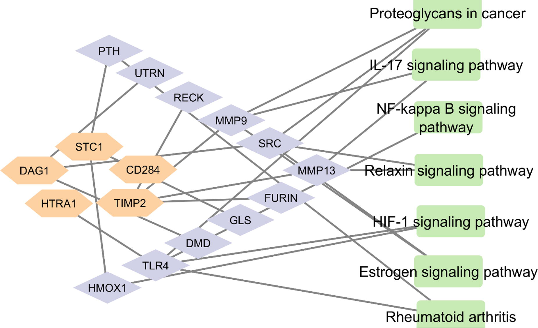
Figure 1: Pathways induced by proposed therapeutic cocktail from ADMSCs derived secretome. Orange nodes show proteins in cocktail, magenta nodes represent their direct protein targets and Green nodes are their RA associated KEGG pathways.
A347 - Proteomic Analysis Of Umbilical Cord-derived Mesenchymal Stem Cell Secretome For Clinical Applications
D. ADVANI, N. Kohli
Khalifa University, Abu Dhabi, United Arab Emirates
*Purpose/Objectives: Mesenchymal stem cells (MSCs) secretome based cell free therapies have been widely used for the treatment of various human diseases. Umbilical cord-derived MSCs (UC-MSCs) are the second leading source explored in various clinical studies, due to their accessibility and sustainable properties. However, the exact molecular and functional response of the secretome repertoire is still not fully understood. This study aimed to assess the functional biological role of the proteins present in UC-MSC secretome to better understand the use of MSC secretome in therapies.
*Methodology: We systematically searched the PubMed, Scopus and Embase databases until January, 2024 using the keywords related to UC-MSCs and secretome. Studies were selected and filtered based on the predefined inclusion and exclusion criteria. The secretome profile of the eligible studies was evaluated and analyzed by using ShinGO tool for functional enrichment and STRING/ClueGO tool for network analysis.
*Results: A total of 129 studies were examined of which four studies that met the eligibility criteria were finally reviewed. A total of 11589 proteins were extracted from the secretome of UC-MSCs. From Venn analysis, we identified 512 secretome proteins that were common in all four studies. Functional enrichment of these proteins revealed that most of the proteins were related to metabolic processes such as protein translation, protein transport, protein folding and amino acid metabolism, assisting cellular growth and proliferation. From network analysis, we pooled out the most significant proteins in the secretome network and identified their functional responses were related to ribosomal metabolism and protein translation processes.
*Conclusion/Significance: The proteins in the secretome of UC-MSCs were enriched in various metabolic processes related to protein synthesis and related metabolic processes highlighting their role in tissue development and regeneration. This study uses bioinformatic tools to warrant the role of UCMSC secretome for potential clinical applications and highlights the functional response of the proteome of the secretome of UCMSCs.
A354 - Cell-based Abiotic Materials For Immunomodulation And Tissue Regeneration
M. B. Oliveira, A. R. Sousa, A. F. Cunha, A. Santos Coquillat, J. F. Mano
University of Aveiro, Aveiro, Portugal
*Purpose/Objectives: The immunomodulatory and pro-regenerative properties of stem cells have been explored for a wide range of healthcare applications. Living cells are usually administered in the form of single cell-based suspensions, aggregates with sheet or spherical shape, or encapsulated in biomaterial matrices. Using such setups, living cells can often migrate, possibly to unwanted locations, and may acquire unpredictable phenotypic features. Therefore, the design of formulations based on abiotic cell-derived products with controlled shape and biological interaction with tissues may enable the processing of functional materials with improved safety and predictable therapeutic potential.
*Methodology: Structural shape- and size-controlled cell-based biomaterials were manufactured based on a method that promotes cell assembly and maturation using a biomaterial-free approach. Two different strategies that enable the preparation of stable biomaterials comprising distinct fractions of cells were developed. The methods were compatible with the preparation of biomaterials with spherical and pliable centimetric fiber shapes. Mesenchymal stem cells were used as a cell model with wide therapeutic potential for the treatment and management of a variety of diseases and conditions, including the ones involving the poor control of inflammatory processes. Living cells were pre-conditioned in vitro using cytokines (e.g., IFNγ), addressing the hypothesis that such short-term exposure would culminate in the tailoring of the therapeutic action of abiotic cell-derived products. The immunomodulatory activity of the different classes of materials were tested with different immune cells from the innate and adaptive systems, including activated T lymphocytes and macrophages in different states of polarization.
*Results: Abiotic cell-derived biomaterials were processed to preserve different fractions of cells and its derivatives, including the cell membrane and/and the extracellular matrix, while maintaining the original shape of the in vitro aggregated tissues. The processed living micro-to-macro tissues were pre-conditioned with pro-inflammatory cytokines, and prepared with rationally designed sizes and shapes. All formulations, regarless of their in vitro-programmed composition, promoted the adhesion and grow of seeded cells (see Figure). The analysis of the immunomodulatory properties of cell-based materials prepared with different in vitro biochemical pre-conditioning, structural properties, and cell-derived compositions showed that both chemical and physical features can be used to modulate the immunomodulatory response triggered by different materials. In particular, for monocytes/macrophages the response of one set of abiotic biomaterials was directly correlated with their ability to hamper the activation of the NFkB pathway after cell exposure to LPS. Interestingly, these materials showed significant activity used in small doses.
*Conclusion/Significance: Cell-derived biomaterials with phenotypic stability were prepared using different methodologies and are here suggested as new classes of materials for tissue regeneration and immunomodulation. Those rapidly fabricated materials are easily programmed in vitro to showcase specific immunomodulatory profiles, while keeping easy handeability, injectability, potency even at low doses, and overall proved safety. Such materials are expected to soon achieve utility in the treatment of inflammatory/immune diseases, and for tissue regeneration.
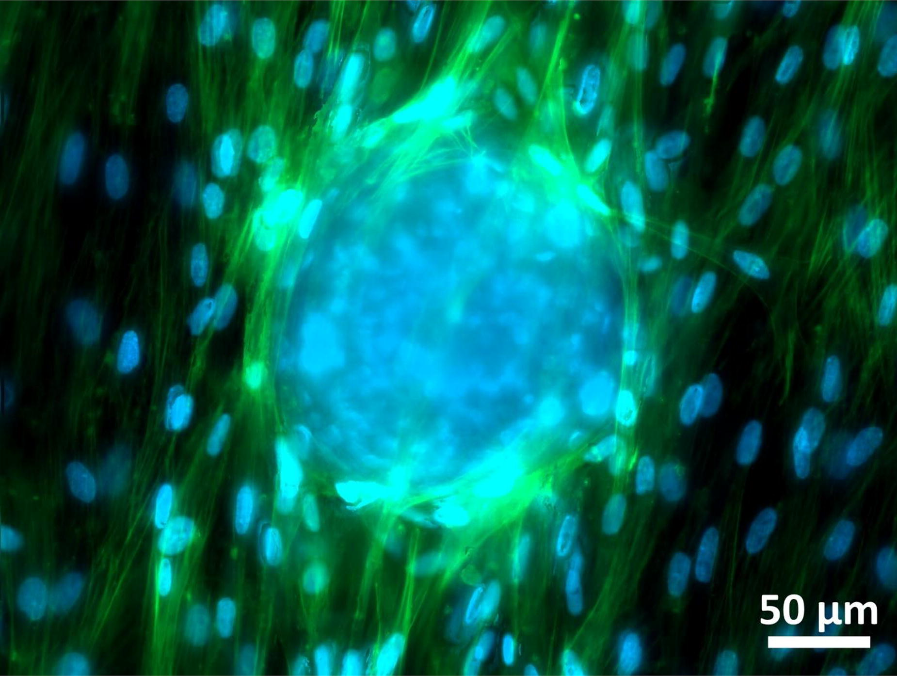
A355 - Comparative Evaluation Of Bone Marrow-derived Mesenchymal Stem Cells, Nerve Growth Factor, And Laminin In The Regeneration Of Acute Peripheral Nerves In Guinea Pig Model
M. A. Rafee1, A. Amarpal1, M. K M1, A. M. Pawde1, V. Chandra1, T. Sharma1,2
1
ICAR-Indian Veterinary Research Institute, Bareilly, India,
2
National Institute of Animal Biotechnology, Hyderabad, India
*Purpose/Objectives: Peripheral nerve affections are quite common in animals, where the nerves of the fore and hind limbs are affected most commonly. The sciatic nerve is the most frequently involved nerve. Sciatic nerve crush injury is a widely used experimental model in nerve regeneration studies of the peripheral nervous system. The mechanism of nerve healing by stem cell therapy is not fully understood. Sciatic nerve injury in guinea pigs seems to be a good experimental model for nerve injury to study different treatment modalities.
*Methodology: The potential of mesenchymal stem cells (MSCs) and nerve growth factor (NGF) with laminin gel for comprehensive healing of nerve injury in guinea pig sciatic nerve injury model was evaluated separately and in combination. The animals were evaluated and the groups were compared for functional recovery, histopathological changes in the sciatic nerve, ultrastructural changes by scanning electron microscopy (SEM), and early gene expression in response to injury and different treatments.
*Results: Significantly better results were obtained when BM-MSCswere combined with NGF and laminin than when given alone. Local application of culture-expanded BM-MSCs augments peripheral nerve regeneration in guinea pigs, which was further enhanced by the addition of laminin and NGF. Laminin gel could be used as a suitable vehicle for BM-MSC delivery at the lesion site to enhance peripheral nerve regeneration. A single injection of NGF at the site of injury was not an effective for peripheral nerve injury; however, the nerve healing induced by BM-MSCs in laminin gel could be accelerated further by the addition of NGF.
*Conclusion/Significance: Enhanced nerve regeneration in this study supports the fact that MSCs injected into an injured nerve can improve both functional and histological regeneration. The better functional recovery obtained with BM-MSCs combined with laminin and NGF could be due to better survival of stem cells and early signaling by NGF to differentiate into Schwann cells. The addition of NGF could have stimulated the differentiation of MSCs into Schwann cells. The laminin used with stem cells further improved the clinical parameters, indicating enhanced nerve repair. Application of MSCs in laminin over the nerve acts as a scaffold for sustained release of cells over the injured nerve. The study concludes that the local application of the culture-expanded BM-MSCs can augment peripheral nerve regeneration in guinea pigs, which can be further enhanced by the addition of NGF and laminin.
A356 - The Effect Of Hypoxic Conditions Oncharacteristics Of Human Nasal Turbinate Stem Cells
S. Hwang
Bucheon saint hospital, Bucheon, Korea, Republic of
*Purpose/Objectives: This study aims to elucidate the characteristics of mesenchymal stem cells (MSCs) derived from the human nasal inferior turbinate tissue (referred to as human nasal inferior turbinate-derived stem cells; hNTSCs) under the influence of hypoxic culture conditions. he investigation focuses on understanding how such conditions impact the characterization, proliferation, and differentiation potential of hNTSCs in comparison to their counterparts cultivated under standard oxygen saturation levels.
*Methodology: Following the isolation of hNTSCs from samples obtained from five patients, the collected specimens are divided into two distinct cohorts: a study group exposed to hypoxic cultivation and a control group maintained under typical normoxic conditions. The evaluation of MSC surface marker expression is performed through fluorescence-activated cell sorting (FACS) analysis. The quantification of cell proliferation dynamics is achieved using the cell counting kit-8 assay. Additionally, the study delves into the intricate landscape of cytokine secretion by employing multiplex immunoassays. The differentiation potential of hNTSCs into osteogenic, chondrogenic, and adipogenic lineages is explored through meticulous histological examination and comprehensive gene expression profiling
*Results: The hNTSCs subjected to hypoxic conditions exhibit an enduring manifestation of MSC characteristics, as corroborated by FACS analysis, showcasing a notable absence of hematopoietic markers coupled with robust expression of quintessential MSC markers. Intriguingly, the proliferation kinetics of hNTSCs remain remarkably unaltered within the hypoxic environment. Over the course of cultivation, a striking parallelism is observed between the cytokine expression profiles of the hypoxic and normoxic groups. Notably, while the potential for osteogenic and adipogenic differentiation in hNTSCs is moderately attenuated under hypoxic conditions, the chondrogenic potential remains relatively stable.
*Conclusion/Significance: In light of these findings, it can be conclusively established that exposure to hypoxic conditions exercises minimal influence over pivotal aspects of hNTSCs, such as expansion capabilities, multipotential characteristics, cytokine and chemokine secretion patterns, and underlying genetic makeup. This study provides a comprehensive perspective on the resilience and adaptability of hNTSCs under varying oxygenation contexts.
A357 - Cryopreservation Of Equine Monocyte Derived Dendritic Cells For Cancer Therapy
T. Deeb1,2, F. Schwebel3, B. Glasmacher1,2
1
Leibniz University Hanover, Garbsen, Germany,
2
Lower Saxony Center for Biomedical Engineering, Implant Research and Development, Hanover, Germany,
3
PetBioCell GmbH, Osterode am Harz, Germany
*Purpose/Objectives: Cellular treatments are gaining an increasing importance in many medical fields. Cancer therapy using dendritic cells is one of these treatments and is showing a promising potential to be an alternative to conventional therapies. Nevertheless, and as such as many cellular products the short life span and the on-shelf availability of dendritic cells is still a big obstacle. One solution is the cryopreservation of the cultivated cells. The objective of this study is the cryopreservation of equine monocyte derived dendritic cells.
*Methodology: By Using a freezing protocol with a cryoprotective agent consisting of 10% (v/v) DMSO and 90% (v/v) autologous serum, a cooling rate of 1 K/min in a controlled rate freezer, storing the samples for up to 12 weeks in liquid Nitrogen (LN2) and then thawing them in a water bath at 37 °C. Besides that, we examined the use of a higher cooling rate (2 K/min) and the use of the combination of passive cooling containers and mechanical freezers (-80 °C) for freezing and storing and lastly we examined varying the water bath temperature (50 °C and 65°C).
*Results: The established control protocol showed ca. 80% recovery rates. The use of passive cooling container and the storing using mechanical freezers at -80 °C did not have a significant effect on the recovery rate within the 12 weeks neither did the increase of the cooling rate. Increasing the thawing temperature from 37 °C in the control protocol to 50 °C and 65 °C enhanced the recovery rates significantly.
*Conclusion/Significance: We were able to establish a control protocol for the cryopreservation of EQ-Mo-DC and still working on optimizing it. We aim to exclude the DMSO from the cpa and revalidate the results.
A358 - Recombinant Human Annexin A5 Accelerates Diabetic Wounds Healing By Regulating Skin Inflammation And Epithelialization
B. Kang, W. Zhang
Shanghai Jiao Tong University School of Medicine, Shanghai, China
*Purpose/Objectives: Chronic wounds are a common complication of diabetes and remain a significant challenge in clinical practice. One of the key obstacles to the healing of diabetic wound is the persistence of active inflammation. We previously demonstrated the potential of cell-free fat extract (CEFFE) to promote the healing of diabetic wounds, and annexin A5 (A5) is a crucial anti-inflammatory protein within CEFFE. This study aimed to evaluate the therapeutic potential of A5 in diabetic wounds.
*Methodology: A5 was loaded into GelMA hydrogels and applied to skin wounds of diabetic mice in vivo. The wounds were observed for 14 days, with photographs taken on days 0, 3, 7, 10, and 14. On the 14th day, wound tissue samples were harvested and stained for histological assessment. Evaluations of inflammation regulation were conducted through anti-CD68 immunohistochemistry staining, anti-CD86, CD206 immunofluorescence staining, and qRT-PCR of wound tissue. In vitro, bone marrow-derived macrophages in presence of A5 and stimulated by LPS (lipopolysaccharide) in vitro, and detected through qRT-PCR, flow cytometry, and immunocytofluorescence staining. Besides, epithelial cells were co-cultured with A5for epithelialization regulation by CCK-8 assay and cell migration assay.
*Results: Through continuous observation and histological assessment, we confirmed that A5 could promote diabetic wound healing. Further analysis revealed that A5 treatment regulated inflammations by promoting the transition of macrophages from M1 to M2 phenotype, which contributed to tissue generation. In vitro experiments demonstrated that A5 exerted a significant effect on reducing pro-inflammatory factors, including IL-1β, IL-6, and TNF-α, and effectively inhibited the polarization of macrophages from the M0 phenotype toward the M1 phenotype. A5 significantly promoted the migration of epithelial cells, which enhances wound epithelization.
*Conclusion/Significance: This study provides compelling evidence that annexin A5 has a significant impact on the regulation of macrophage inflammation and promotion of epithelialization, leading to substantial acceleration of the healing of diabetic wounds.

A360 - Exploring Cellular Fitness Gradients In Adipose-derived Mesenchymal Stem Cells: An Analysis Of Individual And Pooled Settings
D. Kukaj, S. Niebert, C. Schüler, J. Burk-Luibl
Vetmeduni Vienna, Vienna, Austria
*Purpose/Objectives: Adipose-derived mesenchymal stem cells (MSCs) are increasingly recognized for their promising potential in regenerative medicine. Yet, the influence of individual cellular fitness on their functional outcomes remains a crucial area of investigation. This study systematically evaluates the impact of cellular fitness on adipose-derived MSCs, employing a range of assays and categorizing cells into distinct cellular fitness groups.
*Methodology: In the initial phase of the study, MSCs collected from nine human donors underwent the MTS assay to assess cell viability, the colony-forming unit (CFU) assay to measure clonogenic potential, and the population doubling time (PDT) calculation to evaluate proliferative capacity. Based on the results, cells were categorized into three cellular fitness groups: low, middle, and high. Building upon these classifications, the second phase involved the creation of cell pools. Three cell pools were formed, each comprising three donors from their respective low, middle, and high-fitness groups. This approach allowed for the examination of collective effects within each pool. Subsequently, experiments were carried out simultaneously for both individual donors and cell pools, by conducting an in-depth characterization that included cell function studies such as cell migration studies, cell senescence measurement, osteogenic differentiation potential of the cells, as well as the MTS assay and PDT calculation.
*Results: Statistical analyses, particularly one-way ANOVA, uncovered interesting findings. As expected, in the MTS assay, the high-fitness group demonstrated statistically significant increased cell viability compared to the low and middle groups. Accordingly, this group exhibited significantly shorter PDT compared to both other groups. Furthermore, the cell senescence markers showed statistically significant lower values in the high-fitness group compared to both other groups. However, osteogenic differentiation and cell migration studies through scratch assay did not show statistical significance across groups. Importantly, the results of the pooled cells corresponded well to the results of the individual donor cells within fitness groups.
*Conclusion/Significance: In conclusion, this comprehensive analysis provides valuable insights into the influence of cellular fitness on adipose-derived MSCs. The observed lower senescence rate in the high-fitness group suggests a favorable condition where cells within this group exhibit an extended duration before entering senescence. These results are indicative of potential superiority in cell health and functionality. Moreover, the cell pools were indeed representative of the individual donors within their respective cellular fitness groups. However, this may change when pooling cells from different fitness groups. Current investigations therefore include utilizing cell pools with diverse donor fitness levels, to explore how donor composition influences results, aiming to assess if the potency values would then still align with individual donor means. This study lays the groundwork for further exploration of the intricate interplay between cellular fitness and the therapeutic potential of adipose-derived MSCs.
A361
A362 - Matrix Vesicles From Hypertrophic Cartilage Microtissues As A Cell-free Therapy For Bone Regeneration
K. Man, A. J. Rosenberg, D. Gawlitta
UMC Utrecht, Utrecht, Netherlands
*Purpose/Objectives: Extracellular vesicles (EVs) are cell-derived lipid nanoparticles which have garnered growing attention as promising acellular tools for musculoskeletal repair, overcoming hurdles associated with the translation of cell therapies. It is thought that EVs are fundamentally involved in fetal bone development and secondary fracture repair, as extracellular matrix-bound vesicles are critical for endochondral ossification (EO). These matrix vesicles are key players in the development of the growth plate during endochondral bone formation. They are involved in the turnover of the extracellular matrix and its mineralization, as well as being a vehicle for chondrocyte communication and regulation. In this study, we aimed to manufacture EO-mimetic matrix vesicles as a cell-free approach to recapitulate endochondral bone formation.
*Methodology: Microtissues were created using human bone marrow-derived multipotent stromal cells (hBMSCs), which were then differentiated in chondrogenic medium with/without BMP2 for 21 days. Following this, constructs were either maintained in chondrogenic medium or switched to hypertrophic medium till day 28. Spheroids were prepared for biochemical analysis of glycosaminoglycans (GAGs) via DMMB assay and histological analysis. Matrix vesicles were isolated via the enzymatic digestion of cartilage spheroids, followed by differential ultracentrifugation. Isolated matrix vesicle size, morphology and concentration were assessed via nanoparticle tracking analysis (NTA), transmission electron microscopy (TEM), protein quantification and CD63 ELISA. The recruitment and osteogenic differentiation of hBMSCs treated with matrix vesicles was evaluated by biochemical and histological/immunohistochemical analysis. The angiogenic potential of matrix vesicles on human endothelial colony-forming cells (hECFCs) was assessed via tube formation analysis.
*Results: Our findings showed that incorporating BMP2 in chondrogenic medium substantially promoted the size of cartilage spheroids generated, in addition to upregulating the expression of cartilage markers (i.e. GAGs, collagen type II) confirmed via DMMB analysis, Safranin O histology and Collagen II immunostaining. The expression of chondrogenic markers was further improved in the spheroids cultured in hypertrophic medium. NTA and TEM analysis showed that isolated matrix vesicles exhibited a typical size distribution and morphology indicative of nano-sized vesicles (50 - 200 nm). EV protein quantification and CD63 ELISA demonstrated that BMP2 supplementation significantly increased the quantity of matrix vesicles produced when compared to chondrogenic culture alone, with hypertrophic conditioning further enhancing EV yield. Biochemical analysis confirmed all isolated matrix vesicles exhibited positive alkaline phosphatase (ALP) activity, with BMP2 and hypertrophic conditioning further promoting ALP activity. Matrix vesicles substantially enhanced the recruitment of hBMSCs through accelerated migration and proliferation. Importantly, nanoparticle treatment significantly increased hBMSCs osteogenic differentiation and extracellular matrix mineralisation demonstrated via enhanced ALP activity, collagen production and calcium deposition. Additionally, matrix vesicles substantially improved the angiogenic tube formation of hECFCs when compared to the EV-free control.
*Conclusion/Significance: Taken together, our findings demonstrate the induction of hypertrophy within the cartilage microtissues significantly promoted the yield of matrix vesicles produced, in addition to improving these nanoparticles’ osteogenic and angiogenic potency. Thus, these findings showcase the considerable potential of harnessing matrix vesicles derived from hypertrophic cartilage microtissues as a cell-free strategy to promote bone regeneration.
Poster Session II
Wednesday, June 26, 2024, 6:15 PM - 7:45 PM
B2 - Synergising MW And Gelma Functionalisation To Design Advanced Bioinks And Bioresins For Multi-material Bioprinting And To Target Specific Tissue Properties
D. Ribezzi1, J.-P. Zegwaart2,1, S. Florczak1, T. Van Gansbeke2, A. Tejo-Otero3, J. Olijve2, R. Levato1,4
1
Department of Orthopaedics, University Medical Center Utrecht, Utrecht University, Utrecht, Netherlands,
2
Rousselot BV, Expertise Center, Gent, Belgium,
3
BIOMAT Research Group, University of the Basque Country (UPV/EHU), Escuela de Ingeniería de Gipuzkoa, Donostia-San Sebastián, Spain,
4
Department of Clinical Sciences, Faculty of Veterinary Medicine, Utrecht University, Utrecht, Netherlands
*Purpose/Objectives: Gelatin-based hydrogels are widely used in the biomedical and tissue engineering fields, due to their biocompatibility, cell-interactivity and biodegradability. However, batch-to-batch variation is still a hindrance to properties standardization and protocols reproducibility. Besides consistent molar mass (MM) and degree of modification (DoM) it is important to consider polymer, salt and photo-initiator (PI) concentrations and temperature control during hydrogel processing. The work presented herein focusses on the interplay between polymer concentration, MM and DoM of the GelMA specifically, in relation to their suitability as bioinks or bioresins for different multi-material bioprinting approaches, and to optimize cell culture environments to target the mechanical profile of soft tissues, like the native-pancreatic one.
*Methodology: Starting from GelMAs that have molar masses of 90 and 160kDa, with various degrees of modification (40, 60 or 80%), we generated a library of mechanical properties for the different GelMA hydrogels formulations. Photo-rheology curves for the two MM with different degrees of modification were performed at different polymer concentrations (20%w/v, 15%w/v, 10%w/v and 5%w/v) and at different temperature (4°C, 21°C and 37°C), to further investigate the effect of temperature on the mechanical properties and crosslinking kinetics. We therefore synergized the tunability of the GelMAs formulation’s properties and the potential of volumetric bioprinting (VBP) in its combination with suspended bath extrusion printing, termed Embedded extrusion Volumetric printing (EmVP), to build anisotropic multi-material and multi-cellular gelatin-based constructs. Finally, via tuning of the GelMA properties via molecular weight and degree of functionalization modulation, we optimized a 3D cell culture environment for a pancreatic cell line (iβ-cell line, engineered to release insulin upon blue light exposure).
*Results: The collected data showed how increasing the DoM, the storage modulus of the GelMA hydrogels increases too. Similarly, increasing GelMA concentrations, the storage modulus of the hydrogel will increase. For the 90kDa GelMAs a near perfect power-correlation was established. The 160kDa GelMAs showed a linear type of correlation. Moreover, with increasing concentrations of PBS the hydrogel stiffness decreases, and lowest crosslinking temperatures leads to faster rates of crosslinking. 90kDa gelatins could be used as optically transparent fluid bulk support baths for embedded extrusion, showing self-healing like properties at room temperature and without any mechanical damage caused by the movment of an immersed nozzle. We managed to extrude features of 80 µm and generated complex structures obtained via EmVP and via multi-material sequential VBP of different cell types. Finally, we targeted the mechanics of the native endocrine pancreas microenvironment by leveraging 90p60 GelMA bioresins at 5%w/v concentration. Data showed how different MM affected the metabolic activity and ability to proliferate and form islet-like clusters of iβ-cell line.
*Conclusion/Significance: In conclusion, a 95% difference in GelMA hydrogels mechanics can depend on the temperature in the preparatory phase, the concentrations and types of PI, the MW, the DoM, and concentration. The interplay of these factors will induce dissimilar cellular responses, as hydrogel final properties are affected by the polymer characteristics and the printing setup.

B3 - Glycation Promotes Oral Biofilm Formation On Microfabricated Bioengineered Dentin Substrates
S. Álvarez1, J. Morales1, A. Teuschl-Woller2, C. Schuh3, S. Aguayo1
1
UC Chile, Santiago, Chile,
2
FH Technikum Wien, Vienna, Austria,
3
Universidad del Desarrollo, Santiago, Chile
*Purpose/Objectives: Dentin is a highly organized mineralized dental tissue that has a complex microarchitecture that consists of micrometer-sized dentinal tubules within a type-I collagen matrix. As it is the most abundant dental tissue, once it becomes exposed (i.e., such as root surfaces in older individuals) it is an important substrate for the adhesion of microbial colonizers that can lead to the development of dental caries. Therefore, it is important to characterize biofilm formation on aged dentin surfaces in order to develop novel strategies to inhibit this process with the goal of preventing root caries development and progression in the elderly. However, limitations such as lack of clinical specimens, biological variability, and ethical concerns restrict the availability of dentin for in vitro experiments. Likewise, there is a lack of current effective biomimetic in-vitro dentin models that facilitate the study of oral microbial adhesion by considering the surface architecture at the micro- and nanoscales. Thus, the objective of this study was to develop a novel in-vitro microfabricated biomimetic dentin surface that simulates the complex surface microarchitecture of exposed aged dentin, in order to explore biofilm formation of caries-relevant oral microbes associated to dentinal glycation.
*Methodology: Soft lithography microfabrication and biomaterial science approaches were used to construct a PDMS substrate with 4um micropits that were further functionalized with mineralized type-I collagen. Subsequently, substrates were glycated with methylglyoxal (MGO) to simulate dentin aging, and age-associated modifications were analyzed utilizing atomic force microscopy (AFM), elemental analysis, and electron microscopy. Finally, dual-species biofilms of the caries-relevant microbes Streptococcus mutans and Candida albicans were grown with and without artificial saliva coating and characterized on the biofunctionalized PDMS microchips by using confocal scanning laser microscopy (CLSM) and epifluorescence microscopy. Changes in biofilm formation between the non-glycated and glycated analogous dentin substrates were quantified via relative fluorescent units (RFU).
*Results: By employing a combination of interdisciplinary approaches, a mineralized type-I collagen matrix in a micropitted arrangement was generated, that showed similar mechanical properties to native dentin when probed with AFM. The microstructure of the dentinal matrix was also seen to represent the morphology of dentin specimens, and the mineral phase was dominated by the clinically relevant hydroxyapatite. Finally, glycation with MGO led to an increase in the Young’s modulus of the constructs, as well as to an increase in the formation of C. albicans - S. mutans biofilms on the surface, both with and without previous artificial saliva coating.
*Conclusion/Significance: Here we have described our initial attempts at biofabricating an in-vitro dentin model with morphologic and nanomechanical characteristics that simulate aged dentin for the formation of oral biofilm. Also, glycation was shown to increase biofilm formation on aged in-vitro dentin, which could have implications in root caries prevention in the elderly. Overall, we believe that these novel biomimetic dentin constructs can serve as an in-vitro platform to study oral biofilm formation (or biomaterial bonding) without the need for animal or human tooth specimens in the future.
Acknowledgements: ANID FONDECYT #1220804, and ANID FONDEQUIP #EQY230010 and #EMQ210101.
B4 - Biomimetic Molecular Nanotraps From Silk
D. Maniglio1, A. Bossi2
1
University of Trento, Trento, Italy,
2
University of Verona, Verona, Italy
*Purpose/Objectives: Silk fibroin gained attention as a natural biomaterial thanks to its outstanding biocompatibility, processability into varieties of shapes, possibility to tune its mechanical, biological and optical properties, and biodegradability. Fibroin can be easily functionalized, adding methacrylate reactive side-groups (SilMA), which undergo a light mediated radical activation enabling chemical crosslinking. Hence, SilMA further expands the fibroin applications towards the bio-fabrication of scaffolds for tissue engineering, or as component of bioinks for 3D bioprinting. SilMA’s peculiar features make it ideal for the exploration of new exotic applications such as molecular imprinting. MIPs are functional materials with stamped binding cavities possessing high affinity and selectivity for their target. NanoMIPs are currently synthesized starting from Acrylamide-monomers, yielding to nanomaterials with high affinity and selectivity for the target molecule, rivaling with antibodies in applications, such as assays, sensing, and imaging. Although polyacrylamide-nanoMIPs demonstrated non-toxicity in vitro, safety concerns might rise for the possibility of specific organ accumulation and for the lack of degradation in vivo. NanoMIPs composed of natural and biocompatible materials would revolutionize the current scenario, opening to their translatability to clinical settings. On these premises we developed an original protocol using SilMA as a starting material to prepare biocompatible and biodegradable protein molecularly imprinted nanoparticles, named bioMIPs.
*Methodology: BioMIPs were synthesized using SilMA concentrations of 0.3% or 0.03% w/v in 10 mM Acetate Tris pH 3.5, or 5 mM Sodium Phosphate pH 7.0, in the presence of 3 nmol human serum albumin (HAS). LAP photoinitiator was added at the final concentration of 0.2% or 0.04% v/w and photo-polymerized (λ = 365 nm; 10 W, 600 s). Rhodamine-tagged bioMIPs were prepared in same manner, adding 12 nmol of acryloxyethyl-thiocarbamoyl-rhodamineB. Fibroin microfibers were recovered from raw cocoons after sericin removal, while electrospun nanofibers were fabricated starting from a fibroin solution in formic acid, obtaining a non-woven tissue with average fiber dimension of 300 ± 75 nm. Fluorescent BioMIPs binding to micro and nanofibers was made using EDC/NHS chemistry.
*Results: Photo-crosslinking of SilMA (0.03 and 0.3%) in the presence of albumin yielded to imprinted bioMIPs with a Zaverage of 52.8±0.1 and 94.6±1.3 nm, respectively. The specificity of the binding of alb-bioMIPs for its template was investigated using a fluorescein labelled albumin. Moreover, affinity and selectivity were studied using isothermal titration calorimetry. BioMIPs suspended in PB buffer and titrated with increasing quantities of HSA or of non-template proteins (human transferrin). Calorimetric interaction data resulted in a dissociation constant KD = 57.0±2.7 10-9 M, moreover the synthetic process permits to stamp a quasi-single binding site per bioMIP particle. BioMIPs proved selective for their template. Finally, we integrated rhodamine-tagged bioMIPs to the micro and nanofibers to embed albumin recognition to the fabrics. The decoration and the effective albumin recognition by the bioMIP-functionalized fibers was confirmed by imaging.
*Conclusion/Significance: BioMIPs demonstrated selective and specific binding for their target together with non-toxicity, thus embodying the concept of tailor made functional biomimetics. The example of all-silk bioMIPs-fiber superstructures indicates the possibility to add specific extra-functionalities to biomaterials, with foreseen impact on TERM.
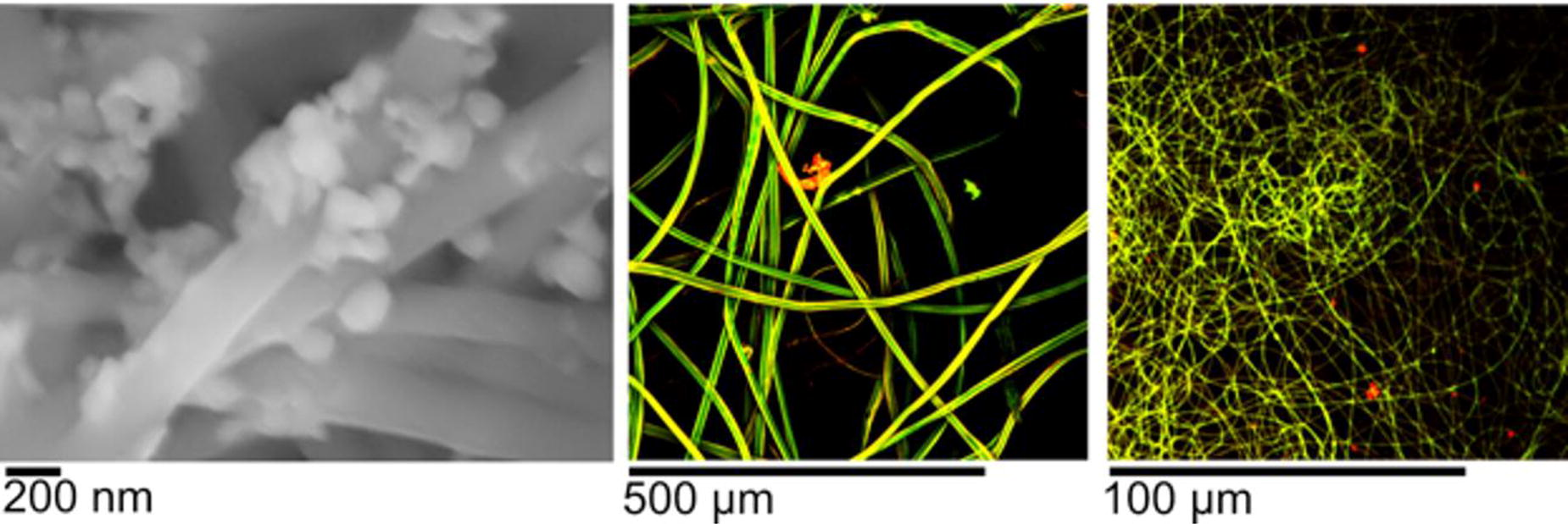
B5 - Light-based Enzymatic Bioprinting With Unmodified Native Fibrin
J. P. Sampson, D. Rütsche, E. Marani, Z. Ji, A. Langat, A. Archer, J. Du, S. G. Boxer, M. A. Skylar-Scott
Stanford, Stanford, CA
*Purpose/Objectives: Light-based printing achieves excellent spatial resolution and scale, making it a fitting approach for generating multiscale architectures such as vasculature, which spans from capillaries (order 10μm) to great vessels (order 1cm). While natural unmodified ECM scaffolds are to date unmatched for generating functional engineered tissues—for example demonstrating the highest burst pressure tissue engineered vascular grafts and most mature cardiac microtissues—they cannot currently be printed using light. We aimed to unite the fabrication advantages of light-based printing with the material advantages of native ECM.
*Methodology: From the literature reporting azobenzene-based light control of thrombin, the Razo & Itelo (Tian et al. 2016) system was tested and selected. In this system (see figure 1A), near-UV (365nm) exposure slows thrombin activity, while blue exposure returns thrombin to full activity. Using light scattering by fibrin as a measurement of coagulation progress, the concentration of each system component (fibrinogen, thrombin, Itelo, Razo, buffer, potassium) was optimized for printing, with trade-offs including dynamic range of thrombin activity, print speed, and cell viability. A custom, multi-wavelength stereolithography printer and fluidic print chambers were constructed.
*Results: Single layer prints of native fibrin were characterized. Importantly, the natural fibrillar, microporous, degradable structure was maintained (figure 1F), inviting cell spreading by HNDFs by 24 hours (figure 1C). Arbitrary print patterns could be created (figure 1E), and cell viability from the total printing process was high (figure 1B).
*Conclusion/Significance: Light-based printing of natural, unmodified fibrin gives ability to construct arbitrary patterns at high resolution in wholly biological, microporous fibrin gel. This natural fibrin gel gives cells the ability to remodel their local environment and interface with neighboring cells—important, for example, for neighboring cardiomyocytes or heart muscle organoids to join to form a cohesive muscle tissue. Future work will be aimed to create multi-layer prints for constructing relevant three-dimensional structures (e.g. multiscale vascular networks). This printing process combines the fabrication advantages of light-based printing with the material advantages of native ECM.

B20
B21 - Towards An Immunogenic Bioprinted Skin Model For Psoriasis
A. Ulloa Fernández1,2, L. Kleissl3,4, M. Markovic1,2, G. Stary3,4, A. Ovsianikov1,2, J. Fernández-Pérez1
1
TU Wien, Wien, Austria,
2
Austrian Cluster for Tissue Regeneration (https://www.tissue-regeneration.at), Wien, Austria,
3
Medical University of Vienna, Wien, Austria,
4
CeMM Research Center for Molecular Medicine of the Austrian Academy of Sciences, Wien, Austria
*Purpose/Objectives: Psoriasis is a common chronic inflammatory skin disease. Immune cells affect epidermal keratinocytes, resulting in cytokine-induced keratinocyte overgrowth. Genetic and environmental factors, as well as gut and cutaneous microbiome changes, also promote disease progression. The treatment for many patients consists of drugs that control the lesions until a new lesion develops. Achieving a long-term remission remains a significant challenge. To understand the pathomechanisms of psoriasis, several preclinical mouse models that resemble the characteristics of the disease have been developed, from spontaneous gene mutations to transgenic and knockout models that provide insight into the pathogenic role of cytokines, cells, and their synergistic effects. Nonetheless, increasing interest has been focused on developing physiologically relevant human-derived in vitro models that could substitute animal models. The emergence of engineered skin constructs for psoriasis based on tissue engineering and 3D printing strategies could increase the complexity and accuracy of the in vitro models. Incorporating various immune cell subsets into these models could allow us to identify their contribution to the disease’s pathomechanism and might lead to more effective treatments. This work aims to use extrusion-based bioprinting techniques to create a psoriasis model. By incorporating Th17 immune cells, the primary immune cell type involved in psoriasis physiopathology, and comparing them with an interleukin-induced model, we achieved a more physiological setup to mimic and study disease progression.
*Methodology: Skin equivalents were prepared using several biomaterials, including GelMA-based inks (BIO INX®) and nanofibrillated cellulose-based material GrowDex®-T (GD-T, UPM Biomedicals), to encapsulate immortalized human skin fibroblasts HDF/TERT164 (Evercyte). The scaffolds were bioprinted using an extrusion-based bioprinter (BioXTM, Cellink). After seeding immortalized human keratinocytes NHEK/SVTERT 3-5 (Evercyte) on top, the scaffolds were cultured in an air-liquid interface phase for seven days to induce epidermal differentiation.
*Results: Unlike GrowDex-T scaffolds, scaffolds generated with GelMA-based bio-inks expressed epidermal differentiation markers like cytokeratin 10 and filaggrin. Additionally, when skin equivalents were treated with IL-17, we observed an increase in the epidermal thickness and the number of Ki67+ cells compared to their non-treated counterparts. Furthermore, a protocol to activate Th17 cells has been tested, obtaining more than 30% of IL-17-producing cells. Adding Th17 cells will enable us to achieve a psoriatic-mimicking immunocompetent skin model. Different strategies to incorporate these cells into the constructs will be tested to create a more relevant physiological model.
*Conclusion/Significance: This research provides insights for creating a 3D bioprinted immunocompetent psoriasis skin model. It allows us to develop and enhance clinical strategies for psoriasis treatments and better understand disease-driving factors and their roles in disease development.
B23
B24 - Human Bone Organoids With Osteoclastic Activity: Emulating Fracture Phenotypes In An In Vitro Model Of Bone Remodeling
C. Steffi1, A. de Leeuw1, G. N. Schädli1, X.-H. Qin1, M. Rüger1,2, R. Müller1
1
ETH Zürich, Zürich, Switzerland,
2
Department of Pediatric Orthopaedics and Traumatology, University Children’s Hospital, Zürich, Switzerland
*Purpose/Objectives: Bone metabolism is a dynamic process characterised by continuous remodeling cycles. Imbalances in these cycles can lead to conditions like osteoporosis, where increased bone resorption often leads to fractures. Current in vivo rodent models used for drug screening, targeting osteoblasts, osteocytes, and osteoclasts, do not fully replicate human bone physiology. To address this gap, we developed an in-depth human bone organoid model that not only encompasses the key cell types involved in bone remodeling but also specifically focuses on osteoclastic activity and its role in inducing fracture-like phenotypes. This model aims to provide a more accurate representation of human bone behaviour in vitro, potentially reducing reliance on animal experimentation and better mirroring the fracture risks associated with osteoporotic conditions.
*Methodology: Our bone organoids mimic both organic and inorganic matrices of osseous tissue. The composition of our bioink, a combination of collagen-analogous gelatin, alginate for enhanced mechanical integrity, and graphene oxide for augmented printing fidelity, integrates with human mesenchymal stem cells (hMSCs) for three-dimensional bioprinting. A pre-culture period of four weeks, under cyclic mechanical compression, facilitates osteogenic differentiation within these mesenchymal stem cells. The introduction of osteoclast precursors, CD14+ monocytes (hMn), and hMSCs in specific ratios on day 27 of the culture, followed by continued culture under mechanical stress for four weeks, enabled the replication of bone resorption dynamics. The hMn and hMSC were introduced in the ratios of 0.5:1 and 1:1. The non-reseeded group was treated as a control (0:0). Organoid mineral density (OMD), total mineral volume (TMV) and mineral turnover rate were monitored using non-invasive time-lapsed micro-CT scans. Time-lapsed organoid stiffness was monitored non-invasively with customised mechanical stimulation unit.
*Results: From week 5 to week 8, we observed notable changes in the bone organoids with osteoclasts. The decrease in OMD and TMV became more pronounced by week 8 (Figure 1A,1B). OMD in experimental groups 0.5:1 and 1:1 was 124 ± 8.7% and 123.7 ± 3.8%, respectively, compared to 142.5 ± 9.5% in the control group. Similarly, TMV was observed to be 152.0 ± 12.3% for 0.5:1 and 201.2 ± 44.8% for 1:1, versus 242.6 ± 22.2% in the control. Furthermore, the mineral turnover rate was decreased with the addition of osteoclasts (Figure 1C) and the stiffness of the organoids (Figure 1D). The fracture phenotype observed in the organoids with the osteoclasts (Figure 1E) was likely due to reduced mineral density and stiffness.
*Conclusion/Significance: Our study demonstrates the significant impact of osteoclasts on mineral composition in bone organoids, with TMV and OMD notably lower in groups with osteoclasts, a trend that intensified over time. The model effectively simulates bone resorption and fidelity loss, mimicking fracture phenotypes through reduced mineral density and stiffness. This in vitro approach, utilising human cells, closely emulates the fracture risk associated with osteoclastic activity in human bone conditions like osteoporosis. It offers a promising alternative to animal models, more accurately replicating human bone pathophysiology and highlighting the potential for minimising animal use while acknowledging the limitations of in vitro systems compared to in vivo models’ complexity.
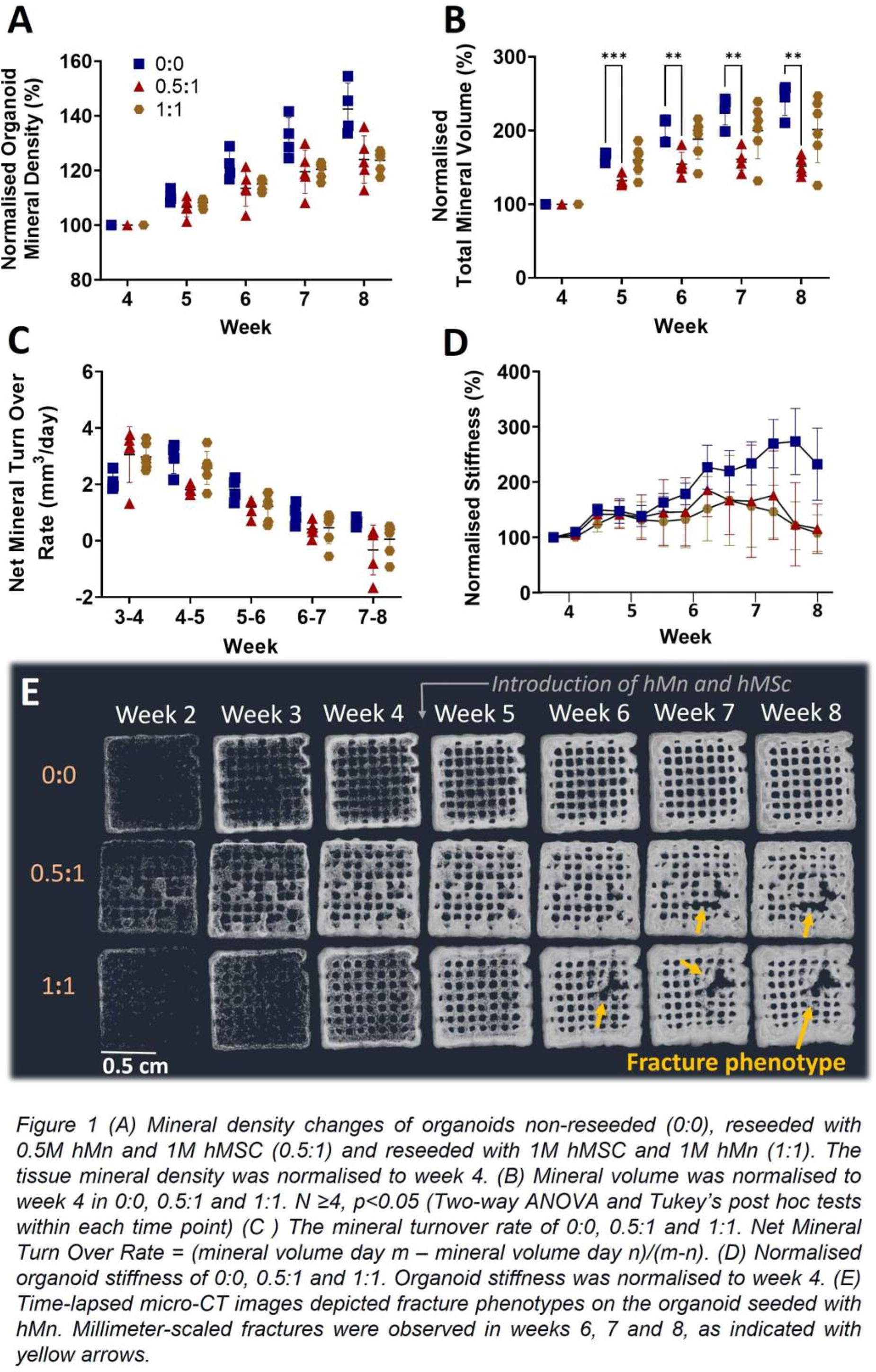
B25 - Ninja Spheroids: Highly Translucent Spheroids Suitable For Optical Observation
H. Kazuki, K. Nobuhiko
yokohama city university, Yokohama, Japan
*Purpose/Objectives: Multicellular spheroids are often used in three-dimensional cell culture due to their highly differentiated functions. The enhanced physiological properties of spheroids are utilized in microphysiological systems (MPS). Experiments using MPS are limited by the volume of medium that can be sampled. This limitation makes it difficult to perform traditional assays that require a certain volume of sample, such as lactate dehydrogenase assays. In addition, traditional methods need several steps, for example, sample application, stirring, and measurement. Optical observation will solve these problems. However, multicellular spheroids are not suitable for such examination because they are thick and do not allow light to pass through. If we could visualize the inside of spheroids, it would provide valuable information for evaluating toxicity. The goal of this study is to form translucent spheroids, i.e. NINJA spheroids, consisting of cells and alginate hydrogel beads.
*Methodology: We used a highly differentiated human hepatocellular carcinoma cell line, HuH-7. Non-beads spheroids, representing normal spheroids without beads, were created by dispensing 0.5 µl of cell suspension (5000 cells/0.5 µl) into a 3% methyl cellulose (MC) medium as we reported in our previous report [1]. Beads spheroids, meaning spheroids including beads, were created by dispensing 0.5 µl of cell-bead suspension (5000 cells: 5000 beads/0.5 µl) into a 3%MC medium [2]. The beads were made of an aqueous solution of sodium alginate, approximately 20 µm in diameter. After culturing each spheroid in MC medium for 24 h the MC medium was recovered. Both types of spheroids were transferred to a 96-well U-bottom plate for easy focusing, and images were captured using a phase-contrast microscope. The obtained images were processed using an image analysis software, Fiji, to generate brightness value histograms.
*Results: The brightness distribution was revealed through image processing. The analysis results are as follows: non-beads spheroids: Max brightness 155, min 11, mode 39, median 64.738, std dev 27.791; bead spheroids: Max brightness 254, min 30, mode 73, median 98.916, std dev 41.627. The median, maximum value, variance, and mode of brightness were higher in beads-included spheroids. The incorporation of beads improved the translucent of the spheroid. The increase in internal light transmission expanded the range of brightness values, suggesting the potential for providing more information.
*Conclusion/Significance: Incorporating beads into the spheroids has allowed light to penetrate deeper into the spheroids. This enables the acquisition of internal information from the spheroids. Currently, we are attempting to establish toxicity tests using image-based assays with NINJA spheroids.
B29 - Vitamin C Regulates Bone Functionality And Tissue Regeneration
M. Curtis, Y. Kim, S. Alimperti
Georgetown University, Washington, DC
*Purpose/Objectives: Bone fractures caused by injuries due to aging require reconstruction to restore tissue function. Bone homeostasis declines due to aging as the tissue undergoes changes in its extracellular matrix (ECM) composition, impacting the bone cells that contribute to mineralization and tissue function. One vital component for maintaining bone homeostasis is a sustained and adequate dietary intake of micronutrients including Vitamin C. Severe Vitamin C deficiency leads to scurvy, a serious disease characterized by skeletal fragility and bone fracture. Herein, we aim to examine the role of Vitamin C in bone functionality and regeneration in normal and aged conditions, such as osteoporosis.
*Methodology: To understand the role of Vitamin C in bone function, we developed a 3D printed micro-physiological system recapitulating the 3D composition/structure of bone ECM. Specifically, a bioink consisting of hydroxyapatite (HA) [ 1, 2.5, and 5 % w/v] dissolved in Ascorbic Acid (AA) at concentration 5, 15, 25% w/v was mixed with collagen I (3mg/ml) and fibronectin (5μg/ml), was extruded via 210um nozzle. Human primary osteoblasts (HOBs) at density 0.1 million/ml were embedded in the ECM. The mechanical properties of the 3D printed structure were measured by SEM, FTIR, and rheology. Finally, the biocompatibility and osteoconductive properties of the 3D printed construct was by caspase-3 staining and via Von Kossa (VK) staining, respectively. In addition, transcription levels of early and late osteogenic markers such as runt-related transcription factor 2 (RUNX2) and osteopontin (OPN), were evaluated by Real-time quantitative polymerase chain reaction (RT-qPCR).
*Results: The concluding results highlight the most optimal condition for engineering bone ECM is 3mg/mL COL with 1-5% HA w/v and 9 umol/g AA. Specifically, SEM highlights how the high [HA] in matrix enhances matrix stiffness and adherence to COL fibers as they polymerize and shows how increasing the pH before adding COL will lead to the formation of more COL bundles. AFM of 1% HA samples revealed the stiffness of dry bone ECM samples E=0.74kPa at a pH of 3, and E=0.49kPa at pH of 6. Consistent with these conclusions, rheometer measurements yielded the highest and most consistent storage/loss modulus ratios for the 9 umol/g AA at a pH of 3 pre-COL, which are the same parameters as found in natural tissues. Lastly, FTIR graphs confirmed the presence of HA in samples with the observed peak at ∼960/cm. With the concluding compositions, we generated 3D-bioprintable bone ECM scaffolds containing HOB cells and implanted in murine models for biocompatibility, tissue regeneration, and bone integration enhancement after bone defects, projecting the incorporation of AA into the scaffolds not just supports better mechanical properties, but also integration and regeneration of host tissues with the scaffold.
*Conclusion/Significance: Our 3D printed micro physiological systems permit us to investigate the role of Vitamin C in bone microenvironment that cells are sensing and measure the dynamic changes of bone regeneration in vitro, avoiding animal studies. Finally, these platforms will enable us to identify new molecular mechanisms in aging bone diseases and develop new bioengineered tools for bone regeneration and host integration.
B31
B32 - Coaxial 3d Bioprinting Of Hydrogels With Small Diameters To Mimic Microvasculature
B. Uysal, S. Madihally
Oklahoma State University, Stillwater, OK
*Purpose/Objectives: Coaxial bioprinting is explored to print tubular structures, which is especially useful for printing organs with vasculature so that they could be similar to how the organs operate within the human body. However, one issue is that due to the varying sizes of blood vessels, not everything is easily replicable, especially at reduced diameters. Most commercially available coaxial nozzles allow printing tubular structures as low as 200 µm, which is still much higher than the lowest limits of blood vessels.
*Methodology: In this work, we have custom built a nozzle that could provide hollow tubular structures with inner diameters less than 100 μm. In continuation to previous work from our group [1], we can achieve vasculature using a chitosan-gelatin thermosensitive hydrogel in combination with poloxamer.
*Results: Using much simpler procedure of varying the inner and the outer flow rates, we can vary the tube diameters.
*Conclusion/Significance: Cell culture work demonstrates feasibility of printing cell-laden structures and scaling to include multiple cell types.
[1] Roehm K, Madihally SV. Bioprinted chitosan-gelatin thermosensitive hydrogels using an inexpensive 3D printer. Biofabrication, 10(1), 015002, 2017
B41 - Comparative Studies On Antibacterial Activities Of Chitosan, Silver Nanoparticles And Maggot Based Chitosan-silver Nanocomposites Against Fish Pathogens
J. A. Olugbojo1, A. a. Akanji2, O. S. Olubodun2, E. o. Dare2
1
Bells University Of technology, Ota, Nigeria,
2
Federal university of Agriculture, abeokuta, Abeokuta, Nigeria
*Purpose/Objectives: Abstract The present study explores the ex- situ bio-fabrication of chitosan-silver nanocomposites, maggot-based chitosan and silver nanoparticles. These studies were conducted based on the increasing demand on the use of nanomaterials as alternative antimicrobial agents against fish pathogens, which are eco-friendly, biocompatible and non-hazardous.
*Methodology: Silver nanoparticles were prepared using Cassia fistula leaf extract as bio-reductant, chitosan nanoparticles were prepared from maggot chitin, while Chitosan-Silver nanocomposites were synthesized through ex-situ bio-fabrication technique involving chitosan and silver nanoparticles to form polymer composites. The Integrity of the formed nanoparticles and composites (Nanohybrids) was confirmed through UV-Visible Spectrophotometry, Fourier Transform Infrared (FTIR) spectroscopy, X-ray Diffraction (XRD) analysis, Scanning electron microscopy, and energy dispersive X-ray analysis. Antibacterial sensitivity test was carried out on Aeromonas schubertii, Aeromonas hydrophila, Vibrio parahaemolyticus, Klebsiella aerogene and proteus mirabilis strains using agar well diffusion technique. Data collected were subjected to analysis of Variance and means were separated using Duncan multiple range test.
*Results: The average particles size for AgNP, Chitosan and CS-AgNC are 24.94 nm, 30.95 nm and 65.03 nm respectively as observed through Field Emission Scanning Electron Microscope (FESEM). FESEM micrograph also shows that Silver nanoparticles were embedded in the chitosan matrix to form the polymer composites. Silver nanoprticles shows both hexagonal and spherical shape while chitosan revealed a cuboidal shape. The in-vitro antibacterial activities showed that there was significant difference (P<.0.05) in the zone of inhibition when tested the nanomaterials on different species of bacterial fish pathogens (Aeromonas schubertii, Aeromonas hydrophila, Vibrio parahaemolyticus, Klebsiella aerogene and proteus mirabilis strains) across the column, except between CS-AgNC and Chitosan, showing no significant difference (P>.0.05) on V. parahaemolyticus (16.00b±1.15 and 15.50b±1.73) and P. mirabilis (19.00b±2.31 and 19.00b±1.15).
*Conclusion/Significance: Consequently, this finding showed that Chitosan-silver nanocomposites were the most potent nanomaterial against the five test organisms with the highest inhibition zones (15.00 mm, 17.50 mm, 16.00 mm, 22.50 mm, and 19.00 mm) followed by chitosan (12.50 mm, 13.00 mm, 15.50 mm, 15.25 mm, and 19.00 mm) and then AgNPs (8.00 mm, 8.50 mm, 12.50 mm, 13.00 mm, and 13.00 mm). This study shows that nanoparticles can be an effective alternative antibacterial against fish pathogen especially where antibiotics fail. Also, while each of the tested nanoparticles produced good results, CS-AgNP was found to be most effective and highly potent against the tested fish pathogens than AgNP and Chitosan.
B43 - Microfluidic Printheads For Multi-Material Extrusion-Based Bioprinting
F. Hofmann1, J. Faber2, S. Budday2, T. Jüngst1
1
University of Würzburg, Würzburg, Germany,
2
Friedrich-Alexander-University of Erlangen-Nürnberg, Erlangen, Germany
*Purpose/Objectives: One aim of the biofabrication research area is the automated generation of functional tissue models with a hierarchical arrangement of cells and materials. These models can be used to replace animal experiments in pharmaceutical and cancer research, but also for regenerative therapies. As most human tissues contain cellular, structural, mechanical or biochemical gradients, mimicking these in a well-defined and reproducible manner, can lead to advanced models. A possible approach to generate gradients using automated processes, like 3D-bioprinting, and to achieve a gradual transition of material, microfluidic printheads can be applied. However, their use in extrusion-based bioprinting is limited by the viscoelastic properties of bioinks and requires customized mixers. The microfluidic printheads developed in this study are used to generate defined mixing ratios for bioinks. The viability of cells after extrusion and the quality of the deposited structures are analyzed.
*Methodology: The printheads were made using digital light processing (DLP), a resin-based additive manufacturing technique. To assess the print quality, the mixers were cut in half, imaged and compared to the CAD-data (figure 1). Simulations were used to estimate the mixing potential and compared to deposited patterns of dyed solutions with variable viscosities. Images were captured and evaluated using FIJI. Materials with different stiffnesses and various cells types were printed, including U87 (human glioblastoma cells) and NIH3T3 (mouse fibroblast cells). Live-dead staining was used to analyze cell damage through mixing and evaluate the cell viability.
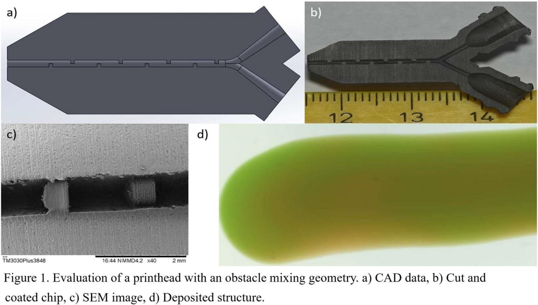
*Results: Constructs with five different mixing structures, including obstacles, sinusoidal, thread and split and recombine channel geometries could be printed with diameters as small as 300 µm. The comparison of the CAD-data and the microfluidic chips revealed an increasing shrinkage of 5% to 25% with decreasing channel diameter. Straight channels had a higher permeability and lower mixing ratios than curved sections, with decreasing channel diameter. The simulations showed, that the obstacle geometry had the best mixing ratio and the sinusoidal structure the worst. This was confirmed by deposited patterns of colored solutions. The mixed area was evaluated for a desired mixing ratio. The obstacle mixing unit achieved a mixing ratio of 70%, which is similar to commercial printheads, whereas a fully homogeneous mixed pattern would give a result of 100%. The cell viability of the self-designed mixers was analyzed after printing and compared to commercial printheads for U87 and NIH3T3 cells. The sinusoidal structure showed the highest, the obstacle geometry a lower and the commercial mixers the worst rate of living cells. This confirmed the first hypothesis, that a higher mixing rate leads to higher shear stress for cells and thus to a lower cell viability rate.
*Conclusion/Significance: This study shows, that DLP printing is suitable to generate perfusable mixing units. Their ability to mix inks with different viscosities depends on the used geometry. Compared to commercial mixers, a similar mixing ratio can be achieved, but the self-designed microfluidic printheads show a higher cell viability. Future experiments will implement mounting the printheads to an extrusion-based 3D-bioprinter, to generate different types of gradients including cells.
B46 - All-aqueous Inspired Fluidic Scaffolds For Biomaterials
H. Shum
The University of Hong Kong, Hong Kong, Hong Kong
*Purpose/Objectives: Design tunable, biocompatible, complex hierarchical structures for facilitating culture of cells for sophisticated functional tissue, organoid, organ-on-chip applications
*Methodology: We take an all-aqueous approach to form completely aqueous liquid-liquid interfaces, and combine microfluidic and printing techniques to design hierarchical customizable hydrogel structures with embedded channels of desired shapes and morphology. The methodology is based on our group’s unique expertise and track records in all-aqueous microfluidics, and aqueous phase separation.
*Results: The resulting fluidic structures enabled by integration of aqueous phase separation, microfluidic control and printing are completely bio- and cyto-compatible as substrates for cells, with potential towards growing of large organoids and organ-on-chips, as confirmed by our cell data. In addition, different fluidic contents can be exchanged across the fluidic channels bounded by our custom-developed membrane walls. The fluidic structures can also be spatiotemporally functionalized, for instance, with different enzymes, on demands, enhancing their versatility and adaptability to different applications.
*Conclusion/Significance: Our works hold great potential as a demonstration of the combined synergies between different disciplines, ranging from fluidics, physico-chemistry, cell culture, tissue engineering and others. The ability to tailor large networks of fluidic channels with spatially controllable functional and surface properties creates new opportunities to culture cells of different types for applications whereas precise control of cells and fluids are required.
B49 - Development Of Fat And Muscle Bioinks On The Basis Of Gellan Gum And Plant Proteins For Cultivated Meat Hybrid Products
J. O. Wollschlaeger, S. Heine, P. J. Kluger
Reutlingen University, Reutlingen, Germany
*Purpose/Objectives: Global meat production is experiencing a surge in response to the escalating demand for meat consumption driven by a rapidly growing world population. Cultivated or in vitro meat presents an opportunity to produce meat through tissue engineering, eliminating the necessity of raising entire animals. This innovative approach involves cultivating cells in vitro using stem cells derived from a small animal biopsy. To replicate a product closely resembling conventional whole meat cuts, the utilization of not only cells but also a supporting matrix is imperative. These biomaterials must possess qualities such as edibility, sustainability, wide availability, animal-free composition, non-toxicity, cost-effectiveness, and processability. Cutting-edge 3D printing techniques, along with suitable bioinks, can be employed to create a conducive 3D environment for muscle and fat cell development to form a hybrid Cultivated meat product.
*Methodology: In the evaluation of different co-culture systems, a murine myoblast cell line (C2C12) and primary isolated bovine adipose-derived stem cells (bASCs) were employed. Initially, a co-culture medium was developed to enable the differentiation of both cell types into myotubes (C2C12) or preadipocytes (bASCs) within a 2D culture. Confirmation of differentiation was achieved through myosin antibody staining for muscle cells and BODIPY staining for fat cells. Subsequently, the cells were incorporated into spheroids through suspension cultures, with the aim of potential scaling up in bioreactors. Three 3D printing approaches were tested to obtain different hybrid products. Scenarios 1 and 2 involved the printing of undifferentiated or differentiated bASC and C2C12 spheroids with 1% (w/v) gellan gum using an extrusion-based 3D bioprinter, with viability and differentiation characteristics checked over 14 days (undifferentiated) or 1 day (differentiated). In Scenario 3, a more complex CAD model with plant protein inks as the shell and a differentiated bASC spheroid containing gellan bioink as the inner part was designed and tested for co-printability, cell viability, fat characteristics, and cookability.
*Results: Notably, both C2C12 and bASCs spheroid cultures remained viable and continued differentiation for 14 days. In the first scenario, undifferentiated spheroids of C2C12 and bASCs were co-printed within a 1% gellan gum bioink and subsequently differentiated. In the second scenario, the spheroids of the two cell types were separately differentiated before being printed together. Both scenarios demonstrated the viability of printing live spheroids and achieving constructs consisting of differentiated muscle and fat cells. In the third scenario, printing with two inks, a protein plant ink encasing the gellan ink containing differentiated bASC spheroids, was successful, with the cells maintaining viability and fat characteristics. The co-printed CAD model was then fried, exhibiting an expected weight loss but retaining form stability.
*Conclusion/Significance: All three scenarios showcased distinct approaches to creating a hybrid product for cultivated meat applications. The ability to co-differentiate fat and muscle in the same constructs represents a groundbreaking step in the field of Cultivated Meat, as does the utilization of spheroids as a crucial building block and a means of scaling up the entire process. However, integrating all aspects remains a challenge in achieving a fully developed hybrid Cultivated meat product.
B50 - Establishment Of Primary Culture Method Of Bovine Skeletal Muscle Cells And Investigation Of Culture Temperature Conditions For Development Of Cultured Meat
A. Yoshida, H. Takahashi, T. Shimizu
Tokyo Women’s Medical University, Tokyo, Japan
*Purpose/Objectives: Alternative technology for meat production holds the potential to alleviate ethical, environmental, and public health concerns associated with conventional meat production. For the future of the food industry, cultured meat promises to become a viable alternative to meat harvested from livestock. This new strategy gained widespread attention after the Mark Post group produced the first cultured meat prototype. Muscle tissue engineering has proven to be a key technology in this innovative system of meat production. However, to produce cultured meat, skeletal muscle cells must be efficiently harvested and propagated. Therefore, we decided to establish an original primary culture method to efficiently collect skeletal muscle cells from bovine meat. Furthermore, considering that the body temperature of bovine organisms differs from that of humans, which is around 39°C, we investigated the optimization of culture temperature for expansion of the bovine muscle cells.
*Methodology: In this study, primary cultures of bovine skeletal muscle cells were grown by enzymatic treatment of bovine cheek meat collected from living animals with pronase, mechanical treatment by pipetting using a female pipette, and iMatrix coating on culture dishes. The cells obtained by this method were passaged repeatedly to confirm their ability to proliferate and differentiate into myotubes. In addition, the bovine skeletal muscle cells were cultured at a temperature of 37°C or 39°C during primary and 1st passage culture to determine the effects of culture temperature conditions on proliferation and differentiation abilities. Furthermore, bovine skeletal muscle tissues were created by culturing the collected bovine skeletal muscle cells with fibrin gel. To confirm maturation level of the muscle tissues, we measured the contractile force of the muscle tissues and analyzed the microstructures in the tissues using immunofluorescence staining.
*Results: Using our primary culture method, we succeeded in collection of highly purified bovine skeletal muscle cells from bovine meat. It was confirmed that the collected bovine muscle cells had the proliferation and differentiation abilities at the same level until the 7th passage. In examining temperature conditions, the cell proliferation rate was higher at 39°C than at 37°C. On the other hand, there was no significant difference in the proliferation ability after subculture. This indicates that our bovine muscle cells can be cultured at 37°C for large-scale expansion in future works. Furthermore, when we created bovine skeletal muscle tissue using our tissue engineering technology, the bovine muscle tissues produced muscle contractions by electrical stimulation. Immunofluorescence staining showed the formation of sarcomeres and native-like membrane structures was in the bovine skeletal muscle tissue.
*Conclusion/Significance: This study demonstrated that our primary culture method at 37°C was effective to collect large number of bovine muscle cells with maintaining differentiation ability. Furthermore, the bovine muscle cells had the ability to mature into contractile muscle tissues. These results indicate that our bovine muscle cells will be a useful cell source for sustainable cultured meat production in the future.
B51 - Food Industry By-products And Additives As Possible Serum-alternatives In Cultivated Meat
T. H. Baldeweg, S. Heine, A. Leikeim, P. J. Kluger
Reutlingen University, Reutlingen, Germany
*Purpose/Objectives: As one of its main goals, cultivated meat wants to improve animal welfare by reducing the need for conventional animal farming and slaughter. However, many cell culture methods are still dependent on the use of animal derived products. Most prominent example is fetal calf serum (FCS), which involves the slaughter of both, mother cow and unborn calf. Consequently, serum reduction or replacement in cell culture for cultivated meat is crucial. A possible way to achieve this is to use inexpensive and sustainable supplements that are already food safety approved and to use waste or by-products of the food industry.
*Methodology: To evaluate this potential, different commonly available by-products of the food industry and widely used food additives were tested on the C2C12 cell line. The effects of these differently prepared supplements on the cells proliferation, metabolism and viability in serum-reduced media was assessed by measurement of the metabolic rate using resazurin conversion and by determination of the cell count over time. Additionally, the myogenic potential of C2C12 cells after proliferation under these serum-reduced conditions was studied by differentiating the myoblast into myotubes and subsequent immunofluorescence staining of myosin.
*Results: Intriguingly, several potential serum replacements enabled partial serum reduction without affecting cellular viability. Furthermore, differently processed by-products of the food industry that promoted cellular growth under serum-free conditions were identified. Screening of different basal media allowed for maximization of this effect. In addition, no morphological changes were observable after complete serum withdrawal. Subsequent differentiation of the myoblast showed the formation of myotubes and expression of myosin after 14 days in our standard differentiation medium.
*Conclusion/Significance: In conclusion, we were able to show that materials known from the food industry can be a promising way to reduce or replace FCS in the production of muscle tissue for cultivated meat. In particular, by-products of different foods can allow for a more sustainable and cost-effective production of cultivated meat by harnessing the potential of otherwise discarded substances.
B53 - Expansion Of Bovine Muscle Cells With Maintaining Differentiation Ability In 3D Culture System For Production Of Tissue-engineered Meat
H. Takahashi, R.-i. Tanaka, A. Yoshida, T. Shimizu
Tokyo Women’s Medical University, Tokyo, Japan
*Purpose/Objectives: Alternative technology for meat production holds the potential to alleviate ethical, environmental, and public health concerns associated with conventional meat production. For the future of the food industry, cultured meat promises to become a viable alternative to meat harvested from livestock. To produce high-quality cultured meat, techniques for expansion of muscle cells harvested from livestock animals is required. 3D suspension culture system will be one of the most appropriate methods for large-scale cell expansion. In this study, we have developed a 3D suspension culture method to efficiently expand the bovine muscle cells with maintaining their differentiation ability, which is required for production of biomimetic culture meat “tissue-engineered meat”.
*Methodology: Bovine muscle cells are not as commercially available as rodent and human cells. Therefore, we first developed a primary culture method to efficiently harvest muscle cells from bovine meat. This primary culture method allowed us to efficiently collect large numbers of primary cells from bovine meat and purify the muscle cells from the cell mixture. The muscle cells were seeded onto polystyrene microcarriers and cultured in a 3D bioreactor. In this study, a commercially available edible basal medium (IMEM) supplemented with 10% bovine serum (BS) was used as a cost-effective and food-grade culture medium. Bovine muscle cells were suspended at as density of 4 × 10⁴ cells/mL and microcarriers were added at 10 mg/mL to the medium in a 30 mL stirred bioreactor. After several days, the cells were harvested from the microcarriers and reseeded to new carriers for passaging. To collect large numbers of muscle cells, the passaging procedure was repeatedly performed for 24 days. In addition, by culture of the passage cells in 2% horse serum-containing medium for differentiation induction, we determined the differentiation ability of the bovine muscle cells into myotubes after the repeated passages.
*Results: The bovine muscle cells cultured on microcarriers continuously proliferated during the repeated passages, and the cumulative proliferation rate was approximately 1.5 × 10⁵ times. In addition, the cells cultured in the 3D culture system showed differentiation ability into myotubes at the same level even after several passages. It is known that myogenic cells gradually lose the differentiation ability in in-vitro culture process. On the other hand, our muscle cells have the proliferation and differentiation abilities in the large-scale cell expansion, required to produce tissue-engineered meat.
*Conclusion/Significance: In this study, bovine muscle cells continuously proliferated with maintaining the differentiation ability into myotubes. In muscle tissue engineering, the differentiation ability is necessary to mimic the bundle structure of myofibers in native muscle. We demonstrated that our bovine cells have the potential to become cell sources for cultured meat having biomimetic structures, which is expected that the new type of meat has all the same characteristics including taste, aroma, texture, and nutrients as traditional meat. Although currently this cultured meat technology is still limited, our expansion technique of bovine muscle cells is important for production of cultured meat.
B55 - Cold Plasma-based Redox Therapy For Breast Cancer Bone Metastasis Tumor Growth Control
L. M. Bouret1, J.-B. Billeau1, M. Weber2, D. Rosenzweig2, S. Reuter1
1
Polytechnique Montreal, Montreal, QC, Canada,
2
McGill University, Montreal, QC, Canada
*Purpose/Objectives: Bone, especially the spine, is a common site of metastasis for breast, lung and prostate cancers. These tumors pose a significant challenge, demanding aggressive treatments like chemotherapy and invasive surgery. To fully remove metastatic lesions, surgical procedures need to extend onto healthy tissue which reduces the probability of remaining malignant cells. Such surgery often involves the removal of healthy tissue, necessitating reconstruction and carrying a risk of infection. Cold plasma therapy, operating at temperatures below 40°C, offers a non-invasive solution by delivering reactive oxygen and nitrogen species (RONS) locally. While research shows promising results, the reaction mechanism between plasma and tissues, and proper treatment dosage and reactive species composition to reach the right effects are still topic of current research. The purpose of this project is to develop and characterize a cold plasma source and investigate its potential in mitigating bone cancer metastasis, hypothesizing its anti-tumor properties. The overall objective is to create a tissue-plasma platform for cold plasma therapy, aiming to control the metastatic spread of breast cancer cells to bone tissue.
*Methodology: We have developed a platform combining tailored plasma reactivity through a kHz coaxial dielectric barrier discharge source and a highly reproducible bioprinted circular bone tissue model. The bone tissue model was bioprinted (Cellink BioX) using cell-laden hydrogel made of 1% alginate and 7% of gelatin (A1G7). Thus, a multi-well plate was generated with identical “breast-to-bone” metastasis as a coculture model of MDA-MB-231 and human bone marrow mesenchymal stem cells (hbmMSCs). Liquid RONS are measured by UV-VIS colorimetry, achieving selectivity through a combination of scavengers, colorimetric probes, and distinguishing against RONS from the gas phase. For each set of plasma treatment at different parameters (i.e. energy, gas composition, distance and time of treatment), metabolic activity through Alamar blue assays at day 1, 2 and 3 and live/dead measurements are used to detail the biological response of the tumor cells.
*Results: Results have shown that A1G7 cell-laden hydrogel was bioprinted with reproducible results in a model of cocultured MDA-MB-231 breast cancer cells and hbmMSCs. Dose responses of plasma on cancer cells and healthy cells were assessed in 2D and 3D cultures. Furthermore, plasma showed a selective antitumoral effect on MDA-MB-231 cancer cells over hbmMSCs healthy cells in both 2D and 3D cultures. Colorimetric assays have also confirmed that long-lived species (H2O2 and NO2-) can be tailored through the energy, the distance, the duration of treatment and the composition of the atmosphere around the plasma.
*Conclusion/Significance: In essence, our innovative platform allows us to create unique biological chemistry, observing its impact on cancerous tissue for groundbreaking redox-based treatment. Using a bioprinted model ensures reproducibility and precise control, enabling detailed studies of tumor migration. With a tailored plasma jet, our platform is crucial for exploring novel therapeutic approaches using exogenous reactive species. This aims to develop a personalized, non-invasive treatment avenue to reduce metastasis growth in bone cancer, with the potential to facilitate the technology’s integration into clinical applications.

B56
B58 - Characterization Of The Bioink Containing Decellularized Tissue Powder Mixed With Cancer Spheroids
M. Kobayashi1, K. Tsutahara1, R. Kawabata1, Z. Ng1, Y. Hashimoto2, T. Kimura2, A. Kishida2, M. Yamamoto1
1
Tohoku University, Sendai, Japan,
2
Tokyo Medical and Dental University, Tokyo, Japan
*Purpose/Objectives: Bioinks are one of the essential components in 3D bioprinting, which should possess sufficient physical properties for micro-patterning and a suitable environment for cells. Among the currently available bioink, the recently introduced decellularized extracellular matrix hydrogel bioink (d-ECM bioink) has gained attention. However, its poor printability and weak mechanical properties remain limited to fabricating complex constructs. Further, pepsin-digestion which is needed for solubilizing d-ECM for preparing bioink, results in the denaturing of the bioactive components within ECM. Therefore, in this research, d-ECM powder which previously showed good in-vivo performance, was mixed with cancer cells, and used for bioink. The bioink which consists of cancer spheroids with d-ECM powders was used to explore the factors that contribute to the construction of the in vivo-like cancer microenvironment.
*Methodology: Decellularized small intestinal submucosa was prepared with high hydrostatic pressure decellularization methods. Those tissues were then lyophilized and ground into powder. 50 µL of HepG2 cells (2.0 × 10⁴ cells/mL ) were cultured with those powders (0.5 µg/well). HepG2 spheroids with tissue powder were evaluated by hematoxylin-eosin (H-E) staining and scanning electron microscope (SEM) observation. Prepared spheroids were embedded in collagen gel to prepare bioink and examined for suitable printability and mechanical properties for bioink.
*Results: Various sizes of decellularized tissue powder were prepared and a sieve was used to equalize the size of the powder. Around 50 µm of tissue powders were collected and cultured with HepG2 cells. After 5 days of incubation of the spheroids with powders, observation with a phase contrast microscope confirmed the presence of d-ECM powders in the spheroids. The size of the spheroids to which d-ECM powder was added tended to be larger than those to which it was not added. SEM and H-E staining also showed that the cells and powders were homogeneously distributed. Results of the viscoelasticity measurement, the addition of spheroids with d-ECM powders into collagen gel makes the gel stiff. The behavior of spheroids after printing as bioink will also be presented in the presentation.
*Conclusion/Significance: The cancer spheroids with d-ECM tissue powder were prepared and the addition of cancer spheroids with d-ECM powder enhanced the mechanical properties of the bioinks. The findings obtained through the investigation of the 3D bioprinting method of d-ECM powder mixed cancer spheroids are expected to lead to the realization of the design of a 3D cancer microenvironment in vitro.
B59 - Metastatic Breast-Bone Carcinoma Model Obtained Through 3D Bioprinting And Tissue Engineering
S. Pontara1, M. Mastrogiacomo2, C. Gentili1
1
Department of Experimental Medicine (DIMES), University of Genoa, Genoa, ITALY,
2
Departiment of Internal Medicine and Medical Specialties (DIMI), University of Genoa, Genoa, ITALY
*Purpose/Objectives: Bone metastases are a debilitating complication of breast cancer. One of the main limitations of the in vitro models currently used is the inability to perfectly mimic physiological conditions. In this scenario, 3D bioprinting technology has revolutionized the field of tissue engineering due to its excellent potential in reproducing the geometry and complexity of constructs. This project aims to develop a dynamic cell culture system, using a fluid bioreactor, a sophisticated tissue engineering and 3D bioprinting approach, to recreate a human bone tissue and human breast cancer model in vitro, which will be connected in order to mimic a metastatic environment.
*Methodology: An alginate-bioink enriched with laminin combined with MDA-MB-231, a metastatic breast cancer cell line, had been printed to obtain repeatable scaffold of human breast cancer tissue. The model is cultured for 3 weeks to evaluate cell proliferation. For the bone tissue we have developed two different models. The first consists of seeding hMSCs on beta-tricalcium-phosphate (bTCP) aggregated with TissueCol. The second model use 3D bioprinting to create a scaffold using an alginate-based ink enriched with tricalcium-phosphate and hydroxyapatite, combined with hMSCs. Both bone models are cultured for 8 weeks in an osteogenic medium.Cell viability is evaluated through MTT, Live/Dead assays. The differentiation process is assessed using histological techniques (DAPI, Hematoxylin and Eosin, Mallory Trichrome, and specific collagen type staining) and Real-time PCR.
*Results: The MTT and Live and Dead assays shows a thriving population in all the models (exponential growth and vitality >90% at different timepoint). Regarding the breast cancer tissue, histological testing shows that MDA-MB-231 tends to migrate to the periphery of the scaffold and form colonies due to the increased presence of nutrients and to the metastatic nature of the cells. As regards bone tissue, for both models histological analysis demonstrates the formation of a tissue-like structure, characterized by organized cellular networks and extracellular matrix production. Real-time PCR data further supports successful osteogenic differentiation, with the regulated expression of osteoblastic markers. The difference between the two models compared is that in the first the composition is simpler to obtain, but the resulting construct present great variability, in the second the model allows a reproducible shape and the differentiation is more marked with a high deposition of collagen.
*Conclusion/Significance: In conclusion, while the bTCP model offers a more conventional but effective approach, the bioprinted model allows to develop a precise 3D construct with high reproducibility. To improve the breast model we are introducing human decellularized breast tissue in the ink in order to better mimic the microenvironment, meanwhile to improve the bone model we are introducing the vascularization because it is a fundamental element for the establishment of an ossicle. We are investigating to promote vasculature in two ways: a coculture in vitro between the bone model and the HUVEC cells using a bioreactor or alternatively the culture of our bone construct in ovo. We are also conducting preliminary tests with a laminar flow bioreactor to simulate the cell migration and mimic the metastatic environment.
B60 - Evaluation Of Three Hydrogel Systems As Substrates For Breast Cancer Cells
E. Capuana, F. Lopresti, C. Di Marco, M. Gaggianesi, C. D’Accardo, G. Porcelli, A. Turdo, M. Todaro, G. Stassi, V. La Carrubba
University of Palermo, Palermo, Italy
*Purpose/Objectives: Breast cancer is still a leading cause of cancer-related deaths among women worldwide with tissue engineering showing promising potential for therapeutic approaches [1]. This study examines the suitability of three different hydrogel systems obtained from the same formulation as substrates for breast cancer cells 3D cultures.
*Methodology: The research analyzed three hydrogels based on a 1.25% w/V alginate solution: two cell-free systems and one cell-laden (Fig. 1A). The first cell-free system was obtained by injecting the mixture of alginate and water into a mold, followed by ionic cross-linking. The second cell-free scaffold is the freeze-dried matrix obtained from the first structure. The cell-laden hydrogel resulted from mixing a 2.5% w/V alginate solution in autoclaved PBS with a suspension of cells in a 1:1 ratio. CaCl2 aqueous solutions at 1% or 3% w/V were used to crosslink the systems [2]. The lyophilized structures were characterized by scanning electron microscopy (SEM) and swelling analysis. Rheological and mechanical evaluations were performed on the cell-free hydrogels to investigate any changes caused by the autoclave process. 5x10⁵ HCC1937 breast cancer cells transduced with an RFP lentiviral vector were cultured on each scaffold for 8 days, and their ability to attach and grow was evaluated by using a fluorescence microscope.
*Results: Rheological tests showed a reduction in the complex viscosity of the polymeric solution (not cross-linked) due to the autoclave process. In contrast, the viscosity of the crosslinked gel remained unchanged. Swelling tests revealed that the swelling ratio reached a plateau after only one hour, suggesting a rapid water absorption of the freeze-dried matrix. SEM analysis of cell-free lyophilized structures showed a superficial skin layer (Fig. 1Bi) but an internally porous structure with stratified pores (Fig. 1Bii) [3]. Mechanical compression tests indicated that the compressive stress-strain curves did not change following autoclaving, with gels showing unaltered Young’s moduli (≈12 kPa). Surprisingly, biological tests showed that the injection molded gel had maximum proliferation after 3 days (Fig. 1Ci) but hindered fluorescence visualization of cells (Fig. 1Cii), suggesting a potential shielding effect of the structure [4]. The lyophilized scaffold was stable for at least 8 days of culture, promoting the formation of cell aggregates despite the absence of proliferative behavior (Fig. 1Ciii, iv). The cell-laden hydrogel showed intermediate proliferative behavior (Fig. 1Cv, vi), with minor problems in post-analysis handling because of its consistency.
*Conclusion/Significance: Overall, this study provides preliminary information on the critical role of the specific processing of hydrogel as an environmental response of breast cancer cells. Further in vitro studies will be used to select the optimal system for 3D drug testing. This discovery will have significant implications for developing innovative breast cancer therapies and refining the design of tumor engineering scaffolds.
Acknowledgments: The research was supported by the Italian Ministry of Health in the frame of the project “PJ MIN SALUTE METODI SOSTITUTIVI 2022”, by Complementary Operational Programme (POC) 2014/2020 of the Sicily Region, and by “SiciliAn MicronanOTecH Research And Innovation CEnter ”SAMOTHRACE" (MUR, PNRR-M4C2, ECS_00000022).
References: [1]doi.org/10.1021/ACSAMI.2C00920; [2]doi.org/10.1088/1758-5090/ac6c4c; [3]doi.org/10.1007/s10616-015-9895-4; [4]doi.org/10.3389/fbioe.2017.00013.
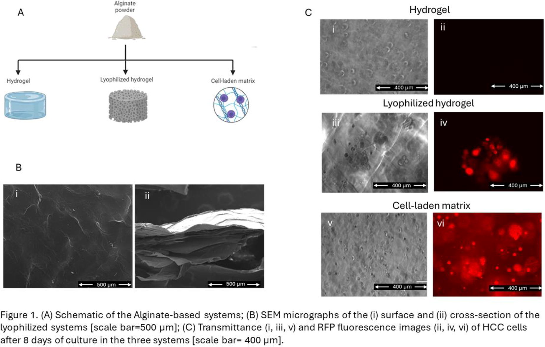
B61 - Key Interactions Between Tumour Cells And Mesenchymal Stem Cells In The Osteosarcoma: Exploring In Vitro Co-culture Models In 2d And 3d Scaffold-based Environments
G. Bassi1,2, A. Rossi1,3, M. Saqawa1,3, E. Campodoni1, M. Sandri1, D. Cochonneau4, D. Heymann4,5,6, S. Fulle2, M. Montesi1
1
National Research Council of Italy (CNR), Faenza, Italy,
2
University of G. d'Annunzio, Chieti, Italy,
3
University of Messina, Messina, Italy,
4
Institut de Cancérologie de l’Ouest, Nantes, France,
5
University of Sheffield, Medical School, Sheffield S10 2RX, United Kingdom,
6
Nantes Université, Nantes 44322, France
*Purpose/Objectives: Osteosarcoma (OS) is a primary malignant bone tumour with a 5-year survival rate of 70% for localized tumour. However, long-term efficacy remains unsatisfactory. It is suggested that Mesenchymal Stem Cells (MSCs) may be the origin of OS cells, resulting from malignant transformation induced by various factors such as mutations and the tumour microenvironment (TME). MSCs play a role in OS progression as, under the influence of OS cells and TME, normal MSCs are recruited to tumour lesions and “educated” into tumour-associated fibroblasts (TAFs), encouraging disease progression. Despite previous investigations, many questions remain about the real impact of MSCs on OS. While various studies have examined the MSCs role in OS using two-dimensional (2D) in vitro cell cultures, a more comprehensive investigation of OS cell-MSC interactions in three-dimensional (3D) in vitro cell cultures with a relevant TME-like matrix remains unexplored. Here, OS cells were co-cultured with MSCs in vitro on 3D hybrid hydroxyapatite-based scaffolds (MgHA/Coll) and investigated.
*Methodology: In this study, GFP-MNNG HOS OS cells were co-cultured with MSCs at 1:1 ratio on MgHA/Coll scaffolds for a total of 50.000 cells/scaffold and compared to scaffold-free and mono-culture conditions. The MgHA/Coll scaffold was selected for its ability to resemble the biochemical composition and the biomineralization process of bone extracellular matrix (ECM). The scaffolds, already numerously published, were designed, synthesized and charaterized by the ISSMC, CNR by a well-established protocol. After 7 and 14 days, the cell morphology and colonization of the scaffold (Actin/DAPI staining and SEM), MMPs/TIMPs release (zymography and ELISA kits) and gene expression (qRT-PCR) were investigated.
*Results: OS cells co-cultured with MSCs exhibited favourable cell morphology and a huge scaffold colonization. The results suggested a potential transformation of MSCs, as in the presence of OS cells MSCs significantly reduced the stemness features in both 2D and 3D conditions. Furthermore, higher expressions of MMP-9 and MMP-2 were observed in 3D conditions compared to 2D, along with the absence of related TIMPs.
*Conclusion/Significance: This study offers promising prospects for the future development of sophisticated systems to explore malignant tumour behaviours and drug screening applications.
B63 - Evaluating The Role Of Cx43 Inhibition In Glioblastoma Invasion
E. Stagner, A. Skardal
The Ohio State University, Columbus, OH
*Purpose/Objectives: Glioblastoma (GBM) is an aggressive form of cancer that is characterized by its highly dynamic and heterogenous nature. Although GBM was first documented in the early 1800’s, there has been minimal advancements in treatment options, which remain limited to surgical resection with adjuvant radiation and chemotherapy. Connexin 43 (Cx43), the most prevalent connexin found in the human body, has been implicated as a contributor in tumor invasion, progression, and treatment resistance in GBM with recent evidence suggesting that reactive astrocytes, a prominent component of the GBM tumor microenvironment which widely expresses Cx43, may play a key role. Previous work in our lab has shown that the Cx43-mimetic peptide αCT1 increased the efficiency of TMZ in glioblastoma stem cell (GSC)-like cells. In this study, we describe the use of bioengineered 3D tumor constructs to investigate the invasive potential of GBM cells when cultured with human astrocytes. We will then explore the feasibility of inhibiting Cx43 hemichannels using Cx43-mimetic peptides JM2 and αCT1 in order to decrease GBM invasion.
*Methodology: Using static conditions in a well plate, invasion studies were conducted using GBM cell lines A172s, U373s, U87s, and BT169s. Tumor cells were be labeled using a cell membrane intercalating dye (DiD, Thermofisher). Two hydrogels aliquots composed of 0.05% thiolated hyaluronic acid and 1.5% gelatin methacrylate were prepared, after which the first, containing tumor cells, was injected into the center of the other, which contained astrocytes. The constructs were then crosslinked with ultraviolet light pulses for 2s at 1 W/cm2. On day 4, constructs were treated with JM2 or αCT1 at a concentration of 0, 1, 10, or 100 μM. Invasion distances were measured from the bulk of the tumor cells to the invasive edge using ImageJ software. Samples were fixed on day 8 and Cx43, actin, and vimentin expression was determined by immunofluorescent staining and confocal microscopy.
*Results: In our initial studies we observed that the more invasive GBM cell line, U373s, decreased in migration distances when treated with 10 and 100 μM concentrations of αCT1 compared to the control. Less invasive cell lines, such as U87s, showed no significant differences between groups. In addition, when astrocytes were present, Cx43 was increasingly expressed both in astrocytes and GBM cells near the leading edge of invasion. In future studies, we plan on exploring the proliferative potential of GBM cell lines when cultured with astrocytes and the ability to inhibit this proliferation with the use of Cx43-mimetic peptides.
*Conclusion/Significance: GBM has a low 5-year survival rate of 5.1% and the current standard of treatment for GBM hasn’t advanced in the last two decades. Therefore, there is a need now to improve clinical outcomes. Targeting Cx43 expression may offer a new mechanistic target for the treatment of GBM.
B64 - Drug Diffusivity In Bioprinted Gelma Constructs: Implications For Biomimetic Tumor Modeling
E. Dogan1, A. K. Miri1, C. Holshue2, A. Bhusal2, R. Shukla1
1
New Jersey Institute of Technology, Newark, NJ,
2
Rowan University, Glassboro, NJ
*Purpose/Objectives: Light-assisted bioprinted gelatin methacryloyl (GelMA) constructs have been utilized for creating cell-laden microtissues and organoid models. GelMA is commonly infused with various cells, which can influence the biophysical properties of the bioprinted constructs. Controlling drug diffusion is a critical design criterion in GelMA biofabrication. Cell encapsulation, which occurs during photocrosslinking, can affect the drug’s diffusivity in GelMA, presenting a point of interest for biomimetic modeling of tumors. Predicting the drug particle diffusivity in 3D hydrogels and understanding how it is affected by various perfusion conditions has been a significant challenge. The findings of this study could assist bioengineers in developing more reliable drug transport models for cell-laden GelMA tumor models.
*Methodology: In this study, we examine the roles of the degree of methacrylate (MA), GelMA mass concentration, and the cell density on the apparent mass transport properties. We use a fluorescent-microscopy-based method to report the apparent diffusion coefficients of Rhodamine-B (RhD-B) with an improved sensitivity when compared to traditional method of particle tracking.
*Results: The diffusion properties of high-MA significantly decreased compared to low-MA. Opposed to a steady range of linear elastic moduli, the diffusion properties in GelMA varied when cell densities ranged 0-10x106 cells/ml [1].
*Conclusion/Significance: The results of this study can help bioengineers and scientists to predict the mass transport properties of in light-assisted bioprinted GelMA constructs. The next step of research will involve assessing how drug diffusivity is influenced by diverse perfusion conditions, including variations in channel size, flow rate, and electrical charge.
B65 - Unravelling Cell-ECM Interactions In Spatially Advanced Multicellular Pancreatic Cancer 3D Models
A.-D. Kataki1, P. Gupta1, U. Cheema1, A. Nisbet1, Y. Wang2, H. Kocher2, P. Pérez-Mancera3, E. Velliou1
1
UCL, London, United Kingdom,
2
Queen Mary University, London, United Kingdom,
3
University of Liverpool, Liverpool, United Kingdom
*Purpose/Objectives: With a 5-year survival rate of only 11%, Pancreatic Ductal Adenocarcinoma (PDAC) is the 7th leading cause of cancer related deaths worldwide. This is partly attributed to the complex tumour microenvironment (TME) and its resistance to treatment. The TME consists of various structural, cellular and biochemical components. Advanced studies of the disease, require the development of TME biomimetic models. We have previously developed polyurethane (PU) based 3D pancreatic cancer models using (i) pancreatic cancer cells (monocellular model) and (ii) pancreatic cancer cells, stellate and endothelial cells, i.e., all of which are important elements of the pancreatic cancer cellular tissue microenvironment (multicellular model).We have shown long term physiological maintenance, feasibility of extracellular matrix (ECM) mimicry, mimicry of PDAC fibrosis/desmoplasia, and we have mapped the effect of hypoxia.
Building on our pancreatic cancer versatile platform, the current work focusses on screening and systematically comparing the effect of different ECM proteins on the long-term cell evolution in both our single and zonal multicellular pancreatic cancer models.
*Methodology: PU scaffolds were prepared as we have previously described, in two architectural configurations: (i) a single scaffold and (ii) a zonal scaffold comprised of an inner (cancer) compartment and an external (stroma) compartment. Absorption-based surface modification of the scaffolds enabled coating with ECM proteins, i.e., laminin, collagen I and/or fibronectin. The cell-specific effect of ECM coatings and their combinations in both model architectures were systematically assessed (> 4 weeks). Imaging of cellular proliferation/spatial organisation and ECM secretion was carried out, along with q-PCR for assessment of EMT/chemoresistance-related biomarkers.
*Results: Our study demonstrates that cancer cells and stromal cells show preference to different ECM proteins for optimal spatial organisation. The ECM coating of the scaffolds led to different levels of ECM secretion by the cells, and affected the upregulation of chemoresistance-related biomarkers. Specifically, pancreatic cancer cells exhibited a denser spatial organisation, secretion of collagen-I and upregulation of biomarkers related to chemoresistance in presence of fibronectin (metastatic cells) and laminin (early cancer cells). Stroma cells showed high viability and dense spatial organisation for all ECM coatings studied. However, fibronectin-coated scaffolds resulted in lower collagen-I secretion by the stroma, in contrast to collagen-I coating which resulted in much higher collagen-I secretion and a better mimicry of fibrosis/desmoplasia. Laminin coating promoted secretion of both collagen-I and fibronectin by the stroma. In the multicellular model, cancer cells showed further upregulation of EMT biomarkers in presence of the stroma compartment, as opposed to the single scaffolds.
*Conclusion/Significance: Our data show the significance of ECM proteins tailoring for the growth, proliferation and cellular protein and biomarker secretion for both cancer and stromal cells in vitro. Additionally, our work highlights the importance of developing zonal/spatial cellular and matrix structures in 3D to achieve a more biomimetic tumour tissue architecture as well as to tailor the scaffold ECM to specific cell types, enabling more accurate PDAC studies, including treatment screening.
Acknowledgements:
E.V. (PI) and P.P.M. (co-I) are grateful to the Medical Research Council UK for a New Investigator Research Grant (MR/V028553/1), which also financially supports P.G.
B66 - 3D In Vitro Model For The Evaluation Of Targeting Senescence To Prevent Breast-to-bone Metastasis
E. Hamburger1,2,3, D. Rosenzweig1,3,2, L. Haglund1,3,4
1
Surgery, McGill University, Montreal, QC, CANADA,
2
Research Institute of The McGill University Health Center, Montreal, QC, CANADA,
3
Orthopaedic Research Laboratory, Montreal, QC, CANADA,
4
Shriner’s Hospital for Children, Montreal, QC, CANADA
*Purpose/Objectives: Current limitations in cancer research show that standard 2D cell cultures do not represent complex interactions and molecular crosstalk between tumour cells, the stromal compartment, and the extracellular matrix. Doxorubicin is a standard chemotherapy for breast cancer, used at suboptimal doses to avoid toxicity. A side-effect of this treatment is increased cellular senescence and therapy-resistance. Cellular senescence is characterised by irreversible cell cycle arrest accompanied by a pro-inflammatory and pro-tumourigenic profile called senescence-associated secretory phenotype (SASP) known to increase growth and spread of cancer cells. The spine is one of the most common sites of bone metastasis even following appropriate standard therapies, leading to decreased function and mechanical strength. The principal objective is to develop a high-throughput 3D physiological co-culture spine tumour microenvironment model to screen anti-tumor efficacy of doxorubicin and novel senolytic compounds. Senolytics specifically induce apoptosis in senescent cells. Our first aim is to determine an in vitro doxorubicin dose reflective of clinical administration. Our second aim is to determine the combined effect of doxorubicin and senolytic treatment on tumour growth, SASP release and cancer cell migration. Our third aim will be to continue the evaluation of the synergistic effects using a bioprinting model of breast to spine metastasis. We hypothesize that senolytics will specifically remove senescent cancer and stromal cells to improve overall doxorubicin efficacy.
*Methodology: The model is constructed of GFP-tagged MDA-MB-231 cell spheroids with surrounding primary spine osteoblasts embedded in a type I collagen matrix. Chemotherapy-induced senescence and combination effects analyzed using immunohistochemistry (IHC) staining with Ki-67, caspase-3, p16
INK4
to measure proliferation, apoptosis, and senescence respectively. The senolytics evaluated are RG-7112 (p53-MDM2 inhibitor restores p53 activity), and o-Vanillin (natural anti-inflammatory senolytic). Fluorescence imaging, alamarblue, and cytokine arrays used to analyze spheroid size/outgrowth, fluorescent intensity, metabolic activity, and SASP factor release respectively.
*Results: IHC p16INK4a showed doxorubicin induces senescence in both cell types significantly as the concentration is increased 0-1µM (p=0.0030, p=0.0058, n=5). After 14days of treatment there was a significant reduction in senescence in the combination treatment compared to doxorubicin alone for both cell types (p=0.0003, p=0.0001, n=4), Ki-67 percent proliferation and total cell count were significantly decreased in the combination treatment (p=0.0006, p=0.0204, n=4), as well as fluorescence intensity and alamarblue metabolic activity as compared to doxorubicin alone (p=0.0001, p=0.0006, n=4). Data was analyzed using GraphPad Prism 10.0. Statistical analysis with p-value < 0.05, +/- SEM.
*Conclusion/Significance: On the socio-economic front, cancer kills 84,600 patients annually in Canada of which about 7% are caused by breast cancer. There has yet to be an effective way of targeting and eradicating senescent cells to avoid tumour reactivation and (skeletal) metastatic disease. We propose that chemotherapy-induced cell senescence is linked to secondary tumour growth and metastasis. The novelty of this work lies in developing an efficient and physiological in vitro drug screening tool representative of the human tumour microenvironment. If successful, our work could establish a reliable pipeline for screening new drug combinations that could have future technological, health, economic and social benefits.
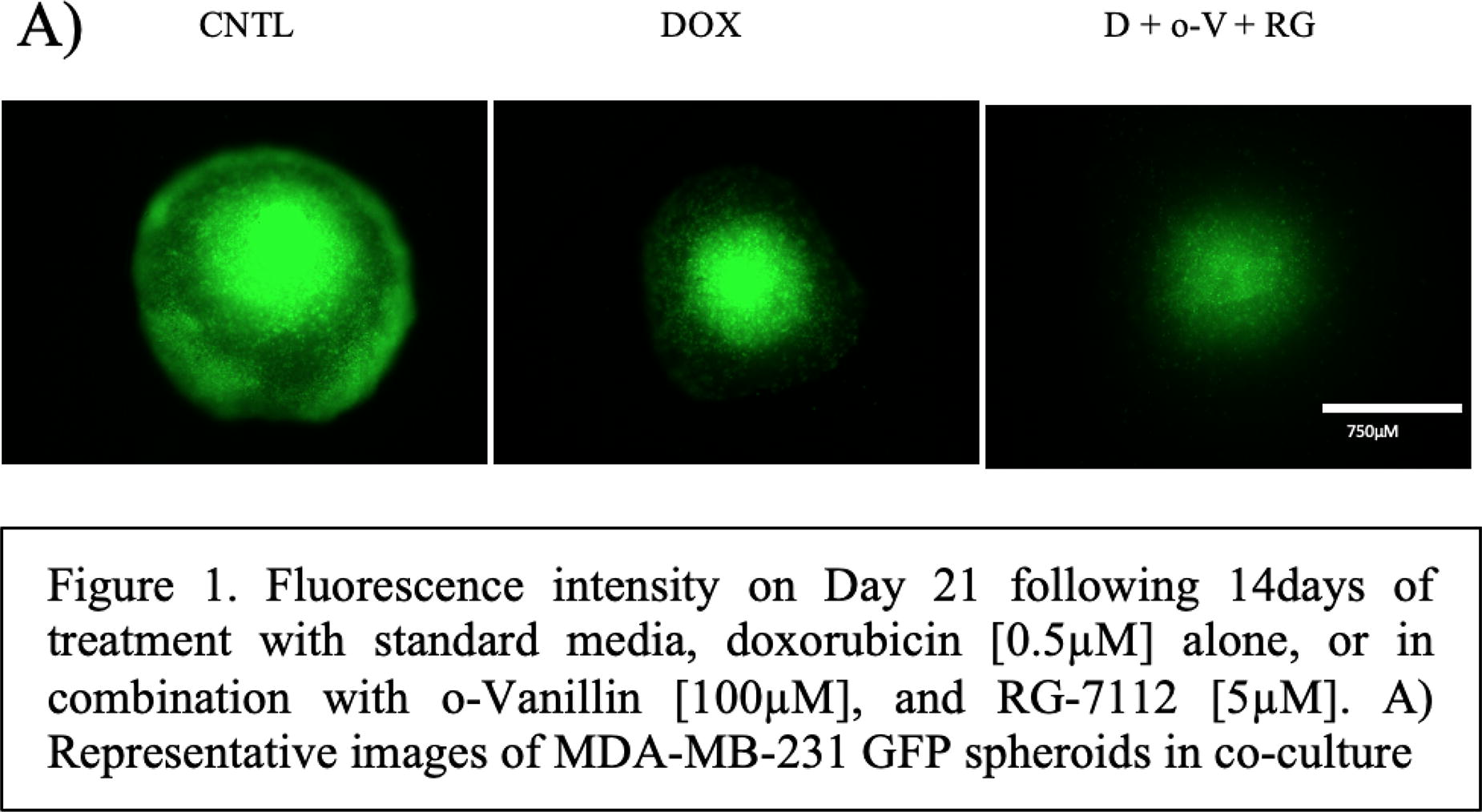
B71 - Photodynamic Therapy To Treat Triple Negative Breast Cancer In Vitro
H. S. Warren1, L. Ethredge1, V. Bliznyuk2, B. W. Booth1
1
Bioengineering, Clemson University, Clemson, SC,
2
Environmental Engineering and Earth Sciences, Clemson University, Anderson, SC
*Purpose/Objectives: Triple negative breast cancer (TNBC) is the most resilient form of breast cancer, being one of the leading causes of death for women and making up 7% of all cancer deaths. Photodynamic therapy (PDT) offers a minimally invasive therapeutic option for TNBC patients as a passive-targeting treatment that reduces the need for other well established yet harsh treatments. PDT involves the use of a high-energy red light to activate injected photosensitizers (PS) at the area of a tumor. Activated PS generate reactive oxygen species (ROS) in the tumor, triggering cell death. The PS tetra(hydroxyphenyl)chlorin (m-THPC) was used in this study due to its high singlet oxygen yield activated by 660-670 nm light. m-THPC’s hydrophobic nature requires the use of the hydrophilic carrier dextran-grafted-polyacrylamide (D-g-PAA) which allows for systemic circulation of m-THPC. D-g-PAA’s pH sensitivity encourages the release of m-THPC from D-g-PAA’s active amide group when in the presence of low extracellular pH typical of tumors. Gold nanoparticles (AuNPs), also used in this study, are effective in enhancing tumor cell death when in the presence of the thermodynamic changes during PDT treatment due to their radiosensitive surfaces. The goal of this study was to determine an ideal concentration of PDT nanocomposite comprised of D-g-PAA synthesized with AuNP and loaded with m-THPC for PDT treatment of human and mouse TNBC cell lines with minimum or no damage to human and mouse normal mammary epithelial cell lines.
*Methodology: PDT treatments were tested on cell lines with minimal amounts of ambient light and 660-670 nm light exposure as well as varying combinations of the nanocomposite components. Ideal concentrations of D-g-PAA synthesized with AuNP loaded with m-THPC that accomplish the killing of TNBC over normal mammary epithelial cell lines were established for both human cell line and mouse cell line. Viability after PDT treatments with the nanocomposite were measured via absorbance. The effect of our nanocomposite with PDT in vitro was also assessed to determine necrosis or apoptosis. PDT treatments induced apoptosis in the cancer cells. The specific pathways of apoptosis were then determined.
*Results: Ideal concentrations of our nanocomposite significantly reduced TNBC cell populations without significantly impacting normal mammary epithelial cells. The results of this study also point to apoptosis as the primary mode of cell death caused by our PDT treatments.
*Conclusion/Significance: These conclusions represent a significant step forward in breast cancer treatment by demonstrating PDT efficacy against TNBC. Additionally, the possibility of using our nanocomposite with PDT has promise of being effective in killing other forms of breast cancer cells.
B74 - Dehydroleucodine Promotes Adipogenic Differentiation In Osteosarcoma Via PPARγ Modulation
T.-M. Chang, Y.-S. Sun, J.-F. Liu
Taipei medical university, Taipei, Taiwan
*Purpose/Objectives: Osteosarcoma is characterized by high heterogeneity and poor differentiation. The metastasis, drug resistance, and recurrence of osteosarcoma remain challenges in treating patients with advanced and metastatic disease. Peroxisome proliferator-activated receptor γ (PPARγ) serves as a nuclear receptor factor crucial for cellular functions, including promoting the adipocyte differentiation and maturation. Previous studies have demonstrated that the use of full PPARγ agonists, such as thiazolidinediones (TZD), promotes the transformation of cancer cells into adipocytes and mitigates drug resistance. However, given the numerous adverse effects associated with TZD, there is an urgent need to find compounds with lower toxicity. Some sesquiterpenes exhibit an affinity for PPARγ and regulate lipid metabolism in cancer cells. This study indicates sesquiterpenes can act as potential ligands for PPARγ, thereby regulating the transcriptional activity of PPARγ to promote adipogenic differentiation in osteosarcoma cells.
*Methodology: The prediction of affinity between sesquiterpenes and PPARγ was assessed using the iGEMDOCK software. Cytotoxicity assessment of sesquiterpenes was conducted through the CCK-8 assay. The oil red O staining method was employed to observe lipid accumulation in osteosarcoma cells. The expression of adipogenesis-related genes and drug transporter-related genes were quantified using qPCR. Additionally, osteosarcoma cell lines were transfected with luciferase plasmids expressing the PPARγ response element to measure PPARγ transcriptional activity. Furthermore, the combined anticancer effect of sesquiterpenes inducing adipogenesis in collaboration with chemotherapy was evaluated using both oil red O staining and the CCK-8 assay.
*Results: PPARγ modulates diverse cellular functions, and the comprehensive activation of PPARγ has been associated with adverse effects linked to TZD. Sesquiterpenes have demonstrated anticancer activity in various cancer cells and play a role in regulating lipid metabolism. Our study was to identify sesquiterpenes with potential binding activity to PPARγ. Analysis of iGEMDOCK software confirmed that sesquiterpenes, including zerumbone, dehydroleucodine, parthenolid, dehydrocostus lactone, alantolactone, isoalantolactone, germacrone, and β-caryophyllene, exhibited the binding affinity for PPARγ. Furthermore, sesquiterpenes were verified to induce adipogenic differentiation in osteosarcoma cells. Notably, some sesquiterpenes exhibited features supporting the promotion of adipocyte differentiation. Among them, dehydroleucodine significantly enhanced the expression of adipocyte differentiation-related genes and PPARγ transcriptional activity in osteosarcoma. Additionally, treatment with dehydroleucodine led to a significant increase in adipogenesis and improved susceptibility to doxorubicin in osteosarcoma. Subsequently, dehydroleucodine was found to inhibit the expression of drug transporters in osteosarcoma. Moreover, dehydroleucodine sensitized doxorubicin-resistant osteosarcoma cells to the effects of doxorubicin following adipogenic differentiation. These findings underline the potential of dehydroleucodine as a PPARγ agonist, revealing its multifaceted impact on adipogenesis, drug susceptibility, and drug transporter expression in osteosarcoma cells.
*Conclusion/Significance: The present study highlights the prospective affinity of sesquiterpenes for PPARγ, exhibiting their potential to initiate adipogenic differentiation in osteosarcoma cells. Especially, dehydroleucodine evolves in promoting osteosarcoma adipogenesis by modulating PPARγ activity and promoting the expression of adipogenic genes. These findings illustrate the capacity of dehydroleucodine as a PPARγ agonist. Moreover, the induction of adipogenesis by dehydroleucodine enhances the responsiveness of osteosarcoma cells to doxorubicin. Our results suggest the prospect of employing dehydroleucodine-induced adipogenic differentiation to against malignant cancer cells in osteosarcoma.
B77 - Transcriptomic Analysis Of Mineralized Tissue-engineered Sheets For Calcific Aortic Valve Disease Modelling
A. Brodeur1,2, V. Roy1,2, L. Touzel-Deschênes1,2, S. Bianco2,2, A. Droit2,2, J. Fradette1,2, J. Ruel1,2, F. Gros-Louis1,2
1
Laval University, Quebec, QC, Canada,
2
CHU de Quebec Université Laval Research Centre, Quebec, QC, Canada
*Purpose/Objectives: Calcific aortic valve disease (CAVD) is characterized by fibrosis and mineralization of the aortic valve, which leads to aortic stenosis and heart failure. At a cellular level, this is due to osteoblastic-like differentiation of valve interstitial cells (VICs), resulting in the calcification of the tissue. Unfortunately, human VICs are not readily available to study CAVD pathogenesis and implicated mechanisms in vitro. However, adipose derived stromal/stem cells (ASC), carrying the patient’s specific genomic features, have emerged as a promising cell source to model cardiovascular diseases due to their multipotent nature, availability, and patient-specific characteristics. We describe a comprehensive transcriptomic analysis of tissue-engineered, scaffold-free, ASC-embedded mineralized tissue sheets using bulk RNA sequencing. We propose that the mineralized tissues produced from ASCs will serve as a suitable model for calcific valve disease.
*Methodology: Osteoblastic differentiation of tissue engineered ASC-embedded sheets was induced by soluble factors in the culture media for 28 days. Calcification was observed and quantified by alizarin red staining and alkaline phosphatase activity. Transcriptomics were studied by bulk RNA sequencing of the tissues. Bioinformatic analysis and gene set enrichment analysis was done using various online databases: the Gene Ontology (GO) Knowledgebase, Kyoto Encyclopedia of Genes and Genomes (KEGG) pathway database, WikiPathway, Reactome Pathway Knowledgebase, MatrisomeDB and Transcriptional Regulatory Relationships Unraveled by Sentence-based Text mining (TRRUST) database. Network analysis was performed using Ingenuity Pathway Analysis software.
*Results: Bioinformatic and gene set enrichment analyses revealed the upregulation of genes associated with the extracellular matrix (ECM) organization, suggesting that ECM could play a vital role in the enhanced mineralization observed in these tissue-engineered ASC-embedded sheets. Upon comparison with publicly available gene expression datasets from CAVD patients, striking similarities emerged regarding cardiovascular diseases and ECM functions, suggesting a potential link between ECM gene expression and CAVDs pathogenesis. A matrisome related sub-analysis revealed the ECM microenvironment promotes transcriptional activation of the master gene runt related transcription factor 2 (RUNX2), which is essential in the CAVD development.
*Conclusion/Significance: We characterized an all-human 3D tissue-engineered model based on ASC to study CAVD in which the disease transcriptomic signatures can be recapitulated. We also showed that the ECM microenvironment established upon induction of mineralization in ASC led to transcriptional regulation by master gene RUNX2. The pathophysiology of CAVD is still an active area of research, and ongoing studies aim to better understand the molecular and cellular mechanisms involved in the disease. This model, which can be made from the patient’s own cells, offers several advantages in studying the pathophysiology of CAVD and could pave the way for tailoring therapies based on an individual’s unique genetic and molecular profile.
B79 - Growing Aortic Valve: Development Of A Biohybrid Heart Valve With Growth Capability For The Treatment Of Paediatric Patients
A. Loewen, N. Dincer, S. Jockenhoevel
Department of Biohybrid & Medical Textiles (BioTex), AME-Institute of Applied Medical Engineering, Helmholtz Institute, RWTH Aachen University, Aachen, Germany
*Purpose/Objectives: Heart valve replacement in paediatric patients imposes unique challenges in terms of material and design requirements. While mechanical heart valves require permanent anticoagulation, they are not commonly used in children due to the high risk of life-threatening haemorrhages. On the other hand, biological heart valves do not require anticoagulation but degenerate quickly in children. Furthermore, both types of heart valves lack adaptation to the growing size of the child’s heart, leading to frequent reinterventions to adjust the valve size.The objective of this research study is to develop a biohybrid aortic valve that can adapt to the growing size of children's hearts. The biohybrid implant consists of a matrix of Elastin-Like Recombinars (ELR), which is being biologized by means of in-situ tissue engineering, and a growth capable textile scaffold structure, which gives the implant mechanical stability.
*Methodology: In order to create a textile scaffold structure that can adapt in a time-controlled manner to the increasing diameter and orifice area of the valve, a warp knitted structure was developed using non-degradable yarns (polyethylene-terephthalate & polyvinylidene-difluoride) that was specifically reinforced with biodegradable fibres (polylactide, polylactide-co-glycolide, polycaprolactone, silk). For integrating degradable fibres, direct integration into the warp-knitting process by means of combined lappings as well as subsequent tailored integration of reinforcing fibres into the warp-knitted structure were investigated. Samples were stored in temperature-controlled phosphate-buffered-saline in order to simulate degradation in-vivo.
*Results: Geometric and mechanical parameters of the scaffold structure were evaluated at different degradation points, including diameter, surface-area, tensile strength, elongation and radial compliance. Selective combination of fibres degrading at different rates and different geometric structures of the reinforcement structures resulted in a stepwise controlled increase of the valve diameter by up to 50 % and the valve orifice area by up to 60 %. Radial elongation of the scaffold structure at an inner mean pressure of 100 mmHg after degradation of the reinforcement fibers was 5 %, 26 % and 50 % for three different geometries of tailored integrated reinforcement structure. Degradation of directly integrated reinforcement fibers in a warp-knit structure with combined lappings led to an increase of diameter of 10 % and 22 % respectively for two different stitch densities of the textile structure. The achieved growth capacity of the scaffold structure corresponds to the growth of a paediatric heart valve over ten years with an implantation within the first year of life.
*Conclusion/Significance: By combining a non-degradable textile base structure with a controlled degrading reinforcement structure, it was possible to fabricate a textile scaffold structure capable of growing with the valve in a defined time-controlled manner. These outcomes represent a decisive step towards a heart valve implant with growth capability for the treatment of young children.

B81 - Rapid Generation Of An Autologous, Non-thrombogenic Endothelium Facilitated By Co-culture Across An Electrospun Polyurethane Scaffold
K. D. MacQuarrie1, K. A. D’Costa1, X. Lei2, G. Zhu2, S. O. Hofer3, H. Ni2, J. P. Santerre1
1
University of Toronto, Toronto, ON, Canada,
2
Unity Health Toronto, Toronto, ON, Canada,
3
University Health Network, Toronto, ON, Canada
*Purpose/Objectives: Small diameter (< 6 mm) synthetic vascular grafts lose patency, preventing the long-term revascularization of coronary, pulmonary, and peripheral arteries. A mature endothelium regulates arteries’ permeability, vasoactivity, and thrombogenicity, and endothelialized grafts have been demonstrated to maintain patency longer. However, the time required to achieve endothelialization limits its practical clinical application. Additionally, it is important that grafts are seeded with autologous cells to avoid immune rejection.
Previous work from our group has demonstrated that indirect co-culture of autologous adipose tissue-derived stromal cells (ASCs) and human adipose tissue-derived microvascular endothelial cells (HAMVECs) across an elastomeric polyurethane scaffold facilitates confluent endothelialization within 48 hours. When HAMVECs are seeded alone, they take 14 days to gain confluence. Our group has also shown that ASCs can be differentiated towards a vascular smooth muscle cell-like phenotype (ASC-VSMCs). ASC-VSMCs express late-stage smooth muscle cell markers at similar levels to native VSMC controls, and contract in response to vasoconstrictors more effectively than ASCs. When ASC-VSMCs are directly co-cultured in a 4:1 ratio with monocytes, more collagen and elastin are produced to mechanically stabilize the graft. In this study, we compare ASCs, ASC-VSMCs, and ASC-VSMCs with monocytes as contralateral cells to support rapid endothelialization and assess endothelial functionality in terms of its thrombogenicity.
*Methodology: ASCs and HAMVECs were isolated from human adipose tissue (approved protocol #13-6437-CE, University Hospital Network, Toronto) with magnet-assisted cell sorting, while monocytes were isolated from whole human peripheral blood (approved protocol #19264, University of Toronto). ASCs were differentiated to ASC-VSMCs by supplementing culture medium with transforming growth factor β and retinoic acid. ASCs, ASC-VSMCs, or ASC-VSMCs with monocytes were cultured contralaterally with HAMVECs across a nano-fibrous polyurethane membrane. Endothelialization and HAMVEC proliferation were measured with immunofluorescence staining and confocal microscopy. To assess thrombogenicity, whole blood or platelet-rich plasma collected in sodium citrate tubes was applied to endothelialized scaffolds alongside acellular and Dacron controls. Platelet activation was measured with flow cytometry and scanning electron microscopy (SEM).
*Results: Here, we show that differentiated support cells do not hinder accelerated endothelialization; ASCs, ASC-VSMCs, and ASC-VSMCs with monocytes support approximately 70-80% HAMVEC coverage within 24 hours. Preliminary results (N=1) suggest that the endothelialized surfaces induce less platelet activation, evidenced by lower P-selectin expression and PAC-1 binding, than in the acellular and Dacron controls (data currently being reproduced). SEM imaging also indicates that fewer blood constituents adhere to the endothelialized portions of the scaffolds (data not shown).
Figure 1. Support cells (red = ASCs, green = ASC-VSMCs, purple = ASC-VSMCs and monocytes) accelerate the endothelialization (percentage of area that is CD31+) of polyurethane scaffolds with HAMVECs over 24 hours. N=3 biological replicates.
*Conclusion/Significance: We have demonstrated that differentiated and non-differentiated ASCs facilitate rapid endothelialization within 24 hours and establish the potential of autologous cell use. Preliminary results suggest that these endothelialized surfaces are less thrombogenic than acellular polyurethane or Dacron, which are among the clinical standards for synthetic vascular grafts. This work may enable the future production of an endothelialized, biomimetic, and non-thrombogenic small diameter vascular graft.

B82 - Dynamic Biofabrication Of Highly Compliant Small Diameter Tissue-engineered Vascular Grafts
A. Weekes1, J. Wasielewska2, J. Davern1, G. Wehr3, N. Pinto4, J. Jenkins4, J. Patel5, Z. Li5, T. Klein1
1
Queensland University of Technology, Kelvin Grove, Australia,
2
Herston Biofabrication Institute, Herston, Australia,
3
University of Queensland, St. Lucia, Australia,
4
Royal Brisbane and Women's Hospital, Herston, Australia,
5
Queensland University of Technology, Brisbane, Australia
*Purpose/Objectives: Regenerative medicine represents a promising avenue for the production of vascular grafts. However, although current tissue-engineered vascular grafts (TEVGs) frequently fulfil certain functional requirements of native blood vessels, many fail to overcome poor mechanical compliance. Here, TEVGs were cultured from placental mesenchymal stem cells (MSCs) and commercially available smooth muscle cells (SMCs) in a dynamic, pulsatile bioreactor system to facilitate enhanced compliance, using biomimetic melt-electrowritten (MEW) tubular scaffolds as substrates for tissue production. Through dynamic culture, we aimed to investigate differences in extracellular matrix (ECM) production and mechanical performance, with a specific focus on graft compliance. Further, the efficacy of decellularization was characterised, toward the development of non-immunogenic, off-the-shelf TEVGs suitable for use in small diameter bypassing.
*Methodology: Small diameter tubular scaffolds (4.0 mm diameter, 80 mm length, 400 µm thickness) were manufactured via rotational MEW from medical grade polycaprolactone (PCL). Scaffold substrates were designed to exhibit biomimetic mechanical properties through perpendicular straight and sinusoidal fibre architecture, resulting in highly consistent porous morphology. MSCs and SMCs were seeded separately onto scaffold substrates and cultured in vitro for 6 weeks in dynamic and static conditions, prior to decellularization with Triton-X100. Biochemical quantification of ECM components and mechanical testing enabled assessment of TEVG properties, alongside histological and immunofluorescence imaging and analysis.
*Results: Mechanical testing indicated enhanced performance in dynamic cultured TEVGs, irrespective of cell type, with negligible loss in structural integrity as a result of decellularization. Dynamic TEVGs exhibited physiologically relevant compliance of 12.4 ± 0.8 % per 100 mmHg, burst pressure of 1225 ± 196 mmHg, and suture pull-out force of 3.1 ± 0.3 N, all significantly greater than static cultured TEVGs. Histological and immunofluorescence imaging supported these findings, as dynamic TEVGs exhibited greater ECM accumulation, with MSC-cultured TEVGs producing a more dense, and collagen-rich ECM, coinciding with greater GAG and hydroxyproline content as quantified via biochemical assays. TEVGs were effectively decellularized via TX100 treatment, assessed via DNA quantification and imaging of the remnant ECM, with negligible influence on mechanical performance.
*Conclusion/Significance: Highly compliant MEW scaffolds cultured in dynamic pulsatile conditions enabled the biofabrication of small diameter TEVGs which exhibited physiologically relevant mechanical performance and enhanced ECM compared to static cultures. Additionally, placental MSC-based TEVGs demonstrated denser collagenous ECM than commercial SMC-based TEVGs. Finally, graft decellularization enabled successful production of matrix-rich acellular TEVGs, with these findings providing motivation for future work toward the development of the next generation of small diameter TEVGs.
B83 - Structural Characterization Of Fresh Human, Porcine, And Ovine Heart Valve Tissue For Engineered Valve Application
P. Caruso1,2, M. DI Giuseppe2, L. Modica De Mohac2, F. Cosentino2, V. Balashov2, M. Barbuto1,2,3, M. Pilato4, G. Raffa4, B. Zuccarello1, W. Wagner3,5, A. D’Amore2,3,5
1
University of Palermo, Palermo, Italy,
2
Fondazione Ri.MED, Palermo, Italy,
3
McGowan Institute for Regenerative Medicine, Pittsburgh, PA,
4
IRCCS - ISMETT, Palermo, Italy,
5
University of Pittsburgh, Pittsburgh, PA
*Purpose/Objectives: The four human heart valves (HVs, Fig 1.1) exhibit substantial differences in their physiology which are reflected in their structural and mechanical properties. HV diseases remain among the most challenging clinical problems in cardiovascular medicine. Human native HVs mechanical and structural properties such as tensile and flexural stiffness, anisotropy, or even simpler factors such as leaflet thickness are largely neglected in the design of commercially available prosthetic devices and often remain poorly characterized, with data being extracted only from animal or cadaveric tissues. This gap in knowledge in valve physiology and prosthetic devices has increasingly made human cardiovascular tissues characterization a priority in biomechanics and tissue engineering.
The purpose of this study is to fully characterize human HVs structure at the organ and tissue scale and to compare these measurements with equivalent data obtained from porcine and ovine tissue. To achieve this aim, fresh human samples were obtained from heart-transplanted patients and tested within 24 hours from the tissue explant. At the organ scale, thickness topology for the aortic, mitral, pulmonary, and tricuspid (AV, MV, PV, TV) HV was measured and compared; at the microscopic scale, quantitative histology and collagen bundle diameter were quantified and compared.
*Methodology: Native leaflets were dissected from the valves and leaflet thickness (HVs from n=9 patients) was measured with a dial gauge (Mitutoyo) at fifteen points spanning from the free edge to the belly and commissural regions, full topology was derived using biquintic interpolation. Human HVs leaflets (n=6) were histologically evaluated using: Hematoxylin and Eosin (H&E) and Masson's trichrome staining to assess the cellularity general morphology of the leaflets’ extracellular matrix (ECM) and collagen fiber architecture.
*Results: Results showed thickness topologies that correlate with the transvalvular pressures acting on the AV, MV, PV, and TV different anatomical locations. All four valves exhibited greater thickness in the coaptation and free edge regions (Fig. 1.2) with the MV showing a higher average thickness compared to the other valves. Collagen quantity was estimated by digital image processing. In order to measure collagen fiber bundle diameter a novel decellularization protocol, specific for HV leaflet, was developed. Next scanning electron microscopy (SEM) analysis was performed on the decellularized leaflets (Fig. 1.3), fiber angle distribution and diameter were quantified using a modified, custom-made algorithm (D’Amore et al. Biomat. 2010) and ImageJ (Fig. 1.4). Human data were finally analyzed and compared to equivalent metrics obtained from porcine and ovine HV tissue.
*Conclusion/Significance: The results: quantified important differences in morphology of fresh human samples that match with the different anatomical locations of the four valves; provided a comparative analysis of human vs. porcine and ovine valve tissue. The ultimate goal of this study is to establish a thorough database of human HVs structure-function properties. This resource will contribute to advance the biomimetic-engineered tissue paradigm by providing a comprehensive description of HV structural and biomechanical parameters that can be potentially duplicated by material processing methods currently available or yet to be developed.
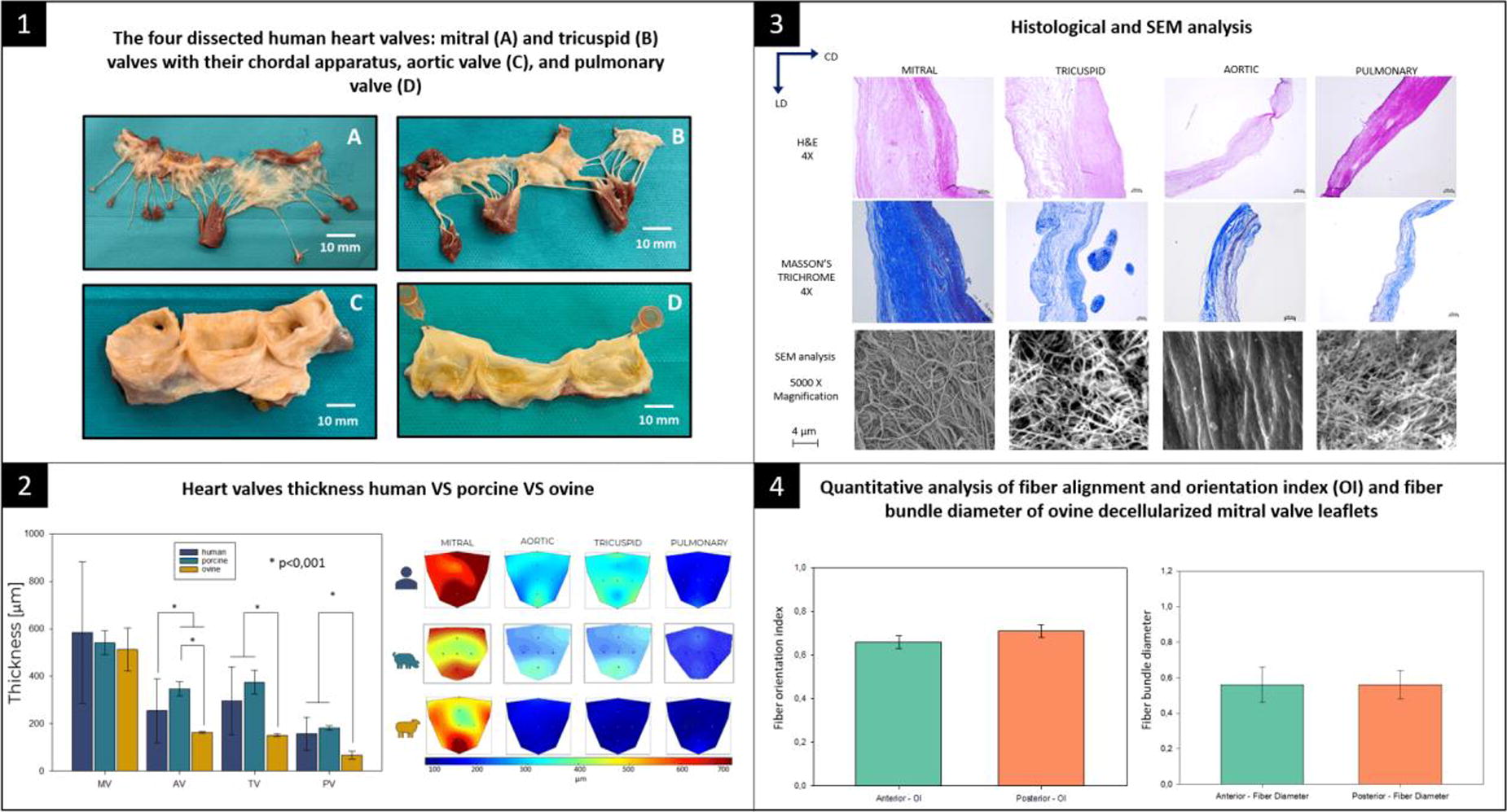
B84 - Biomechanical Characterization Of 3D Bioprinted Collagen-based Small Diameter Resistance Vessels
A. J. PereiraTavares, B. D. Coffin, D. J. Shiwarski
University of Pittsburgh, Pittsburgh, PA
*Purpose/Objectives: Hypertension is a vascular disease affecting ∼116 million Americans per year with healthcare costs of $131 billion/year. Considerable effort has been made toward developing pharmaceutical therapeutics for patients with hypertension, but we are increasingly learning that human specific genomic variations alter drug efficacy and treatment. For this reason, mouse models are becoming less relevant in predicting clinical outcomes. We can begin to address this challenge with tissue engineered blood vessel (TEBV) systems capable of incorporating both the complex 3D microenvironment and human specific physiology. Prior research has generated larger vascular scaffolds (> 1 mm) via folding 2D sheets, electrospinning, and sacrificial molding; however, these techniques are inadequate in generating multilayered small diameter arteries (< 400 µm ID) which are the major functional units regulating vascular resistance. An ideal small diameter TEBV to study hypertension would comprise biomaterials that can be mechanically tunned to mimic both the native and diseased states of the vasculature, and amenable to remodeling by vascular cells to investigate ECM deposition and measure vascular reactivity. Using Freeform Reversible Embedding of Suspended Hydrogels (FRESH) 3D bioprinting, we printed an all ECM-based small diameter resistance artery scaffold capable of connecting to a perfusion bioreactor to study how ECM composition, endothelization, and fluid flow contribute to the development of native and hypertensive disease like small diameter resistance arteries.
*Methodology: FRESH bioprinting allows for high fidelity printing of collagen and other ECM bioinks to generate multi-material scaffolds in almost limitless geometries. A single-channel perfusable 3D vascular model featuring an inner diameter of ∼300 µm suspended within a frame was designed using 3D CAD modeling (Figure 1A). The model was FRESH printed on a custom open source bioprinter with 23 mg/mL collagen-I. Biomechanical evaluation of burst pressure and peak tensions were conducted with pressure and wire myography according to the manufacturer’s protocol (DMT Systems).
*Results: Printed vessel models were patent and perfusable (Figure 1B). The three major vascular layers; adventitia, media and intima are designed into the 3D model to replicate the internal structure of native blood vessels (Figure 1C). Myography (Figure 1D-E) revealed that our FRESH printed artery scaffolds are comparable to mouse mesenteric resistance arteries with a burst pressure of >140 mmHg and an average peak tension of 600 mg, (Figure 1F-G). Additionally, larger printed models with ID > 1 mm exhibited peak tension similar to decellularized mouse aortas, ∼3600 mg, (Figure 1H).
*Conclusion/Significance: This work highlights the exciting potential of applying FRESH bioprinting to ECM-based perfusable scaffolds to generate physiological relevant human resistance artery models that are responsive to both vasodilation and constriction. We demonstrate our ability to alter the mechanical properties by changing model dimensions, ECM composition, or by fixation of scaffolds. Future work will utilize these findings to create microphysiological models of healthy and diseased resistance arteries. These printed collagen vessel models can be applied beyond TEBVs to study cell proliferation, metastasis, fluid transport, and ECM remodeling in large vessel and other organ systems.
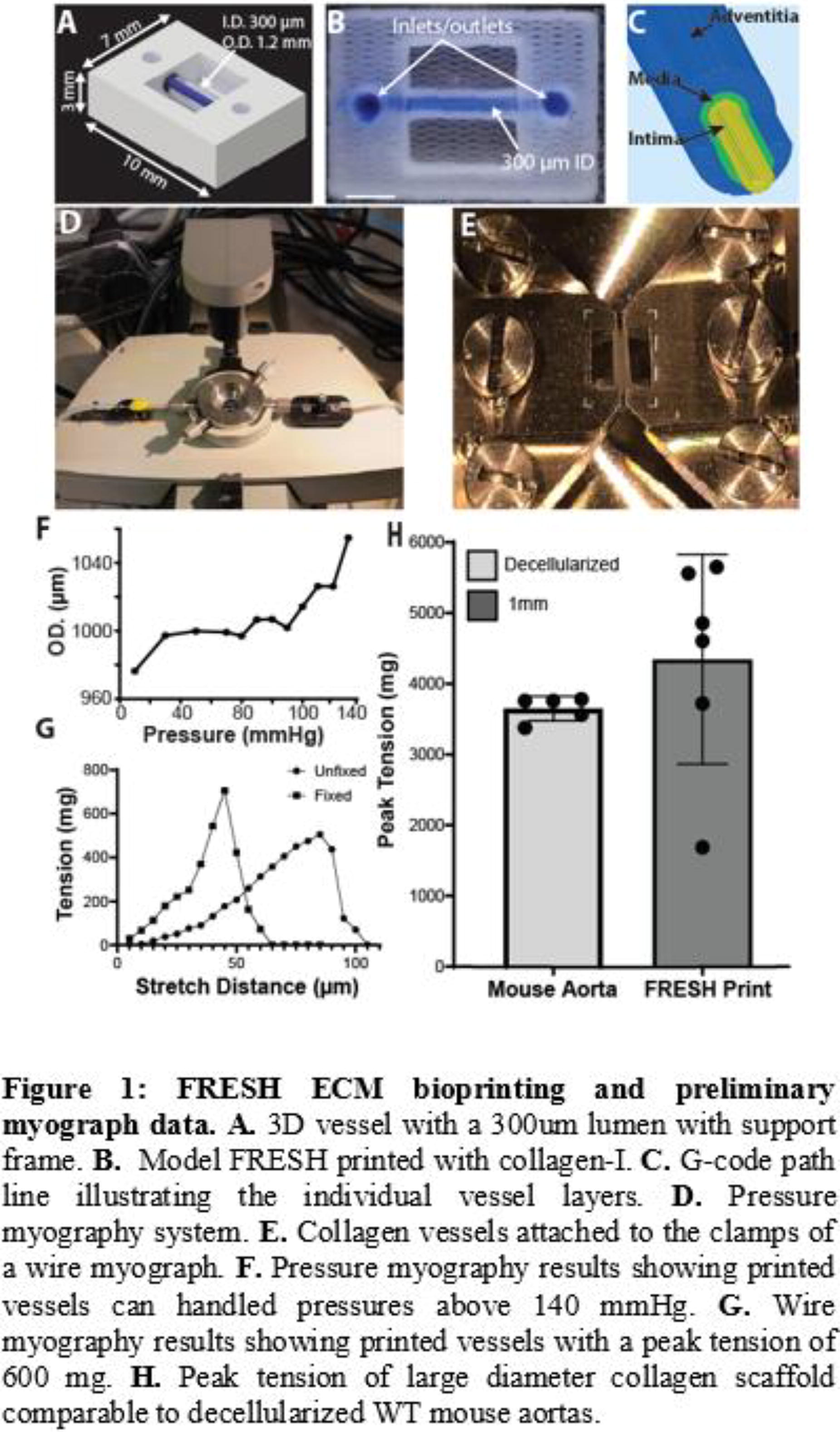
B85
B86 - Assessment Of Bioinks Compatibility With Endothelial Progenitor Cells For Bioprinting Applications
T. AbuSamaan1, C. Triggle1, I. Marei2,3
1
Weill Cornell Medicine Qatar, Doha, Qatar,
2
Imperial College London, london, United Kingdom,
3
Weill Cornell Medicine, Doha, Qatar
*Purpose/Objectives: 3D bioprinting of vascular grafts has the potential to improve the current methods for drug discovery and testing. The use of stem and progenitor cells adds the advantage of a more personalized assessment as these cells can mirror the in vivo environment and could reflect the phenotype of certain diseases. Endothelial progenitor cells (EPCs) are cells that circulate in the blood and show high proliferative and reparative abilities. Additionally, they possess endothelial-to-mesenchymal transition potential, making them an attractive source for 3D bioprinting of vascular grafts. This study aimed to assess the biocompatibility of different types of hydrogel bioinks with cord blood EPCs (CB-EPCs), using 2.5D and 3D cultures, and in co-culture with human coronary artery smooth muscle cells (HCASMCs).
*Methodology: 3 hydrogels were assessed including decellularized vascular extracellular matrix (ECM) GEIMA (TissueLabs), porcine decellularized skin ECM GEIMA (TissueLabs), and rat tail collagen (R&D systems). CB-EPCs were cultured in 2.5D format at different densities (5,10, 50 X103 cell/well), or were embedded within the hydrogel (densities: 2,4,6 X106 cell/ml). Cultures were maintained for 4 days at 37C and 5%CO2. Co-cultures were performed using CB-EPCs cultured on top of the gel and HCASMCs embedded within the gel (1.5X10⁶/ml). Viability was assessed using AlamarBlue assay and cytotoxicity using Live/Dead Viability/Cytotoxicity Kit (Invitrogen). Phenotypic characteristics were evaluated using immunostaining of CD309 and CD31 followed by confocal microscope imaging. Experiments were performed at least 3 times (n=3).
*Results: Decellularized vascular ECM GEIMA and collagen hydrogels both resulted in an increase of cell viability/proliferation with increasing seeding densities, both in 2.5D and 3D formats. No cytotoxicity was observed in both bioinks (Figure 1). Skin ECM on the other hand didn’t support the proliferation/viability of CB-EPCs. Collagen resulted in better morphology and spreading of the cells. Co-cultures of CB-EPCs with HCASMCs resulted in 2 defined layers, with increased proliferation in correspondence with the seeding densities.
*Conclusion/Significance: CB-EPCs showed compatibility with decellularized vascular ECM and collagen hydrogels, with collagen resulting in better compatibility (phenotypic stability, morphology and cell spreading, and enhanced viability both in 2.5D and 3D). Assessing the biocompatibility of bioinks with EPCs is an important step towards the optimization of 3D bioprinting of donor-derived vascular grafts. Such grafts could be used to model diseases and test the vascular cytotoxicity of drug developments prior to translating their use to the clinic, thus improving the outcomes of pre-clinical testing.
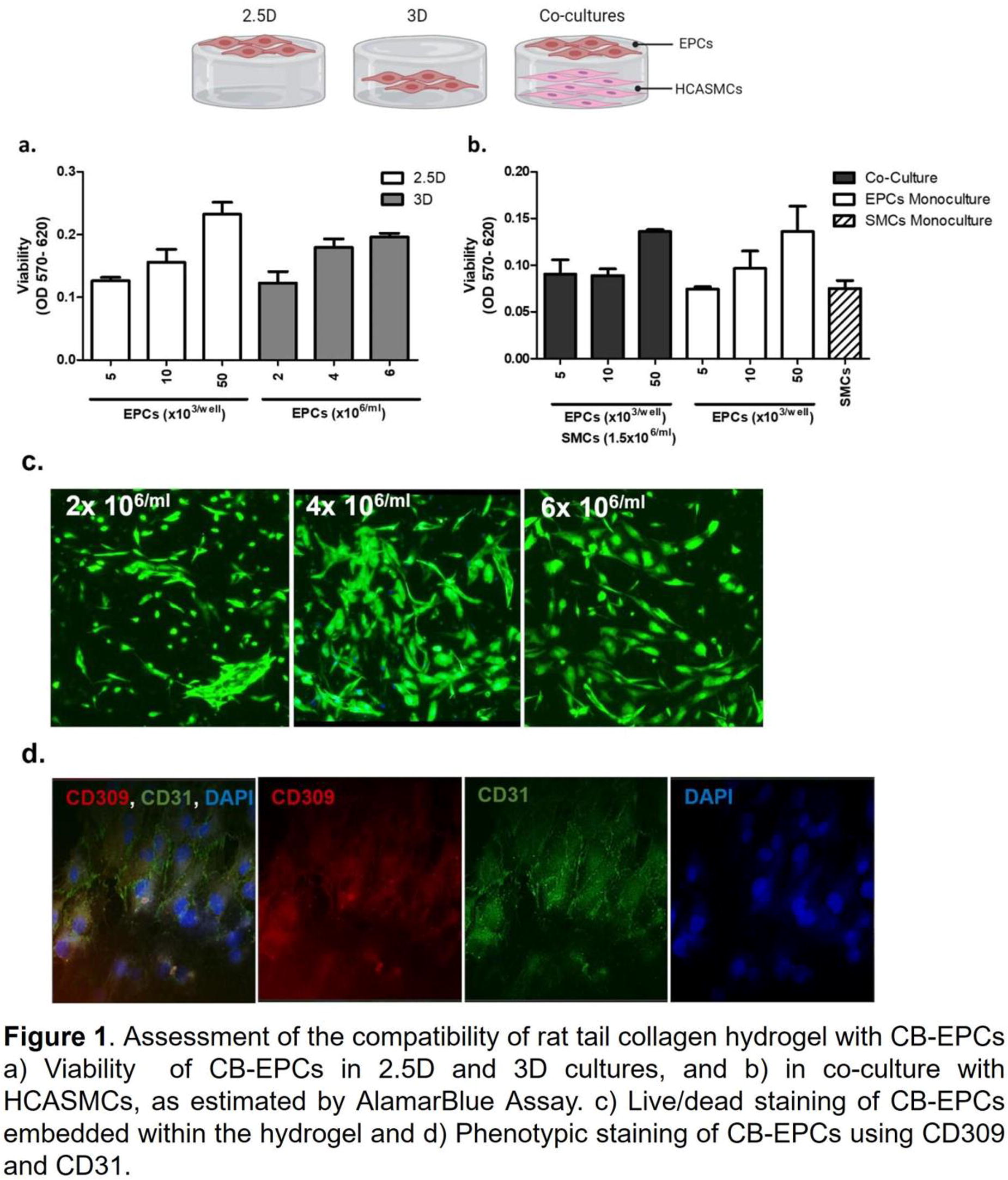
B87 - Engineering Of Bioinspired, Polymeric, Stent-less Mitral Valve: Performance Comparison Of Flat Vs. Saddle-shaped Annulus
E. Lanzalaco1, J. D. Laubrie2, F. Cosentino2, V. Balashov2, G. Raffa3, M. Pilato3, W. R. Wagner4, V. La Carrubba5, A. Pantano5, A. D’Amore2
1
Ri.MED Foundation and University of Palermo, Palermo, ITALY,
2
Tissue Engineering Group, Ri.MED Foundation, Palermo, ITALY,
3
ISMETT, Palermo, ITALY,
4
University of Pittsburgh, Pittsburgh, PA,
5
University of Palermo, Palermo, ITALY
*Purpose/Objectives: In the USA, mitral regurgitation (MR) affects 6% of adults >65 years old and it represents the most prevalent type of valvular disease. 65% of all MR patients are affected by functional (secondary) regurgitation, which is generally associated with a worse prognosis when compared to primary regurgitation. Rather than a leaflet disease, secondary MR, results from remodeling and malfunction of the left ventricle (LV), producing deficient coaptation of leaflets due to tethering of the papillary muscles (PMs). Current treatments to address this pathology include mitral valve (MV) repair or replacement, however, these patients must follow a life-long anticoagulant treatment and they may require additional surgical procedures due to a compromised harmonization of the LV-leaflet mechanics. MV replacement with currently available technologies alters the interaction with LV, due to the use of trileaflet valves, insertion of stents, flattening of the annulus and absence of the chordal apparatus (CA). To address these limitations, this work aims to evaluate the mechanics of two stent-less valve designs:(FSA) flat-shaped and (SSA) saddle-shaped annulus. The numerical models are meant to facilitate the design and fabrication of a polymeric mitral valve able to restore the native ventricle-leaflet continuum. In addition to the annular shape, this work assesses the optimal number and locations of chordae tendineae(CT) by evaluating tissue mechanics and organ-level function.
*Methodology: The biomechanics of the models is assessed in-silico by using Abaqus-explicit, where the two annular shapes are evaluated. The valves were attached to the ventricle by cords varying in insertion points, in quantity (2,4,6,8) and length (80,100,120%, of the native length), for a total of n=48 configurations, 24 FSA and 24 SSA designs. Leaflets and cords were modelled with structural elements, shell (S4) and truss (T3D2), respectively. In both parts, the material was modeled with isotropic behavior(Yeoh) and with mass Rayleigh damping. The annulus and PMs are static, with PMs held in systolic position. The leaflets were loaded with physiological atrial and ventricular timewise pressure. Multiple steps of cyclic pressure were applied over the valve until the cardiac cycle reached the steady state.
*Results: MVs with SSA generally showed lower stresses than FSA valves with delta-stress (max-min von Mises equal) equal to 1.7-MPa and 2.7-MPa, respectively. The stress peaks were in the commissural region and chordae insertion points. The bending deformation index was equal to 0.7 while the orifice area was estimated as 2.5-cm2 for both geometries. The valve designs that included two cords only presented the highest stresses and leaflet prolapse.
*Conclusion/Significance: Overall, this work shows that the SSA architecture has lower magnitude and more homogeneous stress maps than the FSA model. The comparative analysis of the CA showed that two cords are not enough to confer mechanical stability to the engineered valve with at least four cords being necessary to provide good function. Also, is estimated that six cords may be the maximum needed for the engineered valve apparatus, as the stress-strain distribution for the configurations with six and eight cords did not significantly differ when compared to the four-cord design.

B88 - Characterizing The Degradation Behavior Of Various Nanofiber Scaffolds In Vivo
S. Kunioka1, T. Yoshida2, D. Naruse3, Y. Setogawa1, Y. Kikuchi4, M. Tsutsui1, N. Wakabayashi5, K. Oyama1, H. Kamiya1
1
Asahikawa Medical University, Asahikawa, Japan,
2
Toyama Industrial Technology Research and Development Center, Nanto, Japan,
3
IAAZAJ HOLDINGS CO., LTD., Toyama, Japan,
4
Emory University Structural heart and valve, Atlanta, GA,
5
Sapporo Cardio Vascular Clinic, Sapporo, Japan
*Purpose/Objectives: Synthetic small-caliber vascular grafts occlude quickly due to thrombus. One of the causes of this occlusion is a compliance mismatch between native vessels and vascular grafts. Therefore, nanofiber grafts, which degrade and replace with native vascular tissue when appropriate timing, potentially solve this problem. There are a few studies that have described the degradability of nanofibers in vivo. However, objective and quantitative data is limited. In this study, we aimed to examine the degradation behaviors of various nanofibers in vivo.
*Methodology: Poly (ε-caprolactone) (PCL), poly (D, L-lactide) (PLA), poly (glycolic acid) (PGA), and PCL/PLA polymer alloy (PCL/PLA) were examined. Nanofiber sheets were created using electrospinning methods, and four nanofiber sheets (40 x 40 x 0.5 mm) were implanted subcutaneously in the backs of mice. In vivo degradability was evaluated up to 24 weeks after the implantation. Changes in the molecular weight of the nanofibers were measured by size exclusion chromatography (SEC) as an indicator of nanofiber degradation in vivo. Morphological changes of nanofiber sheets were also evaluated by histological staining and scanning electron microscopy imaging.
*Results: PGA was utterly degraded in 3 weeks; however, the other nanofibers remained until 24 weeks. In PCL, PLA, and PCL/PLA, the SEC showed decreases in the number average molecular weight (Mn) and the weight average molecular weight (Mw) while an increase in the polydispersity index (Mw/Mn). Reduction rate of the main peak height comparing 24 weeks against no-implanted control in PCL/PLA was larger that that in PCL and PLA alone, suggesting that PCL/PLA degraded faster than PCL and PLA alone. In the histological examination, the nanofiber sheets became partially positive for Pico-Sirius red staining, suggesting collagen tissue deposition into nanofibers. When performing SEC, nanofiber scaffolds are dissolved in organic solvent, and the total amount and molecular weight of dissolved nanofibers are measured. Although SEC showed a reduction of the total amount of nanofiber scaffolds (60-80% compared to no implanted control), some insoluble scaffold chanks remained in the SCE solvent at 24 weeks after implantation, in contrast to the no implant controls, which were wholly dissolved.
*Conclusion/Significance: In this study, we quantitatively demonstrated that PCL, PLA, and PCL/PLA degraded in vivo and that PCL/PLA had higher degradability than PCL and PLA alone. Also, consistent with autologous tissue-like regeneration, the degradation was accompanied by collagen deposition in all materials we tested. Surprisingly, we found that implanted nanofibers partially became resistant to dissolving in the organic solvent used in the SEC assay. We speculate that the resulting low molecular weight nanofiber materials are cross-linked and changed to some insoluble form in vivo. Although further study will be needed to understand the mechanism, this phenomenon could explain others’ observations that these nanofiber-based scaffolds remained for an unexpected long term in vivo. Thus, this study provides new insight into the degradation behavior of nanofibers in vivo.
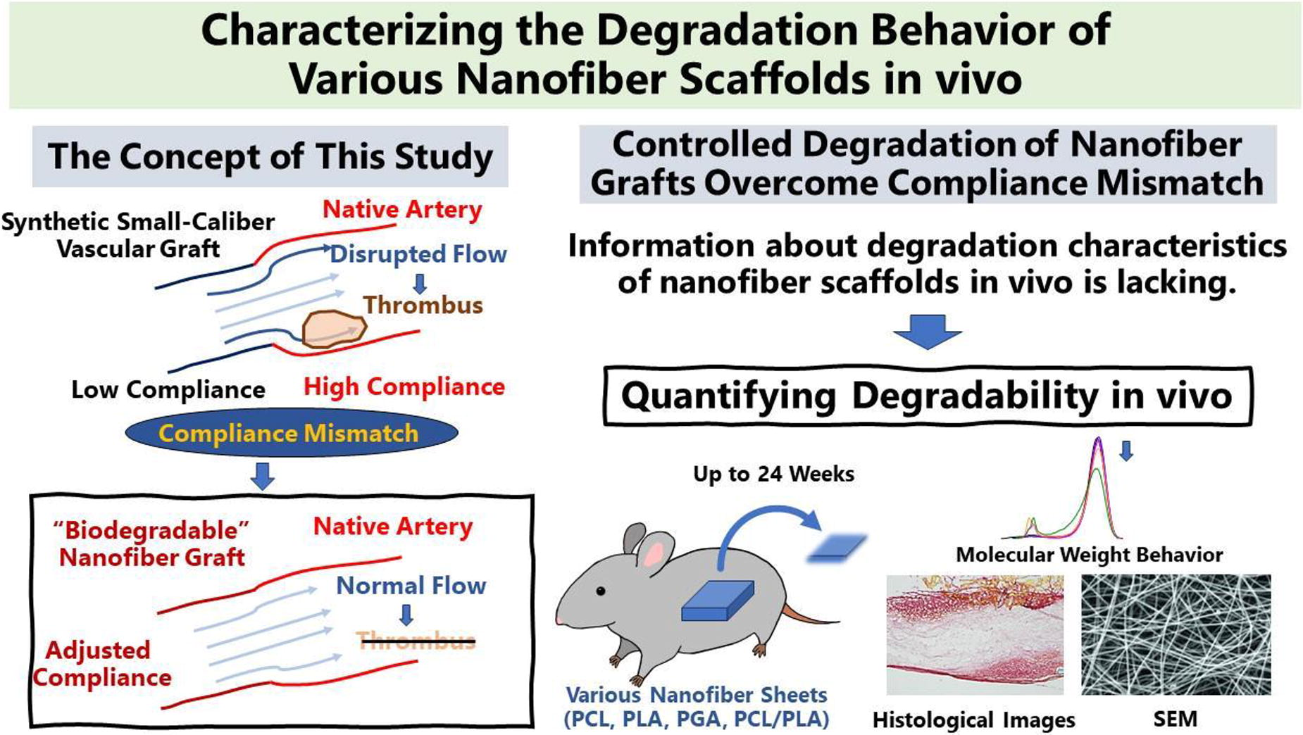
B89 - Alginate Porogen For Microvascularisation In 3D Bioprinted Structures
F. Vanlauwe1, S. Van Vlierberghe2, P. Blondeel2
1
Ghent University, Gent, Belgium,
2
Ghent University, Ghent, Belgium
*Purpose/Objectives: Within the context of bioprinting human-scale organ structures, there is a need for a bioprintable biomaterial that facilitates efficient vascular development by encapsulated cells. This vascular assembly is aided by the creation of pores in the biomaterial to enhance cell migration and signalling. Various methods are available to create pores in hydrogels. However, they either make the hydrogel unsuitable for bioprinting or the pore-creation methodology is detrimental for the viability of encapsulated cells. In this research project, a calcium-crosslinked alginate porogen is combined with a GelMA/cell mixture to induce pores in the hydrogel post-printing and enhance vascular development.
*Methodology: The porogen was created by the mechanical disruption of calcium-crosslinked alginate, resulting in small particles with a diameter of approximately 50-100 µm. The effectiveness of various calcium-chelating agents in dissolving calcium-crosslinked alginate was investigated using a degradation assay generated through a design of experiments. To investigate the leaching of alginate porogen from the hydrogel, time-lapse epifluorescence imaging was performed during the dissolution process of fluorescein-alginate porogen encapsulated in 10 w/v% rhodamine-GelMA. 3D bioprinting was conducted using a Regemat BioV1 3D bioprinter. Bioprintability was assessed by quantifying the similarity of a printed grid structure to a perfect square shape. The possible negative effect of the dissolution condition on the viability of encapsulated Adipose Derived Stem cells (ASCs) and Human Umbilical Vein Endothelial cells (HUVECs) was evaluated through a LIVE/DEAD assay. To assess vascular development capabilities, de novo vascular self-assembly and angiogenic sprouting from pre-existing vascularized tissues are examined in a vasculogenesis and angiogenesis assay, respectively. The vasculogenesis assay involved encapsulating ASCs and HUVECs, as single cells, in hydrogels. The angiogenesis assays was performed by encapsulating precultured spheroids composed of a combination of ASCs and HUVEC.
*Results: A combination of phosphate-buffered saline (PBS) and sodium citrate successfully dissolved 1600 mg of calcium-crosslinked alginate within 40 mins. The leaching experiment demonstrated a significant reduction of fluorescein-alginate, encapsulated in a 10 wv% gelMA hydrogel, after 40 mins of immersion in a PBS/sodium citrate solution. Under this condition, both HUVECs and ASCs were found to be equally viable compared to the control. The 3D bioprintability of the porogen mixed with 10 w/v% GelMA showed no significant difference compared to a well-known printable hydrogel, such as 10 w/v% gelMA without porogen. To assess the impact on vascular development, vasculogenesis and angiogenesis assays were first performed on non-bioprinted hydrogels. In both assays, hydrogels with porogen showed a significant higher total length of vascular structures compared to the control without porogen. These assays were repeated by 3D bioprinting the single cells or spheroids. Once again, the addition of porogen increased the total length of vascular structures significantly compared to the control.
*Conclusion/Significance: The combination of the alginate porogen and GelMA enables the bioprinting of a porous hydrogel while preserving the viability of encapsulated cells. Additionally, the pores contribute to the development of vascular structures. This methodology enhances the toolkit required for bioprinting human-scale vascularized organs.
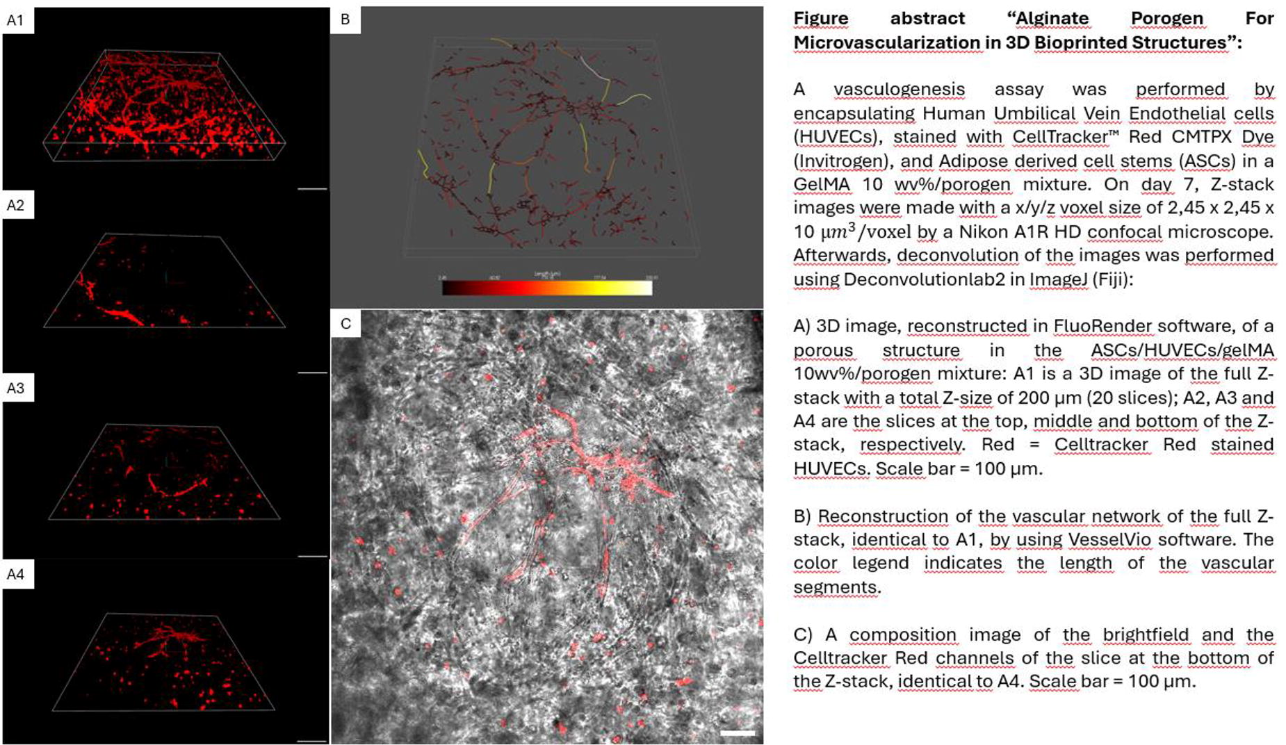
B90 - Engineering Hyaluronan Hydrogels To Deliver Endothelial Progenitors For Therapeutic Angiogenesis
Y. Yao1, I. Fernandes2, G. Keller2, M. Shoichet1
1
University of Toronto, Toronto, ON, Canada,
2
University Health Network, Toronto, ON, Canada
*Purpose/Objectives: Coronary artery disease is the leading cause of mortality worldwide. Therapeutic angiogenesis is a potential strategy to foster new vessel formation in the infarcted tissues and subsequently improve tissue remodeling and cardiac function following myocardial infarction. Human pluripotent stem cell (hPSC)-endothelial progenitors have shown ability to revascularize ischemic tissues, offering a unique opportunity to develop new therapies. However, their application potential is limited by low cell survival and low engraftment upon transplantation. To overcome these challenges, we designed an injectable hyaluronan (HA)-based hydrogel matrix as cell delivery vehicle. The hydrogel harnessed cell adhesion sites and promoted viability and angiogenic function of endothelial progenitors.
*Methodology: Sodium hyaluronate of different molecular weights (10kDa, 40kDa, and 250kDa) were modified with either norbornene (HAN) or methyltetrazine (HAT) functional groups. HA hydrogels were crosslinked using inverse electron-demand Diels-Alder chemistry by mixing HAN and HAT at a 4:1 molar ratio. To enable the adhesion of endothelial progenitors, we screened 24 extracellular matrix (ECM) mimetic peptides. Two cell-adhesive peptides, collagen-derived KGHRGF and laminin-derived NWRHSIYITRFG, were selected and chemically immobilized to HA. To further promote cell adhesion, collagen was physically mixed in HA hydrogels. hPSC-derived endothelial progenitors were seeded on HA gels. After 3 days of culture, viability of cells on HA gels was determined using live/dead assay. Angiogenesis was assessed by analyzing tube formation of the endothelial progenitors. Parameters, including the number of junctions, the number and length of segments and number of meshes, were quantified.
*Results: The viability and adhesion of endothelial progenitors were significantly affected by the molecular weight of HA. Cell viability drastically decreased as the molecular weight of HA increased to 250kDa. Cells on 40kDa HA exhibited highest viability and spreading. Further enhancement of cell adhesion and viability was achieved by increasing the collagen content in the 40kDa HA gel. The substitution of KGHRGF and NWRHSIYITRFG peptides on HA was measured to be 55 nmol/mg and 60 nmol/mg, respectively. Angiogenesis analysis demonstrated that the immobilized peptides stimulated the formation of tube-like structures by endothelial progenitors. Quantification results indicated that the HA hydrogel with a formulation of 0.75% (w/v) HA-KGHRGF and 0.25% (w/v) collagen had most profound improvement in tube formation of endothelial progenitors. Additionally, the 0.75% HA-KGHRGF/0.25% collagen hydrogel had an average gelation time of 5.1 min and remained injectable through 26G needle during a 1-hour testing period.
*Conclusion/Significance: Our novel formulation of HA-based hydrogels is capable of promoting viability and angiogenesis function of hPSC-derived endothelial progenitors, and is appealing candidate used for biomaterial delivered cell therapies to revascularize infarcted tissues.
B91
B92 - Directed Vascularization In Bone Regeneration: Healing Requires Bone Marrow Reconstitution In A Transplant Model
T. Thiele, J. Mehl, A. Ellinghaus, G. N. Duda
Berlin Institute of Health at Charité Berlin, Berlin, Germany
*Purpose/Objectives: The goal of regenerative therapy approaches is complete reconstitution of structural integrity, which requires scar-free healing. Bone is one of the few organs capable of such in situ scar-free regeneration. Unknown remains so far, however, from where healing occurs. The aim of this study was to identify the directionality of regeneration using bone as a model system. To allow for a distinct analyses of the directionality of healing, we used a murine bone graft transplantation model and fluorochromic cell marker staining to assess cellular migration, vascularization, collagen organization, and bone marrow remodeling; this graft transplantation allowed to analyze in the same animal two spatially distinct bone healing processes, one proximal and one distal.
*Methodology: Black6 mice expressing either ubiquitous red fluorescent protein (RFP) or yellow fluorescent protein (YFP) were matched such that each of the chimeric mice served as donor or recipient: 2 mm bone shaft pieces of RFP-labeled bone were cut by a wire saw and transplanted into YFP mice. This allowed tracking and analysis of RFP-labeled cells during bone regeneration at a proximal and distal osteotomy site synchronously. We employed a distraction system to stabilize both osteotomies and the bone graft. Healing was evaluated after 3, 7, and 14 days. As control served flushed bone shaft pieces, lacking the intact marrow graft. For each time point, cryosections were stained with CD146 for identification of micro-vascular structures and second harmonic generation imaging (SHG) to identify thick collagen bundles.
*Results: Following transplantation, host cells from the bone marrow migrated into the bone graft, progressing from proximal at both osteotomy sites, the proximal and the distal one. By 7 days, host cells had invaded approximately half of the graft, and by 14 days, they completely infiltrated the entire graft, but clearly from proximal. Noteworthy was a thinning of bone marrow cells in front of the invasion zone, eventually indicating apoptosis of resident bone marrow graft cells and successive marrow remodeling. Vascular sprouts originated from proximal host marrow and penetrated the graft resulting in a complete vascularization at 14 days. Overlaying vascular staining and nuclear images indicated reduced nuclear density ahead of the vascular invasion front, supporting the idea that graft cells retracted or underwent apoptosis preceding the invasion of novel vascular sprouts and host bone marrow cells. In the control group with flushed bone marrow vascularization was increased within the transplanted bone compared to the non-flushed group.
*Conclusion/Significance: Our study demonstrates (1) the clear proximal to distal invasion of regenerating cells and vasculature in a scar-free bone regeneration model. (2) In front of the invading microvascular structure and marrow cells, the transplanted bone marrow decomposes and gives way to the invading cell front. (3) A flushed bone marrow actually speeds up the vascularization and host invasion and thus accelerates bone regeneration in the presented mouse transplantation model. Our findings have the potential to inform and optimize strategies to achieve efficient and directed scar-free bone regeneration and sheds light on endogenous healing strategies that may be harvested also for clinical applications.
B93 - Design And Construction Of Capillary-Bed-like Structures With Tubular Nanofibrous Meshes
G. E. Kole1,2, V. Hasirci1,2,3, D. Yucel1,2,2
1
Acibadem University, Istanbul, Turkey,
2
Acıbadem University, Istanbul, Turkey,
3
Middle East Technical University, Ankara, Turkey
*Purpose/Objectives: In tissue engineering and in vitro tissue models, the formation and integration of capillary-bed-like constructs are essential for the effective gas exchange and nutrient supply for the survival of 3D tissues. This study aimed to design and construct capillary-bed-like structures with tubular nanofibrous mesh that mimics the basement membrane of the native vascular tissue.
*Methodology: The sacrifical, 3D tubular mold with hierarchical branching was modelled by applying the Murray’s Law. Two different models, basic model (BM) and complex model (CM), were designed with Fusion360. Symmetrical systems with two branches that had equal radii of the daughter branches were planned and the length and diameter of the branches were calculated. Computational fluid dynamic (CFD) was employed to simulate fluid behaviour within BM and CM. The sacrificial models were fabricated by extrusion-based 3D printing using polyvinyl alcohol (PVA). Tubular nanofibrous meshes of polycaprolactone (PCL) and PCL-Collagen were fabricated by electrospining. The morphological and mechanical properties of these meshes were studied, and also the endothelial cell behavior on these meshes was investigated. The branched, tubular nanofibrous meshes were collected over BM and CM sacrificial molds by electrospinning. Afterward, the sacrificial molds were removed with ddH2O, and luminal space was formed in the tubular nanofibrous meshes. The BM and CM of capillary bed-like constructs were examined with SEM. Perfusability of these constructs throughout their lumen was followed with dye injection.
*Results: The BM with two branches split from a parent tube, and the CM in which further branching was applied to the two branched tubes to obtain finally four branched tubular constructs were designed (Figure1a,b). Based on the Murray's Law, the radii of the tubes were calculated as 800, 630, and 500 μm. The BM was composed of 9.3mm parent tube, and 11.8mm branched tubes, while the CM consisted of 5 mm parent tube, 6.3 daughter branches and 8mm sub-tree branches. CFD analysis of both models revealed that the pressure of the fluid remained highest at the starting point, and the pressure gradually decreased as the fluid progressed through the model (Figure1a,b). BM and CM were printed successfully at high quality and with desired dimensions by altering the height, layer width, printing speed, print accleration, print jerk, etc. The uniform, bead-free PCL and PCL-Collagen nanofibrous meshes were successfully fabricated, and Young`s Modulus were 534.04±29.50 kPA and 299.99±30.05 kPA, respectively. The endothelial cells were attached to the nanofibrous meshes and increased in number within 7 days. To form the capillary-bed-like structures with tubular nanofibrous mesh of PCL and PCL-Collagen, the sacrificial molds were effectively removed with water and left a luminal structure. Ink perfusion studies showed that ink flowed throughout the lumen, split into the branches, converged at the connection point of branches, and flowed out from the outlet of the model.
*Conclusion/Significance: The obtained capillary-bed-like structures with tubular nanofibrous mesh are promising to be incorporated into 3D tissue-engineered substitutes or in vitro tissue models to provide vascularization. These nanofibrous mesh luminal surfaces supported the endothelial cell vitality and growth like vascular basement membrane.

B94 - Construction Of A Three-dimensional Tissue With A Dense Vascular Network By Culturing A Vascular Endothelial Cells / Mesenchymal Stem Cells Cocultured Cell Sheet Under Intermittent Positive Pressure
M. Katsuura1,2, J. Homma1, Y. Higashi3, H. Sekine1, S. Nagata2, T. Shimizu1
1
Tokyo Women’s Medical University, Institute of Advanced Biomedical Engineering and Science, shinjuku, Japan,
2
Tokyo Women’s Medical University, Shinjuku, Japan,
3
Tokai Hit Co., ltd, Fujinomiya-shi, Japan
*Purpose/Objectives: Constructing a dense vascular network into an ex-vivo engineering tissue is a crucial factor for the successful engraftment of the tissue. Previous studies have reported that coculturing vascular endothelial cells with mesenchymal stem cells (MSCs) promotes vascular network formation due to paracrine effects. However, this paracrine effect is unstable without additive factors. To create a dense vascular network in vitro, many reports have indicated that mechanical forces, such as shear stress, stretch stress, and hydrostatic pressure, are crucial factors. However, constructing systems to apply such mechanical forces to cells would be challenging due to their complexity. Thus, we have developed a pressurizing system that enables the easy application of external positive pressure to cells created by compressed air. The aim of this study was to investigate the effects of intermittent positive pressure culturing on co-cultured cells in two different conditions; a plane culture condition and a cell sheet condition.
*Methodology: Green fluorescent protein-expressing human umbilical vein endothelial cells (GFP-HUVECs) were co-cultured with human adipose-derived stem cells (hASCs). A comparison was conducted between the control group (atmospheric environment) and the intermittent positive pressure (IPP) group (repeated 80mmHg for 60sec and atmospheric pressure for 60sec ) in different conditions as follow. (1: a plane culture conditions) The vascular network, cell counts, and Lactate production/Glucose consumption (L/G) ratio. (2: a cell sheet condition in vitro) The vascular network, cell counts, morphological changes, and compositional changes of cell sheets. (3: a cell sheet condition in vivo) The transplanted co-cultured cell sheets of both two groups were composed of Gaussia luciferase (GLuc)-expressing hASCs and GFP-HUVECs and were transplanted into an athymic rat. The concentration of GLuc in the blood serum and the perfusion ratio of the cell sheet were evaluated.
*Results: (1: a plane culture conditions) In the IPP group, GFP-HUVECs formed a denser vascular network [total network length (mm/mm²); 4.191vs3.456 n=8, p=0.09, total number of junctions (counts/mm²); 9.383vs7.063 n=8, p=0.10], and significantly higher cell density [(counts/mm²); 1360 vs 1165 n=8, p<0.01]. Additionally, the IPP group demonstrated significantly lower L/G ratio (1.7vs1.9 n=8, p<0.01), which indicated a more aerobic state in an intermittent pressure environment. (2: a cell sheet condition in vitro) In the IPP group, GFP-HUVECs formed significantly more dense vascular network [total network length(mm/mm²); 13.74vs11.24 n=8, p<0.01, total number of junctions(counts/mm²); 87.65vs65.92 n=8, p<0.01] and significantly higher cell density [(counts/sheet); 4328vs3604 n=8, p<0.01]. The cell sheet in the IPP group was thicker [average thickness (μm); 45.70 vs 36.35 (n=8), p<0.01)] and contained a greater amount of collagen [collagen(μg/sheet); 11.03 vs 9.07 (n=4), p<0.05]. (3: a cell sheet condition in vivo) In the IPP group, the GLuc levels in the rat blood serum were higher (Luminescence Intensity: 2824 vs 2600 (n=6), p=0.13), despite no differences in perfusion ratio.
*Conclusion/Significance: The results indicate that applying intermittent positive pressure to the coculture condition of vascular endothelial cells and MSCs contributed to the formation of a dense vascular network and a thicker cell sheet. The pressurized cell sheet is expected to have better paracrine function.
B96 - Breaking Barriers: An Organised Complex 3D In Vitro Coculture System Including Key Components Of The Blood-Brain Barrier
A. Seijas-Gamardo1, P. Fucile1, J. Kim1, V. de la Rosa2, R. Hoogenboom3, S. Foulquier4, L. Moroni1, P. Wieringa1
1
MERLN Institute, Maastricht, Netherlands,
2
Avroxa BV, Ghent, Belgium,
3
Ghent University, Ghent, Belgium,
4
Maastricht University, Maastricht, Netherlands
*Purpose/Objectives: Creating realistic in vitro models of the blood-brain barrier (BBB) is essential for better understanding the neuroimmune communication and developing new effective drugs that can reach the brain. Current models have experimental shortcomings when mimicking key components present in the native system due to their lack of complexity or experimental control. Here, we present a novel in vitro BBB model featuring bespoke pattern of cell-lined perfusable microchannels formed in a cell-laden hydrogel, creating an organized 3D coculture.
*Methodology: Our key innovation lies in the use of a novel thermoresponsive polymer (OXA), which can be triggered to undergo a transition from hydrophobic to hydrophilic by lowering the ambient temperature. With OXA we can generate high resolution microfilaments as sacrificial templates that can be thermally triggered to dissolve. The template remains sufficiently stable and hydrophobic at cell culture temperatures, uniquely supporting the adhesion and growth of hCMEC/D3 brain endothelial cells.
*Results: After embedding and dissolving these cellularised microfilaments within a biomimetic hydrogel, we were able to form patent endothelialised microchannels. These endothelial-lined microchannels are perfusable, mimicking blood flow in the brain’s microvasculature. For the BBB, astrocytes are key, they help regulate blood vessel permeability and stabilize the microvascular structure. The biocompatibility of this templated system allows us to include astrocytes within the embedding hydrogels, thereby surrounding the microchannels and interacting with the hCMEC/D3 lumens. The diameter, flow, and geometry of the microvascular channels together with the hydrogel composition can be adapted, generating a versatile system that can potentially mimic different in vivo conditions. Finally, this coculture system is integrated into a customize microfluidics chamber, supporting both real-time imaging and fluidic pump perfusion.
*Conclusion/Significance: This new biofabrication approach addresses the main limitations of current BBB models by offering a more accessible model compared to animal experiments and more controlled complexity than classical in vitro models. It introduces an organized 3D coculture microsystem that can accommodate changes in ECM properties and cell types, while remaining perfusable. This flexibility and supported functional complexity provide a better in vitro model to study of brain drug delivery methods and BBB permeability related disorders.
B97 - Computer Vision To Predict Cell Seeding Coverage In Re-endothelialized Mouse Lungs
J. Paciocco1, A. Zargar1, A. Moayad1, J. K.D. Chan1, D. Taniguchi2, C. Amon1, G. Karoubi1,2, A. Bazylak1
1
University of Toronto, Toronto, ON, Canada,
2
Toronto General Hospital Research Institute, Toronto, ON, Canada
*Purpose/Objectives: Transplantation of donor grafts recellularized with recipient-derived cells or non-immunogenic universal cell lines is a potential means of reducing the graft rejection and post-transplant complications in lung transplantation. Achieving a fully recellularized lung, however, remains a major limitation. A key parameter of evaluating recellularization efficacy in the field of tissue engineering is the cell seeding coverage (CSC), which is the ratio of seeded cell area to the total area of the lung scaffold. To calculate the CSC from a histological image, the lung scaffold and the seeded cell areas must be quantified. In this work, the U-Net and LinkNet semantic segmentation models are applied to re-endothelialized mouse lung images, generating pixel-wise classifications of the lung scaffold and the seeded cell areas to automate the labour-intensive, and error-prone analysis of histological images.
*Methodology: Raw histological images of recellularized mouse lungs were obtained, preprocessed, and annotated. The histological images contained noise in the form of dark regions, dust, and/or artifacts, as observed in Fig. 1a. Artifacts were removed through denoising operations, resulting in Fig. 1b. The denoised image underwent Otsu thresholding to separate seeded cells (high intensity) from tissue (low intensity). However, heart tissue was also grouped in with the high intensity pixels, as shown in Fig. 1c. To correct this, a mask was manually drawn using the APEERTM software by ZEISS to limit the area of analysis to lungs alone, as observed in Fig. 1d. The area of analysis mask and the Otsu-thresholded mask were then multiplied, generating the product mask observed in Fig. 1e. Semantic segmentation was performed using the U-Net and LinkNet models. To train and test these models, k-folds cross validation was employed. Techniques including patch-based learning, data augmentation, and transfer learning were utilized to compensate for the low image quantity. Vanilla and patch-based models were compared in terms of their ability for classification of lung scaffold and seeded cell pixel as well as CSC prediction accuracy.
*Results: The precision, recall, and intersection over union (IOU) of the vanilla and patch-based semantic segmentation models are averaged across four cross-validation iterations. The patch-based U-Net model returns the lowest root mean square error (RMSE) of 1.57% ± 0.36% for cell seeding coverage, which is noteworthy because the vanilla U-Net returns the worst RMSE of 3.66% ± 4.33%. This improvement demonstrates the advantages provided by a patch-based learning approach when training on limited data. The patch-based LinkNet also shows an improvement over the vanilla LinkNet, reporting a 1.76% ± 0.31% RMSE over the 2.20% ± 0.36% RMSE.
*Conclusion/Significance: Each of the semantic segmentation models satisfactorily predicted CSC, although the accuracy of the patch-based models, particularly the patch-based U-Net model, outperformed the vanilla models in the pixel-wise classification of lung scaffold area and seeded cells and in the prediction of CSC. These results show semantic segmentation's potential to automate pixel-wise analysis of re-endothelialized images, saving time, reducing workload, and minimizing subjective manual errors.
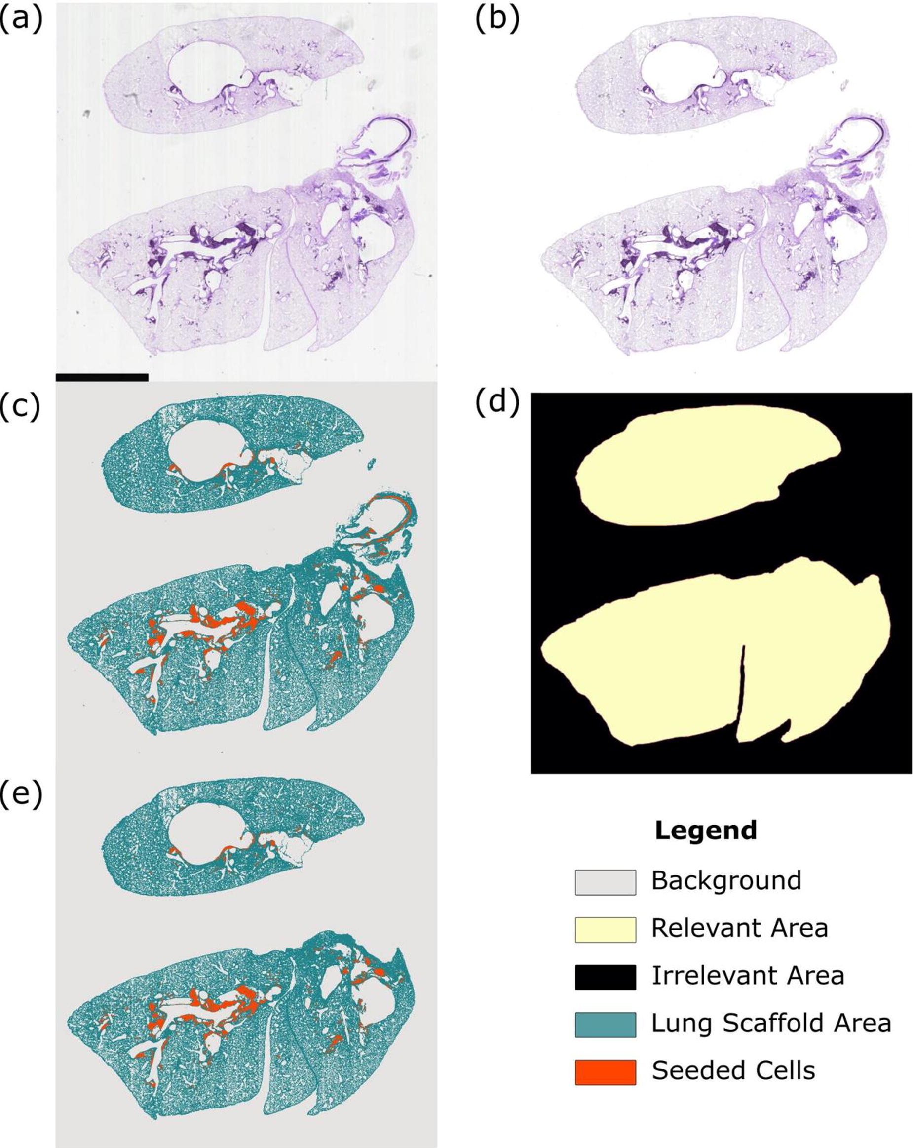
B98 - In Silico Model Development of Induced Pluripotent Stem Cells Differentiation to Definitive Endoderm
A. Mostofinejad1, D. A. Romero1, D. Brinson2, T. K. Waddell2,3, G. Karoubi2,3, C. Amon1,2
1
Mechanical and Industrial Engineering, University of Toronto, Toronto, ON, CANADA,
2
Institute of Biomedical Engineering, University of Toronto, Toronto, ON, CANADA,
3
Latner Research Laboratories, University Health Network, Toronto, ON, CANADA
*Purpose/Objectives: Differentiation of stem cells into definitive endoderm is a key step of the directed differentiation of embryonic stem cells (ESCs) and induced pluripotent stem cells (iPSCs) into endoderm derivates, e.g., thyroid, thymus, lungs, liver, pancreas, and the epithelial lining of the respiratory and digestive systems. Significant research has been conducted on establishing protocols for producing iPSC-derived definitive endoderm. These endeavours have primarily relied on in vitro and ex vivo trial and error experiments, with limited attention to utilizing mathematical modelling needed for accelerated discovery by reducing the time, cost, and variance and improving cell yields. In silico models also enhance our understanding of the complex population dynamics processes involved in this phenomenon and guide the optimization of these protocols.
*Methodology: In this work, we utilize a model development approach outlined in a previous paper by the authors, here focusing on modeling multicellular populations undergoing differentiation. Our strategy includes proposing multiple biology-informed differential equation-based models, considering different assumptions about the differentiation process. These assumptions encompass the various states stem cells transition through to reach a differentiated state, along with different growth models. We then calibrate these models using in vitro data from BU3 NGST iPSCs, NKX2.1-GFP and SFTPC-tdTomato reporter cells, for tracking and quantification of the differentiation process. Finally, we identify the most suitable model by subjecting these models to rigorous selection processes and ensuring model interpretability using identifiability analyses.
*Results: Our analysis shows that models without transitory cellular states perform better in predicting cell population evolution, compared to models with one transitory state or models with two parallel transitory states. Also, in the observed models, logistic growth outperforms exponential and Gompertz growth models. To make the loss function stable with a smaller number of needed observations, we incorporate the concept of variance models into our previously introduced model development methodology. Here, we show that a combined error function is the best in defining model variance.
*Conclusion/Significance: To the best of the authors' knowledge, this project presents the first mathematical model of the population dynamics of multi-cellular cultures of iPSCs differentiating into definitive endoderm. This model will be a tool for furthering our understanding of the differentiation process and optimizing it for various applications.
B99 - Establishing A Fluid-structure Interaction Model For Examining Structural Deformations In Decellularized Mouse Lungs During Re-endothelialization
A. Moayad1, J. K.D. Chan1, A. Zargar1, D. Taniguchi2, D. Romero1, C. Amon1, T. K. Waddell2, G. Karoubi1,2, A. Bazylak1
1
University of Toronto, Toronto, ON, Canada,
2
Toronto General Hospital Research Institute, Toronto, ON, Canada
*Purpose/Objectives: Endothelialization of acellular lung scaffolds remains a significant challenge hindering the generation of bioengineered lungs. Specifically, there is little known of the interactions between delivered cells and the scaffold microenvironment. Herein, we employ fluid-structure interaction (FSI) to investigate the structural changes of lung vasculature during the re-endothelialization process of a decellularized lung scaffold with the objective to optimize the re-endothlialization procedure. Addressing the limitations of rigid wall assumptions in conventional computational fluid dynamics (CFD) studies, this research incorporates deformable and porous structures to more accurately model re-endothelialization.
*Methodology: This study utilized COMSOL Multiphysics for analysis. Intact pulmonary systems from 12-week-old adult mice (C57Bl/6) were employed to create three lung geometries. The systems underwent cannulation and cleansing through a detergent-based decellularization process to remove cellular material. A contrast agent (Microfil) was then injected into the vasculature for micro-CT X-ray imaging. Image processing utilized for vasculature isolation and segmentation. The fluid domain was established within the vasculature, while the solid domain was assigned to the porous parenchyma. An interface at the fluid-solid contact point was designated, enabling fluid property extraction for finite element analysis (FEA). A dynamic mesh was designed for the parenchyma to measure vasculature deformation arising from fluid-solid interactions during the recellularization process. The structural deformation of the vasculature and parenchyma resulting from fluid-induced stresses is calculated assuming linear elastic behavior and isotropic material properties. A laminar flow condition was assigned for the modeling of the fluid as Reynolds number is less than 1000. The pulmonary artery functioned as the fluid flow inlet, while the pulmonary vein and parenchyma surface served as outlets.
*Results: In this study, we explored three re-endothelialization methods: positive pressure seeding, negative pressure seeding, and dual pressure seeding. Positive pressure seeded cells directly into the lung scaffold via the pulmonary artery. Negative pressure seeding used a vacuum around the parenchyma and pulmonary vein, with atmospheric pressure in the pulmonary artery. Dual pressure seeding combined negative pressure conditions with pulmonary artery cell infusion. The computational model for porous flow enables the examination of region-specific flow across the vascular walls. In every re-endothelialization instance, the arterial walls exhibited the greatest flow rate, followed by the venous and then distal vessel walls. This is primarily attributed to the fact that the pulmonary artery (inlet) experienced maximum flow dynamic pressure. Conversely, the pulmonary vein exhibited a reduced flow rate due to the resistances it faced, as the fluid flow lost some momentum when traversing the distal vessels. Furthermore, in all cases, the distal vessels consistently displayed the highest wall shear stress (WSS) values across all geometries. This is presumably a result of microfluidic flow's influence within smaller vessels, where shear viscous forces prevail.
*Conclusion/Significance: By implementing a more accurate fluid-structure interaction model for the re-endothelialization process, this research has the potential to significantly impact the development of bioengineered lungs, paving the way for more effective and efficient tissue engineering strategies, ultimately improving patient outcomes in the field of regenerative medicine.
B100 - Analysis Of The Impact Of Vascular Somatic Growth
R. dos Santos1, A. Dave2, U. Siddiqi3, K. Khabaz1, N. Nguyen1, L. Pocivavsek1, N. Hibino1
1
University of Chicago, Chicago, IL,
2
University of California San Francisco School of Medicine, San Francisco, CA,
3
Yale University, New Haven, CT
*Purpose/Objectives: Children experience changes in vascular shape and size with age. For patients with congenital heart defects, surgical procedures involving vascular grafts aim to resolve the disease yet may have unwanted effects on the surrounding growth and morphology of native vasculature. This study aims to simulate how a child’s growth will affect long-term results of heart surgery, particularly for those born with single-ventricle defects. For these patients, the Fontan procedure presents a surgical solution to reroute systemic venous blood by connecting the inferior vena cava (IVC) to the pulmonary artery (PA). This is commonly done using a synthetic vascular graft that acts as a conduit. A key challenge regarding the Fontan extracardiac conduit is that it lacks growth potential, which is concerning for young children in which cardiac regions surrounding the conduit continue to grow as normal. There has been a limited understanding of how the non-proportional relationship between the conduit's lack of growth and native tissue growth impacts Fontan hemodynamics. We hypothesize that as patients grow, the conduit will not be able to adapt, resulting in shape and stress changes in the IVC and PA. Analysis of these changes in response to pressure over time will inform optimal conduit placement and size to improve surgical outcomes.
*Methodology: We studied three patients with extracardiac Fontan conduits sized from 18 to 20 mm with at least two time points in their MRI data to better characterize patient conduit wall morphology over time (1-4 years). Utilizing MRI imaging, each patient's Fontan route was created into a 3-D model using SimpleWare ScanIP, a software that enables the creation of models from imaging data. Utilizing Abaqus CAE, we simulated exercise pressure conditions for each patient model at all available time points and compared morphometric changes as illustrated by Figure 1. At the regions of major change (circled regions in Figure 1), we calculated the average Von Mises stress (distortion energy density of a region) and utilized percent difference to compare the change in pressure load of those areas over time.
*Results: The morphometric changes and average Von Mises Stress at exercise conditions for specified regions were different over time for each patient. There was noted morphological change and movement of the PA in relation to the conduit, which varied significantly among patients. Patients who displayed greater changes in PA geometry and movement also had increased Von Mises stress over time. Von Mises stress underwent significant variation over time, spanning a wide range of -29% to +169% percent difference.
*Conclusion/Significance: Somatic growth could be a factor in conduit wall shape change over time, specifically skewing the conduit in a preferential manner by increasing the stress experienced by the walls of the PA. This may be related to the disproportion created by the conduit's lack of growth and native tissue growth. The changing morphology of the Fontan route in different patients over time noted in this study suggests further work on the long-term hemodynamics of Fontan circulation.
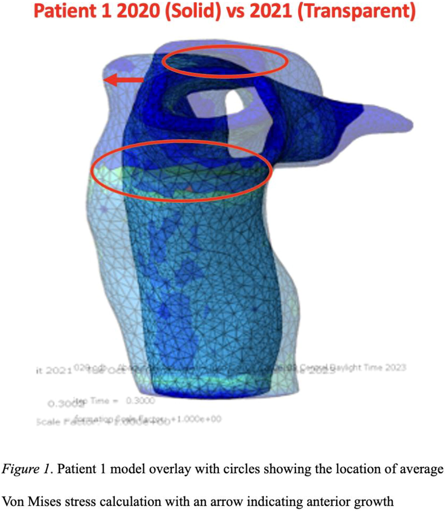
B101 - Machine Learning-based Optimization Of Ceramic Scaffolds For Biomanufacturing
Q. Li1, C. Wu1, A. Entezari2, J. Zhong1, B. Wan1
1
University of Sydney, Sydney, Australia,
2
University of Technology, Sydney, Sydney, Australia
*Purpose/Objectives: Treatments of critical size bone defects caused by trauma, diseases, or cancer signify a major clinical challenge. While autograft transplantation remains a gold-standard treatment option for bone defects at present, its use has been largely restricted by various drawbacks such as the need for secondary procedures, morbidity of donor site, and limited availability of bone grafts. To address these limitations, tremendous research effort has been devoted to scaffold-based tissue engineering, in which ceramic additive manufacturing (AM) technologies have exhibited great promise to create functional scaffolds with tailored biomechanical properties. Nevertheless, the true potential of the advanced biofabrication and AM techniques has not been fully exploited yet due to lack of clinical-driven optimization approaches available. This paper aims to develop a novel machine learning based design procedure for biofabrication of tissue scaffold.
*Methodology: To address the challenge, a clinic-based machine learning (ML) design approach is proposed herein, where 3D ceramic printing technique is combined to fabricate scaffolds with triply periodic minimal surfaces (TPMS), aiming to fulfil the anticipated biomedical requirements for the desirable bone regeneration outcomes. The proposed ML based design strategy couples a Bayesian optimization (BO) algorithm to enable time-dependent mechano-biological optimization of the 3D printed ceramic scaffolds at a reasonably low computational cost. Furthermore, a lithography-based ceramic manufacturing (LCM) technique is employed to fabricate the optimised scaffolds based on the proposed ML-based design framework. The micro-CT analyses of the additively manufactured ceramic scaffolds are used to assess their geometric qualities obtained from additive manufacturing.
*Results: Two representative clinical examples are adopted here, which use the ceramic scaffolds for treating segmental defects of sheep tibia and mandible, respectively. The animal in-vivo study results are used to correlate with the in-silico modelling results. It is demonstrated that the optimised TPMS scaffolds can enhance bone ingrowth outcomes significantly. This study is expected to gain new insights into design strategy and provide an effective design tool for ceramic additive manufacturing.
*Conclusion/Significance: This study proposed a machine learning (ML) based design approach to ceramic additive manufacturing (AM) of bone tissue scaffolds. A mechano-biological optimization of the 3D printed ceramic scaffolds is considered with cheap-to-evaluate cost for in-vivo study. It is shown that the optimized design of the scaffolds performed substantially better for treating the critical size segmental defects in a sheep tibia and mandible. The study is expected to leverage advanced ML and ceramic AM techniques for design and fabrication of porous scaffolds for clinical applications with ease.
B102 - Transplantation Of Conformal Coated Islets In A Pre-vascularized Cell Pouch™ Device For Beta Cell Replacement In Diabetic Rats
G. C. Gonzalez1, A. V. Hernandez1, N. H. Dehaseth2, N. M. Ziebarth2, A. A. Tomei1
1
Diabetes Research Institute, University of Miami, Miami, FL,
2
Department of Biomedical Engineering, University of Miami, Miami, FL
*Purpose/Objectives: Type 1 diabetes (T1D) is currently managed with daily insulin injections and in limited cases, intrahepatic allogeneic islet transplantation. Islet transplantation, however, requires chronic systemic immunosuppression to prevent rejection and recurrence of autoimmunity limiting its applicability to a small cohort of patients with severe diabetes. Here we evaluated the capability of conformal coated islets to reverse diabetes in rats after transplantation in the prevascularized subcutaneously implanted retrievable Sernova Cell Pouch device. This combination product has the potential to eliminate the need for chronic systemic immunosuppression after islet (donor or stem cell derived) transplantation, increasing the safety of the procedure and its applicability to larger numbers of patients with T1D.
*Methodology: Conformal coating (CC) with polyethylene glycol (PEG) hydrogels was achieved with a custom flow focusing device through coaxial flow of the coating solution with immiscible polypropylene glycol. Thickness and mechanical/permeability properties of single conformal coated (CC) model beads were characterized up to 100 days after fabrication in vitro by FRAP and AFM. Cell Pouch devices provided by Sernova Corp. were placed subcutaneously in female Lewis rats for five weeks. Then, diabetes was induced by streptozotocin injection and CC Lewis male rat islets were transplanted into the prevascularized pouch. Abatacept was given intraperitoneally (2mg/rat every other day starting 14 days before islet transplant). Blood glucose was measured 3-times/week and serum C-peptide weekly through ELISA; IPGTT was conducted 34 and 98 days after transplantation. Grafts were removed 101 days after transplantation to confirm CC islet graft-mediated diabetes reversal.
*Results: We found a linear correlation between islet diameter and CC islet diameter confirming that the coatings conform to the size and shape of the islets. PEG coatings were permeable to insulin and glucose but impermeable to IgG and diffusion coefficients were stable between 10 and 100 days after coating. CC capsules were mechanically stable for at least 100 days during in vitro culture. Transplantation of 4000 IEQ/rat syngeneic islets in the subcutaneous prevascularized pouches with Abatacept reversed diabetes in 100% of rat recipients. We also observed weight gain, serum C-peptide and CTLA-4 levels higher than 1ng/mL and 200µg/mL, respectively, and normal glucose tolerance in the recipients. Graft removal at 101 days after transplantation caused recipient rats to revert to hyperglycemia confirming graft function.
*Conclusion/Significance: CC minimizes coating thickness and transplant volume, enabling encapsulated islet transplantation into retrievable and confined pre-vascularized subcutaneous cell pouches. By optimizing the CC islet dosing and immunomodulation regimen in the syngeneic rat islet model we showed safety and efficacy of this combination product for at least 100 days with ongoing studies in allogeneic rat islet transplant models.
B103 - Antipsoriatic Patches Loaded With Polyphenols Applied On Psoriatic Skin Substitutes
Y. Ruel1,2, F. Moawad3, J. Alsarraf4,5,6, A. Pichette4,5,6, J. Legault4,5,6, D. Brambilla3, R. Pouliot1,2
1
Faculté de pharmacie, Université Laval, Québec, QC, Canada,
2
Centre de recherche en organogénèse expérimentale de l’Université Laval/LOEX intégré au Centre de recherche du CHU de Québec-Université Laval, Québec, QC, Canada,
3
Faculté de pharmacie, Université de Montréal, Montréal, QC, Canada,
4
Département des Sciences Fondamentales, Université du Québec à Chicoutimi, Chicoutimi, QC, Canada,
5
Centre de recherche sur la boréalie (CREB), Chicoutimi, QC, Canada,
6
Laboratoire d’Analyse et de Séparation des Essences Végétales (LASEVE), Chicoutimi, QC, Canada
*Purpose/Objectives: Psoriasis is an inflammatory skin disease characterized by red thick plaques and by important leucocyte infiltration, especially dendritic cells and T lymphocytes. Moreover, the risk of toxicity using systemic or biological treatments for a long-term use is high. Consequently, topical treatments are usually the first choice of therapy because of their local effects. However, ointments contain fatty excipients and require several daily applications to the psoriatic skin, which eventually lead to poor patient adherence and drug underdosage. Therefore, novel custom-made separable and biodegradable microneedle patches were used to control the release of antipsoriatic drugs. Recently, we have studied the antipsoriatic effects of polyphenols on T cells and psoriatic keratinocytes coculture. The expression of several pro-inflammatory cytokines including TNF-α and IL-17A was reduced, as well as cell proliferation, decreasing Ki67 and PCNA levels. Thus, these polyphenolic compounds were loaded into the microneedle patches. The antipsoriatic effect of this treatment was evaluated on immunocompetent human psoriatic reconstructed skin enriched with T lymphocytes. The objectives of this project were to: 1) Develop a method to apply these microneedle patches on human reconstructed skin, and 2) Evaluate the antipsoriatic effects of these microneedle patches applied on human psoriatic reconstructed skin and loaded with either polyphenols or methotrexate, a reference treatment for moderate to severe psoriasis, by comparing the epidermal and the skin total thickness with their respective counterparts.
*Methodology: 1) Biodegradable microneedle patches in PLGA/PLA loaded with Cy5-COOH were applied on the reconstructed skin substitutes (N = 3 donors, n = 3 reconstructed substitutes per condition). Each patch held 100 microneedles. Different combinations of time and temperature were evaluated to enable the detachment from the upper part of the patch leaving the microneedles in the skin. The percentage of successfully implanted microneedles in the skin was determined by fluorescence imaging with a Typhoon scanner. 2) Psoriatic skin substitutes enriched with T cells were cultured for 49 days and then, the microneedles loaded with polyphenols were inserted into the skin for a one-week treatment. At the end of the experiment, skin biopsies were harvested; skin samples directly in contact with the patches and beside the patches were compared. The thickness of the epidermis and of the total living skin with and without the treatments were determined (N = 3 psoriatic donors, N = 3 healthy donors, n = 2 substitutes per condition).
*Results: 1) After 20 minutes at 20 °C followed by 5 minutes at 37°C, the patches were detached from the substitutes for which fluorescent imaging showed that 96% of the microneedles were successfully implanted into the epidermis of the substitutes. 2) The epidermis thickness of the psoriatic substitutes was reduced of 57% by the application of the microneedles loaded with polyphenolic compounds (biopsies beside the patches) compared with psoriatic control. Our results show that there was diffusion of the polyphenols into the skin substitutes. This reduction was also more significative compared with methotrexate.
*Conclusion/Significance: Microneedle patches loaded with polyphenols are promising for psoriasis treatment and could improve the compliance of patients.
B104 - Sustainable Delivery Of Retinoic Acid Enhances The Functional Maturation Of Human Stem Cell-derived Motor Neurons
J. Seong1, C. Chun2, A. S. Smith2, M. Kim1, T.-E. Park1, D. L. Mack2, J. Joo1
1
Ulsan National Institute of Science and Technology(UNIST)), Ulsan, Korea, Republic of,
2
university of washington, seattle, WA
*Purpose/Objectives: Human induced pluripotent stem cells (hiPSCs) and differentiation technology have provided remarkable access to the study of neuromuscular diseases. However, the technology of differentiation still needs to address the insufficient maturation of differentiated neurons. This problem is, in part, due to the absence of replicating the spatiotemporal gradient of morphogens. For instance, retinoic acid (RA) is one of the essential ingredients for neural differentiation. However, it suffers from a short half-life in the physiological environment. Herin, we suggest the use of biodegradable porous silicon microparticles (pSiMPs) to maintain and regulate intact RA supplements during in vitro differentiation.
*Methodology: Hydrophobic RA is securely loaded into pSiMPs to demonstrate suitable conditions for motor neuron differentiation. Porous silicon substrates are fabricated via an electrochemical etching process, followed by lift-off and ultrasonic fracture to form microparticles. The pores safely protect RA molecules via a self-sealing process to form a thin oxide layer onto the silicon well. RA-loaded pSiMPs are added to the hiPSCs, thereby sustainably releasing the intact RA molecule over time while minimizing direct contact with hiPSCs. Using microelectrode arrays (MEA) and whole-cell patch clamps confirm the electrophysiological function of the neurons. Single-cell RNA sequencing is also performed to track transcriptomic changes.
*Results: We found that RAs were sufficiently loaded into pSiMPs and released with intact activity during neuronal differentiation. The hiPSCs are cultured under sustained release of RA over 12 days without changes in cell viability and differentiation efficiency caused by dissolved silicon components from pSiMPs. RA from the pSiMPs maintains a constant concentration over 24 hours in the media environment. The neurons are well-differentiated under these conditions in the morphological aspect. Moreover, the differentiated neurons show improved electrophysiological functions, not only in individual neuron activity but also in neuronal network activity. Furthermore, at a gene level, sustainable RA supplement leads to increased axon genesis and neurofilament bundle assembly genes.
*Conclusion/Significance: Biodegradable pSiMPs provide sustained release of active RA, advancing in vitro neuronal differentiation from hiPSCs without any side effects on cell viability and differentiation efficiency. Also, not only at the functional phase but also at the gene level, maintaining RA concentration during differentiation is critical for producing matured neurons. This platform serves as an advanced tool for understanding in vivo neuronal differentiation by regulating neurotrophic factor concentration in the in vitro environment.
B106 - Vessel On A Chip System For Study Of Nanotherapeutics-Wall Interactions For Abdominal Aortic Aneurysm Repair
J. Yau, X. Qin, C. Marquez, Y. Liu, A. Ramamurthi
Lehigh University, Bethlehem, PA
*Purpose/Objectives: Abdominal Aortic Aneurysms (AAAs) are localized, rupture-prone expansions of the vascular wall in the abdominal aorta due to degradation of elastic and collagen fibers, proteins that impart elasticity and stiffness respectively, by overexpressed matrix metalloproteinases (MMPs). Currently, invasive and risky surgery is the only intervention for AAAs and no drug-based therapies exist to reverse these conditions. Previous in vitro work on aneurysmal smooth muscle cells (aSMCs) has shown promise in using nanotherapeutics as a method to manage AAAs by both promoting elastogenesis and inhibiting further proteolysis through release of therapeutics and stabilization of the extracellular matrix (ECM). In this work, we expand on previous results to investigate both targeting and pro-elastogenesis capacities of spherical and rod-shaped nanotherapeutics using a bilayer microfluidic device that incorporates hydrodynamic flow and cell-cell interactions.
*Methodology: Nanoparticles (NPs) were formulated using an emulsification method with PLGA (poly(lactic-co-glycolic acid), 0.5-0.7 dL/g carboxyl terminated) and 1% polyvinyl alcohol (PVA) surfactant. Rod-shaped NPs were created by embedding spherical NPs into a 10% PVA film and stretching the film while placed in a 70°C mineral oil bath. The bilayer microfluidic device was created by pouring 10:1 polydimethylsiloxane (PDMS)-curing agent onto molds for the bottom and top layers and aligned on a glass slide. The top layer of the microfluidic device was seeded with Rat Aortic Endothelial Cells (RAOECs) while the bottom layer was seeded with elastase induced rat aneurysmal smooth muscle cells (EaRASMCs). For targeting studies, coumarin 6-tagged PLGA NPs were added in low serum media and quantified for fluorescent intensity. Similarly, for studies investigating ECM stabilization, Doxycycline (Dox) encapsulated NPs were added in similar manner and analyzed for ECM proteins.
*Results: Dynamic light scatting, scanning electron microscopy, and atomic force microscopy studies were conducted to verify and quantify the shape, size, and charge of the spherical and rod-shaped PLGA NPs. Endothelial disfunction was initiated using cytokines and confirmed using immunofluorescent staining for Integrin αVβ3 and VE-Cadherin. Initial results in solely RAOECs indicate increased NP endocytosis and transmigration in cytokine activated ECs vs. healthy ECs with further enhanced endocytosis and transmigration when using rod-shaped NPs. As NP endocytosis was investigated in a microfluidic device incorporating hydrodynamic flow, we notice similar trend of increased NP endocytosis and transmigration through the cytokine activated ECs vs. healthy ECs (Figure 1). Additionally, there was an increase of uptake of NPs in the microfluidic vs. no flow conditions indicating flow facilitates particle endocytosis by ECs. These results translate to increased transmigration of the Dox-NPs to the aSMCs injury site.
*Conclusion/Significance: Our results demonstrate the potential of using nanotherapeutics as a management method for AAAs. We see rod-shaped NPs have increased capability for uptake and translocation through RAOECs. These same results were confirmed using a microfluidic co-culture system of ECs with SMCs. Results also demonstrate ECM stabilization by Dox-NPs by improving EC health to synergistically enhance EaRASMC recovery. This information may be leveraged for development of nanotheraputics to actively target and recover the AAA segment.

B107 - Poly(lactic-co-glycolic Acid) Microparticles For The Delivery Of Model Drug Compounds For Applications In Vascular Tissue Engineering
J. M. Wyse, B. Sullivan, P. Lopez, C. Rathbone, M. E. Wechsler
University of Texas at San Antonio, San Antonio, TX
*Purpose/Objectives: Localized delivery of pro-angiogenic factors to promote the repair and regeneration of damaged tissues remains a challenge in vascular tissue engineering as current delivery methods fail to maintain the necessary sustained release profile and often rely on supraphysiologic doses to achieve the desired therapeutic effect.;;By implementing a microparticle delivery system, localized delivery can be coupled with sustained controlled release to mitigate the risks involved with the high dosages currently required. For this purpose, poly(lactic-co-glycolic acid) (PLGA) microparticles were fabricated and the loading, release, and delivery of model angiogenic molecules specifically a small molecule, nucleic acid, and protein, were assessed in vitro using microvascular fragments (MVF).
*Methodology: Unloaded and loaded microparticles were fabricated via anti-solvent microencapsulation using standard techniques. Particles were loaded with either methylene blue (MB), microRNA mimic tagged with Cy3 (miRNA), or bovine serum albumin (BSA). Microparticle size was determined using scanning electron microscopy. Drug release was performed from microparticles alone (in PBS at 37°C over 14 days), as well as from microparticles imbedded in fibrin gels. At select time points, PBS supernatant was removed and analyzed for the concentration of the loaded model. Lastly, MVFs were incubated with the miRNA-loaded microparticles in fibrin gels over 5 days. MVFs were fixed, stained with lectin, and fluorescently imaged to visualize miRNA distribution and MVF viability.
*Results: PLGA microparticles were spherical, polydisperse, (Figure 1A) and diameters directly related to the size of the loaded model drug (Figure 1B). The release study revealed that loaded microparticles alone, across all model drugs, exhibited burst release that plateaued over time which was also observed by the model drugs alone in fibrin gel (Figure 1C). However, the microparticles in the fibrin scaffold resulted in a much slower release and did not have as large of an initial burst. This suggests that the addition of the particles within the scaffold, which mimicked the MVF study conditions, allowed for a more sustained release profile. Lastly, the MVF study revealed that MVFs were visible and distributed throughout the entire fibrin gel in both miRNA alone or loaded microparticle trials, indicating the microparticles and miRNA did not hinder MVF viability (Figure 1D). With the miRNA-loaded microparticles there was increased signal and distribution of Cy3 fluorescence compared to miRNA alone. Therefore, microparticle use improves the introduction of miRNA to MVFs.
*Conclusion/Significance: The use of PLGA microparticles as a carrier for a variety of therapeutics ranging in size for vascular disease treatment is promising as the fibrin scaffold extended the controlled release and the MVF study demonstrated microparticle drug delivery would be viable. Moving forward, the system will be further optimized towards translational efforts for in vivo and clinical studies to develop revascularization strategies using biomaterials.
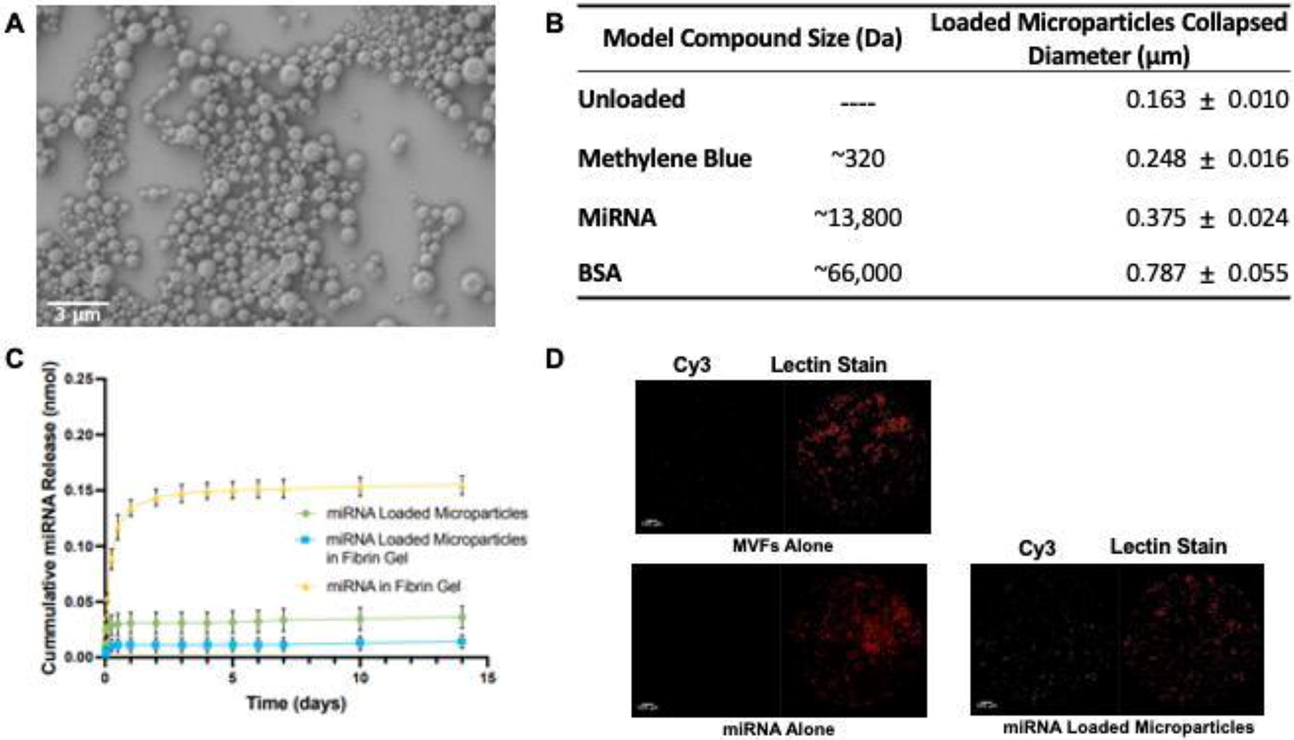
Figure 1. A) SEM image of miRNA-loaded microparticles. 5KX. Scale bar=3µm. B) Table of model compound sizes and collapsed microparticle diameters. C) Release profile of miRNA from microparticles, microparticles in fibrin gel, and miRNA in fibrin gel. Data reported as mean ± SEM (n=3). D) Fluorescent images of MVFs in fibrin gels. Scale bar=500µm.
B108 - Red Blood Cell Carrier For Controlled Delivery Of Therapeutics: The Effect Of Human Phenylalanine Hydroxylase Concentration On Their Metabolic Activity
Y. KISHITANI, N. KOJIMA
Yokohama city university, Yokohama, Japan
*Purpose/Objectives: Phenylketonuria (PKU), an inborn error of metabolism, causes central nervous system damage by increasing the concentration of phenylalanine (Phe) in the blood. PKU can be treated symptomatically by decreasing the blood Phe concentration. PEGylated phenylalanine ammonia lyase (PEG-PAL), which became available in Japan in May 2023, is a formulation based on phenylalanine ammonia lyase (PAL) from Anabaena variabilis. It was created with the expectation of decreasing immunogenicity and prolonging half-life in the blood. PAL is suitable for administration into the blood for decreasing Phe levels due to its activity in monomeric form. However, this formulation does not provide therapeutic benefit in all PKU patients, and there are concerns about the negative effects of administering a cyanobacteria-derived enzyme. We have investigated the development of human phenylalanine hydroxylase (hPAH) therapy to increase treatment options. However, hPAH may not be sufficiently active at low concentrations because it functions in a tetrameric form. It is necessary to administer the enzyme into the blood by using a red blood cell carrier to maintain high enzyme concentrations. In this study, we aimed to clarify the effects of changing concentrations of hPAH on their metabolic activity.
*Methodology: hPAH were recombinant Escherichia coli enzymes with a His-tag modification at the C-terminus. The enzyme activity was measured in a 200 µL system at 37°C after loading Phe into a reaction solution containing enzyme and coenzyme. Experiments were performed under three conditions of human PAH concentrations of 0.95 mg/mL (low concentration PAH) 2.74 mg/mL (medium concentration PAH) and 48.6 mg/mL (high concentration PAH) in the reaction solution and four conditions of coenzyme concentrations of 0, 100, 500 and 1000 μM for a total of 12 conditions.
*Results: Preliminary studies suggest that the metabolism of Phe by hPAH stopped within 2 hours and equilibrium was reached. The equilibrium concentration of Phe for each PAH concentration was examined. In the 100 μM coenzyme condition, the equilibrium concentration for low hPAH concentrations was approximately 600 μM, 500 μM for medium concentrations, and 300 μM for high concentrations. In general, the enzyme does not alter the equilibrium concentration. However, the result indicated that metabolic activity of hPAH possibly was changed by enzyme and coenzyme concentrations. Further investigation revealed that the reaction proceeded at higher coenzyme concentrations. Under high concentration conditions, the equilibrium concentration was below 100 μM at coenzyme concentrations above 500 μM.
*Conclusion/Significance: In terms of treatment of PKU, the equilibrium concentration to be below 100 µM is desirable. hPAH should not be administered directly into the blood to enable enzyme replacement therapy. It was suggested that it should be administered by enclosing in an erythrocyte carrier to maintain a high concentration with coenzyme.
B109 - Sustained GFP Protein Delivery To Hard-to-transfect Cells Using Biocompatible Polymeric Nanoparticle Emulsion
R. Negru, F. Teymour, G. Papavasiliou, M. Vaicik
Illinois Institute of Technology, Chicago, IL
*Purpose/Objectives: Immense potential for disease treatment and management lies in the area of intracellular nanoparticle delivery. Current methods are limited by the cell type and the dosing strategy. Developing strategies applicable to all cell types that allow for sustained therapeutic delivery would lend to a more effective and expansive application of therapeutics for the treatment of chronic diseases. Biocompatible nanoparticle emulsions (BCNEs), synthesized via inverse-phase miniemulsion polymerization, resulting in poly(ethylene) glycol (PEG) hydrogel nanoparticles with alterable mesh size and release kinetics and capable of encapsulating hydrophilic therapeutics may demonstrate success towards this feat.
*Methodology: BCNEs with encapsulated green fluorescent protein (GFP-BCNE) will be defined in terms of their release kinetics and the bioactivity of the therapeutic load using a previously published “surrogate scaffold method” to demonstrate release and electrophoresis and circular dichroism to evaluate bioactivity of encapsulated GFP post-reaction. Following verification of a mesh size design allowing for 20+ days of therapeutic release, GFP-BCNE will be delivered to hard-to-transfect cells (keratinocytes, stem cells, neuronal cells, and adipocytes), the cells will be observed in real-time using confocal microscopy for GFP release into the cytoplasm, and the cells will undergo Fluorescence Activated Cell Sorting (FACS) analysis to quantify the presence or lack thereof of GFP in cells at 5 time points: (1) two hours after initial treatment, (2) 50% GFP release, (3) 100% GFP Release, (4) 50% reduction in GFP signal, and (5) 100% reduction in GFP signal.
*Results: In preliminary studies, we have seen successful GFP delivery via BCNE into human-derived primary adipose tissue (Figure 1), which encourages our hypotheses. Remaining GFP-BCNE delivery and quantification experiments are on-going studies, and results will be presented upon completion of the study. We expect that the nanoparticle will cross the cellular membrane of each cell type, swell in the cellular cytoplasm, release the GFP into the cytoplasm of at least 90% of cells, and reach and maintain a maximum cytoplasmic fluorescence upon complete nanoparticle swelling and cargo diffusion for 14+ days.
*Conclusion/Significance: The successful transfection of keratinocytes, neurons, adipocytes, and stem cells along with the sustained delivery of GFP using GFP-BCNE will provide promising and supportive evidence for the future study of therapeutic delivery using BCNEs to cells in vitro and in vivo towards the improvement of cell and tissue functionality.

B110 - Development Of Nanocapsules Containing Nicotine, Nornicotine, Anatabine And Anabasine For The Treatment Of Central Nervous System Injuries
R. Zimmermann1, R. P. Silveira1, T. F. de Oliveira2, L. E. Sperling1, P. Pranke1
1
Universidade Federal do Rio Grande do Sul, Porto Alegre, Brazil,
2
Universidade Federal de Ciências da Saúde de Porto Alegre, Porto Alegre, Brazil
*Purpose/Objectives: Several studies have provided clear evidence of nicotine anti-inflammatory, neuroprotective and neuroregenerative activities. It is expected that the derivatives nornicotine, anatabine and anabasine also have these important properties. The objective of this work was to characterize the nanocapsules and evaluate their cytotoxicity in the PC12 cell line.
*Methodology: Nanocapsules were prepared using poly (lactic-co-glycolic acid) (PLGA) through the interfacial polymer deposition technique. They were characterized in terms of diameter, polydispersion index (PdI), zeta potential and encapsulation efficiency. Cytotoxicity was evaluated by MTT and LIVE/DEAD assays.
*Results: The nicotine nanocapsules presented a mean diameter of 227±0.09 nm, PdI of 0.281±0.03, zeta potential of -16.7±0.03 mv and encapsulation efficiency of 51.6%. The nornicotine nanocapsules had a mean diameter of 115±0.03 nm, PdI of 0.210±0.05, zeta potential of -26,9±0.03 mv and encapsulation efficiency of 46.5%. The anatabine nanocapsules presented a mean diameter of 128±0.04 nm, PdI of 0.216±0.04, zeta potential of -19.5±0.06 mv and encapsulation efficiency of 48.9%. The anabasine nanocapsules had a mean diameter of 158±0.05 nm, PdI of 0.227±0.05, zeta potential of -15.5±0.03 mv and encapsulation efficiency of 52,9%. The PC12 cell line was treated with nanocapsules containing nicotine, nornicotine, anatabine and anabasine in the concentrations of 1 µM, 5 µM and 10 µM, and cell viability was assessed at day 1, 3 and 7 by MTT assay and compared with cells cultivated on a culture plate without nanocapsule treatment (control) and cells treated with empty PLGA nanocapsules (equivalent to each nicotinoid concentration - blank). In general, treatment with higher concentrations of nicotine and its derivatives decreased cell viability when compared to the control. Therefore, the highest concentration of the nanocapsules that did not reduce the viability was used for further analyses. The LIVE/DEAD assay was performed with PC12 cells treated with: 5 μM nicotine; nornicotine 10 μM; anatabine 1 μM and anabasine 10 μM, and the results corroborated what had been demonstrated in the MTT assay.
*Conclusion/Significance: The results show that the nanocapsules have adequate size, zeta potential and polydispersion, which attests to their stability, as well as good encapsulation efficiency. In the cytotoxicity tests, it was observed that the nanocapsules containing nicotinoids were not toxic to cells at the concentrations tested, therefore, they may be a promising system for treatment of central nervous system injuries.
B111 - Sustained Release Of Anti-inflammatory And Bactericidal Agents From Coaxial Nanofibrous Scaffold For Burn Wound Healing
M. Kamboj, B. Das, V. Rathi, A. Poundarik
Indian Institute of Technology Ropar, Rupnagar, India
*Purpose/Objectives: Burn injuries have been prone to be a potentially fatal injury that can occur to any individual, regardless of age. Post burn injury shows significant higher level of systemic inflammatory cytokine and bacterial growth as well. Herein, we developed and incorporated Flurbiprofen as well as ciprofloxacin in core-shell nanofibrous scaffold respectively to achieve anti-inflammatory and antibacterial property.
*Methodology: The core-shell was developed from hydrophobic polycaprolactone (PCL) and hydrophilic Chitosan (CS) that shows co-delivery of two different therapeutic agent. The physiochemical and in-vitro biocompatibility were investigated accordingly. Additionally, contact angle measurement, swelling property and enzymatic degradation was studied. In-vitro antibacterial properties were also investigated against gram positive and gram negative bacteria
*Results: TEM, FTIR, FESEM and AFM results confirms the uniform fabrication of core-shell fiber loaded with drug having average diameter of 350-400nm. The core and shell diameter were controlled in such a way that it show significant loading and sustained release of ciprofloxacin upto 65 ± 2.1 % whereas Flurbiprofen upto 88 ± 1.9 % in 4days. Disc diffusion and optical density method against S. aureus and E. coli that specifies its LD 50 in 24hr against both strain. In-vitro biocompatibility studies was carried out using cell adhesion, MTT and live-dead assay against Normal Human Dermal Fibroblast cells. The cells formed a complete sheath on scaffold seen in electron microscopy with more than 80% cell viability in 7 day study that shows remarkable cell adhesion and proliferation over scaffold. Further, Flurbiprofen delivery and its effect was determine against macrophage cells. It signifies the M1 to M2 transition visualize with immunostaining and PCR confirm anti-inflammatory property
*Conclusion/Significance: The nanofibrous scaffold developed here could be an adequate delivering agent for sustained release of various therapeutic agent simultaneously. It notably promote infection control and burn healing.
B113 - In Vitro;Biocompatibility Assessment Of Living Therapeutic Materials: A High Throughput Strategy
K. K. Desai
Leibniz Institute for New Materials, Saarbrücken, Germany
*Purpose/Objectives: Living Therapeutic Materials (LTMs) are advanced materials comprising engineered cells entrapped in either self-produced or polymer-based biocompatible matrices. Particularly, LTMs containing drug-eluting bacteria have the potential to revolutionize drug delivery due to their ability to produce drugs in situ which eliminates costly post-production steps such as purification. In addition, genetically modified bacteria can produce drugs on demand or in response to a pre-defined trigger. This allows for treatments with more complex drug regimes. Until now, most research has focused on the design and fabrication of these LTMs systems and proof-of-concept devices for a variety of biomedical applications. However, in vitro biocompatibility validation of these devices to the host has been widely overlooked.
*Methodology: Here, for the first time, we present a 96-well plate-based method to screen LTMs to determine their biocompatibility potential in vitro. With this approach, we were able to encapsulate different strains of engineered bacteria in a model hydrogel (polyvinyl alcohol (PVA)-based) with a core/shell architecture. We showed the proliferation of bacteria in 3D confinement at different timepoints using alamarBlue assay and compared it with their growth in suspension. We could also track the morphology of mCherry-expressing bacteria using fluorescence microscopy. In addition, we studied the toxicity potential of our LTMs to fibroblasts and monocytes. This was quantified by lactate dehydrogenase and alamarBlue assays using culture supernatants of the ELMs. We further investigated the likelihood that our LTMs trigger inflammation. This was achieved by tracking cytokines such as interleukin-6 on monocytes.
*Results: Our results suggest that the LTMs were cytocompatible and did not trigger strong immune responses. We also demonstrated the versatility of our approach by examining three PVA-based LTM systems, embedding either gram-positive bacteria (Corynebacterium glutamicum or Lactiplantibacillus plantarum) or gram-negative bacteria (Escherichia coli).
*Conclusion/Significance: Our work illustrates an easy-to-follow and replicable high-content workflow for determining the in vitro biocompatibility of LTMs, a crucial step in advancing LTMs for clinical translation.
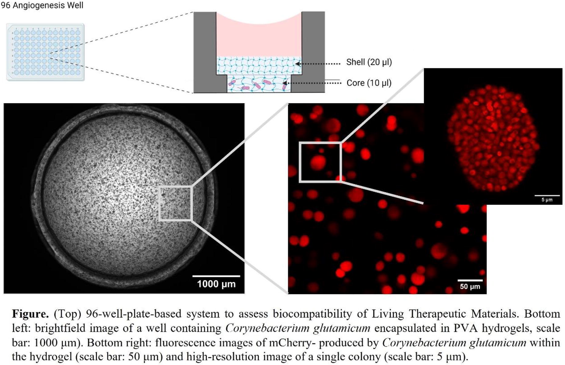
B117 - Functionalized Silk Nanoparticles As Immunotherapy Delivery Systems In Cancer Therapy
S. J. Peters, N. Murugan, N. Rouleau
Wilfrid Laurier University, Waterloo, ON, Canada
*Purpose/Objectives: Metastatic melanoma is an extremely aggressive cancer with estimated 5-year survival rates as low as 5%. Conventional treatments are cytotoxic, prone to drug resistance, and have poor efficacy in treating malignant tumors in the mid-to-late stages. Therefore it’s imperative that we devote resources into the development of novel treatments for melanoma. Nanoparticle-based therapies are a promising alternative as they have the advantages of improved biocompatibility, targeted drug delivery leading to higher tissue specificity, controlled drug release, and optimized permeability and retention in the tumor microenvironment (TME). Nanoparticles (NPs) can passively target tumors by exploiting their poor lymphatic drainage and leaky blood vessels, leading to the natural accumulation of NPs in the TME. Silk-NPs display pH-dependent drug release kinetics, releasing larger quantities at faster rates as pH decreases. This allows for further targeting of melanoma cells, as the TME is acidic. Silk-NPs have the advantages of being biodegradable and biologically inert. These properties are specific to biomaterial-based NPs and lowers their toxicity. Multidrug resistance can be overcome using double-stranded RNA (dsRNA) which is known to activate innate immune responses in the TME. Silk-NPs are extremely versatile and can be altered to deliver any molecule. Therefore, dsRNA analogues can be attached to silk-NPs to induce an innate immune response by activating innate pathogen recognition receptors (PRRs) found in the endosome and cytoplasm. Activation of these PRRs leads to pro-inflammatory responses and eventually apoptosis of cancer cells. The objective of this study is to develop a tunable silk-NP delivery system for the release of immunomodulatory nucleic acids within melanoma cells.
*Methodology: Silk-NPs are made with size ranges of 40nm to150nm from silk fibroin (Bombyx mori). Variation in silk-NP size made it possible to determine the optimal size for melanoma cell transfection. The silk-NPs are electrostatically coupled to dsRNA analogues using cationic polymers. Activation of the innate immune system is assessed by measuring levels of innate immune proteins via enzyme-linked immunosorbent assay (ELISA), after exposure to silk-NPs. Cell death and cell stress assessments are conducted using viability and oxidative stress assays. The pH-dependent release mechanism of silk-NPs is tested using dialysis tubes at several pHs relevant to the TME. The in vitro studies have been translated into murine models because cell cultures cannot fully model the complexities of cellular signalling involved in the innate immune response. Cell cultures and mice from both sexes are used to elucidate any sex-dependent mechanisms of melanoma. Expression of sex hormones are monitored overtime using ELISA and analyzed for correlations with levels of innate immune system expression.
*Results: Development of a novel silk-based drug delivery system that confers improved bioavailability and biocompatibility to chemotherapeutic drugs, leading to better prognosis for melanoma patients.
*Conclusion/Significance: A better understanding of the factors involved in the interferon response and immune cell recruitment. With this knowledge modulation of the innate immune response can be improved to overcome the colossal barrier of multidrug resistance found in many cancers.
B118 - A Kirigami Strategy For 4D Tissue Formation
K. L. Gasvoda, A. Ding, O. Jeon, A. Sterenberg, E. Alsberg
University of Illinois-Chicago, Chicago, IL
*Purpose/Objectives: Shape evolution, such as bending and folding geometries, in tissue development is crucial for functionality. Traditional tissue engineering systems composed of hydrogels and/or cells have been developed to replicate distinctive curvatures created during developmental processes, but some complex architectures are still difficult to achieve. Approaches utilizing 4D systems have facilitated the formation of unique tissue geometries; however, it is still challenging to achieve some complicated shapes. Recently, Kirigami strategies (KSs) that enable 4D complex shape transformations have been employed for other biomedical applications, such as nanosensors, but KSs have not yet been implemented in cytocompatible approaches for tissue engineering. Here, we report a cytocompatible Kirigami system for the creation of complex construct architectures using different spatially patterned hydrogels with pre-designed differences in swelling ratios.
*Methodology: The system is comprised of two main biomaterial components: 1) a hydrogel composed of photocrosslinked alginate with controlled swelling that undergoes the shape transformation of the constructs and 2) a rigid and minimally swelling support ring composed of photocrosslinked polyethylene glycol and calcium-crosslinked alginate that maintains the desired curvature of the swelling alginate-based component throughout the culture. Photolithography was used to crosslink the hydrogels and engineer the specific original shapes required for the resulting desired shape transformations, which were controlled by the original swelling alginate shape and by changing the swelling properties of the alginate.
*Results: This system supports deformation throughout the alginate portion of the construct, which is sustained for the duration of the culture time. Encapsulated cells within the highly swelling alginate undergoing shape transformation maintained high viability after two weeks of culture. Importantly, encapsulated human mesenchymal stem cells (hMSCs) in the deforming hydrogel could differentiate down the chondrogenic lineage over 3 weeks in concert with the construct architectural changes. The curvature of the resulting cartilaginous tissue remained stable after being released from the rigid support.
*Conclusion/Significance: The cytocompatible KS system enables the formation of constructs that can undergo multiple different deformations within the same deforming component without breaking, while at the same time allowing for new tissue formation. This KS has great potential for engineering tissues that require distinctive curvatures.
B119 - Microfluidic Assembly Of Mitochondria-loaded Microparticles For On-demand Delivery
R. C. Miller, J. S. Temenoff
Georgia Institute of Technology, Atlanta, GA
*Purpose/Objectives: With the discovery that mitochondria can be actively transferred from one cell type to another and promote wound healing, new therapeutic approaches have been developed to harness the benefits of this phenomena. Nevertheless, promoting sufficient levels of endogenous mitochondria transfer to observe tissue level changes can be both variable and difficult to predict. Therefore, enzyme-directed delivery of encapsulated mitochondria may improve the observed therapeutic efficacy. Additionally, uptake of free mitochondria post-release is still a major limitation affecting the therapeutic potential. As such, the goal of this work is to (1) increase mitochondrial uptake through polymer-based mitochondrial surface modification and (2) enable targeted mitochondria release through the encapsulation in matrix metalloproteinase (MMP) cleavable hydrogel microparticles (Figure 1a). Using this system, we hypothesized that increased mesenchymal stem cell (MSC) derived mitochondria transfer to smooth muscle cells will support the energy demands for myogenesis in vitro.
*Methodology: Mitochondria (stained with MitoTracker Red) were isolated and purified via density gradient centrifugation from human MSCs (RoosterBio, Inc). To facilitate free-mitochondria uptake, TAT-peptides (QPRRRQRRKKKRG) and triphenol phosphate (TPP) were conjugated to dextran via carbodiimide chemistry (TAT-dextran-TPP). The isolated mitochondria were incubated with 2.5wt% TAT-dextran-TPP for 12 hours and subsequently encapsulated in MMP-cleavable hydrogels (1µg/mL total mitochondria protein) using droplet microfluidics (Figure 1b). Either polymer-free mitochondria, TAT-dextran-TPP mitochondria, or TAT-dextran-TPP mitochondria-loaded microgels were incubated with smooth muscle myoblasts (C2C12) for 24 hours. The mitochondria uptake was characterized based on the mean pixel fluorescent intensity of MitoTracker Red using confocal microscopy (LSM 710). To determine the effect of mitochondria uptake on C2C12 myogenesis, 80% confluent C2C12 cells were allowed to differentiate into myotubes for 5 days and imaged. One-way ANOVA with Tukey’s post hoc test was performed for statistical analysis. p≤0.01 was considered significant and a minimum of 5 replicates was used.
*Results: MSC-derived mitochondria encapsulated in ∼90µm diameter MMP-cleavable microgels were actively released over 48 hours when in the presence of 1mg/mL collagenase. No release was observed in a collagenase-free solution, illustrating selective release in the presence of MMPs (Figure 1c). Furthermore, following 24-hour incubation with free-mitochondria, we observe a 7.5-fold increase and 6.8-fold increase in mitochondria uptake by C2C12 cells when functionalized with TAT-dextran-TPP polymer or released from microgels respectively (Figure 1d). After 5 days, we observed greater differentiation of myoblasts into myotubes for both the TAT-dextran-TPP mitochondria and TAT-dextran-TPP mitochondria-loaded microgel conditions (Figure 1e). We attribute the increase in myogenesis to the increased uptake of functional mitochondria.
*Conclusion/Significance: From this study, we are able to illustrate that mitochondria can be isolated from MSC culture, loaded into MMP-cleavable microgels and released, all while preserving their function. Moreover, decoration of the mitochondria with TAT-dextran-TPP leads to a noticeable increase in internalization by C2C2 cells, resulting in boosted myogenesis. We note that while these results are most directly applicable for muscle regeneration, on-demand mitochondrial release for localized cellular uptake has applications in a wide range of regenerative medicine applications.

B120 - A Bioreactor For Magnetomechanical Dynamic Scaffold Stimulation And Acceleration Of Bone Tissue Formation
M. Kalogeropoulou1, M. Takhsha2, F. Albertini2, P. Fucile1, L. Moroni1
1
MERLN Institute, Maastricht, Netherlands,
2
Institute of Materials for Electronics and Magnetism (IMEM), National Research Council (CNR), Parma, Italy
*Purpose/Objectives: Within the broad spectrum of strategies for successful tissue regeneration, physical stimuli have received significant attention owing to their capacity to create a more biomimetic environment. Dynamic mechanical loading has been identified as a crucial stimulus for stem cell differentiation and bone tissue maturation. Recently, magnetic fields have been recognized as another stimulation type capable of promoting osteogenesis. However, the combined effects of magnetomechanics (i.e. simultaneous magnetic and mechanical stimulation) in bone regeneration remain unclear. Moreover, a significant discrepancy exists between suggested experimental systems for magnetic stimulation in vitro, impeding reliable comparison across different studies. Addressing these challenges, we present a comprehensive guide for the fabrication and customization of a robust bioreactor for high-throughput, in vitro magnetomechanical stimulation. By combining the bioreactor with magnetic, cell-laden scaffolds, we investigate the synergistic effect of magnetic and mechanical stimulation on the osteogenic differentiation of human mesenchymal stem cells (hMSCs).
*Methodology: The bioreactor features a moving magnet array aligned with a stationary culture stage. The magnetic stage motion is controlled by a stepper motor connected to a 3D printing motherboard, allowing the frequency and magnetic field strength to be customized using open-source software. Magnetic scaffolds with low (5%) and high (20%) magnetite content were 3D printed using PLA/PCL/Fe3O4 nanocomposites. Mechanical tests, magnetic characterization, and multiphysics simulations were combined to predict scaffold response to external magnetic stimulation. For the validation of the magnetomechanical effect, scaffolds were exposed to oscillating magnetic fields, and their deformation was documented using a confocal laser. In vitro, hMSCs seeded on the magnetic scaffolds underwent magnetomechanical stimulation every second day for 28 days, with static conditions serving as controls. Biological assays of osteogenic and mechanobiological markers were conducted in order to understand the synergy of the two stimuli.
*Results: The bioreactor was programmed to utilize a 50 mm maximum vertical distance for oscillatory motion of permanent magnets, resulting in a magnetic flux density between 0 mT and 275 mT at the bottom of each culture well. Magnetic characterization of the scaffolds showed a homogeneous magnetization profile across all conditions, which depended solely on their magnetic content. Confocal measurements indicated approximately 1% maximum compressive strain for high magnetic content scaffolds at low oscillation frequencies. A physiologically relevant (walking) frequency of 1 Hz was, thus, selected for cell experiments. In vitro, hMSCs displayed excellent viability and matrix production for all conditions. Scaffolds exposed to stimulation exhibited enhanced expression and secretion of osteogenic markers, compared to static controls, suggesting the potential of magnetomechanical stimulation for accelerating bone tissue formation.
*Conclusion/Significance: We have presented a versatile bioreactor for in vitro magnetomechanical stimulation of 3D structures and have used it for investigating the synergistic effect of cyclic, magnetomechanical stimulation in bone formation. Our results suggest that this stimulation type enhances osteogenic differentiation and could have a significant impact on bone formation in vitro. Moreover, the simple design and open-source approach of our bioreactor facilitate its use with various magnetic or nonmagnetic biomaterials and cell types, at a host of field intensities and frequencies.
B121 - Remote Activation Of Mechanotransduction Via Integrin Alpha 5 Aptamer Conjugated Magnetic Nanoparticles Promotes Osteogenesis
H. Hajiali, M. Rotherham, A. J. El Haj
Healthcare Technologies Institute, Institute of Translational Medicine, School of Chemical Engineering, University of Birmingham, Birmingham, United Kingdom
*Purpose/Objectives: Magnetic particles (MNP) are used for specific targeting of regions of cell membrane based receptors by conjugation on the surface with different types of biomolecules. Our research has investigated the use of MNP targeting for applying mechanical force directly to mechano-sensitive regions on the cell receptor thereby stimulating mechanotransduction and downstream signalling [1]. The force is applied remotely using dynamic field gradients which is currently being translated to the clinic for novel therapies for bone regeneration in Spinal fusion and Vertebral repair. Remote activation of mechanosensors such as ion channels and growth factors has been demonstrated to promote differentiation toward musculoskeletal cell types from multiple stem cells sources used for cell therapies [1,2]. A current challenge of these approaches using antibodies for targeting is non specific binding. In this presentation, we present our work using aptamers as targeting agents. Aptamers are short nucleic acid sequences capable of specific, high-affinity molecular binding which bind to proteins and receptors with a higher affinity compared to antibodies. Aptamers have several advantages, including small size, good biocompatibility, and low immunogenicity [1]. In addition, inactive aptamer clones can be selected which bind to specific regions enabling controlled mechano-activation without generic direct binding activation of key receptors.
*Methodology: The MNPs were conjugated with Cd49e antibody for binding to the integrin α5, and CD49e/ CD29 aptamer for binding to the integrin α5β1 receptors. To explore the potential variation in aptamer binding and antibody strategies, antibody and aptamers were conjugated to MNPs and their signalling efficacy was investigated using Y201 TCF/LEF GFP reporter cells. Aptamer concentration (2.5, 5, and 10 μg/mg) was optimized. Gene expression of RUNX2 and osteopontin, ALP activity, and Alizarin red staining was analyzed.
*Results: The TCF/LEF reporter cell line demonstrated that mechano-activation of the cells by Fz aptamer MNPs resulted in significant upregulation of the Wnt signaling pathway relative to the activation of the cells by Fz antibody MNPs. Furthermore, the mechano-activation of MSCs by the CD49e/CD29 MNP-aptamers significantly upregulated the expression of the osteogenic marker, RUNX2 compared to the mechano-activation of MSCs by the CD49e MNP-antibodies. In addition, our findings indicate a concentration-dependent response in ALP activity. This trend was consistently reflected in the level of alizarin red staining, as well as the expression levels of osteopontin and RUNX2 (Figure 1). Our results show the responses are upregulated by low concentrations of aptamers ensuring high efficacy with low immunogenicity for osteogenic differentiation for Orthopaedic applications.
*Conclusion/Significance: ConclusionOur studies provide valuable insights into the use of mechano-targeted MNP platforms for bone regeneration. The study highlights the potential for using aptamer bound platforms for these applications in promoting signaling pathways related to bone formation. Acknowledgments This work was funded by ERC Advanced Grant DYNACEUTICS (no. 789119).

Figure 1. Western Blot images of magnetically stimulation hMSCs by different concentrations of aptamers loaded on MNP for GAPDH, RUNX2, and Osteopontin after 7 days of stimulation; Data represented as mean ± SEM (n = 3), * for p < 0.05, and ** for p < 0.01.
B123 - Marine Biological Coproduct As A Source Of Hydroxyapatite (HA) For Dental Pulp Regeneration
N. P. Nosal
University of Bergen, Bergen, Norway
*Purpose/Objectives: The pulp within the tooth provides nutrients and detects potential pathogens, and its loss will make the tooth fragile. Traditionally, infected dental pulp is removed and replaced with inorganic materials. Using regenerative endodontics, inflamed/necrotic pulp tissues are replaced with regenerated pulp-like tissues to revitalize teeth and improve their health. The inherent properties of marine-derived hydroxyapatite (HA) make it particularly promising for dental pulp regeneration because of its multi-doped ion composition and micro- and nano-scale architecture. Hence, the study investigates the odontogenic potential of dental pulp stem cells (DPSC) when exposed to HA doped with essential ions, including manganese (Mn), strontium (Sr), and zinc (Zn).
*Methodology: In this study, HA was doped with different concentrations of Mn, Sr, and Zn. Dental Pulp Stem Cells (DPSC) were cultured up to 21 days, to investigate their behavior. Cell viability, proliferation and morphology were assessed in vitro using live/dead stain, PicoGreen quantification assay and PrestoBlue. Additionally, cell morphology was examined using a confocal microscope. Cell differentiation was measured by quantitative real-time reverse transcriptase-polymerized chain reaction analysis (qRT-PCR), alkaline phosphatase test and Alizarin red staining. These methods provided insights into the cellular responses and differentiation patterns when the cells were subjected to marine HA, which aids in understanding their potential applications.
*Results: According to the PicoGreen assay, there were varying levels of dsDNA among the groups. Sr 3% has the highest dsDNA content after 14 days, however, experienced a decline after day 21. In contrast, Sr 6% demonstrated high levels of dsDNA with a persistent grow even after day 21. The PrestoBlue assay showed an increased proliferation across all the different groups. Notably, Sr 3% and Sr 6% had the highest proliferation after 14 days, whereas HA doped with Zn 1.5% demonstrated a continued growth even after day 21. Alkaline phosphatase staining revealed significant differentiation patterns in the doped groups. Importantly, Zn 3% and Zn 1.5% showed the highest peaks after day 14, while Sr 3% and Sr 6% displayed the most prominent peaks after day 21. Additionally, Sr 1.5% exhibited the most calcium deposits after 21 days. These findings demonstrate the effect of marine HA doped with varying concentrations of strontium (Sr), zinc (Zn), and manganese (Mn) and their influence on cellular proliferation, differentiation, and calcium deposition. Specifically, Sr 3% and Zn 1.5% showed promising outcomes on proliferation and differentiation respectively using multiple assays. Hence, the results indicate their potential on enhancing the properties of hydroxyapatite in tissue regeneration.
*Conclusion/Significance: Based on these results, marine HA enhanced proliferation and differentiation of DPSC in response to ion doping in a concentration-dependent manner. The increased proliferation and differentiation of DPSC when exposed to specific concentrations of Sr and Zn highlight their potential application in dental pulp regeneration.
B124 - Thiolated Tendon Tissue Particle Hydrogels With BMP-2 Microspheres For TBI Treatment
J. Townsend1, J. Deng2, B. Andrews3, S. Barbay4, R. Nudo4, M. Detamore1
1
University of Oklahoma, Norman, OK,
2
University of Oklahoma, Lawrence, KS,
3
University of Iowa Health Clinic, Iowa City, IA,
4
University of Kansas Medical Center, Kansas City, KS
*Purpose/Objectives: Traumatic brain injury (TBI) is a life-threatening condition characterized by internal brain herniation and commonly treated by a two-stage surgical intervention. In the first surgery, decompressive craniectomy is performed to reduce intracranial pressure and permit the brain to swell beyond the confines of the calvarial vault. In the second surgery, spaced weeks to months after the first, cranioplasty is performed to restore the cranial bone. During the time between surgeries, patients often develop the neurologic condition termed “syndrome of the trephined” characterized by headaches, dizziness, mood swings, and fine motor dexterity issues. Hydrogels are well-positioned for severe TBI treatment following decompressive craniectomy, given their innate flexibility and potential for bone regeneration to treat TBI in a single-stage surgery. We hypothesized that a tendon tissue particle hydrogel with encapsulated BMP-2 microspheres would enhance osteogenesis of human bone marrow mesenchymal stem cells (hBMSCs) and promote bone regeneration in a rat TBI model.
*Methodology: A pentenoate-modified HA (PHA) polymer was covalently linked to thiolated tissue particles of devitalized tendon (TDVT), thereby forming an interconnected hydrogel matrix. Pig Achilles’ tendon (Male, 6 months old) tissue was thiolated by reacting with N-succinimidyl S-acetylthioacetate. PHA was created by reacting HA with pentenoic anhydride. BMP-2 microspheres were fabricated using a Buchi B-390 encapsulator consisting of 1 mg of BMP-2 per gram of PLGA microspheres. In vitro groups received 4% PHA and 15% TDVT particles with 1, 5, or 10% (w/v) BMP-2 microspheres. Hydrogels seeded with hBMSCs was evaluated for osteogenesis using qPCR for COLI, OCN, and RUNX2 gene expression. Hydrogel formulations were evaluated in a rat TBI model for motor skill recovery using the reach test method over 8 weeks and cranial bone regeneration using microcomputed tomography after 10 weeks of recovery (5 x 8 mm defect, n = 6 or 7).
*Results: Gene expression for the TDVT group containing 10% BMP-2 microspheres significantly upregulated COLI, OCN, and RUNX2 expression compared to the TDVT group with soluble BMP-2 (p<0.05). Microcomputed tomography after 10 weeks of recovery demonstrated non-significant bone regeneration for hydrogel groups compared to the sham defect. 8-week motor skill recovery using the reach test method demonstrated reduced impairment for the TDVT group containing BMP-2 microspheres compared to the TBI group without treatment (p<0.05).
*Conclusion/Significance: The TDVT hydrogel containing 10% BMP-2 microspheres demonstrated attractive in vitro osteogenic gene expression; however, minimal new bone formation was observed after 10 weeks of recovery in the rat TBI model. Although bone regeneration was minimal, controlled release of BMP-2 from microspheres reduced motor skill impairment in the rat TBI model. A combined BMP-2 delivery approach of controlled release from microspheres and conjugation to the hydrogel matrix may potentially improve bone regeneration while maintaining reduced motor skill impairment.
B126 - Cell Viability With Exposure To 3D Printed Titanium Alloy Metal Mesh For Craniofacial Reconstruction
A. A. Cahoon, M. K. Vaicik, A. Mostafaei
Illinois Institute of Technology, Chicago, IL
*Purpose/Objectives: Metal implants are commonly selected for those with critical sized defects requiring a craniofacial implant. Much research has focused on modifying titanium implants with bioactive components to improve osteointegration. Recent studies have provided evidence that metal mesh implants can provide mechanical strength and have potential for bone tissue regeneration. The rationale for this research is that a 3D printed titanium alloy (Ti-6Al-4V) mesh implant would allow for a customized shape for the specific patient and allow for tissue regeneration from the host for better integration of the implant. Our long-term goal is to develop a 3D printed craniofacial implant made with a metal mesh structure that supports osteointegration. The objective of this research is to evaluate 3D printed titanium alloy nanostructures and mesh geometry on cell viability and behavior (adhesion, growth rate). These results are expected to have an important positive impact in developing personalized craniofacial implants with less inflammatory side effects to improve care for patients needing craniofacial reconstruction.
*Methodology: Titanium samples were 3D printed and then autoclaved to achieve sterilization. Samples were submerged in Dulbecco’s Modified Eagle Medium (DMEM) for 24 hours at 37°C to create Ti-6Al-4V-exposed leachate. In vitro cytotoxicity assays were performed on 3T3 fibroblast cells to determine the cell viability when exposed to metal sample leachate. 3T3 fibroblasts were cultured in 100% complete growth medium (control group), a mixture of 50% complete growth medium and 50% leachate (group A), and 100% leachate (group B). Cells were seeded in a 24-well tissue culture-treated plate at seeding density of 0.05 x 10⁶ cells/well. Cells were incubated at 37°C and 5% CO2. At day 5 of culture with the leachate live/dead staining assay (Thermo Fisher Scientific) was conducted to assess cell viability using Calcein AM and Ethidium Homodimer-1. Cells were imaged to determine percentage of cells viable by calculating the area of cells in the well stained green/live and red/dead. Cell attachment was assessed by staining 3T3 fibroblast cells with PKH26 red fluorescent cell linker (Sigma-Aldrich) and seeding these cells directly onto the surface of the Ti-6Al-4V samples. After 24 hours cells were imaged and processed to obtain the average area of cell coverage on the sample.
*Results: Average percentage of live/dead cells were recorded in three groups. Results of preliminary experiments were 97.14±2% live for control group, 87.46±12% live for group A, and 86.65±5% live for group B. These results suggest good cell viability with Ti-6Al-4V leachate. Results from the attachment experiment proved 3T3 cells successfully attached to the Ti-6Al-4V sample.
*Conclusion/Significance: This study investigated in vitro cytotoxicity of 3T3 fibroblasts to provide insight of cell viability when exposed indirectly (leachate) or directly to 3D printed Ti-6Al-4V samples. Cell viability results indicated that cells survived exposure to the titanium alloy leachate and were able to grow. Cell attachment results indicated that cells remained attached to the metal samples. Both results suggest that 3D printed Ti-6Al-4V samples are compatible with 3T3 fibroblasts. This study provides promising foundational results that encourage further studying of metal mesh craniofacial implants.
B127 - Development Of Biomaterials For Guided Bone Regeneration
G. M. Machado1, N. Maurmann2, R. Hartmann Kasper1, C. Gomes3, J. Girón2, P. Pranke2, M. Camara Brew1
1
Universidade Luterana do Brasil (ULBRA), Canoas, Brazil,
2
Universidade Federal do Rio Grande do Sul (UFRGS), Porto Alegre, Brazil,
3
Art Lounge, Brasilia, Brazil
*Purpose/Objectives: Guided bone regeneration is a crucial method for treating bone defects caused by trauma, disease, or surgery. This study aimed to create biomaterials in the form of chitosan-based composite particulate powder and electrospun polycaprolactone (PCL) membrane for guided bone regeneration.
*Methodology: The particulate biomaterial was obtained in the form of chitosan and evaluated. Additionally, it was complexed with ascorbic acid 2-phosphate (ASAP) and β-tricalcium phosphate (β-TCP) to form the chitosan/ASAP and chitosan/ASAP/β-TCP complexes. The particles were characterized using Fourier Transform Infrared Spectroscopy (FTIR) and zeta potential. The cell viability assay was conducted using 3-(4,5-dimethylthiazol-2-yl)-2,5-diphenyltetrazolium (MTT), while live cell staining was performed with fluorescein diacetate (FDA) and nuclear assay with 4',6'-diamino-2-phenyl-indole (DAPI). The membranes were developed using the electrospinning method. A 14% w/v polycaprolactone (PCL) membrane was treated with ASAP at a concentration of 300 μg/ml. The membranes underwent characterization through contact angle measurement, and a cell viability analysis using MTT was conducted. Additionally, a comparison was made with a commercially available collagen membrane. Statistical analysis was performed using ANOVA, followed by the Tukey test.
*Results: The FTIR results indicate a complexation process between chitosan, ASAP and β-TCP. The zeta potential measurements showed that chitosan particles had a value of 25.3 mV, while chitosan/ASAP and chitosan/ASAP/β-TCP had values of -3.2 mV and 1.2 mV, respectively. These results suggest that there was particle aggregation. Cell viability was assessed by measuring the absorbance after MTT. The mean and standard deviation values for absorbance were 0.096 ± 0.03, 0.096 ± 0.01, 0.129 ± 0.01, and 0.130 ± 0.01 in the control group, chitosan-treated cells, chitosan/ASAP group (p < 0.05), and chitosan/ASAP/β-TCP group (p < 0.05), respectively. In the electrospun scaffold, cell viability was not significantly affected compared to the control. The percent absorbance and standard deviation values relative to the control of MTT were 100 ± 29 for the cells in tissue culture plate (control), 80 ± 4 for the scaffolds PCL, 98 ± 23 for the scaffolds PCL/ASAP and 85 ± 10 for commercial membrane. The adsorption of ASAP on the membrane with PCL increased its hydrophilicity.
*Conclusion/Significance: The study concluded that all samples were biocompatible in terms of cell viability. Therefore, the particulate complexes and the membrane with adsorbed ASAP show promise as biomaterials for guided bone regeneration. Financial support was provided by the National Council for Technological and Scientific Development (CNPq), the Financier of Studies and Projects (FINEP), the National Institute of Science and Technology for Regenerative Medicine (INCT-Regenera) and the Stem Cell Research Institute (IPCT).
B128 - In Vitro Study Of The Cytotoxicity Of Resins For Restorative Dentistry
N. Maurmann1, G. M. Machado2, R. P. Silveira1, E. N. Santos2, E. R. Santos2, P. Pranke1,3
1
Universidade Federal do Rio Grande do Sul (UFRGS), Porto Alegre, Brazil,
2
Universidade Luterana do Brasil (ULBRA), Canoas, Brazil,
3
Instituto de Pesquisa com Células-tronco (IPCT), Porto Alegre, Brazil
*Purpose/Objectives: The use of composite resins has revolutionized restorative dentistry. However, concerns have been raised about their potential cytotoxicity due to the release of residual monomers, particularly Bisphenol A (BPA) and its derivatives, following polymerization. This study aims to evaluate the cytotoxicity of two commonly used resins on mesenchymal stem cells.
*Methodology: Specimens of Vittra APS® and Opallis® resins, each measuring 5.2 mm³, were produced, LED polymerized, and then immersed in a culture medium (DMEM supplemented with fetal bovine serum) to obtain extracts after 2 and 8 days. Dental stem cell viability was evaluated using 3-(4,5-dimethylthiazol-2-yl)-2,5-diphenyltetrazolium bromide (MTT) and live/dead cell staining with fluorescein diacetate (FDA) and propidium iodide. The experiment involved testing the cells after 24 hours of contact with extracts from 2 and 8 days. The study included a negative control (cells with culture media), a cell-free culture medium (white control), and treatment with Triton X-100 to induce cell death (positive control). The results were expressed as the mean ± standard deviation (SD) and evaluated using ANOVA followed by the Tukey test.
*Results: After 24 hours, the 2-day extract from Vittra® resin light-cured resulted in 85% viability, which decreased significantly (p<0.01) compared to the negative control group. The average absorbance concentration and standard deviation after 1 day of cultivation with 2 days of the extract were 0.529 ± 0.077, 0.537 ± 0.015, 0.450 ± 0.017 (p < 0.01), 0.073 ± 0.004 (p < 0.01), and 0.047 ± 0.013 (p < 0.01) at control, Opallis®, Vittra®, white, and death control, respectively. With the 8-day extract, all treatments significantly reduced cell viability compared to the negative control group (p < 0.01). The MTT absorbance and SD values were 0.615 ± 0.042 for the control group, 0.544 ± 0.039 for the Opallis® group, 0.498 ± 0.024 for the Vittra® group, 0.069 ± 0.016 for the white control, and 0.077 ± 0.026 for the death control. The 8-day extract of Vittra® resulted in the lowest viability percentage of 81% compared to the negative control group among all extracts studied.
*Conclusion/Significance: The study found that the 2-day extract from Vittra® resin caused a statistically significant decrease in cell viability within 24 hours. Additionally, both resins showed a reduction in cell viability compared to the negative control group when exposed to the 8-day extract. This highlights the importance of evaluating cytotoxicity over an extended period. However, neither resin caused a decrease in cell viability greater than 70% and therefore were not considered cytotoxic. Further research and development aimed at reducing cytotoxic effects and identifying alternative materials could enhance the safety and effectiveness of dental restorative procedures. Financial support: This work was supported by the National Council for Technological and Scientific Development (CNPq), the Financier of Studies and Projects (FINEP), the National Institute of Science and Technology for Regenerative Medicine (INCTRegenera), and the Stem Cell Research Institute (IPCT).
B130 - BONE REMODELING IN ORTHODONTICS. A NOVEL APPROACH
C. R. Gomes, C. M. Duarte
ArtLounge Odontologia, Brasilia, Brazil
*Purpose/Objectives: The goal of this presentation is to demonstrate a clinical outcome of how orthodontic movement with skeletal anchorage may improve bone remodeling. This technique may improve patient's lives without undergoing through orthognatic surgery.
*Methodology: Patient had a vertical growth of maxilla, with full exposure of central incisors in resting position. While in full smilling, she had a 4to 5 mm of gingival exposure.In order to improve this condition, 6 miniplates were placed two on zygomatic crest an two more anterior, on canine Pilars.Vertical forces were applied until the desired outcome was reached.
*Results: After 19 months of orthodontic treatment, the incisors exposure were diminished to a 3 mm level. Also the paciente related to feel much more comfort on speaking, eating and also a greater social comfort, because she suffered from bullying her hole life.
*Conclusion/Significance: This technique is reliable, reproducible and is proven to be a great alternative to patients who do not want to undergo an orthognatic surgery. Also these results show a new field of future studies in orthodontics associated with skeletal anchorage
B131 - New Strategies For Oral And Maxillofacial Tissue Regeneration
X. Jiang
1,2
1
College of Stomatology, Shanghai Jiao Tong University, Shanghai, China,
2
Shanghai Ninth People’s Hospital, Shanghai Jiao Tong University School of Medicine, Shanghai, China
*Purpose/Objectives: Oral and maxillofacial bone tissue constitutes the framework of facial appearance and has complex structure and function. Tumors, inflammation, trauma, and congenital diseases can lead to the loss of large bone in maxillofacial region, which seriously affects the physical and mental health of patients. In the repair of bone and dentition defects, how to utilize scaffolds and implants to reconstruct oral function need to be explored. Besides, how to achieve higher and faster soft tissue regeneration in a moist oral environment also needs to be solved urgently. Therefore, new engineered strategies need to be deliberately designed to stimulate the regeneration speed and quality of soft and hard tissues in the oral and maxillofacial region.
*Methodology: This review will focus on the combined application of seed cells, inducing factors, scaffold materials, and implant surface modification, starting from the optimization of seed cells, efficient targeted delivery of bioactive factors, and synergistic regulation of biomaterials and the structure/composition of dental implants, with the aim of clinical application and functional restoration of soft and hard tissues of oral and maxillofacial region. At the same time, the new progress of achieving rapid and accurate bone regeneration in clinical trials to promote the recovery of maxillofacial morphology and function is introduced.
*Results: The seed cells were screened and optimized to promote bone regeneration in oral and maxillofacial region. Biomimetic strategy was used to construct scaffold and implant surface structure conducive to bone defect repair, forming a microenvironment promoting bone regeneration. After exploring the key molecules and mechanisms in bone regeneration process, corresponding delivery and release strategies were designed to improve the speed and quality of local bone regeneration. Advanced materials were make full use and novel strategies of suitable materials for oral environment were designed to effectively play the function of promoting tissue regeneration under the interference of complex environment.
*Conclusion/Significance: With the deepening of multidisciplinary cognition on stem cell research, drug delivery, gene therapy, nanotechnology, digital medicine and the active development of clinical translation projects, researchers and clinicians are expected to finally realize the physiological regeneration of oral and maxillofacial region through collaborative research.
B132 - Non-invasive Imaging For Systematic Identification Of Trabecular Orientation For Bone Tissue Engineering
C. M. Micheler1,2, K. Yavaşoğlu1, J. Kollofrath2, F. Charitou1, R. von Eisenhart-Rothe1, C. Große2, M. Zäh2, R. Burgkart1, J. J. Lang1,2
1
Klinikum rechts der Isar (Technical University of Munich), Munich, Germany,
2
Technical University of Munich, Munich, Germany
*Purpose/Objectives: This study develops a methodology that uses non-invasive imaging techniques to systematically identify the preferred orientations of trabecular structures in bone. These structures are central to load distribution in bone and enable the development of tissue engineered bone substitutes that mimic natural bone mechanics. This will significantly improve the integration of bone substitutes and thus the treatment of large bone defects in regenerative medicine. The aim of this study was therefore to develop a methodology for the systematic identification of possible preferred directions of trabecular structures in bone. Previous research has mainly focused on the trabecular structure of the femur. However, further knowledge of the structure in other bones is needed. The methodology has been developed using the complex pelvic bone as an example and can be applied to other bones.
*Methodology: In this study, 5 human pelvises (female:4, male:1) were examined using micro-CT (Fig.a)) to obtain high-resolution images of the trabecular bone structure (resolution 100µm). The images were then segmented using Dragonfly software (U-Net++) to isolate the cancellous structure of the pelvic bone. For detailed analysis of the cancellous bone, defined volume elements (5x5x5mm) were generated and analysed using an adapted Mean Intercept Length (MIL) method in Dragonfly. This analysis allows the anisotropy of the trabecular structures in the pelvis to be visualised spatially as a vector field.
*Results: The vector fields of the cancellous bone structure of the pelvis were analysed visually. Figure b) shows an evaluation cube (5x5x5mm) over the acetabulum in which the vectors are colour coded according to their directional components: blue for x-component, red for y-component and green for z-component. This figure shows a recognisable vector component, particularly in the z-component, indicating the flow of force to the sacroiliac joint. Figure c) on the right shows the preferred directions of a hemipelvis and the vectors are colour coded according to their degree of anisotropy. This visualisation helps to understand the complex anisotropy of the trabecular structures. The analyses revealed different preferred directions of the trabeculae, corresponding to the attachment points of muscles and ligaments and the flow of force from the sacroiliac joint to the acetabulum.
*Conclusion/Significance: In this study, the use of high-resolution imaging and multiple segmentation iterations played a critical role in providing detailed anisotropy analysis. In the pelvis, trabeculae were found to be primarily aligned along the force flow from the sacroiliac joint to the acetabulum. Our methodological approach included MIL analysis for quantitative assessment, complemented by visual and qualitative assessment of the vector fields. Although a comprehensive quantitative analysis of each vector strand is pending, the data obtained so far has provided important visual insights. This approach can be applied to other parts of the bone. This methodological framework is important not only for a better understanding of bone mechanics, but also for the development of tissue-based bone substitutes. By identifying the preferred orientations of trabecular structures, this will enable the development of appropriate bone graft substitutes to assist in the treatment of large bone defects in regenerative medicine.
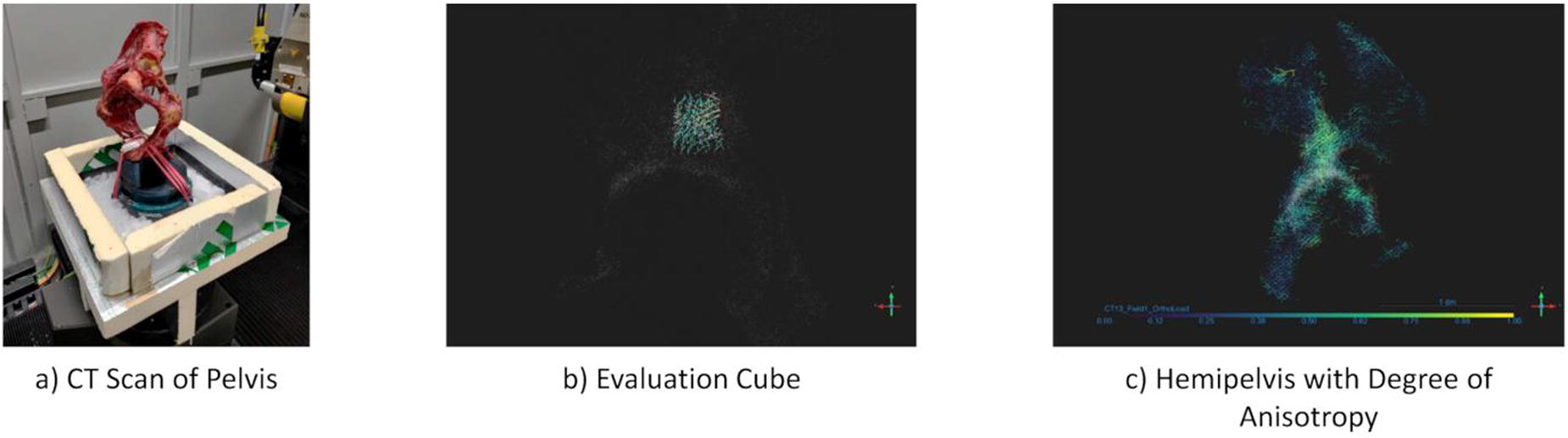
B133 - Exploring Extracellular Matrix Patterns And Remodelling In 2d And 3d Patient-derived Models Of Chronic Liver Disease In Response To Matrix-targeting Therapeutic Therapies
S. Campinoti1, U. Rastovic1, L. Wei1, B. Almeida1, S. F. Bozzano1, R. Amiri2, N. Harris1, T. S. Koay1, O. Ajayi1, C. Kerins3, F. Kenny4, A. Zamalloa4, L. Adofina4, R. Miquel4, Y. Zen3, P. Srinivasan4, K. Menon4, N. Heaton4, C. Luni2, E. Gentleman4, T. Shaw4, D. Ure5, S. Chokshi1, E. Palma1, L. Urbani1
1
The Roger Williams Institute of Hepatology, London, United Kingdom,
2
University of Bologna, Bologna, Italy,
3
King’s College London, London, United Kingdom,
4
King’s College London, London, United Kingdom,
5
Hepion Pharmaceuticals, Edison, NJ
*Purpose/Objectives: Fibrosis results from long-term liver damage in chronic liver disease (CLD), marked by hepatic stellate cell (HSC) activation and altered extracellular matrix (ECM) deposition, impairing liver function. Evidence suggests that factors like aetiology and fibrotic stage influence the biochemical and biophysical remodelling of deposited ECM in CLD, but mechanisms remain unclear due to the lack of relevant in-vitro/ex-vivo patient-derived models combined with robust ECM analysis methodologies. Here, we investigate ECM production, composition and remodelling in patient-derived 2D and 3D models of chronic liver disease using decellularisation combined with quantitative fibrillar image analysis, and validated by testing the ECM remodelling activity of a pan-cyclophilin inhibitor (Rencofilstat), peptidyl-prolyl isomerases that facilitate protein folding and collagen cross-linking currently in clinical development for metabolic-associated steatohepatitis (MASH) patients.
*Methodology: Precision Cut Liver Slices (PCLS) and primary HSCs were prepared from human liver tissue with different fibrotic stages, and exposed to pro-fibrosis stimuli (TGFβ, ethanol, IL-6, free fatty acids, high glucose/insulin), mimicking Alcoholic Liver Disease and MASH, in the presence/absence of Rencofilstat. Fibrosis induction in HSCs and PCLS were assessed by gene (RNAseq, Quantigene) and protein expression (proteomics), immunofluorescence and soluble factors secretion, quantified by Luminex. ECM remodelling in PCLS and in vitro culture of HSCs (produced upon HSCs stimulation) was characterised for its biochemical composition (via proteomics) and fibrillar alignment via immunofluorescence and confocal imaging. PCLS’s ECM and decellularised cell-derived matrix (CDM) confocal images were analysed using two fibrillar network analysis bioinformatic pipelines (TWOMBLI and AFT).
*Results: Pro-fibrotic stimuli induced patient-derived HSC activation in culture, resulting in increased proliferation and high ECM deposition. Different combinations of fibrotic insults generated distinct levels of cell activation and fibrillar ECM arrangements, assessed via computational image analysis. Interestingly, the fibrotic score of the liver tissue of origin influenced HSC activation and matrix architecture, indicating the suitability of our model to mimic patient- and aetiology-specific fibrotic features. Proteomic analysis on CDM revealed qualitative and quantitative differences in CDM compositions between different fibrotic scores (stages) and treatments. Rencofilstat profoundly and consistently reduced pro-fibrogenic markers in both 2D and 3D models. Remarkably, cyclophilin inhibition significantly reduced fibrogenic pathways in PCLS and reduced ECM deposition in HSCs, combined with significantly less organised ECM fibre bundles, indicating remodelling towards a softer 3D-matrix structure. Proteomic analysis showed qualitative and quantitative changes in CDM and PCLS ECM composition upon cyclophilin inhibition.
*Conclusion/Significance: Our study highlights patient-derived models’ utility in investigating ECM remodelling in diverse fibrotic settings. Our unique combination of models, decellularisation and bioinformatic pipelines allows the study of ECM remodelling patterns associated to specific liver disease stages/aetiologies, and in response to microenvironment-targeting therapy. The information on fibrillar quantity and alignment between patients and upon different pro-fibrotic insults provides new crucial information on the molecular mechanisms of ECM deposition in chronic liver disease and allows for a deeper understanding of drugs’ mechanisms of action, as well as the discovery of novel therapeutic targets.
B134 - Influence Of GAG Content On Strain Transfer In Cartilage Tissue Models
M. A. Boos1, S. R. Lamandé2, K. S. Stok1
1
The University of Melboune, Parkville, Australia,
2
Murdoch Children’s Research Institute, Parkville, Australia
*Purpose/Objectives: A thorough understanding of mechanobiological processes and remodelling networks and mechanoregulatory communication are essential for future tissue engineering approaches. Glycosaminoglycans (GAGs) play an important role in the mechanical properties and strain transfer in cartilage as they have shown to have strain attenuating properties [1]. However, it remains unclear to what extent GAGs contribute to the heterogeneous strain transfer and how this affects remodelling and tissue health. Therefore, the aims of this study were to investigate the influence of GAG content on the influence on micromechanics and strain transfer in cartilage tissue models.
*Methodology: Bovine chondrocytes were encapsulated in allylated gelatin (gelAGE) hydrogels containing thiolated chondroitin sulphate (CSSH) were cultured over 35 days the change in strain transfer were assessed. Using FELIX, a custom-built device for non-destructive image-guided micromechanical evaluation of biological tissues and tissue-engineered constructs (combining multiphoton microscopy with non-destructive mechanical testing of native soft tissues) [2], samples were subjected to stepwise unconfined compression at 5% and 10% strain while simultaneously imaging the cell nuclei (Hoechst 34580) under a multiphoton microscope.
The imaged volume of the samples was acquired, and slices of z-stack were generated to show displacement of nuclei. Images of the same nucleus under 5 mN preload, 5% strain, and 10% were isolated and an overlay was created to visualise displacement. The absolute displacement between 5 mN preload and 10% strain were measured and calculated as a ratio relative to 10% of the sample height. No statistics were performed on this data due to low sample size of n = 2-3.
*Results: All groups showed less displacement after 35 days indicating strain attenuation. Hydrogels containing CSSH seemed to attenuate strain at 10% more compared to the control at day 1 (Figure 1). After 35 days of culture all samples showed decreased displacement of nuclei relative to 10% of the sample height.
*Conclusion/Significance: Recent studies have shown that cells in biomaterials deposit matrix around the cells and create their own microenvironment that shields them from the mechanical influence of the original hydrogel network. In this study, a decrease in nuclei displacement could be observed after 35 days of culture in all groups. This suggests that the strain transfer is changing with different ECM composition and a potential influence of these microenvironments on strain transfer in the tissue. The question arises as to how these new microenvironments contribute to the bulk tissue mechanics. Another question this poses is, to what extent do the GAGs in the ECM contribute to cellular activities and is it only the microenvironment around the cells contributing, and how do the properties of the biomaterials impact the cells long term as the tissue-engineered samples mature.
References
[1] Han et al. Nature Materials, 2016. 15(4): p. 477-84.
[2] Boos et al. .Journal of the Mechanical Behavior of Biomedical Materials, 2023.142: 105868.

B135 - Oxygen Sensor Integrated Scaffolds For Tissue Engineering Applications
T. AV1, J. Berilla1, R. Somoza1, S. Cho1, G. J. Christ2, H. Baskaran1, J. Welter1
1
Case Western Reserve University, Cleveland, OH,
2
University of Virginia, Charlottesville, VA
*Purpose/Objectives: Cell fate in a tissue-engineering (TE) bioreactor is at least partially determined by nutrients and metabolites. State-of-the-art bioreactors use perfusion or medium changes to maintain a constant biochemical environment, but this does not guarantee sensing or control at or within the tissue, where small variations can trigger large changes in cell viability and differentiation. The goal of this project is to develop an ultra-sensitive oxygen optode sensor integrated with scaffolds to detect oxygen levels in TE constructs in real time.
*Methodology: We have developed electro-sprayed 250 µm diameter PDMS sensor beads incorporating an in-house developed ruthenium chloride-based complex. The beads were adhered to or embedded in collagen scaffolds intended for muscle tissue engineering. Sensing is based on the time constant of excitation/emission, and is both highly accurate and insensitive to changes in spot sizes, dye stability, and intensity, that plague intensity-based methods of O2sensing. A blue (457 nm) LED array emits a 30 µs pulse for excitation. A fluorescence decay phase follows the end of the excitation pulse and follows an inverse exponential curve. High-speed data for the dynamics of excitation and emission at 600 nm are collected through an optic fiber and a photomultiplier tube. Data are collected on a custom 25 MS/s A/D system and analyzed off-line. As the O2 partial pressure increases, the time constant (τ) of the decay decreases. Sensor beads were incorporated in a porous collagen scaffold (1 cm x 1 cm x 100µm) in a petri dish and seeded with 106 passage 2 human MSCs. Controls included O2-sensing beads with and without scaffold, and without cells. O2levels were measured every 90 seconds continuously for 14 hours in the experiment shown (Fig. 1).
*Results: Our analysis shows that oxygen levels in scaffolds without cells remained stable at ∼20% while levels in cell-seeded scaffolds dropped over 6 hours to a new equilibrium level near ∼6%. Sensor beads remained in place and did not interfere with cell attachment, cells remained adherent to the scaffolds and did not migrate off to the petri dish. The beads did not interfere with standard live-dead assays.
*Conclusion/Significance: This advanced oxygen-sensing technology can play a significant role in the manufacturing of TE medical products. Initial experiments showed a significant decrease in oxygen concentration over the course of 14 hours. Improvements are being made to increase the resolution and repeatability. It will allow the implementation of real-time optimization and correction procedures during the fabrication of TE constructs.
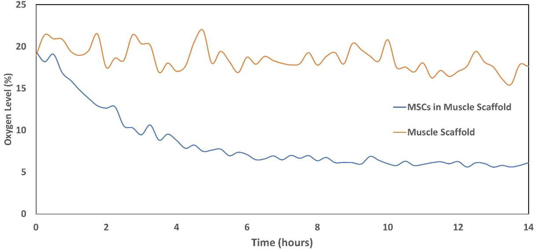
B138 - Multiparametric Assessment Of Cells In 3D Biomaterials
V. Lehnhardt1,2, I. Schmidt1, C. Burkhardt1, H. Hartmann1, J. Marzi1,2
1
NMI Natural and Medical Sciences Institute at the University of Tübingen, Reutlingen, Germany,
2
Eberhard Karls University, Tübingen, Germany
*Purpose/Objectives: Additive manufacturing using bioinks, comprising cells and biocompatible polymers, is an emerging biofabrication strategy to produce functional tissue models. Despite advancements in building increasingly sophisticated objects, biological and cellular analyses in printed constructs remain challenging and often imply endpoint analyses. Methods that enable non-invasive monitoring of embedded cells in bioprinted constructs would provide further insights on functionality and viability as well as cell material interactions.
*Methodology: To generate process-relevant information at different scales of bioprinting, our goal is to develop a multimodal, correlative imaging pipeline to determine and predict quality aspects of the bioprinted object. Therefore, we established a workflow to combine conventional immunofluorescence staining, molecular-sensitive Raman imaging and high resolution electron micoscopy for high content analysis of the same sample.
*Results: Different aspects such as culture format, cell type and the selection of the biopolymer were considered and evaluated. Furthermore, technical barriers such as sample transfer, penetration depths and background signal interferences were assessed and aligned between the microscopic techniques. Our multimodal approach enabled for multiparametric evaluation and monitoring of cellular distribution within the biomaterials, morphology, viability, phenotype and metabolic state. For example, Raman microscpetroscopy allowed for time-resolved monitoring of stress-induced cellular responses to hypoxia or sample stiffness.
*Conclusion/Significance: We aim to further expand the established benchmarks and transfer the protocols to more complex 3D structures. Automation and correlative data analysis will enable to evaluate bioinks and access cellular information in 3D culture in situ, representing a promising tool for the quality assessment of bioprinted objects.
B139 - Development Of Aptamer-CRISPR-12/13 Biosensors For Chondrogenic Biomarkers
A. Breitman, K. Zaman, S. Balachandar, J. Welter, Y. Fortenberry, R. Somoza
Case Western Reserve university, Cleveland, OH
*Purpose/Objectives: Biosensors that allow the identification and amplification of biological signals into physicochemical outputs can be a formidable application for Tissue Engineering applications. We propose the developing of molecular biosensors to detect specific molecules produced by cells in real-time, through fluorescence-based aptamer-CAS sensing platform. This sensor technology comprises the use of a CRISPR-Cas12 or Cas13 designed to recognize any DNA (Cas12) or RNA (Cas13) specific aptamer. This recognition activates a secondary trans-nuclease activity of Cas12 and Cas13 which is coupled to a fluorescent reporter. This sensing platform is used to assess the production of specific biomolecules known to be produced during the differentiation process that identify specific biomolecules produced during the differentiation process.
*Methodology: To develop the aptamer-based CRISPR-CAS12a molecular sensor we used an aptamer that recognizes hTGFB1. A PAM (protospacer adjacent motif) sequence was added to the aptamer sequence to be able to be recognized by the cas12a enzyme. A crRNA that binds to the aptamer was designed. For the reporter molecule we used a short ssDNA sequence with 6-FAM at the 5’ end and a BHQ quencher at the 3’ end. Upon recognition of the aptamer, the trans-cleavage activity of the enzyme is activated, allowing the ssDNA to be cut and the fluorescence to be acquired. As expected, in the absence of TGFB1 a high signal intensity is obtained. The signal decreases when TGFB1 is present. A non-target DNA sequence was used as a negative control. For the CRISPR-CAS13a sensor we developed an RNA aptamer that recognizes collagen type II and collagen type X via systematic evolution of ligand by exponential enrichment (SELEX). A CAS13 binding motif is added to the aptamer sequence and a crRNA is designed to recognize the aptamer sequence.
*Results: As expected, in the absence of TGFB1 a high signal intensity is obtained (more free aptamer to be recognized by the cas12a enzyme. The signal decreases when TGFB1 is present. A non-target DNA sequence was used as a negative control. The developed aptamer that recognizes collagen type II was incorporated into the CRISPR-CAS13 sensing platform. The sensor system was able to detect collagen 2 produced by chondrogenic differentiated human mesenchymal stem cells. Recombinant collagen 2 was used as a positive control.
*Conclusion/Significance: The aptamer-CRISPR sensing platform showed to be usefull to assess specific extracellular biomolecules produced during the chondrogenic differentiation process. This biosensing strategy has several advantages compared to conventional approaches (e.g., ELISA) in terms of time and real-time acquisition of data. The proposed sensor platforms could be used to detect a broad range of analytes (nucleic acids, proteins, whole-cell detection, etc.), at a much higher resolution given by the specificity provided by aptamers and sensitivity of CRISPR-Cas. The ability to resolve small changes in the concentration of many biomarkers secreted into the media non-invasively and in real time offers the prospect of monitoring and fine-tuning the cell culture environment.
B140 - A Surgical Genetically Engineered Mouse Model (surgi-gemm) Shows Anti-tumor Immunity With Combined Mesh Implantation And Cancer Immunotherapy
M. T. Wolf, S. Pal, I. Baurceanu
National Cancer Institute, Frederick, MD
*Purpose/Objectives: Tumor resection and subsequent biomaterial mesh implantation are ubiquitous to cancer treatment. Over 90% of non-metastasized solid tumors are treated with surgery, and biologic scaffold “mesh” materials composed of decellularized tissues are necessary to repair tissues damaged by surgical trauma. Local tumor recurrence after resection is also common, with 3-50% incidence across tumor types, necessitating complements to surgery such as immunotherapy. Genetically Engineering Mouse Models (GEMMs) mimic the genetic profile of human tumors in immune competent animals making them superior to cell lines, however, there are no GEMM models of biomaterial mesh implantation. We therefore (1) developed a melanoma GEMM of tumor resection and mesh implantation to fill this gap in translation and (2) used this model to test the combination of mesh implantation infused immunotherapy for local delivery.
*Methodology: We created Surgi-GEMM using the Cdk4R24CHgf-tg Melanoma that replicates familial human melanoma genetics, is highly recurrent after tumor resection, and is uncured with checkpoint immunotherapy. Melanoma tumors were initiated by carcinogen exposure, and subsequently passaged in syngeneic wild type C57Bl/6 mice without in vitro culture to prevent artifactual selection. We resected tumors and implanted a 1x1 cm biologic mesh (decellularized urinary bladder) and compared tumor growth and immune response via IHC. Finally, we used the Surgi-GEMM to evaluate co-delivery of immunotherapy agents with ECM mesh after tumor resection; a cancer vaccine using the STING pathway agonist cyclic di-AMP (CDA) and autologous whole lysed tumor vesicles prepared by sonicating melanoma tumors.
*Results: Melanoma tumors were highly recurrent, with 19/20 showing local recurrence when tumors were resected at 200-400 mm3 size. We created a subcutaneous Surgi-GEMM model wherein the melanoma tumor and adjacent healthy tissue margin are resected followed by mesh implantation infused with cancer immunotherapy (Fig1A,B). Immunohistochemistry showed that the biologic mesh recruited numerous leukocytes to the post-resection environment primarily F4/80+ macrophages and CD3+ T cells (Fig1C), as well as relatively smaller numbers of natural killer cells and negligible neutrophils. We then infused the biologic mesh with immunotherapy by adsorbing a cancer vaccine consisting of tumor lysate and immune adjuvant CDA, or individual components. Implanting biologic mesh alone after resection prevented recurrence in 25% of animals compared to 0% with resection only (Fig1D). We generated tumor vesicle lysate by sonicating whole tumors until vesicles smaller than 250 nm were obtained via dynamic light scattering. CDA immunotherapy was the most effective agent in conjunction with biologic mesh implantation; survival with or without the addition of tumor lysate was 55% and 50%, respectively. Further histologic analysis showed that the mesh and vaccine co-delivery significantly reduced lung metastases from greater than 10 % of histologic area to 0.1%, suggesting systemic immune protection.
*Conclusion/Significance: We found that a pro-regenerative biologic mesh implant induces inflammation that enhances immune surveillance and is effective with local immune adjuvant delivery. This is the first model of surgical tumor resection and biomaterial repair using GEMM tumors and may enable discovering synergies between tissue engineering and cancer immunotherapy.
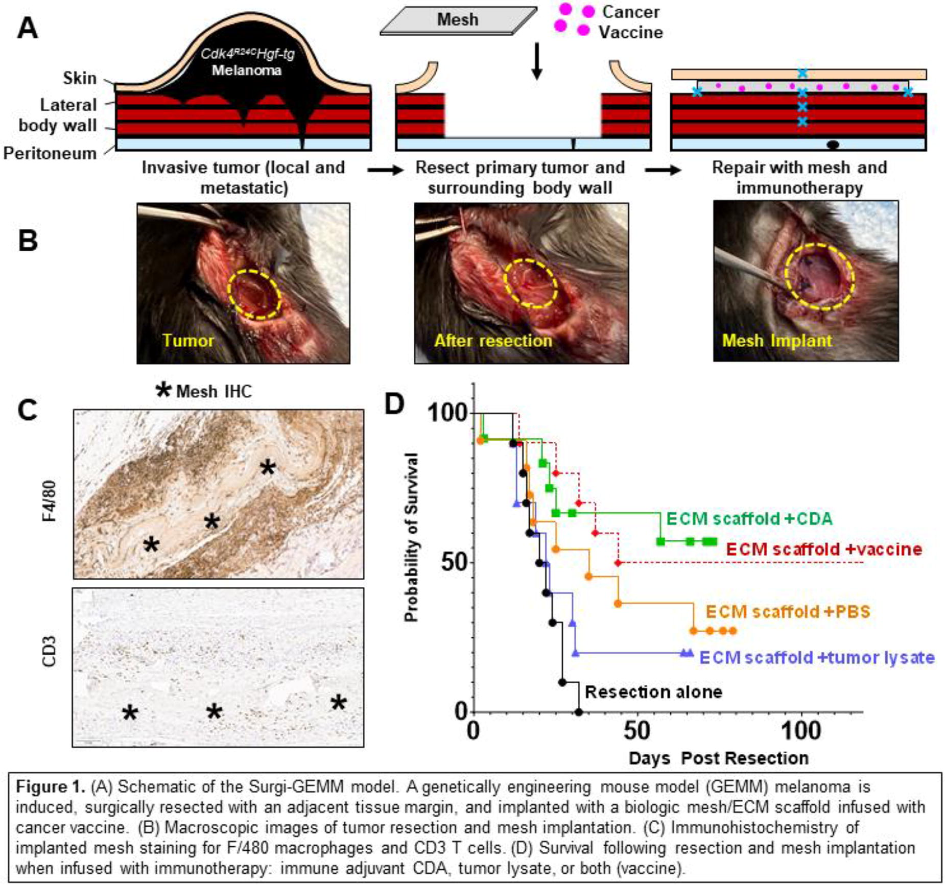
B141 - Length Of Peg Linkers Modulate Checkpoint Protein Immunoregulatory Function In The Subcutaneous Space
N. M. Racca1, A. Dontu1, K. Riley1, E. S. Yolcu2, H. Shirwan2, M. M. Coronel1
1
University of Michigan, Ann Arbor, MI,
2
University of Missouri, Columbia, MO
*Purpose/Objectives: Checkpoint proteins have taken a prominent role in immunotherapies aiming to balance reactive T cell targeting and tolerogenic regulatory T cell (Tregs) responses. Program death-1 (PD-1) and its ligand, programmed death ligand-1 (PD-L1), comprise a checkpoint inhibitor pathway that is known to have a pronounced role in the development and maintenance of induced Tregs. We have previously demonstrated that synthetic delivery of these proteins can lead to immunoregulatory functions both in vitro and in vivo. Yet, it remains unknown how protein immobilization through synthetic linkers and changes in their mechanical supports may alter the ligand-receptor interactions and downstream signaling.
*Methodology: We hypothesize that by altering the physical properties of the linker, such as the length of the PEG molecule, we can manipulate the way the PEG immobilized immunomodulatory proteins interact with cells. To test this hypothesis, PEG linkers of length 6, 30, and 60nm were used to capture the chimeric streptavidin PD-L1 proteins. The effect of linker length was evaluated in vitro to understand its capacity to convert naïve T cells to Tregs. We further validated the impact of the linker on immobilized PD-L1 and its immunomodulatory function in vivo by implementing a subcutaneous transplant model, where the protein was delivered through previously described microgel carriers with varying PEG linker lengths.
*Results: In vitro, a longer more flexible linker (60 nm) was found to promote a 25% higher conversion of naïve T cells into Tregs than control conditions. Furthermore, in vivo assessment of these linkers demonstrated a dynamic immune microenvironment which peaked at day 7 post-implantation (n=4) and resulted in elevated immune infiltration, in the T cell compartment only, in the group with the longer flexible linker. Notably, just a 10-fold change in linker size was sufficient to boost PD-L1 regulation of the dynamic balance between T effector and Tregs in vivo, with a 15-fold increase in comparison to control conditions.
*Conclusion/Significance: Overall, we show that PD-L1 function can be modulated by adjusting the length of the grafting linker providing a promising translational strategy to harness the therapeutic potential of checkpoint inhibitors without changing immunogenicity or attenuating protein bioactivity.
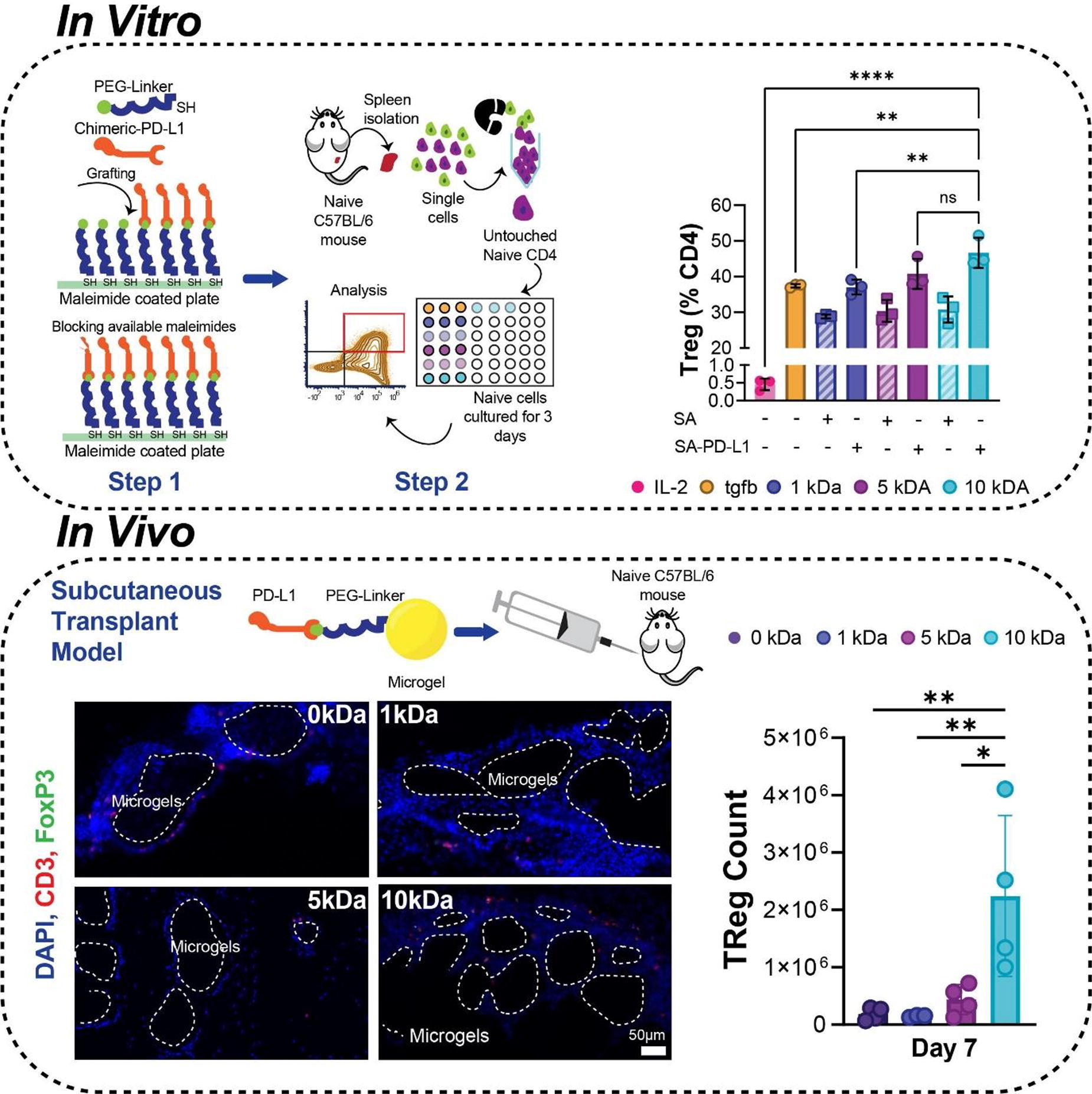
B142
B143 - Tolerogenic Human Placental Trophoblast Survival And Engraftment In A Xenograft Model
S. C. Hiremath, C. Chilimba, J. D. Weaver
Arizona State University, Tempe, AZ
*Purpose/Objectives: Cell and tissue transplantation is hindered by graft rejection, which necessitates lifelong chronic systemic immunosuppression. Placental pregnancy is the only natural state of de novo tolerance against allogeneic tissue in adult mammals, and is mediated by placental trophoblast cells, which use diverse mechanisms to induce antigen-specific tolerance. Understanding trophoblast tolerogenic mechanisms holds potential implications for eliminating immunosuppression in allogeneic transplantation. In this study, we explore xenograft ectopic survival and engraftment of alginate macroencapsulated human placental trophoblasts in immune competent and immune compromised murine models.
*Methodology: In this study, we aimed to evaluate the potential of human trophoblast model cell line, JAR, to evade graft rejection via tolerogenic soluble factors in an immune competent xenograft model (C57BL/6 recipient). To evaluate this, we macroencapsulated JAR cells in alginate spiral devices (4.5x10⁶ cells per spiral) and validated their survival via live/dead imaging and metabolic activity in vitro over 7 days of culture. JAR cells maintained metabolic activity and viability over the culture period.
*Results: Macroencapsulated JAR cells transfected with nanoluciferase enabled longitudinal in vivo imaging and quantification of trophoblast survival. To evaluate successful engraftment in the absence of immune rejection, encapsulated and unencapsulated JAR cells were transplanted into immune deficient NSG mice in the subcutaneous (SUBQ) or epididymal fat pad (EFP) sites with a vasculogenic VEGF-delivering degradable hydrogel to enhance localized vascularization (Fig. 1A). Unencapsulated groups maintained higher luminescent signal than encapsulated groups, increasing gradually over time, and formed palpable tumors SUBQ over 3 weeks. Encapsulated groups exhibited ∼74% and 213% signal relative to day 0 imaging in SUBQ and EFP groups, respectively. Overall, encapsulated JAR cells exhibited highest survival in the EFP transplant site. Next, we evaluated unencapsulated and encapsulated nanoluciferase JAR survival in an immune competent C57BL/6 xenograft model. JAR were transplanted in SUBQ and EFP sites, with and without vasculogenic degradable hydrogel. VEGF can both enhance engraftment via improved vascularization, and enhance lymphatic development, which could accelerate graft rejection. The EFP groups experienced rapid rejection, with reduction to near-baseline signal in all groups by day 10 (Fig. 1B). In the SUBQ groups, rejection was observed between day 14-28, with the exception of the encapsulation + vasculogenic hydrogel group, which demonstrated significantly elevated signal comparable to the NSG group out to day 63 (Fig. 1C). Encapsulated non-tolerogenic control human HEK cell controls were rapidly rejected within 10 days (data not shown), suggesting that trophoblast-secreted soluble factors contributed to JAR survival.
*Conclusion/Significance: Macroencapsulated trophoblast in vivo imaging demonstrates high cell survival in immune compromised mice and comparable survival in immune competent mice in the SUBQ site. Ongoing studies are evaluating mechanisms of trophoblast survival and immune modulation via histology and flow cytometry.
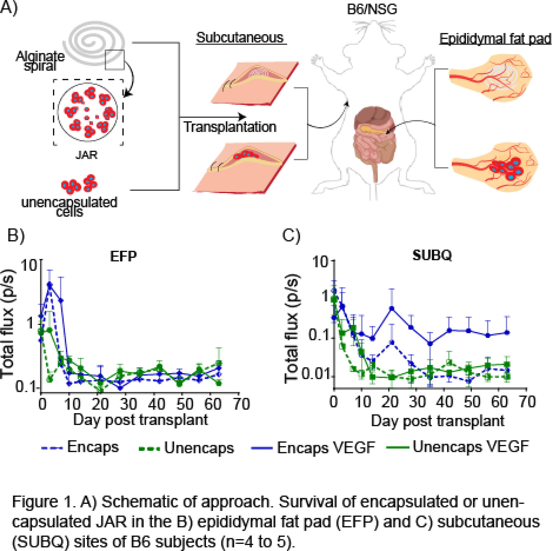
B144 - The Impact Of Exosomes Derived From Cd4+ T-cell Subsets On Satellite Cell Activity In Vitro
A. Jain, R. J. DiPaolo, J. Kornbluth, K. Garg
Saint Louis University, Saint Louis, MO
*Purpose/Objectives: Severe and persistent inflammation is a major impediment to muscle recovery post-volumetric muscle loss (VML)[1]. Helper CD4+ T-cell subsets (Th1, Th2, Th17, and Treg) [2] accumulate in the VML defect, but their exact role in muscle repair is largely unknown [3]. Studies have shown that Th1 produces IFN-γ and TNF-α to promote inflammation and satellite cell (SC) proliferation [4], while the release of interleukin (IL)-4 and IL-13 by Th2 and amphiregulin by Treg suppresses inflammation and promotes the differentiation of SC [5]. Th17 produces IL-17 A, which promotes angiogenesis and myogenesis [6]. The goal of this study was to determine the role of CD4+ T-cell derived exosomes in supporting SC activity in vitro. We hypothesize that exosomes derived from Th2 and Treg will support SC differentiation and fusion, while Th1 and Th17 subsets will enhance SC proliferation.
*Methodology: CD4+ T-cells were isolated from rat spleens and polarized into Th1, Th2, Th17, and Treg, as previously described [7]. Unpolarized CD4+ T-cells (Th0) served as controls. T-cell conditioned media (CM) was collected from each subtype on days 5 and 7 of culture and was used for exosome isolation using Exo-Quick TC (System Biosciences). Exosomes were characterized using Exo-Check Array and Nanoparticle Tracking Analysis (NTA). SCs isolated from the hindlimb muscles of rats were seeded in a 48 well plate at a density of 100,000 cells/well (n=4/group) and incubated with growth media containing PBS (control group) or 25 µg/ml exosomes derived from Th0, Th1, Th2, Th17 or Treg for 72 hours, following which supernatants were collected and stored at -80 deg C for ELISAs.
*Results: The expression of IFN-γ by Th1, IL-4 and IL-13 by Th2, IL-17 A and IL-22 by Th17 and FoxP3 by Treg was confirmed. Exosomes from all helper T cell subsets were within 30-150 nm in size. Th0 derived exosomes expressed characteristic surface markers such as TSG101, CD81, and ICAM. The release of IL-6, IGF-1, and VEGF by SCs response to helper T cell exosomes was quantified. IL-6 promotes activation and proliferation of SCs [8, 9], and its production was significantly increased by SCs in response to Th2 or Th17 exosomes. Insulin-like Growth Factor (IGF-1) enhances both proliferation and differentiation of SCs [10]. SCs produced more IGF-1 in response to Th0, Th1 or Th2 exosomes relative to Treg exosomes or PBS. Vascular Endothelial Growth Factor (VEGF) enhances SC proliferation [11] but its production was similar between groups.
*Conclusion/Significance: All Th subsets promote proliferation of SCs by promoting VEGF production. Th0, Th1 and Th2 derived exosomes increased the production of IGF-1 in SCs, thus enhancing their proliferation and differentiation. Although Th2 and Th17 are anti- and pro- inflammatory, respectively, exosomes from both increased SC activation. Treg exosomes did not influence SC activity. These results were unexpected. A possible reason behind this could be that the biological cargo of T-cell exosomes and CM is different. Future studies will investigate the role of T-cell subsets and their secreted products (exosomes vs. CM) on muscle regeneration in a rat model of VML.
B145 - Antigen-Conjugated Scaffolds For Enrichment Of Disease-Specific T-cells
S. N. Wheeler, A. H. Morris
University of Michigan, Ann Arbor, MI
*Purpose/Objectives: T-cells are critical to immune functions from protecting against pathogens to inducing tissue repair and regenerative responses. Blood draws are the gold-standard for collecting immune cells, and while easy to perform, cannot readily isolate sufficient T-cell clones of interest due to the rarity of disease-specific T-cell populations in circulation. For autoimmune diseases like multiple sclerosis, MS, improved tools to isolate and study specific T-cells would be a boon to our understanding of how these cells contribute to disease progression. We aim to use biomaterial scaffolds as a tool to enrich specific clones of T-cells. Specifically, we propose to conjugate antigens to polymer backbone prior to scaffold fabrication, allowing for precise loading of antigen, while avoiding burst release and systemic T-cell stimulation.
*Methodology: In this study, we developed a tool for studying antigen-specific T-cells. We chose two peptide antigens: OVA323-339, a model antigen, and PLP139-151, an MS-relevant antigen. Carbodiimide crosslinking was used to conjugate peptide antigens to poly (lactide-co-glycolide) (PLG) and conjugation was confirmed using nuclear magnetic resonance. Antigen-conjugated scaffolds were fabricated via solvent evaporation and salt leaching techniques, yielding porous scaffolds that release antigen slowly as polymer degrades (peptide release kinetics evaluated with CBQCA assays). To characterize interactions between scaffolds and cells in vitro, antigen-loaded scaffolds were co-cultured with either antigen-specific cell lines or splenocytes. Activation of antigen-specific cells was then confirmed through IL-2 ELISAs. To measure proliferation of antigen-specific cells, scaffolds were co-cultured with CFSE-labeled OVA-reactive primary cells for 72 hours, followed by flow cytometry. To assess the ability of scaffolds to recruit antigen-specific cells in vivo, OVA-loaded scaffolds were implanted subcutaneously in mice, followed by adoptive transfer of GFP-labeled OVA-specific cells. After 7 days, scaffolds were explanted and flow cytometry was used to quantify OVA-specific cell enrichment. In future studies, antigen-conjugated scaffolds will be implemented in an EAE mouse model of MS for detection and monitoring of disease-specific cells in scaffolds over the course of disease progression.
*Results: Carbodiimide chemistry successfully conjugated PLP139-151 and OVA323-339 peptides to PLG with efficiency rates of 81.94% and 75.21%, respectively. CBQCA assays demonstrate sustained peptide release through 112 days. Increases in IL-2 production and CFSE dye dilution indicate that antigen-conjugated scaffolds release antigen that retains its activity and activates T-cell populations of interest in vitro. Following subcutaneous scaffold implantation and adoptive transfers, flow cytometry confirmed preferential trafficking of antigen-specific CD4+ T-cells to implants loaded with target antigen over control scaffolds.
*Conclusion/Significance: In conclusion, carbodiimide crosslinking chemistry can be utilized to conjugate disease-relevant peptide antigens to PLG at high efficiency, allowing for precise loading of antigen in the scaffolds. Additionally, we have shown that this conjugation does not alter the activity of the peptide antigen and allows for recruitment and engagement with antigen-specific T-cells in vitro and in vivo. Although this study investigates the use of OVA323-339 and PLP139-151 peptide antigens, this system is modular and allows other peptides of interest to be incorporated into the scaffolds, allowing for the study of a variety of inflammatory diseases, autoimmunity, cancers, and other pathologies.
B146 - IL-23 Responsive Cellular Sensors For Autoimmune Monitoring
G. L. Garza, A. H. Morris
University of Michigan, Ann Arbor, MI
*Purpose/Objectives: Autoimmunity is a growing global concern. Multiple sclerosis (MS) specifically is a neurodegenerative disease in which the immune system attacks the myelin sheaths of cells in the central nervous system (CNS) and disrupts signal transduction, affecting over 2.5 million people worldwide. The current gold standard for detection and diagnosis of MS is brain imaging through MRI to look for plaque formation. Unfortunately, damage to CNS tissue appearing on MRI is often irreparable. The purpose of this project is to engineer cellular sensors for early detection and continuous monitoring of MS, and autoimmunity broadly, to affect earlier and more precise treatment and improve patient outcomes.
*Methodology: We chose to target the cytokine interleukin-23 (IL-23) as it a protein involved in adaptive immune function and important in MS. Our group assembled 24 Modular Extracellular Sensor Architecture (MESA) receptor designs specific to IL-23 using molecular cloning. MESA receptors are synthetic cell surface receptors composed of two chains that dimerize upon binding a soluble ligand, similar to receptor tyrosine kinases. Each receptor includes two naturally derived transmembrane domains, two different nanobody binding regions for IL-23, a Myc tag, and a transcription factor that induces expression of a fluorescent reporter upon activation. Plasmids encoding each receptor design were transiently transfected into HEK293FT cells in high-throughput using polyethylenimine and evaluated for surface trafficking and IL-23-induced activation using flow cytometry. To measure cell surface trafficking, transfected cells were stained with an anti-Myc antibody prior to flow cytometry analysis. Functional tests were conducted by comparing the fluorescent signal of transfected cells in the presence vs. absence of IL-23. Future efforts are devoted to transitioning this technology to in vivo to monitor immune fluctuations in an MS mouse model.
*Results: All receptor designs traffic to the surface significantly better than the negative control. Successful receptor designs were classified as having a fold change of 1.5 or higher between background noise and inducible signal. We identified seven of the 24 designs that meet this criterium. The greatest number of successful receptor designs combined a nanobody binding to the N-terminal domain of the p40 subunit and a nanobody capable of binding to either the p19 or p40 subunits of IL-23.
*Conclusion/Significance: These results identify at least seven promising receptor designs that activate an internal cell circuit in the presence of IL-23. These receptor designs will progress through further functional testing and translation in vivo to validate our cell sensors in the context of monitoring MS. These results also inform future receptor engineering for IL-23 as the receptor designs with the most pronounced signal-noise ratio targeted both the p19 and p40 regions of IL-23. Moreover, we are one step closer to building a real-time monitor of autoimmunity to help patients identify and manage their diagnosis.
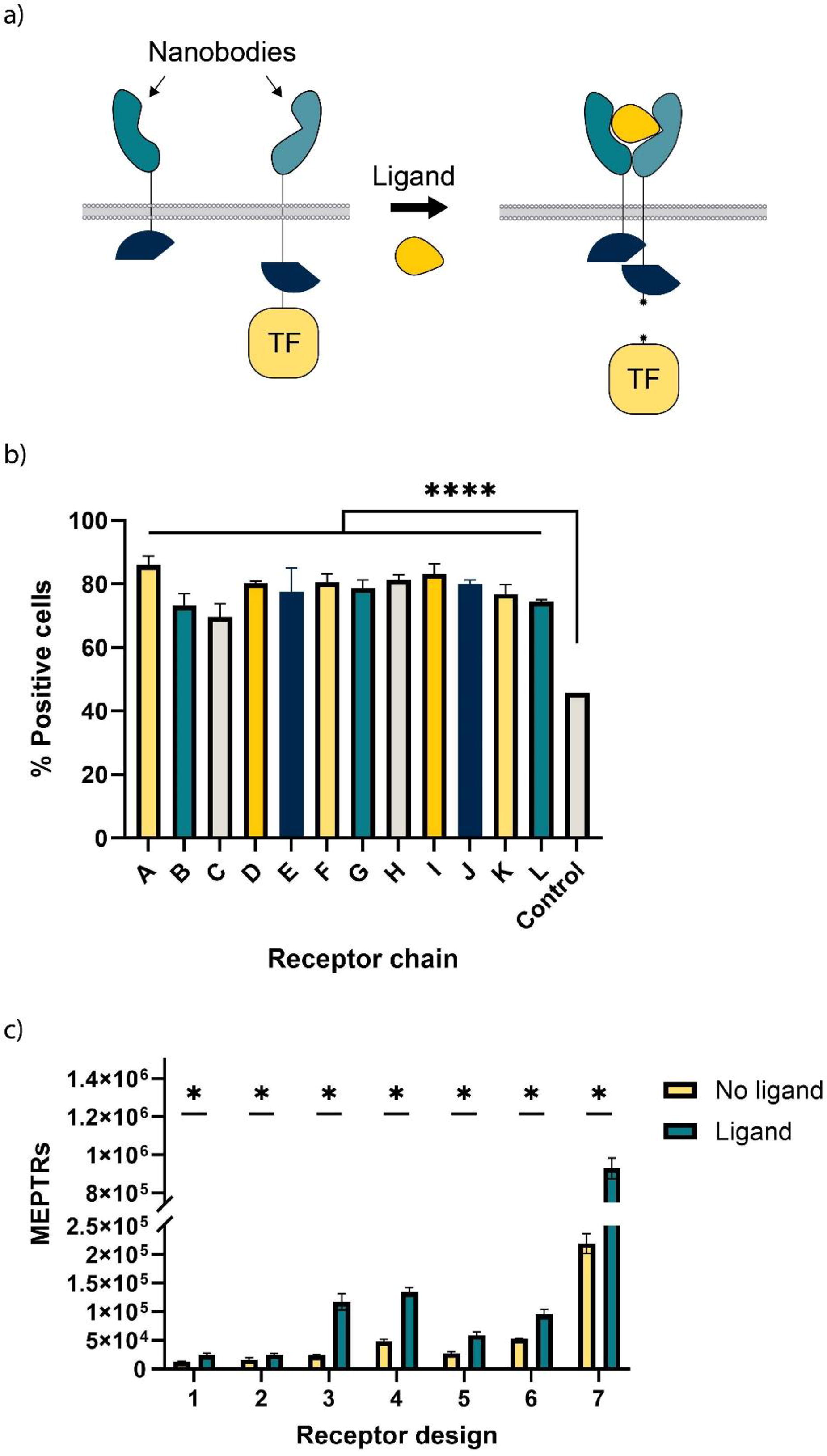
B147 - Pilot Investigation Of Pattern Recognition Receptor Agonists On Tendon Healing In Vitro
S. E. Winston, A. Stoner, J. Kurihara, L. Chow, L. Pezzanite, S. Dow, K. McGlvray
Colorado State University, Fort Collins, CO
*Purpose/Objectives: The more than 250,000 surgeries occurring annually in the United States, rotator cuff (RC) repair surgeries fail at unacceptable rates ranging from 20-94%. Histological analysis of these failed repairs show that the surgically repaired tendon is not returning to its original tissue composition and is instead scarring. Tendon healing overall, is a largely unknown cascade, however, it is known that the immune response to the injured tendon has a large effect on whether the tendon scars or regenerates. This has brought interest in modulating immune responses to improve clinical success of these repairs. Our novel approach is to functionalize Pattern Recognition Receptor Agonists (PRRA), which are molecules that have been used in cancer research effectively as an immunotherapy approach. These agonists activate the innate immune system through the Toll-like and Nod-like receptor pathways. We hypothesize that PRRAs can be utilized to activate the immune system to increase clinical outcomes of tendon healing. To investigate this, we aim to examine how gene expression changes in tendon when exposed to PRRAs in vitro.
*Methodology: Biopsies were taken (N=3) from (N=4) sheep (ovis aries) supraspinatus tendons. Tendon samples were cultured in media with 50 ng/ml concentrations of the selected PRRAs. PRRAs were ALOH (Invivogen), TLR3 Poly(I:C) HMW (Invivogen), STING (Invivogen), and CpG (Invivogen). Tendons were cultured overnight then frozen. Tendon samples left in media with no agonists were controls. RNA was then extracted, purified, and sent for genomic sequencing (Novogene Inc.). Gene expression data were analyzed using Partek Flow. Gene Ontology of Biological Processes (GOBP) was used to analyze gene enrichment of each agonist group compared against the controls using gene set enrichment analysis (GSEA) to determine the overall effect of the applied agonists.
*Results: ALOH, CpG, Poly(I:C), and STING treatments had 146, 119, 213, and 259 significantly differentially expressed genes (Figure 1a-d). GOBP analysis of Poly(I:C) revealed upregulation of proinflammatory pathways such as IFNα and IFNγ and TNFα signaling (Figure 1e). It also upregulated pathways relating to cartilage development such as chondrocyte differentiation and downregulated adipogenesis.
*Conclusion/Significance: PRRAs had meaningful effects on genetic changes in the tissues. In the controls TNFα, IFNα, and IFNγ (all proinflammatory cytokines) are known to play an important role in the early stages of wound healing, were upregulated. In comparison Poly(I:C), gave a higher proinflammatory response. Poly(I:C) also had a mixed effect on specific tendon healing markers including fatty infiltration and an increase in cartilage formation, which are signs of tendon degeneration. Poly(I:C) downregulated adipogenesis and upregulated pathways related to cartilage formation. Taken together these data indicate Poly(I:C) is directly affecting tendon health in a positive way through the limiting of fat production and the increase in immune cytokines that are instrumental in proper wound healing. TLR3 activation through Poly(I:C) has been shown to improve wound healing times in human skin, but its effects on tendon healing have not been characterized. These preliminary data illustrate that the regulation of extracellular matrix production and immunomodulatory properties of Poly(I:C) have the potential to be a novel tendon therapy.
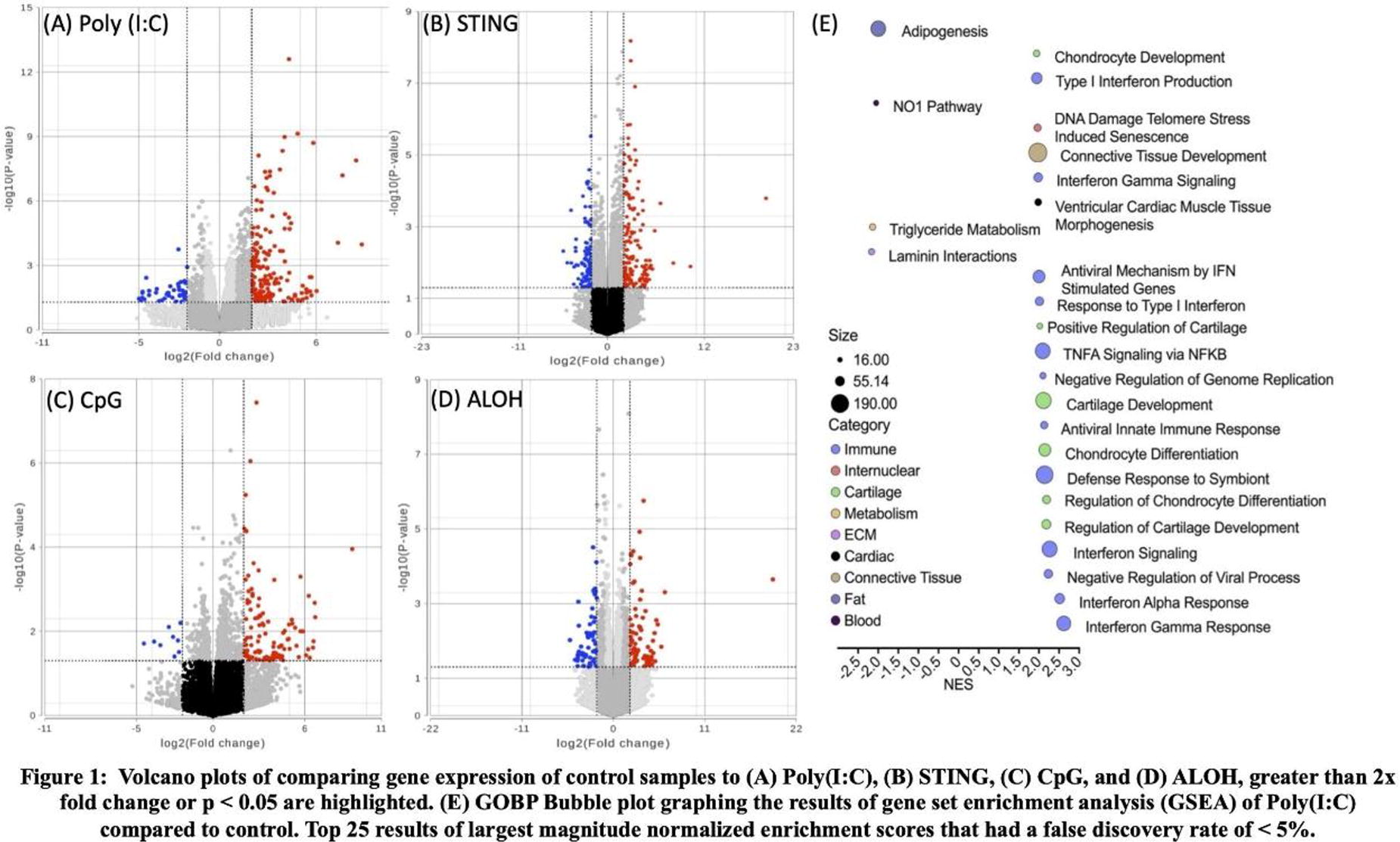
B149 - Engineering Immunomodulatory Materials For Regenerative Medicine
D. Salthouse
Newcastle University, Newcastle upon Tyne, United Kingdom
*Purpose/Objectives: Collagen is extensively utilised in tissue engineering and regenerative medicine due to its highly desirable properties, including biocompatibility, biodegradability, and ability to provide structural support and flexibility. However, collagen is commonly derived from mammalian sources (typically of bovine or porcine origin), which poses several limitations, including high cost, potential risk of immunogenicity and transmission of infectious diseases, and ethical and religious constraints. Jellyfish collagen represents a more sustainable and economical alternative collagen source and has also demonstrated biocompatibility and potential immunomodulatory and regenerative capabilities. Therefore, we investigated the potential of jellyfish collagen hydrogels, obtained from Rhizostoma pulmo (R.pulmo) jellyfish, to be utilised in tissue engineering and regenerative medicine.
*Methodology: A variety of R.pulmo collagen hydrogels (RpCol hydrogels) were formed through the addition of various chemical crosslinking agents in a range of concentrations and ratios: carbodiimides, multi-arm functionalised polyethylene glycol (PEG)-derivatives, and genipin. Degradation profiles of RpCol hydrogels were assessed by monitoring hydrolytic degradation, while the chemical structures of RpCol hydrogels were analysed by Fourier-transform infrared spectroscopy (FTIR). Rheological and morphological properties of RpCol hydrogels were evaluated by rheology and scanning electron microscopy (SEM), respectively, while the endotoxin levels of RpCol was determined by limulus amoebocyte lysate (LAL) assay. Morphology and viability of human mesenchymal stem/stromal cells (MSCs) encapsulated in RpCol hydrogels were evaluated by immunostaining, and the metabolic activity and interleukin (IL)-10 production of encapsulated MSCs were assessed by fluorescent spectrophotometry and enzyme-linked immunosorbent assay (ELISA), respectively.
*Results: RpCol alone did not form stable hydrogels, however, hydrogel stability was increased through the addition of a variety of chemical crosslinking agents. The characteristic triple helix structure of RpCol was confirmed by FTIR, and the degradation profiles, morphological, and rheological properties of RpCol hydrogels were shown to be adaptable for different applications through the addition of specific chemical crosslinking agents. The endotoxin levels of RpCol were below regulatory limits (<0.5 endotoxin units (EU)/ml), thus allowing the potential use of RpCol ;in vivo.; 8-arm polyethylene glycol succinimidyl carboxyl methyl ester (PEG-SCM)-crosslinked RpCol hydrogels were stable, relatively strong, allowed rapid glucose diffusion, and preserved the viability and induced a significant increase in the metabolic activity and interleukin (IL)-10 production of encapsulated MSCs.
*Conclusion/Significance: Therefore, RpCol hydrogels crosslinked with 8-arm PEG-SCM demonstrate potential to be utilised in a wide range of tissue engineering and regenerative medicine applications. Depending on the intended application, different immunomodulatory molecules could be incorporated into RpCol hydrogels to modulate the host immune response and promote tissue regeneration.

B150 - Two-Photon Polymerization
J. Fernandez Perez, A. Ovsianikov
TU Wien, Wien, Austria
*Purpose/Objectives: MSC are known to have immunomodulatory capacity and induce regeneration, which is enhanced with 3D culture and with hypoxia. Two-photon printing (2PP) is a high-resolution additive manufacturing technique as only a small voxel is polymerized at a pulsed laser’s focal point. We have previously reported the use of this technique to produce hybrid spheroid microscaffolds which support MSCs for musculoskeletal tissue engineering applications. This work aims at studying the immunomodulatory profile of MSC in hybrid spheroid microscaffolds.
*Methodology: Human adipose MSC, hTERT immortalized (Evercyte) were seeded into bucky-ball (BB) microscaffolds at normal density (4,000 cells/BB) or low density (2,000 cells/BB). Likewise, cells were seeded onto round bottom lipidure-coated well plates to induce spheroid (SPH) formation (at same cell densities as for BB). 2D controls were set-up at the same time. After 3 days of aggregate formation, samples were switched to serum-free medium (DMEM only) for 24 hours, then harvested for RNA isolation and further qPCR.THP1 cells were differentiated towards “naïve” macrophages by exposure to 20 ng/ml PMA during 24h. After a 24 h resting period, i.e., the PMA was removed and substituted for growth medium, M1 or M2 polarization was induced by addition of 100 ng/ml LPS and 5 ng/ml IFNγ, or 20 ng/ml IL4 and 20 ng/ml IL13, respectively, for 48 h. M0 controls were kept in growth medium. Polarization was analyzed via qPCR. Differentiated macrophages were treated with MSC-conditioned medium (microscaffolded spheroids, spheroids, and 2D) and their polarization analyzed.
*Results: Immunomodulatory genes stanniocalcin (STC1) and TNF alpha induced protein 6 (TSG1) presented similar levels of expression in all four conditions (Figure 1). Quite marginally, the low densities seemed to express slightly less STC1, albeit not statistically significant. TSG-1 was slightly, but significantly, upregulated in SPH-H compared to BB-H, BB-L and SPH-L. VEGF-A had a higher expression in all 3D conditions, compared to the 2D, indicating a higher pro-angiogenic phenotype. N-Cadherin (CDH2) was expressed in 3D but not in 2D controls which could be expected as the cell-cell contacts are less abundant in 2D. Polarization of macrophages was confirmed by detecting an upregulation of CXCL10 and TNFα for M1, and of CCL22 for M2. These markers were used to analyze the macrophages upon treatment with conditioned medium (data not shown).
*Conclusion/Significance: These data seem to indicate that the formation of spheroids within microscaffolds retains MSC immunomodulatory capacity and pro-angiogenic potential. Future experiments will focus on studying the influence of MSCs in hybrid spheroid microscaffolds towards improving and enhancing wound healing and angiogenesis, as potential therapeutics.
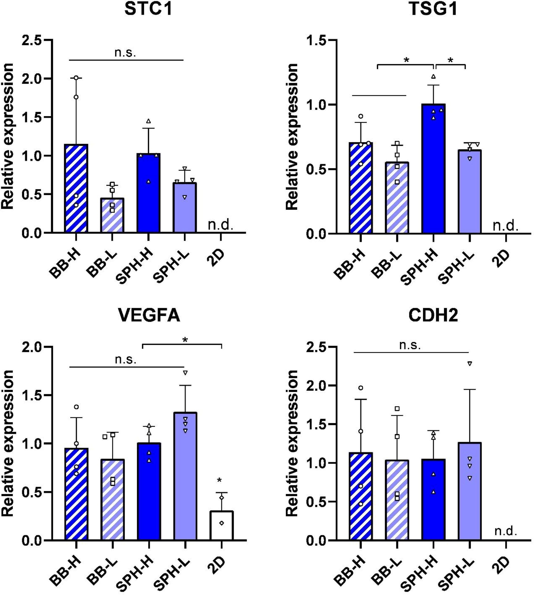
B151 - 3D Printing Of Salt Template For Nanoparticles Reinforced Gyroid Scaffold For Bone Regeneration
S. S. Lee1,2, I. Kim1,2, N. Kleger3, D. Tran3, A. R. Studart3, S. J. Ferguson1
1
Department of Health Sciences and Technology, ETH Zurich, Zurich, SWITZERLAND,
2
Department of Biomedical Engineering, Dongguk University, Seoul, KOREA, REPUBLIC OF,
3
Department of Materials, ETH Zurich, Zurich, SWITZERLAND.
*Purpose/Objectives: Osteoporosis, a prevalent condition, is associated with the impairment of bone tissue formation, increasing the risk of bone fractures. The disease commonly hampers the natural self-healing mechanism of bones, necessitating the use of bone implants. Regrettably, the current state-of-the-art practices in the field lack any tissue-engineered solutions. The rise of additive manufacturing (AM) has spurred the development of advanced technologies dedicated to crafting orthopaedic implants and scaffolds which play a pivotal role in augmenting bone tissue regeneration. This study introduces an inventive methodology for magnesium oxide nanoparticles (MNP) reinforced poly-ε-caprolactone (PCL) based gyroid-structured scaffolds via the infiltration of sacrificial salt templates.
*Methodology: The production of templates involves using a masked stereolithography apparatus (MSLA) with optimized printing parameters. Injection molding is employed to fabricate salt-template-infiltrated scaffolds, featuring pore sizes ranging from 250 µm (SP) to 400 µm (LP). The scaffold material comprises PCL compounded with 20 wt% MNP to create a composite with biodegradability and notable osteogenic effects. The subsequent testing groups (SP, LP, MNP reinforced SP (M-SP), MNP reinforced LP (M-LP)) were 3D printed and characterized by experiments such as mechanical testing, finite element analysis, inductively coupled plasma (ICP), protein adsorption and in vitro 3D culture using human mesenchymal stem cells (hMSCs).
*Results: All testing groups were 3D printed, showcasing gyroid structures without defects and confirming the successful integration of MNP reinforcement. ICP analysis validates controlled magnesium ion release, and the mechanical testing reveals higher compressive moduli in MNP-reinforced groups, showcasing structural stiffness irrespective of pore size. Additionally, biocompatibility assessments using hMSCs illustrate favorable outcomes, with cell attachment and collagen synthesis across all groups. While cell proliferation tends to be higher in groups without MNP, detailed analyses, including Alizarin Red S Staining (ARS) and RT-qPCR, expose superior performance in calcium deposition and osteogenic gene expression for M-SP and M-LP compared to SP and LP.
*Conclusion/Significance: The study demonstrates the successful utilization of salt templates for 3D printing complex structured scaffolds. The incorporation of MNP enhances osteogenic differentiation, with M-SP and M-LP outperforming SP and LP in calcium deposition and osteogenic gene expression. Mechanical testing reveals superior stiffness in MNP-reinforced groups, confirming the structural integrity of the scaffolds. In summary, the results emphasize the promising potential of this innovative AM method in advancing bone tissue regeneration. The findings contribute valuable insights to the development of tissue-engineered solutions for bone-related conditions, addressing the limitations of current practices in the field.
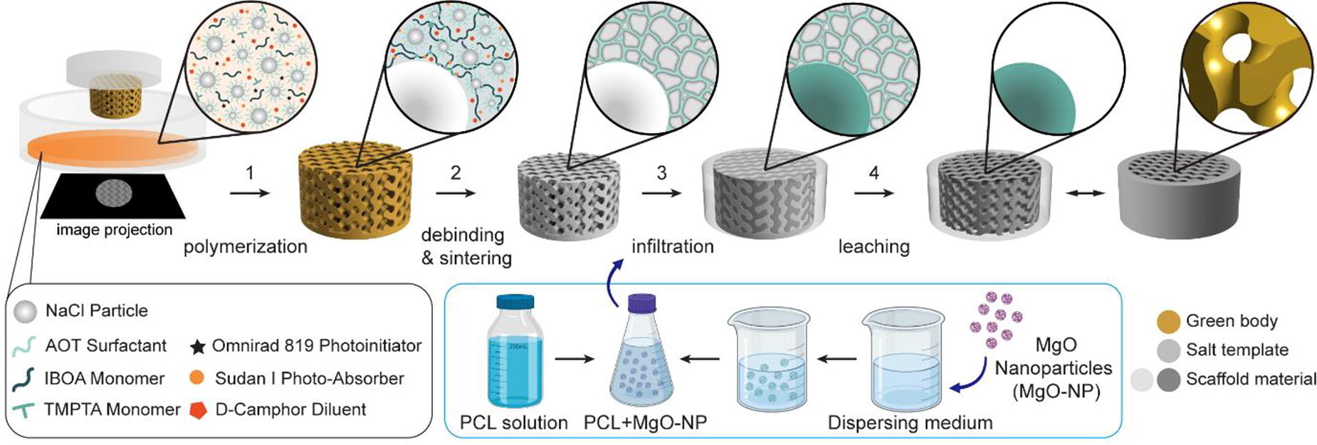
B155 - Reconstruction Of The Intra-articular Environment Of Osteoarthritis With A 3d Co-culture System Of Patient-derived Cells
G. IM
Dongguk University, Goyang, Korea, Republic of
*Purpose/Objectives: The inflammatory environment of osteoarthritis (OA) is a three-dimensional (3D) environment formed by the interaction of various cells within joint cavity. Several in vivo and in vitro models have been developed to study the pathophysiology of the disease and test the efficacy of new drugs. Animal model in OA is cost-intensive and time consuming as well as associated with ethical concern and greart inter-species difference. On the other hand , 2D in vitro model of OA has been widely used in drug development, offering a simple, low-cost option. However, it does not reflect the influence of microenvironment/ role of external factors. Also, loss of articular chondrocyte phenotype in the culture makes it a low predictive measure for OA disease models and drug screening.In comparison, 3D in vitro OA models can provide a cheaper, ethical and faster approach. Also, as multi-component and real-time/bidirectional crosstalk is possible in the model, it may fill the gap between clinical trial and conventional preclinical in vitro/in vivo models. Osteoarthritis on chip (OoC) models have been devised by several authors. Unfortunately, they are mostly complicated models while not reflecting all intraarticular variable. To develop a simple, applicable 3D in vivo OA model, we devised a co-culture system that mimic the intraarticular environment in OA. It is called “Pizza chip” in which articular chondrocytes, synoviocytes, adipose stromal cells (ASCs) were cultured in spheroid from in separation from one another, but sharing culture media.
*Methodology: Articular cartilage, adipose, and synovial tissue samples from osteoarthritis patients were obtained during total knee arthroplasty. After the cells were isolated from each tissue, they were cultured in the 3D co-culture system, OA patient-derived synovial cells, chondrocytes, and ASCs (4x10⁵ cells for each lineage) were inoculated in 400 µL of culture medium. After 24 hours of culture, IL-1β (10 ng/ml) was treated for 48 hours to induce an inflammatory response. Then, each cell and culture medium were collected and cell viability (MTT assay), nitric oxide (NO) synthesis by inflammatory stimuli, and secretion of inflammatory cytokines (ELISA assay) was measured.
*Results: Cell viability was not different in spheroids derived from chondrocytes, adipose stromal cells and synoviocyte while NO synthesis was significantly greater in spheroids derived from chondrocytes in the inflammatory environment induced by IL-1β treatment. No significant difference in cell proliferation/viability according to the cell culture method (2D monolayer or spheroid chondrocyte culture) or presence of co-culture (spheroids of chondrocytes only or co-culture of 3 cell lineages). Level of inflammatory markers (IL-6, PGE2 and MMP-13) varied significantly with different culture method / the presence of co-culture.
*Conclusion/Significance: We developed a simple 3D culture model model which mimic interaction within IA cavity, and is easily applicable without much technical experience. Shortcomings are 1) biomechanical factor not applicable 2) difficult to include immune response. With this reconstruction of closer-to-real IA environment of OA with spheroids derived from OA patients’ cells, limitations and problems of drug screening using 2D monolayer culture can be improved and the use of experimental animals may be replaced with this system.
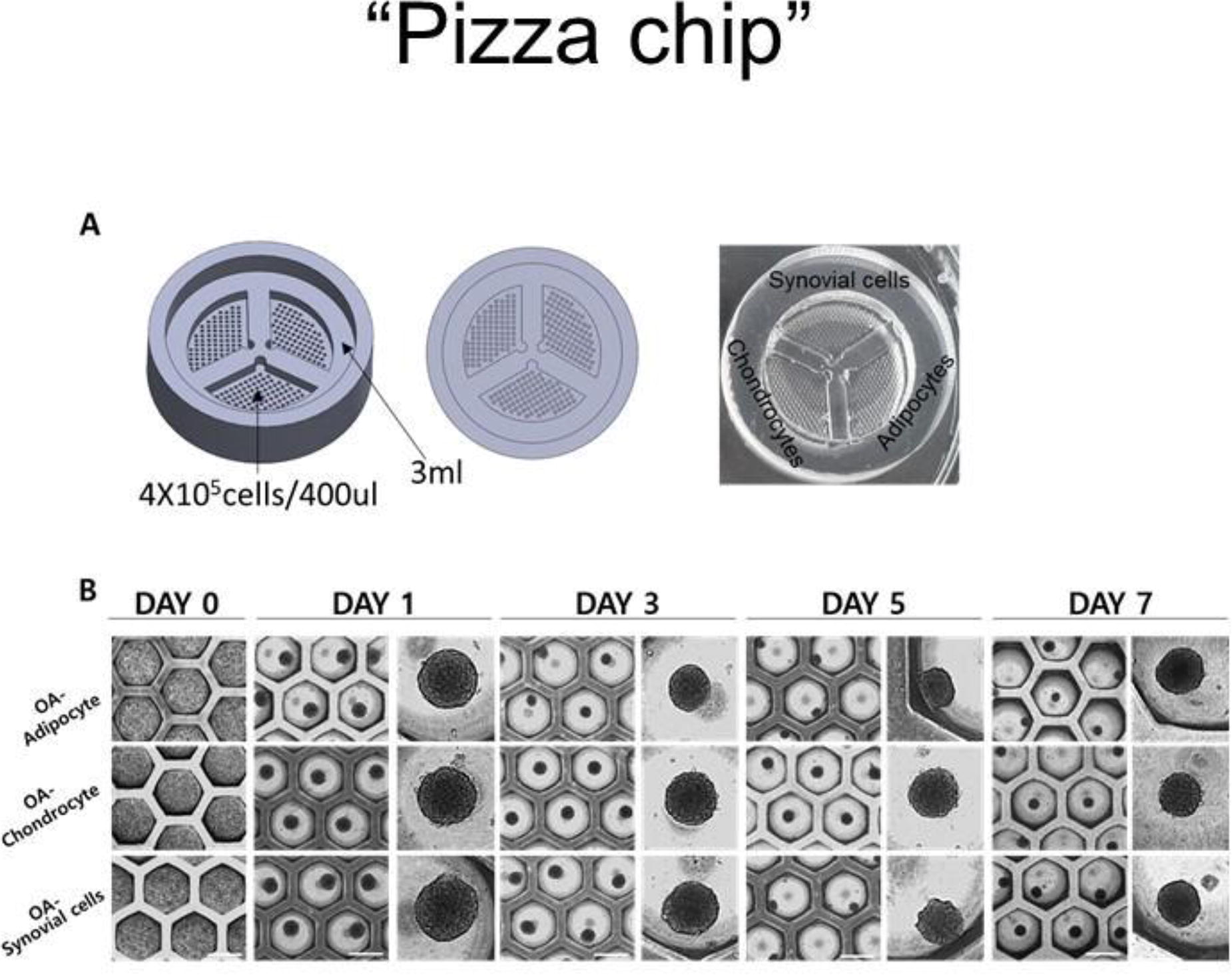
B156 - IPSC-derived Chondrocytes And Periosteum-derived Cells In GelMA Hydrogel For In Vivo Cartilage And Bone Regeneration
H. Agten1, J. Van Hoorick2, S. Van Vlierberghe3,2, F. P. Luyten1, V. Bloemen1
1
KU Leuven, Leuven, Belgium,
2
BIO INX, Zwijnaarde, Belgium,
3
Ghent University, Ghent, Belgium
*Purpose/Objectives: Deep osteochondral defects, i.e. defects affecting the articular cartilage and the underlying subchondral bone, caused by a number of conditions including acute trauma and osteochondritis dissecans, do not heal spontaneously. Hydrogel-based tissue engineering strategies hold great promise in cartilage and osteochondral tissue engineering, due to the resemblance of hydrogels to the cartilage extracellular matrix and their compatibility with 3D bioprinting. Here we evaluate the use of a dual construct encompassing two cell types and investigate different degrees of maturity of implanted tissue engineered cartilage.
*Methodology: Cell-laden gelatin methacrylol (Gel-MA INX X210 (BIO INX)) hydrogels containing either human periosteum-derived progenitor cells (PDCs) or human induced pluripotent stem cell (iPSC)-derived chondrocytes were evaluated in vitro and in vivo for their potential to regenerate the subchondral mineralized tissue and the articular cartilage on the joint surface respectively. Cell-laden hydrogel constructs were directly implanted ectopically or cultured in a single serum-free chemically defined medium and implanted after 21 days. Additionally, periosteum-derived cell-laden constructs and iPSC-derived chondrocyte-laden constructs were combined in a dual strategy and implanted orthotopically in a critically-sized osteochondral defect in nude rats. We investigated the difference between directly implanted chondrocyte-laden hydrogel constructs and precultured constructs, to determine the level of maturity needed for joint healing. Explants were obtained 4 and 16 weeks after surgery. The constructs were analysed before and after implantation with qualitative and quantitative methods such as histology, immunostainings, gene expression analysis and microcomputed tomography (µCT).
*Results: In periosteum-derived cell-laden constructs, histological stainings showed the formation of a cartilage-like template after 21 days in vitro, undergoing in vivo endochondral ossification in an ectopic assay. Gene expression analysis detected the upregulation of (pre)hypertrophic genes (COL10A1, IHH, CNMD) at the end of the in vitro culture period. Tissue engineered constructs with iPSC-derived chondrocytes formed a cartilage-like matrix, high in sulphated GAGs (Safranin O) and collagen type II (immunostaining) after 21 days in vitro. These constructs form a dense cartilage matrix after 4 weeks in vivo, even without in vitro pre-culture. Orthotopic implantation of both cell-laden hydrogel constructs resulted in consistent repair of the subchondral bone, as shown and quantified by µCT. Interestingly, histology suggested that direct implantation of chondrocyte-laden hydrogel constructs is more favorable for articular cartilage regeneration than the implantation of pre-cultured constructs.
*Conclusion/Significance: Taken together, the data show that iPSC-derived chondrocytes encapsulated in Gel-MA can generate hyaline cartilage-like tissue constructs with different levels of maturity, while using periosteum-derived cells in the same construct type generated mineralized tissue and cortical bone in vivo. Moreover, we demonstrated the importance of defining the proper tissue intermediate and level of maturity for successful orthotopic in vivo repair. In conclusion, the aforementioned cell-laden hydrogels can be potent partners in a multi-component strategy for the treatment of deep osteochondral defects.
B157 - Growth And Differentiation Factor 5 (gdf5) Loaded Nano-fibrous Scaffold In Combination With Mesenchymal Stem Cells For Full-thickness Cartilage Defect Regeneration In A Rabbit Model
M. Mamachan, A. Banu Shajahan, S. Khan, K. Singh, S. Maiti
ICAR-Indian Veterinary Research Institute, Bareilly, India
*Purpose/Objectives: To evaluate the chondrogenic potential of poly (glycerol sebacate) (PGS) co-polymer nano-fibrous (NF) scaffold loaded with growth and differentiation factor-5 incorporated sugar glass nanoparticles (SGnP-GDF5) along with bone marrow-derived mesenchymal stem cells (BM-MSC) in full thickness articular cartilage defect of rabbits.
*Methodology: Fifty adult healthy New Zealand White rabbits of either sex were allocated randomly into five treatment groups. A full-thickness chondral defect (4 mm diameter and 2 mm depth) was surgically created in the trochlear facet of the left femur using a Brad point drill bit and power drilling machine. Group A was assigned as control without receiving any treatment. The defects were implanted with various biomaterials, including PGS co-polymer NF scaffold (group B), BM-MSC laden PGS co-polymer NF scaffold (group C), SGnP-GDF5 loaded PGS co-polymer NF scaffold (group D) and BM-MSC laden SGnP-GDF5 loaded PGS co-polymer NF scaffold (group E). Five animals from each group were sacrificed on the 60th day and 90th day of post-treatment. The samples were evaluated based on gross morphology, histopathology and scanning electron microscopy (SEM) analysis to evaluate cartilage regeneration.
*Results: Superior quality cartilage regeneration simulating the hyaline cartilage was observed in groups D and E. Whereas the rest of the treatment groups exhibited inferior quality repair compared to groups D and E. Amelioration in repair was evident in the 90th day tissue samples compared to 60th day in all treatment groups. The formation of a better-quality extracellular matrix with optimum proteoglycan and collagen levels was remarkable in the safranin O staining of groups D and E. The hematoxylin & eosin (H & E) staining has highlighted a well-developed tide mark zone in both groups. The RGB trichrome staining has underscored an explicit columnar distribution of chondrocytes in group E. Moreover, SEM analysis suggested a higher number of chondrocytes in these groups, inferring that these groups support healthy cartilage regeneration. The results obtained from the gross morphology, histopathology, and SEM analysis consistently aligned with each other.
*Conclusion/Significance: The sustainable release of GDF-5 from SGnP and the microenvironment provided by the NF scaffold enhanced the therapeutic potential of stem cells in cartilage regeneration in our present study. The controlled release of growth factors throughout the healing period facilitated superior quality regeneration in groups D and E. This research highlights the utility of nanoparticles for delivering growth factors to replace the conventional method of repeated intra-articular injection of growth factors. Moreover, this technique paves the way to counteract the short half-life of significant growth factors involved in cartilage regeneration. The chondrogenic potential of stem cells was effectively complemented by the biochemical stimulation of GDF-5 and the mechanical robustness of the NF scaffold. This research concludes the potential of SGnP-GDF-5 in combination with MSC for superior cartilage healing.
B158 - Creating A Mechanically Functional Artificial Pericellular Matrix Using A Modular Hydrogel System For Articular Cartilage Tissue Engineering
M. van Mourik1, J. Spierings1, P. Koca2, F. Abinzano1, R. van Donkelaar1, J. Foolen1, K. Ito1
1
Eindhoven University of Technology, Eindhoven, Netherlands,
2
University of Stuttgart, Stuttgart, Germany
*Purpose/Objectives: Articular cartilage has mechanical properties which could be considered conflicting. While the bulk tissue has a high stiffness to withstand joint loading, chondrocytes are surrounded by a softer pericellular matrix (PCM). The apparent stiffness of the extracellular matrix (ECM) is around 10x higher compared to the PCM. This creates a challenge when developing a tissue-engineered construct that meets both the mechanical demand of the joint and provides the cells with a favorable microenvironment, which is often overlooked in current tissue-engineering approaches. By combining a high-stiffness bulk hydrogel with cells encapsulated in low-stiffness microgels, this study aims to overcome this challenge.
*Methodology: Articular chondrocytes (bACs) and articular chondroprogenitor cells (bACPCs) were isolated from bovine articular cartilage. Osteochondral plugs (ø = 10 mm) with a chondral defect (ø = 6 mm) were harvested from porcine femoral condyles. Using droplet-based microencapsulation, bACs and bACPCs were encapsulated in 1% agarose microgels, which were pre-cultured for 3 days. Chondral defects were filled with either bACs, seeded in low-stiffness (LS) 10% GelMA (∼30 kPa) or high-stiffness (HS) 10% GelMA (∼300 kPa), or pre-cultured encapsulated bACs or bACPCs, seeded in HS GelMA. Samples were cultured for 28 days in a compression-sliding bioreactor mimicking human gait. A compression ramp test was used to compare mechanical properties of uncultured and day 28 samples. Cell viability, content, and matrix synthesis were assessed using biochemical analyses. Matrix formation was visualized using (immuno-)stainings for sGAGs, type-I/II/VI collagen, and perlecan. The expression of genes related to chondrogenesis, dedifferentiation, hypertrophy, and catabolism were analyzed with qPCR.
*Results: Alcian blue and type-II collagen immunostaining showed matrix formation in all four groups, however, large differences were observed between groups. bACs in LS GelMA produced more GAGs and type-II collagen compared to bACs in HS GelMA, although the matrix was not uniformly distributed for LS GelMA. When encapsulated bACs were embedded into HS GelMA, more GAGs were produced compared to non-encapsulated cells in HS GelMA, but not as much as in LS GelMA. However, type-II collagen staining intensity was similar to LS GelMA and the cell response was more uniform throughout the construct. Encapsulated bACPCs showed less GAG and type-II collagen staining compared to encapsulated bACs, and zonal distribution seemed less distinct. Immunostainings for PCM markers type-VI collagen and perlecan showed similar intensity and distribution for all groups.
*Conclusion/Significance: This study suggests that combining different microenvironmental and bulk mechanical properties can control the synthesis and distribution of matrix components by cartilage cells. Providing a soft microenvironment is beneficial for chondrogenesis, as this improves sGAG and type-II collagen production by bACs in HS GelMA. Additionally, a high-stiffness bulk hydrogel decreases the zonal differences observed in LS GelMA, indicating improved strain distribution throughout the depth of the material. The use of ACPCs seems promising, although the stimulation of these cells needs further investigation to obtain proper tissue formation. When optimized, utilizing this concept could improve the functional outcomes of articular cartilage tissue engineering.

B159 - Bioadhesive Protein-Engineered Microcapsules For Spheroid Delivery As A Novel Treatment For Diffuse Cartilage Lesions
D. Venegas-Bustos1, A. Vega1,2, M. Alonso1, J. Rodríguez-Cabello1
1
BIOFORGE Lab, University of Valladolid − CIBER BBN, Valladolid, Spain,
2
Servicio de Cirugía Ortopédica y Traumatología, Hospital Clínico Universitario de Valladolid, Valladolid, Spain
*Purpose/Objectives: Degenerative joint disease, particularly of the knee, presents as a spectrum ranging from focal articular cartilage defects to more extensive diffuse cartilage lesions (DCL). These lesions, precursors to osteoarthritis (OA), challenge treatment due to the limited self-healing capability of cartilage. OA, characterized by the progressive breakdown of cartilage, leads to joint pain and functional impairment. Current treatments with mesenchymal stem cells (MSCs) show promise but are limited by poor cell retention at the target site. Addressing this, our study focuses on enhancing cell delivery systems for DCL regenerative therapies.
*Methodology: We aim to develop advanced cell microcarriers enhancing MSC spheroid localization and retention within cartilage lesions. Utilizing protein-engineered polymers, our spheroid nano-coating system incorporates cell-binding motifs (e.g., RGD), matrix metalloprotease (MMP)-sensitive sequences (e.g., GTAR), and specific binding sequences targeting hyaline cartilage components (e.g., collagen type II) into elastin-like recombinamers (ELRs). The recombinant production of these ELRs, followed by chemical modification with azide and cyclooctyne, facilitated a layer-by-layer spheroid nanocoating approach using click chemistry.
*Results: Our ELR coatings exhibited effective articular cartilage binding, as demonstrated by immobilizing collagen type II and chondroitin sulfate on gold sensors and measuring ELR interactions. In vitro assays showed that our microcarrier system not only localized cells accurately but also promoted spheroid proliferation and differentiation, aiding tissue regeneration.
*Conclusion/Significance: This research marks a significant leap in cell-based therapy for DCL. Our innovative microcarrier system addresses the critical challenge of cell retention, offering a novel and more effective approach for cartilage regeneration. These advancements hold great potential for improving treatment outcomes in patients suffering from OA and related degenerative joint diseases.

B160 - Biopolymer Based Biphasic Scaffold For Efficacious Regeneration Of Osteochondral Defect In Rat Model
S. Pathmanapan1,2, S. Anandasadagopan1,2
1
CSIR- Central Leather Research Institute, Chennai, India,
2
Alagappa College of Technology, Anna University,, Chennai, India
*Purpose/Objectives: Trauma, ageing, arthritis and sports injuries are the provoking conditions for cartilage and bone damage. Injuries/Defects in cartilage will eventually degenerate to Osteoarthritis (OA), which damages both cartilage and its subchondral bone, if left untreated, which may affect millions of populations and creates a socio-economic burden on society. It is prominent that cartilage is avascular and aneural tissue that retards intrinsic healing and regeneration. Besides, the cartilage and subchondral bone hold different hierarchical structures and physiological nature which possess a significant hindrance to osteochondral regeneration in clinics. The prevailing treatments only focus on managing the pain and improving the patient’s quality of life, whereas they failed to promote the complete restoration of osteochondral defects (OCD). At present, tissue engineering has emerged as an appropriate choice in regenerative medicine, focusing on restoring damaged tissue with its physiological function. Thus, in our study, we have fabricated and evaluated a biopolymer-based biphasic scaffold as a potent biomaterial for osteochondral tissue regeneration which focuses on restoring both chondral and subchondral defects.
*Methodology: We have fabricated a biphasic scaffold by sol-gel method with both organic and inorganic phase. The organic phase consists of collagen and fibrin which mimics the Extra Cellular Matrix (ECM) of cartilage, whereas the inorganic phase consists of Graphene oxide (GO) and nanohydroxyapatite (nHAP) which mimics the ECM of subchondral bone. Then the scaffolds were subjected to structural and physicochemical analysis to evaluate their pore structure, porosity, water absorption, degradation potential and mechanical strength. The biocompatibility of scaffolds was evaluated by cell viability and cell adhesion assays. Then the osteogenic and chondrogenic differentiation potential of the biphasic scaffold were analyzed at both cellular and molecular level by Alkaline phosphatase assay ALP, Alizarin red staining, Glycosaminoglycans (GAG) analysis and gene expression studies. Further the in vivo osteochondral regeneration potential of the scaffolds was evaluated in the rat model and the results were analyzed by X-ray, Micro CT and histology studies.
*Results: Our findings exhibits that the fabricated biphasic scaffold is biocompatible with optimum pore structure, porosity and degradation potential, also the subchondral layer exhibited an enhanced biomineralization potential. The engineered biphasic scaffold exhibited an effectual osteogenic and chondrogenic potential which were supported by upregulation of osteochondral specific marker genes such as COL1A1, RUNX2, ALP, SOX9 and COL2A1. The X-ray and Micro CT scan exhibited the healed defects at 12th week post-surgery which were also correlated with histology and immunohistochemical results.
*Conclusion/Significance: Thus, we have fabricated a biphasic scaffold as a cell-free biomaterial that holds potent advantages in promoting tissue repair with economic and low risk. The findings suggest that the fabricated bilayer scaffold has an effectual osteoinductive and chondroinductive potential which will be a potent biomaterial for complete restoration of osteochondral defect.
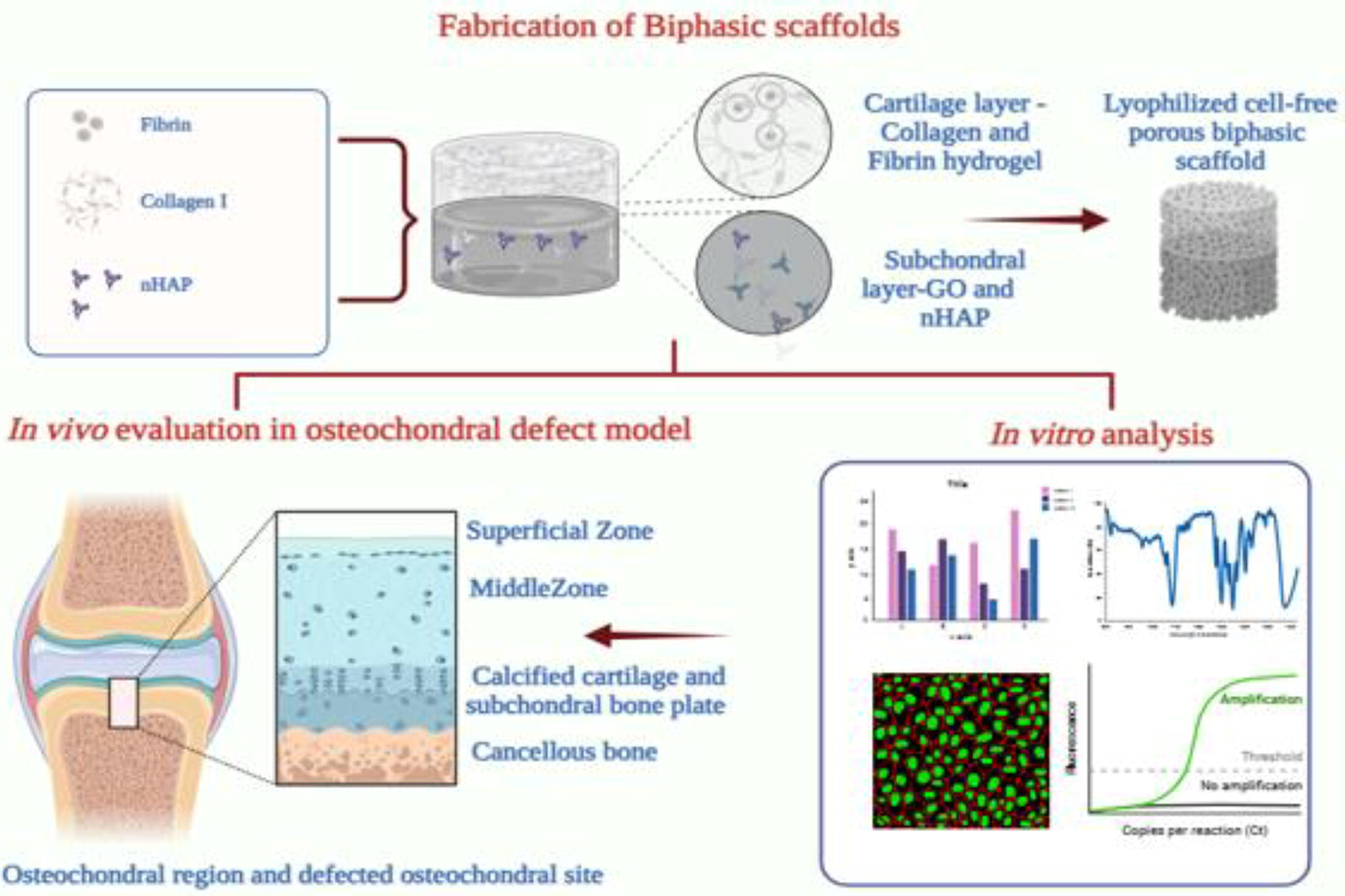
B161 - Microstructural Analysis Of The Tibial Condylar Subchondral Bone Of Knee OA: Micro-CT And Clinical CT
L. Ng, M. Tamaddon, C. Liu
University College London (UCL), London, United Kingdom
*Purpose/Objectives: Osteoarthritis (OA) is the most common degenerative joint disease that affects synovial joints as a whole joint. Multiple structures within the joint may be involved in the degenerative process. OA is generally diagnosed using conventional radiographs such as MRI, X-ray, and CT. The classic hallmark of the radiograph includes osteophytes, subchondral sclerosis and the narrowing of joint space are used to define and classify OA severity. In bone microstructural research, imaging techniques such as micro-CT (µCT) are favourable for bone microstructural analysis. This study aims to evaluate the microstructural values of the OA tibial condyle plateau and analyse whether µCT and clinical CT have a similar pattern microstructurally and whether it’s size-dependent.
*Methodology: 20 OA knee specimens were collected from knee total arthroplasty surgery patients. The specimens were stored in 4% paraformaldehyde (PFA) and stored at 4oC at the time of retrieval for at least 48 hours. CT scanner (SOMATOM Definition Edge 128, Siemens) with settings 120 kV, 169 mA, and Micro-CT (Skyscan 1172, Bruker Micro-CT) with settings: 40.38um image pixel size, 90kV, and 123uA for scanning. The scanned clinical CT data and micro-CT data were exported, processed, reconstructed, and analysed via Mimics 21.0 software (Materialise, Leuven, Belgium), Fiji/ImageJ (National Institutes of Health, Bethesda, MD, USA), and CTAnalyser (Bruker micro‐CT, Kontich, Belgium). The parameters analysed in the project: total volume of interest (TV), bone volume (BV), bone volume in relation to the total volume (BV/TV), bone surface density in relation to the total volume (BS/TV), trabecular thickness (Tb.Th), trabecular separation (Tb.Sp), alignment of trabeculae (DA), structure model index (SMI), total porosity (Po(tot)), and connectivity (Conn).
*Results: The whole tibial condyle comparison between CT and µCT BV/TV was shown to be greatest in Conn, Po(tot) and BS/TV for CT, whereas Tb.Th and Tb.Sp were greater in µCT. Comparison between the medial and lateral side of the tibial condyle with CT and µCT shows that Tb.Th, Tb.Sp and Conn were greater with µCT than CT, but both share a similar pattern where Tb.Th and Conn are greater on the medial side and Tb.Sp is greater on the lateral side. BV/TV, Po(tot) and BS/TV were observed to be greater in CT, but Po(tot) and BS/TV are greater on the lateral side, whereas BV/TV is greater on the medial side. In the analysis where regions of interest (ROI) were further specified into smaller loading regions and other regions, The results were observed to fluctuate with unstable trends between CT and µCT in different parameters apart from Tb.Th, Tb.Sp, BV/TV, and BS/TV. Value differences are observed between CT and µCT, but similar trends.
*Conclusion/Significance: µCT data showed greater consistency with minimal differences between groups, whereas clinical CT showed greater variability. Although some parameters follow similar trends, not all clinical CT measurements are suitable for effective monitoring and diagnosis of osteoarthritis (OA). Clinical CT may lack accuracy for microstructural analysis of small samples (≤10 mm), indicating its limited ability to conduct in-depth analysis of such regions of interest.
B163
B164 - Different Expression Of RANKL In Osteoblasts And Osteoblast/ Endothelial Cell Co-Cultures In Response To Dexamethasone And VEGF
K. Wirsig, N. Bürger, A. Bernhardt
TU Dresden Faculty of Medicine Carl Gustav Carus, Dresden, Germany
*Purpose/Objectives: Bone formation requires a close interaction between osteogenesis and angiogenesis. Therefore, in vitro bone models should include both bone cells and endothelial cells to closely mimic this tissue. Dexamethasone (Dex) is used as an anti-inflammatory and antiallergic drug with osteoporosis as known side effect. However, cellular mechanisms of Dex causing the loss of bone density are still unclear. We investigated the impact of Dex on co-cultures of osteoblasts isolated from primary human tissue and human umbilical vein endothelial cells (HUVEC) in comparison to osteoblast monocultures to gain insides into mechanisms of action of Dex on bone metabolism. Furthermore, the effect of externally added vascular endothelial growth facor (VEGF) in combination with Dex was analysed.
*Methodology: Osteoblasts were isolated from human femoral heads. Commercially available HUVEC were seeded on top of osteoblasts. Co-cultures of osteoblasts and HUVECs, in comparison to osteoblast monocultures, were treated with Dex or the combination of Dex and VEGF compared to a Dex/VEGF free control over a time period of 14 days. Microscopic investigations, gene expression analysis, quantification of cell type-specific enzyme activities and protein secretion were performed.
*Results: As expected, Dex stimulated alkaline phosphatase (ALP) of osteoblasts in monoculture and co-culture of osteoblasts and HUVEC. The combination of Dex and VEGF showed the highest ALP expression and activity of osteoblasts. Dex inhibited RANKL expression of osteoblasts in monoculture and in co-culture with HUVEC. Interestingly, this effect was reversed when co-cultures of osteoblasts and HUVEC were treated with the combination of Dex and VEGF. In contrast, osteoblast monocultures treated with Dex and VEGF did not show induced RANKL expression. Moreover, Dex reduced tube formation of HUVEC significantly.
*Conclusion/Significance: RANKL expression of osteoblast-HUVEC co-cultures in the presence of Dex is only induced when additional VEGF is supplied. Osteoporosis is characterised by bone loss and therefore enhanced activity of bone resorbing osteoclasts, which are dependent on RANKL. Therefore, we conclude that endothelial cells play an important role in the mechanism of Dex-induced osteoporosis.
B165 - Correlation Between The Cartilage Degeneration And The Subchondral Bone Remodelling In Osteoarthritic Joints
S. Ajami, L. Ng, M. Zhang, C. Liu
University College London, London, United Kingdom
*Purpose/Objectives: Osteoarthritis (OA) is a degenerative joint disease that affects both cartilage and subchondral bone [1, 2]. The osteochondral junction is the transition between soft and hard tissues and so is critical in absorbing the stresses during joint loading. Abnormal loading leads to microfractures within the osteochondral junction and within the subchondral bone. With progression of OA, as results of changes in the loading pattern, bone remodelling and resorption occur in the joint [3]. This weakens the physical environment that supports the overlying cartilage. In this study, we examined the changes in local distribution of volumetric bone mineral density (vBMD) in the subchondral bone, and the biomechanical properties of the overlying cartilage with an aim to understand the effect of subchondral bone remodelling on the overlying cartilage degeneration.
*Methodology: Human femoral heads were collected during total hip replacement operation due to OA. Cartilage was graded using ICRS classification, and the mechanical property of cartilage was measure by non-destructive cyclic indentation . To determine the remodelling of the subchondral bone, a peripheral quantitative CT (pQCT) was used to assess the vBMD distribution within the subchondral bone. Non-parametric Kruskal-Wallis method was used for statistical analysis (p= 0.05).
*Results: The examination of retrieved tissues revealed cartilage in different stages of degeneration, from normal to severely abnormal. Subchondral vBMD decreased with cartilage ICRS grade from 576 to 253mg/cm3 confirming bone remodeling in all samples. Dynamic modulus of cartilage was mapped and showed a weak positive correlation to ICRS grades (3.34±0.93, 2.86±1.11, 4.64±4.37 and 5.56±1.83 N/mm for grade I,II,II and IV respectively), and a moderate positive correlation to subchondral vBMD (r=0.59), confirming the concurrence of cartilage biomechanics, degeneration and SCB remodelling.
*Conclusion/Significance: We demonstrated that the subchondral plate and trabecular compartments exhibit different characteristics with increasing CLS in OA joints; somewhat expected as they serve differing morphology, physical and mechanical roles. The heterogeneous microstructural changes of subchondral bone that are linked with CLS in different regions of the femoral head could be due to divergence in load distribution in the hip joint and proximal femur. The findings suggest that changes in subchondral plate thickness, porosity, TMD and trabecular bone thickness, TMD and separation could be used as imaging markers for early diagnosis of OA. These data indicate that subchondral bone provides mechanical support to the overlying cartilage. There is a direct correlation between the cartilage degeneration and the subchondral bone remodelling. Subchondral bone remodelling is an integral part of the pathology of OA, and structural, biochemical and biomechanical changes in the subchondral bone is association with OA progression. Acknowledgement: This work was financially supported by H2020-MSCA-RISE (BAMOS, grant no: 734156) & Innovate UK (OScaffold, grant no: A02872).
B166 - Effects Of Co-culture Conditions On Spatial Behavior And Viability Of Mscs/huvecs Spheroids For Bone Tissue Engineering Applications
S. El Hajj1, M. Bankoué Ntaté1, R. Siadous2, M. Dupuy1, H. Duval1, D. Letourneur3, J. Amédée2, B. David1
1
CentraleSupélec, Université Paris-Saclay, Gif-sur-Yvette, France,
2
Université de Bordeaux, Bordeaux, France,
3
Université Paris Cité, Université Sorbonne Paris Nord, Paris, France
*Purpose/Objectives: It is crucial when designing bone tissue substitutes to successfully induce vascularization of the scaffold at the injury site which can be facilitated through prevascularization protocols. Earlier co-culture research in bone applications showed that 3D culture offers a more advantageous and physiologically-relevant microenvironment favoring cell-to-cell interaction, cell-to-matrix interaction, as well as the reorganization of endothelial cells leading to vascular formation. In this work, we explored the influence of MSCs/HUVECs co-culture in a 3D scaffold on osteogenic differentiation in prolonged in-vitro dynamic culture while identifying the optimal culture conditions and their effects on cells’ spatial behavior and viability.
*Methodology: MSCs and HUVECs labeled with PKH 26 and 67, respectively, were seeded (1:1) on porous pullulan/dextran hydrogel scaffolds supplemented or not with hydroxyapatite crystals (HAp). Each scaffold was seeded with increasing cell densities of either 400,000, 600,000, 800,000, or 1,000,000 cells. 24 hours post-seeding, 23 hydrogels were stacked, centrally aligned, then transferred and cultured up to 21 days in a custom-made perfusion bioreactor under a steady flow of 10 mL/min of culture media. Otherwise, they were left under static conditions in 6 well plates. Samples were collected on days 1, 3, 7, 14, and 21 to assess the spatial reorganization of spheroids using CLSM (Zeiss) and OCT (Thorlabs). The images were processed and analyzed using imageJ and R-spatstat package. Cell viability was measured using a Live/Dead kit (Invitrogen™). The early osteogenic marker alkaline phosphatase (ALP) was assessed using the colorimetric ALP Assay Kit (Abcam). Cell proliferation was tracked using the CyQUANT™ Cell Proliferation Assay (Invitrogen™). Hydrodynamics and oxygen transport within the bioreactor were simulated using Lattice-Boltzmann methods (LBM).
*Results: Spatial clustering of HUVECs at the center of the spheroids was observed across all culture conditions and seeding densities starting D7. It can indicate an endothelial/angiogenic affinity to hypoxic environments, as LBM simulations showed lower O2 levels at the spheroidal core. After 2 weeks of culture, the viability was significantly higher in dynamic conditions (>94%) when compared to static conditions (<75%) with dead cells being more present at the center. Additionally, ALP expression was drastically higher in the presence of HAp regardless of perfusion flow and peaked at D7 with an average value of 2.37 nmol/min/µg. Moreover, cellular proliferation was significant in dynamic conditions, leading to a 2-fold increase in initial cell number, particularly on the inner and outer peripheries of the discs. Nevertheless, LBM simulations showed that the wall shear stress remained negligible in the hydrogel's pores where cells proliferated.
*Conclusion/Significance: Our findings show that the co-culture of HUVECs and MSCs in a porous PUDNA hydrogel results optimally in homogenously distributed and viable spheroids for bone tissue engineering applications in the presence of HAp and under a steady perfusion flow. Next, we plan on assessing more osteogenic markers and the effects of culture conditions on their regulation and overall matrix deposition. We also plan on closely mapping the glucose gradients at the scaffold and spheroid levels to better understand the emerging spatial patterns in relation to local biochemical factors.
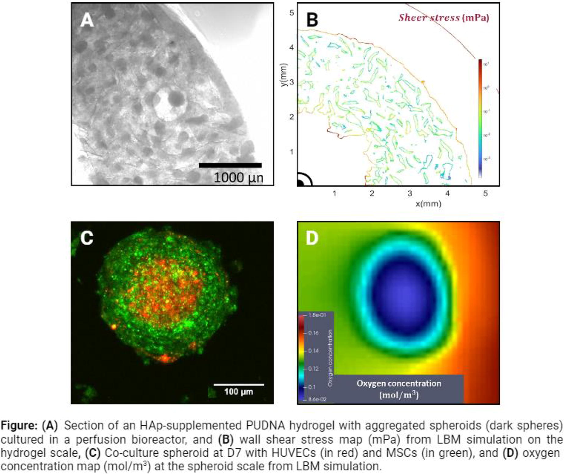
B167 - Examining The Interplay Between Collagen Alignment And Bioceramic Incorporation On Osteoblast Cell Proliferation, Differentiation And Mineralization
N. Y. Patrawalla, K. Bock, K. Liebendorfer, V. Kishore
Florida Institute of Technology, Melbourne, FL
*Purpose/Objectives: Biomimetic scaffolds provide the essential biophysical (e.g., surface topography, stiffness) and biochemical cues (e.g., composition) to guide cell morphology, proliferation, and differentiation. Although the effects of biomaterial-directed cues on cell response have been widely reported, few studies have sought to decouple these effects to better understand the interplay between the different physicochemical factors on tissue-specific cell function. In this study, beta-tricalcium phosphate (β-TCP) was incorporated into electrochemically aligned collagen (ELAC) and random collagen threads, and the individual and interactive effects of collagen alignment (i.e., biophysical) and bioceramic incorporation (i.e., biochemical) on osteoblast proliferation, differentiation, and mineralization were investigated.
*Methodology: ELAC threads were prepared using the principles of isoelectric focusing by combining dialyzed collagen solution (3.1 mg/ml) with β-TCP (10% w/w) and subjecting the mixture to an electric field of 20 V for 1 min. Pure ELAC threads were prepared in a similar manner but without the addition of β-TCP. Random collagen threads with and without β-TCP were prepared by casting neutralized collagen solution into a PLA mold and incubating at 37 ºC for 60 min to induce fibrillogenesis. All threads were crosslinked using 0.0625% genipin in 90% ethanol for 2 h. Confirmation of β-TCP incorporation and validation of collagen fiber alignment was carried out using scanning electron microscopy (SEM) and polarized light microscopy (PLM). Physical characterization of collagen threads included assessment of swelling capacity, stability, and crosslinking degree. Tensile tests were performed to assess the effects of collagen alignment and incorporation on collagen thread mechanical properties. Saos-2 cells were seeded at 10,000 cells/cm2 on the threads and the individual and synergistic effects of collagen alignment and β-TCP incorporation on cell proliferation, differentiation, and mineralization was investigated. Data were analyzed using one-way ANOVA with Tukey posthoc test. Statistical significance was set at p < 0.05.
*Results: Results from SEM and PLM confirmed that collagen fiber alignment in ELAC was retained upon β-TCP incorporation. ELAC threads exhibit greater water uptake capability and stability than random collagen counterparts. TNBS assay showed that the crosslinking degree was comparable for all threads (∼30%) indicating that collagen alignment and β-TCP incorporation had no effect. ELAC threads were significantly stronger and stiffer (p < 0.05) than random collagen threads. β-TCP incorporation had no effect on random collagen threads but significantly decreased (p < 0.05) the tensile strength and stiffness of ELAC threads. Alkaline phosphatase (ALP) activity was significantly higher (p < 0.05) in ELAC threads compared to random threads. In addition, β-TCP incorporation into ELAC significantly augmented (p < 0.05) cell metabolic activity, ALP activity, and cell-mediated calcium deposition.
*Conclusion/Significance: In conclusion, results from this study indicate that topographical cues from aligned collagen significantly enhance osteoblast differentiation suggesting that collagen anisotropy is a key driver of osteogenesis. In addition, there exists a synergy between collagen alignment and β-TCP incorporation as evidenced by enhanced cell differentiation and mineralization. Overall, β-TCP incorporation into aligned collagen framework can yield biomimetic functional scaffolds for bone regeneration applications.
B168 - Enhancing Titanium Alloys With Graphene Family Nanomaterials For Bone Repair
H. Lord-Moulding, L. Biant, J. Gough, M. McNicholas
The University of Manchester, Manchester, United Kingdom
*Purpose/Objectives: Bone defects are debilitating and common. Over a critical size, they will not spontaneously heal meaning some intervention is necessary. Joint replacements (arthroplasties) are also extremely common. Surgically implanted biomaterials are required to encourage osteogenesis in the case of large defects and total or partial arthroplasty is the only option in end stage osteoarthritis. This is becoming more prevalent as the population ages. Titanium (Ti) alloys are widely used in current surgical interventions for replacing bone. The metal has good mechanical properties and is largely bioinert meaning it will not be rejected. However, this inertness means there is room for improvement - if Ti were to have enhanced bioactivity, bone regeneration and osseointegration of implants could be improved. Two-dimensional graphene family nanomaterials (GFNs) have become of great interest in medicine due to their unique and powerful properties. This is due to their structure, which can be hydrophilic as well as allowing excellent electrical conductivity which may aid cell communication. It is also easy to produce in large enough quantities for medical applications. Therefore, GFN coatings offer one potential avenue for enhancing titanium.
*Methodology: Before coating, GFN flake height and lateral dimensions were analysed by atomic force microscopy (AFM) and scanning electron microscopy (SEM) respectively. Following this, GFNs were deposited onto Ti64 (surgical Ti alloy) via electrodeposition. The surface was analysed by SEM and Raman spectroscopy, gleaning qualitative topographical information and confirming the presence of GFNs respectively. The wettability of coated and uncoated surfaces were also assessed by water droplet contact angle measurement and the roughness of the surfaces by white light interferometry, both of which affect protein adsorption and cell adhesion. Cells were cultured on the materials and LIVE/DEAD, alamarBlue, PicoGreen and DAPI/Phalloidin assays performed in order to assess cell viability, proliferation and morphology.
*Results: GFNs were confirmed by Raman mapping to deposit effectively on the alloy using our method. The wettability of the materials saw a gradual increase after longer GFN deposition times implying that, as expected, more GFN present on the samples improves hydrophilicity. Roughnesses were confirmed to be generally equivalent between samples after the preparation and coating. The morphology of cells was seen to be more fibroblastic and proliferation was enhanced on the GFN-coated materials compared with uncoated Ti. PicoGreen analysis showed a consistently higher DNA content for cells cultured on the coated materials compared to the uncoated over at least 14 days.
*Conclusion/Significance: If bone regeneration can be made more effective then patient quality-of-life can be improved and the socioeconomic burden can be lightened. GFNs provide a promising platform for enhancing current materials with a large scope for more advanced modifications such as the functionalisation of the material with active biomolecules. GFNs are biocompatible and easily produced meaning they are suitable for scaling up and for use in healthcare, we have shown that they can readily be deposited on Ti and the scaling up of this process is also very achievable. Next steps include assessment of bone matrix deposition by stem cells on the materials.
B169 - Enhancing Bone-on-a-chip Development: A Proposed Sequential Co-culture Process For Optimizing The Microenvironment Of Osteoblast And Osteoclast Cells
K. Nae-Un1, M. Quan1, G. Kim2, H.-H. Park1, Y. Kim2, Y.-S. Cho1
1
Wonkwang university, Iksan, Korea, Republic of,
2
Mary’s Hospital, Daejeon St, Korea, Republic of
*Purpose/Objectives: Bone-on-a-chip is a technology that mimics the biological microenvironment of bone within a chip and attracts attention as a technology that can replace basic and in-vivo tests for new drug evaluation. To develop of the bone-on-a-chip, it is important to mimic the biological environment of bone. Bone is a dynamic tissue that is continuously bone remodeling process throughout life. The bone remodeling process refers to the process by which old or damaged bone is removed and replaced with new bone. The bone remodeling process is performed by osteoblasts and osteoclasts. Osteoclasts perform the resorption of bone by secreting proteolytic enzymes that dissolve bone matrix proteins. Osteoblasts perform the new bone generation, ultimately differentiating into osteocytes. Additionally, osteoblasts and osteoclasts affect the differentiation of each cell through interactions between cells. Therefore, the development of a new co-culture method that can implement the interaction between osteoblasts and osteoclasts must be accompanied. In this study, we developed a sequential co-culture process of osteoblasts and osteoclasts that can mimic the biological microenvironment of bone.
*Methodology: To implement the interaction between osteoblasts and osteoclasts, MC3T3-E1 cells were used for osteoblasts and RAW263.7 cells were used for osteoclasts. To optimize the co-culture conditions of osteoblasts and osteoclasts, co-culture was evaluated at the same cell number and various cell ratios (1:1, 1:10, 1:20 (osteoblast: osteoclast)). Co-culture was performed by dividing up traditional and sequential methods, and traditional co-culture was compared and evaluated as a control. To confirm the differentiation and cell-to-cell interaction between osteoblasts and osteoclasts, osteogenic and osteoclastogenic differentiation markers and interaction markers were evaluated.
*Results: By co-culturing osteoblasts and osteoclasts at various ratios and evaluating differentiation and interactions between cells, the ratio of osteoblasts to osteoclasts was optimized to 1:10. In addition, based on the optimized cell ratio, the culture medium composition was optimized by co-culturing osteoblasts and osteoclasts in various culture medium compositions. As a result, through the evaluation of differentiation markers and interaction markers of osteoblasts and osteoclasts, it was confirmed that sequential co-culture implemented cell-to-cell interactions better than traditional co-culture.
*Conclusion/Significance: In this study, we developed a sequential co-culture process of osteoblasts and osteoclasts that can mimic the biological microenvironment of bone. It is judged that sequential co-culture of osteoblasts and osteoclasts can be used to develop bone on a chip as a culture method that can realize cell-to-cell interaction in-vitro.
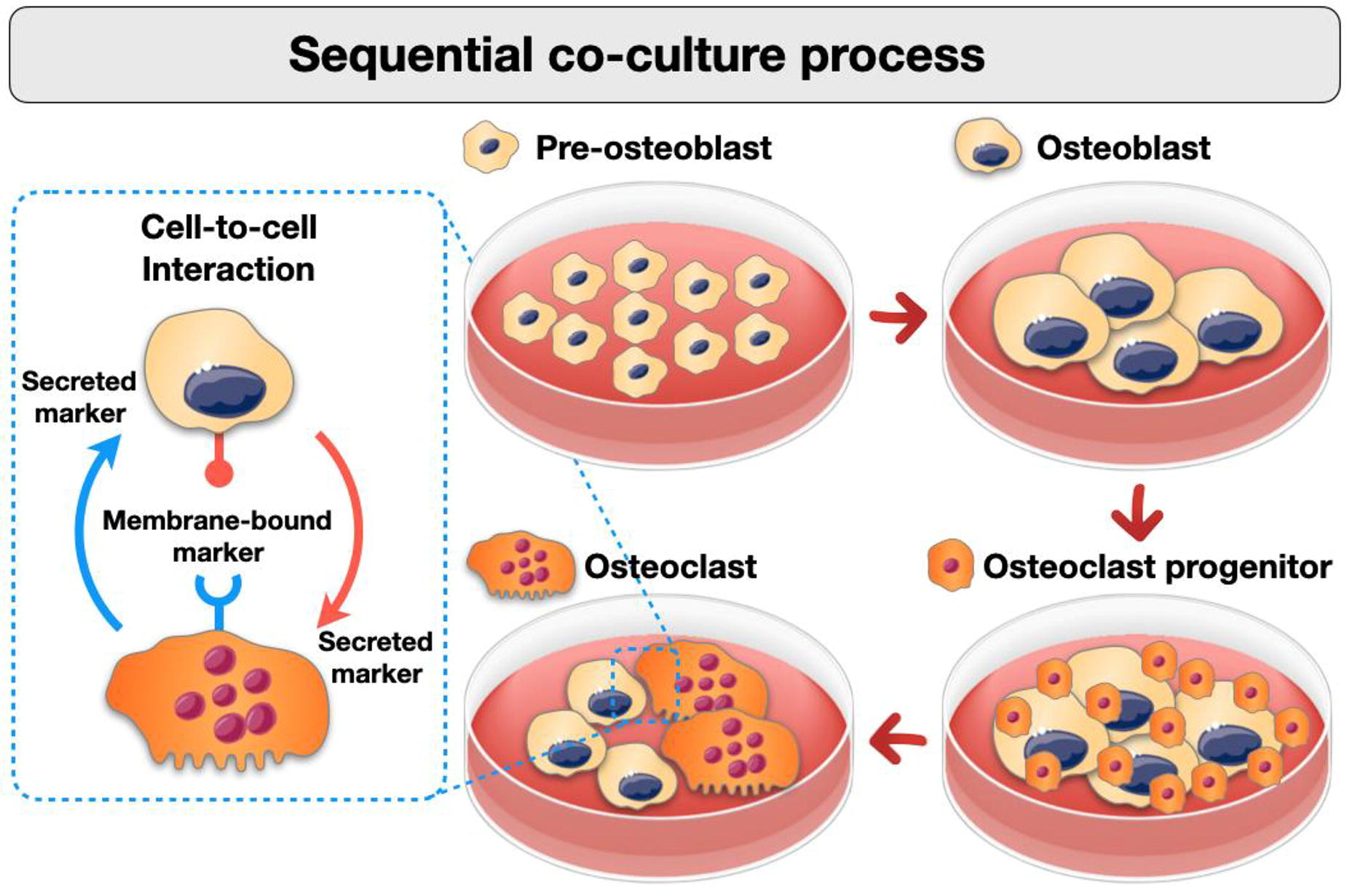
B170
B171 - Bone Sweet Bone - A Novel Organotypic Culture Model Of The Diabetic Bone
P. S. Gomes, R. Araújo, M. Fernandes
Faculty of Dental Medicine, U Porto, Porto, Portugal
*Purpose/Objectives: Bone regeneration is potentially compromised in the context of various systemic diseases, leading to the crescent necessity of developing innovative therapeutical strategies. Diabetes mellitus holds particular significance due to its association with compromised bone metabolism and healing. Hyperglycemia, a hallmark of diabetes, significantly influences tissue metabolism, leading to structural alterations and a subsequent reduction in biomechanical properties. As such, it is essential that experimental models reflect bone tissue functional and morphological changes elapsing from the pathological process, providing robust data for evaluating potential therapies. However, the reliance on in vivo models has raised societal and ethical concerns, prompting the exploration of alternative and representative models of these conditions. In this frame, this study aims to establish and characterize an innovative organotypic model of the diabetic bone, reaching hand of the ex vivo embryonic chicken femur grown in hyperglycemic conditions.
*Methodology: Embryonic femurs from Gallus domesticus were isolated and cultured at the air/liquid interface for eleven days under hyperglycemic (25mM glucose) or normoglycemic (5.5 mM glucose) conditions. Following the culture period, femora were processed for subsequent analysis. Histomorphometric assessment of tissue features were assessed upon differential histochemical stains, including Alcian Blue/Sirius Red and Massońs trichome, to differentiate collagen deposition within glycosaminoglycan enriched matrix; and Von Kossa staining was employed to differentiate mineralized tissue. Microtomographic analysis was conducted to evaluate structural parameters related to the mineralized tissue portion. A carbonyl content assay was performed to assess potential oxidative stress-induced damage. Additionally, gene expression was conducted, focusing on the evaluation of osteogenic, osteoclastogenic, and inflammation-related markers.
*Results: Exposure to high glucose levels lead to increased collagen deposition, accompanied by elevated carbonyl content, indicating structural damage attributable to heightened oxidative stress response to the excessive microenvironmental glucose levels. Gene expression analysis revealed that high glucose levels upregulated the expression of pro-inflammatory and early osteogenic markers, simultaneously suppressing the expression of late osteogenic markers. These molecular alterations were corroborated by histomorphometric and microtomographic analysis, further demonstrating a significant impairment of the mineralization process. In addition, osteoclastic activity was increased by the hyperglycemic condition. These results align with the previously acknowledged hyperglycemia-induced alterations in bone tissue structure and functionality, as observed in both human and experimental animal data.
*Conclusion/Significance: The model's ability to comprehensively express hyperglycemia-induced tissue alterations, as well as the molecular patterns of gene and protein expression, highlights its integrative responsiveness and solidifies its relevance in research. This multifaceted approach not only provides a holistic understanding of the effects of hyperglycemia on bone tissue but also positions the model as a valuable tool for exploring and advancing regenerative strategies in diabetic conditions. The comprehensive insights garnered from this study lay a robust foundation for future investigations, aiming to unravel the intricacies of bone regeneration and develop targeted interventions to mitigate the impact of diabetes mellitus on skeletal health.
B172 - 3D Printed Polylactic Acid/Cuttlefish Bone Biocomposite Scaffolds Promote Osteogenesis In Vitro
A. Gialouri1, K. Loukelis2, N. Bouropoulos1, M. Chatzinikolaidou2,3
1
University of Patras, Patras, Greece,
2
University of Crete, Heraklio, Greece,
3
FORTH, Heraklion, Greece
*Purpose/Objectives: Green chemistry includes processes that aim towards the reduction of waste products by reintegrating them in other useful production cycles, thus providing financial benefits and minimizing environmental pollution levels. Marine by-products derived from shrimps, crabs, squids, lobsters, and cuttlefish can partially include wastes, which are usually discarded away without further utilization. Cuttlefish bone has a unique porous structure, mainly consisting of aragonite, a calcium carbonate polymorph. Taking these into consideration, the aim of this work was to develop a functional biomaterial based on the thermoplastic polylactic acid and the cuttlefish bone with tailored properties for bone tissue engineering.
*Methodology: Purification of the aragonite powder from organic compounds and impurities was achieved by alkaline treatment in boiling NaOH followed by immersion in boiling methanol. PLA and composite PLA/aragonite filaments were prepared, containing 2.5, 5.0 and 10% w/w of aragonite. 3D printed scaffolds were manufactured using the fused filament fabrication (FFF) technology. All materials were characterized by Differential Scanning Calorimetry, Thermogravimetric analysis, X-ray Diffraction, Scanning Electron Microscopy and Fourier Transform Infrared spectroscopy and water contact angle measurements. Dynamical Mechanical Analysis performed on scaffolds to examine their mechanical properties. Finally, the scaffolds biocompatibility, cell adhesion and osteogenic potential were evaluated by detecting alkaline phosphatase activity, collagen and calcium production by pre-osteoblasts.
*Results: It was found that alkaline treatment led to the removal of organic compounds and did not alter the aragonite polymorph. Water contact angle measurements showed that the addition of aragonite increased not only the hydrophilicity of PLA but also the storage modulus. Adhered pre-osteoblastic cells cultured on PLA/aragonite scaffolds exhibited high biocompatibility. Alkaline phosphatase activity showed that an increased aragonite content enhances osteogenic differentiation. Calcium production, detected by alizarin staining protocol, showed an almost two-fold increase from day 7 to day 14, indicating that PLA/aragonite scaffolds stimulated matrix mineralization. Collagen production was increased in all PLA/aragonite scaffolds demonstrating their capacity to support extracellular matrix formation.
*Conclusion/Significance: In this study, different composite scaffold formulations were produced containing thermoplastic PLA matrix and aragonite from cuttlefish as a filler. The results demonstrate that 3D biocomposite PLA/aragonite scaffolds promote osteogenesis in vitro.
B173 - Enhancing Bone Regeneration Through Bioactive Ca3Si Scaffold In Ovine Large Volume Segmental Bone Defects
F. Medeiros Savi1,2, S. Saifzadeh1,3, O. Richardson1,4, M. Pandian1,4, J. Chang5,6,7, C. Wu5, C. Yang6, C. Vaquette8,9, D. W. Hutmacher1,2
1
Queensland University of Technology, Brisbane, Australia,
2
Max Planck Queensland Center for the Materials Science of Extracellular Matrices, Brisbane, Australia,
3
Medical Engineering Research Facility, Brisbane, Australia,
4
Centre in Regenerative Medicine, Brisbane, Australia,
5
Shanghai Institute of Ceramics, Shanghai, China,
6
The First Affiliated Hospital of Wenzhou Medical University, Wenzhou, China,
7
Zhejiang Engineering Research Center for Tissue Repair Materials, Wenzhou, China,
8
The University of Queensland, Brisbane, Australia,
9
Herston Biofabrication Institute, Brisbane, Australia
*Purpose/Objectives: The human body possesses remarkable regenerative capabilities, particularly in the context of bone tissue regeneration. However, due to pathological conditions where extensive bone loss occur, the body's intrinsic healing capacity may be insufficient. Currently, autologous bone grafts (ABG) stand as the gold standard treatment for large segmental bone defects. Unfortunately, their application is hindered by limitations in availability and associated donor site morbidities. Our research employs a well-established sheep animal model to explore Scaffold-Guided Bone Regeneration (SGBR) concepts, focusing on medical-grade polycaprolactone scaffolds combined with tricalcium phosphate and calcium phosphates (mPCL-TCP-CaP) composite scaffolds. This composite exhibits exceptional physicochemical and osteoinductive properties. Within this framework, our study investigates the regenerative potential of 3D-printed tricalcium silicate (Ca3Si) scaffolds combined with a mPCL-CaP mesh for the reconstruction of a sizable 3cm segmental bone defect.
*Methodology: A 3cm defect was created in 12 sheep tibiae. The defects were treated with two experimental groups: I) Ca3Si combined with an mPCL-CaP mesh and II) Ca3Si + mPCL-CaP mesh with an additional 1mg of bone morphogenic protein 2 (BMP2) and compared against autologous bone grafts (ABG) and mPCL-TCP scaffold controls. Over the course of 3, 6, 9, and 12 months, X-rays, mechanical testing, micro-computed tomography (µCT), histological, immunohistochemical and scanning electron microscopy (SEM) analyses were conducted.
*Results: In alignment with X-ray and µCT assessments, the histological and immunohistochemical results showed complete bridge of the defect site after 12 months for groups I and II. Enhanced mechanical stability and significantly higher bone volumes were achieved for group II when compared to the ABG and mPCL_TCP scaffold only. SEM evaluation showed osteocytes in direct contact with the Ca3Si scaffold and with the mPCL-CaP mesh, as well as secondary osteon formation.
*Conclusion/Significance: These findings underscore the potential of 3D-printed Ca3Si scaffolds, in conjunction with the SGBR concept, as an effective approach for reconstructing large segmental bone defects.
B175 - Engineering Of Extracellular Matrix From Human Ipsc-mesenchymal Progenitors To Enhance Osteogenic Capacity Of Human Bone Marrow Stromal Cells Independent Of Their Age
D. Hanetseder1,2, T. Levstek1,2, A. H. Teuschl-Woller3,2, J. K. Frank1,2, B. Schaedl1,2,4, H. Redl1,2, D. Marolt Presen1,2
1
LBG Ludwig Boltzmann Institute for Traumatology. The Research Center in Cooperation with AUVA, Vienna, Austria,
2
Austrian Cluster for Tissue Regeneration, Vienna, Austria,
3
Department Life Science Engineering, University of Applied Sciences Technikum Wien, Vienna, Austria,
4
University Clinic of Dentistry, Medical University of Vienna, Vienna, Austria
*Purpose/Objectives: Regeneration of bone defects is often limited due to compromised bone tissue physiology. Previous studies suggest that engineered extracellular matrices enhance the regenerative capacity of mesenchymal stromal cells. In this study, we used human-induced pluripotent stem cells, a scalable source of young mesenchymal progenitors (hiPSC-MPs), to generate extracellular matrix (iECM) and test its effects on the osteogenic capacity of human bone-marrow mesenchymal stromal cells (BMSCs).
*Methodology: iECM was deposited as a layer on cell culture dishes and into three-dimensional (3D) silk-based spongy scaffolds.
*Results: After decellularization, iECM maintained inherent structural proteins including collagens, fibronectin and laminin, and contained minimal residual DNA. Young adult and aged BMSCs cultured on the iECM layer in osteogenic medium exhibited a significant increase in proliferation, osteogenic marker expression, and mineralization as compared to tissue culture plastic. With BMSCs from aged donors, matrix mineralization was only detected when cultured on iECM, but not on tissue culture plastic. When cultured in 3D iECM/silk scaffolds, BMSCs exhibited significantly increased osteogenic gene expression levels and bone matrix deposition. iECM layer showed a similar enhancement of aged BMSC proliferation, osteogenic gene expression, and mineralization compared with extracellular matrix layers derived from young adult or aged BMSCs. However, iECM increased osteogenic differentiation and decreased adipocyte formation compared with single protein substrates including collagen and fibronectin.
*Conclusion/Significance: Together, our data suggest that the microenvironment comprised of iECM can enhance the osteogenic activity of BMSCs, providing a bioactive and scalable biomaterial strategy for enhancing bone regeneration in patients with delayed or failed bone healing.
B177
B178 - Deciphering The Transcriptional Landscape Associated With Endochondral Bone Formation Potential In Marrow Stromal Cells
A. Lolli1, E. Ji1, J. Witte-Bouma1, P. de Jongh1, R. Hoogenboezem1, E. M. Bindels1, M. H. Raaijmakers1, G. J. van Osch1,1,2, N. Fahy1,1,3, E. Farrell1
1
Erasmus MC University Medical Center Rotterdam, Rotterdam, Netherlands,
2
University of Technology Delft, Delft, Netherlands,
3
Technological University of the Shannon: Midlands Midwest, Limerick, Ireland
*Purpose/Objectives: Endochondral ossification is the critical process in bone formation and regeneration whereby transient cartilage is remodelled into bone. We and others previously succeeded in tissue engineering bone via endochondral ossification by implanting chondrogenically-primed human marrow stromal cells (hMSCs) in mice. Unfortunately, the lengthy phase of in vitro priming (3-5 weeks) needed to obtain mature cartilage hinders the translation of such approaches for the treatment of large bone defects. Interestingly, little attention has yet been paid to the extent to which the priming may be shortened and how the in vivo dynamics of cartilage remodelling may be affected. We set out to elucidate the effect of chondrogenic priming duration on the dynamics of hMSC-mediated endochondral ossification. We further investigated the changes in cell transcriptome linked to the acquisition of bone formation potential, which still remain largely undefined.
*Methodology: hMSC pellets (2x10⁵ cells/pellet) were cultured in the presence of the chondrogenic inducer Transforming Growth Factor(TGF)-β3 for 1,3,5,7 or 21 days. The pellets were subjected to histological analysis, total RNA isolation for bulk RNA-sequencing, or implanted subcutaneously in athymic mice for 12 weeks. In vivo mineralisation was analysed by µCT. The implants were retrieved at several time-points post-implantation (d3,d7,d14,d28,d56,d84) and subjected to (immuno)histological characterisation to monitor bone formation, or flow cytometry to determine the recruitment of innate immune cell subsets.
*Results: During chondrogenic priming, noticeable cartilage (glycosaminoglycan) formation in the pellets started on d5-d7 and increased henceforth. After implantation, 7d- and 21d-primed pellets consistently led to bone formation (N=4 hMSC donors). While priming the cells for less than 7 days led to extremely limited or no bone formation, we occasionally observed signs of bone formation even when hMSCs were chondrogenically primed for only 1 day (Fig.1). Next, we investigated whether a brief priming of 7 days impacts the dynamics of bone formation. In vivo, chondrogenic pellets primed for 7 or 21 days were similarly encapsulated by fibrous tissue (d3-d7 post-implantation), underwent mineralisation (d7-14) and microvascular infiltration (d14-d28), forming bone with marrow (d56-d84) (N=2 hMSC donors). These pellets exhibited comparable persistence of human/donor-derived cells, identified by human-specific GAPDH staining, and recruitment of host-derived innate immune cells, determined by flow cytometry. However, longitudinal µCT demonstrated significantly increased bone volume for 21d- vs 7d-primed pellets (∼5-fold). To determine the transcriptional signature associated with bone formation capacity, we compared the full transcriptome of 7d- and 21d-primed pellets with that of 3d-primed pellets. We identified a panel of 102 transcription factors and 63 microRNAs specifically modulated in bone-forming samples, reconstructing regulatory networks that can direct bone formation.
*Conclusion/Significance: We provide proof-of-concept that a very brief chondrogenic priming reliably leads to bone formation without impacting the dynamics of endochondral ossification. We further identified the gene signature and transcriptional regulatory networks associated with bone formation potential, providing a number of new targets that may be exploited in combination with short priming strategies to scale-up bone volume or speed-up bone formation. Our data raise exciting opportunities for developing translational and improved endochondral ossification-based tissue engineering approaches for bone defect repair.
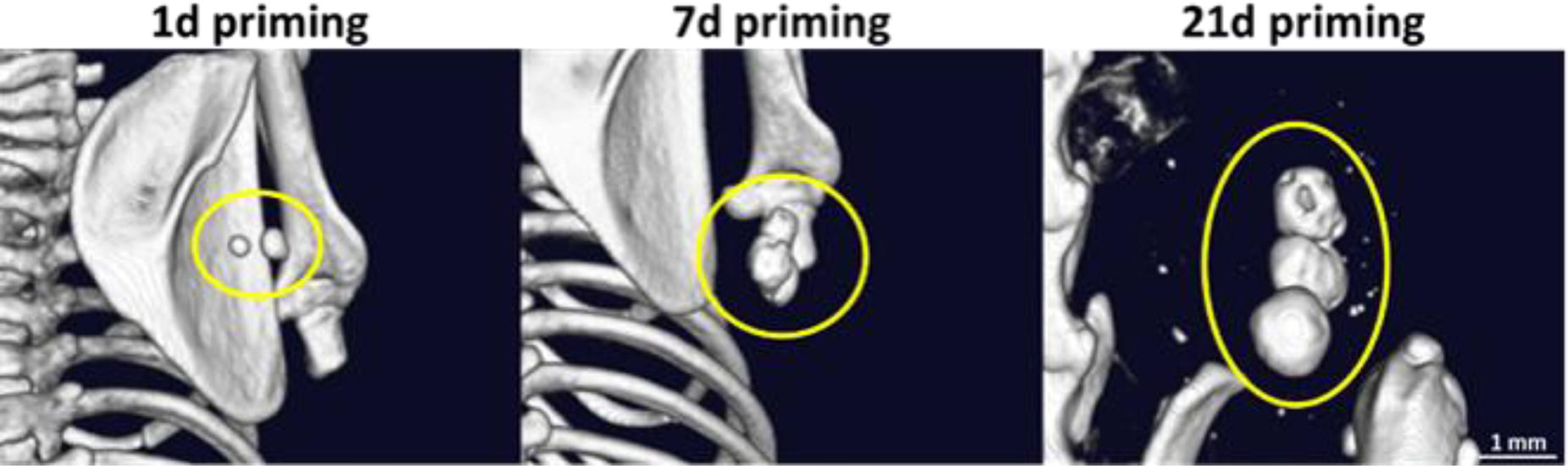
B179 - Annexin A5 Prevents Bone Erosion In Collagen-induced Mouse Arthritis By Regulating GAS7 As A Brake Protein
W. Zhang, Z. Jia
Shanghai Ninth people’s hospital, Shanghai, China
*Purpose/Objectives: Rheumatoid arthritis (RA) is a chronic inflammatory disease that often leads to localized joint bone damage and systemic bone loss, resulting in decreased bone mass and osteoporosis. Inflammation activates osteoclast generation, causing bone resorption, while inhibiting the differentiation and bone-building capacity of osteoblasts. Current RA therapies target inflammation and osteoclast generation, considered breakthroughs in treatment. Our previous research indicated a crucial role of Annexin A5 (AnxA5) in macrophage-mediated inflammation regulation, but its potential in RA remains unknown.
*Methodology: To assess the therapeutic effect of AnxA5 on RA, we utilized a collagen-induced arthritis (CIA) mouse model. We evaluated the impact of AnxA5 on CIA mouse symptoms, gait, and Micro-CT, and performed structural (HE; Safranin O-fast), macrophage-related (CD86/CD68), and osteoclast-related (TRAP) staining on ankle and paw joint sections. Additionally, mRNA analysis was conducted on synovial and bone tissues. In vitro experiments focused on the impact of AnxA5 on osteoclast development, including morphological, functional, and genetic changes during bone marrow-derived macrophage (BMM) induction of osteoclast differentiation. Potential targets and mechanisms of AnxA5 affecting osteoclast differentiation were also investigated.
*Results: AnxA5 significantly ameliorated symptoms in CIA mice, potentially through macrophage regulation and inhibition of osteoclast differentiation. In vitro experiments revealed that early application of AnxA5 markedly suppressed osteoclast differentiation, with GAS7 protein identified as a potential interacting target. By expressing GAS7 protein, osteoclast differentiation was halted, while knockdown of GAS7 protein allowed osteoclasts to differentiate under low concentrations of RANKL.
*Conclusion/Significance: AnxA5 exhibits targeted effects at inflammatory injury sites, potentially improving RA symptoms and bone erosion through regulation of the inflammatory microenvironment (macrophage polarization) and inhibition of osteoclast differentiation. It may hold significant therapeutic value in inflammatory and osteolytic bone diseases.
B182 - DEVELOPMENT OF A COMPOSITE BONE PLATE - TOWARDS BIOMEDICAL VIBRANCY AND ECONOMIC VIABILITY
M. Hoque, A. Rayhan, A. Rashid
Military Institute of Science and Technology (MIST), Dhaka, Bangladesh
*Purpose/Objectives: Titanium, stainless steel, tantalum etc. are commonly used for bone plates. However, the metallic bone plates inherit some fundamental issues namely, stress shielding, bone atrophy etc. due to their incomparable strength to the natural bones. This work aims to develop an alternative composite bone plate that should be biofunctional and economical.
*Methodology: Firstly, different types of composite samples were fabricated using available different natural fibers like jute, silk, kenaf, etc. which were embedded into matrices such as epoxy resin, polypropylene, etc. Secondly, a bone plate was fabricated after obtaining the required bone healing properties in the composite sample. Different tests including mechanical tests (Tensile, Flexural and Compression), cytotoxicity test, microstructural analysis, and moisture absorbability, etc. were performed. Besides, the mechanical properties were verified through the finite element analysis module of Ansys.
*Results: The kenaf/epoxy biocomposite could be produced from locally available and low-cost resources (natural fiber and epoxy resin), which demonstrates high potential to be used as an alternative to conventional metallic bone plates. Figure 1 presents the fabricated biocomposite bone plate samples. The kenaf/epoxy biocomposite meets the fundamental mechanical property requirements of a bone plate. Thus, the further procedure of finding the potentiality started. This composite material introduces itself as non-cytotoxic, which was evidenced by the cytotoxicity test. The water absorption test and microstructural observation further signified the potentiality of the fabricated composite bone plate.
*Conclusion/Significance: This work verifies the potential of natural fiber-reinforced polymer composites as an alternative to metallic/conventional biomaterials used for bone plates. The renewability, local availability, and biocompatibility of the natural fiber (kenaf), and the non-cytotoxicity of the fabricated composite (kenaf/epoxy) make it suitable for the bone plate application. The mechanical properties of the composite bone plate were also investigated computationally and experimentally that match with the natural bone properties. The preliminary results suggest that the fabricated composite bone plate is promising for the fixation of bone fracture.
B183 - Ready-to-use Calcium Sulfate Based Cement Paste Functionalized With Strontium For Osteoporotic Bone Defects
R. F. Richter1, C. Vater1,2, M. Gelinsky1, A. Lode1
1
TU Dresden, Dresden, Germany,
2
University Hospital Carl Gustav Carus and Faculty of Medicine of TU Dresden, Dresden, Germany
*Purpose/Objectives: Osteoporosis as a systemic disease is characterized by reduced bone density and altered bone architecture, resulting in an increased risk of fractures. The cause of these changes is a disturbed balance between bone resorption and bone formation, caused either by an increased activity of the resorbing osteoclasts or by a reduced activity of the bone-forming osteoblasts. Due to their dual effect, strontium ions are particularly interesting for osteoporosis therapy, as they promote bone formation by stimulating osteoblasts and at the same time inhibit osteoclast resorption. In this study, we developed and investigated a fast degrading and ready-to-use calcium sulfate (CaS) based cement paste combined with strontium-modified calcium phosphate cement (CPC) for local strontium ion release in osteoporotic bone.
*Methodology: Calcium sulfate hemihydrate powder was mixed with an already established CPC precursor mixture (α-tricalcium phosphate based), different amounts of strontium carbonate as strontium ion source and a cytocompatible oil based carrier liquid to form a ready-to-use cement paste that starts setting on contact with aqueous liquids. Composites with varying strontium amount and a pure CaS paste as well as a pure CPC paste as reference were investigated regarding their degradation behavior and mechanical properties. Therefore, samples were incubated for 28 days at 37 °C in human plasma-like medium. At defined time points, supernatants were taken and analyzed regarding ion release (Ca, P, Sr). For the samples themselves, mass loss and porosity as well as compressive modulus (CM) and compressive strength (CS) were measured. Additionally, all material groups were investigated regarding cytocompatibility and the influence on proliferation and osteogenic differentiation of human mesenchymal stem cells.
*Results: Calcium sulfate based pastes showed a significantly different degradation behavior compared to the pure CPC reference. For all CaS containing groups a higher mass loss over time and an increased porosity was observed. While this improved degradation resulted in a decrease in the mechanical properties over time for the pure CaS, the CaS-CPC composites showed stable or even increasing values for CM and CS which were comparable to the pure CPC. Ion release for CaS based pastes was also distinctively different as they all showed a strong calcium release and a phosphate uptake, while in contrast for pure CPC a calcium uptake and a phosphate release were observed. More importantly all CaS-CPC composites showed a significantly increased release of strontium ions compared to the CPC samples and reached the effective level to have the desired positive influence on cell behavior. All components and composites showed no detrimental effect on cell viability.
*Conclusion/Significance: The developed CaS-CPC composites are a promising material system for bone replacement in an osteoporotic environment as it effectively combines the advantages of CaS and CPC cements. The strong degradation of the CaS phase allows a faster scaffold resorption and leads to a higher local release of strontium ions to address the disturbed bone remodeling process more effectively. The CPC phase on the other hand still provides mechanical stability and due to the slower resorption process a long lasting source of strontium ions.
B184
B186
B187 - Developing An In Vitro Osteochondral Micro-physiological System For Modelling And Testing Therapeutics For Osteoarthritis
K. W. Smith1,2, S. L. Fung1, H.-F. Wu1,2, I. Chiesa3, G. Vozzi3, C. De Maria3, R. Gottardi1,2
1
Children’s Hospital of Philadelphia, Philadelphia, PA,
2
University of Pennsylvania, Philadelphia, PA,
3
University of Pisa, Pisa, Italy
*Purpose/Objectives: Currently, animal models remain popular for complex musculoskeletal research, even though they may not accurately represent human biology and are costly. In vitro models could be used to replace certain animal studies but struggle to capture the three-dimensional nature of musculoskeletal tissues as well as their crosstalk. Here, we develop a micro-physiological in vitro system that uses a biphasic bioreactor, multiple cell types, and scaffolds to mimic native biology and crosstalk of the osteochondral unit of articular joints. We validate the efficacy of the model to emulate osteoarthritis (OA) by treating with inflammatory factors and show our in vitro model can recapitulate cross talk across tissue types, enabling the testing of candidate therapeutics.
*Methodology: For the cartilage region, human bone-marrow derived mesenchymal stem cells (BM-hMSCs) are seeded in photo-crosslinkable hydrogels and differentiated chondrogenically. For the bone region, BM-hMSCs are seeded onto a 3D printed gelatin-hydroxyapatite scaffold and osteogenically differentiated. Human umbilical vein endothelial cells (HUVECS) and BM-hMCSs are seeded into the pores of the scaffold in a fibrin/gelatin hydrogel. These components are combined in the biphasic bioreactor. To induce inflammation, the cartilage constructs are treated with either: control (no additives), cytokine cocktail (20ng/mL interleukin 1β, 100ng/mL interleukin 6, and 1mg/mL tumor necrosis factor α), or M1 macrophage conditioned medium. Chondrogenesis, osteogenesis, and inflammation are assessed by RT-qPCR and staining.
*Results: Cartilage Gels: There is strong collagen II deposition in the matrix of the gel, and only cellular expression of collagen I, indicative of chondrogenesis (Figure 1A).
Vascularized Bone:
There is strong calcium deposition in the osteogenic region of the scaffold, seen by Alizarin Red staining. Collagen I staining allows visualization of the vasculature formed by the HUVECS (Figure 1A, white arrow) as well as matrix deposited by the osteogenic BM-hMSCs (Figure 1A, yellow arrow).
Structure of Complete Construct:
The combined osteochondral construct shows region specific expression of bone and cartilage markers respectively (Figure 1B). Masson’s trichrome staining shows strong collagen deposition in both cartilaginous and osseous regions, with calcification visible in the osseous region. This is confirmed by collagen I and collagen II staining, the latter only in the cartilaginous region. Alcian blue staining shows GAG deposition in the cartilaginous region. This confirms the biphasic bioreactor supports osteochondral constructs and a separate cartilaginous and vascularized osseous region reminiscent of the osteochondral junction in articular joints.
*Conclusion/Significance: Our preliminary data show that we have developed a multi-tissue micro-physiological system simulating OA. Moving forward, we will probe the role of the osseous region in regulating cartilage response to pro-inflammatory stresses. Additionally, we will test OA therapeutics like Rapamycin and TNF inhibitors. Using a micro-physiological system to test therapeutics for OA prior to animal studies can both minimize the number of animals required for preclinical testing and save on animal costs. These in vitro systems will help advance knowledge of musculoskeletal diseases development as well as allow rapid testing of potential therapeutics for treatment.

B188 - Establishment Of An Ex Vivo Cartilage Degeneration Model By Combined Collagenase Treatment And Mechanical Loading
L. Wen1,2, S. Grad1, L. Creemers2, M. Stoddart1
1
AO Foundation, Davos Platz, Switzerland,
2
University Medical Center Utrecht, Utrecht, Netherlands
*Purpose/Objectives: Osteoarthritis (OA) is the most common degenerative joint disease, affecting millions of people worldwide. Ex vivo explants models are essential for screening potential therapy for cartilage, as they are also in accordance with the 3Rs principles of animal testing. The aim of this study is to generate a bioreactor loaded ex vivo cartilage degeneration model, which can be used to evaluate approaches towards extracellular matrix regeneration and inhibition of degeneration for osteoarthritis therapy.
*Methodology: Osteochondral plugs were harvested from bovine joints by using a trephine with an inner diameter of 8 mm. The subchondral bone was cut with a circular saw to reach a height of 5 mm. A platform from LifeTec Group (Fig A) which could separate cartilage and bone was used to culture the osteochondral explants. Cartilage medium included High-glucose DMEM, 100 μg/ml sodium pyruvate, 100 U/ml Penicillin-Streptomycin, 50 μg/ml ascorbic acid, 2.5% HEPES, 1% ITS + Premix and 1% nonessential amino acids. Medium for bone was High-glucose DMEM, 100 μg/ml sodium pyruvate, 100 U/ml Penicillin-Streptomycin, 50 μg/ml ascorbic acid, 2.5% HEPES, 10% FBS and 5mM β-glycerophosphate. Cartilage was treated with 0.1% Collagenase (285 units/mL) for five minutes, then refreshed with culture medium. A cartilage bioreactor system (Fig C) was applied to load the osteochondral plugs. Loaded plugs were exposed to ball oscillation of ± 25° at 1 Hz. Simultaneously, dynamic compression was applied at with 20% to 40% sinusoidal strain of cartilage thickness. Mechanical load was performed for 1 h a day for 1, 2 or 3 days. After 1, 2, 3 or 7 days, cartilage was collected for histology. To distinguish live and dead cells, lactate dehydrogenase (LDH) and ethidium homodimer staining was performed. Safranin O and Fast Green staining were used to visualize the glycosaminoglycan (GAG) and collagen content in the extracellular matrix. COL2A1 was visualized by immunohistochemistry.
*Results: Collagenase treatment maintained high viability of chondrocytes but resulted in significant loss of GAG in the extracellular matrix (Fig B). Notably, mechanical loading led to chondrocyte death at the degenerated cartilage surface from day 2 on (Fig D), which simulates the in vivo cartilage degeneration.
*Conclusion/Significance: Intra-articular injection of collagenase has been used to induce osteoarthritis in vivo. In this study, we established a collagenase-induced ex vivo cartilage degeneration model, combined with kinematic load, which could reduce live animal use in experiments. The additional application of load led to an increased cell death at the upper cartilage surface. Extracellular matrix regeneration and preserving chondrocyte viability are key factors for successful cartilage therapy. Therefore, this model is promising to evaluate the efficacy of matrix regenerating and anti-catabolic drugs in the future.
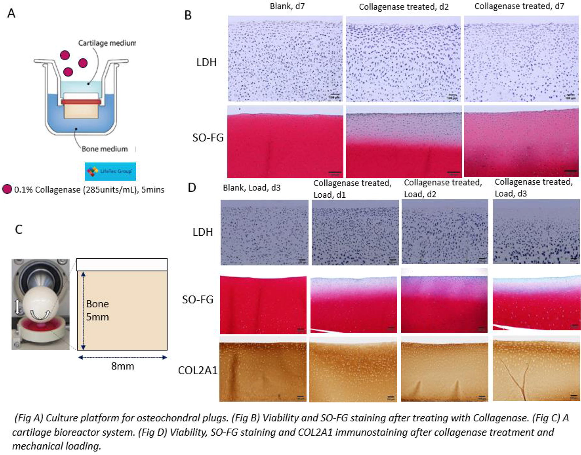
B189 - An Ecm Scaffold Combined With A Compliant 3d Printed Spring-shaped Reinforcement For Cartilage Engineering Applications
P. J. Díaz-Payno1, W. Solorzano Requejo2, J. Llorca1,2, A. Díaz Lantada2, J. Patterson1
1
IMDEA Materials, Getafe, Spain,
2
Polytechnic University of Madrid (UPM), Madrid, Spain
*Purpose/Objectives: Articular cartilage is a soft tissue lining the ends of the long bones in our joints. Even minor lesions in articular cartilage (AC) can cause underlying bone damage creating an osteochondral (OC) defect. OC defects can cause pain, impaired mobility and can develop to osteoarthritis (OA). OA is a disease that affects nearly 10% of the population worldwide, and represents a significant economic burden to patients and society. While significant progress has been made in this field, realising an efficacious therapeutic option for unresolved OA remains elusive and is considered one of the greatest challenges in the field of orthopaedic regenerative medicine. Therefore, there is a societal need to develop new strategies for AC regeneration. In recent years there has been increased interest in the use of tissue-specific aligned porous freeze-dried extracellular matrix (ECM) scaffolds as an off-the-shelf approach for AC repair, as they allow for cell infiltration, provide biological cues to direct target-tissue repair and permit aligned tissue deposition, desired in AC repair. However, most ECM-scaffolds lack the appropriate mechanical properties to withstand the loads passing through the joint. One solution to this problem is to reinforce the ECM with a stiffer framework made of synthetic materials, such as polylactic acid (PLA). Such framework can be 3D printed to produce anatomically accurate implants, attractive in personalized medicine. However, typical 3D prints are static, their design is not optimized for soft-hard interfaces (OC interface), and they may not adapt to the cyclic loading passing through our joints, thus risking implant failure. To tackle this limitation, more compliant or dynamic designs can be printed, such as coil-shaped structures.
*Methodology: Thus, in this study we use finite element modelling to create different designs including single triple, single quadruple, double triple and double quadruple helix and prototype them in PLA. The optimal design is combined with an ECM slurry. Briefly, the ECM slurry is combined with the PLA coil and freeze-casted under directional freezing prior to freeze-drying the samples to obtain an off-the-shelf scaffold with a dynamic reinforcement. The scaffold will be combined with mesenchymal stem cells (MSCs) to investigate the chondrogenic potential of such metamaterial.
*Results: The double helix has a higher stiffness modulus than the single helix and the quadruple helix a higher stiffness modulus than the triple helix. The single helixes have a better recovery after compression, while the doble helixes have a higher plastic deformation under compression. The directional freeze-casting results in ECM scaffolds (either alone or PLA reinforced) containing a tailored microarchitecture mimicking aspects of native AC.
*Conclusion/Significance: To conclude, it was possible to design and simulate stiffness of different coil-shaped reinforcements and 3D print the PLA prototypes without support material. We were able to isolate and incorporate ECM into the coil structure and produce dynamic scaffolds that have the potential to be used in cartilage tissue engineering.
B192 - Human Cartilage- And Synovium-derived Matrix Bound Nanovesicles As Potential Therapeutic Approaches For Osteoarthritis
C. Pineda Molina1, J. Mitchell2, M. Floren2, K. Hansen3, G. S. Hussey1, S. F. Badylak1
1
University of Pittsburgh, Pittsburgh, PA,
2
AlloSource, Centennial, CO,
3
University of Colorado, Aurora, CO
*Purpose/Objectives: Osteoarthritis (OA) is a prevalent and debilitating condition affecting over 500 million individuals worldwide. The progression of OA is characterized by a chronic low-grade inflammation affecting the entire joint. Pathological changes associated with OA progression include articular cartilage destruction, subchondral bone thickening, osteophyte formation, synovitis, ligament and meniscus degeneration, and joint capsule hypertrophy. Contributing factors to OA include joint injuries, obesity, aging, and genetics, among others. Despite the prevalence of OA, the mechanisms of its progression, especially considering patient age and health, remain poorly understood, with current clinical options focused on symptom management rather than addressing the causes.Recently, a subset of extracellular vesicles named as matrix-bound nanovesicles (MBV) has been identified as a key component of extracellular matrix (ECM). MBV are tightly bound to collagen fibers and are released as part of tissue turnover or in response to traumatic injuries and pro-inflammatory events. MBV present a variety of surface markers and cargo molecules capable of regulating cellular phenotypic responses with immunomodulatory effects on damaged tissues. For instance, MBV isolated from porcine urinary bladder matrix (UBM) have been shown to mitigate synovial inflammation and bone damage in a pristane-induced rat model of rheumatoid arthritis. Despite these findings, research on the immunomodulatory role of MBV, especially those derived from human tissues, has not been widely studied.
The objective of the present study is to evaluate MBV from human articular cartilage and synovium, with the potential to develop new biomarkers for understanding the progression of OA and novel therapeutic treatments using MBV.
*Methodology: The study is divided into three main parts. First, isolation and molecular and biochemical characterizations of MBV from human articular cartilage and synovium are performed. Secondly, the role of MBV isolated from cartilage and synovium in the progression of OA in an in-vitro model is evaluated: Using normal chondrocytes, the effect of MVB isolated from OA cartilage and synovium in the progression of OA is investigated. Gene expression profile and proteomics analysis are used to determine the mechanisms of the MBV modulation. Finally, using a destabilized medial meniscus (DMM) mouse model of OA, the immunomodulatory effects of human normal cartilage and synovium MBV are evaluated in-vivo.
*Results: Specifically, the study compares MBV from tissue specimens, considering variables such as age (pediatric vs. young adult vs. adult vs. elderly specimens) and OA progression (healthy vs. OA specimens) to elucidate the roles of aging in OA progression.
*Conclusion/Significance: The successful isolation and identification of MBV from human articular cartilage and synovium mark a significant achievement. This study holds meaningful promise as it addresses the critical gap in understanding the immunomodulatory roles of MBV in OA. By comparing MBV populations from different age groups, the research addresses the complexities of OA progression. Ultimately, this work has the potential to inform the development of innovative therapies for OA with MBV-mediated strategies.
B193 - 3d-printed Osteochondral Platform Reveals New Biomechanical Insights In Interfacial Bone-articular Regions
Y. Kim, S. Alimperti
Georgetown University, Washington, DC
*Purpose/Objectives: Osteochondral fractures due to trauma, aging, or surgical procedures led to loss of the homeostatic interactions between articular and subchondral compartments. Existing therapeutics may include anti-inflammatory drugs, while in severe cases, surgical restoration of cartilage and bone is considered a better solution than only cartilage repair. To address these limitations, it is essential to develop novel therapies for osteochondral fractures by engineering and applying new in vitro strategies based on advanced manufacturing approaches. In this study, we developed a novel 3D printed bilayer platform to investigate the 3D interfacial biomechanical properties of the osteochondral junction area involved in the initiation and progression of osteochondral fractures.
*Methodology: To address this hypothesis, we engineered and utilized a 3D printed bilayer scaffold consisting of a hard compartment, mimicking the subchondral tissue, and a soft compartment, mimicking the chondral part. The hydroxyapatite units shown in Figure 1(a) were created by utilizing composite inks of hydroxyapatite (HA) and Polyvinyl butyral (PVB). Next, collagen bioinks (1.5, 3, and 6 mg/mL) with embedded human articular chondrocytes (HAC) (at a concentration of 0.1 million) were 3D printed on top of the 3D printed hard compartments. After developing this bilayer structure, the bilayer interactions of the collagen matrix and the hydroxyapatite scaffolds were examined via SEM, AFM, and micro-CT. The bilayer adhesion of those two compartments with different stiffness was measured via the pressurized blister test method (Figure 1(b-d)). This method introduces a gas under a collagen film to form a blister, then increases the gas pressure until the collagen film delaminates from the substrate. Finally, the HAC viability and functionality were tested via live/dead assay and Alcian Blue staining, respectively.
*Results: The total stress response via the shear modulus demonstrated that collagen gel at a concentration of 6 mg/mL showed approximately 9 times higher stiffness (p < 0.05) compared to collagen gel at a concentration of 3 mg/mL and approximately 45 times higher stiffness (p < 0.05) at a concentration of 1.5 mg/mL. Also, our SEM data showed that the formed collagen layer with 0.5 mm thickness at concentrations 1.5 and 3.0 mg/mL was deformed and burst before delamination from the hard compartment. Conversely, the layer of high collagen concentration (6 mg/mL) with a thickness of 0.5 mm was deformed and detached from the HA compartment after 57.2 secs. Also, the bilayer adhesion energy of the soft and hard compartments was measured at 0.063∼0.106 J/m2, indicating that high collagen concentration preserves bilayer interactions and mechanical strength in the interfacial area. Also, under those conditions, the Alcian blue staining showed 6 times higher HAC functionality (p<0.05) in collagen gels at 6mg/mL compared to other conditions. Finally, we found that by increasing the collagen film thickness up to 1 mm and polymerization time up to 24 h, the adhesion strength was increased by 10 and 35 percent, respectively (p<0.05).
*Conclusion/Significance: Overall, this study serves as the foundation to better understand mechanisms involved in osteochondral defect initiation and progression in the osteochondral junctional area by using 3D printing technology.

B194 - Optimizing Culture Conditions For The Operation Of A Parallel Flow Bioreactor For Anisotropic Cartilage Engineering
L. Metzler, C. Fauteux-Lefebvre, J.-P. St-Pierre
University of Ottawa, Ottawa, ON, Canada
*Purpose/Objectives: Tissue engineering of the articular cartilage has resulted in clinical successes for many patients. Nevertheless, poor outcomes still occur for some, highlighting the need to continue to improve the quality of engineered tissues. Replicating the anisotropic organization of native articular cartilage in engineered cartilage to improve the biomechanical performance of repair cartilage has been proposed as a promising direction to achieving functional repair. Biomolecular gradients have been posited to play a key role in chondrocyte specification into zonal phenotypes and anisotropic tissue maturation. We recently developed a parallel flow bioreactor to generate biomolecular concentration gradients across hydrogels, with the aim of guiding anisotropic cartilage formation. Previous work has demonstrated the ability to establish and maintain such biomolecular gradients in acellular hydrogels. In this study, we are presenting our recent work in order to optimize culture conditions within the bioreactor to avoid inhomogeneities in cell distribution within the hydrogel and to identify biomolecules that can be exploited to drive the formation of anisotropic cartilage.
*Methodology: In a first study, the parallel flow bioreactor was operated under conditions to generate a glucose gradient across an alginate hydrogel to investigate the effects of cell concentration (1 or 10 M chondrocytes per mL) and medium supplementation (2.5% FBS or 0.2% FBS + 1% ITS) on cell responses. Briefly, glucose concentrations in the bioreactor outlets was measured throughout the operation using the hexokinase assay, cell distribution and viability was done by imaging the hydrogel cross-section following live/dead staining, and ECM accumulation was carried out in two hydrogel halves representing the construct portions interfacing each channel of the bioreactor by measuring DNA (Quant-iTTM PicoGreenTM dsDNA assay), sulfated glycosaminoglycan (sGAG; DMMB assay), and collagen (hydroxyproline assay) contents. A second study investigated the effects of glucose, pyruvate, and glutamine concentrations on ECM accumulation using a design-of-experiment approach for chondrocyte culture in alginate beads.
*Results: Substantial chondrocyte proliferation resulting in cell masses occurred on the surfaces of hydrogels cultured at low initial cell density (1M per mL) and/or in the presence of 2.5% FBS, but this was prevented by increasing the initial cell density or supplementing the media with lower FBS levels combined with ITS. The establishment of a glucose gradient on its own was insufficient to drive anisotropic ECM accumulation in the constructs. Bead cultures suggested effects of both pyruvate and glutamine concentrations on DNA, sGAG and collagen contents, providing opportunities for further tuning of the culture conditions within the bioreactor.
*Conclusion/Significance: Careful tailoring of the media composition and cell density within the hydrogel can allow to achieve a uniform distribution of viable chondrocytes through the depth of the constructs, thereby supporting the evaluation of anisotropic tissue formation as a function of culture conditions. Future work will investigate the effects of pyruvate and glutamine gradients formed by operation of the parallel flow bioreactor on cartilage formation.
B196 - Adaptation Of Near Infrared (NIR) Spectroscopy For In Situ Monitoring And Optimisation Of Tissue Engineered Cartilage Growth
H. Wickramaarachchige1,2,3, N. Sadeesh1, O. Elkadi1, A. Eskelinen4,2, E. Nippolainen1, A. Gebraad1,2,3, S. Miettinen2,3, I. O. Afara1
1
University of Eastern Finland, Kuopio, Finland,
2
Tampere University, Tampere, Finland,
3
Tampere University Hospital, Tampere, Finland,
4
Coxa Hospital for Joint replacement., Tampere, Finland
*Purpose/Objectives: Articular cartilage lesions are frequent in adults. They may remain clinically silent and unnoticed for decades while morphologically progressing to osteoarthritis (OA). As current cartilage repair procedures often fail to restore long-term physiological tissue function, tissue engineering (TE) poses an attractive alternative for creating viable replacement tissues. However, current approaches in TE often fail to create biocompatible patient- and site-specific replacement cartilage that meets the functional demands of natural joint environment. Optical spectroscopic techniques, such as near infrared spectroscopy (NIRS) can be used to evaluate connective tissue integrity to assess changes in different biomarkers during tissue growth. This study aims to adapt NIRS as a tool for rapid in situ monitoring of neo-cartilage growth during TE, without disrupting the culture process.
*Methodology: Cartilage TE was conducted using human bone marrow mesenchymal stem/stromal cells (BMSCs, 5M cells/mL) seeded in Gelatin methacrylate (GelMA) hydrogel scaffolds and cultured for up to 28 days in established serum-free chondrogenic differentiation under free-swelling conditions. NIR spectra were acquired from the constructs in situ every 24h during culture period using a custom fibre optic probe (Fig. 1a, b). Spectroscopic measurements were conducted directly on the hydrogel constructs. At specific timepoints (1, 3, 7, 14 and 28 days), constructs were extracted and subjected to biomechanical and biochemical tests to assess the functional and compositional properties of the constructs which are important biomarkers of cartilage formation and development. To assess the relationship between the spectra and culture duration, we conducted preliminary data analysis using different machine learning methods (Fig. 1e).
*Results: The hydrogel constructs subjetced to Live/Dead staining showed that the constructs maintained its viablility for 28 days (Fig 1d). Preliminary results show a strong relationship between the spectra and culture duration (Fig. 1c), suggesting that NIRS is sensitive to matrix deposition in the construct during tissue formation. Spectral absorption in regions (1600-1800 nm) indicative of cartilage matrix ECM (collagen and proteoglycans) can be observed to have higher in weeks 3 and 4 compared to weeks 1 and 2. Results of spectral data analysis show that NIRS can predict the duration of culture, which is indirectly linked to the quantity of matrix deposition.
*Conclusion/Significance: NIRS is a promising tool for monitoring the growth and development of engineered cartilage during tissue engineering. This optical method could be integrated into a bioreactor for in situ online monitoring of tissue growth during tissue engineering, potentially providing real-time information on the efficiency of the culture process. Such information could be extremely useful for optimizing and potentially personalizing the TE process. Ultimately, this study will enable development.
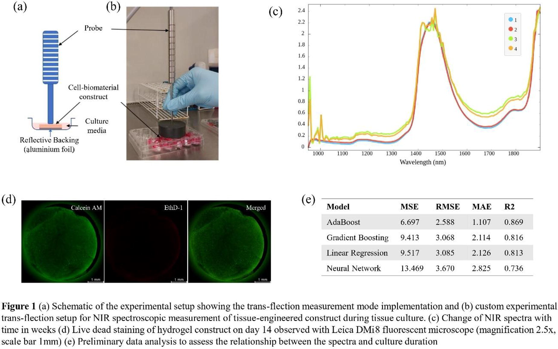
B198 - Comparison Of Cartilage Pre-digestion Techniques For Auricular Cartilage Tissue Constructs
N. E. Brown1, V. Dominick1, S. Kulkarni1, A. Frankel1, S. J. Hollister1, J. S. Temenoff1,2
1
Georgia Institute of Technology and Emory University, Atlanta, GA,
2
Georgia Institute of Technology, Atlanta, GA
*Purpose/Objectives: Microtia is a congenital deformity that affects the development of cartilage tissue of the outer ear. Auricular cartilage has limited regeneration capacity; therefore, researchers have explored reconstruction strategies like chondrocytes in 3D printed scaffolds, minced cartilage embedded in fibrin gels, and isolated chondrocytes encapsulated in hydrogels. As per these examples, different cartilage pre-treatment types can be used, so the goal of this study was to compare different levels of cartilage pre-digestion (isolated chondrocytes (isolated), partially digested minced cartilage (partially digested), minced cartilage (minced)) within poly (ethylene glycol) (PEG)-based hydrogels in terms of neo-cartilage formation within the constructs. Specifically, in this study, we developed a method for a partially digested minced cartilage matrix that we hypothesized can support chondrocyte phenotype maintenance due to the presence of native cartilage extracellular matrix (ECM).
*Methodology: Cartilage was harvested from the auricle of Yucatan pigs (6-9 mo.). The cartilage was cut into 1mm2 and 2mm2 pieces for the isolated and partially digested groups, respectively, and then treated with a 0.6 wt.% collagenase solution for 4 hours. Chondrocytes were isolated and cultured over 2 weeks while the digested pieces were rinsed with phosphate-buffered saline before encapsulation. All cartilage digestion levels were encapsulated in a 15 wt.% PEG-diacrylate (PEGDA) solution and crosslinked via radical polymerization. Cell viability was measured using Live/Dead staining (n=4). Over 4 weeks in vitro, gene expression of collagen 2 (COLII) and aggrecan (ACAN) was measured using PCR (n=4). In addition, hydrogels were stained for H&E and Safranin O and underwent immunohistochemistry (IHC: COLI, COLII, ACAN) (n=4). One-way ANOVA and Tukey’s multiple comparison tests were used to determine possible significant differences (p < 0.05)).
*Results: Brightfield imaging demonstrated that we successfully developed a partially digested cartilage matrix, based on the transparency of the pieces, indicating the loss of some ECM (Figure 1A). All groups encapsulated within PEGDA hydrogels remained viable over 7 days (Figure 1B). Isolated and partially digested groups showed a significant increase in COLII expression by week 2 (isolated:1018 ± 265, partially digested: 90 ± 56; (fold change over passage 2 chondrocytes)), while the isolated group expressed significantly more COLII (74 ± 16) and ACAN (7 ± 2) by week 4 (Figure 1C). H&E and Safranin O revealed a homogenous distribution of isolated chondrocytes and glycosaminoglycan deposition in the isolated group (Figure 1D). IHC of the isolated group showed intense COLI, COLII, and ACAN staining around nuclei and additional ACAN deposition in the hydrogel matrix at week 4.
*Conclusion/Significance: We demonstrated that PEGDA hydrogels can support chondrocyte viability and neo-cartilage matrix production over 4 weeks in vitro, particularly in the isolated chondrocyte group. Of interest, the partially digested group could be prepared in under 4 hours and showed chondrogenic potential and, thus, could be an exciting source for use in the operating room, but needs further optimization to improve matrix production over time. This novel study provided a head-to-head comparison of different cartilage digestion levels as tissue sources for various cartilage tissue engineering applications.
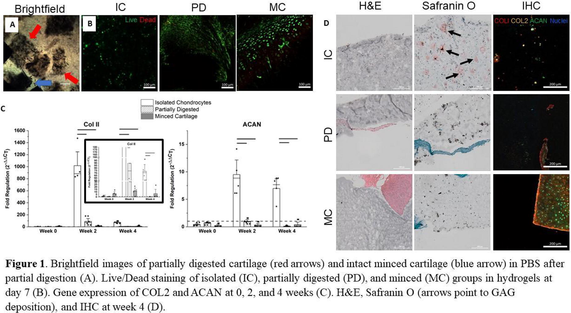
B199
B200 - Platelet-rich Plasma (prp) On Co-culture Of Chondrocytes And Osteoblasts And Its Positive Role In Gap Repair Between The Bone And Articular Cartilage
J. YOO, S. Lee, H. Kim, J. Nam
Seoul National University College of Medicine, Seoul, Korea, Republic of
*Purpose/Objectives: We aimed to investigate the influence of platelet-rich plasma (PRP) on the co-culture of chondrocytes and osteoblasts as well as its potential positive effects on gap repair using a co-cultured scaffold, expanding on the previous research.
*Methodology: Allogenic leukocyte-depleted PRP was obtained from blood supernatants after centrifugation at 150G for 10 minutes. PRP was then incubated with the PLGA mesh (Ethicon, USA). Human chondrocyte cells harvested from femoral head resected during total hip arthroplasty were co-cultured with commercially available hFOB1.19 osteoblast cell line on the PLGA mesh. The experimental group utilized the PRP-treated PLGA mesh, while the control group used only the PLGA mesh. For proliferation analysis and phenotype expressions, we counted cell numbers and employed RT-PCR with specific primers representing chondrocytes and osteoblasts. Subsequently, gap-mimic constructs, composed of devitalized human bone discs, the PLGA mesh, and cartilage, were implanted into subcutaneous pouches of nude mice. The gap-mimic constructs consisted of two groups: PRP-free scaffold (n=8) and PRP-treated scaffold (n=8). After 4 and 8 weeks, the constructs were harvested for gross and microscopic analysis. The attachment between bone and cartilage in the harvested constructs was assessed by categorizing the entire construct length into 0-25%, 25-50%, 50-75%, and 75-100%. The presence of cellular invasion at the interface was also examined.
*Results: In the PRP-free group, the number of chondrocytes and osteoblasts approximately increased 3.7-fold (7.5x10⁴ ± 1.4x10⁴ cells) seven days post-seeding, whereas, in the PRP-treated group, the number of cells increased an approximately 4.5-fold (9.1x10⁴ ± 0.6x10⁴ cells). FE-SEM findings also highlighted a more dense and expansive coverage of the mesh surface in the PRP-treated group (Figure 1a). Furthermore, the phenotypes of both chondrocytes and osteoblasts in each group were well preserved, as evidenced by the results of RT-PCR. At the 4-week assessment, both groups had similar attachment levels (PRP-free: 2 cases with 0-25%, 1 case with 50-75%, and 1 case with 75-100% attachment; PRP-treated: 2 cases with 0-25%, and 2 cases with 75-100% attachment). Cellular invasion was not observed in any of the four cases in the PRP-free group, while in the PRP-treated group, it was observed in one out of the four cases. At the 8-week assessment, both groups showed comparable attachment (1 case with 25-50%, and 3 cases with 75-100% attachment in each group). However, cellular invasion was observed in 2 out of 4 cases in the PRP-free group, while it was present in all 4 cases in the PRP-treated group (Figure 1b).
*Conclusion/Significance: In the co-culture process of chondrocytes and osteoblasts, the use of PRP resulted in a higher cell count compared to when PRP was not used, while still maintaining the phenotypes of each cell. Moreover, PRP is presumed to play a positive role in inducing the healing of the gap between subchondral bone and articular cartilage. Therefore, cell transfer technique with the use of PRP can play a positive role in repair the gap between subchondral bone and articular cartilage.
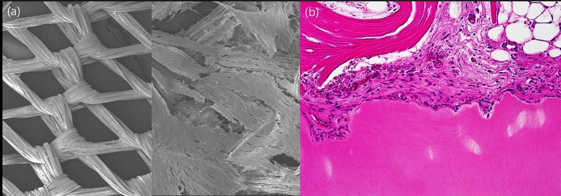
B201 - In Vivo Gap Repair Between Bone And Articular Cartilage With Co-cultured Human Articular Chondrocyte And Human Osteoblast On Plga Mesh Scaffold
J. YOO, S. Lee, H. Kim, J. Nam
Seoul National University College of Medicine, Seoul, Korea, Republic of
*Purpose/Objectives: We hypothesized that human chondrocytes and osteoblasts would maintain their cellular phenotype after co-cultured on PLGA mesh. Then, we evaluated the regeneration potential of these cell-scaffold constructs for gap repair between bone and cartilage in vivo.
*Methodology: Co-culture of chondrocyte and osteoblast & proliferation analysis: Human chondrocyte cells were harvested from the resected femoral head of ONFH patients during total hip arthroplasty. We used commercially available hFOB1.19 cell line for osteoblast. We performed co-culture technique with two cell lines. For proliferation analysis and phenotype expressions, we counted cell numbers and used RT-PCR with specific primers representing chondrocytes and osteoblasts: type II collagen, aggrecan, alkaline phosphatase, and osteocalcin. Producing a gap-mimic construct and in vivo experiment: We produced a gap-mimic construct comprising three layers: cartilage, scaffold, and bone (Figure 1a). We used mesh-type PLGA (Ethicon, USA) with dimensions of 10x8 mm as a scaffold. Gap-mimic constructs comprised two groups: Acellular scaffold (n=10) and Co-cultured cells on scaffold (n=10). The constructs were implanted into subcutaneous pouches of nude mice and harvested after 4 and 8 weeks for gross and microscopic analysis. Gross evaluation and microscopic analysis: The vertically divided constructs were stained with hematoxylin and eosin for histological analysis. To evaluate the gap healing, we assessed the degree of attachment and the presence of cell invasion at the interface. We determined the proportion of the length of contact between bone and cartilage within the construct, and categorized it into ranges of 0 to 25%, 25 to 50%, 50 to 75%, or 75 to 100%. Cell invasion was also inspected at the interface.
*Results: Chondrocytes and osteoblasts successfully adhered and proliferated on the mesh-type PLGA. Proliferation assays conducted seven days post-seeding showed that the number of chondrocytes increased by approximately 4.3 times (4.3 x 104 ± 0.5 x 10⁴ cells) and osteoblasts (1.2 x 10⁴ ± 0.1 x 10⁴ cells) by about 1.2 times. The RT-PCR affirmed that both chondrocytes and osteoblasts were proliferating while maintaining their respective phenotypes. The results of the gross and microscopic analysis of the harvested construct are as follows. In the case of the gap-mimic construct without cells, all constructs harvested at 4 weeks showed no attachment or cellular invasion. Some of the constructs harvested at eight weeks showed attachment (4 out of 6 constructs with 0-25%, and 2 with 75-100%), but no cellular invasion was observed. In the case of the gap-mimic construct with co-cultured cells, 2 case with 0-25%, 1 case with 50-75%, and 1 case with 75-100% attachment was observed at four weeks. Cellular invasion was observed in 1 out of 4 cases. At eight weeks, 1 case with 0-25%, 2 cases with 50-75% and 3 cases with 75-100% attachment were observed. Cellular invasion was detected in 4 cases out of 6 (Figure 1b).
*Conclusion/Significance: Human chondrocytes and osteoblasts can be co-cultured on PLGA mesh with their own phenotype. Both cells-carrier complex showed healing potential between the bone and cartilage in vivo.
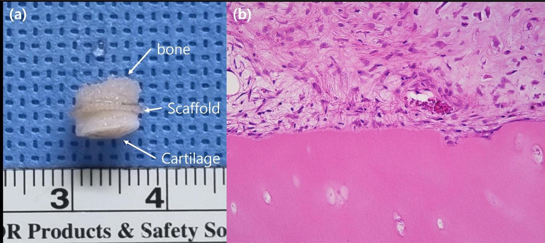
B202 - Non-destructive Monitoring Of Tissue-engineered Cartilage Growth And Development From Culture Media Using Near Infrared Spectroscopy (NIRS)
N. Sadeesh1, H. Wickramaarachchige1,2,3, O. Elkadi1, A. Eskelinen4,2, A. Gebraad1,2,3, E. Nippolainen1, S. Miettinen2, I. O Afara1
1
University of Eastern Finland, Kuopio, Finland,
2
Tampere University, Tampere, Finland,
3
Tampere University Hospital, Tampere, Finland,
4
Coxa Hospital for Joint Replacement, Tampere, Finland
*Purpose/Objectives: This research aims to develop a rapid and non-destructive protocol for monitoring the development of tissue engineered cartilage construct using Near Infrared Spectroscopy (NIRS).
We hypothesize that the presence and quantity of different biomarkers tissue growth and development in the culture media can be detected and quantified in real-time and without labels using NIRS.
*Methodology: Bone marrow mesenchymal stem cells (BMSCs) were cultured with three experimental setups: (1) BMSCs in Gellan Gum hydrogel (with spermidine crosslinker), (2) BMSCs in Gelatin methacryloyl (GelMa), and (3) scaffold-free setup with only BMSC cells (a seeding volume of 5 million was used in all setups). The setups were monitored for 21 days, with media collected every two days to study changes in the constructs’ composition. Media samples were subjected to NIRS measurements using a custom setup (Figure 1a) consisting of an Avantes NIR (900 to 2500nm, Avaspec -NIR256-2.5-HSC) spectrometer equipped with a fiber optic probe. Hyaluronan (HA) levels in the media samples from the Scaffold-free setup were assessed using Hyaluronan-ELISA kits (Department of Biomedicine, University of Eastern Finland). The acquired NIR spectra and biomarkers (HA) function as key reference points to evaluate and monitor tissue growth without damaging its integrity during the TE process. Spectral and biomarker data were preprocessed and analyzed using the QUASAR-Orange tool to assess the capacity of NIRS to detect changes in culture media during tissue growth [via estimation of HA]. Standard machine learning algorithms Random Forest, Adaboost, KNN, and Gradient Boosting are employed to predict changes in spectra over the 21-day period.
*Results: The machine learning models consistently identified substantial spectral variations across the 21-day period for all three setups. The R2 values from machine learning models ranged between 0.4 and 0.8, indicating significant spectral changes over time (Figure 1b, Figure 1d). The cumulative HA levels were determined through ELISA kits, demonstrating a consistent decrease over the 21-day period, suggesting ongoing tissue growth. Subsequent linear regression analysis of the HA (ng/ml) data spanning the 21-day period revealed a statistically significant deviation over time (p-value = 0.0016 and R2 value = 0.8) (Figure 1c).
*Conclusion/Significance: This research, based on the hypothesis that NIRS enables real-time, label-free assessment of diverse biomarkers in culture media, shows immense promise. The primary goal is to predict cartilage tissue growth by estimating key biomarkers in culture media using NIRS. Given that the composition of culture media consists of more than 99% water, preliminary results reveal that NIRS can detect low biomarker concentrations in biofluids, offering comprehensive insights into changing media composition during tissue growth. Ongoing preliminary studies aim to estimate other biomarkers, such as collagen, from the culture media using NIRS, potentially enhancing the monitoring of cartilage growth during TE. The outcome of this study could address the demand for functional cartilage replacements in the treatment of OA and present a protocol that can be adapted for personalized cartilage TE. Moreover, the innovative optical monitoring approach has broader implications for revolutionizing the engineering of diverse biological tissues.

B204 - A Functional In Vitro 3D IPSC-derived Neuromuscular Junction Model For Use In Neurotoxin Potency Testing Or Preclinical Drug Development
J. Fleming1, K. Gray1, D. Nash1, V. Leung1, C. Gelber1, C. Michas1, S. Luttrell1, A. Smith2, D. Mack2, N. A. Geisse1
1
Curi Bio, Seattle, WA,
2
University of Washington, Seattle, WA
*Purpose/Objectives: Diseases of the neuromuscular junction (NMJ) have devastating impacts on individuals’ quality of life and are often ultimately fatal. The development of effective therapeutics necessitates disease models that more closely mimic in vivo conditions. To date, models of the NMJ lack clear functional outputs, or require bespoke low-throughput manufacturing techniques to create. Here, we present a novel in vitro functional neuromuscular junction model derived from human iPSCs, comprising motor neurons and skeletal muscle 3D tissue constructs cultured within a commercial grade cell culture consumable. Furthermore, we validate the utility of this model by demonstrating Botulinum toxin (Botox) potency for blocking neuron-mediated muscle contractions, and identifying pre and post synaptic markers using immunohistochemistry.
*Methodology: Human iPSCs were differentiated (separately) into motor neurons bearing a blue light-activated channel (ChR2) and skeletal muscle cells. Functional engineered skeletal muscles were generated in the commercially available MantarrayTM two-post tissue platform, allowing temporal label-free functional measurements. Following 10 days of differentiation, these skeletal tissues were combined with 100 neuronal spheroids using a specialized consumable, removing the requirement to directly handle spheroids. Following 1 week of co-culture 488mn light application, tissues produced synchronized functional responses from the underlying muscle at 5, 2, and 1 second intervals. Neural-driven functional responses exceeded 30% of maximal skeletal muscle force via broad field electrical stimulation, demonstrating high levels of innervation.
*Results: Application of 10nM botulinum toxin A (BoT) to co-culture tissues ablated blue light responses within 4 hours, reducing both force magnitude and fidelity of blue light synchronized force production. This demonstrates the specificity of the motor neuron - skeletal muscle interactions within these tissues, assuring that force generation is dependent on motor neuron derived acetylcholine.
*Conclusion/Significance: We present this iPSC-based model of the NMJ as a novel platform for preclinical development of drugs targeting currently intractable diseases of the NMJ, to improve clinical translation of novel therapeutics. Through the application of iPSC derived motor neurons containing disease associated mutations, diseases such as ALS can now be studied in a dish. Additionally, healthy NMJs present a platform for toxicity and potency testing on known and unknown neurotoxins in a human-relevant system.
B205 - Regulating Microenvironment Of Spinal Cord Injury And Delivering Drug To Promote Nerve Regeneration
Y. Chen
Suzhou Institute of Nano-Tech and Nano-Bionics, Chinese Academy of Sciences, Suzhou, China
*Purpose/Objectives: Spinal cord injury (SCI) commonly disrupts the neuronal connections between the brain and the periphery, leading to temporary or permanent loss of sensory and motor functions. Initial mechanical trauma to the spinal cord initiates a secondary injury cascade, including ischemia/ reperfusion injury, inflammation, reactive oxygen species (ROS) generation, and glutamate-mediated excitotoxicity, forming a harsh inhibiting regenerative microenvironment of SCI, which have worse effects than the initial traumatic injury. In addition, endogenous neural stem/progenitor cells (NSCs/NSPCs) have been confirmed that they may migrate toward the lesion site, with the maximum peak occurring in 3-8d after injury. It makes them a promising cell source for the treatment of SCI. Several clinical drugs have been proven to mediate NSCs to give rise more neurons, such as paclitaxel (PTX). However, the innovative strategy of remodeling the SCI microenvironment and simultaneously delivering drugs to regulating endogenous NSCs is still desirable for SCI repair.
*Methodology: We chose the exosomes derived from NSCs and mesenchymal stem cells, respectively. We verified the effects of stem cell-derived exosomes on migration, proliferation and differentiation fate of NSCs by CCK8, immunofluorescence staining, and transwell, respectively. The dual bio-specificity peptide BSP was synthesized in chemical solid-phase peptide synthesis. One end of it was the collagen-binding domain, and another end was a 7-peptide that specifically binds to transferrin on exosomes. We applied both confocal microscopy and scanning electron microscopy to prove that BSP can effectively link exosomes with collagen materials. Furthermore, we chose a clinically approved PTX as the exosome-loaded model drug, resulting in a functional collagen scaffold. Completely transected SCI rats were used to verify the therapeutic effect of the functional collagen scaffold by virtue of mediating endogenous NSCs.
*Results: We found NSCs-derived exosomes promote the proliferation of NSCs by regulating the MEK/ERK/CREB signaling pathway, and mesenchymal stem cell-derived exosomes can promote the migration of NSCs both in vivo and in vitro.
*Conclusion/Significance: Based on the exosomes to delivery PTX, the obvious nerve regeneration is observed, which further promote the motor function recovery of complete SCI rats.
B206 - Cmcht/pamam Nanoparticles Loaded With Manganese For Magnetic Resonance Imaging Applications
E. P. Oliveira1,2, L. Colás3, F. R. Maia1,2, R. L. Reis1,2, P. Ramos-Cabrer*3,4, J. M. Oliveira*1,2
1
3B’s Research Group, I3Bs – Research Institute on Biomaterials, Biodegradables and Biomimetics, University of Minho, Headquarters of the European Institute of Excellence on Tissue Engineering and Regenerative Medicine, Barco, Guimarães, Portugal,
2
ICVS/3B’s–PT Government Associate Laboratory, Braga/Guimarães, Portugal, Braga, Portugal,
3
CIC biomaGUNE, Basque Research and Technology Alliance (BRTA), San Sebastian, Spain,
4
Basque Foundation for Science, Ikerbasque, Bilbao, Spain
*Purpose/Objectives: MRI is a real-time and non-invasive imaging technique used extensively in clinics for disease diagnosis and treatment. Contrast agents are used to enhance the differences in signal intensity of body regions, resulting in clearer images with smaller details being visible[1]. Manganese-based imaging has emerged as an alternative to standard contrast agents, due to its lower toxicity and ideal magnetic properties. Nevertheless, manganese has a short half-life in vivo and diffuses quickly from tissues or biomaterials, decreasing its efficiency[2].
*Methodology: Herein, we developed manganese-loaded carboxymethylchitosan/polyamidoamine nanoparticles (CMCht/PAMAM NPs[3]) to overcome this problem, by stabilizing the manganese and promoting a sustained release, which will prolong its effect in vivo. The CMCht/PAMAM NPs were dissolved in water and mixed with a solution of manganese (0.4 mM), with equal volumes of each solution. The final solution was stirred for 24 hours and dialyzed for another 24 hours. The manganese incorporation into the NPs was assessed through 1H-NMR, XPS, SEM-EDS, and ICP-MS, while the particles were characterized by DLS, FIB-SEM, and AFM. The Mn-CMCht/PAMAM NPs effect on the cells’ metabolic activity, proliferation, and internalization was analyzed by culturing the nanoparticles with Adipose Stem Cells (hASCs), and their hemolytic properties were assessed by culturing them with human erythrocytes from whole blood. The T1 and T2 magnetic relaxivities were characterized in vitro at 1.5T, and phantom images were performed at 7T and 11.7T MRI machines.
*Results: The presence of Mn in the NPs was confirmed by 1H-NMR, XPS, SEM-EDS, and ICP-MS. The NPs size was also analysed by DLS, FIB-SEM, and AFM. DLS analysis showed the NP size did not alter upon manganese bonding. However, their charge was dependent on the quantity of available manganese. Regarding their magnetic properties, the r1 relaxivities were of 17.5 mM 17.5 mM-1s-1 (1.5 T), 9.7 mM-1s-1 (7T), and 6.8 mM-1s-1 (11.7 T); r2 relaxivities were of 136.1 mM-1s-1 (1.5 T), 80.9 mM-1s-1 (7T) and 75.0 mM-1s-1 (11.7 T) and r2/r1 ratios of 2.1, 8.4 and 11.1, respectively. The Mn-CMCht/PAMAM NPs displayed a typical T1 contrast agent at lower magnetic fields, while at higher magnetic fields, they behave as a dual T1-T2 contrast agent. The nanoparticles’ phantoms showed a concentration-dependent decrease in MRI signal upon different dilution ratios. Moreover, the Mn-CMCht/PAMAM NPs presence did not affect the cells’ metabolic activity or proliferation, were easily internalized, and did not present a hemolytic behavior.
*Conclusion/Significance: In brief, the successful incorporation of manganese into the nanoparticles was confirmed by several techniques, showing only differences in their charge related to the electrostatic charges involved in the reaction. Furthermore, the nanoparticles are non-cytotoxic and non-hemolytic, and easily internalized by the hASCs, demonstrating promising features for biomedical applications as a contrast agent for MRI studies.
B209 - Development Of An In Vitro Model Of The Outer Annulus Fibrosus-Cartilage Endplate Interface Using A Perfusion Bioreactor
C. Thom1,2, R. Kandel1,2, J. Santerre1
1
University of Toronto, Toronto, ON, Canada,
2
Mount Sinai Hospital, Toronto, ON, Canada
*Purpose/Objectives: Lower back pain is a leading cause of disability worldwide and is often associated with intervertebral disc (IVD) degeneration. IVDs are fibrocartilaginous tissues between the vertebral bodies of the spine which facilitate load transmission and enable spinal flexibility. Once damaged there is a lack of treatment options that permanently restore functionality. Tissue engineering aims to address the limitations of existing therapeutics by generating living tissue constructs that could replace the diseased disc. Given the complex disc architecture coupled with the limitations of conventional static culturing methods, the production of tissue constructs that recapitulate the structural features and mechanical strength of the native disc has yet to be achieved. The current study aims to develop an in vitro-formed model of the outer annulus fibrosus (OAF)-cartilage endplate interface, which is often overlooked in current research despite its critical role in maintaining structural and biochemical integrity of the IVD. It is hypothesized that in vitro-formed OAF can be integrated with in vitro-grown cartilage tissue, such that the interfacial strength of the tissue interface is significantly enhanced when cultured under perfusion flow within a bioreactor.
*Methodology: OAF cells are isolated from bovine caudal discs and seeded onto polyurethane scaffolds, as described previously1, in a spinning bioreactor to generate in vitro-formed OAF tissue. These tissues are then co-cultured with deep zone articular chondrocytes that have been isolated from bovine caudal metacarpal-phalangeal joints and cultured on PTFE membranes. OAF-chondrocyte constructs are co-cultured statically for 7 days in mineralizing media containing β-Glycerophosphate. Co-cultures are either maintained in static culture or transferred to a perfusion bioreactor system in which they are dynamically cultured using a flow rate of 5mL/min, for an additional 7 days before performing histological or mechanical assessment. All experiments, except for mechanical testing, were repeated with 3 biological sets, with each condition done in quadruplicate. Statistical analysis was performed using a T test and significance assigned at p<0.05.
*Results: Co-culture of in vitro-formed OAF tissue and chondrocytes generated an integrated tissue interface composed of cartilage and AF tissue. More tissue appeared to be present, histologically (Figure 1), in constructs grown in the perfusion bioreactor versus static conditions. The interface was also stronger in the perfused constructs as determined using a pull-apart testing, where the average interfacial strength of perfused and static constructs was 53.3kPa and 18.9kPa, respectively (preliminary data; N=1). Immunostaining of the OAF-cartilage constructs demonstrate collagen type I, collagen type II, and aggrecan distribution for both conditions appear similar to that of the native OAF-CEP interface (Figure 1). Von Kossa staining demonstrates cartilage mineralization under both perfusion and static culture conditions. However, OAF-cartilage constructs that undergo perfusion appear to have more mineral deposition.
*Conclusion/Significance: This study provides a first-generation in vitro OAF-CEP model that incorporates key structural properties of the native interface, and demonstrates that the use of a perfusion bioreactor enhances development of in vitro-grown IVD tissue interfaces. These findings fill a translation gap in the field of IVD tissue engineering and will support the scale-up of such tissues to physiological sizes.

B210 - A Degradable Nanofibrous Scaffold Of Poly(ε-caprolactone-co-lactide) For Intervertebral Disc Regeneration
M. Chaaban1, C. Falcoz2, C. Pinese2, C. Paniagua2, J. Clouet1, M. Fusellier1, B. Nottelet2, J. Guicheux1, C. Le Visage1, X. Garric2,3
1
University of Nantes, Oniris, INSERM, Regenerative Medicine and Skeleton, RMeS, UMR 1229, Nantes, FRANCE,
2
Department of Polymers for Health and Biomaterials, IBMM UMR 5247, University of Montpelier, CNRS, ENSCM, Montpellier, FRANCE,
3
Department of Pharmacy, Nîmes University Hospital, Nîmes, FRANCE.
*Purpose/Objectives: The intervertebral disc (IVD) consists of a gelatinous core (nucleus pulposus, NP) surrounded by a fibrous ring (annulus fibrosus, AF). Accumulation of micro-cracks and tears in the AF can lead to NP herniation outside the disc space, compressing nerve roots and causing pain. IVD herniation is a major cause of low back pain, affecting 90% of people in their lifetime. Although conventional discectomy effectively relieves pain, it fails to address the underlying structural damage, resulting in re-herniation and accelerated disc degeneration. Here, we designed a dual-repair approach targeting: i) the NP with the injection of a commercially available hyaluronic acid gel, and ii) the AF by implanting a bio-inspired multi-lamellar scaffold with oriented microfibers, self-anchoring system, and controlled biodegradability to promote the regeneration of a spatially organized AF tissue.
*Methodology: To this aim, we synthesized a novel poly(ε-caprolactone-co-lactide) (PCLA) with various molar percentages of lactide (10, 20, and 30 %) and structures (copolymer or blend). These polymers were electrospuned into sheets of aligned nanofibers.
*Results: Thermal, mechanical, and structural properties were analyzed, showing a Young’s Modulus ranging from 20 MPa to 37 MPa, close to that of the targeted AF tissue. Moreover, gamma irradiation (25 kGy) was employed for sterilization, causing no significant impact on cytocompatibility, thermal properties, except for a Young’s Modulus increase by 21-31 MPa when lactide > 20%, or molar masses of the polymers. The in vitro degradation of gamma sterilized PCLA showed a molar mass loss of 70 ± 0.4% after 6 months for the copolymer and an average of 53 ± 17% for blends. Furthermore, scanning electron microscopy revealed visible fiber breakage in the case of the copolymer, potentially facilitating the in vivo deposition of newly formed AF tissue over time. PCLA sheets guided the in vitro alignment and proliferation of ovine AF cells and maintained the expression of AF markers for 2 weeks (collagen type I, type II and aggrecan). Furthermore, a multi-lamellar 3D implant was produced by stacking sheets of PCLA and incorporating a self-anchoring system. The multi-lamellar scaffold was maintained within a full-thickness annular defect (4mm biopsy punch) of bovine tail IVDs during 4 weeks of culture and cell infiltration between the layers of the scaffold was observed. Further analysis of cellular phenotype and matrix deposition on PCLA sheets using Elisa and RT-qPCR is ongoing.
*Conclusion/Significance: Overall, this study highlights the ability of the newly designed implants to instruct tissue regeneration and future work will evaluate the long-term efficacy in a sheep annular defect model.
B211 - Characterization Of Bovine Intervertebral Discs And Novel Hydrogels For The Repair Or Replacement Of Spinal Discs
K. J. Donovan1, L. A. Brunmaier1, T. W. Walker1, W. E. Rochefort2
1
South Dakota School of Mines & Technology, Rapid City, SD,
2
Oregon State University, Rapid City, SD
*Purpose/Objectives: Herniated spinal discs affect approximately 75% of the population. The herniation of a disc is the weakening of the annulus fibrous that causes the nucleus pulposus to extrude, thereby compressing the spinal nerve and inducing severe back pain. One of the most popular treatments is spinal fusion which is an invasive procedure that removes the extruded portion of the disc and joins two or more vertebrae. Unfortunately, this procedure results in reduced functionality and mobility of the spine. To overcome these deficits, our long-term goal is to re-inflate the spinal disc with a hydrogel rather than leave a flattened disc. As a part of this effort, we have synthesized an injectable hydrogel that models the nucleus pulposus of a bovine spinal disc, which is being used as a surrogate for a human spinal disc.
*Methodology: Literature has suggested that mature runt cows are a good model for human spinal discs, as the cow discs are comparable in size to humans. The nucleus pulposus of bovine-tail spinal discs and novel foam hydrogels were characterized with dynamic mechanical analysis (DMA) via small-amplitude oscillatory compression and with unconfined steady compression.
*Results: Compressive modulus from the dynamic oscillatory axial compression frequency sweep at 1% strain is reported. The bovine discs compare favorably with the human values reported in Freeman, et al. The reproducibility of the bovine data and favorable comparison to human data confirms the viability of bovine tails as a model. The foam gel and simple gels demonstrated reproducible mechanical properties but were not as comparable to the reported human values.
*Conclusion/Significance: The results from the bovine studies are reproducible and compare favorably to the human data. Bovine discs are a promising model for the hydrogel synthesis. The hydrogels have the potential to obtain the desirable rheological properties for spinal disc replacement.
B212 - Enhancing Axonal Guidance Of iPSC-derived Spinal Cord Progenitor Cells With Anisotropic Microporous Annealed Particle Scaffolds For Spinal Cord Injury Repair
C. Kwokdinata, K. Chai, S. Chew,1,1
Nanyang Technological University, Singapore, Singapore
*Purpose/Objectives: Spinal cord injury (SCI) results in irreversible impairment of sensory, motor and autonomic functions. While neural stem cells (NSCs) encapsulated in bulk hydrogels have been used for spinal tissue regeneration, ethical concerns arise from the use of NSCs derived from fetal and embryo sources. Furthermore, the lack of topographical cues and porosity in these hydrogel scaffolds leads to non-aligned axonal growth and clumped neuronal extensions that impede the effective reconnection of neuronal pathways in the spinal cord in vivo. Here, we hypothesize that integrating microchannels into gelatin microporous annealed particle (MAP) scaffolds would facilitate the neuronal differentiation of human induced pluripotent stem cells (iPSC)-derived spinal cord progenitor cells (SCPCs), provide axonal guidance and promote neurite outgrowth.
*Methodology: Gelatin MAP microchannel scaffolds were constructed by annealing gelatin microparticle slurry via Ruthenium-catalyzed photocrosslinking. We mechanically characterized the slurry and scaffolds of varying packing densities by rheology and compression tests. The incorporation of microchannels within these scaffolds was achieved by printing sacrificial Pluronic F127 within the gelatin microparticle slurry prior to photocrosslinking. We evaluated the behavior and cell fate changes of SCPCs within these anisotropic MAP scaffolds in terms of cell viability, attachment and differentiation.
*Results: By varying the degrees of microparticle packing, the interstitial spacing between the microparticles could be controlled. Correspondingly, the microscale porosity (5 - 30%) as well as the compressive modulus of the scaffolds could be altered. Moreover, the compacted gelatin microparticle slurries exhibited shear-thinning properties and self-recovery behavior, demonstrating its suitability as a support bath for freeform printing of sacrificial channels in the fabrication of microchannel MAP scaffolds. The combination of micropores and microchannels facilitated the migration and neuronal differentiation of SCPCs. Notably, axonal alignment was observed within these channels. Additionally, the extent of neurite outgrowth within the scaffold walls was significant in MAP scaffolds with lower packing density as compared to SCPCs encapsulated in bulk gelatin hydrogels and MAP scaffolds with higher packing density.
*Conclusion/Significance: Our study highlights the potential of MAP scaffolds in spinal cord tissue engineering applications, which is attributed to their tunable porosity and channel architecture for guiding cell migration and promoting neuronal differentiation.
B216 - Eco-AI in Ophthalmic Tissue Engineering: Large-Scale Fabrication of Keratoprostheses from Industrial Biowaste
P. R. Corridon1,2, X. Wang1, I. V. Pantic3,4,5, J. Cumic3, S. Valjarevic3, H. Vurivi2, S. Daoud6, V. Chan1, M. G. Shibru1, Z. M. Ali1, D. Nesic3, A. E. Salih2, H. Butt2
1
Khalifa University, Abu Dhabi, United Arab Emirates,
2
Khalifa University of Science and Technology, Abu Dhabi, United Arab Emirates,
3
University of Belgrade, Belgrade, Serbia,
4
University of Haifa, Haifa, Israel,
5
Ben-Gurion University of the Negev, Be’er Sheva, Israel,
6
Cleveland Clinic Abu Dhabi, Abu Dhabi, United Arab Emirates
*Purpose/Objectives: In the realm of ophthalmic tissue regeneration and bioengineering, the cornea presents unique challenges due to its distinct tissue composition and functionality. There is a global need for advancements in reconstructing these specialized tissues, particularly in addressing end-stage conditions that result in blindness. This research leverages the underutilized biological waste from slaughterhouses to create high-throughput platforms for producing corneal grafts. By repurposing these materials, we are addressing the critical demand for keratoplasty and contributing to environmentally sustainable practices.
*Methodology: Over the past two years, 1,000 discarded eyes from Arabian sheep breeds in the UAE were utilized to develop native and acellular corneal keratoprostheses. The acellular scaffolds were produced using a zwitterionic biosurfactant, ensuring an eco-friendly and cost-effective approach. Key components of keratograft formation, including ECM fibril organization, ocular transmittance, mechanical integrity, and cellular reintegration, were assessed manually and using artificial intelligence approaches.
*Results: Decellularized scaffolds retained the microarchitecture integrity that supported appreciable optical light transmission (over 70%) after reversing opacity, a well-established hallmark of decellularization and long-term native corneal storage, with glycerol. Blinded semi-quantitative analyses also revealed notable variations in the degrees of intactness of lamellae alignment and damage. Likewise, machine-based approaches provided a means to provide fully automated scaffold categorization and helped discern sites for repopulation, which was then gauged experimentally via the integration for substantial epithelial cell integration.
*Conclusion/Significance: Our approach underscores emerging technologies in ophthalmologic tissue engineering, focusing on innovative solutions for corneal transplantation. We thus demonstrate the possibility of a sustainable and unconventional biomaterial source that can serve across a range of biomedical applications, namely Ophthalmic Tissue Regeneration and Bioengineering.
B217 - Waffle-inspired Hydrogel-based Macrodevice For Spatially Controlled Distribution And Potentialvascularization Of Encapsulated Therapeutic Microtissues
C. H. Pham1, Y. Zuo2, Y. Chen1, N. M. Tran1, D. T. Nguyen1, T. T. Dang1
1
Nanyang Technological University, Singapore, Singapore,
2
Shenzhen Maijie Life Science Ltd, Shenzhen, China
*Purpose/Objectives: Macro-encapsulation systems for delivery of cellular therapeutics in diabetes treatment offer major advantages such as device retrievability and high cell packing density. However, microtissue aggregation and the absence of vasculature have been implicated in the inadequate transfer of nutrients and oxygen to the transplanted cellular grafts. Herein, we develop a waffle-inspired hydrogel-based macrodevice to encapsulate therapeutic microtissues positioned in homogeneous spatial distribution to mitigate their aggregation while concurrently supporting an organized intra-device network of vascular-inductive cell, termed WIM device. This platform comprised two components with complementary topography features that fit together in a lock-and-key configuration.
*Methodology: We fabricated waffle-inspired hydrogel networks (“lock” component) interspersing microwells of defined dimension by photolithography, in which vascular-inductive cells (HUVECS) were embedded for potential vascularization. Insulin-secreting INS-1E microtissues in alginate solution (“key” component) were dispensed on the “lock” component in a lock-and-key configuration. Subsequent gelation of alginate by barium ions resulted in entrapment of microtissues in alginate at desired locations. In vitro studies included design optimization for spatial distribution efficiency, assessment of structural interlocking, cellular viability and functionality of microtissues and vascular-inductive cells encapsulated in WIM device. Islets-loaded WIM device was subcutaneously transplanted in diabetic mouse model to assess the ability of WIM device in allowing insulin secretion for glycemic control.
*Results: The micropattern with square microwells effectively entrapped 90% of INS-E microtissues in controlled locations while the interlocking design placed them in a co-planar spatial arrangement with proximity to vascular-inductive cells. The WIM device co-laden with INS-1E microtissues and HUVECs maintained desirable cellular viability in vitro with encapsulated microtissues retaining their glucose-responsive insulin secretion while embedded HUVECs express angiogenic marker. In vivo, device encapsulating primary rat islets achieved blood glucose control for 2 weeks in chemically induced diabetic mice.
*Conclusion/Significance: Overall, this macrodevice design lays foundation for a cell delivery platform, which has the potential to facilitate nutrient and oxygen transports to therapeutic grafts and thereby might lead to improved disease management outcome.

B218 - Engineered Human Cardiac Tissues To Assess The Cell-specific Contribution Of Bag3 In Dilated Cardiomyopathy And To Identify Therapeutic Targets
M. Morsink, B. Z. Wang, Y. Kim, L. Luo, J. Watkins, X. Zhang, B. Fine, G. Vunjak-Novakovic
Columbia University, New York, NY
*Purpose/Objectives: Dilated cardiomyopathy (DCM) is the most common cause of heart failure in the US. Mutations in Bcl2-associated athanogene 3 (BAG3) are associated with DCM. While BAG3 mutations in cardiomyocytes have been widely studied, their effect on non-myocyte cardiac cells is poorly understood. As fibrosis is a pathological hallmark of all end-stage hearts, there is a critical clinical need to uncover the contribution of BAG3 mutations to the matrix-secreting cardiac fibroblasts. Human induced pluripotent stem cells (iPSC) and engineered heart tissues (EHTs) provide unprecedented opportunities to study human cardiac biology in a cell-type specific manner. This study aims to investigate the contribution of cardiac fibroblast-specific BAG3 to heart function and to identify suitable drug candidates to treat DCM.
*Methodology: To assess the cell-type specific contributions of BAG3 to cardiac function, iPSC-derived cardiomyocytes and cardiac fibroblasts with either wild-type or BAG3 knockout genotype were systematically combined to generate four types of EHTs. The tissues were analyzed for differences in force production and collagen deposition, as hallmarks of DCM. To gain mechanistic insight into the cardiac fibroblast-specific alterations upon BAG3 loss, we performed several proteomic analyses. Lastly, to explore suitable drug candidates for DCM, a machine-learning algorithm was developed to distinguish between wild-type and BAG3 knockout fibroblasts by immunofluorescence. We performed a high-throughput drug screen and scored the BAG3 knockout fibroblasts based on their similarity to the wild-type phenotype. The top hits were further validated by their ability to rescue the phenotype of BAG3 knockout in cardiac fibroblasts in EHTs.
*Results: The loss of fibroblast-specific BAG3 led to a reduction in force production and increased collagen deposition, recapitulating the phenotype of DCM. Proteomic analysis of the wild-type and BAG3 knockout cardiac fibroblasts revealed increased proliferation and sensitivity to TGFβ signaling upon loss of BAG3. We found that BAG3 binds the type II receptor of TGFβ and mediates its degradation, providing a mechanistic understanding for the observed phenotype. Leveraging these phenotypic differences, the machine-learning algorithm we developed identified the top 12 drug candidates in the compound library that induced knockout cardiac fibroblasts to exhibit the greatest similarity to the wild-type phenotype. We selected the top three drug candidates by their ability to induce a comparable level of proliferation and TGFβ sensitivity in knockout fibroblasts. Lastly, 1 drug improved the force production of the EHTs with fibroblast-specific BAG3 knockout, thereby providing a possible therapeutic candidate for BAG3-mediated DCM.
*Conclusion/Significance: This study highlights the ability of iPSC-based human cardiac tissue models to unravel the cell-type specific contributions to cardiac disease. Expanding on the known role of BAG3 in cardiomyocytes in DCM, we found that BAG3 in cardiac fibroblasts plays a pivotal role in cardiac tissue dysfunction. Thus, interventions to treat BAG3-mediated DCM should extend beyond cardiomyocyte-centric research. We further demonstrated that the EHTs provide an excellent platform to perform in vitro drug testing. Together with machine-learning, we identified 1 drug candidate that can rescue the BAG3-mediated DCM phenotype and thus has the potential to ameliorate DCM in patients and reduce mortality rates.
B219 - Volumetric Bioprinting Of An Endocrine Pancreas Platform For Drug Screening Using Functional Human Ipsc-derived Islets
P. Catala1, D. Ribezzi1, C. Brice1, O. Dudaryeva1, G. Groessbacher1, M. Caiazzo2,3, R. Levato1,3
1
University Medical Center Utrecht, Utrecht, Netherlands,
2
University of Naples Federico II, Naples, Italy,
3
Utrecht University, Utrecht, Netherlands
*Purpose/Objectives: Pancreatic islets of Langerhans finely regulate circulating glucose levels. A loss or dysfunction of beta cells present in the islets results in diabetes mellitus, which affects over 500 million people worldwide. The current treatment for diabetes relies on the delivery of insulin, sometimes in combination with other insulin-sensitizing drugs, but does not fully recapitulate the innate glycemic control. Thus, novel therapies to address the limitations of the current treatments are needed. Generating functional islets from human induced pluripotent stem cells (iPSC) could unlock an unlimited cell source to engineer a cutting-edge platform for disease modelling and drug testing. Yet, despite significant progresses in the protocols for the generation of islets from iPSC, current cell cultures cannot replicate intricate islet-environment interactions, lowering the predictive power for drug testing. This absence of reliable, native tissue-like in vitro models limits the early identification of unsuitable therapeutics. Bioprinting technologies, biomaterials, and (stem) cell engineering provide a novel technological toolbox to pave the ground towards the design of bioinspired systems that recapitulate salient organ functions. This study combines the generation of induced pluripotent stem cells (iPSC)-derived functional islets of Langerhans with volumetric light-based bioprinting with the aim of engineering an advanced in vitro model of the endocrine pancreas.
*Methodology: Human pancreatic islets were generated from iPSC following a seven-stage protocol (Figure 1A). When reaching stage 7, iPSC-derived islets were collected and pancreatic constructs were generated via layerless and shear stress-free, light-based Volumetric Bioprinting (VBP). Concentrations ranging from 2%-7% (w/v%) of optically tuned bioresins, namely gelatin methacrylate (GelMa) and alginate methacrylate (AlgMa), were used for bioprinting. iPSC-derived islets were characterized via immunofluorescence and single cell transcriptomics. Insulin release on bioprinted constructs upon glucose exposure was assessed with a static and dynamic glucose stimulated insulin secretion (GSIS) assay. For the dynamic GSIS, volumetrically printed endocrine pancreatic constructs were perfused at a flow of 100 µL/min for two hours.
*Results: VBP allowed to produce 50-60 mm3 pancreatic constructs in 30 s and posed no mechanical or chemical stress on iPSC-derived islets, which were highly viable for 21 days after printing (Figure 1B). Stiffer, higher polymer content GelMa hydrogels presented the highest islet viability. Islet metabolic activity remained constant over time during 21 days of culture. Volumetric bioprinted iPSC-derived islets showed mature, insulin, glucagon and somatostatin, single-hormone producing cells after 21 days (Figure 1B) both characterized via immunofluorescence and single cell transcriptomics. Bioprinted islets are functional and possess a glucose-responsive insulin secretion profile both in static and in perfusion systems. Finally, the system is compatible with supplementation with anti-diabetic drugs for drug screening.
*Conclusion/Significance: Overall, this technology opens up to new possibilities on developing a novel platform for the in vitro testing of islet functionality paving the way for a patient-specific drug testing platform.

B220 - Safer Spaceflight: Exploring Hibernation Induction With Human Organoid Models
J. Diaz1, R. Ramamurthy1, C. Heathershaw1, G. Nomdedeu-Sancho1, O. Latham1, J. Roddey1, F. Welker2, J. Bethencourt2, V. Gorantla1, T. Shupe1, A. Jacobson1, P. F. Wilson3, X. Han1, J. T. Maxwell1, A. Atala1, G. Almeida-Porada1, C. D. Porada1
1
Wake Forest Institute for Regenerative Medicine, Winston Salem, NC,
2
Globe Institute, Copenhagen, DENMARK,
3
Pacific Northwest National Laboratory, Richland, WA.
*Purpose/Objectives: Astronauts on missions in deep space face significant health risks due to exposure to radiation in the form of Solar Energetic Particles (SEP) and Galactic Cosmic Rays (GCR). One promising solution to mitigate these risks is inducing a torpor-like hypometabolic state, a technique mimicking hibernator species’ natural responses to extreme environments. Hypometabolic induction in rodents has been shown to decrease cell replication while maintaining DNA repair, which could potentially enhance the body’s resilience to space radiation. However, the potential protective effects of inducing a torpor-like state have never been evaluated in human tissues, and ethical issues obviously preclude conducting such experiments on human subjects. Our research aims to induce metabolic depression in human organoids to understand and control metabolism for astronaut health and medicine. Studying fossilized remains of extinct hibernating hominins (S. hominin) may provide insights for inducing torpor in modern human organoids.
*Methodology: Our methodology involved using organoids of human liver, heart, and trabecular bone (for direct comparison to fossilized S. hominin remains) to model the normal physiological conditions of each native tissue. We subjected these organoids to reduced glucose environments and varying levels of 5’-AMP to induce metabolic depression, then assessed organoid survival, functionality, and metabolism. By evaluating organoid resilience and the sustainability of hypometabolic states, we were able to determine the thresholds of metabolic depression possible in each tissue while preserving viability. Building upon these foundational experiments, we plan to cryopreserve organoids and expose them to simulated SEP and GCR radiation at NASA’s Space Radiation Laboratory, assessing the radioprotective effects of metabolic depression and suspended animation on their survival and functionality. This research will contribute critical data to NASA’s risk assessment models regarding human tissue response to space radiation.
*Results: Our research has revealed key findings. Heart organoids displayed a threshold of reduced contractility as glucose levels decreased, setting a baseline for managing metabolism in space and medical situations. 5′-AMP helped liver organoids survive and remain functional while inducing and maintaining a hypometabolic state with reduced glucose. In preparation for analyzing ancient human remains, we successfully analyzed the proteome of trabecular bone organoids, obtaining high protein concentrations and MS2 spectra, suggesting well-preserved proteomes. The organoids closely resembled native bone, containing approx. 160 proteins, including bone-specific and ECM-associated ones.
*Conclusion/Significance: Inducing hypometabolic states and prolonged viability at low-temperatures in human organoids represents a significant advancement in space medicine and regenerative medicine, addressing key challenges of long-duration space missions. Our research paves the way for inducing human hibernation-like states, potentially revolutionizing resource conservation and astronaut health. Moreover, precise metabolic control has broader implications beyond space exploration, enabling organ preservation for transplants and advancing medical applications. Additionally, this metabolic control offers promising strategies in trauma care, potentially allowing for a ‘metabolic pause’ in critically injured patients, thus buying valuable time for life-saving interventions. This research thus represents a pivotal connection between space health studies and the fields of tissue engineering and regenerative medicine, providing novel approaches to address worldwide patient care challenges.
B221
B222 - Reproducible Generation Of Human Liver Organoids (hlos) On A Pillar Plate Platform Via Microarray 3D Bioprinting
M.-Y. Lee1,2, S. Shrestha1, V. K. Lekkala1, P. Acharya1, S.-Y. Kang1, M. G. Vanga2, P. Joshi2
1
University of North Texas, Denton, TX,
2
Bioprinting Laboratories Inc, Farmers branch, TX
*Purpose/Objectives: Human liver organoids (HLOs) hold significant potential for recapitulating the architecture and function of liver tissues in vivo. However, conventional culture methos of HLOs, forming Matrigel domes in 6-/24-well plates, have technical limitations such as high cost and low throughput in organoid-based assays for predictive assessment of compounds in clinical and pharmacological lab settings.
*Methodology: To address these issues, we have developed a unique microarray 3D bioprinting protocol of progenitor cells in biomimetic hydrogels on a pillar plate with sidewalls and slits, coupled with a clear bottom, 384-deep well plate for scale-up production of HLOs. Microarray 3D bioprinting is a droplet-based printing technology demonstrated on the pillar plate to generate a large number of small organoids for predictive hepatotoxicity assays.
*Results: Foregut cells, differentiated from human iPSCs, were mixed with Matrigel and then printed on the pillar plate rapidly and uniformly, resulting in coefficient of variation (CV) values in the range of 15 - 18%, without any detrimental effect on cell viability. Despite utilizing 10 - 50-fold smaller cell culture volume compared to their counterparts in Matrigel domes in 6-/24-well plates, HLOs differentiated on the pillar plate exhibited similar morphology and superior function, potentially due to rapid diffusion of nutrients and oxygen at the small scale. Day 25 HLOs were robust and functional on the pillar plate in terms of their viability, albumin secretion, CYP3A4 activity, and drug toxicity testing, all with low CV values. From three independent trials of in situ assessment, the IC50 values calculated for sorafenib and tamoxifen were 6.2 ± 1.6 µM and 25.4 ± 8.3 µM, respectively.
*Conclusion/Significance: Our unique 3D bioprinting and miniature organoid culture on the pillar plate could be used for scale-up, reproducible generation of HLOs with minimal manual intervention for high-throughput assessment of compound hepatotoxicity.

B223 - Free-standing 3d-bioprinting Of Heart Tissue Utilizing Electrically Conductive Hydrogel Microparticles And Hipsc-derived Cardiomyocytes
K. Roshanbinfar, A. Anspach, T. U. Esser, F. B. Engel
Friedrich-Alexander-Universität Erlangen-Nürnberg, Erlangen, Germany
*Purpose/Objectives: 3D-bioprinting is a promising technology to generate geometrically controlled hierarchical structures of human tissues. However, direct printing of living cells, especially in the case of soft tissues, has proven difficult. The evolution of in-support bath printing has provided significant advances in this field to take first steps towards generating anatomically accurate simplified heart models. However, scaling up the support bath production and required steps in removing the support path pose challenges to this approach. Here, we present a method to directly and free-standing 3D-bioprint functional cardiac rings and ventricle-shaped cardiac tissues in an accurate and reproducible manner that can be cultured for at least one month and respond to pharmacological stimuli.
*Methodology: We have generated hydrogel microparticles from gelatin methacrylate (GelMA), GelMA-- Poly (3,4-ethylenedioxythiphene) poly styrene sulfonate (PEDOT:PSS), and GelMA-polyethyleneimine-coated gold nanoparticles (bPEI-AuNPs) utilizing complex coacervation method (Figure 1). We have analyzed particle size and rheological properties and electrical conductivity of each bioink. The biocompatibility of microparticles was tested with NIH3T3 and human induced pluripotent stem cell (hiPSC)-derived cardiomyocytes before we 3D-bioprinted cardiac rings and ventricles by directly resuspending hiPSC-derived cardiomyocytes within hydrogel bioinks.
*Results: We have shown that based on the reaction criteria it is possible to control particle size in each bioink. More importantly, we have optimized printing approaches for the nozzle dimensions and type, achieving the best printing reproducibility with 27G tapered tips and a particle size of 40 - 70 µm. Live and dead staining images showed that fibroblasts (day 1) and hiPSC-derived cardiomyocytes (day 4) survived the printing process with a significant overall viability. Moreover, immunostaining images of hiPSC-derived cardiomyocytes showed well striated sarcomeric apparatus in these cells which were wrapping around particles. The 3D-bioprinted constructs started to beat after 3 days and maintained their contractile function during the 28 days of analysis. Importantly these tissues responded to adrenergic pharmaceutical treatments, showing their potential to be used as a model of cardiac tissue.
*Conclusion/Significance: We have developed a novel approach to generate different types of microparticle hydrogels and controlled their morphological and dimensions. These bioinks provide a significant step forward in 3D-bioprinting of cardiac and potentially other electrically sensitive tissues and make it possible to directly free-standing 3D-bioprint hiPSC-derived cardiomyocytes to generate models of human cardiac tissues.

B224 - Generating Human Ipscs-derived Cerebellar Organoids And Their Application In Testing Gene Therapeutics For Rare Disease Treatment
S. Ryu, J. Inman, H. Hong, Y. Gedik, Y. Jethmalani, V. Jovanovic, J. Ferreira De Sousa, A. Simeonov, P. Ormanoglu, I. Singec, C. Tristan;
NIH, Rockville, MD
*Purpose/Objectives: The cerebellum plays a crucial role in motor coordination and is implicated in several neurological conditions. Most of our current knowledge of the human cerebellum primarily relies on data obtained from postmortem tissues, animal models, or immortalized cell lines cultured using traditional 2D methods, each of which has shown limitations in recapitulating the intricacies of human cerebellar development and disease pathophysiology. In recent years, various types of human induced pluripotent stem cell (hiPSC)-derived organoid models have shown great potential as better models for in vitro studies, as they more accurately mimic in vivo development. However, there has been little progress in effectively generating cerebellar organoid models from hiPSCs. Among various neurodegenerative disorders affecting the cerebellum, Friedreich’s ataxia (FRDA) is a particularly debilitating disease characterized by a rare genetic mutation in the FXN gene, leading to severe degenerative changes within the cerebellum. Presently, there is no effective cure for this condition, underscoring the urgency of developing more advanced models to better understand its underlying mechanisms and explore potential therapeutic interventions.
*Methodology: In this study, we have successfully developed a novel 60-day protocol to generate a cerebellar organoid model derived from hiPSCs. This innovative model serves as a valuable tool for investigating cerebellar biology, disease progression, particularly in FRDA, and testing gene therapeutic modalities.
*Results: Our results, based on hiPSC lines from both healthy individuals and FRDA patients, confirmed the generation of two primordial regions of the cerebellum, giving rise to its main neuronal cell types, including Purkinje cells, granule cells, deep cerebellar nuclei, and interneuron cells. Additionally, with our new protocol, we were able to successfully generate Bergmann glia-like cells that play a crucial role in the cellular architecture of the developing cerebellum as a native living scaffold to guide migratory neuroblasts to their destination. Furthermore, we found that patient-derived FRDA cerebellar organoids displayed disease-specific signatures such as reduced expression of the mitochondrial enzyme ACO2, increased apoptosis, and abnormal mitochondrial morphology. Notably, cerebellar organoids with long GAA repeats, which correlate with clinical disease severity, also exhibited a higher degree of mitochondrial abnormality in vitro. Ultimately, we explored the application of this model for testing gene therapeutics, including Adeno-Associated Virus (AAV)-mediated transgene and CRISPR-Cas9 gene delivery, with the aim of advancing our understanding of FRDA and identifying potential treatment strategies.
*Conclusion/Significance: In summary, we have successfully established a robust protocol to generate a cerebellar organoid model that could be applied as a valuable tool to better understand the disease signature in cerebellum-targeting rare diseases and explore innovative therapeutic approaches.
B225 - 3D Printed Anisotropic Heart-on-a-chip As An In Vitro Platform For Drug Research
Y. Wu
University of Oxford, Oxford, United Kingdom
*Purpose/Objectives: Organ-on-chip (OoC) technology has recently become an increasingly popular tool in drug development research. Despite heart-on-a-chip (HoC) technology having become an increasingly popular tool for evaluating drug-induced cardiotoxicity, its development remains a challenge owing to the anisotropic cardiac structure of the native myocardium.
*Methodology: A hybrid biofabrication method that combined with 3D printing and electrospinning technology was developed for preparing an in vitro 3D bioengineered cardiac tissue. The in vitro 3D bioengineered cardiac tissues were then fabricated by encapsulating three-layer multiscale scaffolds within a photocurable methacrylated gelatin hydrogel shell. More attractively, a 3D anisotropic heart-on-a-chip platform was established with the integration of 3D bioengineered cardiac tissues. The heart-on-a-chip was used as an ex vivo model for further evaluating drug-induced cardiotoxicity and cardioprotective efficacy of doxorubicin (DOX) and dexrazoxane (DEX), respectively.
*Results: The 3D multiscale modular cardiac scaffold consists of the 3D-printed micrometer-scale scaffold frames that can mimic the aligned myocardium anatomical structure and the branched-aligned electrospun nanofibers network that enables to directionally guide cellular arrangements. The multilayer bioengineered cardiac tissue was further developed by encapsulating three individual layers of multiscale scaffolds within a hydrogel shell. Our newly developed multilayer 3D scaffold promoted the maturation of aligned and elongated CMs on each layer and individually controlled the cellular orientation on different layers in a 3D environment. When integrating the multilayered bioengineered cardiac tissue into the self-designed microfluidic chip system, an anisotropic heart-on-a-chip was obtained to use as an ex vivo platform for evaluating drug-induced cardiotoxicity. Our evaluation of the cardiotoxicity of DOX and the cardioprotective effect of DEX in anti-cancer combined therapies on the heart-on-a-chip confirmed that our model could recapitulate the clinical manifestations of both drugs. The enhanced performance of CMs on the chip may be attributed to the enhanced maturation of CMs within the 3D anisotropic heart-on-a-chip.
*Conclusion/Significance: These results indicated that our heart-on-a-chip model developed by integrating the 3D bioengineered cardiac tissues could effectively recapitulate the clinical manifestations, thereby highlighting their efficacy as a valuable preclinical platform for testing drug efficacy and cardiotoxicity.
B226 - Influence Of Cell Seeding Density And ECM Type On Engineered Heart Tissue Behavior
M. Obenreder1, A. Dhand2, L. Leinwand1, J. Burdick1
1
University of Colorado Boulder, Boulder, CO,
2
University of Pennsylvania, Philadelphia, PA
*Purpose/Objectives: Heart disease is the leading cause of death worldwide. In recent years, in vitro cardiac models known as Engineered Heart Tissues (EHTs) have been developed to better understand the etiology and pathology of cardiac diseases using human induced pluripotent stem cell (iPSC) derived cardiomyocytes (CMs). Multiple groups have developed EHT models that use different cues to increase iPSC maturation; however, there is no standard method to generate EHTs and features such as the type of extracellular matrix (ECM) are variable across groups. To fill this gap in knowledge, this work elucidates the influence of cell seeding density and ECM composition on iPSC-CM EHT properties.
*Methodology: Our EHT model was generated using iPSC-derived cardiomyocytes in a Digital Light Processing (DLP) printed mold with flexible posts generated using a poly(ethylene glycol) diacrylate resin (figure A). The human derived iPSCs were differentiated into CMs using a well established WNT pathway modulation protocol and then mixed (10 and 60 million cells/mL) within an ECM solution of either type 1 collagen (1 mg/mL) or fibrinogen (4 mg/mL) and then added to the mold for culture. The EHTs were maintained for 10 days without stimulation to allow for compaction and spontaneous beating and then maturation media was added for two weeks. The functionality of the iPSC-CM EHTs was measured using brightfield and immunofluorescent calcium imaging (figure B). The effect of cell seeding density was compared before (day 22) and after (day 36) maturation and the role of ECM composition was assessed at the day 22 timepoint.
*Results: iPSC-CM EHTs with low and high seeding densities (10 and 60 million cells/mL) had comparable functional contractility outcomes of action potential duration (APD), force, and relaxation time before maturation was induced. The time to peak of contraction was significantly greater for the high seeding density condition. After maturation, both seeding densities achieved the expected results of increased force and relaxation time; however, only the high seeding density had an increased APD and a decreased time to peak associated with mature iPSC-CMs (figure C). For exploring the role of ECM, the iPSC-CM EHTs were generated with 1mg/mL collagen or 4mg/mL fibrinogen and underwent similar compaction during the 10 days in culture. On day 22, there were significant differences in all functional contractility outcomes assessed (results not shown). For example, the collagen EHTs had 60.4% lower time to peaks and 90.5% greater relaxation time compared to fibrinogen, while the fibrinogen EHTs exhibited greater force of contraction and 14.4% longer APD. All iPSC-CM EHTs exhibited compaction and spontaneous beating at the day 22 timepoint.
*Conclusion/Significance: DLP printed iPSC-CM EHT models exhibit functional differences and altered responses to biochemical maturation given different EHT compositions. By elucidating the effects of cell seeding density and ECM composition over time, we will be able to advance on current EHT models, as well as provide the field a platform to study a variety of cardiac diseases and therapies within a mature model.

B227 - High Fidelity Cardiac Ventricle Fabricated From Porous Elastomer Promotes Vasculogenesis
S. Okhovatian1, R. Khosravi2, S. Landau3, Y. Zhao4, M. Radisic1
1
Univerisity of toronto, Toronto, ON, Canada,
2
Toronto General Hospital Research Institute, University Health Network,, Toronto, ON, Canada,
3
Univeristy of Toronto, Toronto, ON, Canada,
4
Toronto General Hospital Research Institute, University Health Network, Toronto, ON, Canada
*Purpose/Objectives: In 2021, approximately 20.1 million people passed away from cardiovascular disease, accounting to almost one third of overall death tolls worldwide.[1] Whilst engineered cardiac constructs hold a great promise for minimally invasive cardiac regeneration, current cardiac models are not yet able to fully model native myocardium to serve as a regenerative therapy.[2] Engineered left ventricles (LV) recreate cardiac cavity and are able to model fluid ejection parameters such as ejection fraction.[3] Micro-scaled surface topographies on these scaffolds would help fiber orientation and hence pumping capability of the model.[4, 5] Here, we propose that by fabrication of a highly porous, elastomeric scaffold, we can establish microvasculature within the cardiac construct in vitro and induce de novovascularogenesis in vivo.
*Methodology: Macro- and Micro-scaled porosity provide physical cues to promote vascularization.[6] To achieve this, a sacrificial negative mold shaped like adult heart was printed and a solution of polycaprolactone (PCL) was injected into the mold. Following solvent removal by lyophilization, and overnight wash of the sacrificial mold, scaffolds with 85-99% void have been created. (Figure 1a) The resultant scaffold was imaged by scanning electron microscopy (SEM) and Keyence microscopy revealing its topographical features. Mechanical properties of the scaffold were measured by biaxial and uniaxial stretching and as well as compression testing. It was found that a preseeding of scaffold with dental pulp stem cells (DPSC) or mixture of DPSC and endothelial cells (ECs) will provide better cardiomyocyte survival and retention. By using GFP-tagged ECs, progress of vessel development and maintenance, eccentricity, length and number of vessel junctions is tracked over time. Functionality of the cardiac tissue have also been measured by image analysis and electrical stimulation to obtain excitation threshold (ET), maximum capture rate (MCR), and force frequency relationships, as well as calcium handling with/without electrical stimulation. These scaffolds have been implanting in rat omentum evaluate their neovascularization, vascular anastamosis and cell retention post explant. Both in vitro and in vivo samples shall be stained for general filament, F-actin, cardiomyocytes, Troponin T and fibroblasts, Vimentin, general macrophages, CD68, M2 macrophage marker, CD206.
*Results: Our results indicate that by utilizing this novel technique, we can get scaffolds with upto 99% porosity and 50%± 12% surface porosity with elongated fibers and pores (circularity of 0.75±0.05). (Figure 1b) Uniaxial stretching of the scaffold has shown that apparent elasticity of the scaffold is around 25 to 75 kPa and its mechanical anisotropy of approximately 2. Additionally, 2-step seeding, and fibrin encapsulation has shown to provide improved cell retention, survival and elongation. These scaffolds were seeded with EC/DPSC/CMs to achieve beating scaffolds with elongated and oriented microvasculature. (Figure 1c) The scaffolds were implanted with different cell mixtures such as EC/DPSC/CMs, DPSC/CM or EC/DPSCs and observed formation of micro- and macro- vasculature around the implanted construct after 14 days. (Figure 1d)
*Conclusion/Significance: Our results provide a proof-of-concept methodology for creation of highly porous tissue engineering constructs with complex details. It has proven to support vascularogenesis in vitro and neovascularization in vivo, hence having the potential for regenerative applications.

B228 - Microfabrication Human Ips-derived Cardiomyocyte Tissues For Cardiotoxicity Assays
K. Oiso, T. Sekiwa, J. Fukuda
Yokohama National University, Yokohama, Japan
*Purpose/Objectives: Cardiotoxicity assays using human iPS-derived cardiomyocytes are expected to be an alternative method to animal experiments in drug discovery. However, a lack of tissue engineering approaches sufficiently replicating microstructures and in vivo responses using human cardiomyocytes is one of the key issues. The purpose of this study is to find an approach to engineering 3D tissues using human iPS-derived cardiomyocytes for cardiotoxicity assays.
*Methodology: Spheroid is a typical 3D culture method for various cell types including cardiomyocytes. Using iPS-derived cardiomyocyte spheroids, we first investigated effects of fatty acid-based medium without glucose on cardiac differentiation. Considering that the oxidation of fatty acids requires a large amount of oxygen, the microdevice for spheroid array culture was fabricated with oxygen permeable silicone rubber. We then designed a microdevice to apply tension to cardiac tissues in a uniform direction by self-contraction.
*Results: The oxygen-permeable spheroid culture device induced formation of cardiomyocyte spheroid with a uniform diameter and without hypoxic necrosis. Expression of cardiac differentiation makers, such as MYL2 in the spheroids were improved in spheroids cultured on fatty acid-based culture medium compared to typical glucose medium. Further maturation with oriented sarcomere structures was also observed under tension culture in the microdevice.
*Conclusion/Significance: The microdevice may provide suitable conditions for in vitro cardiotoxicity assays. Our next subject is to miniaturize and arrange microdevices in parallel, and examine tissues with cardiotoxic compounds.
B229 - Defining The Safety And Efficacy Of AAV Gene Therapy Using Human Liver Tissue Equivalents (hLTEs)
R. M. Ramamurthy, W. Zheng, S. K. George, M. Wan, Y. Zhou, B. Lu, C. E. Bishop, A. Atala, C. D. Porada, G. Almeida-Porada
Wake Forest Institute for Regenerative Medicine, Winston-Salem, NC
*Purpose/Objectives: Clinical trials employing adeno-associated virus (AAV)-derived vectors for gene delivery have been impeded by unanticipated immunological/inflammatory responses and lower transgene expression levels than what was expected based on preclinical studies. The poor correlation observed in in vitro vs. in vivo studies as well as species-species differences in AAV-vector tropism question the accuracy with which animal models will likely predict human-specific response to AAV and eventual treatment success. Human liver tissue equivalents (hLTEs) are fabricated using major cell types in the liver in physiologically relevant frequencies (75% hepatocytes, 10% stellate, 10% Kupffer, and 5% endothelial cells) and possess the ability to recapitulate the biology and function of native human liver. This study aimed to utilize hLTEs to define the safety and efficacy of human liver-directed AAV gene therapy.
*Methodology: hLTEs were fabricated by self-aggregation in Elplasia® plates (96 x 79(micro) wells) and were transduced (MOI: 105 vg/cell) on the same day of fabrication with clinically relevant serotypes, AAV5 and AAV3b, encoding a GFP reporter.
*Results: Transmission electron microscopy of hLTEs revealed presence of microvilli and bile canaliculus-like structures, demonstrating the formation of a rudimentary biliary system and providing evidence of hLTEs resembling native liver. Incucyte® imaging used to track aggregation and GFP expression showed impaired aggregation in AAV5 and AAV3b groups (57.57 ± 2.42 and 24.57 ± 4.01 spheroids/well, respectively) compared to control hLTE(-) (74.86 ± 3.8 spheroids/well) and most efficient transduction in AAV5 group compared to AAV3b (fold change in GFP expression compared to control: 2.73 ± 0.09 and 1.19 ± 0.03 for AAV5 and AAV3b, respectively). Liver function biomarkers measured from day 6-10 showed that both transduced groups exhibited decreased albumin and alpha-1-antitrypsin (A1AT) levels at the earliest time points compared to hLTE(-). Albumin levels recovered by day 10 but A1AT levels remained significantly lower in both groups. ALT was significantly elevated in AAV5 group on day 6, and AST was elevated on day 6 in both AAV3b and AAV5 groups. However, ALT and AST fell to match control by day 7 and 9, respectively. RT-qPCR analysis revealed significant dysregulation of hepatotoxicity-related genes in both AAV5 and AAV3b groups, while only AAV3b group exhibited significant dysregulation of inflammation-related genes. Histological staining and RT-qPCR were used to investigate potential AAV-induced fibrosis. ImageJ® analysis of Masson Trichrome and Sirius Red staining showed no significant increase in fibrotic area compared to control hLTEs, and no significant dysregulation of pro-fibrotic genes was observed with RT-qPCR. Integration site analysis revealed possible integration sites (ISs) in the genome (4591 ISs for AAV5 and 9283 for AAV3b), as well as their proximity to transcription start sites.
*Conclusion/Significance: In summary, data show successful recapitulation of native liver biology and increased transduction efficiency of AAV5 compared to AAV3b. No compelling evidence of AAV-induced fibrosis was observed. However, impaired self-aggregation, significant dysregulation of hepatotoxicity- and inflammation-related genes, and possible ISs warrant further investigation. Data collected thus far demonstrate the potential of hLTEs in providing critical knowledge regarding the safety and efficacy of AAV gene therapy in human liver.
B230
B231 - Functional Materials For High Resolution 3D Bioprinting In Kidney Engineering
M. Neumann1, D. Kozinec1, G. Slaats2, T. Vermonden1
1
Utrecht University, Utrecht, Netherlands,
2
UMC Utrecht, Utrecht, Netherlands
*Purpose/Objectives: This project aims to develop new functional biomaterials suitable for high resolution 3D bioprinting. The main focus lies on the reproduction of kidney-mimicking structures, in particular kidney tubes, at a physiologically relevant size to promote their function.
*Methodology: To achieve the necessary resolution, hydrogel-based tubes can be printed in a coaxial manner and then shrunk using an external trigger. We have developed shrinkable hydrogels that can shrink trough charge compensation reactions or exposure to cytocompatible temperatures (up to 37°C). This shrinking process is enabled by the increased hydrophobicity of crosslinked polymers, which leads to water expulsion and size reduction. In a cytocompatible manner, we have achieved up to a five-fold volume reduction within 24 hours.
*Results: One major challenge in developing these hydrogels is finding the right balance between mechanical stability and visco-elasticity for ideal cytocompatibility. To address this, we have combined non-dynamic bonds, such as polymerized methacrylates, with dynamic covalent bonds, such as boronic ester bonds. The presence of dynamic bonds allows for reversible detachment and reforming, providing space for incorporated cells to migrate and proliferate. We have incorporated kidney cells, specifically kidney tubuloids, into these scaffolds and assessed their viability, metabolic activity, and function. Tubuloids are three-dimensional, polarized, cystic structures comprising epithelium from different segments of the nephron. They can be derived from patient kidney tissue or urine samples and offer good proliferation potential while remaining genetically stable. Unlike other kidney cell lines, these organoids can thrive in a 3D culture environment. We incorporated such kidney tubuloids into our HA-based hydrogels and our findings have demonstrated excellent cytocompatibility of the tested materials. Hydrogels with low polymer content and the presence of dynamic bonds have shown positive effects on cell viability, tubuloid morphology, and cell proliferation. Furthermore, we are currently investigating the potential impact of dynamic bonds on the vascularization of these hydrogel implants.
*Conclusion/Significance: In conclusion, the here presented hydrogel presents high potential for regenerative medicine purposes as it can be coaxially printed into kidney-like tubular structures, shrunken to approach physiological size ranges of the nephron and it offers a cytocompatible environment for kidney tubuloids.
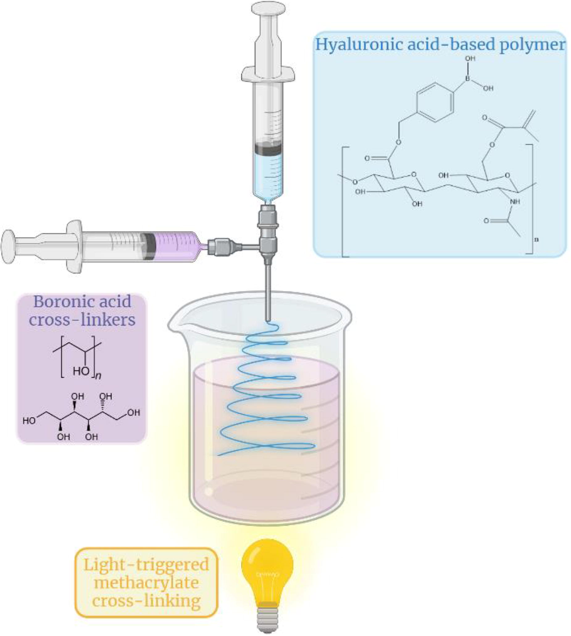
B232 - Development Of Ms-on-a-chip; Effects Of Microfluidic Device Structure
P. C. Hurley1,2,3, P. M. Holloway4, J. McMahon5,1,3, J. Hampson6, G. M. O’Connor2,1, U. FitzGerald6,1,3
1
CÚRAM SFI Research Centre for Medical Devices, University of Galway, Galway, Ireland,
2
National Centre for Laser Applications, University of Galway, Galway, Ireland,
3
Galway Neuroscience Centre, University of Galway, Galway, Ireland,
4
Radcliffe Department of Medicine, University of Oxford, Oxford, United Kingdom,
5
School of Biological & Chemical Sciences, University of Galway, Galway, Ireland,
6
School of Biomedical Engineering, University of Galway, Galway, Ireland
*Purpose/Objectives: Multiple sclerosis (MS) is a chronic inflammatory condition affecting the central nervous system (CNS). An immunological hallmark of the disease is the presence of oligoclonal bands in the cerebrospinal fluid (CSF), suggesting a survival niche for B cells within the CNS. Highly organised follicle-like structures have been detected in several chronic inflammatory diseases and in a large proportion of people with MS. The structures known as lymphoid-like follicles (LLFs) consist of B-cells and stromal support cells and are often found on the surface of the brain and on the meningeal membranes. The overall aim of our project is to determine how LLFs form in people with MS by developing a microfluidic model of the niche in which they are found.
*Methodology: We aim to 1) use MRI and post-mortem tissue images to define the physical properties of regions of the brain commonly found to harbour these structures and 2) use these properties to develop both models and devices that will mimic the subarachnoid space in this disorder. In this study, we will investigate cellular interactions and assess the suitability of our various device designs for modelling this aspect of disease pathology.
*Results: Guided by the physiology, we have developed COMSOL Multiphysics® models of our devices, as well as models of CSF flow within the sulci of the brain. We have estimated that cells in the sub-arachnoid space may be subjected with shear stresses of up to 0.123 Pa, following input velocities of 1.6mm/s. Using these in silicoresults, we have developed numerous iterative designs and will study the effects of these on modelling LLF formation in vitro.
*Conclusion/Significance: This study will contribute toward the project goal of developing an Organ-on-a-chip (OoC) model of LLF formation. This device optimisation stage will accelerate the realisation of a high-throughput device mimicking LLFs in MS. To our knowledge this is the first time that this aspect of disease pathology has been modelled using OoC technology.
B233 - Dynamic Culture Of Cerebral Organoids Using A Pillar/perfusion Plate For The Assessment Of Developmental Neurotoxicity
M.-Y. Lee1,2, P. Acharya1, S. Shrestha1, P. Joshi2, N. Choi1, V. K. Lekkala1, S.-Y. Kang1, G. Ni1
1
University of North Texas, Denton, TX,
2
Bioprinting Laboratories Inc, Farmers branch, TX
*Purpose/Objectives: Despite the potential toxicity of commercial chemicals to the development of the nervous system (known as developmental neurotoxicity or DNT), conventional in vitro cell models have primarily been employed for the assessment of acute neuronal toxicity. On the other hand, animal models used for the assessment of DNT are not physiologically relevant due to the heterogenic difference between humans and animals. In addition, animal models are low-throughput, time-consuming, expensive, and ethically questionable. Recently, human brain organoids have emerged as a promising alternative to assess the detrimental effects of chemicals on the developing brain. However, conventional organoid culture systems have several technical limitations including low throughput, lack of reproducibility, insufficient maturity of organoids, and the formation of necrotic core due to limited diffusion of nutrients and oxygen.
*Methodology: To address these issues and establish predictive DNT models, cerebral organoids were differentiated in a dynamic condition in a unique pillar/perfusion plate, which were exposed to test compounds to evaluate DNT potential. Briefly, embryonic bodies (EBs), initially formed in an ultralow attachment (ULA) 384-well plate, were successfully transferred to the pillar plate containing Matrigel and subsequently differentiated into cerebral organoids in the perfusion plate with test compounds.
*Results: The pillar/perfusion plate facilitated uniform, dynamic culture of cerebral organoids with improved proliferation and maturity by rapid, bidirectional flow generated on a digital rocker. Day 9 cerebral organoids in the pillar/perfusion plate were exposed to ascorbic acid (DNT negative) and methylmercury (DNT positive) in a dynamic condition for 1 and 3 weeks, and changes in organoid morphology and neural gene expression were measured to determine DNT potential. As expected, ascorbic acid didn’t induce any changes in organoid morphology and neural gene expression. However, exposure of day 9 cerebral organoids to methylmercury resulted in significant changes in organoid morphology and neural gene expression. Interestingly, methylmercury did not induce adverse changes in cerebral organoids in a static condition, thus highlighting the importance of dynamic organoid culture in DNT assessment.
*Conclusion/Significance: We envision that the pillar/perfusion plate could reduce the cost of cell culture media, enhance reproducibility and scalability of dynamic organoid culture and analysis, and simplify the organoid culture process with minimal manual intervention for high-throughput compound screening.

B234 - Vascularization Based On Sacrificial Templating For Bioreactor And On-chip Applications
M. Ryma1, S. Heilig2, Y.-Y. Dai3, I. Cicha3, T. Scheibel1, J. Groll2
1
University of Bayreuth, Bayreuth, Germany,
2
University Clinic Würzburg, Würzburg, Germany,
3
University Hospital Erlangen, Erlangen, Germany
*Purpose/Objectives: Organ-on-a-Chip technology is a valuable tool to provide more transferable test results regarding basic research, drug development or other applications than classic 2D cell culture. However, bottlenecks are the limited availability of complex and fully embedded vasculature in 3D tissue compartments, which hinders the generation of bigger and more natural tissues. We investigated the potential of Melt Electrowriting (MEW) (Figure Top) and Freeform Printing (FFP) (Figure Bottom) to generate sacrificial microfiber networks for bioreactor and on-chip applications regarding vascularization. These scaffolds can be embedded and dissolved on demand in crosslinkable hydrogel matrices for the generation of accurate and perfusable microchannel networks.
*Methodology: For this, we introduced biocompatible poly(2-oxazoline)s as a material for sacrificial scaffolds. These polymers are thermoresponsive in aqueous solutions, meaning that they dissolve on demand by simple temperature reduction. These poly(2-oxazoline)s were fabricated via FFP and MEW to generate highly accurate microfiber networks in 2D and 3D. The bioreactors were fabricated via
*Results: The poly(2-oxazoline) scaffolds were placed in dedicated bioreactors and chips. In aqueous environments, e.g. upon embedding in hydrogels, the fibers swelled, leading to fusion of adjacent filaments. Subsequent lowering of the temperature led to scaffold dissolution on-demand, without leaving non-dissolvable remnants and resulting in a leakage-free connection to the medium reservoirs. This approach enabled the generation of interconnected channel networks with bifurcations according to Murray’s law, resembling the natural vascularization. Importantly, the developed method is compliant with many of currently used hydrogels (e.g., methacrylated gelatin or collagen), suitable for vascularized tissue engineering approaches. Furthermore, the scaffolds with more complex 3D geometries could be produced using poly(2-oxazoline)s in combination with FFP method.
*Conclusion/Significance: Adding sacrificial scaffolds for channel generation in chips and bioreactors opens up multiple possibilities in creating viable 3D constructs of larger dimensions, containing microvascular network to support cell survival and tissue maturation.

B235 - Application Of A High Throughput Electrical Stimulation Bioreactor To Investigate The Role Of Exogenous Voltage Regimes Affecting In Vitro Cellular Functions
C. Reghukumar
The University of Manchester, Manchester, United Kingdom
*Purpose/Objectives: Electrical stimulation plays an increasingly important role in the field of tissue engineering. Benefits can include influencing cell proliferation, migration, differentiation, improving vascularisation and tissue integration, as well as extracellular matrix production. Exogenous electrical stimuli have been widely applied in clinical interventions such as in cardiac pacemakers, transcutaneous electronic nerve stimulator devices and more recently considered in chronic wound dressing products. For advancing electrical stimulation-based therapies, it is essential to study the underlying signalling mechanisms affected by exogenous electrical stimuli, at both the cellular and tissue level. In this project, the role of externally applied electrical stimuli on cell activity, proliferation, and trilineage differentiation of human mesenchymal stem cells (hMSCs) is investigated.
*Methodology: A simple, customised 24-well plate electrical stimulation bioreactor with platinum wire electrodes was developed for applying direct current (DC) electrical stimuli on bone marrow-derived hMSCs cultured in a 2D environment. Three different DC electrical voltage regimes (50, 100 and 150 mV/mm) were applied to the cells for 60 min per day, for 7 consecutive days using a ‘9130’ programmable direct current (DC) power supply. Cell metabolic activity, relative gene expression and extent of mineralisation during trilineage differentiation were investigated during various time points post electrical stimulation until day 21, to understand the effect of externally applied electrical stimuli on the growth and differentiation of hMSCs.
*Results: A 24-well plate electrical stimulation bioreactor was developed using a Computer-Aided Design (CAD) model. Its biocompatibility was tested to demonstrate the advantage of using platinum electrodes as it showed minimal corrosion effects and generated less harmful faradaic by-products than other electrode candidates. Further, the role of externally applied electrical stimulation showed improved cell proliferation and metabolic activity in hMSCs when differentiated into adipogenic, chondrogenic and osteogenic lineages with the differentiation media compared to the control (samples with no electrical stimulation but cultured with differentiation media).
*Conclusion/Significance: The study demonstrates the promising application of the custom-designed 24-well plate electrical stimulation bioreactor for in vitro high throughput electrical stimulation experiments. The effect of direct DC voltage shows enhanced cell metabolic activity with increased DNA content per cell, during the trilineage differentiation of hMSCs. The effect of a particular voltage regime is significantly different for different differentiation pathways which can be further correlated to its dependency on intrinsic transmembrane potentials for lineage commitment in stem cells. Also, the bioreactor can be further extended to be used for incorporating additional electrical stimulation parameters such as frequency, waveforms, duty cycle, etc. to investigate further the effect of electrical stimulus on stem cell-based tissue engineering technologies.
B236 - Biomimetic M-type Interface Integrated Into Precise Co-culture System As Muscle-tendon Junction-on-a-chip For Drug Development
L. Wang
Southern Medical University, Guangzhou, China
*Purpose/Objectives: Individual skeletal muscle and tendon models failed to capture the intricate cellular organization and interaction at the interface between the muscle and zones, which limited the understanding of muscle-tendon junction (MTJ). So, it is essential to develop an appropriate approach to construct an MTJ-on-a-chip platform that permits the precisely compartmentalized co-culture of myocytes and tenocytes of MTJ tissues for mimicking the physiological microenvironment of native MTJ tissue.
*Methodology: The multiscale MTJ scaffold was fabricated by the aligned electrospun nanofiber yarns via an interwoven weaving approach. To further investigate the cellular morphologies and differentiation of tendon stem/progenitor cells (TSPCs) and muscle satellite cells (SCs) on these MTJ scaffolds, the TSPCs and SCs were compartmentalized seeded on the zoon of TNFYs and MNFYs, respectively. To resist the interference of the external environment, we further encapsulated GelMA hydrogel onto the surface of MTJ scaffold to develop engineered MTJ tissue. Crucially, to overcome the co-culture issue of the muscle and tendon zones, a microfluidic device with a tissue co-culture chamber was designed, enabling the precisely compartmentalized co-culture of SCs and TSPCs within a single system. Clinically approved ciprofloxacin (CIP) was chosen as a model drug to examine the toxic effect on engineered MTJ tissue in MTJ-on-a-chip, while mitoquinone (MitoQ) was used as the protective one.
*Results: These multiscale MTJ scaffolds with an anisotropic interwoven “M-type” interface in five different ratios (3:1, 2:1, 1:1, 1:2, 1:3) were successfully fabricated via the interwoven weaving approach. TSPCs and SCs gradually transitioned and contacted on the interface of MTJ scaffolds, the “M-type” structure could enhance the cellular organization and interaction between muscle and tendon zones and form a cellular “M-type” interface. The engineered MTJ tissue could facilitate the investigation of intricate cellular organization and interaction between the muscle and tendon zones due to the biomimetic “M-type” interface, and then could induce myogenic and tenogenic differentiation due to the aligned nanofibrous structure. Compared to 2D models of TCP, the performance of engineered MTJ tissue in our MTJ-on-a-chip showed higher sensitivity of MTJ toxic or protective efficacy.
*Conclusion/Significance: In summary, we successfully fabricated a 3D engineered MTJ tissue with aligned nanofibrous structures and a biomimetic “M-type” interface, and further develop an MTJ-on-a-chip for drug research. The electrospun nanofiber yarns with a biomimetic “M-type” interface enable a promising approach for engineered MTJ tissue development, and the microfluidic device promoted the precisely compartmentalized co-culture of muscle and tendon zones, which suggested that the MTJ-on-a-chip performed a promising in vitro model for biomedical research of MTJ injuries.
B238 - A Cellularized Marine Collagen-based Wound Dressing For The Treatment Of Skin Wounds: Observations In A Rodent Animal Model
A. Carolo1, G. Zivelonghi1, M. Roncoroni2, S. Marzorati2, T. Martinello3, A. Porzionato4, R. Sacchetto1, F. Perozzo5, M. Sugni2, M. Patruno1, L. Melotti1
1
Department of Comparative Biomedicine and Food Science, University of Padova, Legnaro, ITALY,
2
Department of Environmental Science and Policy, University of Milan, Milano, ITALY,
3
Department of Veterinary Medicine, University of Bari, Valenzano, ITALY,
4
Department of Neurosciences, University of Padova, Padova, ITALY,
5
Plastic and Reconstructive Surgery Unit, Padova University Hospital, Padova, ITALY.
*Purpose/Objectives: Skin wound healing is a dynamic process that comprises different overlapping phases that aims to restore the skin integrity. However, a minimal impairment (e.g., infection) may lead to hard-to-heal ulcers or open non-closing wounds in a constant state of inflammation. For this reason, an ideal treatment able to contain inflammation while promoting healing is necessary in the clinic. Wound dressings are materials used to cover and protect the wound from damage along with maintaining its moisture, hence they are one of the gold standard treatments for wound care management in Veterinary Medicine. Besides dressings, cell therapy has also shown promising results, in particular the application of mesenchymal stem cells (MSCs). This cell population can release a plethora of soluble molecules that modulate different biological processes such as inflammation. In this work we describe the application of a marine collagen-based wound dressing (MCDT) alone or in combination with allogeneic adipose-derived MSCs (aMSCs) for the treatment of skin wounds in a rodent model.
*Methodology: A total of 48 rats were included in the study. On the back of each animal two round wounds were surgically induced: to one aMSCs or the biomaterial, with or without cells (MCDT or MCDT+aMSCs), was applied (n = 16 per treatment) while one was left untreated. Wounds were checked daily for signs of adverse reactions or infection. After 5 and 10 days, skin samples were collected and processed for histopathological (inflammation and granulation tissue (GT) presence) and gene expression analysis for factors involved in the skin wound healing process (extracellular matrix proteins, growth factors, and pro-inflammatory cytokines).
*Results: Clinically, at day 5 wound closure rate was similar among treatments. At day 10, MCDT-treated wounds showed a slightly lower wound closure rate compared to treatment containing cells. At the histological level, the presence of inflammatory cells was comparable among untreated and MCDT-treated wounds while cell-based treatment showed a lower level of inflammation in the healing tissue. This observation is also reflected in the mRNA level of TNF-α and, especially, IL-1β, both well-known pro-inflammatory factors, which was lower in wounds treated with aMSCs and MCDT+aMSCs. All treated wounds also showed a higher amount of GT on day 5 compared to untreated lesions. Moreover, while treated wounds showed a more mature GT at day 10, untreated lesions still displayed a high amount of immature GT. These results are supported by the high expression of collagen type I and III in untreated wounds, while treatments controlled collagen expression to base levels at day 10. Moreover, higher levels of angiogenesis were observed in treated wounds, especially with MCDT+aMSCs, which were also characterized by a higher presence of vessels, especially in the lower layers of the tissue, and higher levels of PDFGb and VEGF.
*Conclusion/Significance: In conclusion, all treatments led to better outcomes compared to physiological healing. Indeed, lower inflammation and better deposition of structural proteins are crucial factors to reach a proper level of skin wound healing after wounding and both elements were observed in this work.
B239 - Optimized Ultrafiltration-based Depletion Of Extracellular Vesicles From Cell Culture Supplements Increases Metabolic Activity And Viability Of Equine Adipose-derived Mesenchymal Stem Cells
A. Ramesova, S. Kau-Strebinger, J. Burk-Luibl
University of Veterinary Medicine Vienna, Vienna, Austria
*Purpose/Objectives: Extracellular vesicles (EVs) are critical components of intercellular communication with immense potential in diagnostic and therapeutic applications. Mesenchymal stem cell-derived EVs (MSC-EVs) are of particular interest, due to their regenerative features. However, to harness the full potential of MSC-EVs, it is necessary to standardize cell culture conditions to avoid culture media contamination with EVs originating from cell culture supplements, e.g. fetal bovine serum (FBS). These xenogeneic EVs bias downstream analysis and subsequent use of MSC-EVs. Thus, it is important to effectively deplete EVs from supplements before use. A challenge represents the question of how to separate only EVs so that other nutrients would stay in the supplement to support cellular health. This study aimed to optimize previously published ultrafiltration-based EV depletion protocol by prior centrifugation and dilution of FBS and to assess the effect of using this prepared FBS on metabolic activity and viability of equine adipose-derived (eAD-)MSCs.
*Methodology: FBS (Lot: 2078409, Gibco®, ThermoFisher Scientific) was pre-centrifuged at 2.000 x g (5 mins) and 10.000 x g (30 mins) at RT. Non-diluted (ND) or 1:2 and 1:5 diluted (D) was centrifuged using Amicon® Ultra-15 Centrifugal Filter Units (100 kDa MWCO) for 55 mins at 3.000 x g, 4 °C. Total protein concentration in retentate and filtrate was measured using BCA assay and adjusted for comparability of protein filtration efficiency between conditions. To assess the effect of EV-depleted FBS on cell metabolic activity and viability, eAD-MSCs were seeded on 96 well plates (day 0) in standard cultivation media (full FBS). At day 1, cells were treated with media containing: i) full FBS, ii) EV-depleted, non-diluted FBS (ND-FBS), iii) EV-depleted, 1:2 diluted FBS (D2-FBS), iv) EV-depleted, 1:5 diluted FBS (D5-FBS). At days 1 and 5, cell metabolic activity and viability were evaluated by MTS Assay and compared between groups i-iv.
*Results: Dilution of FBS prior to ultrafiltration increased protein filtration efficiency (fold change to ND-FBS: 7.1 in D2-FBS and 4.8 in D5-FBS). Total protein concentration of the filtrate increased from 2.1 ug/uL in ND-FBS to 12.4 ug/uL in D2-FBS and 10 ug/uL in D5-FBS. The volume of the filtrate increased from 10.5 mL in ND-FBS to 11.6 mL in D2-FBS and 12.7 mL in D5-FBS. Reversely, retentate volumes decreased. Macroscopically, the ND-FBS filtrate was almost see-through, indicating retention of other components of FBS, not only EVs. eAD-MSCs treated with D-FBS in standard cultivation media showed a higher metabolic activity than eAD-MSCs treated with ND-FBS, but this effect was overall lower than in eAD-MSCs cultured in standard media containing full FBS.
*Conclusion/Significance: Cell culture supplements contain xenogeneic EVs, which hinder the analysis and usage of cell-originating EVs. One way how to effectively deplete EVs from supplements is by ultrafiltration. A good method how to avoid possible congestion of the filter and retaining of proteins, and possibly other nutrients, is by diluting the FBS before ultrafiltration, which increases protein concentration in the filtrate. Subsequent usage of this D-FBS filtrate in eAD-MSC culture improves cell metabolic activity and viability as compared to ND-FBS filtrate.
B240 - Breaking The Glass Ceiling Of WomenS Entrepreneurial Intention In Bio&Medtech
J. N. Cervantes Guzman, S. Mathews, D. W. Hutmacher;
Queensland University of Technology (QUT), Brisbaine, Australia
*Purpose/Objectives: In this comprehensive study, the researchers undertake an examination of women’s entrepreneurship across two distinct domains: STEM & Bio&MedTech. The central objective of this research is to explore the various factors that exert influence on women’s entrepreneurial intentions within the Bio&MedTech sector, effectively bridging the chasm between academic inquiry & practical implementation. This endeavor is underpinned by the overarching goals of facilitating well-informed decision-making, advancing gender diversity, nurturing innovation, & stimulating economic growth. Bridging the Gap: The research conducted seeks to empower female researchers, providing them with the means to pursue their entrepreneurial aspirations & translate scientific innovations into viable business opportunities. To achieve this, the study aims to discern the pivotal elements that mold women’s entrepreneurial intentions in the Bio&MedTech sector. These insights are poised to pave the way for customized programs tailored to address specific challenges, such as securing funding, forging networks, & accessing mentorship. Addressing Research Gaps: A noticeable research gap emerges from the limited attention directed toward the unique constraints & obstacles encountered by female entrepreneurs in the Bio&MedTech sector. Research Questions & Aims: The primary research question (RQ1) revolves around the identification of factors that either encourage or impede women’s entrepreneurial aspirations within the Bio&MedTech sector. To tackle this overarching question, the study is guided by two specific aims: 1.1: To scrutinize &elucidate the distinctions characterizing entrepreneurship in STEM & the Bio&MedTech sector. 1.2: To ascertain the motivating factors underpinning entrepreneurial intentions in the Bio&MedTech sphere. The Managerial Problem: the Bio&MedTech sector suffers from a marked underrepresentation of women among its entrepreneurial ranks. This discrepancy represents a significant missed opportunity, resulting in the underutilization of valuable resources within the biotechnology business.
*Methodology: The researchers employ a systematic literature review methodology, adhering to the PRISMA (Preferred Reporting Items for Systematic Reviews & Meta-Analyses) approach, to consolidate existing knowledge & identify research gaps. Complementing this approach is the utilization of bibliometric analysis, facilitating insight into publication, citation, & co-citation patterns. This analysis provides the researchers with the ability to pinpoint influential authors, publications, & research themes within the Bio&MedTech sector.
*Results: The research outcomes underscore the pivotal role played by individual & contextual factors in shaping women’s entrepreneurial intentions within the Bio&MedTech realm. Factors such as self-confidence, propensity for risk-taking, creativity, social support, & a strong passion for entrepreneurship emerge as key determinants. Additionally, societal attitudes, cultural norms, industry characteristics, & supportive policies are identified as influential drivers of entrepreneurial intentions. While some factors affecting entrepreneurial intentions appear to overlap between STEM & Bio&MedTech, the research underscores the presence of unique challenges specific to the Bio&MedTech sector. These include a prevailing male-dominated culture & a pronounced lack of gender diversity in leadership positions.
*Conclusion/Significance: this study serves as a guiding beacon, illuminating the path toward gender diversity & the empowerment of women in the Bio&MedTech entrepreneurial landscape. By addressing these critical issues, the research contributes substantively to the advancement of this field, driving innovation & economic growth while ensuring that no talent remains untapped.
B241 - Utilizing A Bioreactor For Ex-vivo Perfusion To Model Mouse Kidney Disease
J. D. Castillo Prado1,2, A. Bhadwal1,2, I. Rogers1,2,3
1
University of Toronto, Toronto, ON, Canada,
2
Mount Sinai Hospital, Toronto, ON, Canada,
3
Department of Obstetrics and Gynecology, University of Toronto, Toronto, ON, Canada
*Purpose/Objectives: Chronic kidney disease (CKD) affects 11-13% of the population worldwide. A hallmark of CKD is fibrosis which exacerbates kidney dysfunction and is linked to increased mortality. Limited studies have been conducted utilizing ex-vivo cultures in kidney fibrosis research. Animal models of kidney fibrosis involve invasive procedures that can affect the animal’s health and experiment results. Also, disease stages are missed since the organs of interest cannot be analyzed without invasive techniques. Ex-vivo whole-organ perfusion (EVOP) provides a solution by enabling real-time, longitudinal analysis at the whole-organ level. Also, controlled culture conditions lead to reduced alterations between experiments. Similarly, experimental treatments can be directly administered to the organs while avoiding extrarenal stimuli. For this purpose, we designed, built and validated a bioreactor to support mouse kidney ex vivo perfusion. We hypothesize that a mouse kidney can be maintained using EVOP for 10 days and that renal fibrosis can be reproduced in an EVOP system through ureteric obstruction (UO) and cisplatin-induced nephrotoxicity.
*Methodology: Following kidney isolation and renal artery canulation, kidneys were cultured under EVOP for 4, 7, and 10 days. Kidneys were also subjected to UO and cisplatin treatment to induce fibrosis and were then cultured ex-vivo for 4 and 7 days.
*Results: We determined that tissue viability and proper marker expression were sustained for up to 10 days in the EVOP kidneys. We determined that our system meets the oxygen demand of a mouse kidney. Urinalysis demonstrated urine production. Preliminary results revealed higher amounts of collagen deposits present in UO and cisplatin kidneys at days 4 and 7. Similarly, fibrosis-treated kidneys secreted pro-fibrotic cytokines into their culture medium, indicating fibrosis development.
*Conclusion/Significance: Surface-to-volume ratio of medium, perfusion flow rates, and oxygen availability were all considered while maintaining the bioreactor small and practical to use. The EVOP system can maintain mouse morphology and function for 10 days. Fibrosis appears to have developed by days 4 and 7 after treatment. Future steps include optimizing the culture medium to extend EVOP times to 14 days. We will also analyze the gene expression of control and fibrotic kidneys to help improve the EVOP model. This ex-vivo perfusion system provides a platform for bioengineering purposes such as disease modelling, physiology research, tissue regeneration studies, cell therapy, drug testing, and recellularization of decellularized mouse organs.
B242
B243 - Surgical Reconstruction Of An Artificially-induced Hypospadias In Rabbits Using A Tissue-engineered Human Urethral Substitute
C. Caneparo1,2, E. Elia1,2, S. Chabaud1,2, J. Fradette1,2, S. Bolduc1,2
1
Université Laval, Quebec, QC, Canada,
2
CHU de Quebec-Université Laval research center, LOEX, Quebec, QC, Canada
*Purpose/Objectives: Urethral reconstruction remains a challenge due to the lack of available donor tissues and complications after surgeries, especially for patients with recurrences or severe anomalies. When the foreskin is unavailable, the oral mucosa remains the gold standard graft for urethroplasty. However, the quantity available for grafting is limited, and it cannot be harvested twice from the same area, leading clinicians to develop new alternatives. We developed a human-derived tissue-engineered urethral substitute (US), devoid of exogenous biomaterials, which showed in vitro that mechanical resistance, histology, and functionality (waterproofness) were similar to native tissues. The present study aimed to evaluate this US in vivo using the most frequently used animal model for urethral reconstruction, the New Zealand rabbit. The engraftment of this substitute in immunosuppressed rabbits and the technical feasibility of correcting an induced artificial penoscrotal hypospadias were evaluated.
*Methodology: US were reconstructed using the self-assembly technique, then grafted in immunosuppressed rabbits. Following current clinical guidelines, a two-stage urethral reconstruction was realized. The US was compared to two control groups (n=6 per group). Control groups: 1- the native urethral plate, resulting from the urethra being longitudinally opened without its removal (positive control), and 2- a “clinically relevant” control group using a donor rabbit oral mucosa, both for a 2-stage tubularized urethroplasty. For the experimental group, we assessed the effectiveness of our US for urethral reconstruction. The urethra was initially removed and replaced with the graft (first stage), and 12 weeks later, a second-stage tubularization urethroplasty was performed. Urethroscopy was performed during follow-up at weeks 20 and 28, and urethrography was performed at week 28.
*Results: US exhibited a native-like histology before and after the graft. During the study, we showed that treatment of the rabbits with 1 mg/kg of tacrolimus, maintained immunosuppression with minimal side effects. We showed that oral mucosa can be harvested from donor rabbits and successfully grafted in recipient rabbits. However, significant contraction upon healing was observed. US were grafted successfully but also exhibited significant contraction upon healing in some rabbits. Tubularization was possible for all but one. Cystoscopy showed that the US was well-healed eight weeks after tubularization. Rabbits from control group 1 (native urethral plate) had no complication at week 28 (success rate 100%). Rabbits from control group 2 (oral mucosa graft) underwent successful tubularization, but two presented urethrocutaneous fistula (success rate 66%). Four of the six (66%) rabbits in the US experimental group had no urethral complication after tubularization. Still, two of these four rabbits had to be euthanized due to unrelated reasons (dental malocclusion and intestinal obstruction (ileus)). However, the two remaining rabbits showed recurrent penoscrotal meatal stenosis at the junction of the US and the native urethra before tubularization, and urethroplasty was not performed (success rate 66%).
*Conclusion/Significance: The self-assembled substitutes produced in vitro represent a promising biomaterial for urethroplasty. However, some adjustments must be made in the preclinical protocol to circumvent the problems encountered due to the use of this rabbit model.
B245 - In Vitro Assessment Of Radiation Exposure And Radioprotection Using A Primary Human Bronchial Epithelial Cell Model
D. Lizanets, S. V. Murphy, A. R. Hall
Biomedical Engineering, Wake Forest Institute for Regenerative Medicine, Winston Salem, NC
*Purpose/Objectives: While radiotherapy (RT) is a standard treatment for lung cancer, proximal healthy tissue irradiation is unavoidable. Because lung tissue is particularly susceptible to off-target radiation damage, this can lead to the significant clinical challenge of radiation-induced lung injury (RILI). Current models for studying radiation damage and radioprotection are limited in their translatability. Existing in vivo animal models lack translational relevance as the animals used are more resilient to the effects of radiation and elicit a different organ response, have different repair mechanisms, and have a different onset timeframe for RILI, thus making them a difficult choice for direct comparison. In vitro models typically employ cell lines which do not provide a comprehensive model for studying radiation damage given their immortalized state which alters their both their genetics and growth rates, two important parameters used to measure relevant effects of irradiation and treatment. It is crucial to develop relevant models that can improve our understanding of the impacts of radiation damage as well as radioprotectant countermeasures in primary cell models as they will ultimately enhance clinical translatability, being more characteristic to those in the native human lungs. In this study, an in vitro lung model comprised of primary human bronchial epithelial (HBE) cells is employed to study radiation exposure and assess the radioprotective capacity of WR-1065, the active metabolite of the prophylactic drug Amifostine.
*Methodology: In vitro lung models were established using primary HBE cells and then exposed to gamma radiation with doses ranging from 0-10 Gy. The cellular impact of exposure to ionizing radiation was explored by measuring DNA damage immediately following irradiation as well as assessing the downstream impacts on cell proliferation, establishing dose-dependent effects of radiation damage. To explore the efficacy of a radioprotectant in this model, prophylactic treatments with WR-1065 were administered prior to irradiation and the concentration-dependent effects were evaluated post-hoc.
*Results: Radiation were found to induce dose-dependent changes in cell morphology along with increased incidence of DNA DSBs, causing a decrease in cell proliferation. It was established that the extent of quantifiable DNA damage reaches a limit at 8 Gy, a dose commonly used in clinical radiotherapy regimens. For WR-1065 treatments, we established that concentrations up to 4 mM were non-toxic and then evaluated its radioprotective capabilities following 8 Gy gamma irradiation, demonstrating a concentration-dependent ability to reduce the consequences of radiation exposure.
*Conclusion/Significance: An in vitro model using primary HBE cultures was established as a tool for observing the effects of radiation exposure of human lung cells and investigating the ability of radioprotective agents to mitigate this damage. This work sets a foundation for ongoing and future experiments that will expand the clinical relevance by integrating fractionated exposure regimens and by increasing model complexity to better match physiological conditions with an advanced airway organoid model at air-liquid interface that incorporates mature, differentiated primary HBEs along with other supportive cells.
B246 - Highly Elastic Electrospun Scaffolds For The Lower Urinary Tract
R. Sturm, Y. Huo, F. Yiu, R. Afshari, Z. Zhong, K. Harrison, Y. Gu, M. Ghovvati, N. Annabi
UCLA, Los Angeles, CA
*Purpose/Objectives: Commonly applied biologic scaffolds and current tissue substitution options have significantly higher stiffness than native urethral or bladder tissue. The mechanical niche is an important parameter controlling cell fate and extracellular matrix (ECM) deposition. Additionally, tissue mismatch of vastly different substrates can minimize early functionality and increase regional wound tension when applied in reconstructive procedures. The aim of this study was to engineer a novel biomimetic, suturable, and biocompatible scaffold with target mechanical properties of lower urinary tract tissue and complete an in vitro analysis of lower urinary tract cell line propagation on these scaffold structures.
*Methodology: Nanofibrous scaffold sheets were synthesized by electrospinning varied concentrations of two naturally derived polymers in random orientation onto a mat: elastin-like polypeptide (ELP) and gelatin methacryloyl (GelMA) to achieve target mechanical properties for 1) soft (tensile modulus, TM: 50-250kPa) and 2) soft and elastic (300-500% extensibility) scaffolds, respectively. Scaffolds were characterized using NMR spectroscopy, degradation, swelling, tensile and suture testing, light and scanning electron microscopy. Human bladder-derived urothelial (UC) and smooth muscle cells (SMC), and neonatal foreskin fibroblasts (Fib) (ATCC) were seeded in mono cell culture conditions using cell-specific media directly onto 1cm2 scaffolds in 24-well plates at a 1x10⁴ cells/mL concentration. A systematic evaluation of cell viability (Presto blue), adhesion/spreading (Actin/dapi), cytotoxicity and proliferation (Live/dead, counts) using light/fluorescent and confocal microscopy occurred between day 1 and 7 endpoints. T-tests were used for pairwise comparisons; one-way ANOVA to evaluate between-group differences.
*Results: The material composition and fabrication process were optimized to achieve target mechanical properties, using a combination of GelMA and ELP. Specifically, percent extensibility significantly increased with the substitution of 3 to 7% ELP for GelMA weight/volume (w/v) versus 10% GelMA alone. Furthermore, 5% GelMA/5% ELP w/v achieved TM and ultimate tensile strength in static and cyclic testing that did not significantly differ from anterior urethral or bladder tissue in the New Zealand White rabbit and was consistent with soft and elastic target parameters. Scaffolds were suturable, with improved performance in suture tensile testing with the addition of ELP. Excellent cell viability was observed from day 1 to 7 across cell lines and scaffolds. Proliferation and spreading of SMCs and Fib was robust, while UCs demonstrated less scaffold adherence and proliferation as compared to the other two cell lines.
*Conclusion/Significance: Highly elastic engineered GelMA/ELP hybrid scaffolds are non-cytotoxic. The nanofibrous scaffolds supported robust Fib and SMC cell line proliferation. Future scaffold modifications are ongoing to optimize parameters for UC proliferation, and in vivo biocompatibility tests are ongoing. In the future, these scaffolds may provide an alternative strategy for suturable, elastic lower urinary tract replacement tissue that can restore early functionality in lower urinary tract reconstructive procedures.
B248
B254
B302 - Development And Characterization Of Graphene Derivative-gelma Hybrid Bioinks For The Generation Of Bioartificial Tissue Substitutes Via 3d Bioprinting
M. P. Lavín1, Ó. D. García-García2, F. Condi de Godoi3, M. Griera-Merino4, I. Jorba5, S. de Frutos6,4, S. Micó7, J. M. Baena7,8, V. Carriel2, N. Campillo7,8
1
Graphenano S.L., Murcia, Spain,
2
University of Granada, Granada, Spain,
3
Tessenderlo Group nv, Brussels, Belgium,
4
Graphenano Medical Care S.L., Madrid, Spain,
5
University of Barcelona, Barcelona, Spain,
6
University of Alcalá, Madrid, Spain,
7
REGEMAT 3D S.L., Granada, Spain,
8
BRECA Health Care S.L., Granada, Spain
*Purpose/Objectives: The fabrication of bioartificial tissue substitutes is a complex process that relies on the application of innovative biomaterials and manufacturing techniques enabling the generation of cell-laden scaffolds mimicking natural tissue interfaces. Among the many biomaterials, gelatin methacryloyl (GelMA) hydrogels have shown great potential for 3D bioprinting tissue engineering applications due to their high biocompatibility, biodegradability and tunable mechanical properties. In this study, the potential use of hybrid hydrogels based on GelMA and a highly-purified graphene-derivative (BioGraph®) as biomaterials bioinks for extrusion-based 3D bioprinting applications in tissue engineering was investigated.
*Methodology: ClaroTM BG800 GelMA (PB Leiner) was blended with BioGraph® (Graphenano Medical Care), a highly-purified graphene-derivative. BG800 prepolymer concentration in the hydrogel was maintained either at 5% (BG800-5) or 10% (BG800-10)(w/v), while growing concentrations of BioGraph (up to 0.3% w/v) were tested. All formulations contained 0.1% (w/v) LAP photoinitiator. Rheological characterization was conducted in a MCR302 rheometer (Anton Paar) equipped with a light source providing an irradiance of 60 mW/cm2 at 405 nm. Printability tests were performed in a REG4Life bioprinter (REGEMAT 3D) equipped with a refrigerated syringe and a 405 nm photocuring module. The biocompatibility of BG800 and BioGraph®-BG800 was investigated both in vitro and in vivo.
*Results: Formulations containing BioGraph® concentrations up to 0.1% are compatible with 3D bioprinting, showing good extrudability with reduced clogging at the printing temperatures, effective photocrosslinking at the irradiances tested, high shape-fidelity and high resolution of the printed scaffold. In situ photocrosslinking tests revealed that BioGraph® concentration decreases the speed of the photocrosslinking and the stiffness of the cured matrix, while GelMA concentration has the opposite effect. In vitro studies indicated that BioGraph® content ≤0.1% (w/v) did not have an adverse impact on the viability and proliferation of rat adipose-derived mesenchymal stem cells (r-AMSCs). Similarly, acellular scaffolds implanted subdermally in rats generated a normal foreign body reaction showing a collagen encapsulation procedure without presence of massive leucocytes nor fibrosis. Higher macrophagic interactions with faster in vivo degradation, and higher r-AMSCs proliferation in vitro were observed when lower concentrations of BioGraph® (0.025% w/v) were added.
*Conclusion/Significance: The GelMA-BioGraph® hybrid hydrogels developed in this work show reinforced rheological and biological properties, tailored for extrusion-based 3D bioprinting with applications in the tissue engineering of soft and hard tissues.
Funding: Study financed by Grant CPP2021-009070 and FIS P20-0318. N.Campillo is granted by PTQ2019-010731.
B303 - Development Of Biodegradable Fiber Stents Using Sheath-core Biphasic Monofilaments For Non-vascular Applications
J. Son1, M. Jang1, G. Kim1, Y. Joung2, K. Kim3, C.-i. Kwon4
1
Korea Textile Development Instutute, Daegu, Korea, Republic of,
2
Korea Institute of Science and Technology, Seoul, Korea, Republic of,
3
M.I.Tech, Co. Ltd., Pyeongtaek, Korea, Republic of,
4
CHA University School of Medicine, Seongnam, Korea, Republic of
*Purpose/Objectives: Biodegradable stents (BS) are highly attractive functional stents that are continuously being studied under this concept, and several clinical results have recently been reported. BS have a wider lumen compared to plastic stents and do not need to be removed. In addition, they can be mixed with drugs to release anti-bacterial and/or anti-tumor compounds. Because biliary BS cannot be made with thinner wire than that used for esophageal BS, clinical attempts with biliary BS have used the percutaneous trans-hepatic biliary drainage (PTBD) route. The clinical outcomes and prevalence of adverse events associated with biliary biodegradable stents can differ according to degradation process and time. The aim of this study was to observe the degradation process and time of different BS prototypes, and to evaluate sequential changes in their mechanical properties
*Methodology: Biodegradable sheath-core monofilaments with PDO as the outer sheath and PLLA or magnesium (Mg) wire as an inner core were developed with a conjugated melt spinning method and a melt coating method, respectively. BSs were made with four different types of Biodegradable monofilaments using cross-and-hook knitting hand-made method. Using an in vitro bile flow phantom model, we compared degradation time, radial force changes, and morphologic changes among four different BS prototypes: polydioxanone (PDO) BS, polyglycolide (PGA) BS, PDO/poly-L-lactic acid (PLLA) sheath-core BS, and PDO/Mg sheath-core BS. Using an in vivo swine bile duct dilation model, we performed a direct peroral cholangioscopy (DPOC) examination to observe the biodegradation process and related adverse events at regular intervals.
*Results: In the bile flow phantom model, the PGA BS and PDO/Mg BS prototypes showed rapid radial force reduction and morphological changes and complete degradation within six weeks. PDO/PLLA BS maintained high radial force and kept their original shape for longer than the PDO BS, up to16 weeks. A total of 24 BS was inserted into the dilated bile ducts of 12 swine. In this animal model, DPOC examination revealed that PDO BS and PDO/PLLA BS maintained their original shapes for approximately 12 weeks, but PDO BS showed a greater degree of fragmentation and induced biliary stones and bile duct obstruction.
*Conclusion/Significance: Our results showed that PDO/PLLA BS maintained their original shape and radial force for a relatively long time and minimized adverse events. Our results could be helpful in providing basic data and suggesting new directions for further development of BS.
Acknowledgment: This work was supported by the Korea Institute of Planning and Evaluation for Technology in Food, Agriculture and Forestry (IPET) through Companion Animal Life Cycle Industrialization Technology Development Program, funded by Ministry of Agriculture, Food and Rural Affairs (MAFRA) (32208604).
B304 - Characterising The Material Properties Of Bioengineered Scaffolds And Human Tissues Using Shear Rheology
A. R. Upadhyay1, D. Bakkalci1, E. Moeendarbary1, M. Butler2, M. Bergin3, M. Loizidou1, U. Cheema1
1
University College London, London, United Kingdom,
2
UCB Pharma, Slough, United Kingdom,
3
UCB Pharma, London, United Kingdom
*Purpose/Objectives: Biophysical parameters are integral to patho-physiologies like fibrosis and cancer. Systematic investigations of these, how they influence cell behaviour and disease progression, are feasible in 3D disease models. It is essential for biomaterial research to consider 3D architecture and mechanical properties for more accurate modelling of cell interactions in the microenvironment. Many techniques have been used to explore biomechanics but limitations surrounding methodology and different elastic moduli, makes direct comparison of measurements difficult. We compared soft and dense collagen I gels to other commonly used biomaterials and human tissue using oscillatory rheometry; establishing a reproducible technique to measure shear elastic modulus, as a measure of material stiffness.
*Methodology: Acellular collagen I hydrogels and dense collagen I gels were manufactured with a starting concentration of 2 mg/ml. Dense constructs were generated by plastic compression of hydrogels, for a final collagen density of 6% v/v. This was compared to acellular Matrigel and Geltrex (1.3ml volume) along with human tissue samples from renal carcinomas and chronic kidney disease patients. Amplitude and frequency sweep testing was conducted using the Kinexus Pro+ Rheometer (Netzsch), at various frequencies and strain rates. Immersion-well geometry was used, baseline testing at 1Hz frequency and 37°C. Storage modulus, phase angle, LVE region and stress-strain rate curves were analysed.
*Results: Acellular collagen I hydrogels are significantly softer, with an average phase angle of 37.2 and an average elastic modulus of 38.6 ± 2.14 Pa, indicating greater viscous potential. Whereas, acellular dense collagen I is much stiffer, with an average elastic modulus of 412.4 ± 41.1 Pa. Matrigel’s elastic moduli averaged at 72.1 Pa ± 8.6, however Geltrex’s moduli measurements were closer to the soft collagen gels (32.9 Pa ± 0.7). The phase angle for Matrigel averages at 5.09 ± 0.27 and Geltrex at 3.21 ± 0.52, in comparison to the dense collagen I model at 12.39 ± 0.31. The lower phase angle indicates that the material behaves more like an elastic solid with less viscous energy. The shear stiffness of human tissue sits around 1.4 kPa ± 337.0 on average; the shape of the linear viscoelasticity curve of kidney tumour samples (Figure 1) is comparable only to the acellular dense collagen model, unlike other biomaterials.
*Conclusion/Significance: Collagen density influences the material’s mechanical properties. Low density, soft collagen gels exhibit high viscoelasticity and low stiffness. Increasing the collagen density in gels alters the material properties and reduces its ability to resist elastic deformation. The complexity of the mechanical forces within bioengineered scaffolds is highlighted by the low phase angle and shear stiffness in Geltrex and Matrigel, compared to dense collagen I which is stiffer but holds greater viscous potential due to its matrix composition. In comparison to all the other biomaterials analysed, the dense collagen model generates more physiologically relevant stiffness and behaves more similarly to human tissue when experiencing increasing strain. These can be used to investigate interactions between key cell types and physical environment during disease. A better understanding of this will guide our design for more biomimetic models.
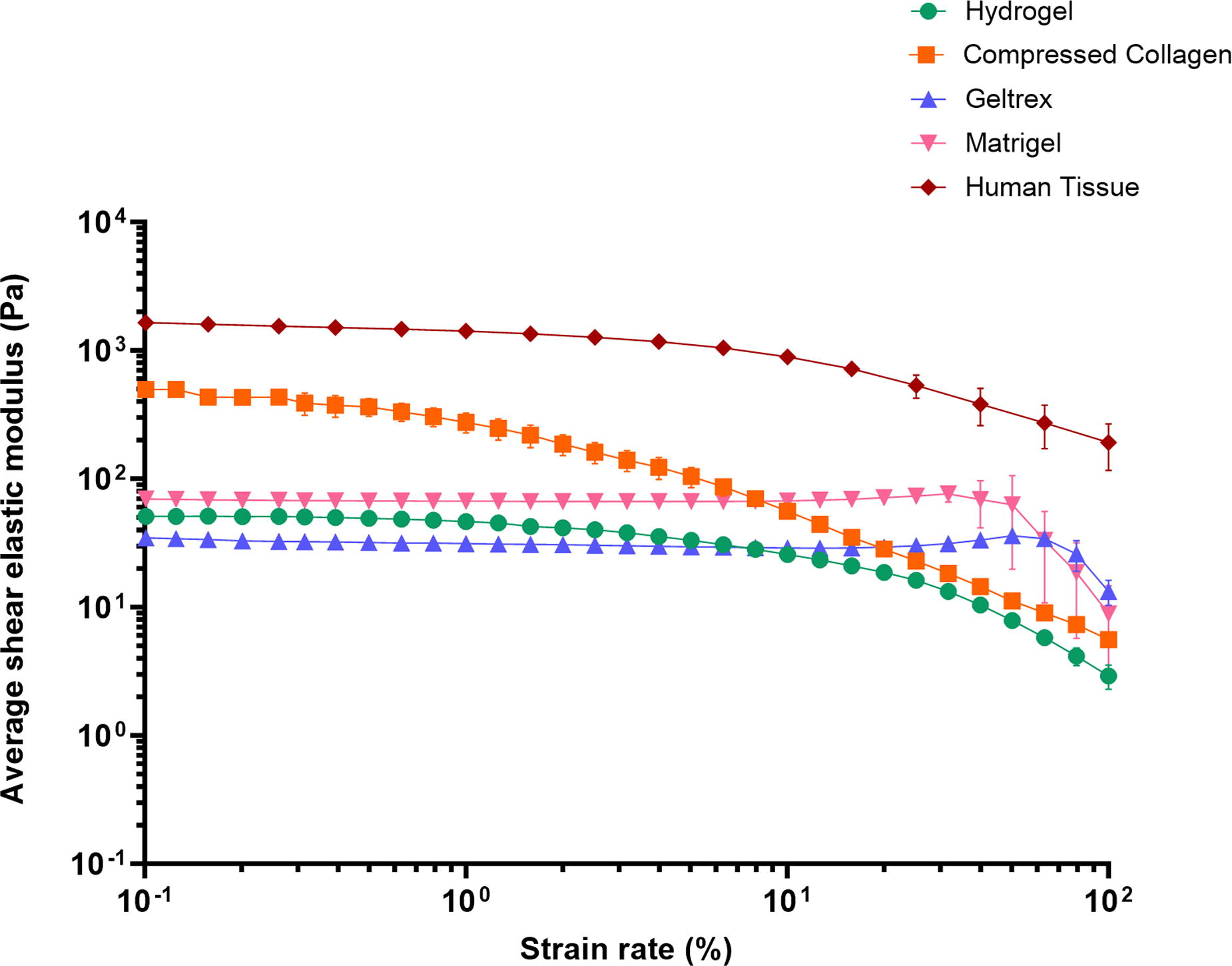
B305 - Ultra-tough Hydrogels With Open-loop Micro Hollow Channels Capable Of Physical Exchange Mimicking Capillaries
S. Lee, C. Park, C. Kim
Jeonbuk national university, Jeonju-si, Korea, Republic of
*Purpose/Objectives: Since cells can survive only within about 200μm from capillary for a prolonged time in the body, regenerating damaged tissue beyond the critical size remains a great challenge. Despite the recent publication of research papers highlighting the significance of angiogenesis in the restoration of damage surpassing a critical threshold, the challenge persists in achieving the formation of hollow channels, reminiscent of capillaries, within hydrogels, particularly when targeting dimensions on the order of tens of micrometers. Furthermore, due to the generally limited mechanical strength exhibited by the majority of hydrogels, it becomes imperative to engineer a hydrogel with mechanical properties adequately tailored for bone tissue applications to stimulate capillary formation within the osseous structure.
*Methodology: We synthesize a UV cross-linkable fibroin hydrogel prepolymer, and then homogeneously embedded PS volumetric microfibers inside the gel. To emulate the topographical features of a capillary structure, Dimethylformamide (DMF), selected for its microfiber removal and physical cross-linking capabilities, was utilized to fabricate tough hydrogels with micro-hollow channels. To improve osteoconductivity, the surface was coated with a high concentration of SBF solution to prepare a tough microchannel hydrogel suitable for hard tissue, and then its activity against cells was evaluated.
*Results: Utilizing punching and selective removal, a tough open-loop hydrogel with a micro hollow channel of approximately 30 μm was formed. The DMF used to form the hollow channel not only selectively removed the volumetric PS microfibers, but also induced a crystalline beta-sheet (>50%). The strategic use of DMF enabled the fabrication of a dual-crosslinked tough hydrogel with a substantial tenfold increase in compressive strength (from 400 kPa to 4 MPa). The resulting micro hollow channels enable the transport of substances such as oxygen and nutrients and the surface-coated calcium-phosphate mineral particles promote cell adhesion and differentiation.
*Conclusion/Significance: We succeeded in fabricating a tough hydrogel with omnidirectional properties and a hollow channel that mimics the diameter of the capillary. The mechanical strength reinforced by the successful induction of crystalline beta-sheets of dual crosslinked SM is expected to play a crucial role in broadening the range of the currently limited application of naturally derived hydrogel in soft tissue.
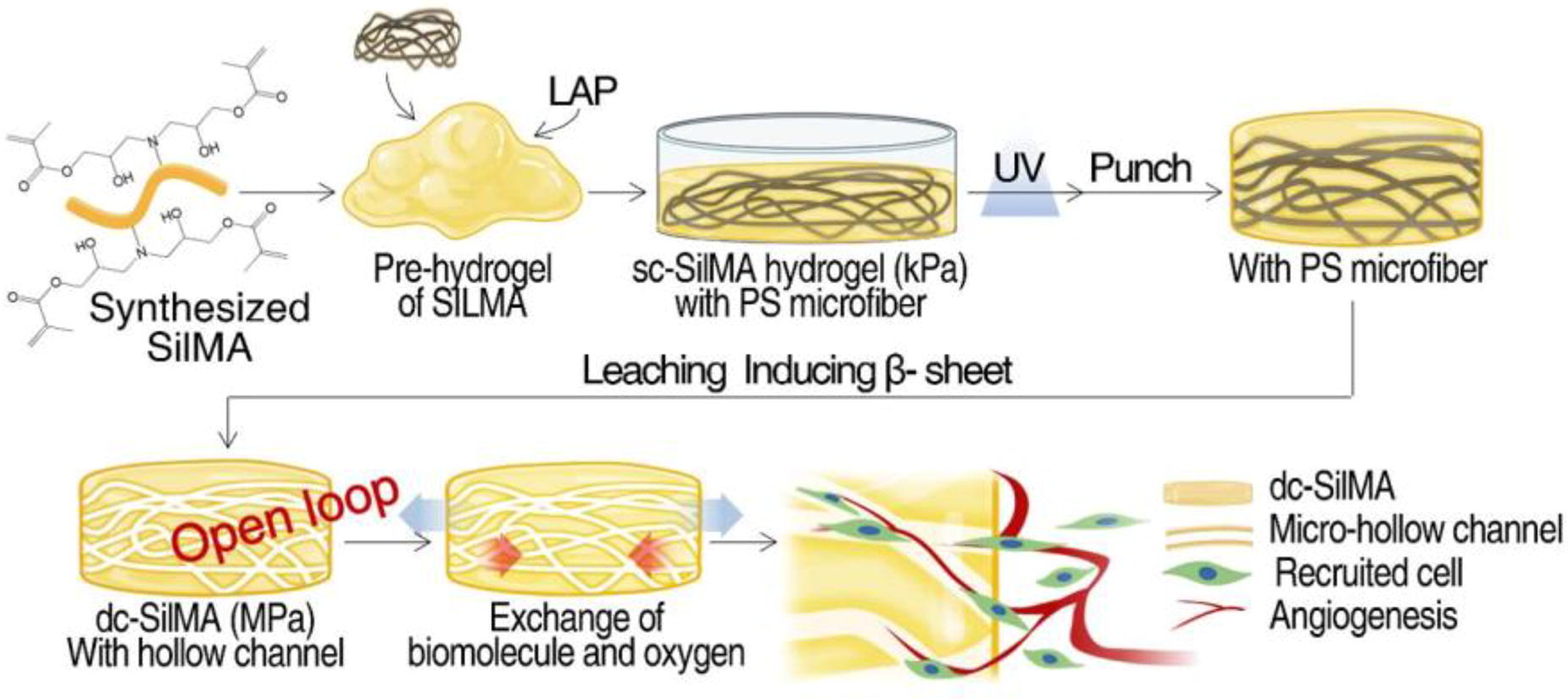
B306 - Blood Outgrowth Endothelial Cells Produce A Healing Response On Novel Ferromagnetic Flow Diverting Stents
A. M. Zielonka1, A. Thome1, A. A. Oliver2, R. Kadirvel2, D. F. Kallmes2, B. J. Tefft1
1
Medical College of Wisconsin, Milwaukee, WI,
2
Mayo Clinic, Rochester, MN
*Purpose/Objectives: Flow diverter implantation is an increasingly popular approach for the endovascular treatment of intracranial aneurysm, a life-threatening condition that affects roughly 1 in 50 people in the United States. When flow diverters are introduced to the site of an aneurysm, endothelial cells must adhere and contribute to the healing response that will generate neotissue to cover the aneurysm neck and provide successful long term aneurysm occlusion. Furthermore, current lack of rapid endothelialization on flow diverting stents necessitates antiplatelet therapy following aneurysm treatment with flow diverting stents. This limits the clinical utility of flow diverters, creating a need to balance bleeding risk against device-related thrombosis risk. Our novel strategy to enhance endothelialization is to use a ferromagnetic flow diverter that can capture magnetically labeled cells. The aim of this study is to characterize the survival and behavior of blood outgrowth endothelial cells (BOECs) in vitro after being seeded onto a novel ferromagnetic flow diverter designed for the in situ capture and retention of patient-specific BOECs after stent placement.
*Methodology: Peripheral blood mononuclear cells were isolated from rabbit blood and cultured in endothelial cell medium until the appearance of BOEC colonies. BOEC phenotype was confirmed using tube formation assay and immunofluorescence staining for endothelial markers CD31 and CD34. BOECs were rotationally seeded onto flow diverters and cultured statically inside wells of a 96 well plate for up to 36 days. Hoechst staining, TUNEL staining, and Live/Dead (calcein-AM and ethidium homodimer-1) staining were used to assess cell proliferation, apoptosis, and viability, respectively. Immunofluorescence staining with antibodies against collagen IV, laminin, and fibronectin was applied to determine extracellular matrix (ECM) deposition by BOECs. Flow diverters were cut longitudinally and imaged en face using a fluorescence microscope.
*Results: BOECs seeded onto the flow diverting stent showed no statistically significant difference in percentage of apoptotic cells compared to control cells grown on culture dishes after 6 days of culture (0.45% ± 0.33% and 0.46% ± 0.09%, p = 0.98). After two weeks of culture, virtually all cells were viable in culture dishes and on the device, and notable proliferation was present in both groups. Immunofluorescence revealed collagen IV, laminin, and fibronectin deposition by BOECs seeded onto the flow diverting stent present at day 7 of culture and increasing through day 36. Importantly, cells and ECM were found both on and between the wires of the device. This indicates the cells can synthesize ECM in a manner that would seal the flow diverting stent and cover the neck of an aneurysm.
*Conclusion/Significance: Cytocompatibility of the novel ferromagnetic flow diverting stent and deposition of ECM proteins by cells on the device demonstrate the potential for this treatment to properly heal the aneurysm site, preventing thrombosis and aneurysm rupture.
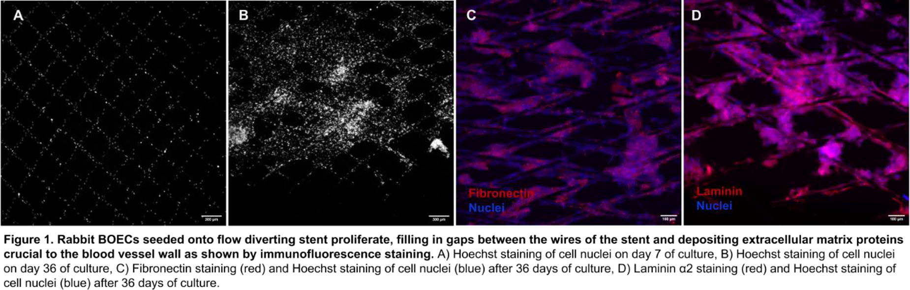
B307 - Study On The Post-anti-adhesion Of The 8-arm PEGNHS-modified Porcine Pericardium
S. Say, M. Suzuki, Y. Hashimoto, T. Kimura, A. Kishida
Material-Based Medical Engineering, Institute of Biomaterials and Bioengineering, Chiyoda city, JAPAN.
*Purpose/Objectives: After liver resection, the formation of post-adhesions poses complications for both patients and surgeons in subsequent operations, leading to prolonged procedures and increased blood loss. The development of an anti-adhesion barrier capable of preventing or easily separating from the liver is necessary. Our team found a potential 8-arm PEGNHS (hexaglycerol octa(succinimidyloxyglutaryl) polyoxyethylene) functionalized decellularized porcine pericardium for this purpose due to selectable size and sutureable membrane, low inflammation, and significant repelling fibroblast cells adherence. Here, we evaluated the post-anti-adhesion ability of our membrane for preventing the post-liver adhesion of rats and compared it with the commercial product Seprafim. The optimum modification molar ratio was also investigated to achieve the most suitable membrane.
*Methodology: The porcine pericardium was decellularized using the high hydrostatic pressure (HHP) method and modified with 8-arm PEGNHS. Samples were designated as 8P2, 8P5, and 8P10: [8: 8-arm PEG] [P: porcine] [2,5,10: molar ratio of NH2 groups to NHS in collagen]. The PEG modification was confirmed using ATR-FTIR and anti-PEG antibodies. In vitro, anti-adhesion was assessed with NIH3T3 fibroblast cells for 7 days. The dPPC, Seprafilm, 8P2, 8P5, and 8P10 were implanted in SD rat liver wounds created by brushing with sandpaper. Without implantation, it served as the control. Rats were sacrificed after 2 weeks, and anti-adhesion efficacy was evaluated through adhesion scores and histological analysis.
*Results: After the modification process, the 8-arm PEGNHS was fully modified to the dPPC evidenced by the brown color covering its entire area. The ATR-FTIR peak intensity of the modified samples at the C-O-C bond (1080 cm−1) increased. On day 1, NIH3T3 cells adhered well to the dPPC and continued to increase, covering the surface by day 7. Minimal cell adhesion on the surface of all 8-PEG-modified samples was observed on day 1, and no signs of cell proliferation were noted until day 7. This result indicates that the similar cells’ repelling surface was maintained even though the modification molar ratio was increased. The adhesion score of 8P5 (0.5/3) was significantly lower than that of the control (2.5/3) and Seprafilm (2/3). Adhesion scores were rated as follows: 0 for no adhesion, 1 for easy separation without blood, 2 for strong and difficult separation, and 3 for inability to separate. The observed gap between 8P5 and the liver confirms the absence of adhesion formation, indicating easy separation and a low adhesion score. Cell infiltration was also observed indicating regenerability of the membrane.
*Conclusion/Significance: Post-adhesion formation of the liver was prevented by using our modified membrane. The easily separable membrane could be beneficial for subsequent liver resection operations. It is expected to be a part of the body membrane once it is transplanted for the long term. Larger animals are required to produce the repeatable procedure in the clinical application.
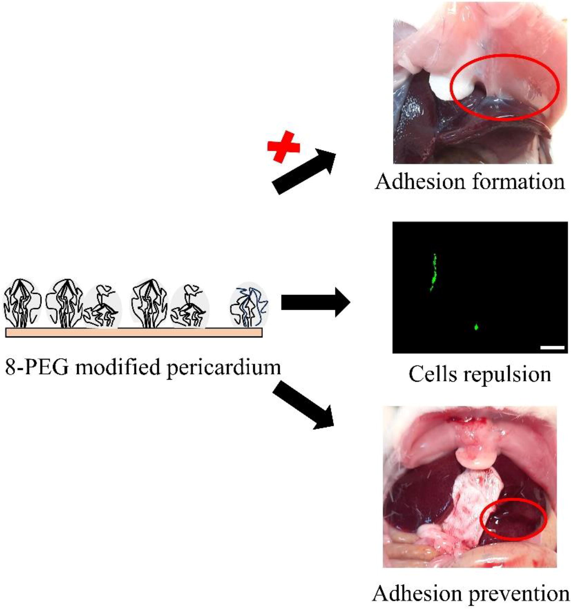
B308 - Development Of A Biodegradable Scaffold Bearing L-phenylalanine For Selective Capturing Of Cancer Cells
R. Itani, R. Mitsuyasu, D.-H. Kang, K. Homma, M. Matsusaki
Osaka university, Suita, Japan
*Purpose/Objectives: The recurrence of cancer is an issue in cancer treatment, and the elimination of cancer cells remaining in the body is desired. Here, we report the development of a novel polymeric scaffold that selectively captures breast cancer cells. The cancer cell-capturing polymer was synthesized by grafting L-phenylalanine onto a biocompatible poly(vinyl alcohol) (PVA-Phe). L-phenylalanine exhibits high affinity to L-type amino acid transporter 1 (LAT1), which is overexpressed on various cancer cells. We evaluated cancer cell-selective adhesion ability of PVA-Phe using MCF-7 (Michigan cancer foundation-7), a human hormone receptor-positive breast cancer model cell expressing LAT1.
*Methodology: Flow cytometry measurements and confocal laser microscopy were performed to confirm LAT1 expression in MCF-7. In order to evaluate the adhesive property of the PVA-Phe to the cancer cells, we fabricated PVA-Phe and PVA films and seeded the cells on top. The films were observed using phase contrast microscopy and scanning electron microscopy (SEM). The adherent cells were observed with a phase contrast microscope, and the number of adherent cells was quantified after harvesting the adherent cells with trypsin.
*Results: From the results obtained by flow cytometry measurement, the proportion of cells with LAT1 fluorescence greater than the maximum IgG1 fluorescence was calculated as LAT1 positive. The percentage of LAT1-expressing cells was 7.1% in normal cells (human mammary epithelial cells; HMEC) and 66.0% in MCF-7. This result confirmed the high expression of LAT1 on the surface of breast cancer cells. From the SEM image of PVA-Phe film, micro-sized grains of various shapes and sizes were distributed in a circle. This observation suggested that the film was not formed by a uniform layer of PVA-Phe grains. The phase contrast microscopic images of MCF-7 and HMECs showed almost no cell adhesion on the PVA film. On the other hand, cell adhesion was observed on the PVA-Phe polymer-coated area only when MCF-7 was seeded. The number of trypsin-treated viable cells supported the image observation results: no MCF-7 cells or HMECs were collected from the PVA-coated samples. In contrast, 11,720 adherent cells were recovered from the PVA-Phe-coated samples (Figure 1). These results indicated that the PVA-Phe may serve to selectively capture cancer cells if the cancer cells are present in the vicinity of the PVA-Phe.
*Conclusion/Significance: In this study, we have reported the selective cancer cell-capturing ability of PVA-Phe, a biocompatible PVA-based polymer grafted with L-phenylalanine. The grafted Phe moiety on PVA-Phe acted as a cancer cell recognition site and interacted with LAT1, a transmembrane protein overexpressed on cancer cells. On the PVA-Phe coated surface, cell adhesion was only observed in the case of the human breast cancer cells (MCF-7), not in the case of non-cancerous human mammary epithelial cells (HMEC). This cancer cell-capturing molecule can potentially contribute to the problem of cancer metastasis in cancer therapy.

B309 - Construction Of Tough Hydrogel Crosslinked Via Ionic Interaction By Protection Effect Of Hydrophobic Domains
S. Bunuasunthon, M. Nakamoto, M. Matsusaki
Osaka University, Osaka, Japan
*Purpose/Objectives: In the field of tissue engineering, hydrogels are well-known for their ability to mimic the extracellular matrix (ECM), referring to their great water content and biocompatibility. However, most hydrogels are struggle with poor mechanical properties. These limitations hinder their application, especially in tissue engineering field requiring durable materials. While various approaches have been introduced to fabricate tough hydrogels, many of them involve the conjugation of non-biodegradable polymers, raising concerns about potential toxicity. Therefore, the development of hydrogels that not only demonstrate great mechanical strength but also consist of biocompatible materials is needed. In this study, we introduced the concept of conjugating hydrophobic groups onto an ionic biodegradable polymer to construct a tough biodegradable hydrogel. Schellman postulated in the 1950s that when aqueous solvated ions approached a hydrophobic surface, interactions between ions would be enhanced. The theory was recently experimentally verified, but to the best of our knowledge, there are no reports on the use of this concept for hydrogel formation. Herein, we introduce the synthesis of biocompatible polymer, poly(γ-glutamic acid) (γ-PGA) together with the conjugation of ionic crosslinked moieties alendronic acid (Aln) and hydrophobic moieties L-phenylalanine (Phe) for construction of biodegradable tough hydrogel.
*Methodology: The synthesis of γ-PGA-Aln-Phe was achieved through an amide coupling reaction, providing a controllable grafting degree for both Aln and Phe. The impact of the hydrophobic domain on the hydrodynamic diameter of the polymer, crosslinking time, and mechanical properties of the constructing hydrogel were investigated.
*Results: Time sweeps rheological analysis of the polymer solution crosslinking with calcium solution indicated that the presence of the hydrophobic domain accelerates the gelation time. Compression testing of the constructed γ-PGA-Aln-Phe hydrogel revealed six times higher in compressive strength compared to the γ-PGA-Aln hydrogel, emphasizing the significant importance of the hydrophobic domain in enhancing the toughness of the gel. Furthermore, stability testing of the constructed gel under EDTA solution demonstrated that the γ-PGA-Aln-Phe gel could maintain the crosslinked site of the gel for over a month. In contrast, without the hydrophobic domain, γ-PGA-Aln showed an unstable gel that dissolved within 10 minutes after immersion. This highlights the crucial role of the hydrophobic domain in protecting the ionic crosslinked site. Interestingly, the investigation of calcium content within the constructed gel using the complexometric titration method showed a lower amount of calcium in the γ-PGA-Aln-Phe gel compared to the γ-PGA-Aln gel. This underscores the dominant role of the hydrophobic domain in protecting and enhancing the toughness of the constructed gel, even with a lower amount of calcium ions within the gel.
*Conclusion/Significance: Based on these distinct findings, it can be concluded that the presence of a hydrophobic domain accelerates the gelation time of the calcium crosslinking site, enhances the toughness of the constructed gel, and protects the ionic crosslinked site to form a stable gel under aqueous solutions. Given the biocompatibility of all functional groups in this synthesized polymer, it is anticipated that this system will be valuable in the field of tissue engineering.
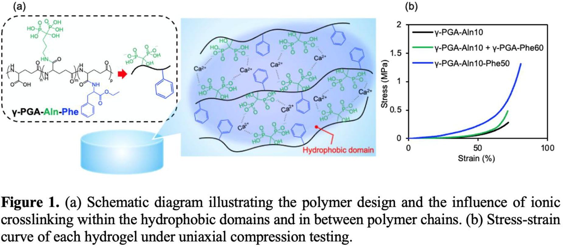
B310 - 3D-Printed Porous Implants Promote Healing And Modulate Foreign Body Reaction
G. Chen, L. Chen, B. Ratner
University of Washington, Seattle, WA
*Purpose/Objectives: The long-term performance of implantable medical devices (e.g., transcutaneous devices and implantable biosensors) is limited by the body’s foreign body reaction (FBR). The body reacts to foreign materials in an inflammatory manner and ultimately walls them off with a dense, avascular scar layer. Previous studies have discovered that materials with uniform interconnected 40 μm (about half the diameter of a human hair) spherical pores reduced fibrosis and seamlessly healed with the body in a vascularized fashion. We hypothesized that the 40um pore sizes of materials determine the tissue healing outcomes. In this study, with commercialized resin materials, we have used a time-saving and cost-effective method to 3D-print uniformed and interconnected 40um cubical implants.
*Methodology: The in situ implant-tissue interactions were compared with the traditional 40um spheric porous and non-porous materials in subcutaneous mouse tissue. Tissue staining techniques including H&E, Masson Trichrome, and Immunohistochemistry will provide invaluable insight into foreign body capsule formation, vascularization, and tissue integration of these novel implants.
*Results: The histology work will be analyzed in the next few months.
*Conclusion/Significance: The study evaluates the foreign body reaction and tissue integration between different shapes of uniform 40um porous materials.
B311 - Prevention Of Postoperative Peritoneal Adhesion By Hepatocyte Growth Factor Dna Aptamer In Rat Cecum Abrasion And Abdominal Sidewall Defect Model
Y. Dai,1, N. Inagaki,1, R. Ueki, S. Sando,1, K. Hasegawa, T. Ito,1,1
The University of Tokyo, Tokyo, Japan
*Purpose/Objectives: Postoperative peritoneal adhesions (PPAs) present a persistent challenge in various types of abdominal surgeries, and up to 90% of patients develop adhesions following open abdominal surgical intervention. Over time, these adhesions cause serious complications, including small bowel obstruction, organ dysfunction, and infertility. Adhesiolysis, the primary treatment involving surgical intervention, is associated with a significant risk of recurrent peritoneal adhesions, causing additional stress and financial burdens to the patients. Hepatocyte growth factor (HGF) is recently reported for its ability to prevent adhesion formation through various mechanisms, including the promotion of fibrinolytic activity and the inhibition of fibrosis. In this study, we focused on the HGF aptamer—a synthetic oligonucleotide designed to bind with c-Met, the HGF receptor specifically. The HGF aptamer, known for its enhanced thermal stability, and versatility, emerged as a promising candidate for clinical application. In this study, we assessed the effectiveness of the HGF aptamer in both in vitro and in vivo experiments and examined its impact on mesothelial mesenchymal transition (MMT) in the development of PPAs.
*Methodology: To examine the functions of HGF aptamer in inhibition of fibrosis, the elevation of fibrinolysis, and the stimulation of wound healing, HGF aptamers were applied to the mesothelial cell line, MeT5A. A scratch assay was performed to evaluate the cell motility. The degree of fibrosis was quantified with Sirius red staining and immunohistochemistry. RT-PCR was conducted to examine the adhesion-related gene expressions. Additionally, HGF aptamer was administered to the rat postoperative peritoneal adhesion-induced model. Two models, including the partial hepatectomy and cecum abrasion models, were performed to confirm the biosafety, adaptability, and comprehensive anti-adhesion efficacy of HGF aptamer in simulated clinical settings.
*Results: HGF aptamer effectively promoted wound healing, raised fibrinolytic activity by regulating the expression of plasminogen activator and its inhibitor and prevented fibrosis by suppressing mesothelial mesenchymal transition and upregulating collagen degradation. In the rat cecum abrasion and abdominal sidewall defect model, HGF aptamer significantly mitigated the severity of postoperative peritoneal adhesions and successfully prevented adhesion formation. Results from both in vitro and in vivo studies collectively suggest that the HGF aptamer is promising in preventing PPA formation, indicating its potential in future clinical applications.
*Conclusion/Significance: The effectiveness of HGF aptamer in preventing PPAs was supported by its ability to regulate multiple pathological cascades. The results from in vivo studies also suggest that HGF aptamer has the potential for clinical application. However, further investigation into optimal dosing and pharmacokinetics is necessary to improve its clinical efficacy.
B312 - Developing New 3D-hydrogel Models Of The Human Mammary Gland To Investigate Breast Cancer Initiation
M. Green, A. Saiani, O. Tsigkou
University of Manchester, Manchester, United Kingdom
*Purpose/Objectives: The ability to culture and analyse cells within a 3D environment that is more representative of the in vivo environment is at the forefront of current research. However, the gold standard for 3D matrices are naturally derived such as collagen and Matrigel® which have significant limitations such as batch-batch variability, poor chemical definition and xenogeneic contaminates. This has led to a shift in focus away from these naturally derived matrices to synthetic ones. Self-assembling peptides or SAPHs are gaining popularity due to their ease of use, biocompatibility, chemical definition, and tuneable properties. I have been using the SAPH hydrogels; PeptiGels® developed by Saiani and colleagues with the aim of developing a synthetic model of the functional unit of mammary gland, breast acini to investigate effect of matrix stiffness on breast cancer initiation.
*Methodology: Several PeptiGels® developed by Saini and colleagues, which vary in their charge and stiffness, where characterised after 24-hour media conditioning through Oscillatory Shear Rheology. The viability of the mouse mammary gland epithelial cells, EPH4 was assessed for a period of 14 days following encapsulation into the PeptiGels® using Live/Dead and PicoGreen assays. Characterisation of the organoid structures formed within the hydrogels was assessed using a number of techniques including brightfield microscopy, immunofluorescence staining and Haematoxylin & Eosin Staining.
*Results: Comparison of the PeptiGels® against the gold standard Matrigel® determined that the PeptiGels® are significantly stiffer, with PeptiGel® Alpha2 being more representative of the tumour environment whilst Alpha7 and EE being more representative of the stiffness of healthy breast tissue. Assessment of viability suggests that the PeptiGels 3D environment alone without any functionalisation can support EPH4 cells formation of spheroidal structures like Matrigel® however assessment of area, circularity, diameter, and number of cells per organoid indicate that these spheroidal structures are more representative of the malignant phenotype rather than biologically relevant acini.
*Conclusion/Significance: All PeptiGels® tested were able to maintain the viability of EPH4 cells for 14 days. The EPH4 cells formed clusters with characteristics more reflective of malignant acini indicating the PeptiGels® are a suitable platform for breast epithelial cell culture and have potential for the development of biologically relevant acini.

B313 - Alternative Strategies For The Recombinant Synthesis, DOPA Modification And Analysis Of Mussel Foot Proteins - A Case Study For Mfp-3 Of Two Mytilus Species
C. Zwies1, A. M. Vargas Rodriguez1, M. Naumann2, A. Hivechi1, F. Seifert1, K. Mäder1, M. Pietzsch1
1
Martin-Luther-University Halle-Wittenberg, Halle (Saale), Germany,
2
Fraunhofer Institute for Cell Therapy and Immunology Department of Drug Design and Target Validation, Halle (Saale), Germany
*Purpose/Objectives: Mussel foot proteins (Mfps) possess unique binding properties to various surfaces due to the presence of L-3,4-dihydroxyphenylalanine (DOPA) [Waite, 2017]. Mytilus foot protein-3 (Mfp-3) is one of several proteins in the adhesive plaque. Its localization at the plaque-substrate interface proved that Mfp-3 plays a key role in adhesion [Silverman & Roberto, 2007]. Therefore, the DOPA-modified protein is suitable for the development of innovative bio-based binders. Commonly, mushroom tyrosinase is used to modify peptide-bound DOPA in Mfp-biopolymers. However, it is limited and results in hydroxylation of 15 - 40 % only [Cha et al., 2009]. Promising alternatives are microbial tyrosinases (mTyr) [Zaidi et al., 2014]. The respective enzyme from V. spinosum was shown to be highly active and is also capable to hydroxylate peptide-bound tyrosines in Mfps in vivo [Axambayeva et al., 2018]. In our study we aimed to address two aspects. On the one hand we wanted to investigate the in vitro modification of Mfp-3 with recombinantly produced His6-mTyr from V. spinosum for the first time. Furthermore, we wanted to overcome solubility limitations of previous recombinant production strategies of Mfps.
*Methodology: We designed Mfp-3s from the genus M. edulis and M. galloprovincialis as SUMO-fusion proteins in order to increase the solubility. His6-SUMO-Mfp-3s were produced in 7.5 L-scale fed-batch bioreactor in TB medium and purified chromatographically via IMAC. His6-SUMO-Mfp-3s were further processed using SUMO-protease to gain tag-free Mfp-3s.
Microbial tyrosinase (His6-mTyr) was produced in E. coli KRX and used for the in vitro hydroxylation of the Mfp-3s. MALDI-MS was used to confirm the presence of DOPA. QCM-D analysis as well as tensile shear tests were performed to test the protein adhesiveness.
*Results: We obtained 55 mg purified Mefp-3 and 100 mg purified Mgfp-3 per liter E. coli culture. Furthermore, we used His6-mTyr to modify Mfp-3 in vitro. MALDI-TOF-MS confirmed that up to twelve hydroxyl groups were incorporated. Considering the fact that our Mfp-3s contain 10 tyrosine residues this finding suggests that not only DOPA but also 3,4,5-trihydroxyphenylalanine (TOPA) was formed. To our knowledge this is the first time that the presence of TOPA within Mfp-3 was shown after in vitro modification by His6-mTyr from V. spinosum.
The adsorption analysis confirmed comparable adhesive properties to SiO2-surfaces like MAPTrixTM. In tensile shear strength tests modified Mefp-3 resulted in a tensile shear strength of 1.68 MPa, which was comparable to the control with a commercial glue.
*Conclusion/Significance: Our protocol for obtaining recombinant Mfp-3 only from the soluble protein fraction of E. coli is robust, reproducible and demonstrates the possibility of economical mass production. A powerful MALDI-MS analysis approach allowed a deeper insight in the modification pattern of the peptide-bound tyrosines within Mfp-3s.
Finally, the highly hydroxylated Mfp-3s exhibited excellent bulk adhesive properties. Mfp-3 and Mfp-3-based polymers modified with His6-mTyr have the potential to become a practical bioadhesive for material science as well as for medical and biotechnological applications for future biomaterials. In first trials modified Mefp-3 was already used for the functionalization of electrospun nanofibers that can be utilized for advanced biomedical applications.

B314 - Extensive Survey Of Antibody Invariant Positions For Efficient Chemical Conjugation Using Expanded Genetic Codes
A. Kato
Kyowa-Kirin, Co.Ltd, Tokyo, Japan
*Purpose/Objectives: Modification of biomaterials with antibodies has widespread applications in many different fields, such as bioactive biomaterials. The site-specific chemical conjugation of antibodies, following synthesis with an expanded genetic code, promises to advance antibody biomaterial conjugates. The incorporation of synthetic designer amino acids into antibodies not only guarantees site specificity but also allows the use of bio-orthogonal chemistry, which achieve high stability and orientation control of immobilized antibodies. However, the efficiency of amino acid incorporation fluctuates significantly among different sites, thereby hampering the identification of useful conjugation sites.
*Methodology: In this study, we applied the codon reassignment technology, E. coli cells in which UAG stop-codon reassigned to designer amino acid, to synthesize of chemically functionalized antibodies containing Nε-(o-azidobenzyloxycarbonyl)-l-lysine (o-Az-Z-Lys) at defined positions in antigen-binding fragment (Fab) constant domains. We then examined o-Az-Z-Lys reactivity in these variants during strain-promoted alkyne-azide cycloaddition (SPAAC).
*Results: 62 positions in the Fab constant domains were analyzed. MS analyses showed that the masses of the variants were equal to those expected from an amino acid replacement by o-Az-Z-Lys at the chosen positions. ELISA showed that the incorporation of o-Az-Z-Lys at any of the chosen sites did not impair the antigen binding activity. The most of the examined positions exhibited highly efficient conjugation ranging from 80% to 90%. Furthermore, some of the Fabs linked via linkers exhibited agonistic activity in cultured cells as opposed to the antagonistic nature of antibodies.
*Conclusion/Significance: The incorporation of o-Az-Z-Lys into Fab using UAG-reassigned E. coli cells greatly facilitated the screening for antibody positions available for chemical conjugation. Experiments utilizing the representative positions demonstrated the utility of the newly identified positions and opened ways to regulate function by the control of the conjugated Fab molecules orientation. These results showed that our approach greatly enhanced the availability of antibodies for chemical conjugation and might aid in the development of new antibody-conjugated bioactive biomaterials.

B315
B318 - Regenerative Role Of Supercritical Carbon Dioxide Derived Collagen Acellular Dermal Matrix On Hair Follicle In Hair Regrowth
Y.-J. Chen, S. Periasamy, D.-J. Hsieh
ACRO Biomedical Co., Ltd., Kaohsiung, Taiwan
*Purpose/Objectives: Hair follicles are principally situated at the head of the scalp, affected by various factors, such as ultraviolet light exposure and temperature. Alopecia areata is a typical hair follicle disease, manifested by hair loss, which primarily affects mental health and quality of life. We plan to develop a collagen acellular dermal matrix (ADM) using supercritical carbon dioxide (SCCO2) extraction technology. Therefore, we hypothesized the hair follicle growth investigation of collagen ADM scaffold in mice.
*Methodology: In the present study, C57Bl6 7-week-old male mice were grouped into four groups as follows: Sham, platelet-rich plasma (PRP), collagen ADM scaffold, and collagen ADM scaffold with PRP. Collagen ADM scaffold injections were done with and without PRP.
*Results: Hair growth was significantly increased in the collagen ADM scaffold with PRP compared to all other experimental groups at different time points. Hair follicle numbers significantly increased as evaluated by H&E staining of both longitudinal and horizontal tissue sections in collagen ADM scaffold relative to all other experimental groups at different time points of the experimental period. Collagen ADM scaffold with PRP was found to modulate the expression of CD34, Wnt, Lef-1, SOX-9, β-catenin and K17 in the hair follicle growth cycle, thereby increasing the hair follicle and hair density.
*Conclusion/Significance: The SCCO2-derived collagen ADM increased hair follicle and hair density and might be therapy for alopecia, without any adverse effects.
B319 - Injectable In Situ Crosslinking Hydrogel For Autologous Fat Grafting
P. Apelgren1, K. Oskarsdotter2, C. T. Nordgard3, K. Säljö1, A. A. Solbu3, E. Eliasson4, S. Sämfors4, H. E. Saetrang5, L. E. Asdahl5, E. M. Thompson6, C. M. Troedsson6, S. Simonsson7, B. L. Strand3, P. Gatenholm8, L. Kölby1
1
Institution of Clinical Science, Gothenburg, Sweden,
2
Institution of Clinical Science, Göteborg, Sweden,
3
NOBIPOL, Trondheim, Norway,
4
Chalmers University of Technology, Gothenburg, Sweden,
5
DuPont Nutrition Norge AS d/b/a NovaMatrix, Billingstad, Norway,
6
Ocean TuniCell AS, Blomsterdalen, Norway,
7
Institution of Biomedicine, Gothenburg, Sweden,
8
CELLHEAL AS, Sandvika, Norway
*Purpose/Objectives: Autologous fat grafting is hampered by unpredictable outcomes due to high tissue resorption. Hydrogels based on enzymatically pretreated tunicate nanocellulose (ETC) and alginate (ALG) are biocompatible, safe, and present physiochemical properties capable of promoting cell survival.
*Methodology: Here, we compared in situ and ex situ crosslinking of ETC/ALG hydrogels combined with lipoaspirate human adipose tissue (LAT) to generate an injectable formulation capable of retaining dimensional stability in vivo. We performed in situ crosslinking using two different approaches; inducing Ca2+ release from CaCO3 microparticles (CMPs) and reliance on physiologically available Ca2+ in vivo. Additionally, we generated ex situ-crosslinked, 3D-bioprinted hydrogel-fat grafts.
*Results: We found that in vitro optimization generated a CMP-crosslinking system with comparable stiffness to ex situ-crosslinked gels. Comparison of outcomes following in vivo injection of each respective crosslinked hydrogel revealed that after 30 days, in situ crosslinking generated fat grafts with less shape retention than 3D-bioprinted constructs that had undergone ex situ crosslinking prior to grafting. However, CMP addition improved fat-cell distribution and cell survival relative to grafts dependent on physiological Ca2+ alone.
*Conclusion/Significance: These findings suggested that in situ crosslinking using CMP might promote the dimensional stability of injectable fat-hydrogel grafts, although 3D bioprinting with ex situ crosslinking more effectively ensured proper shape stability in vivo.
B320 - Tailoring Hydrogel Mechanics And Architecture For Effective Vocal Fold Regeneration
S. Nejati1,2, L. Mongeau1,2
1
McGill University, Montreal, QC, Canada,
2
Centre for Interdisciplinary Research in Music Media and Technology, Montreal, QC, Canada
*Purpose/Objectives: Vocal folds play a crucial role in voice production, phonation, and singing. Pathological conditions can impair their function, leading to voice disorders. Tissue engineering offers promising repair and regeneration strategies, aiming to develop scaffolds that mimic the native vocal fold tissue microenvironment. Tissue regeneration is intricately influenced by the dynamic interplay between the physical attributes of tissue engineering scaffolds and the resulting biological responses. Hence, the successful regeneration of vocal fold tissue necessitates a comprehensive understanding of the effects of substrate properties on cellular behavior. This study focuses on understanding how scaffold mechanics and architecture influence the immunological and reparative activities of vocal fold tissues.
*Methodology: We engineered a tunable macroporous hydrogel system using Gelatin Methacryloyl (GelMA) and Polyethylene Glycol Diacrylate (PEGDA), with Polyethylene Glycol (PEG) as a porogen. By varying PEGDA molecular weights, hydrogels with different mechanical and architectural properties were created. Mechanical characteristics were assessed via tensile strength tests and rheometry, while morphology was analyzed using Scanning Electron Microscopy (SEM) and confocal microscopy. A series of biological assays was conducted: hVFFs morphology, differentiation, and collagen synthesis were evaluated through immunostaining; fibroblast proliferation was studied using the WST-1 assay; cell migration was assessed via the Boyden chamber assay; and macrophage polarization and secretion profiles were investigated using immunostaining and ELISA.
*Results: The results revealed that increasing the molecular weight of PEGDA from 700 Da to 10,000 Da resulted in decreased hydrogel stiffness, from 62.6 kPa to 8.8 kPa, and increased pore dimensions from approximately 64.9 µm to 137.4 µm. Biological evaluations demonstrated that hydrogels with higher stiffness promoted fibroblast proliferation and spreading, albeit with an increased propensity for fibrosis, owing to a surge in myofibroblast differentiation and collagen synthesis. In contrast, hydrogels with greater molecular weights had a softer matrix with expanded pores, enhancing cellular migration and promoting an M2 macrophage phenotype conducive to tissue healing. The findings support the P6000 hydrogels as prime candidates for vocal fold repair and show that the macroporous hydrogels can be tuned to serve in various tissue engineering applications.
*Conclusion/Significance: Understanding the interplay between the physical properties of hydrogels and the immune and reparative activities of vocal folds is paramount in designing scaffolds that not only support but also actively contribute to vocal fold tissue regeneration. This study has elucidated such relationships using macroporous GelMA/PEGDA hydrogels. This research underscores the importance of fine-tuning hydrogel mechanics and porosity to tailor scaffolds specifically for vocal fold regeneration. By focusing on the unique requirements of vocal fold tissue, such as the need for optimal mechanical support and favorable cellular interactions, GelMA/PEGDA hydrogels can be effectively adapted to meet the specific challenges of regenerating this specialized tissue.
B322 - Effects Of Hydrogel Cranioplasty On Motor Recovery
J. Deng1, S. Barbay2, J. Townsend3, B. Andrews4, R. Nudo2, M. Detamore3
1
University of Kansas, Lawrence, KS,
2
University of Kansas Medical Center, Kansas City, KS,
3
University of Oklahoma, Norman, OK,
4
University of Iowa, Iowa City, IA
*Purpose/Objectives: Decompressive craniectomy (DC) is performed to decrease intracranial pressure after severe traumatic brain injury (TBI). This two-step surgical procedure involves removing a large portion of the cranial bone and replacing the cranial bone once intracranial pressure has decreased. The time between removing and replacing the skull gives rise to a condition known as “syndrome of the trephined” that is marked by cognitive and behavioral deficits that can be reversed upon replacement of the removed portion. This project aims to reduce the surgical steps of this intervention using pentanoate functionalized hyaluronic acid (PHA) hydrogel that has osteogenic properties while being elastic to allow time for resolution of intracranial pressure before it hardens. Our group will address the effects of several modifications of PHA to improve motor recovery from a TBI.
*Methodology: Long Evans rats (male, 10-12 weeks old, 350-415g) were trained on a reach task requiring rats to reach and grasp small food pellets placed outside of a Plexiglas behavioral box with the preferred forelimb. Food pellets were singularly presented for > 60 trials per session. Once a rat reached > 60% success rate, a pre-injury baseline was established. A controlled cortical impact (CCI) procedure was then used to deliver a TBI to the rat forelimb motor cortex contralateral to the trained forelimb. A small 5mm diameter craniectomy was made to access the cortex in all groups. Immediately following the CCI injury a DC was performed. Rats were assigned randomly to one of three control groups or one of three experimental (hydrogel) groups. Control groups: 1) non-injured group with a unilateral DC and no cranial repair (noTBI/DC); 2) TBI group with a DC and no cranial repair (TBI/DC); 3) TBI group with only a 5mm craniectomy and acrylic repair to model a closed head injury (TBI closed). Experimental (hydrogel) groups: 1) PHA; 2) thiolated devitalized tendon (TDVT); 3) TDVT with encapsulated bone morphogenetic protein-2 (BMP- 2). Experimental groups were tested for their effect on motor recovery and bone regeneration compared to control groups. Reach performance was assessed once per week for 8 weeks after the CCI injury.
*Results: Our data show a significant motor deficit for each TBI group during the first week post-CCI. The noTBI/DC control group did not show a significant motor deficit over the 8-week assessment period. In the following weeks, compared to no TBI with DC control, the PHA and TDVT groups showed a persistent impairment on the reach task while the TDVT+BMP-2 group had less severe impairment than the other hydrogel groups over time.
*Conclusion/Significance: TDVT+BMP-2 reduced motor impairment the first week post-TBI, before any benefits of bone regeneration would be seen, and was able to facilitate recovery up to the 8 weeks post-TBI. Future directions include understanding BMP-2 effects on motor recovery as a neuroprotectant.
B323 - Development Of Nonvascular Biodegradable Stent Based On Polydioxanone/polycaprolactone Sheath/core Monofilaments
G. Kim1, J. Son1, J. Park1, S. Jang1, S. Park2, K. Kim2
1
Korea Textile Development Institute, Daegu, Korea, Republic of,
2
MI Tech Co., Ltd., Suwon, Korea, Republic of
*Purpose/Objectives: A stent is a structure used to expand and maintain a narrowed area into a space of a certain volume. Biodegradable stents are alternative to the existing metal stents because of easy to remove and a few side effects. In this study, we investigated the novel biodegradable stent the biodegradable monofilaments for non-vascular stents.
*Methodology: Addressing these challenges, we propose a PDO/PCL sheath/core monofilament structure, using melt-spinning for preparation. PDO, with its rapid biodegradability but simultaneously high bending stiffness, is used for the sheath component, while PCL, known for its biostability and flexibility, forms the core. We then fabricate stents from PDO/PCL sheath/core monofilaments and compare their morphological and mechanical characteristics with biological incubations to those of PDO and PCL monofilaments. This investigation reveals varying biostabilities and mechanical properties of these promising biodegradable materials and emphasizes a melt-spinning fabricated sheath/core structure that enhances the advantages of both materials.
*Results: PDO/PCL sheath/core monofilaments improved mechanical properties and stent stability. The retention period of stents has been improved. Inclusion of a PCL core layer has been verified to enhance the mechanical properties, as well as accelerate the decomposition rate and extend the decomposition period. Furthermore, the confirmation of radial force, a crucial factor in stent utilization, revealed a gradual decrease in radial force during biodegradation in comparison to a singular stent fabricated using PDO and PCL.
*Conclusion/Significance: Considering the mechanical properties and degradation behavior, the PDO/PCL sheath/core monofilament was found to be effectively maintained. However, PDO/PCL sheath/core stents tend to have a slightly greater decrease in radial force with disintegration time compared to PDO stents and PCL stents. Based on these results, the optimized biodegradable nonvascular stents will be determined through further research on the area ratio of the sheath and core.
*Acknowledgment: This work was supported by the Technology Innovation Program (20017645) funded by the Ministry of Trade, Industry & Energy(MOTIE, Korea).
B324 - Spatio-Temporal Modulation Of Cellular Behaviors With Shape Memory Polymers
K. Uto, M. Ebara
National Institute for Materials Science, Tsukuba, Japan
*Purpose/Objectives: The interdependent relationship between biomaterials and biology has led to the concept of “materiobiology,” a scientific research field that studies the effects of biomaterial properties on biological functions at the level of cells, tissues, organs, and whole organisms. A series of studies have been conducted to clarify how the physical properties of biomaterials, such as stiffness and topography, affect cell behaviors, including cell adhesion, elongation, proliferation, cell orientation, and the differentiation of stem cells. In most cases, the material properties are treated as static physical properties, and there is a significant gap with the dynamic physical environment in the body. In this presentation, we will discuss the development of biomaterials with shape memory ability that allow spatio-temporal manipulation of topography, and their potential as tools for novel materiobiology research.
*Methodology: Shape memory cell culture substrates based on poly(ε-caprolactone) (PCL) were prepared by photocrosslinking tetra-branched PCL with various degrees of polymerization functionalized with acrylate end-groups in the presence of the photoinitiator. Permanent surface topography was generated by photocrosslinking the PCL macromonomers on a mold. Temporary topography was later embossed into the photocrosslinked PCL above the melting temperature (Tm) and fixed by cooling below the crystallization temperature (Tc). Shape-memory transition from temporary to original permanent topography was triggered by heating again above the Tm. Light-actuated shape memory culture substrates with spatio-temporal manipulability were also prepared by hybridization of PCL and photothermal nanomaterials. NIH3T3 fibroblasts were used to evaluate cellular responses to dynamic and global or local topographic changes on the substrate surface.
*Results: Shape-memory PCL substrates demonstrated the important role of surface topograhy in time-dependent cytoskeleton remodeling under biological relevant conditions. The change in cell orientation induced by the surface topography transition was also revealed to occur in different modes by live imaging under the microscope. In addition, to eliminate the influence of temperature change on cell orientation, we have developed a novel shape memory culture substrate incorporating photothermal nanomaterials such as titanium nitride nanoparticles, and have succeeded in establishing a system that can change the surface topography by light irradiation without the need for temperature change. The developed light-actuated shape memory culture substrates can induce topography changes not only globally but also locally, which is expected to enable spatio-temporal control of cell orientation.
*Conclusion/Significance: We have developed a novel approach to observe time-dependent changes in cell alignment using biomaterials with shape memory ability. We demonstrated that temporary surface topography can be easily programmed on the substrate and can be used as a dynamic culture substrate that rapidly changes to a permanent surface topography in response to temperature change or light irradiation, even in the presence of cells. The versatility and biologically friendly nature of shape memory PCL could enable novel and diverse applications, especially in materiobiology.
B326 - New Topographical Material Inducing Axonal Growth For Corpus Callosum Management
J. Baptiste1,2,3, K. Uto1, A. Nabil1, G. Lambard1, M. Ebara1,2
1
National Institute for Materials Science, Tsukuba, Japan,
2
University of Tsukuba, Tsukuba, Japan,
3
Polytech Grenoble INP-UGA, Grenoble, France
*Purpose/Objectives: The corpus callosum, a neuronal bridge comprising white matter in the central nervous system, is crucial for transmitting nerve impulses and proper brain function. Unfortunately, malformations, particularly partial agenesis, frequently occur during prenatal development, resulting in the absence of one or two ends (splenium or/and rostrum) of the corpus callosum at birth. Such malformations can lead to severe handicaps. To address this issue, we focus on studying and developing a bioactive implant as a topographical material capable of inducing axonal growth in vitro. The final goal is to use this implant to bridge the missing segment of the corpus callosum in vivo. For the preliminary stages of implant development, we designed, characterised and optimised a biocompatible polymer, poly(dimethyl siloxane) (PDMS), to mimic a substitutional corpus callosum via active learning assisted by Bayesian optimisation.
*Methodology: In our study, the two parts of the PDMS kit (Sylgard 184) were mixed in different ratios ranging from 10:1 to 50:1; base:curing reagent. The PDMS elastomers were cured at 60oC overnight. Initially, an evaluation of the isolated material’s behavior, including cross-linking and rigidity in its bulk form, was conducted. Subsequently, an assessment of the polymer’s surface characteristics, such as appearance and adhesion, was performed. Using 3D printing and micro-printing, we aim to design the optimal topography conducive to the controlled development and differentiation of axons on the implant. The objective is to recapitulate the environment of the corpus callosum, considering the mechanical, chemical (by functionalizing fibronectin homogeneously, then under a concentration gradient, to the PDMS surface, with preliminary tests conducted on human mesenchymal stem cells (hMSCs) before testing on a PC12 cell line), and topographical properties of our material.
*Results: Commencing with an initial stiffness value of 1.47 MPa for PDMS with 10:1, we lowered it to 40kPa by decreasing the curing reagent ratio. Still, there was a noticeable change in the surface state of the sample. This change in the surface state makes it difficult to transfer a nanotopography on its surface. An important point of this study is to determine how the dimensions and geometry of the topography can be effectively adjusted to fit this modified PDMS while using Johnson-Kendall-Roberts (JKR) theory to assess the adhesion work of the PDMS. Additionally, noteworthy observations include the expansion of PDMS on the polystyrene substrate in the form of dendrites. We intend to explore the potential utility of this phenomenon within the neural environment. The fibronectin coating to a polystyrene substrate enables much better adhesion of cells such as hMSCs. Leveraging these combined material properties, our study aims to optimise the bio-integration of the implant in a 2D format.
*Conclusion/Significance: Our investigation focuses on the development of a 2D polydimethylsiloxane (PDMS) implant intended to promote axonal growth, with ongoing experimentation employing PC12 cells. Following the identification of the optimal 2D design, our research will progress to the exploration of a 3D implant, aiming to emulate the structural and morphological characteristics of the absent portion of the corpus callosum.
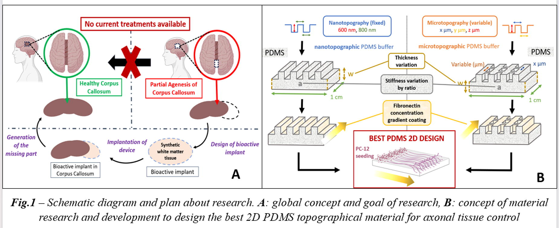
B327 - Identification Of Laminin-binding Oligopeptides In Guiding Cardiac Differentiation And Development
S. Casarella1, F. Ferla1, D. Di Francesco2, F. Boccafoschi1
1
Università del Piemonte Orientale, Novara, Italy,
2
Université Laval, Quebec, QC, Canada
*Purpose/Objectives: The extracellular matrix (ECM) plays a key role in tissue regeneration, in particular interacting with integrins and focal adhesions (FAs). In fact, FAs are complexes that link the cytoskeleton to the ECM, facilitating cell adhesion and guiding cell behavior related to the environmental changes which may modify the substrate characteristics. FAs link to actin stress fibers and include transmembrane integrin receptors, specifically integrins α and β heterodimers, binding the intracellular environment to the ECM. Activating pathways like Focal Adhesion Kinase (FAK) and mitogen-activated protein kinases (MAPKs), integrins induce hypertrophic and anabolic responses in cardiac myocytes. Despite partial knowledge of FAs, studies highlight the pathophysiological roles of integrins in cardiac cells, acting as major mediators between cardiomyocytes and the extracellular environment. In adult cardiac cells, one of the mainly expressed integrins is α7β1, binding specifically to laminin-1, -2, and -4; this integrin has a protective effect on cardiomyocytes and is reduced after a myocardial infarction (MI), indicating an important role in cardiac functionality. Human mesenchymal stromal cells (hMSCs) in cardiac tissue engineering have gained attention, responding to the microenvironment with biophysical forces regulating communication, migration, adhesion, and lineage commitment. Regardless of specific cues, cells interact primarily through FAs, translating biophysical signals via the cytoskeleton to impact gene and protein expression associated with specific cell lineages.
*Methodology: To evaluate the cardiomyogenic potential of the laminin-specific integrin α7β1 derived peptides, KKGSYNNIVVHV (A2G2), YAIFLNKGRLEV (A2G52), immortalized human mesenchymal stromal cells (hMSC 201) and neonatal mouse ventricular cardiomyocytes were used. The optimization of cardiac differentiation protocol was performed using biochemical and growth factors to induce the differentiation towards a cardiac-like phenotype of hMSC Y201. Moreover, isolation protocol for neonatal mouse ventricular cardiomyocytes was optimized using enzymatic tissue digestion with collagenase type II, followed by a discontinuous Percoll gradient centrifugation. Phalloidin and immunofluorescence staining were used to characterize cell adhesion using different concentrations (100 µg/ml and 50 µg/ml) of the peptides A2G2 and A2G52. For the evaluation of cardiomyogenic potential of the two peptides, morpholometrical analyses, rt-PCR and western blot analyses were performed.
*Results: Staining with phalloidin and vinculin revealed morphological changes with enhanced focal adhesion and stress-fibers formation compared to untreated cells, mainly evident at a concentration of 50 µg/ml for both peptides. In particular, results revealed an increased length and number of FA in primary cardiomyocytes, differentiated and undifferentiated Y201. Cardiac and adhesion markers analyses confirmed the role of peptides in cardiomyogenic differentiation, in which treated cells have higher expression of early and late cardiac markers.
*Conclusion/Significance: Cardiac growth and differentiation involve complex interactions among growth factors, proteins, receptors, and specific ligands. Here, we demonstrated how integrin binding peptides influence cell behaviour not only in primary cardiomyocytes but also in guiding cardiac differentiation of hMSC. The peptides derived from cellular matrices may also be used to functionalize matrices such as hydrogel and used for regenerative purposes.

B328
B329 - Altering The Blood Microenvironment With Biodegradable Metals To Reduce Thrombosis
D. Anderson1, H. Le1, H. Vu1, J. Johnson1, J. Aslan1, J. Goldman2, M. Hinds1
1
Oregon Health and Science University, Portland, OR,
2
Michigan Technological University, Houghton, MI
*Purpose/Objectives: Biodegradable metals have potential as cardiovascular stents; however, limited work has examined the blood microenvironment response of these metals, particularly in the absence of antiplatelet or anticoagulant therapies. Our primary objective was to quantify the extent and mechanisms of thrombus formation on pure biodegradable metals (Mg, Zn, Fe, and Mo) and commonly used stable clinical alloys (NiTi, CoCr, and stainless steel). The specific purpose was to quantify the blood microenvironment responses to biologically reactive metal materials through measurement of platelet responses, contact pathway initiation, and inflammation activation.
*Methodology: To accomplish the objective, the pure metal and alloy wires were tested with human plasma using in vitro assays of coagulation factor XII (FXII) activation and fibrin generation to identify mechanisms of thrombus initiation and propagation. Using a clinically-translatable ex vivo model of flowing blood in a non-human primate model, the wires were coiled and placed into an exteriorized arteriovenous shunt loop and exposed to non-anticoagulated blood controlled at 100mL/min for 1hr. Platelet and fibrin attachment were quantified on the wires. Biochemical analyses of boundary layer blood draws from downstream of the metals allowed for quantification of the blood microenvironment, including platelet activation and reactivity, as well as coagulation and inflammation markers. Additionally, metal ions from the blood plasma were quantified with inductively coupled plasma mass spectrometry (ICP-MS) to observe metal degradation. Thrombus formation on the wires was also assessed with scanning electron microscopy (SEM).
*Results: The in vitro activation of coagulation FXII by Mg was decreased compared to Fe, Mo, stainless steel, CoCr, and NiTi. The onset of fibrin generation by Mg was delayed compared to Fe and Mo. Platelet and fibrin accumulation (Fig. 1) on the wires during the whole blood ex vivo shunt was significantly lower for Mg compared to the other metals and alloys. Analysis of the downstream microenvironment indicated an increase in several biological markers for all metals during the 1hr study suggesting an increase in thrombosis potential, including thrombin-antithrombin, platelet factor 4, soluble GPVI, and myeloperoxidase. There were no significant changes between the metal types. ICP-MS data showed no significant changes in metal ion concentrations for any element tested, indicating a lack of degradation during the 1hr test. SEM showed thrombus components on all wires, but with no detectable differences between metal types.
*Conclusion/Significance: Overall, all biodegradable metals and stable alloys showed activation of the blood microenvironment leading to thrombus formation. These results suggest antiplatelet or anticoagulant therapies will be necessary for clinical use. Ongoing work is investigating the blood microenvironment responses to biodegradable alloys and the role of surface modifications to decrease thrombus formation.

B332 - Biomimetic Collagen Peptides Provide Specific Adhesion Motifs For MSCs In Functionalised Hydrogels
I. P. Smith, M. Domingos, S. M. Richardson, J. Bella
University of Manchester, Manchester, United Kingdom
*Purpose/Objectives: Type I collagen is commonly used as a biocompatible substrate in regenerative medicine therapies and in vitro research models. This natural biopolymer, however, is not the sole extracellular matrix (ECM) component found in human tissues and alone cannot fully recapitulate the extracellular landscape that cells interact with in vivo. This highlights an important consideration for cellular therapies - do the adhesion interactions between cells and the ECM components selected for biomaterials matter for tissue regeneration? To help answer this question we have modelled the cell adhesion binding motifs present on mammalian fibrillar collagens by synthesising collagen peptides which provide cellular adhesion in a specific manner to α1β1 integrin, α2β1 integrin and discoidin domain receptors (DDR1/2). Using in vitro modelling, we evaluate the phenotypic changes of human mesenchymal stem cells (MSCs) when cultured on substrates which dictate a single mechanism of adhesion. We also demonstrate the functionalisation of these collagen peptides into viscoelastic hydrogels suitable for 3D bioprinting.
*Methodology: Synthetic collagen peptides with 36-42 amino acids containing binding sequences specific to α1β1 integrin (GLOGEN, GFOGEN; O: hydroxyproline), α2β1 integrin (GROGER) and DDR1/2 (GVMGFOGPRGQO) were obtained commercially (AAPPTec, USA). Circular dichroism (CD) spectroscopy was used to confirm the correct collagen triple helical conformation of these peptides and to measure their melting temperatures. Human MSC, foreskin fibroblast and fibrosarcoma cell lines were cultured on 2D peptide coatings. Integrin-peptide binding specificity was confirmed using integrin subunit blocking antibodies. Commercial acrylamide hydrogels (SoftWell, USA) were functionalised covalently with the collagen peptides before morphological and phenotypic analysis of MSC adhesion. Collagen peptides were also functionalised to gellan gum hydrogels using carbodiimide chemistry, before rheological characterisation and optimisation to create a cell-compatible biomaterial suitable for 3D extrusion bioprinting.
*Results: Molar ellipticity spectra confirmed the triple helical conformation for all collagen peptides. CD spectroscopy was also used to measure the melting temperature of each peptide, which were all sufficiently high (52°C - 64°C) to prevent helical unfolding at physiological temperatures. Fibrosarcoma cell adhesion was only observed to the GROGER peptide, confirming that this motif binds specifically to α2β1 integrin. Inhibition of fibroblast and MSC binding to GFOGEN and G[F/L]OGEN peptides when incubated with α1 and β1 blocking antibodies confirmed the binding specificity of these sequences to α1β1 integrin. The differential expression of focal adhesions and MSC lineage markers were also observed for MSCs cultured on peptide-coated substrates. Rheological characterization of peptide-functionalised gellan gum hydrogels confirmed that this biomaterial maintained its viscoelastic properties post carbodiimide functionalisation and ionic cross-linking. Cell viability was also shown to be unaffected after encapsulation and extrusion of the cell-laden hydrogels.
*Conclusion/Significance: Triple helical collagen peptides capable of binding to specific integrins and discoidin domain receptors 1/2 with sufficient thermal stability for in vitro studies were successfully produced. Peptide functionalised hydrogels were also successfully created, enabling the presentation of specific adhesion motifs to cells in 3D. We intend to further utilise this model to investigate the potential for customising cell adhesion cues to improve the differentiation of MSCs in 3D bioprinted tissue models.
B333 - Single-step Bioprinting Of Cell-laden Collagen In Tannic Acid Bath
S. Palladino1, F. Copes1, G. Candiani2, D. Mantovani1
1
Laval University, Quebec City, QC, Canada,
2
Politecnico di Milano, Milan, Italy
*Purpose/Objectives: The fabrication of cell-laden biomimetic scaffolds is one of the most sought-after approaches in the field of tissue engineering and regenerative medicine (TERM). In the process of scaffold design, the control over shape and micro-architecture, distribution and orientation of both extracellular matrix and cells within the construct still represents a major challenge. In this scenario, extrusion bioprinting gained increasing interest thanks to its ability to control scaffold microstructure. Although extrusion bioprinting leads to remarkable results with numerous polymers, TERM applications, and the tight biological requirements drastically decrease the number of printable biomaterials to only a few. An evident example is provided by collagen. It shows poor printability, mainly due to its low mechanical properties and long gelation time in vitro, while it excels for its biological properties and unique role in human tissues, thus earning the role of gold standard biomaterial for most of TERM applications. For this reason, research is now investigating different approaches to overcome the collagen-bioprinting incompatibility. Despite the presence of some collagen-based bioinks in the literature, bioprinting pure collagen still represents a drastic bottleneck. In this work, pure collagen bioprinting in tannic acid (TA) bath was investigated. The rationale was that the interactions of TA with collagen at the outermost layer guarantees the scaffold shape post-printing, without interfering with the collagen fibers and their freedom to assemble within the core. This strategy allows pure collagen bioprinting without neither the addition of supporting biomaterials nor chemical modifications nor light-based crosslinking.
*Methodology: Collagen-based hydrogels with a range of concentrations (2 to 6 mg/mL) were combined with liquid baths at different concentrations of TA (5 to 20 mg/mL). Each collagen-TA combination was characterized in terms of rheological and mechanical properties, collagen fibers morphology and distribution, and chemical structure. Cell-free hydrogels were then characterized in terms of printability and shape fidelity: for each collagen concentration, the optimal TA concentration and printing parameters were found. Finally, cell-laden formulations were bioprinted using the optimized printing conditions and the biological response was determined in terms of cell viability and distribution within the printed scaffolds.
*Results: Results demonstrate that the interaction between collagen and TA takes place exclusively on the outer layer of the scaffold: While the presence of TA guarantees the shape retention, collagen in the core appears unaltered by the presence of TA. Results show a membrane-like structure on the outer layer, that protects the phenomenon of collagen fibres formation within the core. This provides a physiological environment for cells and minimizes the exposure to acidic medium, while effectively improving printability. Additionally, both printability and biological response were significantly improved by increasing collagen concentration, and they were found to be dependent on TA concentrations.
*Conclusion/Significance: Overall, the external-only interaction with TA protects collagen within the core thus enabling pure collagen scaffolds printability. This strategy provides a physiological environment for cells, thus positively impacting the biological performance of the final construct. It constitutes an innovative approach to respond to the collagen-bioprinting incompatibility for TERM applications.
B334 - Cellularized Collagen-based Vessel Model For The Study Of Early Atherosclerotic Key Events
R. Vinturelle, F. Copes, D. Mantovani
Laval University, Quebec, QC, Canada
*Purpose/Objectives: Atherosclerosis is one of the main pathologies associated with the cardiovascular system. The pathology evolves from a dysfunction in the endothelium, which can be triggered by different stimuli, often associated with increased levels of cytokines such as Tumor necrosis alpha (TNF-alpha). This dysfunction leads to a decreased activity of the endothelial enzyme nitric oxide synthase (eNOS), causing an increase in cell permeability, inflammation, and release of growth factors. Among them, Platelet-derived growth factor (PDGF) is responsible for Smooth muscle cells (SMCs) phenotypic switch, inducing their migration and proliferation towards the artery lumen. The SMCs dysregulation combined with inflammation response and lipid retention contributes to atheroma plaque formation, a major cause of death worldwide. Thus, the development of new therapies is crucial. Currently, to develop new medical treatments, in vitro and in vivo models represent the gold standard screening strategies. However, current in vitro models, based on 2D cell culture, still lack the complex cellular interaction networks found in vivo. Conversely, besides ethical issues, in vivo studies demonstrated that animal models often fail to predict treatment effects on humans. Hence, it is necessary to develop a suitable model bearing the ability to recreate complex physiological conditions in vitro. Vascular tissue engineering (vTE) aims to obtain constructs serving as substitutes for the replacement of damaged vessels. One of the strategies in vTE is to combine human cells and 3D scaffolds to create constructs resembling native tissues. Scaffolds made with collagen hydrogels are widely used in vTE and could be used for developing advanced in vitro models to study pathogenesis and/or the efficacy of new treatments. Therefore, collagen scaffolds can be used as pathological models, such as for recreating key events of the atherosclerotic lesion, including endothelial dysfunction and SMCs’ dysregulation. This work aims to develop a cellularized collagen-based construct recapitulating the vessel wall, and including biochemical cues, such as TNF-alpha and PDGF, to mimic early atherosclerosis key events.
*Methodology: The vascular model was prepared with SMCs embedded in collagen type 1 hydrogel and endothelial cells (ECs) seeded on top. Subsequently, the model was treated with a solution of TNF-alpha, and after incubation, treated with PDGF. The effects on ECs and SMCs were evaluated in the model before and after treatments thought DNA quantification, targetting toxicity in ECs, and proliferation in SMCs. Also, the effect on cell permeability was analyzed by immunofluorescence. Moreover, the expression of eNOs and calponin related to ECs and SMCs dysregulation, respectively, were analyzed.
*Results: Our results showed that the treatment with TNF-alpha increased ECs permeability as it decreased its viability and eNOs expression. In addition, the treatment with PDGF increased SMCs proliferation and showed differences in protein expression related to the phenotype switch of SMCs which is typical of the dysregulation process.
*Conclusion/Significance: In conclusion, using a cellularized collagen-based construct, coupled with biochemical cues, we demonstrated the feasibility of recreating ECs dysfunction and SMCs dysregulation. However, to obtain a more complex and efficient atherosclerosis model, other characteristics such as e.g., inflammation and lipid deposition should be introduced.
B335 - Scaffolds As Regulators Of Cell Morphology: Effects Of Fibrin-based Composites.
F. V. Lali1, A. Keirouz1, N. M. Jalil1, L. Nguyen2, J. F. Dye1
1
Oxartis Ltd, Oxford, United Kingdom,
2
Eastman Dental Institute, London, United Kingdom
*Purpose/Objectives: One strategy for optimizing the cell instructive function of a tissue scaffold is to build complexity of of components; a somewhat opposing strategy is to seek a biomaterial with appropriate intrinsic biological functions for integration and vascularisation. Incorporation of the coagulation protein, fibrin, into scaffold-like biomaterials, gives favourable wound healing properties, especially in providing adhesion signals for the ingrowth of blood capillaries, support of fibrocyte migration and build-up of new tissue in the wound space. However, many biomaterial scaffolds describe such properties, but clinical outcomes, in terms of degree of scarring, from using particular scaffold types for dermal reconstruction, vary widely. Our overall hypothesis is that the extent of scarring or regenerative reconstruction, is determined by morphological and phenotypic responses of cells to a specific scaffold. The aim of this study was to investigate the effect of a second component incorporated to a base fibrin scaffold structure, on the behaviour of primary dermal fibroblasts.
*Methodology: Fibrin-based scaffolds were fabricated by an established process involving emulsion templating, cross-linking and lyophilisation. GMP-grade Chitosan (F-C), gelatin (F-G) or hyaluronan (F-H) were incorporated at 10% w/w, as well as a fibrin (Fbg) only reference. The physical structure and porosity of each scaffold was similar. Primary human dermal fibroblasts (HDF) seeded onto discs of scaffolds in 96-well format were evaluated, by vital staining (CalceinAM/Propidium-iodide), proliferation (alamar-blue), and morphology (SEM).
*Results: HDF adhered on all scaffolds within 1h and were viable. Cell morphology in F-G and F-H became elongate over 24 h, whereas on F-C and F, cells remained rounded. Cells remained viable within scaffolds without proliferation, with cell density highest in F-H. SEM showed HDF in all scaffolds conformed closely to the scaffold structure, with a morphology distinct and characteristic of each scaffold type. Particularly notable morphological spreading behaviour was induced by the addition of gelatin, whereas a more amoeboid morphology was induced by hyaluronan, associated with higher proliferation. Hence, minor compositional modification, with similar nano-and micro-scale scaffold structure, profoundly influences HDF behaviour.
*Conclusion/Significance: ‘Tuning’ tissue scaffold composition determines the pattern of fibroblast behaviour. We speculate that that these effects seem to indicate that different types of cell differentiation are induced by combinations of cell-matrix adhesive signals. Relating the cytoskeletal structure, expression of fibrotic mediators such as alpha Smooth muscle actin in vitro, to assessment of histological and clinical outcomes in terms of scar or regenerated tissue, will establish the predictive value and significance of these responses. This will be important for understanding the role of initial adhesive interactions in determining the trajectory of histological outcome, and for optimizing a clinical scaffold.
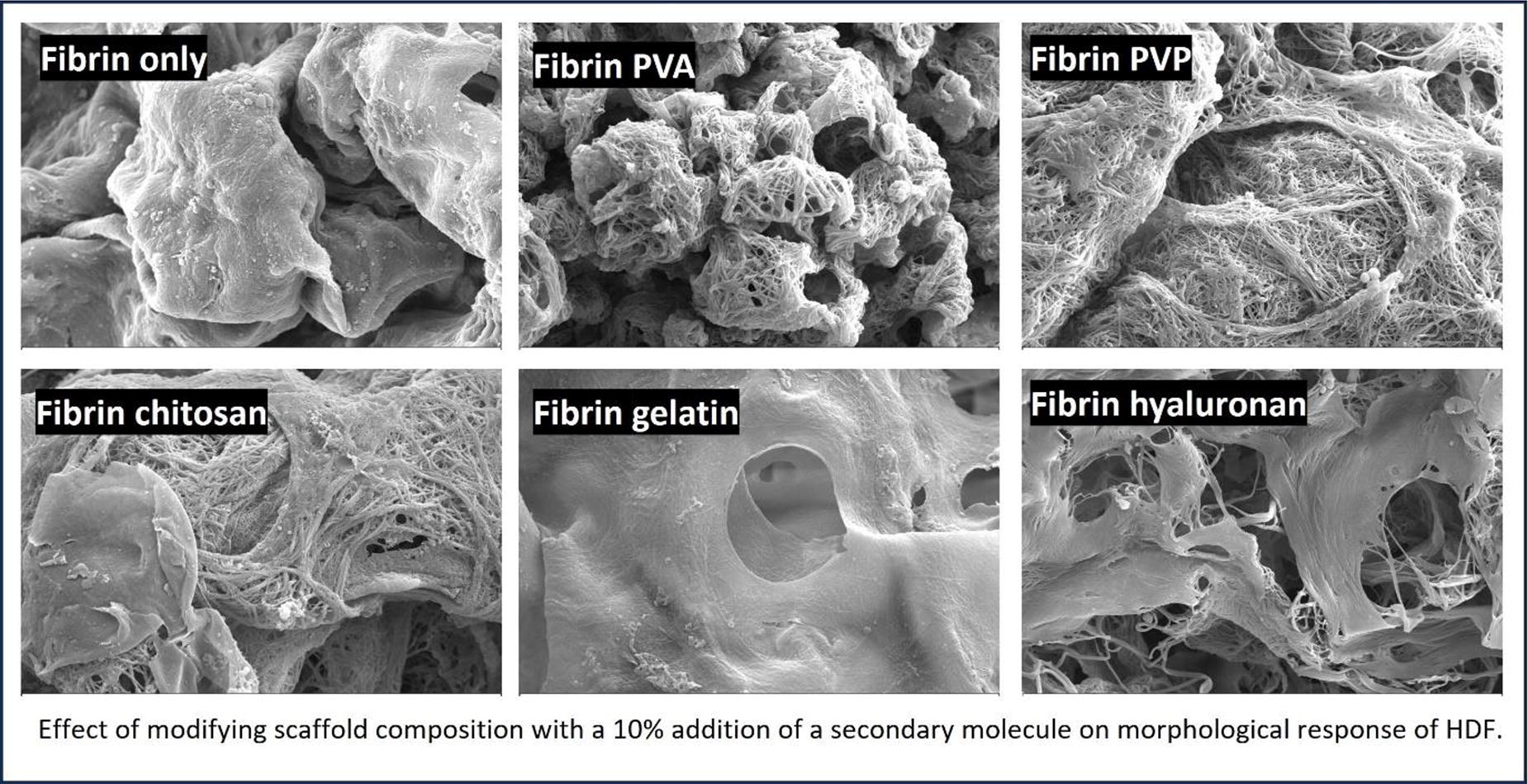
B337 - Development Of Polydeoxynucleotide-loaded And Visible Light Photo-crosslinked Gelatin Methacrylate (gelma) Hydrogel For Cartilage Tissue Engineering
S. Park1, J. Song1, T. Kang1, A. Motta2, R. Reis3, G. Khang1,2,3
1
Department of PolymerNano Science & Technology and Polymer Materials Fusion Research Center, Jeonbuk National University, Jeonju, Korea, Republic of,
2
Department of Industrial Engineering and BIOtech Research Center, University of Trento, Trento, Italy,
3
3Bs Research Group, University of Minho, Guimaraes, Portugal
*Purpose/Objectives: Regenerating cartilage through tissue engineering faces challenges due to its unique avascular nature and low cell density. Polydeoxyribonucleotide (PDRN) shows therapeutic potential for cartilage defects by inhibiting proteoglycan breakdown and reducing MMP13 gene expression. However, PDRN injection has drawbacks like unregulated cargo release. Gelatin methacrylate (GelMA), a light-curable polymer, offers a solution. With the RGD sequence enhancing cell-scaffold interaction, GelMA is biocompatible and biodegradable. UV-based curing may damage cells, so we propose using Riboflavin as a photoinitiator for visible light-induced crosslinking. Our goal is to develop a GelMA hydrogel for controlled PDRN release and induce cartilage formation through visible light crosslinking.
*Methodology: Initially, chemical characteristics were analyzed by confirming the methacrylation of gelatin through NMR and assessing the degree of GelMA methacrylate substitution via fluoraldehyde analysis. Furthermore, chemical bonding between PDRN and GelMA hydrogel was verified through FT-IR, and the release behavior of PDRN over time was examined. Morphological analysis was conducted using SEM, while the physical properties of the hydrogel were evaluated as a support structure for cartilage tissue regeneration through assessments of swelling ratio, degradation rate, and compression strength. In vitro analyses included MTT analysis to confirm hydrogel cytotoxicity, GAG quantification to assess chondrocyte activity. And histopathology Analysis was performed to evaluate in vivo cartilage tissue regeneration.
*Results: The study confirmed a 76 ± 5% MA degree through NMR and fluoraldehyde analysis. PDRN was stable in GelMA hydrogel without chemical binding, as verified by FT-IR. Increasing GelMA ratio led to smaller pore size and higher mechanical strength in hydrogels. Notably, 14% GelMA hydrogel exhibited sustained PDRN release up to 21 days. Higher GelMA concentrations showed a slight increase in cytotoxicity. Considering comprehensive physicochemical analyses, 14% GelMA hydrogel was chosen as an optimal material for cartilage tissue engineering. In vitro experiments demonstrated enhanced GAG content and upregulated expression of chondrocyte-specific genes (COL2, SOX9, AGG). In vivo assessment on a rabbit cartilage defect model confirmed GelMA-PDRN hydrogel’s ability to create a favorable microenvironment for chondrocyte proliferation and attachment at the damaged site.
*Conclusion/Significance: In conclusion, our GelMA-PDRN hydrogel system addresses cartilage regeneration challenges. GelMA’s high methacrylation degree supports visible light-induced crosslinking, ensuring stable PDRN incorporation. Optimal 14% GelMA ratio exhibits sustained PDRN release, suitable mechanical strength, and pore size for cartilage regeneration. In vitro, the hydrogel demonstrates biocompatibility, enhancing chondrocyte activity and gene expression. In vivo, the hydrogel promotes a conducive microenvironment for chondrocyte proliferation in a rabbit cartilage defect model. This study introduces an optimized GelMA hydrogel, overcoming PDRN injection limitations with controlled release and improved efficacy. The results of our research indicate a promising avenue in regenerative medicine, offering innovative solutions for cartilage defects. Hydrogel composition and explore diverse in vivo models to validate clinical potential are in progress.
B339 - Calcium Phosphate Ceramics In Autologous Blood Coagulum Containing RhBMP6 Promotes Healing Of Large Bone Defects In Rabbits
N. Ivanjko1, N. Stokovic1, M. Pecin2, A. Smajlovic2, M. Milesevic1, H. Capak2, Z. Vrbanac2, A. Javor2, D. Maticic2, S. Vukicevic1
1
School of Medicine University of Zagreb, Zagreb, Croatia,
2
Faculty of Veterinary Medicine University of Zagreb, Zagreb, Croatia
*Purpose/Objectives: Large bone segmental defects are among the most complex conditions in orthopaedics. Currently, the gold standard for the treatment of large segmental defects is autologous bone graft (ABG) harvested from the iliac crest bone. We have developed OSTEOGROW, a novel autologous bone graft substitute (ABGS) comprised of recombinant human Bone Morphogenetic Protein 6 (rhBMP6) and autologous blood coagulum (ABC) as a BMP carrier. However, preclinical studies have revealed that in certain indications where compressive forces are present such as large segmental defects, OSTEOGROW implants should be supplemented with compressive resistant matrix (CRM). This study aimed to compare rebridgment of the segmental defect in rabbits following implantation of OSTEOGROW with synthetic ceramic particles as CRM (OSTEOGROW-C), OSTEOGROW (rhBMP6/ABC), and commercially available rhBMP2 delivered on absorbable collagen sponge (rhBMP2/ACS).
*Methodology: In this study, an ulnar segmental defect (15 mm) was created in 18 New Zealand White Rabbits (lat. Oryctolagus cuniculus, 20-week-old, male, a bodyweight of 3-5 kg) and filled with osteoinductive implants according to the experimental design. Experimental animals were assigned to the following four experimental groups: (A) rhBMP6/ABC + tricalcium phosphate ceramics (TCP; particle size 1000-1700 µm) (n=5); (B) rhBMP6/ABC + biphasic calcium phosphate ceramics (BCP with TCP/HA ratio 40/60%, particle size 1000-1700 µm) (n=5); (C) rhBMP6/ABC (n=5); (D) rhBMP2/ACS (n=4). The applied dose of rhBMP6 or rhBMP2 was 100 µg per implant while the amount of ceramics in implants containing CRM was 200 mg. Healing of segmental defect was assessed by in vivo x-ray scans 4 and 8 weeks following the surgical procedure. Experimental animals were terminated after 17 weeks for further assessment of segmental defect rebridgement by ex-vivo x-ray scans, micro CT, and histological analyses. Animal studies were approved by the National Ethics Committee.
*Results: All tested BMP-based osteoinductive therapies promoted the rebridgement of the ulnar segmental defect as observed by in vivo x-ray images. Defect healing was confirmed on ex vivo CT and micro CT images and by palpatory testing. Moreover, microCT analyses revealed that OSTEOGROW with calcium phosphate ceramics (OSTEOGROW-C) induced a larger amount of bone than formulations of osteoinductive devices without CRM (rhBMP6/ABC or rhBMP2/ACS). Importantly, at the end of the follow-up period, TCP particles were almost completely resorbed while there was a large number of residual BCP particles incorporated in newly formed bone. Furthermore, the healing of bone defect was confirmed by histological analyses that revealed complete osseointegration of newly formed bone with native bone.
*Conclusion/Significance: OSTEOGROW-C promoted the healing of large segmental defects in rabbits and was superior to the formulation of osteoinductive devices without synthetic ceramic particles. Therefore, OSTEOGROW-C is a promising therapeutic solution for the treatment of patients with large bone defects.
B340 - The Role Of The Biochemical Composition Of Biomaterials In 3D In Vitro Liver Models
H. Criel1, C. Grootaert1, R. van der Meeren2, J. Van Camp1
1
Ghent University, Ghent, Belgium,
2
Rousselot, Ghent, Belgium
*Purpose/Objectives: High-throughput screening utilizing in vitro liver models is critical to test the pharmacologic and toxicologic effects of drugs and other chemicals. Since research has shown that monolayer-cultured liver cells lack in many cases the metabolic competence to mimic the in vivo condition, several three-dimensional (3D) liver models have been developed to better recapitulate normal liver function and metabolic capacity. These 3D models take into account the interactions between cells and their surroundings, the so-called microenvironment. In tissues, the matrix surrounding the cells is called the extracellular matrix (ECM) which contains a variety of biochemical components. The biochemical composition and mechanical stiffness of the extracellular matrix (ECM) in liver tissue are intricately linked to their specific localization within the lobule of the liver. Concurrently, research indicates that the functionality of hepatocytes is also closely tied to their respective position within this lobule. In contemporary research, an extensive array of biomaterials is employed to emulate the extracellular matrix. Existing studies highlight the significant impact of the chosen biomaterial on cellular functionality. Nonetheless, there remains a substantial gap in our understanding regarding how the biochemical composition and stiffness of the microenvironment in a 3D cell culture model influence hepatocyte responses to drugs or other chemical components.
*Methodology: In this study, we developed different blends of ECM-based biomaterials based on the in vivo biochemical composition and stiffness of the ECM. The mechanical stiffness of the biomaterial hydrogels that were formed was determined using rheology and compression testing. In addition, differentiated HepaRG cells and HepG2 cells were combined with the selected biomaterial blends in a 3D hydrogel. Over time, we tested the effect of the biochemical composition of the biomaterial on the response of differentiated HepaRG cells and HepG2 cells to exposure to chemical (food) toxins. We looked at cell viability, cellular respiration, cell metabolism, cell differentiation and polarity, and cellular functionality including protein production, detoxification capacity, and bile production utilizing techniques such as the IncuCyte S5® Live-Cell Analysis system, Seahorse XF Analyzers, and qRT-PCR.
*Results: Our results showed that differences in the biochemical composition of the tested biomaterial blends did result in significant differences in cell viability, metabolism, protein production, and gene expression of certain CYP450 enzymes after exposure of the differentiated HepaRG cells to some of the indirect-acting toxins.
*Conclusion/Significance: In conclusion, this study offers valuable insights into how the biochemical composition of biomaterials influences hepatocyte responses, contributing significant knowledge to advance research in tissue engineering and regenerative medicine. The choice of biomaterial within a 3D liver model can significantly impact results regarding the hepatotoxicity of specific chemical components found in food and pharmaceuticals. Overlooking the influence of the biochemical composition and stiffness of the extracellular matrix (ECM) on cellular behavior in in vitro studies could lead to misleading conclusions.
B342 - Preparation Of Microcrystalline Cellulose-Graphene Oxide (MCC-GO) Based Composites For Bone Tissue Engineering Applications
N. Zurita-Méndez, G. Carbajal-De la Torre, J. Ortíz-Ortíz, M. Espinosa-Medina
Universidad Michoacana de San Nicolás de Hidalgo, Morelia, Mexico
*Purpose/Objectives: Treatment of large bone defects is one of the most challenging tasks in orthopedics, an estimated of 2.2 million bone grafting procedures are performed worldwide per year [1]. Bone tissue engineering has an increasing interest in the development and construction of analogous bone grafts with osteoconductive, bioactive, biodegradative and mechanical properties. Cellulose (Cel) in its pristine form presents adequate mechanical properties, non-immunogenicity, nontoxicity, source abundance and low production cost, being one of the polysaccharides often studied for treat bone and cartilage defects. By other side, graphene oxide (GO) has shown a broad range of chemical functionalities and excellent thermal, electrical and mechanical properties besides, it has been found that GO creates a favorable micro-environment in scaffolds materials.
*Methodology: In this work, 3D scaffolds conformed by microcrystalline cellulose-graphene oxide 1%/hydroxyapatite/polylactic acid (MCC-GO/HA/PLA) had been developed in three different ratios, and identified as MCCGO10, MCCGO20 and MCCGO30. GO was synthetized by the modified Hummer’s method [2] and in a proportion of 1% was added to cotton solution for the obtainment of MCC-GO composite, posteriorly; HA was synthetized by the sol-gel method [3] and the 3D scaffolds were designed by the solvent-casting/salt leaching technique. The materials were characterized by FTIR, XRD and SEM techniques and as porous scaffolds, bioactivity and biodegradability of the composites were in-vitro evaluated by immersion into simulated body fluid (SBF) and phosphate-buffered saline solution (PBS).
*Results: XRD results showed defined diffraction peaks related to MCC-GO composite with high crystallinity of the MCC phase and the HAp matched with the pattern diffraction file related to Ca10(PO4)6(OH)2. GO particles displayed a bidimensional crumpled-sheet structure with a randomly high-pitched appearance which demonstrates that the oxidation process introduced an imbalance in the oriented layered structure of the precursor graphite and HA with porous morphology is probably that facilitates and enhances the circulation of body fluids, promoting integration, vascularization, and tissue regeneration. These properties make them valuable materials in various biomedical applications, including bone tissue engineering, drug delivery systems, and implants.
*Conclusion/Significance: Graphene oxide, hydroxyapatite and cellulose in a polylactic matrix had been studied for their application in bone tissue engineering. Three different proportions were evaluated with excellent potential properties each; observing that composition MCCGO30 presents the best biocompatibility due to the GO amount enhanced the biocompatibility in bone scaffolds, but also the polysaccharides of the cellulose increment the bioactivity. By other hand, MCCGO10 scaffolds presented the best biodegradability related to the polymeric PLA phase that is present in the scaffolds.
B343 - One-pot Extraction Of Bio-resources From Human Hair Via A Zero-waste Green Route
A. Mukherjee1, S. Pal1, S. Parhi1, P. Ghosh2
1
CSIR-Indian Institute of Chemical Biology (CSIR-IICB), Kolkata, India,
2
Institute of Health Sciences, Presidency University, Kolkata, India
*Purpose/Objectives: Upcycling of bio-derived wastes to value-added materials has received significant researchattention over the past decade. In this work, we report the extraction of keratin and melanin, twoimportant biopolymers, from waste human hair through an environmentally benign and greenextraction route.
*Methodology: The reaction was performed using an ionic liquid (IL), 1-butyl-3-methylimidazoliumchloride solvent due to its previously reported advantages associated with extraction efficiency andreusability compared to conventional solvents. Furthermore, employing imidazolium-based ionicliquid for biopolymers containing amine/amide functionalities has several benefits related to easyprocessing and physicochemical properties such as thermal stability and low melting point. Detailedanalysis of the extracted biopolymers including chemical, thermal, and morphologicalcharacterizations suggest the high-temperature extraction process did not affect the structuralintegrity of the biopolymers.
*Results: The extracted keratin with a yield of around 44% displayed the potential to be employed ina variety of blood-contacting biomaterials, as evidenced by hemolytic potential assays. The otherextract was melanin pigment with preserved ellipsoidal morphology as determined by FE-SEManalysis, which had an approximate yield of 17%. Melanin demonstrated the ability to reduce 2,2-diphenyl-1-picrylhydrazyl indicative of its free-radical scavenging activity. The significantantioxidative ability with strong UV absorption of melanin suggests their utilization for biomedicalapplications. Melanin can be coated onto thin films or fused into dermatological products to beutilized as sun-protective materials. The ILs used for extraction were recovered and recycledmultiple times asserting the sustainability of this ecofriendly procedure. The dialysis remains werefound to be electrically conductive being able to glow an LED light indicating the feasibility ofemploying it in bioelectronics further highlighting its implications on polymer circular economy.
*Conclusion/Significance: Altogether, the work provides a zero waste green approach to extractingbiopolymers from renewable resources and their uses in different biomedical applications.
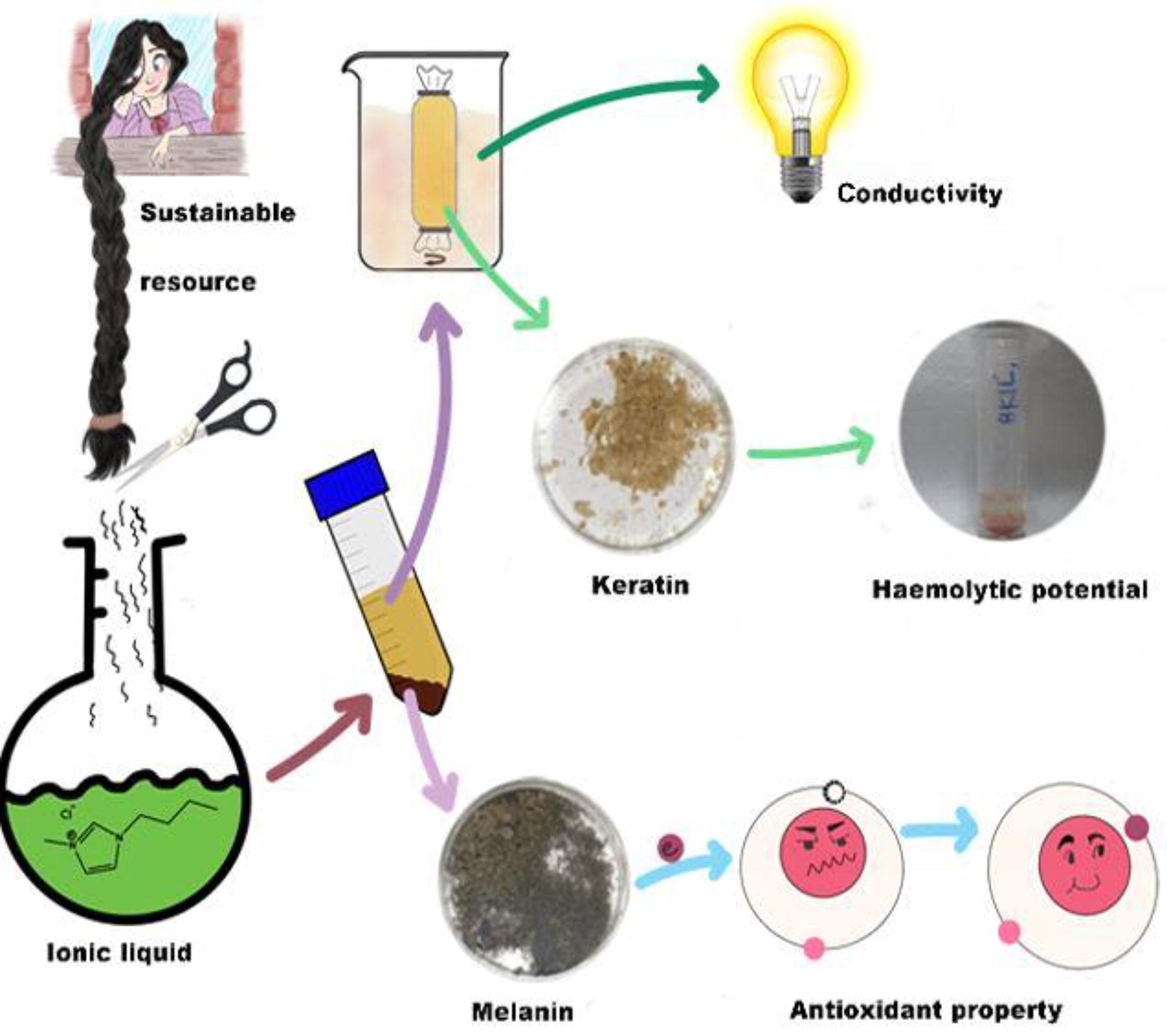
B345
B346
B347 - Designing A Photocrosslinkable Hydrogel For Donor-site Wound Treatment
B. Tabatabaei Hosseini, V. Gabriel, K. Kim, J. Hu
University of Calgary, Calgary, AB, Canada
*Purpose/Objectives: Donor-site wounds arise from the extraction of healthy skin for the purpose of skin grafting, employed to address extensive skin loss resulting from deep burns, ulcers, or trauma. Typically, the recovery period for these wounds spans 7-10 days, although individual variations exist, influenced by factors such as the patient’s age and nutritional well-being. Post-harvesting, donor-site wounds commonly exhibit increased pain levels, attributed to the exposure of dermal nerve endings. Additionally, there is a rapid onset of exudation following the removal of skin grafts, necessitating instant care with an absorbent dressing.
*Methodology: Addressing the main limitation associated with the existing dressings—inefficient wound exudate management— and aiming immediate and tunable wound coverage, this study focused on designing a moist dressing for enhanced donor-site wound treatment. A cellulose derivative was chosen and modified with methacrylate groups to achieve photocrosslinking. The modification process was fine-tuned by adjusting pH and time for optimal methacrylation degree. The modified cellulose was then added to a chitosan base to form an interpenetrating polymer network (IPN). The mechanical and physical properties were evaluated for each composition of the hydrogel. Additionally, the biocompatibility, antimicrobial and homeostatic effect of the best performing hydrogel were assessed.
*Results: Firstly, we successfully modified the cellulose and chitosan components, leading to the formation of a stable and fast crosslinking hydrogel within 1 min. The NMR results indicated a 30% methacrylation degree for the cellulose derivative, outperforming the previously reported values. The tensile strength, rheology, and swelling ratio of the hydrogels were tuneable by adjusting the cellulose concentration in the hybrid hydrogel. Furthermore, the final hydrogel promoted homeostasis and showed excellent cell viability and proliferation.
*Conclusion/Significance: In conclusion, our rapid crosslinking hydrogel, derived from modified cellulose and chitosan, addressed a critical aspect of wound care. With tunable properties and proven attributes supporting wound management, it promises advancement in donor-site wound treatment.
B348 - Imparting Multi-scalar Architectural Control Into Silk Materials Via Ice-templating
H. Joukhdar1, Z. Och1, K. Lim2, J. Rnjak-Kovacina1
1
University of New South Wales, Sydney, Australia,
2
University of Sydney, Sydney, Australia
*Purpose/Objectives: Human tissues and organs exhibit complex hierarchical and gradient structures that are essential to their function and should be recapitulated within biomaterial scaffolds targeting their regeneration. Unidirectional freezing, an ice templating technique where ice acts as a porogen, is uniquely suited to recapitulating the architectural anisotropy, gradients and hierarchical transitions of human tissues, but ice templating of polymeric systems, including silk fibroin, remains less well understood than their colloidal counterparts.
*Methodology: To address this, we developed a versatile and accessible freezing setup for silk that allowed tuning of the freezing parameters including the polymer cooling rate (30 ºC min-1 to 2 ºC min-1) and ice solidification velocity (2.5 mm min-1 to 0.6 mm min-1) using liquid nitrogen. We performed real time visual and thermal monitoring of the freezing process for multiple silk concentrations (2-10% wt/v) and material states (liquid, hydrogels), and correlated these conditions with pore morphology.
*Results: We demonstrate unprecedented control over pore size (100 - 90,000 µm2) and pore morphology (cellular-lamellar), consistent pore alignment, and generation of gradient porosity in silk scaffolds. For the first time we demonstrated the impact of shear thinning behaviour of silk in ice crystal formation, demonstrating the non-linear and complex freezing phenomena is silk. Finally, we demonstrate the utility of ice-templating photo-crosslinked silk hydrogels, which support simultaneous pore alignment, porosity, and concentration gradients.
*Conclusion/Significance: The resultant materials are porous but retain the hydration and elastomeric nature of hydrogels, making them ideally suited for soft tissue engineering.

B349 - Nanocellulose Hydrogels With Improved Properties For Advanced 3d Bioprinting In Innovative Tissue Engineering Applications
E. Usala1, Z. González1,2, N. Campillo3, S. Micó3, J. M. Baena3,4, B. Ferrari2,5, A. Rodríguez1,2, E. Espinosa1,2
1
Instituto Químico para la Energía y el Medioambiente (IQUEMA), Universidad de Córdoba, Córdoba, Spain,
2
Unidad Asociada CSIC-UCO, Fabricación Aditiva de Materiales Compuestos Basados en Celulosa Funcionalizada, Obtenida de Residuos de Biomasa, Córdoba, Spain,
3
REGEMAT 3D S.L., Granada, Spain,
4
BRECA Health Care S.L., Granada, Spain,
5
Instituto de Cerámica y Vidrio, Consejo Superior de Investigaciones Científicas (CSIC), Campus de Cantoblanco, Madrid, Spain
*Purpose/Objectives: Bioprinting is an emerging technology for manufacturing functional tissue constructs to replace injured or diseased tissues. This process offers notable advantages, including high reproducibility and precise control over the automated fabrication of constructs. The bioprinting procedure involves the utilization of a biomaterial solution in hydrogel form, typically encapsulating desired cell types, known as bioink, to generate tissue constructs. Various biopolymer hydrogels derived from biofuel and biorefinery processes have proven successful as bioinks for bioprinting. Examples include agarose, gelatin, alginate, chitosan, cellulose, among others. Despite the availability of diverse natural bioinks and biomaterials for bioprinting, certain limitations persist. These limitations encompass partially weak cell adhesion properties and restricted mechanical stability, which limit the size of artificial tissue constructs achievable through bioprinting. Consequently, alternative strategies must be implemented to address these challenges, with the most common approach involving the addition of additives or other biomaterials, such as graphene and alginate, that offer mechanical support and new properties without adversely affecting the cells.
*Methodology: In this research, two novel hydrogels were formulated by combining cellulose nanofibrils (CNFs), isolated by a biorefinery process starting from wheat straw, with alginate and, on the other hand, the CNFs were combined with reduced graphene oxide (rGO), added by a colloidal process, to create a scaffold suitable for tissue engineering applications. The materials were characterized by XRD, Zeta Potential, FT-IR, cytotoxicity and rheological analysis. In addition, the sterilized hydrogels were extensively evaluated for their printability, with a focus on identifying optimal hybrid hydrogels compositions that demonstrate precision in 3D printing of constructs.
*Results: The synergistic properties of CNF and alginate provide a supportive matrix with enhanced mechanical strength and bioactivity, while the incorporation of rGO into CNF introduces electrical conductivity to the bioinks.
*Conclusion/Significance: This study explored the effects of incorporating alginate and rGO into nanocellulose-based bioinks, providing insights into how it influences both biocompatibility and printability. The resulting hybrid formulations facilitate strong cell growth, show minimal toxicity, and exhibit excellent precision, making them well-suited for applications in tissue engineering.
B351 - Sodium Alginate-gelatin Bead For Spheroid Encapsulation
J. Rios-Vergara, L. Toro, C. Echeverri-Cuartas
EIA University, Envigado, Colombia
*Purpose/Objectives: Biomaterials in cell culture can closely mimic the extracellular matrix (ECM) by providing support and nutrients. Gelatin and sodium alginate are biomaterials widely used in culture since they are biocompatible and have low cytotoxicity. On the other hand, spheroids promote the formation of cell-cell interactions in all three dimensions, like the physiology of organisms. In this study, we developed an Alg-Gel matrix that simulates the three-dimensional conditions of spheroid cell culture
*Methodology: The Alg-Gel matrix was characterized the Alg-Gel matrix using Fourier transform spectrometry -FTIR-, morphology evaluation, swelling and degradation by gravimetry, sterility evaluation, and cytotoxicity evaluation, as well as the spheroid culture in its pre- and post-encapsulation, (optical microscopy, fluorescence microscopy, and MTT assay) to observe the behavior of these and the possible factors associated to the matrix that influence their growth and development.
*Results: The findings showed that the Alg-Gel 80:20 matrix presented the highest percentage of swelling (34.91 %) which indicates a possible greater presence of pores; in addition, we evidenced that the cell culture medium affects the integrity of the matrices, making it difficult to manipulate them in the long term. The size of the spheroid’s pre-encapsulation was evident, demonstrating that the average was 181.85 ± 9.70 μm. Furthermore, we observed that the MTT assay for spheroids evaluation is associated with low absorbance levels, even though when it was a viable spheroid since the formation of formazan crystals was evidenced. Its morphology was observed by scanning electron microscopy -SEM which indicated that this method is not suitable for evaluating spheroids.
*Conclusion/Significance: The encapsulated spheroids in the Alg-Gel matrices were associated with a smaller size than the control (control = 211.55 ± 19.68 μm; 50:50 = 193.77 ± 18.67 μm; 70:30 = 191.20 ± 16.63; and 80:20 = 183.54 ± 19.14 μm). The decrease in size was related to the amount of Alg, which would provide the opportunity for a size-controlled spheroid culture depending on the Alg ratio. All Alg-Gel matrices proved to be viable for both direct and indirect cytotoxicity assays to spheroids, especially the Alg-Gel 80:20 matrix which represented high viability in both cases, with percentages equivalent to 113.58 % (indirect) and 99.36 % (direct). These results suggest that the Alg-Gel 80:20 matrix may have a promising use for the encapsulation of spheroids; however, new approaches should be performed concerning spheroid evaluation methods to corroborate the positive effect of Alg. In addition, it is necessary to implement methods to improve the physicomechanical behavior of the matrix in the presence of the culture medium.
B352
B354 - Magnetic Stimulation Prevents Dorsal Root Ganglion Neuron Death And Enhances Nerve Regeneration To Improve Motor Function In A Sciatic Nerve Injury Rat Model
S. Xu, A. Ito, Z. Zhao, R. Nakahara, C. Tai, F. Miyamoto, H. Kuroki, T. Aoyama
Kyoto University, Kyoto, Japan
*Purpose/Objectives: Severe peripheral nerve injury (PNI) is often associated with retrograde cell death in the spinal cord and dorsal root ganglia (DRG). The reduction in the number of neurons following injury can hinder nerve regeneration and subsequently result in functional deficits. Although nerve autograft is considered the gold standard surgical treatment for PNI, neuronal cell death still poses a challenge to subsequent nerve regeneration and functional recovery. Therefore, if post-injury cell death could be suppressed, it may further promote functional recovery. Magnetic stimulation (MS) has been shown to have a promoting effect on nerve regeneration after PNI. This study aims to apply MS directly to neurons to investigate its effects on post-injury neuronal cell death and nerve regeneration.
*Methodology: In this study, we randomly divided 66 12-week-old Lewis rats into three groups: the MS, control, and intact group. In the MS and control groups, a sciatic nerve autograft model was prepared. All rats in the MS group received MS (20Hz, 1600 pulses, 5 days/week) to the skin above the lumbar spinal cord from the first postoperative day until sacrifice. At 1, 4, and 8-weeks after the injury, we collected and stained the L4-L5 DRG on the same side to count the DRG neuron number (n = 6 per group). Then, we evaluated the apoptotic factors (Bax and Bcl-2) of mRNA expression in DRG by RT-qPCR at 1- and 4-week post-injury (n = 6 per group). And, axon regeneration analysis, electrophysiological, and motor function analyses were performed 8-week post-injury to confirm the promoting effect of MS on nerve regeneration and functional recovery (n = 6 per group).
*Results: Compared to the intact group, a decrease in the number of DRG neurons in the control group was observed 1-week after injury, which persisted up to 8-week post-injury. However, no reduction of DRG neurons was observed at any time point in the MS group, and 1- and 4-week post-injury, the MS group had significantly higher DRG neuron counts than the control group. At 1-week post-injury, Bax mRNA expression was significantly lower in the MS group than in the control group. Conversely, at 4-week post-injury, Bax and Bcl-2 mRNA expressions in the MS group were significantly higher than in the control group. Histological analysis at 8-week post-injury indicated a significantly greater myelinated nerve fiber density in the autografted nerves of the MS group. Additionally, electrophysiological results showed significant improvements in latency. 3-D motion analysis also demonstrated better improvements in the toe angle of toe-off phase of gait cycle in the MS group.
*Conclusion/Significance: Through histological analysis, we confirmed that MS can prevent the death of DRG neurons. At 8-week post-injury, we also demonstrated the promoting effects of MS on nerve regeneration and motor function recovery following PNI. These outcomes may be attributed to the modulation of apoptotic factors mRNA expression by MS, such as Bax and Bcl-2.This study indicates that MS could potentially prevent the death of DRG neurons and facilitate nerve regeneration and functional recovery after PNI by regulating associated apoptotic factors.
B356 - Developing A Hydrogel-based Localised Therapy For Treatment Of Endometriosis
A. Miller
University of Manchester, Manchester, United Kingdom
*Purpose/Objectives: Endometriosis is a common gynaecological condition whereby endometrial-like cells form lesions in extra-uterine locations. Endometriosis occurs in roughly 10% of women of reproductive age. Currently, all medical treatment is symptomatic and often have intolerable side effects. The only curative option for endometriosis is surgical removal of the lesions. However, recurrence rates, particularly after conservative surgery, are high, resulting in re-operation.
The aim of this project is to design a biocompatible, hydrogel-based localised therapy that can prevent lesion recurrence and improve management of this unmet clinical need. A self-assembling peptide hydrogel (SAPH) has been explored to meet this need as they have the potential to be administered during laparotomy, increasing drug availability whilst mitigating systemic side effects.
*Methodology: Analysis of hydrogel performance was achieved through four objectives: to characterise drug- loaded SAPHs; to investigate SAPH localisation and stability in vivo via fluorescence imaging; to establish and validate a murine model of endometriosis recurrence and to utilise this model to test the efficacy of systemic and local therapies.
*Results: In vitro degradation and cell culture studies highlighted the adhesive and bio- compatible properties of the chosen SAPH (Alpha 2 PeptiGel®). Drug-loading of solubilised and lyophilised drugs had a distinct effect on hydrogel structure and drug release, which appeared to be due to changes in electrostatic and hydrophobic interactions between drug and peptide.
The SAPH was successfully functionalised with an infrared tag (IR750) without compromising its physio-chemical properties. In vivo and ex vivo imaging showed that the IR750-tagged SAPH administered onto the parietal peritoneum of mice remained in situ for up to 22 days. There was no visible peptide bio-accumulation within the organs, and accumulation within the cartilage was temporary.
A murine model of endometriosis recurrence was successfully developed by implanting donor uterine tissue onto the parietal peritoneum of recipient mice during laparotomy. A second laparotomy was conducted 14 days later, which confirmed all tissue fragments formed fluid-filled lesions. Lesions that were aspirated and partially removed during this laparotomy consistently recurred after 28 days. There were no signs of distress or disturbances to the oestrous cycle. Therefore, this model was deemed suitable for testing the efficacy of localised and systemic therapies on lesion recurrence.
Neither systemic nor localised treatments completely prevented lesion recurrence, but growth rate was attenuated with localised treatment as the drug loaded hydrogels produced an average final lesion volume significantly lower than hydrogel control. Localised application also significantly reduced final lesion volume compared to volume at removal. This effect was also observed for systemic, however, contraceptive effects associated with the therapeutic were observed via disruption of the oestrous cycle.
*Conclusion/Significance: In conclusion, utilising a drug-loaded SAPH during the peri-operative window could be a suitable alternative to conventional post-operative treatment to prevent lesion recurrence. This local treatment was well tolerated; efficacious in reducing lesion volume, without systemic side effects.
B357 - Replacing Banned Synthetic Meshes For Mid-urethral Slings
D. Brownell, A. Zidat, S. Chabaud, L. Germain, J. Ruel, S. Bolduc
Université Laval, Québec, QC, Canada
*Purpose/Objectives: More than 200 million people worldwide suffer from stress urinary incontinence (SUI). This represents 30-40% of all women during their lifetime. Polypropylene mesh mid-urethral slings (MUS) are the current gold standard treatment for female patients with SUI. They have good short-term cure rates, although their efficacy tends to decrease over time. Campaigns against synthetic MUS led to their abolishment in many countries, including severe restrictions in Canada due to their risk of urethral erosion. The traditional autologous fascial MUS has reemerged as a viable alternative. However, autologous MUS’s readoption is slow due to the surgical technical challenge and perceived higher morbidity rates. Thus, tissue engineering (TE) presents itself as a potential alternative to replace synthetic MUS, while avoiding comorbidities related to the harvesting of the autologous fascia.Biomaterials usually serve as the backbone for the cell-seeded reconstruction of genitourinary tissues. However, cultured fibroblasts can secrete and deposit enough extracellular matrix to form stromal sheets (self-assembly). The mechanical properties of these reconstructed tissues parallel those of native tissues in specific models. Here, we reconstruct a stromal MUS substitute using patient’s cells: autologous and free of exogenous material.
*Methodology: A MUS substitute was constructed by modifying the current self-assembly technique used in the reconstruction of skin for treating burn patients. Surface properties of culture dishes were first optimized by evaluating UV-C or H2O2 treatment to allow for cell attachment. Custom tissue anchors were cut from Whatman paper to allow for stromal sheet handling. Dermal fibroblasts were seeded in optimized supports at confluence and cultured for 4 weeks with 2-phospho-L ascorbate to stimulate matrix deposition. The stromal sheet was then folded for a final ply of 4, 6, or 8 and allowed to fuse over the next 2 weeks. Insulin, IGF-1, and LONG®R3IGF-1 supplementation was also evaluated to boost matrix deposition.
*Results: Fibroblast adhesion was optimal and most homogenous when culture dishes were treated with H2O2 gas. Stromal sheet formation was similar to other self-assembly models. When ply was evaluated, there was no advantage for the 8 vs. 6 ply due to the decreased fusion of layers when the stroma was too thick. There was little difference between 4 and 6 ply constructs with a rupture force of 2.2 N for a 1 cm wide tissue. Interestingly, insulin and LONG®R3IGF-1 increased collagen deposition when evaluated 2 weeks after seeding, but there was no advantage in the final product. Tissues cultured with LONG®R3IGF-1 showed significantly reduced fusion between layers.
*Conclusion/Significance: Our human-derived TE MUS substitute can be entirely autologous and appears promising for treating SUI, with similar mechanical properties to native tissue. The current construct is strong enough to support the theoretical required load but remains to be further optimized to ensure resistance when grafted in vivo.

B358 - Validating The Use Of Extracellular Matrix As Scaffold For In Vitro Endometrial 3D Modeling
E. Ribes Martinez1,2, T. S. Oshiro1,2, G. D. Ferronato3,2, J. B. Stöckl2, T. Fröhlich2, M. Ferraz3,2
1
Ludwig-Maximilians University of Munich, Munich, Germany,
2
Gene Center, Munich, Germany,
3
Ludwig-Maximilians University of Munich, Oberschleißheim, Germany
*Purpose/Objectives: In vivo the embryo-maternal communication in the uterus includes different cellular layers, such as epithelium, stroma, immune cells and endothelium. Such complex structured communication is not achieved in 2D cultures, but by combining a co-culture system with all different cell layers, this dynamic maternal communication can be re-created. Therefore, we aimed at validating the use of decellularized endometrial extracellular matrix (dECM) enriched with alginate as a scaffold for producing 3D endometrium models to support embryo development.
*Methodology: Bovine endometrium was collected from a local slaughterhouse, dissected and treated with sterile ultra-pure water (6h), SDC 4 % (24h), followed by 20 washes in water, DNAse I (5 μg/mL; 3h), another two washes, lyophilized and cryo-milled. 20 mg/ml hydrogels were prepared in 0.1M HCl with 1mg/mL pepsin for 72h. RNA and DNA from tissues and dECM gels were extracted and concentration measured using a Qubit. SDC concentration in washes samples were analysed using a Methylene-Blue assay. LC-MS/MS (Ultimate 3000 nano-LC system, coupled to a Q Exactive HF-X mass spectrometer) of dECM was performed. To test embryo bio-compatibility, day 3 in vitro produced embryos were incubated in a bed of endoECM for 96 h. Mechanical properties were measured by nanoindentation (CHIARO) and porosity by liquid displacement. To validate cell compatibility, 20 uL drops of dECM (10 mg/mL + 1% alginate) with 1x10⁶ primary endometrial fibroblast/mL were crosslinked for 45 min at 38°C followed by 15 min with 11 mM CaCl2, 100 mM HEPES, and 0.1 U/ml of thrombin, and cultured at 38°C and 5% CO2. At days 1, 3, 7, and 14, a live dead staining was performed and at day 7 cell proliferation was determined by an EdU assay. Epithelial glands and stroma cells were isolated from endometrial tissue and embedded in the dECM and co-cultured for 14 days.
*Results: EndoECM had a complete removal of DNA and RNA contents compared to native tissues (n=4). Residual SDC in dECM washes were below 0,001% (n=10). Among the ECM related proteins, EndoECM presented 86.7% collagens, 3.17% glycoproteins and 6,37% proteoglycans (n=6); it also had different isoforms of laminin. Porosity of EndoECM enriched with 1% alginate gels was 59% (n=3), and young modulus was 1.2 kPa (n=3). Elastic and viscoelastic behaviour had similar pattern to native tissue. EndoECM was bio-compatible with embryos and had no effect on blastocyst rates (n=3). Stroma primary cells showed 88, 98 and 98% viability on days 3, 7 and 14, respectively, and 14.6% of proliferating cells were detected on day 7 (n=3). EndoECM allowed endometrial gland spheroid culture for over 10 days without the need of spheroid passaging as seen for spheroids cultured using Matrigel.
*Conclusion/Significance: Here we presented the use of dECM as a natural bioscaffold for creating a 3D endometrium model. The dECM promoted cell viability and proliferation, supported endometrial gland spheroid long term growth and are bio-compatible with embryo development.
B359 - Microporous Immune Isolating Capsule With Improved Diffusion For Restoration Of Ovarian Endocrine Function And Immune Protection
D. Sinko, M. Brunette, D. Pavlidis, B. Ray, M. Tong, A. Shikanov
University of Michigan, Ann Arbor, MI
*Purpose/Objectives: Live saving chemotherapy treatments are toxic to the ovaries leading to premature ovarian insufficiency (POI) in adolescent girls. Young women with POI are treated with hormone replacement therapy (HRT) which is unable to reproduce the dynamic, pulsatile and reciprocal interactions of a functioning ovary with the rest of the body. The lack of physiological ovarian endocrine function leads to elevated risks of various endocrine, cardiovascular and musculoskeletal disorders.
*Methodology: We have developed an immune isolating polyethylene glycol (PEG) hydrogel capsule that protects an ovarian tissue allograft while facilitating the exchange of nutrients and hormones. The capsule consists of two layers: an inner biodegradable layer that interfaces with the tissue and promotes growth, and the outer non-degradable layer that prevents rejection. In order to improve the diffusion across the hydrogel, we incorporated sacrificial gelatin microgels into the outer shell of the capsules. When heated to body temperature, the gelatin becomes soluble, leaving disconnected micropores measuring 30 μm in diameter in the bulk nanoporous hydrogel.
*Results: We analyzed PEG hydrogels loaded with 1%, 5% and 10% micropores (PEGG+) using fluorescence microscopy and confirmed that the microgels were homogeneously distributed throughout the bulk gel, and the gelatin diffused out of the capsules after 24 hours. Rheological characterization of the mechanical properties of the 10% PEGG+ gels indicated a 4-fold increase in shear yeild strain when compared to bulk PEG gels with a 56% increase in elasticity and little change viscosity of the material. Next, we measured the diffusion of 4, 40 and 150kDa dextran. The diffusion of 40 kDa dextran increased from 16% in the control nanoporous bulk gels to 26% after 96 hours. The diffusion of 150 kDa dextran only slightly increased from 10% in the bulk gels to 11% in PEGG+ gels reassuring that the microporous shell could still prevent infiltration of antibodies and cells. No difference in the diffusion of 4 kDa dextran was observed for all gel conditions. Future in vivo testing will be aimed at investigating whether the capsule prolongs the survival of the human ovarian xenografts while maintaining its integrity and immunoisolating properties by implanting the capsules subcutaneously in ovariectomized mice for 2, 10 and 20 weeks.
*Conclusion/Significance: In conclusion, incorporation of sacrificial micropores in a nanoporous bulk hydrogel improves diffusion without compromising its mechanical properties.

B360 - Anti-Inflammatory Properties Of Human Placental Tissue Membranes
B. N. Pfaff, A. J. Costantino, H. J. Lim
LifeLink Tissue Bank, Tampa, FL
*Purpose/Objectives: Placental tissue allografts remain a promising solution for the treatment of burns and chronic wounds due to their unique structure, abundance of nutrients, and ability to reduce inflammation. While amnion and chorion grafts have shown reduced scar tissue formation and accelerated healing in diabetic ulcers, to our knowledge there is no direct comparison to understand the benefits of the various placental tissue membranes available. Our study provides one such comparison by investigating the anti-inflammatory properties of amnion, chorion, amnion-chorion, and umbilical cord allografts.
While acute inflammation is imperative for the wound healing cascade, repair models have shown that prolonged inflammation leads to delayed healing and increased scarring. Additionally, unresolved wounds exhibit chronic inflammation, which may predispose the tissue to cancer development. As such, the anti-inflammatory properties of these wound care allografts, which can be attributed to the placental tissues’ multitude of growth factors and extracellular matrix proteins, are distinctly important.
*Methodology: Placental tissue membranes from 4 donors were processed and preserved using proprietary lyophilization and sterilization methods. Conditioned media were generated from each of the preserved membranes, keeping the membrane surface area constant across groups and donors. Human monocytic cells (THP-1) were stimulated by lipopolysaccharide (LPS) in the presence of each conditioned medium or standard growth medium. After 48 hours, supernatants were tested via enzyme-linked immunosorbent assays designed for pro-inflammatory cytokines TNF-α, IL-1β, and IL-6. A reduction in the presence of these pro-inflammatory cytokines relative to the standard growth medium control indicates a desirable outcome.
*Results: The THP-1 inflammatory response was attenuated when cells were exposed to LPS in all placental tissue-conditioned media. Each pro-inflammatory cytokine was inhibited by at least 25% for all membrane types, with the majority of membranes showing greater than 65% inhibition for all cytokines. As a whole, the tissues provided the strongest response to IL-6, with all four membranes showing a reduction greater than 75%. Individually, the umbilical cord performed the best while the amniotic membrane displayed the least cytokine reduction.
*Conclusion/Significance: All four placental tissue membranes exhibited desirable anti-inflammatory properties. These results indicate each of these placental tissue allografts remains a promising solution for the treatment of burns and chronic wounds. They also highlight a unique advantage or disadvantage that can be considered when determining which type of allograft should be applied. Since mitigating inflammation is just one component of wound healing, future studies evaluating cell proliferation, chemotaxis, post-processing maintenance of tissue structure, and more will provide a more holistic picture of the distinct benefits each membrane possesses.
B361 - In Situ Nitric Oxide Generating Bio-functional Hydrogels With Enhanced Anti-inflammatory, Cell Migration, And Angiogenic Capabilities For Chronic Wound Healing
D. Oh, D. Tran, P. Thi, K. Park
Ajou university, Suwon-si, Korea, Republic of
*Purpose/Objectives: The chronic wound due to the extended inflammatory phase and the reduced vascularization has caused a serious problem to human health. Therefore, the development of a biomaterial that can shorten the inflammatory stage and promote the angiogenic process is a promising therapy for the treatment of chronic wounds. Currently, nitric oxide (NO) is an endogenous gas molecule that involve in many physiological processes such as immune responses, apoptosis, inflammation, etc. Specially, anti-inflammatory and angiogenic activity of NO was widely applied in wound healing process and in tissue repair/regeneration. However, the clinical application of NO is limited due to their burst and short half-life release from the biomaterials. Here, we proposed a novel multifunctional NO-releasing hydrogel that can regulate various wound healing processes, including hemostasis, inflammation, cell proliferation and angiogenesis.
*Methodology: Phenol-rich gelatin (GH) polymer was synthesized by conjugating hydroxyphenyl propionic acid onto gelatin through carbodiimide reaction. GH/Cu was simply prepared by mixing the GH polymer solution containing either enzymes or H2O2 and CuSO4. The physicochemical properties of GH/Cu including degradation rate, mechanical strength, morphology, etc. were characterized. The release of Cu ions and catalytic generation of NO from GH/Cu was measured by modified micro-BCA assay and Griess assay, respectively. For anti-inflammation test, the polarization of monocytes was investigated through TGFß-ELISA and CD163 staining assay. In vitro angiogenesis activities of GH/Cu hydrogel were analysed through endothelial cell migration and tube formation assay. Besides, the in ovo chick chorioallantoic membrane (CAM) assay to confirm the angiogenetic properties was also performed.
*Results: The physicochemical properties of GH/Cu can be well controlled by varying the concentration of H2O2 and HRP. The fabricated hydrogel presented the sustained release of Cu ions, thus catalyzed the generation of NO in the presence of endogenous NO donors. The sustained release of NO for almost 3 weeks within the physiological level has regulated the polarization of monocyte toward the M2 phenotype, which demonstrated the anti-inflammatory properties of GH/Cu. Besides, the synergistic effect of Cu ions and released NO can also stimulate the migration and tube formation of endothelial cells as well as the formation of vascular in CAM, illustrating the in vitro and in ovo effect of GH/Cu on promoting angiogenesis.
*Conclusion/Significance: A novel and simple system for the in situ formation of NO-releasing hydrogel via enzyme-mediated reaction was developed. The resulting GH/Cu hydrogel exhibited the controllable property with in situ NO release. GH/Cu presented both the excellent anti-inflammation and angiogenesis properties. Therefore, we suggest that this hydrogel system would significantly enhance the chronic wound healing process through the synergistic effects of hydrogel and NO, which can be used as an advanced wound dressing material.
Acknowledgement: RS-2020-KD00003, KEIT 20018560, NTIS 1415184668

B362 - Automated In Vitro Wound Healing Assay
J. Cwycyshyn1, C. Stansbury1, W. Meixner1, J. B. Hoying2, L. A. Muir1, I. Rajapakse1
1
University of Michigan, Ann Arbor, MI,
2
Advanced Solutions Life Sciences, Manchester, NH
*Purpose/Objectives: Wound healing is essential for the restoration of the epidermal barrier in response to injury. This dynamic process requires an intricate orchestration of cellular migration and proliferation to facilitate wound closure and reestablish the structural integrity of damaged skin. Disruptions to the healing process can lead to excessive scar formation or the development of chronic ulcers, keloids, and other non-healing wounds. The consequences of impaired wound healing also extend beyond chronic wounds to severe skin injuries where precise cellular responses are paramount for optimal tissue regeneration. In cases of deep skin trauma and thermal burn wounds, the intricacies of the wound healing cascade become even more crucial. Despite its significance, our understanding of the intra-cellular signaling mechanisms that initialize, balance, and stop migration and proliferation during wound healing remains incomplete. In vitro wound healing assays are a frequently used approach to examine this complex relationship. However, existing assays suffer from limited reproducibility, low throughput, or inflexibility in experimental design.
*Methodology: We have developed the Automated Wound Healing (AWH) assay—a high-throughput robotics-assisted technique for reproducible wound creation and automated time-lapse image analysis. Our approach utilizes computer-aided design (CAD) wound models which are recreated in confluent cell layers using the BioAssemblyBot (BAB) 3D-bioprinting platform. The dynamics of migration and proliferation in individual cells are evaluated over the course of wound closure through live-cell fluorescence microscopy and our high-performance image processing pipeline (Figure 1A-B).
*Results: By implementing the BAB platform, diverse wound shapes can be generated in any multi-well plate (Figure 1C). This not only allows for multiple experimental conditions to be analyzed in parallel but also offers versatility in the design of wound healing experiments. Compared to the standard scratch assay, the AWH assay shows enhanced consistency in wound geometry and positioning across all wells. The incorporation of live-cell fluorescence imaging in our automated workflow opens up avenues for assessing additional biological processes during wound healing. This real-time imaging capability enables the observation of dynamic cellular events, such as cell cycle phase transitions, providing a more comprehensive view of cellular behaviors in response to injury.
*Conclusion/Significance: Our method emerges as a valuable tool for the automated execution and analysis of high-throughput, reproducible, and adaptable in vitro wound healing assays. Beyond traditional applications, an exciting prospect of the AWH assay is its applicability to cellular reprogramming studies geared towards accelerating the wound healing process. We envision that the delivery of algorithmically predicted transcription factors to wounded cells can expedite migration and proliferation during wound healing, thereby accelerating the restoration of a healthy cellular state. By leveraging the precision and control of the AWH assay, future iterations of our model can be refined to explore the modulation of cellular states during wound healing through transcription factor-guided therapy.
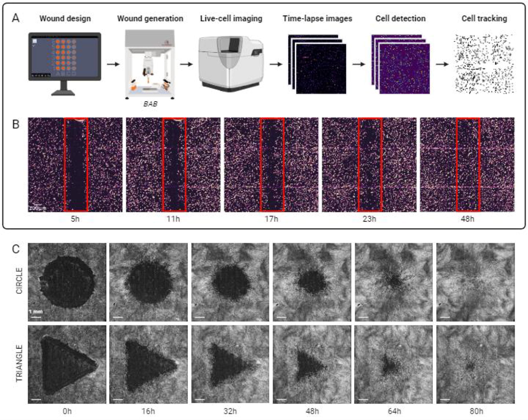
B363 - Leaf-derived Vascular Structures Support Functional Skin Regeneration
B. L. Samolyk1, L. Perreault2, G. R. Gaudette2, G. D. Pins1
1
Worcester Polytechnic Institute, Worcester, MA,
2
Boston College, Chestnut Hill, MA
*Purpose/Objectives: Every year, more than 500,000 Americans are treated for burn injuries and chronic wounds. These injuries often lead to extensive scarring and permanent loss of function. The current standard of care is a split-thickness skin autograft; however, more extensive burns may have insufficient donor sites, requiring an alternative treatment. Dermal analogs have demonstrated some success but require 2-3 weeks for vascular integration. The absence of robust internal vascular networks is typically the cause of graft failure. So, we need to develop implantable skin scaffolds that already contain vascular network structures. The project aims to develop Leaf-derived Vascular Structures (LeaVS) that support skin regeneration and provide provisional vascular support. Previous data showed that decellularized leaf scaffolds retain their vascular network structures while maintaining structural integrity. We hypothesize that the surfaces of these LeaVS can be functionalized to facilitate keratinocyte attachment and epidermal regeneration.
*Methodology: To prepare LeaVS, spinach leaves were subject to cuticle removal, decellularization, and sterilization before being stored at 4°C. Keratinocyte attachment assays were performed by seeding normal human epidermal keratinocytes (NHEKs) at 500,000 cells/cm2 on LeaVS or tissue culture polystyrene (TCP) controls treated with 0, 0.01, or 0.1% poly-L-lysine (PLL) as well as 0 or 50 μg/mL human fibronectin (FN) for 2 hours. Samples were stained with Hoechst, imaged, and quantified for cell density. Epidermal morphology assessments were performed using leaves treated with 0 or 0.01% PLL as well as 0, 10, 50, or 100 μg/mL FN. Neonatal human fibroblasts (NHFs) were seeded at 100,000 cells/cm2 on the underside of the leaves, followed by neonatal human keratinocytes (NHKs) seeded at 1 million cells/cm2 on the topside of the leaves. For decellularized dermis (DED) controls, only NHKs were seeded. Constructs were cultured submerged for 3 days, followed by culture at the air/liquid interface for 7 days. Histological analyses of hematoxylin and eosin (H&E) stained samples were performed to quantify the epidermal thicknesses.
*Results: Keratinocyte attachment assays showed that the TCP positive controls were not significantly enhanced by surface treatment. Interestingly, the LeaVS showed decreasing cell attachment as PLL concentration increased. However, the addition of FN to LeaVS resulted in a significant increase in cell density for PLL groups. Untreated LeaVS did not exhibit a significant difference in NHEK attachment compared to the untreated TCP controls. Epidermal morphology experiments demonstrated that LeaVS support the growth of a contiguous layer of keratinocytes with characteristic cobblestone morphology and progressive epithelial stratification, as well as fibroblast attachment. Interestingly, untreated LeaVS had the thickest epithelial layer (∼40 μm), compared to other treatment conditions and DED controls, although they were not as thick as native epithelial layers (∼63 μm) (Figure 1).
*Conclusion/Significance: This the first study describing the use of LeaVS for skin regeneration. We discovered that untreated leaves displayed the most promise for supporting cellular attachment and proliferation. We anticipate that these scaffolds will enable the future design of a multifunctional engineered skin substitutes for the treatment of traumatic skin injuries by facilitating rapid tissue vascularization and functional regeneration.

B364 - Gene Therapy Of Recessive Dystrophic Epidermolysis Bullosa Skin Cells Leads To Dermo-epidermal Adhesion Strength Restoration In Autologous Bilayered Self-assembled Skin Substitutes
M. A. Barbier1,2, A. Dakiw Piaceski1,2, D. Larouche1,2, K. Ghani3,2, E. Pope4, M. Caruso3,2, L. Germain1,2
1
The Tissue Engineering Laboratory / LOEX and Department of Surgery, Faculty of Medicine, Université Laval, Québec, QC, Canada,
2
CHU de Québec-Université Laval Research Centre, Québec, QC, Canada,
3
Centre de Recherche sur le cancer de l’Université Laval, Québec, QC, Canada,
4
Hospital for Sick Children and University of Toronto, Toronto, ON, Canada
*Purpose/Objectives: Although gene therapy shows great promises for the treatment of Recessive Dystrophic Epidermolysis Bullosa (RDEB), a rare genodermatosis (3/million births) in which minor mechanical stress to the skin results in blisters, scarring and severe pain, permanent restoration of type VII collagen (C7) by genetically modified cells remains a challenge. Here, we investigate the potential of ex vivo gene therapy combined with autologous bilayered skin substitutes both in vitro and in vivo.
*Methodology: Fibroblasts and keratinocytes from either healthy or RDEB patients were extracted and used to produce bilayered skin substitutes using the LOEX’s self-assembly approach. Type VII collagen expression was analysed using flow cytometry and immunofluorescence. Mechanical peeling test using the Instron 1000 (Electropulse) were used to quantify the dermal-epidermal adhesion strength.
*Results: Viral transduction efficiency of C7 in RDEB fibroblasts by gamma-retroviral particles was improved 2.6 folds (p<.05) when using EF-c peptide compared to using polybrene. Moreover, the use of EF-c did not affect fibroblasts proliferation and keratinocytes stem cell content whereas polybrene led to a decrease in both (-20% and -33%, respectively (p<.01)). Overall, we restored C7 production in 55% of fibroblasts and 65% of keratinocytes (N=3). Tissue-engineered skin produced with various combinations of untreated and corrected RDEB cells showed that corrected fibroblasts allows for a higher deposition of C7 compared to corrected keratinocytes in vitro before grafting. Quantitative adhesion strength of the dermo-epidermal junction showed comparable mechanical properties of tissue-engineered skin produced with healthy cells (2.9mN/mm) and those with corrected RDEB cells (3.9mN/mm), but not those produced with untreated RDEB cells (1.5mN/mm). After grafting on athymic mice, C7 deposition at the dermo-epidermal junction and stem keratinocytes were maintained for at least 11 months. After grafting on athymic mice, the surface area of corrected tissue-engineered skin was increased by 4 folds compared to uncorrected RDEB tissue-engineered skin. Moreover, mechanical peeling test 21 days after grafting revealed that healthy tissue-engineered skin had a fast increase in adhesion strength (25mN/mm). In contrast, tissue-engineered skin produced from untreated cells had a low adhesion strength after grafting (4mN/mm) whereas gene therapy treated tissue-engineered skin were similar to healthy tissue-engineered skin (27mN/mm).
*Conclusion/Significance: In conclusion, the ex vivo gene therapy combined with self-assembly approach shows great potential as safe and long-term treatment of RDEB wounds.
B365 - The Therapeutic Effects Of 3d Bio-cell Printed Artificial Skin Mimetic Structure Using Human-derived Skin Cells In Vivo
J. Park1, Y.-r. Kim1, D. Yoon1, J.-S. Lee1, W. Kim2, S. Chae2, W. Chun1, G. Kim2
1
Burn Institute, Hangang Sacred Heart Hospital, Seoul, Korea, Republic of,
2
Sungkyunkwan University School of Medicine, Suwon, Korea, Republic of
*Purpose/Objectives: Various skin grafting procedures, such as autografts and allografts, are being employed to address tissue injury caused by multiple factors. However, these procedures encounter challenges including immune rejection, survival issues, high costs, and limited supply. Consequently, the research and development of an artificial skin with high safety and superior functionality, utilizing biocompatible materials, remains a critical and imperative task. The utilization of 3D cell printing technology is effective in manipulating the porous structure of artificial skin, allowing the creation of diverse structures that mimic human tissue. Recent developments involve the incorporation of human-derived cells, such as keratinocytes and fibroblasts, into bioinks for applications in wound healing and skin regeneration. The objective of our research is to create artificial skin and evaluate the skin regeneration effects in immunocompromised nude mice through the graft of a multilayered biomimetic artificial skin.
*Methodology: This artificial skin is composed of the epidermis layer including keratinocytes, the basement layer infused with KGF (keratinocyte growth factor) and human-derived skin cells, and the dermis layer including fibroblast. The study utilized 3D cell printing technology to fabricate a bioink scaffold composed of collagen and fibrinogen for the epidermis and dermis layers. Three types of grafts were created: 1) a scaffold with the epidermis and dermis layers, 2) a scaffold with the epidermis layer, the dermis layer, and the basement layer containing KGF, and 3) a scaffold with the epidermis layer, the dermis layer, the basement layer containing KGF along with keratinocytes and fibroblasts. These grafts were implanted into the right dorsal skin of BALB/c Slc-nu/nu nude mice with full-thickness wounds. Following transplantation, wound size was measured at 2 and 4 weeks, and tissue samples were collected for subsequent histological analysis.
*Results: We confirmed regeneration effect of artificial skin mimetic structure through the in vivo experiments. Although wound closure time was similar in both the non-treat group and experimental groups, the results revealed that the epidermis and dermis treated with artificial skin showed remarkable regeneration in terms of thickness and density after 4 weeks. Additionally, expression of vimentin, the fibroblast-specific marker, was localized like normal tissue at the artificial skin tissue with human skin cells.
*Conclusion/Significance: It means artificial skin mimetic structure can effectively restore skin wounds. Thus, these results show that artificial skin mimetic structure with human skin cells is effective for the regeneration and reconstruction of skin at the wound.
B366
B367 - Novel Topical Tormulations Based On Salvia Haenkenium Extracts And Hyaluronan For Inflamed Skin Treatment And Dermal Regeneration
M. D’AGOSTINO, C. SCHIRALDI, A. D’AGOSTINO
UNIVERSITY OF CAMPANIA “LUIGI VANVITELLI”, NAPLES, Italy
*Purpose/Objectives: Salvia haenkenium (SH), a native plant of Bolivia is a strong inhibitor of senescence in human primary cells and was recently exploited in wound healing and generally for its potential anti-inflammatory properties. Hyaluronan at high and low molecular weight (HCC), widely explored in diverse cell models, and recently used in the clinical practice, showed beneficial effects in dermo-aesthetic and regenerative injective treatments. In this research work a novel formulation based on HCC coupled with SH were investigated to evaluate its potentiality in counteracting dermal inflammation and improving wound healing.
*Methodology: In vitro scratch test followed by time lapse videomicroscopy (okolab) De-hydration test; 2D in vitro model of psoriasis (LPS based stress), and evaluation of cell viability (hyperproliferation; cytokines production by using RT-PCR, western blot and ELISA. In addition a 3D full thickness skin model was used to further evaluate the bioactivity of the combined HCC and SH in comparison to the sole HCC.
*Results: In vitro wound healing using a time lapse videomicroscopy demonstrated HCC+SH capacity to slightly improve keratinocytes (HaCaT) migration respects the sole HCC. These data were confirmed also by biomarker modulation and especially elastin, integrin αV and aquaporin protein expression, involved in dermal tissue repair and integrity. An in vitro dehydration test, showed the ability of hyaluronan to defend the skin from dryness and its combination with SH extracts even increased the protective effect. Moreover, HaCaT treated with LPS, (E. coli derived), was used to mimic a psoriatic in vitro inflammation model. To evaluate the anti-inflammatory property of HCC+SH Inflammatory biomarkers (e.g. KRT-6, TLR-4 and NFk-B) and specific cytokines (e.g. IL-6, IL-22, IL-23) were quantified. These data helped in obtaining a molecular fingerprint of the pathological model and compare the cell response after treatments. Results by western blotting and ELISA assay confirmed the effect of SH coupled with HCC, in reducing inflammatory mediators. A more complex model, namely 3D-FT skin, was used to better resembling an in vivo condition, and confirmed the efficacy of novel formulation to counteract inflammation similar to what found in 2D model.
*Conclusion/Significance: These results trigger the interest in the novel formulation based on Salvia Haenkenium extract and Hyaluronan for its potential efficacy as natural anti-inflammatory agent for damaged skin, further prompting hyaluronan healing and regenerative properties. 3D skin model, contribute to achieve a better understanding of the biochemical features of the coupling of hyaluronan of diverse MW to a plant extracts.
B368 - Assessment Of Two Dermal Fillers PLLA-based: Size, Zeta Potential, And Impact On Fibroblast Viability
N. Maurmann1, G. M. Machado2, R. P. Silveira1, L. R. Wailer1, P. Pranke1,3
1
Universidade Federal do Rio Grande do Sul (UFRGS), Porto Alegre, Brazil,
2
Universidade Luterana do Brasil (ULBRA), Canoas, Brazil,
3
Instituto de Pesquisa com Células-tronco (IPCT), Porto Alegre, Brazil
*Purpose/Objectives: Biodegradable polymer particles, such as poly(L-lactic acid) (PLLA), have been utilized for their regenerative properties to address the loss of soft tissue volume. PLLA is a biomaterial commonly used as a key component in dermal fillers, contributing to their regenerative and volumizing properties. Two examples of dermal fillers that predominantly consist of PLLA particles are Rennova®Elleva and Sculptra®. The aim of this study was to characterize Rennova® Elleva and Sculptra® and evaluate the metabolic activity of fibroblasts cultured in the presence of these biomaterials.
*Methodology: Optical microscopy followed by ImageJ and zeta potential analysis were utilized to characterize the dermal fillers Rennova® Elleva and Sculptra®. Fibroblasts (MRC5 cell line) were exposed to Rennova® Elleva or Sculptra® at concentrations of 0.5 or 1.0 mg/ml of PLLA. After a 2-day incubation period following exposure to the products, cell viability was assessed through the MTT assay, and cell visualization was conducted using a live-dead stain. The results were expressed as the mean ± standard deviation (SD) and evaluated using ANOVA followed by the Tukey test.
*Results: The microscopy evaluation showed that both products had distinct irregular morphology. Rennova® Elleva had a shape similar to flakes, while Sculptra® looked like crystals. The particle area of Rennova® Elleva was measured to be 833 ± 419 µm², while for Sculptra®, the values were 799 ± 652 µm², with no significant difference between the two. The zeta potential was -40.0 ± 8.4 mV for Rennova® Elleva and -55.5 ± 9.0 mV for Sculptra®. The MTT assay absorbance values for the control (0 mg/ml) after 2 days were 0.187 ± 0.006. Rennova® Elleva did not significantly affect MRC5 viability, with values of 0.207 ± 0.023 for 0.5 mg/ml and 0.187 ± 0.023 for 1.0 mg/ml. However, Sculptra® resulted in decreased cell viability after 2-day contact with MRC5 cells, with values of 0.164 ± 0.018 for 0.5 mg/ml and 0.157 ± 0.017 for 1.0 mg/ml (p < 0.05).
*Conclusion/Significance: This study compared the characteristics and cellular effects of two injectable biostimulators: Rennova® Elleva and Sculptra®. The characterization findings indicate that the particles are micrometric in size, have a negative superficial charge, and are stable against coalescence. According to ISO 10993-5 standards, the viability assessment indicated that the fillers did not have cytotoxic effects on fibroblasts as the cell viability was not reduced by more than 30%. Financial support was provided by the National Council for Technological and Scientific Development (CNPq), Financier of Studies and Projects (FINEP), National Institute of Science and Technology for Regenerative Medicine (INCT-Regenera), and Stem Cell Research Institute (IPCT).
B369 - Assessment Of Hyaluronic Acid Degradation And Cell Viability In Stem Cells, Fibroblasts, And Keratinocytes
N. Maurmann1, E. M. Fiorentini2, G. M. Machado2, M. C. Brew2, P. Pranke1,3
1
Universidade Federal do Rio Grande do Sul (UFRGS), Porto Alegre, Brazil,
2
Universidade Luterana do Brasil (ULBRA), Canoas, Brazil,
3
Instituto de Pesquisa com Células-tronco (IPCT), Porto Alegre, Brazil
*Purpose/Objectives: Facial rejuvenation is a highly desired cosmetic procedure in contemporary society. Hyaluronic acid facial fillers are biomaterials used to enhance and restore facial volume, reduce wrinkles, and rejuvenate the skin. This technique is one of the most commonly used for optimizing facial aesthetics. The study aimed to assess the cellular viability of fibroblasts, stem cells, and keratinocytes when exposed to various commercially available hyaluronic acids, as well as the degradation of the materials.
*Methodology: Degradation was quantified by mixing the compounds with fluorescein and measuring the release in saline phosphate buffer using spectrophotometry at 405 nm. After culturing MRC5 fibroblasts, mesenchymal stem cells, and keratinocytes HaCaT for 24 hours, they were treated with 1mg/ml Juvéderm® Ultra Plus XC, Juvéderm® Volux, Saypha Volume and Saypha Volume Plus. After an additional 24h, cell viability was evaluated using 2,5-diphenyltetrazolium (MTT) and the live/dead assay. The results are presented as the average ± standard deviation of absorbance percentage. Statistical analyses were conducted using ANOVA followed by the Tukey test.
*Results: No significant differences were found between the release of fluorescein from the free dye (control) and the products (p=0.138). The mean and standard deviation values of the percentage of absorbance in relation to the control group as follows: 92±6 (p=0.397) for the Saypha Volume group, 86±8 (p=0.002) for the Saypha Volume Plus group, 78±7 (p=0.000), for Juvéderm® Ultra Plus XC and 72±7 (p=0.000) for Juvéderm® Volux, with the control group having a value of 100±14. Most of the tested hyaluronic acids significantly reduced the viability of fibroblasts. The percentage of absorbance in relation to the control group had the following mean and standard deviation values: 100±14 for control, 92±6 (p=0.397) for the Saypha Volume group, 86±8 (p=0.002) for the Saypha Volume Plus group, 78±7 (p=0.000) for Juvéderm® Ultra Plus XC, and 72±7 (p=0.000) for Juvéderm® Volux. The values for keratinocytes were 100±10, 96±8 (p=0.948), 87±12 (p=0.035), 94±5 (p=0.636), 94±6 (p=0.762), and 1±0 (p=0.000), respectively, for the control group, Saypha Volume, Saypha Volume Plus, Juvéderm® Ultra Plus XC, Juvéderm® Volux, and the death control group. The values for stem cells were as follows: 100±16 for the control, 104±11 (p=0.955) for Saypha Volume, 101±7 (p=1.000) for Saypha Volume Plus, 105±7 (p=0.939) for Juvéderm® Ultra Plus XC, 115±11 (p=0.048) for Juvéderm® Volux, and 1±0 (p=0.000) for the death control.
*Conclusion/Significance: This study did not find any significant differences in the release rates of fluorescein from four hyaluronic acids fillers when compared to the free dye. Saypha Volume did not statistically reduce fibroblast viability. Juvéderm® Ultra Plus XC and Juvéderm® Volux did not affect the viability of keratinocytes. However, Juvéderm® Volux increased stem cell viability. Although the viability of fibroblast and keratinocytes decreased significantly with some hyaluronic acids, the reduction was less than 30%, and therefore they are not considered cytotoxic according to ISO 10993-5. Financial support: National Council for Technological and Scientific Development (CNPq), Financier of Studies and Projects (FINEP), National Institute of Science and Technology for Regenerative Medicine (INCT-Regenera) and Stem Cell Research Institute (IPCT).
B370 - Development Of Stem Cell-derived Recombinant Proteins-loaded Alginate-gelatin Hydrogel For Wound Healing
Y. Ju1, D. Green1, J. Kim1, S. Lee1, B. Vaughan1, J. Jackson1, A. Atala1, C. Nykiforuk2, J. Yoo1
1
Wake Forest Institute for Regenerative Medicine, Winston-Salem, NC,
2
Emergent BioSolutions, Winnipeg, MB, Canada
*Purpose/Objectives: Autografts and allografts are the “gold-standard” for the most implemented clinical treatments for full-thickness skin wounds; however, skin grafts have several treatment limitations, such as limited donor site availability, morbidity, and the need for immunosuppressive drugs to prevent graft rejection. These limitations have led to the development of numerous noncellular dermal substitutes, and cell-based tissue engineering approaches as more suitable wound treatment options for patients. This study aimed to develop a novel therapeutic option using tailored multiple factors (i.e., CMF, conditioned media factor) based on human placental stem cell (HPSC)-derived conditioned media for wound healing therapy.
*Methodology: To deliver CMFs, we prepared an Alginate-gelatin hydrogel combination and investigated their physical characteristics (water absorption, stability). The biological activity and the release profile of the hydrogel delivery system were determined in vitro. To determine the therapeutic efficacy of tailored CMFs, we implanted and evaluated CMFs-loaded hydrogel in a rodent full-thickness wound defect model.
*Results: We observed that the factors-loaded Alginate-gelatin hydrogel provides appropriate physical characteristics, non-cytotoxic, and controlled release of the loaded model drug (Dextran). The therapeutic efficacy study in vivo showed that treatment of CMFs significantly accelerated wound closure, tissue formation, and matured re-epithelialization. In addition, CMFs-loaded hydrogel demonstrated the increased matured vessel diameter and basket-weave collagen network, which is phenotypically similar to native skin tissue.
*Conclusion/Significance: These results suggested that the tailored CMFs-loaded alginate-gelatin combination gel carriers could be a promising and effective therapeutic solution for full-thickness wound healing applications.
B373 - Silk/dopamine Compositedhydrogels With Multi-drugs To Improve Wound Healing For Diabetic Rats
T.-W. Chung, Y.-F. Liu
National Yang-Ming CT University, Taipei, Taiwan
*Purpose/Objectives: To improve wound healing for diabetic subjects, using multifunctional drugs with composited new hydrogels for varing sub-objectives is preferred.
*Methodology: For those objectives, an anti-oxidative and NIR photothermal responsive (Anti-Ox/PTR) SD/FC ((silk/dopamine) / Fucoidan) hydrogels were produced, using catalyzing by horseradish peroxidase (HRP), and H2O2, to produce as a wound dressing and increase the moisture of the wound areas. Multifunctional drugs with SD nano-particles (NP) form were produced, too, which were Neurotensin, Fucoidan and N-Acetyl Cysteine (NT, FC and NAC, as NFA) were coated onto the surface of SD NP using a layer-by-layer technique, To produce Anti-Ox/PTR composited hydrogels, those multifunctional drugs loaded NPs were loaded into Anti-Ox/PTR SD/FC hydrogels.
*Results: SD-NFA NPs, with the particle size/zeta potential for 224.61±2.89 nm/1.36 ± 0.43 mV, repectively. Each drug layer coated onto the surface of SD NP was verified by the changes of particle size/zeta potentials. Moreoevr, the SD-NFA NPs and SD/FC hydrogels were all biocompatible examined by the ISO cell tests.The rates of accumulative drug releases of NAC and NT for SD-NFA NPs, the NPs/hydrogels and the NPs/hydrogels/NIR (e.g., periodical NIR irradiation for 5 min/each time) systems were 54.5 ± 0.94%, 67.7 ± 1.11% and 78.3 ± 1.43%; 27.9 ± 1.42%, 及 43.7 ± 1.45% and 58.7 ± 0.36 %, respectively, with a good linearity for each release for up to 240 h. Hence, the NIR PTR effects on the releases of NAC and NT were significantly triggered for the NPs/hydrogels/ NIR system compared to those for the NPs/hydrogels.
In-vitro
scratch-wound assay showed that the system for SD-NFA NPs in SD/FC hydrogels significantly promoted wound closure than each NP and the hydrogel system.
In-vivo
wound healing for diabetic rats also showed that the system for NPs/hydrogels with periodical NIR irradiations significantly promoted wound closure with growing new blood vessels than other systems.
*Conclusion/Significance: Hence, the system for SD-NFA NPs in SD/FC hydrogels as a wound dressing in corporate with a periodical NIR irradiation is excellent for advanced skin tissue engineering. No disclosure is needed.
B375 - Human Hair Keratin Based Tissue Adhesives For Skin Tissue Engineering
R. TUTAR1, D. CEYLAN TUNCABOYLU2, B. ÇELEBİ-SALTIK3
1
Istanbul University-Cerrahpaşa, İstanbul, Turkey,
2
Bezmialem Vakif University, Istanbul, Turkey,
3
Hacettepe University, Ankara, Turkey
*Purpose/Objectives: Background: Functinal biomaterials as wound dressing are becoming increasingly important in enhancing wound healing. In this study, it was aimed to prepare methacrylate-modified gelatin (GelMA)/human hair keratin1 based tissue adhesives, which were applied in-situ to the wound area and showed chemically crosslinked with light and michael addition reaction in the joint environment. In addition, the prepared tissue sealants adhesion properties were examined for skin tissue engineering.
*Methodology: Methods: A reduction reaction method was followed to obtain keratin from human hair. Chemical cross-linking was created between the active -SH groups in the reduced keratin structure and the methacrylated groups of methacrylated gelatin by Michael addition reaction. In vitro and ex vivo tissue sealing properties of prepared tissue adhesives were performed by using burst pressure, lap shear and wound closure tests.
*Results: The keratin concentration was increased in the prepared tissue sealants, the mechanical strength of the material changed from fracture to ductility. In the presence of keratin, hydrogen bonds in GelMA increase and the polymer chains change the interaction both within and between adjacent polymers. As a result, the GelMA control material broke with the applied compressive force and withstood up to approximately 25 kPa in the burst pressure test. As the Keratin ratio increased in GelMA, the increasing ductility behavior also showed its effects on the in vitro and ex vivo tissue sealing properties.
*Conclusion/Significance: Conclusion: Keratin protein for the application by prioritizing the natural tissue structure for the biomimetic tissue adhesives desired to be created. Finally, a new generation injectable human hair keratin-added tissue adhesives which are biodegradable, compatible with the skin, capable of strong bonding and cross-linking under Michael addition reaction and visible light were prepared.Acknowledgement: This study was financially supported by The Scientific and Technological Research Projects Funding, TUBITAK-1001/121M509.References:1.Tutar R., Koken S.Y., Tuncaboylu D.C.,Çelebi-Saltik B., Özeroğlu C., “In Situ Formation of Biocompatible and Ductile Protein-Based Hydrogels by Michael Addition Reaction and Visible Light Crosslinking”, RSC_New Journal of Chemistry, 2023, 47, 10759-10769, DOI: 10.1039/D3NJ01230A.Keywords: Biomaterials, Human Hair Keratin, Hydrogels, Tissue Sealing
B376 - Title: Evaluation Of Skin Patch Bioprinted With Skin Cells In The Hybrid Hydrogel Of Gelatin And Silk Fibroin
W. Zhao
Wake Forest Institute for Regenerative Medicine, Winston Salem, NC
*Purpose/Objectives: It is still difficult to treat large skin wounds. More efficient therapies are needed. Extrusion-based bioprinting with cell-loaded hydrogel has become an enabled technology for managing skin wounds. It is critical to develop skin-specific hydrogel that is compatible with skin-related cells and possesses proper mechanical properties.
*Methodology: A 3D bioprinted hydrogel patch is designed for accelerating acute wound healing, which is fabricated by bioprinting skin-related cells in methacryloyl-substituted gelatin (GelMA) and silk fibroin (SilMA) and then crosslinked by ultraviolet light (UV). The hybrid hydrogel of GelSilMA exhibits suitable biocompatibility and mechanical properties (See figure A).
*Results: GelSilMA hydrogel displays extended biodegradation and improved mechanical properties. Furthermore, the formulated GelSilMA hydrogel can maintain a structural and moisturized environment persistently and then benefit cell migration and neo tissue formation in wound area. In vivo study demonstrates that the improved microenvironment by the application of GelSilMA hydrogel patch could speed up wound closure (See figure B).
*Conclusion/Significance: We have developed a hybrid hydrogel of GelSilMA. GelSilM hydrogel is gelatinized by one step UV crosslinking. GelSilMA hydrogel presents its therapeutic potential in the management of skin wounds.Figure. (A) Schematic for preparing GelMA, SilMA, and dual-crosslinked GelSilMA hydrogel. (B) Schematic illustration of GelSilMA hydrogel patch in promoting wound healing.
B377 - Insights Into The Use Of Exosomes In Skin Regenerative Medicine: A Brief Review
A. Golchin, A. Mihanfar
Urmia University of Medical Sciences, Urmia, Iran, Islamic Republic of
*Purpose/Objectives: Exosomes are small nano-vesicles found in extracellular space that carry a variety of biomolecules including miRNA, mRNA, DNA, and proteins. Exosomes have undergone extensive investigation, manipulation, and use in biomedicine, serving as a carrier for medication delivery, a means for genetic modification, and as immunizations. Moreover, stem cell-derived exosomes have been suggested as an attractive approach to treating various diseases, particularly wound healing and skin regeneration, without parent cells. Studying exosomal cargo components offers valuable insights for predicting and treating wound healing and skin tissue engineering. It is important to note that the composition of exosomal cargo may vary depending on the parent cells and where they are located.
*Methodology: This is a rapid review of the evidence for the potential effects of exosomes on wound healing and skin tissue engineering. To do so, health technology assessment studies indexed in PubMed, Scopus and Google Scholar were searched using the related keywords.
*Results: Based on our literature review, by developing cell engineered products and developing combination therapies, such as combining exosomes with platelet rich plasma and/or fibrin, these nano vesicles have opened up a new perspective in cell-based therapies. As such, exosomes have the potential to revolutionize the field of skin tissue engineering and wound healing, offering a promising avenue for therapeutic development and medical applications. In recent years, regenerative medicine has witnessed remarkable advancements, largely due to cutting-edge technologies like CRISPR, single cell technology, three-dimensional (3D) printing, organoids, and microfluidics. Among these breakthroughs, exosomes have emerged as a particularly promising option for addressing dermatological conditions, offering unprecedented opportunities for healing and skin tissue engineering.
*Conclusion/Significance: Therefore, exosomes are a promising tool that should further be explored for their potential to improve patient outcomes in the field of skin tissue engineering and wound healing.
B378 - Development Of Fibrin Enriched Dermal Paste As A Treatment For Chronic Non Healing Wounds Biomaterial For Treatment Of Chronic Non Healing Wounds
P. Sawadkar1, K. Greco1, K. Rombouts2, J. Hua1, F. Lali1
1
The Griffin Institute- NPIMR, London, United Kingdom,
2
UCL, London, United Kingdom
*Purpose/Objectives: Chronic non-healing wounds present a significant global health challenge, affecting over $13 million individuals and imposing an economic burden exceeding $15 billion on healthcare systems and are on the rise due to increasing levels of obesity and diabetes in an aging population (Ochoa et al., 2020). Chronic non-healing wounds are severe clinical problems. Grafting is the preferential method when the wound defect is exceedingly large, and wound closure cannot be attained due to primary intention. However, there is a gap between the demand and supply of required tissue. The currently available treatment options have several limitations including technical challenges of surgical interventions, as well as costly hospital bed occupancy followed by nursing care at home, when practicable. To address this issue, we developed an acellular allogeneic fibrin enriched dermal paste (FEDP) using reverse engineering to treat these wounds. The aim of this study was to determine the safety and efficacy of an allogeneic porcine derived FEDP for clinical application to treat human full thickness non-healing wounds.
*Methodology: Material characterization was conducted employing micro computed tomography, scanning electron microscopy (SEM), degradation assays, and mechanical testing to elucidate the paste’s physical and biological attributes. The biological properties, including cellular proliferation, viability, and angiogenic potential, were assessed through proliferation assays, viability essays, and chorioallantoic membrane (CAM) assays. In vivo, efficacy was tested using six porcine models, where circular wounds of 2 cm diameter and 1 cm depth were created and isolated with polytetrafluoroethylene chambers. The wounds were treated with the FEDP and observed over 4 and 8 weeks. Post-termination analysis included proteomics, single cell RNA sequencing, and immunohistochemistry.
*Results: Results demonstrated that the pore range of paste was in the range of 50 -500 µm, which was ideal for wound healing and in vivo results involving comparative analysis with untreated samples indicated that FEDP facilitated comparable wound closure and remodelling within the observed time with a significant (p < 0.05) increase in collagen deposition, angiogenesis granulation tissue formation, and epithelialization, biomaterial engraftment, degradation, and host cellular invasion by 8 weeks compared to 4 weeks. RNA seq data indicated wound base inflammatory reactions were similar between treated and control wounds with migration of various cell populations and proteomics data showed neo collagen deposition at the 8 week than 4 weeks.
*Conclusion/Significance: FEDP was demonstrated to be systemically safe. Clinical pathology data was within the normal range. These findings suggest that FEDP represents a promising alternative for the management of chronic non-healing wounds, potentially overcoming the limitations of existing treatment strategies. Further clinical investigations are warranted to fully establish the therapeutic efficacy and applicability of FEDP in human subjects.

B380
B381 - An Engineered Model To Elucidate Molecular Clutch Mechanisms Of Mechanotransduction During Bone Nanoscale Mineralization
S. M. Vignolo, A. Avathamsa, L. E. Bertassoni
Oregon Health & Science University, Portland, OR
*Purpose/Objectives: Ossification is an important developmental process where bone is created in appositional layers through the gradual calcification of the native extracellular matrix (ECM). This evolution is synchronously orchestrated by biological processes that allow for mesenchymal condensation of stem cells and their differentiation into osteoblasts and osteocytes. While bone mechanobiology has been studied in skeletal development and fracture healing, the interplay between mechanics and biology during the transition of calcifying osteoid to mature bone remains poorly understood. Understanding and replicating bone formation is imperative to support regeneration in critical-sized bone defects. Our objective is to replicate the critical changes in the ECM during bone formation to study the step-by-step process of bone regeneration in vitro, as current models are limited. Here, we employ a nanoscale materials engineering strategy to recreate the densification and calcification of bone tissue in a controllable manner using a polymer-induced liquid-precursor (PILP) based strategy for intrafibrillar mineralization. Ultimately, we hope to advance the fundamental understanding of the mechanobiology behind bone formation, contributing to future research on regenerative therapies.
*Methodology: Collagen hydrogels were fabricated in 4 ways to model the evolving ECM during bone formation: 1) soft collagen, 2) high density fibrillar collagen (HDFC), 3) partially mineralized HDFC, and 4) fully mineralized HDFC. Collagen densification was achieved by compacting the soft collagen via centrifugation. The HDFC hydrogels were mineralized using a modified biomimetic calcification medium (α-MEM, osteopontin, CaCl2, K2HPO4) for 1 and 3 days to represent partial and full mineralization, respectively. The hydrogel model was characterized by alizarin red staining, scanning electron microscopy, and Fourier-transform infrared spectroscopy. Bone-marrow derived human mesenchymal stem/stromal cells (MSCs) were seeded on the surface of the hydrogels. Activation of the molecular clutch and mechanotransductive signaling in response to different stiffness conditions was evaluated through immunofluorescent staining for focal adhesion kinase (FAK), phosphorylated paxillin (p-paxillin), Yes-associated protein 1 (YAP-1), and Ras homolog family member A (RhoA). Similarly, osteogenic differentiation was measured by nuclear expression of Runt-related transcription factor 2 (RUNX2). Quantification was performed using ImageJ.
*Results: Immunofluorescent staining of YAP/RhoA and FAK/paxillin at 2 and 24 hours, respectively, indicated higher expression in dense and calcified samples, evidencing high stiffness sensing, formation of adhesion complexes, and engagement of the molecular clutch under these conditions. Additionally, there was an increased nuclear expression of RUNX2 at 24 hours in dense and calcified samples, signifying a greater differentiation into bone under these conditions. These findings align with cellular morphological response and live cell imaging data, demonstrating robust cellular adherence and motility within one hour of seeding.
*Conclusion/Significance: In summary, we successfully replicated the stepwise changes in the ECM that occur during bone formation, creating a bone-like environment to mimic the ossification process. We observed differences in cellular behavior in response to sequential densification and mineralization in this model which grant the ability to explore further mechanistic questions regarding the cellular response during bone regeneration. The findings of this work contribute to understanding the molecular clutch of stem cells during the complex process of bone regeneration.

B382 - Novel Chick Embryo Window Culture Model And The Effects On Development
C. D. Hart, K. Henderson, R. Efrem, C. K. Kuo
University of Maryland, College Park, MD
*Purpose/Objectives: The chick embryo is an established model to study tissue formation that has led to major discoveries relevant to tissue regeneration, immunology, and more. The chick embryo uniquely enables direct manipulations of the embryo through a window (hole in eggshell) during development. Unfortunately, the standard windowing technique (“side-windowing”) is associated with viability as low as 33%. One potential reason for poor viability is that side-windowing requires partial albumen removal, which is undesirable as removal of 10% of albumen volume leads to 12% viability. Another potential reason is that egg rocking, necessary for normal development, is stopped after side-windowing to avoid spilling the albumen. Side-windowing must be performed by embryonic day (D)3, before the chorioallantoic membrane (CAM) adheres to the shell. Unfortunately, non-rocking during D3-D8 decreases hatchability to 84% of rocked eggs. A window culture technique that overcomes these challenges is needed. Here, we establish “aircell-windowing” at the natural aircell of the egg. We show that this technique can be performed without albumen removal and at later developmental stages to extend the duration of rocking.
*Methodology: White leghorn chicken eggs were incubated with rocking until side-windowing at D3 (SW-D3) or aircell-windowing at D6 and D9 (AW-D6, AW-D9) (Fig. 1a,b). Side-windowing required removing ∼10% of total albumen volume and cutting a hole in the eggshell side. Aircell-windowing involved cutting a hole in the shell at the aircell and puncturing the CAM. Windowed eggs were incubated without rocking to prevent spillage. Non-windowed (NW) eggs in a rocking incubator (NW-Rock) or eggs moved to a non-rocking (NR) incubator at D3, D6, or D9 were controls (NW-NR-D3, NW-NR-D6, NW-NR-D9). At D18, viability was determined, and embryos were sacrificed, staged, and weighed. One-way ANOVA with Tukey’s multiple comparisons or Kruskal-Wallis test with Dunn’s multiple comparisons were used based on normality of the data.
*Results: Viability of AW-D9 was higher than SW-D3, but similar between AW-D6 and SW-D3 (Fig. 1c). Non-windowed groups had 100% viability. Embryo stage and whole embryo mass of SW-D3 were significantly lower and more variable than NW-Rock and AW-D9 (Fig. 1d). The heart masses of all non-rocking groups (windowed and non-windowed) were lower than NW-Rock, but heart masses of windowed groups were no different from non-rocking controls, reflecting that windowing did not impact heart mass. Liver and lung masses were lower than NW-Rock in SW-D3 and AW-D6 but not in AW-D9 (Fig. 1e). AW liver and lung masses were similar to their NW-NR controls, reflecting that aircell-windowing did not impact organ masses. In contrast, the liver and lung masses of SW-D3 were significantly lower than that of NW-NR-D3, indicating that side-windowing compounded the impacts of non-rocking. We attribute higher viability and normal staging of aircell-windowed embryos to windowing after D8 and omission of albumen removal, and recommend aircell-windowing at the latest stage possible to maximize rocking during development.
*Conclusion/Significance: The dramatic improvement in viability and late-stage embryonic development with aircell-windowing overcomes significant obstacles to studying late-stage tissue formation, paving the path to exciting new directions for developmentally-informed tissue engineering and regeneration strategies.
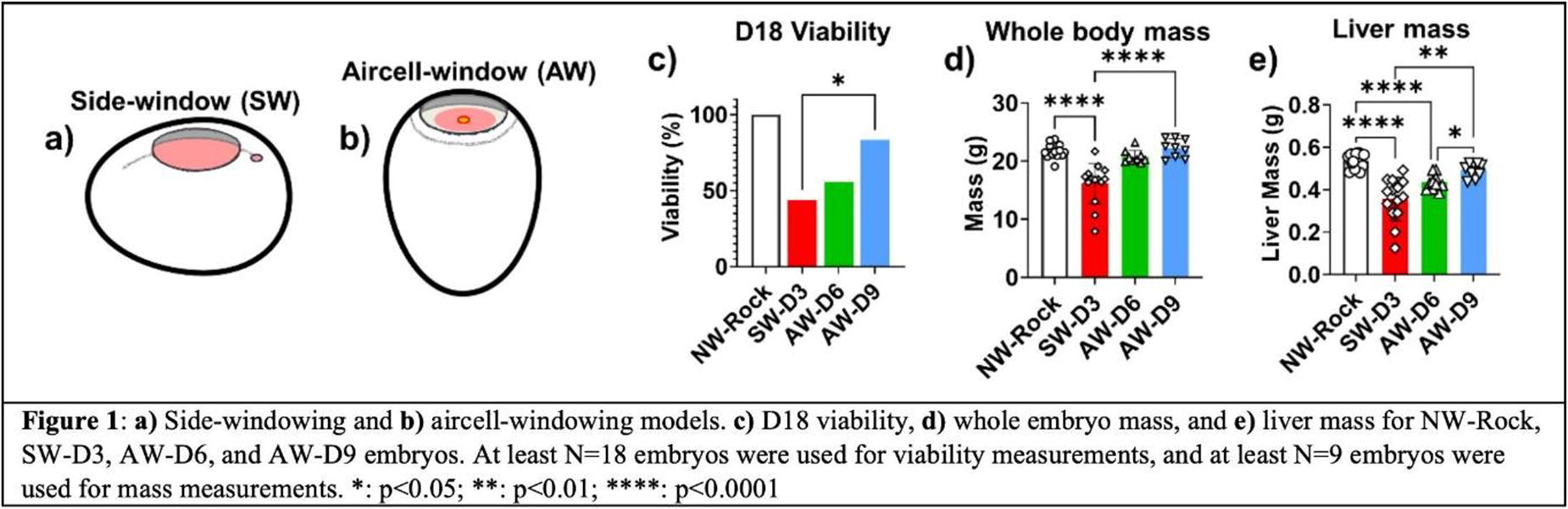
B383 - Development Of Osteoclast-derived Exosomes For Cardiovascular Calcification Therapy
I. Jennings1, M. Donato1, H. Zhou1, S. Kuncorojakti2, M. Purwaningrum3, C. Giachelli1, M. Scatena1
1
University of Washington, Seattle, WA,
2
Universitas Airlangga, Surabaya, Indonesia,
3
Universitas Gadjah Mada, Yogyakarta, Indonesia
*Purpose/Objectives: Extracellular vesicle nanoparticles, like exosomes, have therapeutic potential for drug delivery and regenerative medicine because of their targeting properties, composition depending on the cell source, low immunogenicity and high biocompatibility. Osteoclasts (OC) are mineral resorbing cells indispensable for bone homeostasis. OC-derived exosomes have known bone regulatory properties by directly affecting the function of osteoblasts, the cells depositing the bone mineralized matrix. Specific cargo of OC-derived exosomes has been shown to hinder the mineralization potential of osteoblasts (Li et al, Nat Comm 2016). Ectopic calcification (EC) is defined as the deposition of a mineralized matrix in soft tissues often resembling bone-like structures. EC can occur in the vasculature, kidneys, brain, and joints. Importantly, no pharmaceutical therapies exist to treat soft tissue bone-like mineral deposits. Calcific aortic valve disease (CAVD), a type of EC, is the most prevalent heart valve disorder and the third most prevalent cardiovascular disease in the western world. We and others have shown that valve interstitial cells (VICs) isolated from aortic valves deposited a mineralized matrix when stimulated with elevated inorganic phosphate in vitro (Scatena et al, Card Path 2018). In this study we have characterized the proteome of murine osteoclast-derived exosomes and assessed their function on mineralizing VICs.
*Methodology: Exosomes were purified from conditioned media collected from murine myeloid precursor cells, RAW264.7 (control cells), and OC differentiated from RAW264.7 cells by treatment with 80 ng/ml of murine RANKL. The protein content of control cell- and OC-derived exosomes was determined using proteomic analyses. Non-human primate VICs were cultured in control medium containing 1 mM inorganic phosphate and calcification medium containing 2.6 mM phosphate for 7 days. Control cell- and OC-derived exosomes were added to the VIC cultures for the duration of the mineralization experiment. Calcium deposition in the VIC cultures was determined colorimetrically using the o-Cresolphthalein complexone kit.
*Results: Proteomic analyses revealed that OC-derived exosomes contained elevated OC-specific proteins including TRAP, MMP9, cathepsin K, V type proton ATPase subunits, and several integrins when compared to control cell-derived exosomes. Further, OC-derived exosomes, but not control cell-derived exosomes, prevented calcification of non-human primate VICs cultured in calcification medium. These results suggest that factors contained in OC-derived exosomes have anti-mineralizing properties. Current experiments are addressing possible mechanisms of this novel function of OC-derived exosomes. Further, the RNAs cargo of the different types of exosomes is currently being assessed.
*Conclusion/Significance: Exosomes from OCs and control cells were successfully isolated and demonstrated differential protein content. The novel function of calcification prevention was established for OC-derived exosomes. These exosomes will be further characterized to identify molecular targets with the aim of developing new therapies for CAVD and other EC pathologies.
This work was supported by grant NIH 5R35HL139602 to Cecilia Giachelli.
B384 - Dynamic Cell Culture Promotes Human Tendon Stem/progenitor Cell-derived Extracellular Vesicle Production
M. Clerici1,2, M. C. Ciardulli2, P. Scala2, E. P. Lamparelli2, V. Giudice2, J. Lovecchio3,3, E. D. Giordano4, T. P. Dale1, N. R. Forsyth1,5, N. Maffulli6, G. Della Porta2
1
Keele University, Keele, Staffordshire, United Kingdom,
2
University of Salerno, Baronissi, Italy,
3
Reykjavík University, Reykjavík, Iceland,
4
University of Bologna, Cesena, Italy,
5
University of Aberdeen, Aberdeen, United Kingdom,
6
Sapienza University, Rome, Italy
*Purpose/Objectives: Tendon injuries significantly impact an individual’s quality of life, prompting the exploration of novel strategies beyond conventional surgical methods. Extracellular Vesicles (EVs) present a promising potential strategy for promoting tendon regeneration by delivering specific biological signals to neighbouring cells. This study focuses on isolating and culture-expanding human Tendon Stem Progenitor cells (hTSPCs) from surgical explants and characterizing EV production under both perfused and static conditions.
*Methodology: hTSPCs were isolated from tendon surgery biopsies (Review Board prot./SCCE n.151, 29/10/2020), tested for stemness properties by using flow cytometry (CD14, CD34, CD73, CD90, CD105 and HLA-DR) and trilineage differentiation (adipo, osteo and chondro-genesis), and cultured in Growth Differentiation Factor-5 (GDF-5) supplemented media to promote tenogenic differentiation. Then, hTSPCs were cultured in static and dynamic conditions using a custom perfusion bioreactor. Upon reaching 80% confluence, cells were transitioned to a serum-free medium for 24 hours to collect conditioned media (CM). Ultra-centrifugation was then employed to isolate EVs from the CM, and the resulting pellet underwent comprehensive characterization, including size and concentration measurements using the Nanosight NS300 system, morphology examination via scanning electron microscopy (SEM) and transmission electron microscopy (TEM), and quantification of protein content through Bradford assay, Western blot, and MACSPlex Exosome kit.
*Results: hTSPCs demonstrated stemness and multipotency through the positive expression of CD73, CD90, and CD105, and negative expression of CD14, CD34 and HLA-DR, along with histological confirmation of trilineage differentiation. GDF-5 supplementation upregulated the tenogenic genes SCX-A, TNMD and COL3A1, and cells showed a more elongated tenocyte-like morphology with an aspect ratio increase. After ultracentrifugation, Nanosight analysis revealed the presence of particles with a sub-200 nm diameter and concentration of 7x10^6 particles/mL, in both culture conditions. The dynamic EV pellet showed a three-fold increase in total protein concentration. Western blot analysis confirmed the presence of EV-associated proteins CD81 and TSG-101 exclusively within the dynamic EV pellet. MACSPlex analysis revealed an increased expression of EV markers, particularly CD63 and CD45 in dynamic conditions, while CD9 displayed higher expression in static conditions.
*Conclusion/Significance: Data suggests efficient harvesting of sub-200 nm vesicles from serum-starved hTPSCs, aligning with existing EV research. Dynamic culture conditions appeared to favour EV production, though further characterization is essential for a comprehensive understanding of EVs’ role in tendon regenerative processes. Future plans will include testing their potential effect to instruct stem cells (e.g. mesenchymal stem cells) towards a tenogenic lineage.

B385 - On-chip Loading Of Anti-cancer Drug In Extracellular Vesicles
C. Piunti1,2, E. Cimetta1,2
1
University of Padova, Padova, Italy,
2
Fondazione Istituto di Ricerca Pediatrica Città della Speranza, Padova, Italy
*Purpose/Objectives: Despite contributing to cancer progression, extracellular vesicles (EVs) could serve as potential drug delivery systems in cancer treatment, having the ability to dissolve water-insoluble drugs and facilitating targeted delivery.
However, the clinical translation of EVs is still in its infancy. While traditional methods for EV modifications will remain relevant, microfluidic approaches are expected to replace benchtop methods.
Taking advantage of lab-on-chip devices, passive cargo loading through microfluidic mixing and incubation may be an important strategy to produce functional engineered EVs.
This study focuses on developing a microfluidic device to generate EVs loaded with Verteporfin (VP), a hydrophobic porphyrin with potential applications in Neuroblastoma (NB) therapy, aiming to enhance its tumor specificity and therapeutic effectiveness.
*Methodology: The platform is designed with AutoCAD® and modelled with Comsol Multiphysics® so that both a perfect mixing between EVs and VP occurs within the mixing unit, and the desired incubation time of 10 minutes is achieved. The mold is produced via photolithography, and PDMS devices via replica molding. Finally, plasma treatment is used to form an irreversible hydraulic seal to a glass coverslip. The platform is used to perform both fluid dynamic validations using food coloring and biological validations using Mesenchymal Stem Cells (MSCs)-derived EVs and VP. Once isolated using ultrafiltration, MSC-derived EVs are characterized by Nanoparticle Tracking Analysis, Western Blot and Transmission Electron Microscopy, confirming their successful isolation. The stability of EVs and VP-EVs is measured in PBS using Zeta-Potential. Finally, Immunofluorescence assay and Western Blot are used to evaluate the differences in YAP expression after treatment with VP-EVs in a NB cell line.
*Results: The platform (Figure 1) is formed by two sections: the micro mixing unit (A-a) and the incubation section (A-b) that allows precise incubation times as a function of the imposed flow rate. A detail of a Comsol® simulation is shown in panel B. Fluid dynamic validation confirms both perfect mixing and incubation times (C). Biological experiments are conducted using MSCs-derived EVs and VP. After on-chip loading, the VP-EVs can be easily and efficiently retrieved, making them readily available for subsequent analysis Considering spectral properties, VP-loaded EVs display similar absorption profiles compared to free VP, confirming the highly efficient loading of VP into EVs. The Encapsulation Efficiency (EE%) is significantly higher for our new on-chip protocol, again confirming that efficient loading of VP into EVs is obtained (D). VP loading also increased the EVs’ Zeta Potential (E). With VP identified as a YAP/TAZ complex inhibitor, Immunofluorescence (F-a) and Western Blot (F-b) preliminary results demonstrate that VP-EVs internalized into NB cells effectively alter YAP expression compared to free VP. Thus, VP-EVs treatment could prevent YAP from accumulating and ultimately suppress the progression of NB cells.
*Conclusion/Significance: The loading method here employed is non-destructive and technically simple, allowing application with a broad range of therapeutic molecules. Thus, as a versatile tool, microfluidic technology will help to fully unlock the potential of EVs for speeding up precision medicine and disease treatment.
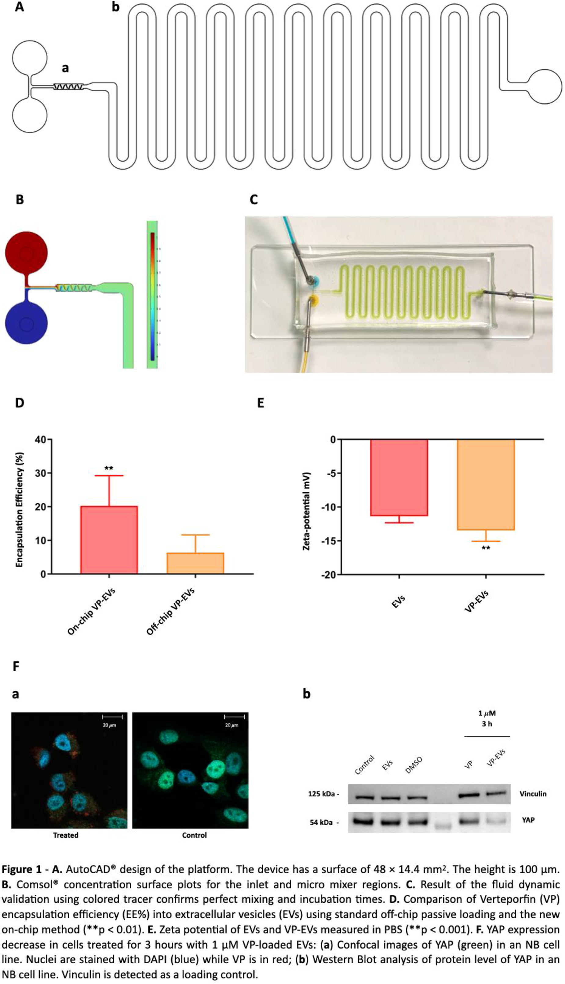
B386 - Detection Of Endothelial-specific Extracellular Vesicles In Saliva For Monitoring Heart Health
W. Meza1, S. Ayus1, J. Jiménez1, M. Buendia1, E. Suarez2, L. Cunci3, D. Freytes4, C. Mora1
1
University of Puerto Rico-Mayaguez, Mayagüez, PR,
2
University of Puerto Rico-Ponce, Mayagüez, PR,
3
University of Puerto Rico-Rio Piedra, San Juan, PR,
4
The University of North Carolina at Chapel Hill, North Carolina, NC
*Purpose/Objectives: Extracellular vesicles (EVs) in saliva, particularly endothelial, are an efficient way to monitor heart health and patient status after a regenerative treatment (e.g., cell therapy, biomaterial injection, etc.). Therefore, monitoring endothelial-related extracellular vesicles (endothelial-EVs), identifiable by endothelial protein markers such as CD144+ and CD31+ in bodily fluids like saliva, blood, and sweat, is a very promising route for real-time diagnosis of heart health status. Our research endeavors to develop a specialized gold biosensor that captures and detects endothelial-EVs within heterogeneous populations obtained from saliva samples. Our study aims to establish a calibration curve utilizing screen-printed electrodes functionalized with antibodies to capture the CD144 protein biomarker found on EVs isolated from human saliva to be used as a critical parameter for monitoring and, therefore, as a diagnosis tool. This approach will enable selective identification and quantification of endothelial-derived EVs, providing crucial insights into their presence and potential diagnostic applications within saliva specimens.
*Methodology: : The human saliva-derived EVs sample was procured from System Biosciences (SBI), while the screen-printed electrodes were sourced from BASI Researcher Products. Fabrication of the biosensors followed the Self-assembled Monolayers (SAMs) technique. First, a Thio alkane carboxylic acid was reacted, followed by CD144 antibody conjugation facilitated by N-(3 dimethylaminopropyl)-N′-ethylcarbodiimide (EDC). Post-functionalization, the electrodes were employed for detecting saliva vesicles. In our analysis, trachea-derived cartilage EVs served as a negative control for the CD144 protein, while serum EVs procured from SBI were used as a positive control. EVs characterization was carried out by Nanoparticle Tracking Analysis (NTA) for particle size distribution and transmission electron microscopy (TEM) was used to define the morphology. The Immunodetection techniques were used to detect and quantify CD144 protein. EVs studies require an orthogonal approach, and we used atomic force microscopy and a potentiostat to characterize the functionalized electrodes and detected EVs.
*Results: The maximum particle size peak of EVs from human saliva was determined as 181 nm via NTA. The TEM images yielded particle sizes of 121 nm and 141 nm. The Dot Blot assay confirmed the presence of CD144 within the saliva EVs, while ELISA assays validated and quantified a concentration of 0.812 ng of CD144 protein/mL. Finally, Electrochemical Impedance Spectroscopy assays demonstrated the functionalization of the electrodes as well as the selective detection of EVs with CD144 protein as cargo. This was demonstrated by topographic analysis with AFM in tapping mode.
*Conclusion/Significance: Successfully developing a biosensor with selective detection capabilities for EVs carrying the CD144 protein marked a significant achievement. This advancement led to the establishment of a promising calibration curve, holding substantial potential for monitoring heart health.
B387 - Human Adipose-derived Stem Cell Exosomes Rescue Airway-related Cells Damaged By Exposure Of So2 Derivatives
J. LEE
Burn Institute, Hallym University, Seoul, Korea, Republic of
*Purpose/Objectives: Inhalation burns, especially when combined with thermal burns, can be fatal and significantly increase the mortality rate through inhaling substances such as carbon dioxide, sulfur dioxide, hydrogen cyanide, and nitric oxide. However, there is no specific treatment to care inhalation burns except for treatments such as relieving bronchospasm and cleaning the airways. Especially, sulfur dioxide (SO2) is a major component of inhalation burn as well as common air pollutant, and these inhaled SO2 can easily be hydrated in the respiratory tract to produce sulfurous acid, which subsequently dissociates to form bisulfite (NaHSO3) and sulfite (Na2SO3) derivatives. Generally, therapeutic effect of stem cells derived-exosome have been reported in different diseases like cardiovascular, heart, kidney, liver, neurological diseases, and wound healing. In this study, we intend to assess whether human adipose-derived stem cells (hASCs) exosomes rescue airway-related cells damaged by exposure of SO2 derivatives.
*Methodology: Process of isolating and purifying exosomes from hASCs conditioned media, Matrigel tubule forming assay (Tube formation assay), Proliferation assay (cell viability assay), RNA isolation and quantitative real-time polymerase chain reaction (PCR) analysis, PKH67 labeling of hASCs exosome and uptake of exosome in cells.
*Results: As a results, hASCs exosomes were well-taken in airway-related cells (e.g. Human Pulmonary Microvascular Endothelial Cells (HPMECs), Human Nasal Epithelial Cells (HNEpCs), Human Bronchial Epithelial Cells (HBEpCs)). hASCs exosome rescue decreased proliferation of airway-related cells by treatment of SO2 derivatives. In case of HPMECs, total tubule length was increased by pretreatment of hASCs exosomes through in vitro angiogenesis assay. Next, we confirmed that pretreatment of hASCs exosomes rescue increased expression of inflammation-related genes by treatment of SO2 derivatives in primary respiratory epithelial cells, like HBEpCs and HNEpC.
*Conclusion/Significance: Taken together, these results suggest that hASCs exosomes can be used as a therapeutic agent in treatment of inhalation burns.
B388 - Biomaterials-based Mass Production Of Mesenchymal Stem Cells And Extracellular Vesicles For Personalized Bone Tissue Regeneration
S.-H. Lee
Dongguk University, Gyeonggi-do, Korea, Republic of
*Purpose/Objectives: Understanding the biophysical relationships between stem cells and applied biomaterials can facilitate the ability to control the functions and behaviors of stem cells. However, the role of biomaterials and especially the 3D microenvironment in stem cell biology remains largely unexplored, compared with that of the 2D cell-culture environment. I would like to introduce 3D hydrogel systems to accelerate cellular reprogramming. Next, I will also introduce a novel technique for mesenchymal stem cells (MSCs) isolation from pluripotent stem cells (PSCs) by utilizing a specific integrin interaction of MSC on a biomaterial. In addition, I will briefly introduce recent works about various approaches to cell and exosome therapy for personalized regenerative medicine. Therefore, I believe these approaches would be very useful in preparing a standard protocol providing personalized mesenchymal stem cells and exosomes for bone tissue regeneration.
*Methodology: We utilized polymer-based photo-cross-linkable hydrogels to encapsulate cells within 3D microenvironments in a biocompatible condition. Before synthesizing the photo-cross-linkable hydrogel, we determined what biomaterials might be suitable for improving the reprogramming or pluripotency. Cell culture plates were coated with gelatin, hyaluronic acid (HA), alginate, chitosan, type I collagen (Col I), fibronectin, or laminin. OCT4-GFP-expressing embryonic stem cells (OG-ESCs) were cultured on these plates without mouse leukemia inhibitory factor (mLIF), a key supplement necessary for the maintenance of pluripotency in ESCs. In the second topic, spontaneously differentiated ESCs were subcultured on different matrixes, specifically none-coated, gelatin-coated (conventionally used ECM), poly-l-lysine (PLL)-coated (no integrin-mediated binding caused by electrostatic interactions), and FN-coated (mainly integrin α;;5β;;1-mediated binding) tissue culture plates, respectively. An efficient and simplified technique for the isolation of MSCs from spontaneously differentiated human embryonic stem cells (hESCs) via integrin
α
;;5
β
;;1 (fibronectin (FN) receptor)-to-FN interactions (hESC-FN-MSCs) is successfully developed.
*Results: Among the various 3D hydrogel systems tested, methacrylated hyaluronic acid (HA) hydrogel significantly improves cellular reprogramming into iPSCs. HA hydrogel with low modulus accelerates reprogramming into iPSCs and thus offers potential advantages for translational applications. Additionally, the initial upregulation of CD44 in encapsulated cells in low-level methacrylated soft HA hydrogel accelerates the reprogramming. Among the various 3D hydrogel systems tested, methacrylated hyaluronic acid (HA) hydrogel significantly improves cellular reprogramming efficiency into iPSCs 10 times higher than conventional 2D conditions. On the other hand, hESC-FN-MSCs exhibit a typical MSC surface phenotype, and cellular morphology, with the whole transcriptome similar to conventional adult MSCs; but show higher proliferative capacity, more efficient trilineage differentiation, enhanced cytokine secretion, and attenuated cellular senescence. This isolation technique on fibronectin shows 30 times higher than that on gelatin. Thus, combining two techniques will provide massive production of personalized MSCs 300 times compared to conventional methods. Based on personalized MSCs, the exosomes were engineered with specific cell/tissue targeting and then applied to bone tissue regeneration.
*Conclusion/Significance: By engineering the MSCs and the exosome, furthermore, the engineered exosomes with cell/tissue targeting greatly enhanced bone tissue regeneration. These novel methods will be useful in the generation of abundant amounts of clinically relevant MSCs and exosomes for personalized bone tissue regeneration.
B389 - Exosomes-derived From Human Fetal Cartilage Progenitor Cells (hfcpcs) Inhibits Osteoarthritis (oa) Phenotypes Of Il-1b-treated Sw982 Cells
J. Lee1, S.-H. Park2, S. Park1, B. Choi1
1
Inha University, Incheon, Korea, Republic of,
2
Pukyong National University, Busan, Korea, Republic of
*Purpose/Objectives: We have previously reported that human fetal cartilage progenitor cells (hFCPCs) have anti-inflammatory and immune-modulatory effects (Cell Transplantation, 28:932-942, 2019). In this study, we investigated anti-inflammatory and immune-modulation effects of exosomes derived from hFCPCs primed with different methods (unprimed control vs IL-1β treatment, hypoxia, and 3D culture) in comparison with those of human bone marrow mesenchymal stem cells (hBM-MSC).
*Methodology: hFCPCs were isolated from the cartilage tissue of GA 12-14 fetuses with IRB approval (AJIRB-CRO-07-139). hBM-MSCs were purchased from CEFO (Seoul, Korea). hFCPCs were cultured in DMEM-HG and unprimed or primed with IL-1β, hypoxia, or 3D culture for 48 hr. hBM-MSCs were cultured in DMEM-HG by the conventional 2D method without priming or IL-1β priming. Anti-inflammatory effect of exosomes was investigated on an osteoarthritis (OA) model using SW982 synoviocytes treated with 0.5 ng/mL IL-1β for 24 hr. Unprimed or primed exosomes (10 mg/mL) were treated to cells for 48 hrs and expression of pro- (IL-1β, IL-6 and MCP-1) or anti-inflammatory (SOCS1 and SOCS3) genes were examined by qRT-PCR. We performed miRNA sequencing (ebiogen, Seoul, Korea) of exosomes to understand the mechanisms of their effects.
*Results: Non-primed hFCPCs exosomes decreased the expression of IL-1β, IL-6, and MCP-1 in IL-1β-treated SW982 cells. Primed exosomes showed stronger inhibitory effects, particularly in the hypoxia and hypoxia/IL-1β combination groups. hBM-MSCs exosomes showed similar results in both non-primed and primed groups. Through miRNA sequencing data of exosomes, we could identify miRNA differentially expressed (DE) in hFCPCs. Confirmation studies were performed to understand the roles and action mechanisms of DE miRNAs and miR-373-3p was shown to be expressed highly in the hypoxia-primed exosomes. Synthetic miR-373-3p mimic showed a significant inhibitory effect on the IL-1β-treated OA model in SW982 cells. miR-373-3p inhibitor was found to partially block the anti-inflammatory effect of the hypoxia-primed exosomes. miR-373-3p mimic or inhibitor of IL-1β suppressed the expression of IRAK4 involved in the NF-kB signaling pathway.
*Conclusion/Significance: This study found that exosomes derived from hFCPCs have anti-inflammatory and immune-modulatory effects in vitro and priming of cells particularly with hypoxia further increases their modulatory activity. miRNA-373-3p could be one of the key molecules mediating the hypoxia-primed exosomes from hFCPCs.
B390 - Genetic Engineering Of The CD81 Large Extracellular Loop For Targeted Delivery Of Mesenchymal Stem Cell Derived Vesicles For Spinal Cord Injury Repair
D. J. Santos1,2,3, D. Hercher2, K. Stadlbauer4, G. Wozniak-Knopp4, J. Grillari2,3,5, A. J. Salgado1,6
1
Life and Health Sciences Research Institute (ICVS), School of Medicine, University of Minho, Braga, Portugal,
2
Ludwig Boltzmann Institute (LBI) for Traumatology, The Research Centre in Cooperation with AUVA, Vienna, Austria,
3
Institute of Molecular Biotechnology, Department of Biotechnology, University of Natural Resources and Life Sciences (BOKU), Vienna, Austria,
4
Christian Doppler Laboratory for Innovative Immunotherapeutics, Institute of Molecular Biotechnology, Department of Biotechnology, University of Natural Resources and Life Sciences (BOKU), Vienna, Austria,
5
Austrian Cluster for Tissue Regeneration, Vienna, Austria,
6
ICVS/3B’s – PT Government Associate Laboratory, Braga/Guimarães, Portugal
*Purpose/Objectives: Spinal cord injury (SCI) is a devastating condition. Its consequences are related with the acute and chronic inflammatory response, as well as the formation of an inhibitory environment for axonal outgrowth that culminates in the formation of a glial scar, limiting the onset of regenerative processes, and thus resulting in lifelong impairments. Systemic administrations of secretome derived from mesenchymal stem cells (MSCs) have been shown to improve motor function in SCI rodent models. However, the extracellular vesicles (EVs) contained within the secretome do not localize specifically to the lesioned spinal cord and are mainly captured by organs of the reticuloendothelial system such as the liver and the spleen. Here, we aim at engineering the large extracellular loop (LEL) of CD81, a molecule present in EVs, in order to increase their homing ability to the lesion site.
*Methodology: To achieve this, we use HEK-293 cells to recombinantly express: 1) proteins of the myelin sheaths that get disrupted during the acute phase of SCI (myelin oligodendrocyte glycoprotein and myelin associated glycoprotein) and 2) chondroitin sulphate proteoglycans that are upregulated at the glial scar in the subacute to chronic phases (neurocan and phosphacan). A yeast display system is then used to select mutant versions of the LEL of CD81 with binding ability for the aforementioned proteins by consecutive rounds of magnetic- and fluorescence-activated cell sorting. The next objective is to do lentiviral transfection of the selected CD81 mutant versions into MSCs. The secretomes from transfected MSCs will be used in organotypic rat spinal cord slice cultures and in a rat model of contusive SCI to assess whether the genetically modified myelin- and glial scar-binding CD81 molecules increase the homing ability of their EVs to the lesioned spinal cord, and whether that enables to increase their therapeutic potential.
*Results: Using a yeast display system expressing mutated versions of the LEL of CD81 (highly expressed in EVs), we are currently selecting for mutant versions with binding ability to myelin oligodendrocyte glycoprotein and neurocan (present in the acute and chronic SCI lesion microenvironments), that were recombinantly expressed in HEK-293 cells.
*Conclusion/Significance: The engineered myelin- and glial scar-binding CD81 versions hold the potential to increase the homing capability of EVs to the SCI site, thereby maximizing the therapeutic impact of systemic MSC secretome administrations for SCI.
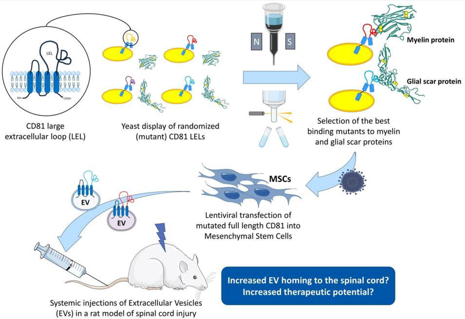
B391 - Matrix-bound Nanovesicles: Ties To Collagen And Encapsulation In Biomaterials
M. J. Dewey1,2, G. S. Hussey2, S. S. Badylak2
1
University of California Santa Barbara, Santa Barbara, CA,
2
University of Pittsburgh, Pittsburgh, PA
*Purpose/Objectives: Matrix-bound nanovesicles (MBV) are nano-scale extracellular vesicles (EV) secreted by cells and isolated from the extracellular matrix (ECM). MBV are similar to exosomes (liquid-phase EV) in size and shape, but these have distinctly different lipid profiles and miRNA cargo. MBV have demonstrated immunomodulatory and wound healing capabilities in applications such as rheumatoid arthritis and optic nerve repair. MBV have the potential to be used as a therapeutic and diagnostic tool; however, we lack an understanding of their biogenesis and how these bind to the ECM. Additionally, these have yet to be incorporated into biomaterial platforms and we aim to utilize these as effective biomolecules for bone regeneration applications.
*Methodology: ECM-related surface markers (integrins) on both MBV and exosomes from the same cell source (human mesenchymal stem cells (MSC), fibroblasts) were assessed by western blotting. Additionally, these cells were cultured on glass coverslips and SEM identified MBV location in regard to collagen production. We assessed whether increases in collagen production related to increases in MBV production by applying various concentrations of ascorbic acid to cell cultures. Furthermore, exosomes and MBV will be added to collagen gels and 3D-printed to create collagen-EV biomaterials. We will examine EV release kinetics, distribution in biomaterials, and impact of collagen-EV biomaterials on MSC osteogenesis. All exosomes were isolated from cell culture media via ultracentrifugation and size exclusion chromatography. ECM deposited by cells was enzymatically digested to release MBV, which were isolated via ultracentrifugation. EV populations were verified by particle tracking analysis, TEM, and surface markers.
*Results: We report a process for isolating MBV and exosomes from the same cell source. MBV were enriched in integrins α5, αV, β1, β5, indicating possible mechanisms for ECM binding. SEM imaging revealed vesicles bound to collagen fibers produced by cells, even after subsequent washing and preparation steps. Increasing ascorbic acid (1000 uM) concentration increased MBV production while this had no impact on exosome production. Furthermore, we will present ongoing work encapsulating MBV and exosomes into 3D-printed collagen gels. We will report EV release profiles, distribution, and impact of exosomes and MBV on MSC viability, proliferation, migration into biomaterials, mineralization, and expression of osteogenic genes and proteins.
*Conclusion/Significance: The present work expands our knowledge of MBV. Tethering of MBV to the ECM may be the result of additional integrins on the surface of MBV, and these could be used to identify MBV from exosomes. MBV may offer an easier method to bind to ECM-based biomaterials for wound repair than other EV. Furthermore, there is a relationship between collagen formation and MBV production, and ascorbic acid can be used to enhance MBV yield. By comparing encapsulation and release of MBV and exosomes in collagen-based biomaterials we will develop an understanding for the affinity and binding of MBV and exosomes to ECM-based biomaterials. As MBV are a critical component of healthy ECM, we expect MBV are a key currently missing component of biomaterials fabrication and will accelerate bone repair strategies.
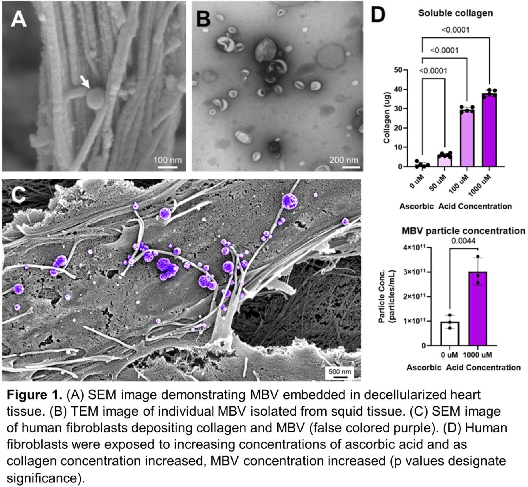
B393 - Assessment Of Personalized EV-Enriched PRF Dressings
K. Spanoudes1,2, A. Samourides3
1
University of Nicosia, Nicosia, Cyprus,
2
VET EX MACHINA LTD, Nicosia, Cyprus,
3
ORGANOGEN LTD, Limassol, Cyprus
*Purpose/Objectives: Delayed wound healing remains a major healthcare burden, which is a cause of patient suffering for large groups of patients, such as diabetics, who are currently 500 million worldwide,The tissue engineering and biomaterials society has dedicated a significant amount of effort to engineer solutions for delayed wound healing and improve patient quality of life. The objective of this work was to create personalized platelet-rich fibrin (PRF) wound dressing materials impregnated with extracellular vesicles (EV’s).
*Methodology: PRF was fabricated from blood collected from purposely-bred adult New Zealand White Rabbits. PRF dressings were subsequently freeze-dried, while EV’s were prepared and loaded at a density of 109 per sample.
For the in vivo assessment the rabbit ear wound was utilized. 9 wounds were inflicted on the ear of each rabbit. The nine wounds were distributed equally between the following three groups:1) EV loaded PRF dressings - three wounds per animal2) PRF dressings (PC) - three wounds per animal3) Untreated wounds - - three wounds per animalThe treatment was not repeated until euthanasia and tissue harvesting on day 14.The wounds were photographed, and wound healing was assessed on days 3,5,7,10,14 with ImageJ software.Upon euthanasia, the tissues were collected, fixed, sectioned and dyed with hematoxylin & eosin for histological assessment.
*Results: Macroscopic assessment revealed that all groups have healed with similar rate, and 100% closure was observed by day 14. However Scar formation was more evident on the NC samples (Fig.1).
*Conclusion/Significance: In conclusion, PRF treatment with EV can reduce scar formation in an in vivo model. In the future, studies in diabetic models, will investigate the benefit of personalized wound dressings for chronic wounds.

B394 - Preparation And Characterization Of Artificial Extracellular Vesicles Using High Hydrostatic Pressurized Cells
K. Kaiden1, T. Kimura1, Y. Hashimoto1, K. Akiyoshi2, Y. Sasaki3, A. Kishida1
1
Institute of Biomaterials and Bioengineering, Tokyo Medical and Dental University, Tokyo, Japan,
2
Department of Immunology Graduate School of Medicine, Kyoto University, Kyoto, Japan,
3
Department of Polymer Chemistry, Graduate School of Engineering, Kyoto University, Kyoto, Japan
*Purpose/Objectives: Extracellular vesicles (EVs) are lipid bilayer particles that mediate cell-to-cell information and are involved in physiological phenomena and disease progression. It is desirable to elucidate the functions of EVs and apply them to novel diagnostic and therapeutic methods. EVs are obtained from cultured cells and biological tissues, but the process is complicated and it is difficult to obtain sufficient yields. To solve these problems, many researchers have been developing methods to artificially produce EVs. One of methods is to produce EVs-mimicking particles from cells by extrusion over polycarbonate membrane filters. On the other hand, it is known that treatment of cancer cells with high hydrostatic pressure (HHP) induces immunogenic cell death (ICD). ICD is cell death that is likely to induce an immune response because antigenic proteins in the dead cell membrane are remained, resulting HHP-treated cancer cells be used to cancer vaccination. From these backgrounds, we hypothesized that the downsizing of HHP-treated cells by filtering using extruder could be used for the preparation of artificial EVs. In this study, artificial EVs with dead cell information were prepared using various HHP-treated cells.
*Methodology: HeLa, MC3T3-E1 and ATDC5 cells were used. Each cell was cultured until 80-90% confluent. Cells were collected by trypsinization and were suspended in medium and subjected to HHP treatment (250, 500 and 1000 MPa). The percentage of dead cells in HHP-treated cells was determined by PI staining and cell number counting. The shape of HHP-treated cells was observed by scanning electron microscopy (SEM). HHP-treated cells were suspended in PBS and were filtered through polycarbonate membranes with pore diameter of 1.0 µm, 0.4 µm and 0.1 µm, stepwisely, using an extruder to obtain artificial EVs. Artificial EVs were characterized by transmission electron microscopy (TEM) and RNA analysis using Bioanalyzer™. Artificial EV size and concentration were measured by nanoparticle tracking analysis (NTA).
*Results: HHP treatment induced cell death of over 90%, irrespective of pressure treatment condition. SEM observation revealed that the cells remained spherical shape after HHP treatment with multiple small holes on the surface. The HHP-treated cells were subjected to filtration using extruder to obtain artificial EVs. For TEM observation, it was founded artificial EVs consisted of lipid bilayer membrane. RNA analysis in artificial EVs indicated that artificial EVs induce small RNAs, with or without HHP treatment. Also, for NTA, it was founded that the artificial EVs of 104 particles with 100-200 nm were obtained from a HHP-treated cell, irrespective of pressure conditions.
*Conclusion/Significance: We have successfully prepared artificial EVs consisting of lipid bilayer membrane by downsizing HHP-treated cells using extruder. The artificial EVs including small RNAs was obtained with the diameter of 100-200 nm. Further research for properties and biological functions of HHP-treated cells and artificial EVs is needed.
B399 - Comparison Of Osteoclast Differentiation From Human Monocytic Precursors And Induced Pluripotent Stem Cells
S. Malayaperumal, J. Simon, A. Blümke, M. Scatena, C. M.Giachelli
University of Washington, Seattle, WA
*Purpose/Objectives: Ectopic Calcification (EC) encompasses a heterogeneous group of pathologies characterized by the extraskeletal deposition of bone-like mineral in soft tissues, such as blood vessels, cardiac valves, joints and muscles. EC may be a consequence of genetics, metabolic disease, trauma or surgery. Risk factors include chronic kidney disease, cardiovascular disease, and diabetes. EC can lead to organ dysfunction and failure and is associated with considerable morbidity and mortality, yet no preventative or therapeutic approaches currently exist. Furthermore, while resection and replacement (in the case of cardiac valves) are commonly used for EC, these surgical methods come with significant risks. Thus, a non-surgical stem cell therapy to remove EC is appealing. Osteoclasts are multinucleated cells that have the ability to resorb bone and play important roles in bone remodeling. Previous studies by our lab and other researchers have shown that engineered murine osteoclast cell therapy could resorb necrotic bone as well as heterotopic ossifications in mouse preclinical models. To translate these finding to the clinic, the production and characterization of human osteoclasts is required. In this study, we aim to investigate the ability of THP-1 myeloid cells and human induced pluripotent stem cells to construct a robust human osteoclast model for further translational studies.
*Methodology: The THP-1 human myleoid cell line was differentiated into osteoclasts with a protein kinase C agonist, phorbol myristate acetate (PMA), followed by RANKL and M-CSF for 14 days. Osteoclasts were differentiated from either human peripheral blood mononuclear cell (PBMC)-derived iPSC or fibroblast-derived iPSCs using an embryoid body-based strategy as previously described. Osteoclast-specific marker staining for Cathepsin K and tartrate-resistant acid phosphatase (TRAP and TRAP-5b) in addition to functional mineral resorption assays were performed. Quantitative real time PCR was used further investigate gene expression. Comparative analyses of gene expression patterns between osteoclasts produced from THP-1 cells, iPSC and native human PBMCs was performed.
*Results: After treatment with PMA, THP-1 cells differentiated into macrophages with elongated lamellapodia, achieving a 95% cell adherence rate. In the absence of PMA, the cells remained small, spherical and non-adhesive. Following RANKL and M-CSF treatment, THP-1 cells fused to form large, asymmetric TRAP+ multinuclear cells. In both iPSC lines, the ALP expression pattern was high during the early expansion phase and decreased during mesodermal and hematopoetic differentiation phases. Following hematopoetic differentation, subpopulations of CD45+, CD14+, and CD11b+ cells were obtained, which differentiated into multinucleated cells that expressed TRAP and Cathepsin K and could resorb mineral. Comparison of gene expression in osteoclasts from Thp-1, human iPSC and native human PBMC-derived OCs is presented.
*Conclusion/Significance: OCs could be successfully differentiated from human iPSCs as well as the Thp-1 myeloid cell line using optimized protocols that included M-CSF and RANKL. These cells will be used for further biological characterization and translational studies with the aim of developing new therapies for EC.
B400 - Novel XEPC Restores Blood Supply By Stimulating Postischemic Neovascularization Under Diabetic Conditions
C. Chang, K. Song, P. Park, J. Lee, H. Lee, H. Cho, S. Yoo
YOUTH BIO GLOBAL, Seoul, Korea, Republic of
*Purpose/Objectives: Diabetes causes poor blood circulation, dull sensation, and less resistance to bacterial infection. Even if there is a wound on the foot, it is difficult to feel, and due to the lack of healing function and resistance to bacteria, mild wounds progress rapidly, resulting in serious complications such as ulcers and gangrene. Severe diabetic-ischemic disease of the lower extremities is a disease of the peripheral artery, and the flow of blood to the end of the leg is caused by vascular obstruction, and in severe cases, there is a risk of limb amputation. Endothelial progenitor Cells (EPC) can be collected from bone marrow, peripheral blood, umbilical cord blood, etc., but there is a disadvantage that it does not exist in large quantities in cells. Xeno-free EPC (X-EPC) we developed is the world’s first vascular stem cell therapy that can be commercialized through mass production of heterogeneous blood-derived and endothelial progenitor cells with stem cell ability. X-EPC has characteristics of both E-EPC and L-EPC. We developed xeno-free medium that did not contain non-human-derived component. When an animal-derived medium is used for cell culture, there is a possibility that heterologous proteins and cells may be injected into the patient’s body, and in this case, unpredictable serious side effects may occur. Therefore, we developed a stem cell culture system that excludes animal proteins and cells from the aspect of stability. In this study, we purposed to evaluate the blood flow recover effect of X-EPC in a diabetic ischemia mouse model.
*Methodology: Seven-week-old nude mice were used. Diabetes was induced by single intraperitoneal injection of streptozotocin(150mg/kg). Four weeks after diabetes induction the animal were divided into 5 groups. X-EPC was cultured by differentiating mononuclear cells isolated from umbilical cord blood, and these cells were injected intramuscularly (0.5x10⁵, 1x10⁵ and 5x10⁵ cells in phosphate buffered saline/head) after one day the femoral artery ischemia of mice. Paw perfusion evaluation was in both hindlimbs using a Laser Doppler, and measurements were performed weekly 1 to 6 weeks after treatment. The femur muscle tissue was then harvested for histological examination. Also, we were measured to the body weight, blood glucose level and foot necrosis of mice.
*Results: The blood glucose concentrations were significantly higher in the diabetic mice than in control mice. Also, body weight was lower in the diabetic mice than in the control. Feet necrosis levels of the diabetic mice did not differ significantly regardless of the treatment given. Treatment of X-EPC in the hindlimb restored paw blood flow in diabetic ischemia mice. The highest blood flow response was noted at a dose of 0.5x10⁵ cells with a response comparable to that in the control group. X-EPC decreased infiltrating cells number in femur muscle tissue. Additionally, X-EPC increased CD31 or α-SMA positive capillary density. However, infiltrating cells number and capillary density was not statistically significant.
*Conclusion/Significance: With the effects of angiogenesis and anti-infiltrating X-EPC reestablishes structural and functional femur vascular. This is highly promising for future treatment of ischemia or foot ulcer from diabetes causes.
B401
B402
B404 - Scale-up And Automation Of Stem Cell-derived Human Pancreatic Islets
L. Smith, D. Soria, F. Aftanas, S. Saxena, D. Hile, T. Bollenbach
ARMI, Manchester, NH
*Purpose/Objectives: Type 1 Diabetes (T1D), which afflicts nearly 40 million Americans, develops in patients in which the beta cells of the pancreatic islets are destroyed by autoimmune attack. Diabetic individuals currently face lifelong dependence on insulin injections; however, implantation of replacement pancreatic islets derived from pluripotent stem cells (SC-islets) holds the promise of a cure. The mission of the Advanced Regenerative Manufacturing Institute is to enable the scalable, consistent and cost-effective manufacturing of cells, tissues and organs. Here, we demonstrate scalable and automated production of SC-islets for distribution to laboratories funded by the Juvenile Diabetes Research Foundation in support of their search for an SC-islet-based cure for T1D. Ultimately, this scalable process will be able to produce SC-islets under GMPs for human therapeutic use.
*Methodology: A published six-stage SC-islet differentiation protocol was transferred to ARMI and was initially scaled from tissue culture flasks to multilayer cell culture vessels, and was then adapted to the institute’s modular automation platform. Development of a scaled, modular, automated and closed manufacturing process is supported through the institute’s Deep Tissue Characterization Center, which enables data-driven identification of in-process and final product markers that support scalability and process control. Comparability between processes at various scales, and between manual and automated processes at large scale was investigated by a combination of predefined quality control specifications for the final product and by untargeted and targeted deep characterization, using a variety of analytical methods, of the differentiation process at each of its stages.
*Results: Using our approaches, we were able to scale up islet differentiation to 1000 fold using multilayered cell culture vessels, using a published protocol previously shown to be effective across multiple pluripotent cell lines. The resulting islets were comparable to those generated in the small scale, manual process in their cellular composition and in their ability to secrete insulin, as measured by the glucose stimulated insulin secretion assay. Untargeted multi-omics analysis in-process has enabled us to distinguish between and predict successful versus unsuccessful production of SC-islets at all scales and manual/automated runs as early as Stage 1 of differentiation. Rigorous untargeted, multi-omics characterization is now providing the opportunity to visualize trends to work towards predictive process models, including those that will shed light on the nature of donor-to-donor variability for autologous production.
*Conclusion/Significance: Using our process optimization, enabled through deep characterization, we are moving towards scaled-up manufacturing of pancreatic islets, which will eventually result in many more patients having access to the therapy, eventually increasing patient quality of life.
Poster Session III
Thursday, June 27, 2024, 6:15 PM - 7:45 PM
C1 - Light-based Bioprinting Of Vascularized Fatty Tissue For The Development Of Biological Breast Implants
N. Hedemann1, A. Thomas2, N. Tribian1, A.-K. Amler2, S. Krüger1, I. Flörkemeier1, J.-P. Weimer1, N. Maass1, L. Kloke2, D. Bauerschlag1, M. van Mackelenbergh1
1
Kiel University and University Medical Center Schleswig-Holstein, Kiel, Germany,
2
Cellbricks GmbH, Berlin, Germany
*Purpose/Objectives: Breast cancer is the most common cancer in the population and cause for the suffering of millions each year, moreover presenting a tremendous burden for our healthcare systems. Resection of the primary tumor is clinical standard procedure and increases treatment success enormously, but leaves the affected with a compromised body integrity. As the patients’ quality of life after cancer treatment has gained more focus in recent years, reconstructive breast surgery following mastectomy is increasing in importance. However, current options including autologous adipose tissue (AT) transfer (flap surgery or lipofilling) and artificial soft tissue implants are insufficient to this end and for soft tissue reconstruction in general, demanding a new generation of breast implants. Bioprinting biological breast implants in life-like size and customizable shape with minor insult to the patient’s body and excellent biocompatibility, exclusively using biomaterials and patient-specific adipose cells, could open up this new chapter for breast reconstruction
*Methodology: In our work, we created vascularized AT constructs using our light-based multi-material bioprinting platform along with an innovative material system and an adapted 3D culture protocol to investigate the potential use of such technology for future reconstructive breast surgery. The fabricated adipose tissue (FAT) models were designed to mimic native human AT by cellular and material composition. Human adipose stem cells, fibroblasts and endothelial cells were bioprinted and differentiated for 4 weeks to form matured constructs with key features of AT.
*Results: Live imaging showed high viability of FAT models over the whole cultivation period and pronounced cellular reorganization. Microscopic and histological analyses revealed a dynamic tissue maturation process with increasing proportions of differentiated adipocytes, ECM remodeling and formation of macro- and microvascular structures within the constructs.
*Conclusion/Significance: This proof of concept of bioprinting vascularized AT could lay the groundwork for the development of all-natural biological breast implants.
C2 - Enabling Efficient Extracellular Vesicle Production Via Osmosis
J. J. Lai1, C. Y. Huang1, H. Nguyen2, D. J. Lundy2
1
National Taiwan University of Science and Technology, Taipei, Taiwan,
2
Taipei Medical University, Taipei, Taiwan
*Purpose/Objectives: Extracellular vesicles (EVs), nanoscale lipid bilayer vesicles secreted by cells, contain rich, complex cargos of peptides and miRNAs, with their surfaces decorated with bioactive proteins. EVs facilitate intercellular communication, making them an attractive option for use as standalone therapeutics or as drug-delivery vehicles. However, there are significant limitations to their use due to technical challenges related to isolation and purification in sufficient quantities and purity. For example, ultracentrifugation is time-consuming with high batch-to-batch variability, and precipitation methods produce low-purity extracts. Our group has developed devices that employ osmosis to concentrate analytes, improving lateral flow assays’ detection limit nearly 100-fold. By selecting membranes with proper pore sizes, this approach can retain EVs while simultaneously removing background molecules from the culture medium, such as free proteins, salts, and small molecules. Therefore, the approach can efficiently isolate EVs from cell culture media at high purity and low cost. The process is spontaneous, rapid, preserves EV integrity, can be scaled for production, and seamlessly interfaces with downstream analytical methods.
*Methodology: The device consists of two parts: a rectangular container for holding the polymer aqueous solution (100kDa polyethylene glycol) and a cassette with sealed membranes on both sides for holding cell culture media. The device parts were fabricated using a laser cutter and 3D printer. PVDF membranes with 100nm pores were glued to the laser-cut acrylic to assemble the cassette. EV isolation was carried out by loading the cassette with 10mL conditioned cell culture media, inserting the cassette into the polymer solution, and collecting the remaining solution (0.5mL) containing the isolated EVs after osmosis (1 - 2 hours).
*Results: The isolation was initially characterized using fluorescent nanoparticles. The device retained ∼80% of the 200nm particles and recovered < 1% of the 20nm particles. For the EV isolation from the cell culture media, the processed solutions were characterized using a bicinchoninic acid (BCA) assay to quantify the total protein content, ELISA to quantify bovine serum albumin (BSA), and nanoparticle tracking analysis (NTA) to determine particle size and concentration. Our results showed > 90% protein reduction, and the BSA amount also decreased by > 90%, confirming the removal of soluble proteins from the conditioned cell culture media. According to the NTA, the particle diameters were mostly between 100 - 200nm. Compared to EVs isolated using ultracentrifugation and precipitation approaches, the particle size distributions are in good agreement.
*Conclusion/Significance: In conclusion, we have demonstrated that the device can be utilized to isolate EVs from cell culture media by removing soluble proteins via osmosis. The device is easy-to-use. The process is spontaneous, instrument-free, rapid, and scalable. The isolated EVs can be directly utilized for downstream characterizations using various analytical techniques. The osmosis approach has the potential to be utilized for manufacturing EVs for therapeutic applications.
C3 - Towards Automated Precision Medicine: Scalable Production Of MSC-derived Microtissues In A Xeno-free 3D Differentiation System
J. Dreger1, J. Moldaschl2, S. Nebel2, C. Kasper2, D. Egger1
1
Leibniz University Hannover, Hannover, Germany,
2
University of Natural Ressources and Life Sciences, Vienna, Austria
*Purpose/Objectives: The increasing demand for 3D in vitro models that replicate human tissues and organs for studying physiological mechanisms, disease progression, and drug target validation, necessitates advanced manufacturing approaches. Although human mesenchymal stem/stromal cells (MSCs) offer a promising solution due to their ethical advantages and multipotency, the automated, standardized, and reproducible creation of 3D models that truly mimic human anatomy and function remains a significant obstacle. In response to this challenge, we present a scalable, xeno-free 3D differentiation process tailored for generating microtissues derived from MSCs, specifically adipocytes, chondrocytes, and osteoblasts.
*Methodology: Our approach involved utilizing micro-structured culture plates for high-throughput spheroid generation and establishing robust differentiation protocols for each lineage within physioxic conditions and xeno-free medium. Additionally, we encapsulated MSCs in alginate liquid-core shell capsules (LCSCs), inducing the formation of multiple spheroids within each capsule. These loaded LCSCs were then transferred to a rotating wall vessel (RWV) bioreactor with six parallelized culture chambers, where differentiation was initiated under conditions established during preliminary static experiments. Assessment methods included histological sections, immunohistochemistry, and secretome analysis. Viability of MSCs in the RWV was confirmed through viability assays, while ongoing efforts focus on evaluating the differentiation process using immunohistochemistry and secretome analysis.
*Results: Successful differentiation across all three lineages (adipocytes, chondrocytes, and osteoblasts) was observed under static conditions. The high-throughput generation of MSC-loaded alginate LCSCs was achieved, and the differentiation process was successfully transitioned to the RWV bioreactor. Although MSC viability in the RWV has been confirmed, ongoing assessments aim to evaluate the differentiation status.
*Conclusion/Significance: Our study introduces a robust, physiologic, xeno-free 3D differentiation protocol within physioxic conditions, seamlessly transferable to a scalable bioreactor. The innovative use of alginate LCSCs for microtissue encapsulation, coupled with separated culture chambers in the bioreactor, allows for the generation of miniaturized adipogenic, chondrogenic, or osteogenic tissues derived from MSCs. This integrated system holds promise for high-throughput production of patient-specific microtissues, serving as valuable tissue models for various applications in biomedical research and personalized medicine.
C5 - The Development Of A Multifunctional Uniaxial Bioreactor With Real-time Monitoring Of Culture Conditions And Tissue Health
A. Mehta1, P.-F. Lee1, E. Renteria1, F. Marini1, J. Kim1, T. Criswell1, T. Shupe1, A. Atala1, J. Hunsberger2, M. Gurcan3, S. Soker1, Y. Ju1, J. Yoo1
1
Wake Forest Institute for Regenerative Medicine, Winston-Salem, NC,
2
RegenMed Development Organization (ReMDO), Winston-Salem, NC,
3
Center for Artificial Intelligence, Wake Forest University School of Medicine, Winston-Salem, NC
*Purpose/Objectives: Bioreactors play a crucial role in moderating the dynamic conditioning of engineered tissues to achieve a required degree of maturation prior to implantation, in vivo. Integrating modern technologies such as cellular stimulators and sensors into bioreactors can enhance tissue development under physiological conditions, and can allow monitoring of tissue constructs within the bioreactor and provide critical information for quality control. This study aimed to develop a standardized self-contained uniaxial bioreactor module for the clinical production of engineered tissue constructs that benefit from unidirectional mechanical and/or electrical stimulation.
*Methodology: To achieve this goal, we have incorporated stimulation and sensing components into the bioreactor design to promote the maturation of muscle tissue construct while maintaining a stable culture environment. The constructed multifunctional uniaxial bioreactor features integrated mechanical and electrical stimulation systems that are easily programmable alongside a force measurement capability. Furthermore, the system includes a sensor loop and a media exchange function to continuously monitor the stability of the culture environment and cellular metabolites over time. An integrated camera system allows for real-time visualization of the tissue maturation process. A media exchange system incorporated into the bioreactor platform is designed to maintain normal physiological conditions over a prolonged time.
*Results: The multi-functional uniaxial bioreactor was tested and validated using native and tissue-engineered muscle constructs. All the bioreactor hardware and software components are appropriately operated as intended. The feedback from sensors ensured that the desired protocols for the tissue stimulation were generated, real-time in-situ measurements of contractile activity and material property were recorded, and stable environment sensing was achieved. Both native and tissue-engineered muscle constructs showed superior tissue morphology and cellular differentiation compared to the non-stimulated control groups.
*Conclusion/Significance: These results validate the effectiveness of the multifunctional uniaxial bioreactor in enhancing muscle cell growth, differentiation, and tissue maturation. This bioreactor may serve as a standard platform for the biomanufacturing of tissues that benefit from unidirectional stimulations and are scaled to accommodate parallel maturation of several constructs or larger tissue volumes.
C6 - Muscle Spheroids For Cultured Meat: From Formation To Differentiation And Scale-up
A. Leikeim, T. H. Baldeweg, L. Van Medegael, J. O. Wollschlaeger, P. J. Kluger
Reutlingen University, Reutlingen, Germany
*Purpose/Objectives: Global demand for meat has been increasing for several decades and will continue to do so as the world’s population grows. However, this development will exacerbate the problems already associated with factory farming: high greenhouse gas emissions and water consumption, contamination of groundwater and farmland by pesticides, fertilizers and manure, inefficient food production due to low protein conversion rates, and increased incidence of epidemics and antibiotic resistance, not to mention animal welfare. There is a need to rethink agriculture and explore new options. One such option is the development of cultured meat. In this area of research, tissue engineering techniques are used to culture animal cells and combined with biofabrication to produce meat-like products. Various methods are being developed to isolate animal cells, promote their proliferation and differentiation in vitro, and subsequently assemble meat-like structures. However, it is still a challenge to achieve sufficient cell masses for a scaled-up production process. The muscle cells required for the production of cultured meat grow adherently, whereas future industrial production requires a suspension culture in large bioreactors. Spheroids are a promising tool as they can be used as building blocks for cultured meat products, while promising a higher yield of cell mass and potentially maintaining the differentiation potential of the cells.
*Methodology: In this study, primary bovine skeletal muscle progenitor cells were isolated from freshly obtained cattle muscle tissue and characterized regarding their proliferative and differentiation potential by fluorescence microscopy and qualitative real-time PCR. They were used to generate spheroids under static and dynamic conditions. After successful spheroid formation, the cells were differentiated and cultured for up to two weeks. Viability was confirmed by live/dead staining and the expression of myogenic markers was analyzed by fluorescence microscopy.
*Results: The primary bovine skeletal muscle progenitor cells successfully formed viable and stable spheroids floating in suspension and easily visualizable by light microscopy. The spheroids could be cultured for up to two weeks and were differentiated into the myogenic lineage as indicated by myosin expression. Static culture generated uniformly sized, round-shaped spheroids, facilitating analysis of individual spheroids and optimization of culture conditions. Dynamic culture conditions on the other side allowed for higher volumes for spheroid production, and thus this method may help to achieve greater cell mass expansion and scalability. Dynamically cultured spheroids showed a more irregular morphology. Moreover, different culture conditions had influence on the initial formation process of the spheroids leading to heterogenous aggregation with various sizes of cellular spheroids and single cells in suspension.
*Conclusion/Significance: These findings serve as a basis for the potential transfer of the spheroid culture to a stirred tank bioreactor, which would allow for easy scale-up in future production processes. In addition, the spheroids can be used as building blocks in biofabricated products. The next steps will be to evaluate the suitability of the differentiated spheroids for 3D printing using a bio-ink, a critical step for potential future production of cultured meat products.
C7 - Shrink Resistant Blue Light Crosslinked Hyaluronan-Collagen Based Conductive Bioink Enables 3D Bioprinting Of Soft Tissue Constructs
K. Roshanbinfar1, A. D. Evans2, M. Kolesnik-Gray3, V. Krstic3, F. B. Engel1, O. P. Oommen2
1
Friedrich-Alexander-Universität Erlangen-Nürnberg (FAU), Erlangen, Germany,
2
Tampere University, Tampere, Finland,
3
Friedrich-Alexander Universität Erlangen-Nürnberg (FAU), Erlangen, Germany
*Purpose/Objectives: Tissue engineering offers a promising avenue for creating viable alternatives to damaged or diseased tissues. Hydrogels stand out as advantageous materials in tissue engineering due to their biocompatibility, adjustable mechanical properties, and capacity to uphold tissue structures while encapsulating cells. Nevertheless, a substantial hurdle in utilizing hydrogels for tissue engineering and 3D bioprinting lies in their susceptibility to cell-induced shrinkage, which adds challenges in designing morphologically intricate tissue constructs. This study addresses this challenge by introducing a novel blue light crosslinking bioink composed of collagen, polyphenol-functionalized hyaluronic acid, and multiwall carbon nanotubes. This innovative bioink exhibits versatility by resisting compaction and shrinkage in both casted and 3D printed tissues.
*Methodology: Employing biomaterial chemistry and tissue engineering techniques, we fashioned and quantified a multifunctional protein-glycosaminoglycan hydrogel system, incorporating conductive carbon nanotubes to emulate the composition of heart tissue. High-density encapsulation of hiPSC-derived cardiomyocytes within 3D hydrogels was performed, followed by evaluation of cardiac tissue structure formation over time. Muscle motion and calcium imaging were used to quantify the beating pulses of the cardiac constructs and we compared our novel bioink with previous hydrogel scaffold materials. Additionally, we 3D bioprinted cell-laden tubular hydrogels and monitored cell growth and construct compaction over time.
*Results: Our efforts yielded a blue light photo-crosslinking, conductive, natural collagen-hyaluronic acid hydrogel scaffold that maintains its geometry during extended culture periods while supporting the creation of 3D cell-laden tissues. Importantly, this material, while resistant to shrinkage, does not impede proper intercellular communication among cardiomyocytes, allowing for their normal growth and contractile function. hiPSC-cardiomyocytes within these hydrogels exhibited comparable rates of cell survival and normal sarcomeric apparatus registers compared to collagen hydrogels. Moreover, these cells demonstrated synchronous calcium flux within the hydrogels. Shrinkage of fibroblast-laden hydrogels decreased significantly from ∼20% for collagen to ∼4% for hyaluronic acid-collagen-MWCNT hydrogels after 15 days in culture. To demonstrate 3D printability, printed human fibroblast-laden tubular hydrogels maintained their shape over 7 days in culture, forming an interconnected network within the 3D rings.
*Conclusion/Significance: In conclusion, we present an innovative blue light crosslinking bioink platform that facilitates tissue growth, function, and architecture without compromising structural integrity. This material mitigates structural deterioration over time in 3D printed in vitro engineered constructs, showcasing its potential for advancing tissue engineering applications.
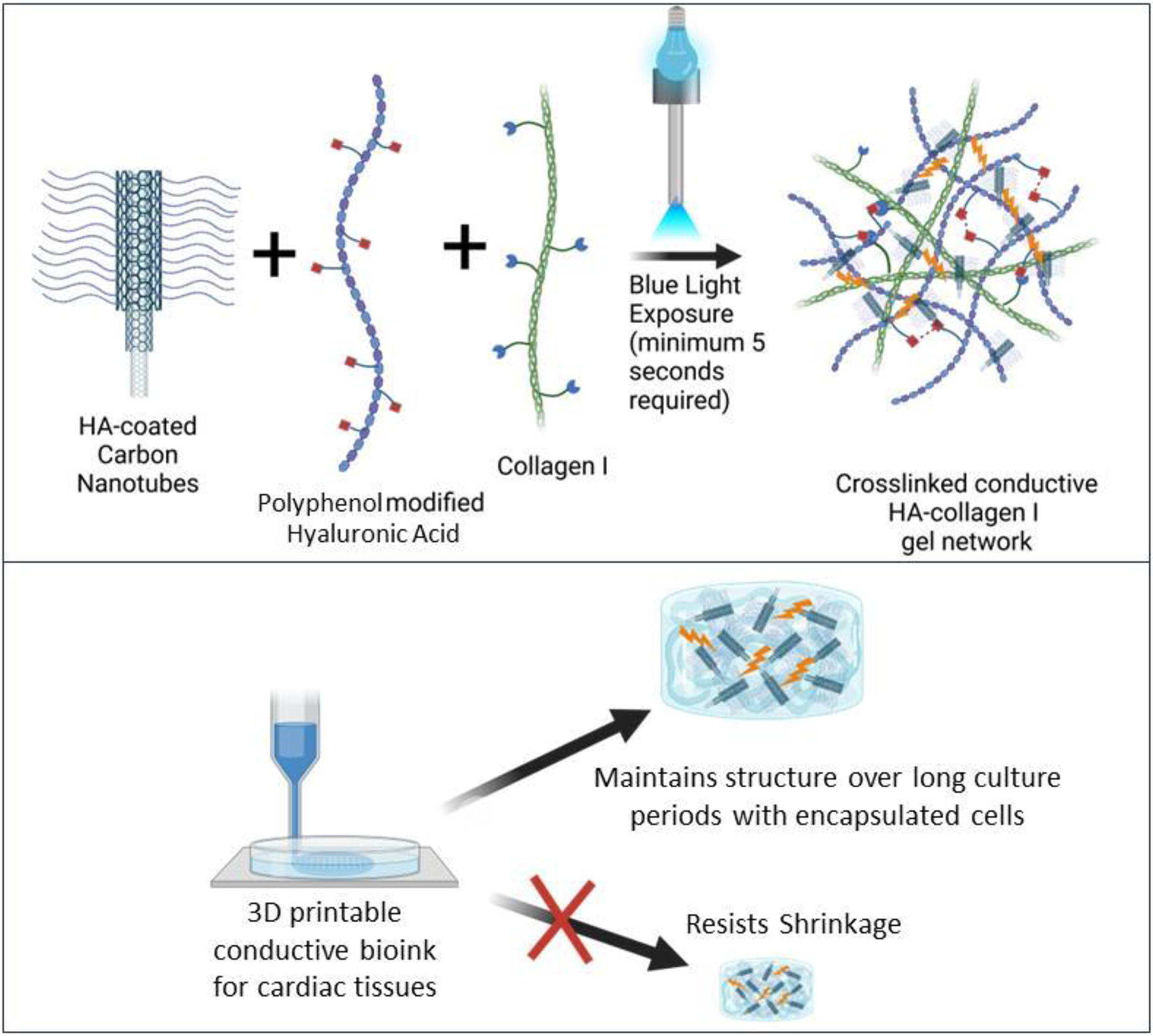
C8 - Light Based-bioprinting Of 3D Heterogenous Tissues Using Poly (n-isopropylacrylamide) As Sacrificial Templates
A. Mishra, N. K. Singh, A. Tahayeri, H. Helms, L. E. Bertassoni
Knight Cancer Institute, Oregon Health & Science University, Portland, OR
*Purpose/Objectives: Sacrificial templates for fabricating engineered tissues have been constrained in architectural intricacy due to the limitations of current extrusion-based 3D printing techniques. Most liquid-phase materials deposited by extrusion are subject to deformation or collapse under their own weight, and their viscosity and surface tension make precise dispensing of small volumes challenging. Here, we show the synthesis and printing of on demand dissolvable photo-PNIPAM that can be patterned to generate complex hierarchical structures using light-based bioprinting.
*Methodology: In brief, the prepolymer solution to fabricate thermoreversible poly(N-isopropylacrylamide) (photo-PNIPAM) degradable dendritic networks and complex sacrificial structures were prepared dissolving NIPAM in dimethyl sulfoxide or isopropanol containing phosphate buffer saline and photoinitiator.
*Results: The composition of prepolymer precursor, solvent ratio and light exposure time were optimized to modulate dissolution and stability of printed complex photo-PNIPAM structures in PBS at low temperature and above room temperature, respectively. Moreover, cell viability and proliferation were evaluated on to the surface of the photo-PNIPAM and results confirm the higher cell viability of MCF-10A, HUVECs and MSCs.
*Conclusion/Significance: This light-based strategies of degradable photo-PNIPAM printing enabled to fabricate thick and dense cellularized engineered tissues with complex hierarchical structures.
C10 - Hyaluronic Acid-based Inks For Stereolithography (Bio)printing: Benefits Of Thiol-ene Vs. Acrylate Functionalized Inks
T. Steudter1, T. Lam2, H. Pirmahboub2, C. Stoppel2, S. Pearson1, A. del Campo1
1
Leibniz-Institute for New Materials, Saarbruecken, Germany,
2
Cellbricks GmbH, Berlin, Germany
*Purpose/Objectives: Most inks in light-based 3D (bio)printing are crosslinked by photoinitiated radical polymerization of methacrylate groups. Despite its widespread use, radical polymerization has some limitations: sensitivity to oxygen and formation of inhomogeneous networks. To overcome oxygen inhibition, a high number of radicals and high light doses are needed for successful photocrosslinking, leading to prolonged printing times and potential increase in cell damage. Inks based on the radical thiol-ene reaction, which involves the addition of a thiyl radical to a vinyl group to form a thioether in a click reaction manner, offer an appealing alternative. The reaction proceeds via a step-growth mechanism and is not inhibited by oxygen. This allows reduction of cell exposure to photons and radicals and expediates printing processes.
This study provides a detailed comparison of hyaluronic acid-based inks (HA-X) for light-based digital light processing (DLP) printing. Hyaluronic acid inks with either norbornene (HA-NB) or allyl ether groups (HA-AE) are benchmarked against methacrylated hyaluronic acid (HA-MA) in terms of storage stability, photocrosslinking kinetics, mechanical properties and printability using DTT as model dithiol crosslinker in the thiol-ene systems.
*Methodology: The syntheses for the different HA-X derivatives were refined from reported one-step processes in non-toxic solvents. The storage stability of precursors was assessed using 1H NMR and GPC analysis. Oscillatory shear rheology with in situ illumination was used to compare photocrosslinking kinetics and the mechanical properties of the methacrylated and thiol-ene inks and hydrogels. Printability was assessed using a DLP-based stereolithographic bioprinter developed and manufactured by Cellbricks GmbH.
*Results: HA-NB, HA-AE and HA-MA were synthesized on gram-scale with approx. 40% modification of the disaccharide repeating units. The synthesized thiol-ene hydrogel precursors HA-NB and HA-AE showed enhanced storage stability allowing long-term storage for up to 15 months at room temperature whereas the 1H NMR spectra of HA-MA showed additional peaks after 4 months. With blue light illumination, gelation of the HA-NB hydrogel occurred 5-times faster and of the HA-AE hydrogel 2-times faster than with HA-MA. Sequential irradiation of hydrogel precursor solutions demonstrated high temporal control over the network formation in the thiol-ene hydrogels. Unlike in HA-MA, which continued to stiffen in the dark state, the storage modulus of the thiol-ene inks increased only during illumination phases. HA-MA formed 2-fold stiffer gels than the thiol-ene systems as expected from the radical chain growth mechanism vs. the step growth mechanism for thiol-ene crosslinking. All inks showed viscosities below 100 mPa s and were therefore suitable for DLP printing. Printing time per layer was 5-fold lower for HA-NB vs. HA-MA, while HA-AE did not show a significant advantage in printing speed. Printing scaffolds containing holes down to 100 µm width confirmed high spatial control and high xy resolution in the photoinduced network formation using thiol-ene inks.
*Conclusion/Significance: Overall, our work demonstrated benefits of thiol-ene versus commonly used methacrylated inks in DLP-based stereolithography printing. Thiol-ene inks show promise for more efficient bioprinting of advanced tissue models by minimizing printing time compared to commonly used methacrylated inks while maintaining high resolution.
C11 - Development Of Bioresins For Volumetric Printing For Vascular Applications And Convergence Of Bioprinting Techniques
C. Gergely
University of Würzburg, Würzburg, Germany
*Purpose/Objectives: For bioresins, which are a bioink formulation used for vat and vial-based biofabrication techniques, a cytocompatible and photocrosslinkable hydrogel precursor needs to be chosen, with methacrylated gelatin (GelMA) as a prototypical representative. GelMA’s thermal gelling behavior, good optical transparency and its properties can be tuned via the formulation of the precursor solution. This results in different mechanical stiffness, printing resolution, and biological behavior to mimic the extracellular matrix (ECM) for the various cell types in volumetric bioprinting. Models for coronary arteries, especially, are needed for medical training and therapeutic replacement purposes. Even 1:1 copies of 3D-scanned tissue, such as CT scans of blood vessel systems of the coronary tree, can be used.
*Methodology: Volumetric printing (VP) is a novel, cell-friendly 3D printing method capable of in situ encapsulation of cells into CAD-designed models in a one-step process within seconds. This leads to the fast manufacturing of usable sample sizes of complex hydrogel scaffolds. With this method, complex objects can be created that were not able to be printed with conventional biofabrication methods, such as multi-material objects or intricate tubular branching constructs.
*Results: The tubular models can be post-seeded with endothelial cells and cultivated under physiological flow to further investigate artificial small-diameter blood vessel models. To prevent cell sedimentation, enhance cell distribution, and provide a beneficial physiological environment for cell maturation, special properties for the resin need to be established. By combining chemistry and biofabrication with tight feedback loops to biology, we are testing advanced in-vitro vasculature models. While resin formulations mimic ECM properties for optimal cell attachment, we have shown, that reinforcement of the tubular GelMA model with different tubular PCL meshes (doi.org/10.1002/adma.202300756) further can come closer to the native mechanical properties of small diameter blood vessels, combining volumetric printing and tubular MEW.
*Conclusion/Significance: However, these properties can only be optimized up to a certain point. Research into alternatives or new polymeric options for photosensitive fabrication needs to be conducted to push the limits of printability and biocompatibility. The goal is to push the limits of biomaterials for additive manufacturing applications by convergence of different bioprinting methods with volumetric bioprinting and taylored polymer resin compositions.
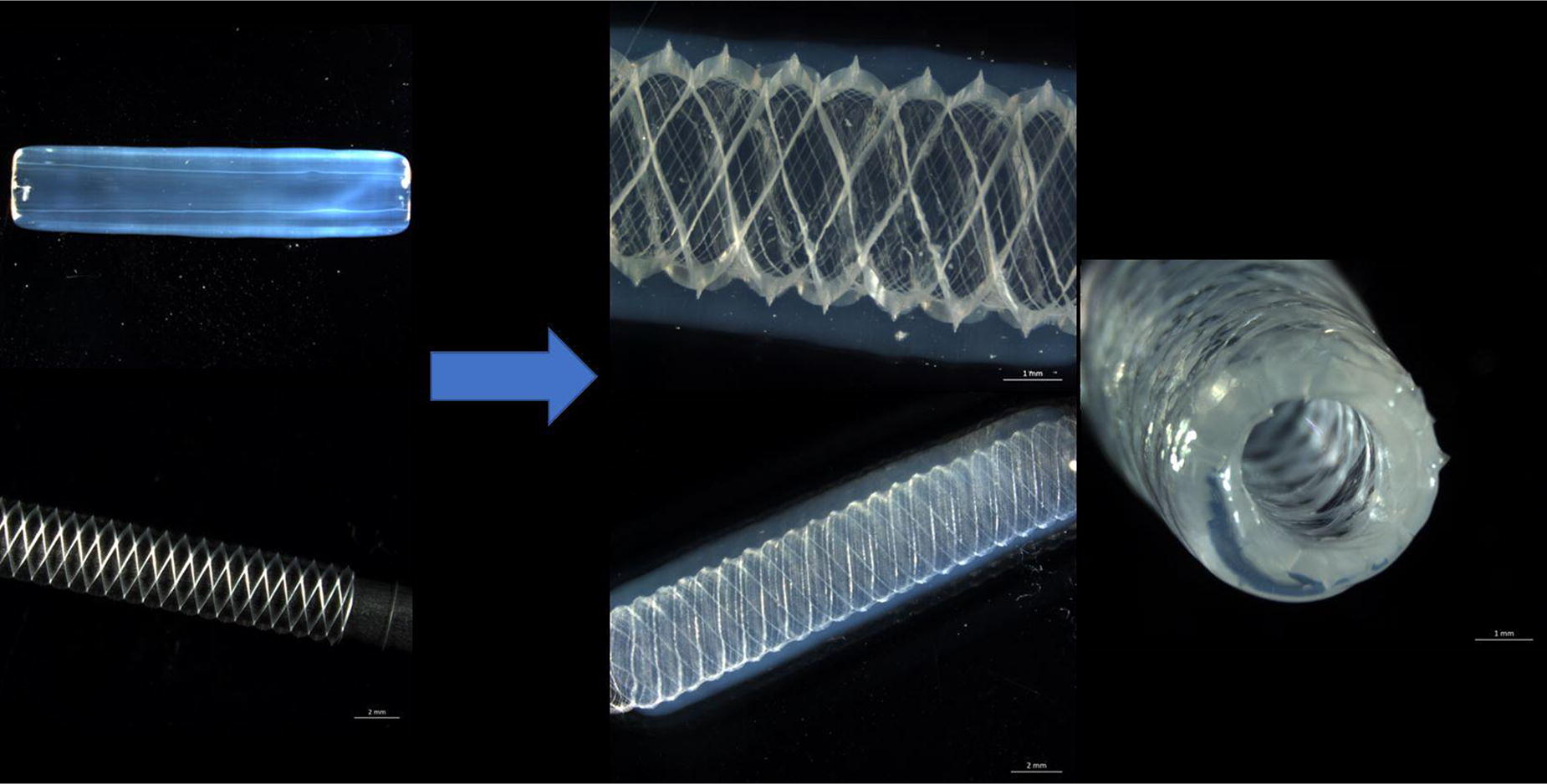
C12 - In Cellulo Photoactivation Of Diverse Protein Targets Enables 4d Spatiotemporal Control Of Cell Behavior And Activity
J. W. Hoye, R. M. Francis, R. P. Brady, B. G. Munoz-Robles, S. Yang, C. A. DeForest
University of Washington, Seattle, WA
*Purpose/Objectives: Proteins play an essential role in many biological processes, including regulating cell behavior, metabolism, and maintaining phenotype. Controlling protein activity spatiotemporally provides a direct route to manipulate biological processes in 4D, including for gene editing and transdifferentiation, all of which are necessary for the creation of engineered tissue.
*Methodology: Towards this goal, we have created a versatile workflow to stably endow cell lines with the ability to inducibly express photoactivatable proteins of interest. Via the installation of non-canonical amino acids containing a photolabile chemical modification at catalytic residues, protein activity can be effectively “caged” until light exposure.
*Results: These cell lines were shown to be photoactivatable on 2D surfaces via mask-based patterning and in 3D culture via multiphoton-based lithography.
*Conclusion/Significance: We anticipate that this pipeline will be readily extendable to photoactivate many functionally diverse proteins of interest, including Cas9 and it’s derivatives to enable 4D control over base editing, indel generation, and CRISPR activation/inhibition.
C15 - Laser-assisted Bioprinting Of Cartilaginous Spheroids Using Image-guided Target And Shoot Strategy
Y. Fan1,2, G. N. Hall1,2, A. Dimopoulos1,2, A. Iazzolino3, B. Viellerobe3, F. Guillemot3, I. Papantoniou1,2
1
KU Leuven, Leuven, Belgium,
2
Prometheus, Leuven, Belgium,
3
Poietis, Bordeaux, France
*Purpose/Objectives: Multi-cellular spheroids such as microtissues and organoids have demonstrated great potential on tissue engineering in recent years as these 3D cellular units enabled better cell-cell and cell-matrix interaction. Current biofabrication methods that use multi-cellular spheroids as building blocks are mainly based on extrusion printing or self-assembling in a customized mold and have very limited precision on the control of the amount and distribution of cell spheroids. In this work, we presented a laser-assisted biofabrication method that use multi-cellular spheroid as building blocks. By using the laser-induced forward transfer (LIFT) technology, we demonstrated the successful printing of cartilaginous multi-cellular spheroids without influencing their biological behavior. Moreover, by combining the LIFT technology and computer vision technology, a novel system called target and shoot system has been implemented and the automatic recognition and transferring of multiple large spheroids in a single sequence was achieved. To our knowledge, this is the first report that successfully printed multiple cell spheroids with a dimension of ∼100µm-300µm by using laser-induced forward transfer technology.
*Methodology: Human periosteum derived cells (hPDCs) were seeded into AggreWell 800 plates and differentiated in a chemically defined chondrogenic medium in order to form callus organoids. The in vitro maturation of the multi-cellular building blocks was monitored by real time PCR and histology during differentiation. Printing parameters such as laser source, volume of sampling, distance between donor and receiver, etc. were defined and tested on hPDC spheroids of different maturity, going from day 3 (mainly cells), day 7 (cells and low ECM) and day 14 (cells and ECM). Finally, viability and histological staining was used to assess functionality after printing.
*Results: Successful transferring of cartilaginous spheroids has been achieved by adjusting the laser printing parameters. The cell spheroids were viable after laser-assisted bioprinting and were able to undergo continued chondrogenic differentiation post printing. By combining the LIFT technology and computer vision technology, a novel system called target and shoot system was employed. The target and shoot system automatically recognized and targeted cell spheroids with a dimension ranging between 100µm-300µm and successfully transferred multiple cell spheroids in a single sequence. To our knowledge, this is the first report that successfully demonstrates multiple cell spheroids printing with a relative large diameter by using laser-induced forward transfer technology.
*Conclusion/Significance: We present here a novel laser-assisted bio-fabrication technology for spheroids. This technique is capable of targeting and transferring multiple cell spheroids within one pulse of laser shot without influencing their biological behavior. Spheroids could be printed at designated locations and patterns. This approach is promising for the automated fabrication of large spheroid based living implants.
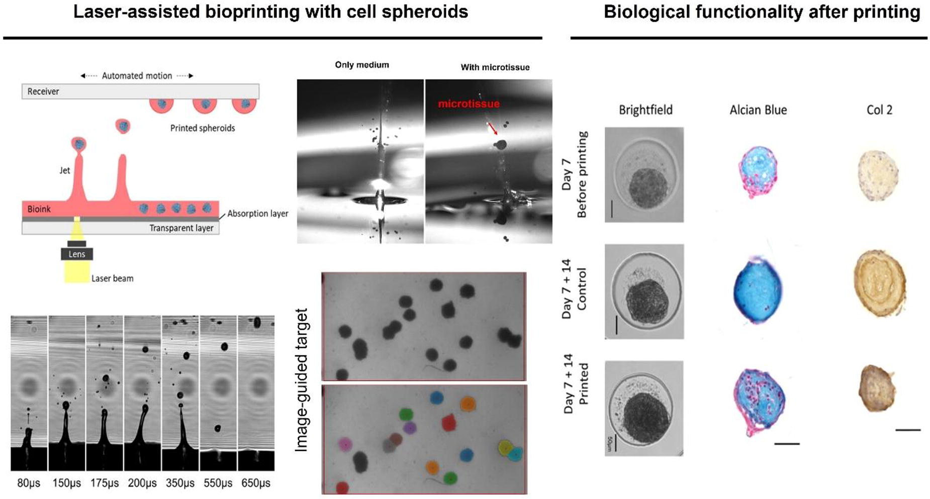
C16 - Nanomaterial Modified Bioinks For Dlp Based Bioprinting Of Bone Constructs: Impact On Mechanical Properties And Mesenchymal Stem Cell Function
J. Kühl1, M. Krümpelmann1, L. Hildebrandt1, M. Bruhn2, S. Gorb2, A. Seekamp1, F. Schütt2, L. Siebert2, S. Fuchs1
1
University Medical Center Schleswig-Holstein, Kiel, Germany,
2
Christian-Albrechts-University, Kiel, Germany
*Purpose/Objectives: 3D printing technologies offer a tremendous potential to produce patient-specific implants for the treatment of critically sized bone defects which vary in size and shape or clinical requirements. Technological progress has been achieved for 3D printing of biomaterial-based bone constructs, however hardly any true biological material has been employed. Nevertheless, an early biologization and vascularization of the implant is essential to ensure and accelerate the formation of functional bone tissue. In this context, bioprinting technologies enable the integration of vital cells or active growth factors into 3D printed constructs. In this study, we established bioinks for digital light processing (DLP)-based bioprinting of bone constructs using gelatine methacryloyl (GelMa) hydrogels as basis for the bioink. The bioink was further modified using either graphene oxide or calcium phosphate nanomaterials to modulate the mechanical and biofunctional properties of the resulting constructs. The 3D model of the porous bone implant was designed computationally. Prior to bioprinting, human mesenchymal stem cells (hMSC) were integrated in the bioinks. After the bioprinting step, the impact of bioinks on cell distribution, viability, cell proliferation and differentiation, and mechanical properties of constructs was measured and investigated over a period of 14 days.
*Methodology: The model was designed using “Shapr3D software” providing a cylindrical shape (d= 7 mm, h= 3.64 mm) and interconnected pores of different sizes. Different bioinks were formulated using GelMa (degree of methacrylation: 80 %), a photoinitiator and photoabsorber. To enhance osteoinductive properties of the GelMa bioink, calcium phosphate nano particles (≤150 nm) were added to the bioink. In addition, another GelMa bioink was modified using graphene oxide. For the bioprinting process the bioinks were loaded with 3 x 10⁶ hMSC /ml bioink. Tissue constructs were printed by DLP using the LumenX (Cellink) followed by evaluation of the resulting constructs by DNA quantification, confocal and SEM microscopy, real time PCR, cryosections followed by alizarin red and sirius red staining and compression testing.
*Results: In comparison to commercially available bioinks used as reference material, the cell viability was higher in the established GelMa bioink and all nanomaterial containing variations along with an even distribution of cells. Morphological data and DNA quantification indicated the highest cell vitality respectively proliferation over time in GelMa basic ink, as well as the best printing resolution of the constructs in comparison to the 3D model. However, CaP modified GelMa bioink indicated the best differentiation of hMSC in terms of gene expression of osteogenic differentiation markers and calcium deposition. Graphene oxide increased the elastic modulus of the printed constructs in control samples printed without cells. However, in initial studies we also observed differences in the stiffness in samples with cells over time which are presently under further evaluation.
*Conclusion/Significance: Overall, nanomaterials showed multiple beneficial effects on DLP-printed bone constructs containing living osteogenic cells.
Acknowledgements: supported by BMBF, BlueBioPol (FKZ 03WIR6207A)
Malte Krümpelmann and Julie Kühl equally contributed.
C17 - Marine Polymer-based Hydrogels For Dlp 3D- Bioprinting
L. Hildebrandt, J. Kühl, A. Seekamp, S. Fuchs
University Medical Center Schleswig-Holstein, Kiel, Germany
*Purpose/Objectives: Osteoarthritis is a degenerative cartilage disease induced by multiple factors including aging and mechanical or inflammatory stress. During disease progression, bone tissue is affected leading to osteochondral defects. Traditional and systemically applied treatments often reveal limited success creating the need for more targeted and individual approaches tailored towards the different stages of osteoarthritis. In this context hydrogels can be used to inject bioactive factors and cells or to manufacture implants e.g. by digital light (DLP) bioprinting technologies. This technology allows to print precise implants for tissue defects by UV mediated gelation of hydrogels. Additionally, cells or bioactive factors can be integrated. Biophysical properties of marine biopolymers might offer advantages for this technology not yet determined. In this study, we printed marine-polymer-based 3D constructs maintaining the viability and functionality of osteogenic cells. In addition, different nanomaterials were investigated to enhance the structural integrity of the 3D printed constructs.
*Methodology: Bioprinting was performed using methacrylated alginate combined with jellyfish collagen for the hydrogels. Bioinks were established by including brilliant black as photoabsorber and Lithium phenyl(2,4,6-trimethylbenzoyl) phosphonate as photoinitiator. Further, bioinks were modified using graphene oxide and 0.3 % fibrillated nanocellulose (NFC) to enhance the stability of the printed constructs. Before printing 3*106 MG63 cells/ml were mixed with the bioinks. The printing process was performed using a Lumen X at 37 °C. The STL 3D file for the construct was designed using “Shapr3D” (h: 3.64 mm, d :7 mm) with pores of 500 and 2000 µm. After printing, the constructs were additionally crosslinked with 50 mM CaCl2. Cellular viability was investigated using calcein-AM, Hoechst and confocal microscopy on the days 1, 7 and 14. Cryosections were prepared to analyze the distribution and cell viability within the constructs. Staining for focal adhesion kinase and actin was performed to analyze cellular morphology and adhesion.
*Results: Methacrylated alginate revealed excellent properties for UV-based DLP printing; however it lacks adequate binding sites for cell adhesion and thus needs to be combined with other extracellular matrix components. The use of mammalian collagen for bioprinting at 37 °C is not feasible due to gelation above room temperature. Our experiments however indicate that the use of marine collagen with a lower melting point is feasible and supports the viability of cells. Independent from the bioink composition, calcein-AM staining indicated cellular viability over 14 days. In addition, cryosections revealed an even distribution of cells inside the bioinks and staining for focal adhesion kinase indicated the interaction of the cells with the materials. GO and NFC reduced swelling of the printed constructs and thus leads to stability of the constructs maintaining their shape. Nevertheless, for all bioinks CaCl2 crosslinking was essential to ensure stability.
*Conclusion/Significance: Marine collagen offers advantages to prepare DLP printed constructs containing viable bone cells. GO and NFC are useful to stabilize the constructs. Further experiments will focus on detailed evaluation of cell and material interaction containing nanomaterials for functionalization.
Acknowledgments: We thank BMBF WIR!-BlueHealthTech-BlueBioPol (FKZ 03WIR6207A.BMBF) for financially supporting this work.
C19 - Digital Light Bioprinting Of A Corneal-Limbal Niche 3D Model
I. Paschalidis1,2,3, B. Souquet2,3, F. Chatelain1, A. Fuchs1, E. Gabison2,3,4
1
CEA Grenoble, Grenoble, France,
2
Hôpital Fondation Adolphe de Rothschild, Paris, France,
3
HIPI UMR976, Stem Cell Biotechnologies, INSERM, Paris, France,
4
AP-HP, Hôpital Saint-Louis, Paris, France
*Purpose/Objectives: The cornea serves as a protective barrier for the eye and facilitates light refraction onto the retina. The epithelium of the cornea is regularly exposed to environmental stresses and its highly dependent on regeneration deriving from the limbal epithelial stem cells (LESCs) found in the limbus - a transition zone surrounding the cornea. Damage to this area can eventually lead to vision loss. Considering the significant lack of advanced in vitro models that mimic the limbal niche, this study aims to reconstruct the limbal microenvironment for homeostasis and disease. To this end, the presented project focuses on the microfabrication of the unique wave-like topography of alternating ridges and crypts that house LESCs and make up the limbal niche. We propose that this topography provides the necessary microenvironment to promote self-renewal capacity and maintenance of the structure and function of the corneal epithelium in vitro.
*Methodology: An initial photocrosslinkable hydrogel consisting of collagen methacrylate (ColMA) and methacrylated hyaluronic acid (HAMA) (both naturally found in the native cornea) was reinforced with methacrylated silk fibroin (SFMA) to produce a suitable cell scaffold. SFMA was employed for the first time in a corneal context, and we demonstrated a dramatic improvement in human primary limbal-derived epithelial cell attachment and growth. Using Digital Light Processing (DLP) technology, UV light was projected in a grayscale manner onto the hydrogel, with spatial control of UV dose producing a map of heterogeneous crosslinking densities that gradually induced hydrogel shape transformation [figure 1A].
*Results: Remarkably, the undulating topographic features resemble the native architecture and promote stratification and cell organization with unique phenotypes across different cell layers, which was not observed on flat gel scaffolds. Specifically, the cells expressed relevant stemness as well as differentiation markers, and produced components of the basement membrane [figure 1B] in a spatially appropriate manner as expected from in vivo observations described in the literature. Cell culture could be maintained for at least 3 weeks, which enables the use of this construct in long-term pathophysiological studies.
*Conclusion/Significance: The format of the hydrogel scaffold (covalently attached to a glass coverslip) also allows easy insertion into a microfluidic device for organ-on-a-chip applications. We thus plan to adapt this system for in vitro modelling and drug screening, taking advantage of this highly biomimetic model of the corneolimbal niche presented above.
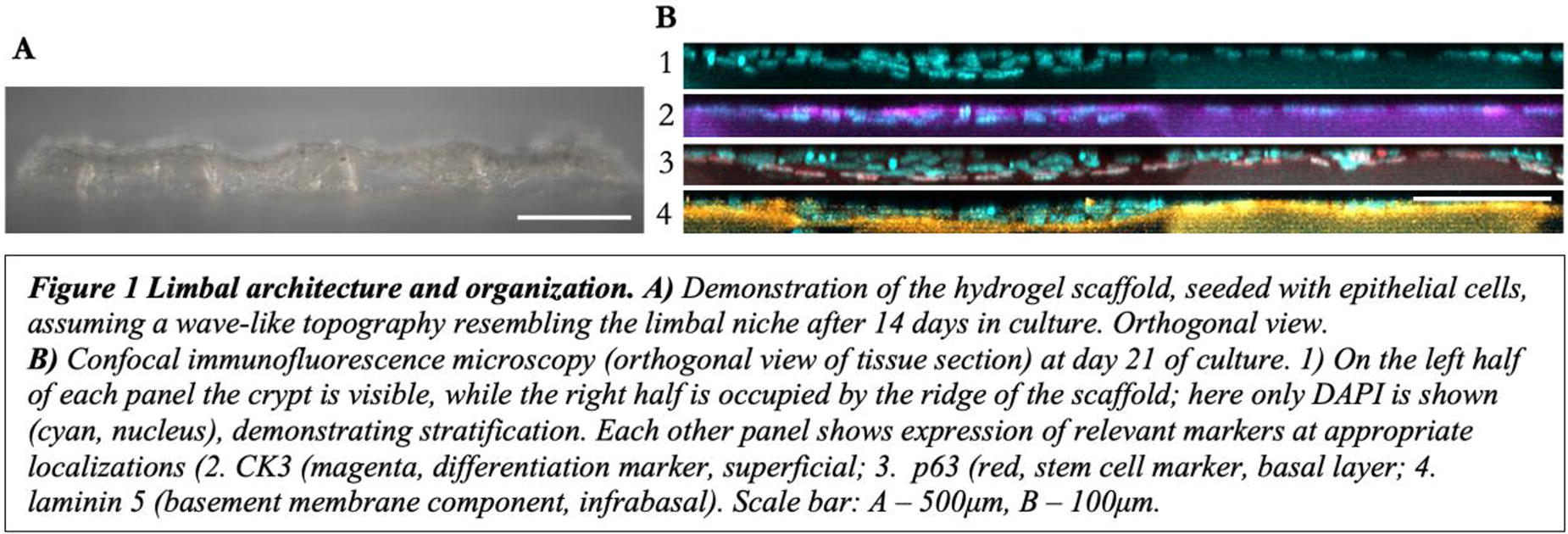
C20 - The Multiaxial Bioreactor Integrated With An Environmental Monitoring System For A Tissue-engineered Skin Graft
S. Jeon1, A. Mehta1, P.-F. Lee1, E. Renteria1, F. Marini1, J. Kim1, T. Criswell1, T. Shupe1, A. Atala1, J. Hunsberger2, M. Gurcan3, Y. Ju1, S. Soker1, J. Yoo1
1
Wake Forest Institute for Regenerative Medicine, Winston-Salem, NC,
2
RegenMed Development Organization (ReMDO), Winston-Salem, NC,
3
Center for Artificial Intelligence, Wake Forest University School of Medicine, Winston-Salem, NC
*Purpose/Objectives: Tissue-engineered skin grafts have emerged as a promising solution for the treatment of chronic wounds. To facilitate the maturation of these grafts, stretch-based bioreactor systems have been employed. However, traditional systems have limited stretching axes as uni- or bi-directional stretching, inducing scar formation in the graft derived from directional cellular orientation. Additionally, the lack of a monitoring system in the bioreactor hindered capturing dynamic environmental change in the culture process.
*Methodology: In this study, we introduced a novel multiaxial stretch-based bioreactor system with the incorporation of continuous environmental monitoring capabilities. To achieve multiaxial cyclic stretching, we designed a unique Hoberman ring structure that transforms linear motor force into radial movement, effectively stretching the skin graft in multiple directions. Additionally, we integrated environmental sensors to monitor critical parameters such as pH levels, oxygen concentrations, glucose, and lactate levels within the bioreactor. The integrated sensor system creates a single flow loop, enabling continuous monitoring of the progress of the culture. Particularly, we also integrated a media level sensor to maintain the air-liquid interface, a crucial environmental factor for preserving the functionality of the skin.
*Results: Through controlled cyclic stretching, the surface area of the skin graft increased by 1.13 compared to the static culture group. And, multiaxial stretching exerted uniform stress within the skin graft inducing homogenous cellular orientation compared to uni- and bi-axial stretching groups. Moreover, stretching induced early maturation of keratinocytes within the skin graft, in conjunction with dermal fibroblasts by observing key protein markers related to skin maturation.
*Conclusion/Significance: These findings highlight the potential of our integrated design of bioreactor in providing new insights for the manipulation of tissue-engineered skin tissue, thus paving the way for clinical applications in wound healing.
C22 - Multi-functional Pulsatile Bioreactor Module Development For Biomanufacturing Engineered Tubular Tissues
P.-F. Lee1, A. Mehta1, E. Renteria1, F. Marini1, J. Kim1, T. Criswell1, T. Shupe1, A. Atala1, J. Hunsberger2, M. Gurcan3, S. Soker1, Y. Ju1, J. Yoo1
1
Wake Forest Institute for Regenerative Medicine, Winston-Salem, NC,
2
RegenMed Development Organization (ReMDO), Winston-Salem, NC,
3
Center for Artificial Intelligence, Wake Forest University School of Medicine, Winston-Salem, NC
*Purpose/Objectives: Bioreactors have become a standard tool in biomanufacturing tissue-engineered medical products for preconditioning and maturing tissue constructs. This study aimed to develop a standardized, self-contained, and modular bioreactor platform capable of establishing scalable and automated processes for the clinical manufacturing of tubular tissue constructs.
*Methodology: To achieve this goal, design parameters specific to tubular tissues, such as blood vessels, were considered and incorporated to develop a novel pulsatile bioreactor prototype. The constructed tubular tissue bioreactor included multiple functions and features, including a pulsatile flow mechanism controlled under specific pressure conditions and the incorporation of real-time monitoring of flow dynamics through an integrated sensor module to assess the culture environment and cellular metabolites. To achieve efficient cell seeding, a machine rotary mechanism has been integrated within the bioreactor chamber box. A novel flow equalizer has been designed to provide equal flow stimulation to multiple vessels from one single pump. The tubular scaffolds were fabricated from a 5% collagen/PCL mixture for validation studies of cell seeding method comparison and vessel maturation under flow stimulation.
*Results: The flow equalizer system can provide uniform flow stimulation across multiple tubular organs within the bioreactor with a minimal flow rate variation of less than 4%. These capabilities represent a significant improvement over the traditional 1-to-3-way connector, which results in a flow rate difference of 16%. To validate the functionality and reliability of the bioreactor, we conducted baseline tests using endothelial cell-seeded electrospun vessel scaffolds. The results demonstrated that the bioreactor module operated effectively, with the integrated sensors providing quantitative data. Furthermore, we implemented a machine rotary mechanism within the bioreactor chamber box, which offers distinct advantages over the traditional manual seeding method. Applying the rotary mechanism for cell seeding resulted in superior endothelial cell coverage. Furthermore, endothelial cells were organized and aligned in the direction of flow after five days of flow stimulation.
*Conclusion/Significance: The multi-functional pulsatile bioreactor has been successfully validated and may be used as a standard tool for biomanufacturing tissue-engineered tubular tissue constructs.
C23 - A Semi-automated System For Biomanufacturing Of A Large-size Hydrogel Sheet
W. Chan1, Z. Lee1, G. Soo2, B. Le1, W. Woo1, B. Salam1, W. May3, M. Tan2, D. Choudhury1
1
Bioprocessing Technology Institute, A*STAR, Singapore, Singapore,
2
Singapore Institute of Manufacturing Technology (SIMTech), A*STAR, Singapore, Singapore,
3
A*STAR, Singapore, Singapore
*Purpose/Objectives: Hydrogel is as a ubiquitous scaffold in tissue engineering and regenerative medicine, playing an important role in maintaining cell and tissue growth. Beyond its scaffolding role, hydrogel has an advantage of serving as a reservoir for nutrients and growth factors crucial for keeping cellular and tissue integrity. Additionally, its high-water content helps maintain high moisture and allow efficient heat dissipation, useful in many skin tissue engineering applications. Researchers have spent significant efforts to enhance hydrogel functionality by integrating diverse factors, including drugs to augment tissue regeneration or angiogenesis. The optimization of hydrogel formulation, cross-linking conditions, and the incorporation of additives has become a focal point in these endeavors. Presently, these processes are manual, involving intricate handling operations that introduce batch variations, time inefficiencies, and necessitate substantial training. To address this need, our objective is to design and construct an automated system capable of executing hydrogel casting, crosslinking, washing, and various other procedures, thereby overcoming the limitations associated with manual handling.
*Methodology: We have successfully engineered an advanced semi-automated hydrogel fabrication system, designed to optimize existing processes and accommodate the potential for scaled-up production. Our system integrated a programmable logic controller (PLC) system complemented by an array of sensors, collectively obviating the need for manual intervention.
*Results: This system automatically handles labor-intensive tasks like dispensing, delaminating, and shaping through seamless automation. The adaptability of the system is another feature; it has the capacity to allow various parameter adjustments, empowering researchers to fine-tune processes. The modular design further enhances versatility, enabling the seamless integration of alternative processes such as UV or chemical cross-linking, thereby amplifying the system’s utility and functionality. Employing this state-of-the-art system, we conducted meticulous optimizations of our hydrogel formulations, generating hydrogel sheets measuring up to 100 × 100×2 mm. Through this system, we achieved a stiffness of 38k Pa (at 10% compression) for the hydrogel.
*Conclusion/Significance: This semi-automated system with flexibility and enhanced production capacity not only empowers us to optimize diverse hydrogel formulations but also significantly reduces batch variations and alleviates the need for extensive user training. This research receives support from A*STAR Grant Project no. A18A8b0059. Figure shows the semi-automated system for manufacturing of large-size hydrogel.
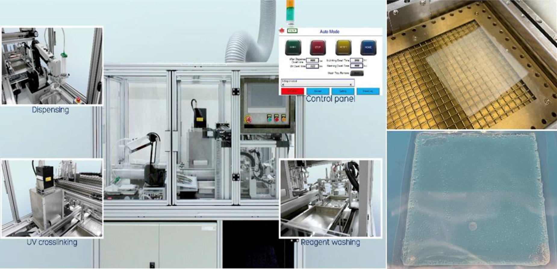
C24
C25
C26 - An Open-source Camera-assisted Alignment System For High-performance Multi-material 3D Bioprinting
S. Ashraf1,2, D. J. Shiwarski2,1, A. PereiraTavares2, J. W. Tashman2, A. W. Feinberg1,1
1
Carnegie Mellon University, Pittsburgh, PA,
2
University of Pittsburgh, Pittsburgh, PA
*Purpose/Objectives: Multi-material 3D bioprinting has shown promise in fabricating complex, 3D tissue constructs with diverse cell types bioinks. For this to be successful, multi-nozzle extrusion requires multiple syringe extruders and a method of calibrating their XYZ positional offsets. Point-based alignment approaches that estimate the extruder nozzle’s position by touching a known marker on the printer bed risk bioink contamination and have ±100 µm error, limiting print fidelity when repeatedly switching between extruders. Precision tools like laser micrometers, costing over $20,000, can improve alignment but pose a significant financial barrier. We aimed to create an affordable, open-source system for sterile calibration of positional offsets between multiple syringe extruder nozzles with ±10 µm accuracy.
*Methodology: We designed an Automated Lens-based Initial Needle Registration System (ALINr) as an affordable, open-source calibration tool powered by the Raspberry Pi Compute Module 4. Our previously published Replistruder 4 syringe pump extruder is used by labs globally for 3D bioprinting. The Replistruder 4.5 was designed to be narrower, enabling more extruders to fit on a platform, while increasing its accuracy by utilizing precision linear motion components. A three-nozzle extrusion calibration print was made to test error reduction in multi-nozzle bioprinting by ALINr. This involved measuring the positions of constructs printed by the second and third extruders to the first. These were printed in collagen using Freeform Reversible Embedded Hydrogels (FRESH) 3D bioprinting on a 3D printer with three Replistruder 4.5 extruders. Prints using point-based alignment were compared with those from ALINr alignment, imaged, and gauged using optical coherence tomography.
*Results: The ALINr system costs $280 in terms of bill of materials and uses two cameras to locate the extruder needle X, Y, and Z positions (Figure A). Powered by a battery, it is wireless and portable for use in cell culture hoods for sterile alignment. A programmed graphical user interface enables simultaneous viewing of XY and Z cameras. It incorporates a machine vision-based automated circle detection program to automatically locate the outer diameter of the needle and plot its coordinates to a center point. The printer controller calculates tool offsets from the coordinates of each extruder needle relative to the first for accurate print pathing. The Replistruder 4.5 costs $90 per extruder and is 20% narrower than its predecessor. Each extruder is attached to a second linear motion system for independent Z height control, thereby enabling the elimination of the Z-offset, which is crucial for multi-nozzle alignment. The Replistruder 4.5 consistently produced collagen filaments 90 µm in diameter and calibrating the three extruders using ALINr resulted in mean X, Y errors of 52 ± 29 µm and 36 ± 26 µm, respectively. ALINr reduced the mean error by 3.5-fold in X and 8-fold in Y and Z axes compared to point-based alignment (Figure B).
*Conclusion/Significance: We developed a high-performance multiple-syringe extruder platform and a wireless camera-assisted calibration tool with automated needle tracking. These affordable, open-source tools broaden access to high-resolution, multi-material tissue scaffold fabrication. They are adaptable for multi-tool fabrication equipment and integration into automatic alignment pipelines.
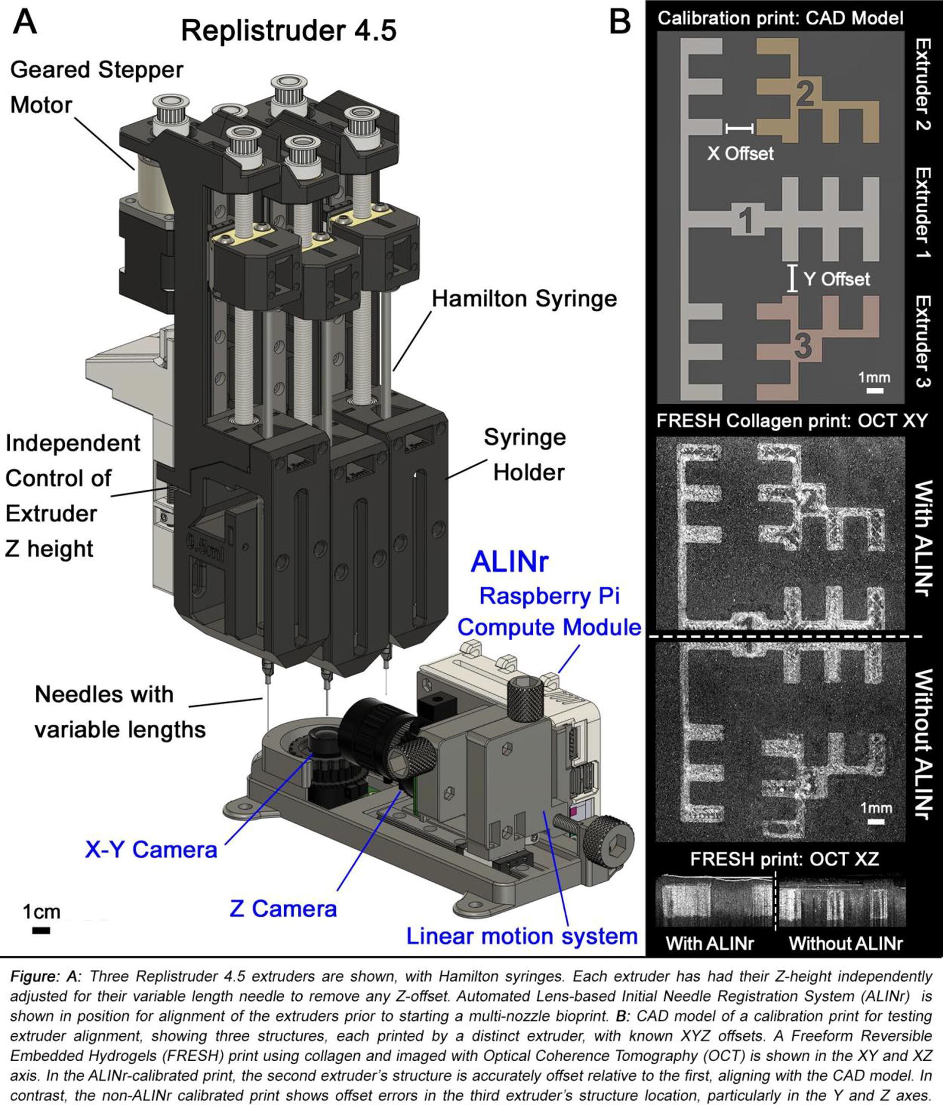
C27 - A Customizable Method For Optimizing Volumetric Bioprinting Shape Fidelity Using Machine Learning
T. W. Wheeler, G. Lindberg
University of Oregon, Eugene, OR
*Purpose/Objectives: Volumetric printing (VP) represents an attractive method to explore for bioprinting applications, as it can additively print entire objects simultaneously without hard limits on overhanging or connected structures. VP faces an especial challenge for optimization compared to other 3D-bioprinting modalities which intrinsically print to a build plate. The lack of an anchor point in VP inhibits the ability to orient, align, and match input to output data, making it especially difficulty to generate sufficient aligned data for machine learning (ML). A particular challenge in applying ML optimization lays in meaningfully generating and aligning input and output data at a scale sufficient to allow for successful model training. Here we propose a high-throughput method to optimize VP with machine learning using randomly generated shapes.
*Methodology: Gelatin Methacryloyl (GelMA) was synthesized (0.43 - 0.56 mmol/g) and used (5wt%) together with LAP (0.1wt%) and a Readily3D Tomolite machine (exposure time 20-33 s, intensity: 250-400 mJ/cm2), with and without Human dermal fibroblast (1x10⁶ cells/ml). In this developed ML method, printing shapes are randomly generated by producing a closed Bezier curve with random permutations, or by overlaying polygons in random configurations. Shapes are batched in the STL file by stacking along the z-axis to increase throughput. These prints are then stained (Coomassie), imaged, and aligned with input masks via rigid registration alignment. These input-output pairs constitute training data to train an Attention U-Net based model to predict an optimal input mask for a desired output shape and scale, with the attention component also incorporating experimental parameters. We tested the outcome of training by printing batches of random shapes using print masks either with ML-adjustment or without (control). We scored outcomes by calculating the similarity of the mean overlap of binary masks of aligned input-output pairs.
*Results: ML-adjusted prints showed 89.9 ± 2.6% similarity to desired shape, versus 70.1 ± 1.7% similarity on control prints (n=7 prints for both groups), indicating a significant improvement in recapitulation of the intended output shape (p<0.001 by two-tailed t-test). Conducting an ablation study of the attention mechanism, we found that prints generated from attention-ablated model only showed 70.5 ± 3.4% similarity, compared to an attention-inclusive control which resulted in 84.8 ± 3.6% similarity. This significant difference (p<0.001 by two-tailed t-test) indicates that an attention mechanism is necessary for integrating data across a variety of print conditions, and further suggests that this method can produce a model that functionally incorporates material-specific metadata to increase accuracy of results.
*Conclusion/Significance: Overall, we demonstrate a ML method to optimize volumetric printing for shape fidelity with a simple, high-throughput image analysis platform that can be readily re-tuned for new materials and processing protocols. This protocol was designed with a general level of technical expertise in mind, such that it can be adapted and run without a deep understanding of data processing or machine learning. As such, this work presents a method which can be further built to minimize the optimization time for new conditions and postprocessing steps with a minimum of requisite operator expertise.

C28 - Rheology-informed Hierarchical Machine Learning Model For Enhanced Prediction Of Bioprinting Resolution
D. Oh1, M. Shirzad1, E.-J. Chung2, S. Nam1
1
Pukyong National University, Busan, Korea, Republic of,
2
Seoul National University College of Medicine, Seoul, Korea, Republic of
*Purpose/Objectives: Extrusion-based bioprinting has been a widely used technique for research and commercial purposes. Recently, the printability prediction of extrusion-based bioprinting attracted considerable attention, which can lead to accurate and rapid fabrication as well as long-term stable functionality. Initially, the physical model was adopted for printability prediction with the rheological modeling, which was significantly sensitive to the power law index obtained by line fitting of the measured viscosity. Thus, small errors in rheological measurement and line fitting significantly impacted prediction accuracy. Recently, multiple studies of printability prediction employed machine learning methods to estimate printing resolution, but they addressed the confined variations in bioink composition and printing parameters. Therefore, in this study, the rheology-informed hierarchical machine learning (RIHML) model was demonstrated to overcome the limitations of conventional printability prediction methods.
*Methodology: The RIHML model was designed with a multi-input neural network to calculate the printing resolution using three types of input data; the printing parameters (nozzle diameter, nozzle length, printing speed, and pressure) and two types of rheological properties (viscosity and storage modulus). Various bioink compositions of Pluronic F-127, gelatin, xanthan gum, alginate, calcium chloride, and cellulose nanocrystal were used to make datasets for machine learning. The rheological properties of the bioinks were measured using a rotational rheometer with a 20 mm parallel plate.
*Results: The printing resolution of constructs fabricated by extrusion-based bioprinting was compared with the printing resolution predicted by various machine learning algorithms. Specifically, five different machine learning models, including the RIHML model as well as two classical machine learning models (random forest and support vector machine) and the conventional models based on artificial neural networks (concentration-dependent and printing parameter-dependent models), were trained and tested using a small dataset of bioink properties and printing parameters. All the prediction results presented RIHML has higher accuracy than conventional models. Furthermore, the RIHML model can predict the resolution of the new compositions due to its intrinsic features in the dataset that are not biased to specific bioinks but rather are general.
*Conclusion/Significance: We developed a rheology-informed machine learning model to improve the prediction accuracy of the printing resolution of tissue-engineered constructs fabricated by extrusion-based bioprinting. The model was used to predict the printing resolution in three different cases including new printing parameters with trained bioink materials, new concentrations of the trained bioink constituents, and untrained bioink compositions with the new material. Interestingly, the RIHML model exhibited the lowest errors in printability prediction for various conditions. Furthermore, the RIHML model can easily generalize and embrace new data, even with a small dataset size. Overall, the study demonstrated that the RIHML model can be a versatile tool for predicting printability in extrusion-based bioprinting.

C29 - Spheroid Viability Prediction With Deep Learning: Automating Quality Control In Tissue Engineering Applications
Z. Sheikh
University of Chicago, Chicago, IL
*Purpose/Objectives: Spheroids are three-dimensional aggregates of cells that are essential for biomaterial-free tissue engineering. They serve as the building blocks for 3D-bioprinted tissue patches. Spheroids larger than 500 μm, the desired size for bioprinting, tend to have a hypoxic core with necrotic cells since nutrient and gas exchanges fail to reach the core. Therefore, it is critical to assess spheroid viability to ensure the success of these patches. However, current viability assays are time-consuming, necessitate significant manual labor, require specialized training, or are subject to human bias. In this study, I propose the use of a deep learning (DL) model to efficiently and accurately predict the viability of spheroids using an image-based classification algorithm.
*Methodology: To assemble a comprehensive dataset for the DL model, over 1,500 mouse mesenchymal stem cell (mMSC) spheroids ranging in size from 300 μm to 600 μm were created using 96-well cell culture plates. A grayscale image of each spheroid was taken on an EVOS imaging microscope. The viability of each spheroid was acquired by using a CCK-8 assay and calculating the viability percentage from the absorbance value. This resulted in each mMSC spheroid image having a corresponding viability percentage that is used to train and validate the DL model. The proposed deep learning model employs convolutional neural networks (CNNs) to analyze the high-resolution spheroid images. The model is built to automatically detect and classify the spheroids as either viable (> 35%) or non-viable (< 35%) based on morphological features such as diameter and mean gray value. The spheroid images were preprocessed for proper formatting and augmented to expose the model to more image variability. The model was then trained and validated on the dataset, using a categorical cross-entropy loss function to measure the difference between the predicted output and actual value. The model’s performance was finally assessed on a test dataset comprised of unseen data, evaluating its ability to make accurate predictions for new images.
*Results: Model accuracy and model loss were used to assess the success of the DL model. After 29 epochs, the model consistently achieved a training accuracy above 70%. However, the validation accuracy exhibited noticeable fluctuations while still on an uphill trajectory. The training loss decreased to approximately 60% over 35 epochs, and the validation loss fluctuated while on a downhill trajectory.
*Conclusion/Significance: With further modifications, this model can address the challenges associated with manual assessment methods and efficiently determine spheroid viability. The next steps include improving the distribution of the data by generating more spheroids with high viabilities to enhance the model’s generalization ability. This also supports the expansion of the model from binary to multi-class classification, allowing for more specific and informative viability outputs. Additionally, utilizing a supercomputer and adjusting the model’s parameters will help determine the best model architecture for decreasing loss and increasing accuracy over a greater number of epochs. This model can be used to streamline the assessment of spheroid quality, thus accelerating the development of engineered therapeutics.
C31
C33 - 3d-bioprinted MPN Hydrogels With Robust ROS-scavenging, Antimicrobial, And Immunomodulating Properties For Diabetic Wound Healing
Z. Sun1,2, X. Chen1,3, C. Zhou1, Y. Yang1
1
Department of Plastic Surgery, Tongren Hospital of Wuhan University (Wuhan Third Hospital), Wuhan, China,
2
Medical College, Wuhan University of Science and Technology, Wuhan, China,
3
School of Medicine, Jianghan University, Wuhan, China, Wuhan, China
*Purpose/Objectives: The wound healing process can be divided into four distinct stages, including coagulation, inflammation, proliferation, and remodeling. On the one hand, eradication of bacterial infections and excess reactive oxygen species (ROS) during the clotting and inflammation phases is critical, as ongoing inflammation and oxidative stress can greatly affect the wound healing process. Metal-phenolic networks (MPNs) has attracted extensive attention in antimicrobial therapy due to its excellent anti-inflammatory, anti-oxidation, antibacterial and good biocompatibility properties. On the other hand, in the phase of proliferation and remodeling, promoting the polarization of M1 macrophages to M2 macrophages and angiogenesis are the keys to accelerate tissue regeneration. Astragaloside IV is widely used in the field of wound healing and can promote angiogenesis by activating small ubiquitin-related modifiers of HIF-1α. Simultaneously, 3D printing technology is also developing rapidly in the field of biomaterials, which holds great promise for wound healing due to its highly adjustable and breathable loose structure. Therefore, it is possible to combine 3D printing technology with a bio-ink with antibacterial, anti-inflammatory, anti-oxidative, pro-angiogenic, and immune-regulating properties to construct a 3D-bioprinted multifunctional hydrogel patch with wide application prospects in promoting wound healing.
*Methodology: In this study, we formed EGCG-Ag MPN nanoparticles by REDOX method with AgNO3 and epigallocatechin gallate (EGCG), and sealed them into Gelatin methacryloyl (GelMA) hydrogel together with astragalus IV (As) to form multifunctional bio-ink, and then placed them into an extruded 3D bio-printer at 10°C to form patches with corresponding wound shape using UV light cross-linking.
*Results: Firstly, the fabrication and physicochemical characterization of the bio-inks were characterized by SEM, FTIR, XRD, TGA, Raman spectrum, UV-vis-NIR absorption spectrum, rheological test, etc. The results showed that we successfully synthesized EGCG-Ag-As-GelMA multi-functional bio-inks. Then, the infrared thermal imaging and live staining of bacteria have proved that it has excellent photothermal effect and antibacterial properties under NIR laser irradiation. Subsequently, the biocompatibility was evaluated by cell death staining and CCK-8, and the antioxidant activity was determined by 1,1-diphenyl-2- picrylhydrazyl (DPPH) radical scavenging activity assay and ROS staining. The results showed that the bio-functional bio-ink had good biocompatibility and active oxygen scavenging ability. Finally, we implanted the 3D-bioprinted patch into the infected wounds and diabetic wounds of rats. Through gross imaging and histological staining, it was confirmed that EGCG-Ag-As-GelMA multifunctional patch could promote the regeneration of hair follicles, glands and collagen, effectively antibacterial, increase the levels of CD206, CD31 and VEGF, and decrease the levels of IL-6 and CD86.
*Conclusion/Significance: We have successfully constructed an 3D-bioprinted EGCG-Ag-As-GelMA multi-functional hydrogel patch, which removes excess ROS through EGCG-Ag MPN, kills bacteria its photothermal effect and slow-release Ag+ in the early stage of wound healing, and promotes angiogenesis through astragaloside IV in the later stage of healing, thus accelerating wound healing. In summary, the 3D-bioprinted EGCG-Ag-As-GelMA patch is a promising multifunctional hydrogel patch with anti-inflammatory, anti-oxidant, anti-bacterial, pro-angiogenesis and pro-immune-regulation in wound healing.
C34 - Advancing Skin Protection: Unveiling The Uvb Protective Role Of Maa And Innovative Strategies For In Vivo Defense
N. Mahajan, A. Gorkun, K. Willson, S. Soker, A. ATALA
Wake Forest University, Winston-Salem, NC
*Purpose/Objectives: The skin, acting as the primary defense against pervasive UVB radiation, plays a critical role in protecting the body from various skin pathologies. Our research is dedicated to enhancing skin protection through a comprehensive approach. The first objective of our study involves the ectopic application of Mycosporine-like Amino Acids (MAA), derived from red algae, to explore their protective effects on mouse skin. Simultaneously, the second objective investigates the efficacy of MAA gene sequences cloned into lentivirus for fibroblast transfection. These genetically modified fibroblasts are subsequently used in the 3D bioprinting of skin, which is implanted onto a nude mouse model. Our main goals encompass obtaining targeted insights into the dynamics of skin damage through in vitro UVB dose optimization and assessing MAA’s effectiveness in nude mice as an innovative strategy for UVB defense. Our hypothesis suggests that the genetic modification of fibroblasts with the MAA gene cluster, used for 3D skin bioprinting and implanted on the mouse model, enhances protection against UVB radiation by reducing reactive oxygen species (ROS) production, thereby preventing skin damage.
*Methodology: To investigate the protective effect of MAA in vivo, twelve mice were subjected to daily UVB radiation (600mj/cm²) for six days. A designated spot was treated with 1mM MAA (Treatment), while another spot was exposed without MAA (No Treatment). Visual and histological examinations at 48, 72 hours, and 6 days post-UVB exposure provide crucial insights into the impact of UVB radiation and the efficacy of MAA in preventing skin damage. The MAA gene cluster was cloned into lentivirus for subsequent transfection of fibroblasts. After doing stable transfection, fibroblasts, both genetically modified with the MAA gene cluster and normal, were collected, mixed in hydrogel, and 3D bioprinted into constructs. The constructs were bioprinted with a diameter of 1cm or a 10mm biopsy punch size, comprising two layers of 0.5mm thickness each. For the control group, normal fibroblasts were used for 3D bioprinting. Following bioprinting, the skin constructs were crosslinked with thrombin for an hour and supplemented with media spiked with aprotinin. The constructs matured in cell culture for 7 days before transplantation onto 12 mice with genetically modified cells and 12 mice with normal fibroblasts. Two constructs were implanted per mouse and were bandaged every third day until 14 days of wound healing, followed by exposure to UVB radiation of 600mj/cm2 for four days (results awaited).
*Results: results awaited
*Conclusion/Significance: Our study marks a significant advancement in comprehending skin responses to UVB exposure with MAA application, laying the groundwork for innovative dermatological interventions. With a focus on in vivo MAA efficacy, our research stands as a crucial step toward the development of targeted strategies for effective skin protection against UVB damage in the field of dermatological research.
C35
C36 - 3d Bioprinting Of Collagen Based Tissue Engineered Vascular Grafts
Z. Liang, A. Rossello, N. Emuna, X. Gong, M. Mak
Yale University, New Haven, CT
*Purpose/Objectives: This study addresses the critical challenge of developing effective vascular grafts for congenital heart disease, the leading cause of newborn mortality. We focus on developing a biocompatible tissue-engineered vascular graft (TEVG) using cutting-edge 3D bioprinting technology. Traditional grafts, often made of synthetic materials, are associated with risks like thromboembolism and require prolonged cell culture, limiting their rapid production and use. Our approach integrates a novel rapid gelation technique for collagen I solutions, which accelerates the gelation rate through macromolecular crowding, with extrusion-based embedded 3D bioprinting to construct TEVGs. These grafts demonstrate high printing fidelity and resolution, ensuring precise replication of desired structures. Post-printing enhancements through rotational dehydration and crosslinking significantly improve their structural integrity and mechanical durability. The resulting TEVGs feature a smooth surface suitable to endothelialization and perfusion, making them highly promising for future in vivo implantation.
*Methodology: In this study, we developed a novel granular slurry-based support for rapid collagen gelation, termed TRACE bath, tailored for extrusion-based embedded 3D bioprinting. We prepared bioink using acid-solubilized type I rat tail collagen. Various tube models were designed in SolidWorks, then 3D printed layer-by-layer using a LulzBot bioprinter with a custom temperature control head within the support bath. Post-printing, the structures were allowed to gel, extracted, and washed with PBS, and their geometric accuracy were evaluated. To assess their structural integrity, we perfused food dye through the TEVG’s lumen, and observed leakage. To closely resemble mechanical strength of natural tissue, we further enhanced the mechanical property of printed collagen tube through rotational dehydration and crosslinking with genipin. We then evaluated suture retention strength and burst pressure for the biomechanical robustness. The luminal surface topography was examined using Scanning Electron Microscopy. Finally, biocompatibility was confirmed by rotational seeding human umbilical vein endothelial cells (HUVECs) into TEVG’s lumen and culturing for 72 hours, followed by immunofluorescence staining.
*Results: We successfully printed tubular structures with varying aspect ratios using collagen bioink in the TRACE bath. These structures were easily extracted following brief gelation periods. Achieved TEVGs had a minimum inner diameter of 1.5mm, a maximum length of 3cm, and wall thicknesses between 0.2 to 0.5mm, fitting the requirements for rat abdominal aortic implantation. The printed TEVGs precisely matched the computer designs, showcasing the effectiveness of the rapid collagen gelation technique combined with embedded 3D bioprinting. Post-printing enhancements significantly improved their mechanical properties, such as suture retention and burst pressure (reaching up to 1600mmHg). Post-modified TEVGs presented smoother luminal surface, and formed uniform endothelial cell monolayer. These results suggest our bioprinted TEVGs are promising for rat implantation.
*Conclusion/Significance: This study introduces a rapid and accurate 3D bioprinting methods for fabricating TEVGs from biocompatible, natural ECM material. The resulting TEVG exhibited high geometric accuracy, enhanced mechanical strength, and biocompatibility, making them suitable for in vivo rat implantation. Future work will focus on characterizing their in vivo performance in rats, contributing to the development of functional and durable vascular grafts for clinical applications.
C37 - Synergizing Bioprinting And 3d Cell Culture To Enhance Tissue Formation In Synthetic Printed Constructs
D. Günther1, C. Bergerbit1, A. Marsee2, S. Vedaraman1, A. Pueyo Moliner2, C. Bastard1, M. Dewerchin3, J. Gerardo-Nava1, P. Carmeliet3, K. Schneeberger2, B. Spee2, L. De Laporte1
1
DWI - Leibniz Institute for Interactive Materials, Aachen, GERMANY,
2
Department of Clinical Sciences, Faculty of Veterinary Medicine, Utrecht University, Utrecht, NETHERLANDS,
3
Department of Oncology, Laboratory of Angiogenesis and Vascular Metabolism, KU Leuven, Leuven, BELGIUM
*Purpose/Objectives: Bioprinting is currently the most promising method to biofabricate complex tissues but predominantly relies on animal-derived bioinks. While these materials have demonstrated efficacy in bioprinting complex tissues, uncertainty about their clinical translation arises due to their animal origin. Alternatively, synthetic bioinks are developed that match the printability of natural bioinks but lack their cell support to facilitate tissue formation. In this context, the stiffness that is needed to ensure the shape fidelity of printed constructs opposes with the soft matrix that cells require to grow. To bridge this gap, we develop a medically approved, PEG-based bioink to promote growth of cell spheroids consisting of human primary endothelial cells (HUVECs) and fibroblasts (NHDFs), while being printable.
*Methodology: To achieve our objective, we focus on the degradability of the PEG-based bioink, which is finely tuned by combining enzymatic and hydrolytic degradation mechanisms. This way, centimeter-scale constructs with high shape fidelity can be printed that subsequently soften rapidly enough over time to create space for cells to grow. Additionally, multi-material printing of PEG with sacrificial cellulose nanofibril and gelatin template inks allows to create perfusable channels within the printed constructs to mimic macrovascular structures.
*Results: Our findings demonstrate that the developed PEG-based bioink with the right parameters results in 3D stable constructs with the required stiffness enabling the printing process, while showcasing efficient spheroid growth, including the formation of endothelial sprouts. Multi-material printing, utilizing sacrifical cellulose nanofibril and gelatin template inks, provide the physical support to allow for the printing of slowly crosslinking bioinks and the formation of perfusable channels after template removal. The addition of Optiprep proves effective in preventing spheroid sedimentation in the syringe while printing and during bioink gelation after printing. Additionally, we optimize the spheroid composition by increasing the HUVEC fraction, resulting in improved overall sprouting and spheroid reorganization into uniluminal structures over time with HUVECs lining the inner spheroid lumen. Printing with high spheroid densities thereby results in spheroid fusion and extensive cellular network formation throughout the constructs.
*Conclusion/Significance: In contrast to traditionally used animal-derived bioinks, our work showcases the development of a PEG-based bioink to print perfusable constructs with high shape fidelity that support the growth of spheroids consisiting of HUVECs and NHDFs at the same time. While 3D cell culture demands a soft environment for cell growth, bioprinting necessitates structural fidelity, which is provided by stiff materials and presents a challenge in reconciling the divergeht requirements. To our knowledge, this is the first study that demonstrates the optimization of a PEG-based bioink to address this challenge and promote the growth of human primary cells while being printable. This way, our work may lay the foundation for future applications of synthetic bioinks in the clinic.

C39 - Development Of Artificial Skin Manufacturing Equipment Using 3D Printing Technology
D.-G. Lee1, H. Lim1, S. Park1, W. Park2, J. Lee1
1
Korea Institute of Machinery & Materials, Daejeon, Korea, Republic of,
2
Yeungnam University, Gyeongsan, Gyeongbuk, Korea, Republic of
*Purpose/Objectives: 3D bioprinting technology is a technique that creates artificial tissues and organs based on 3D printing. It involves layering cells and biomaterials one by one to produce three-dimensional tissues and organs with shapes and functions similar to those of human organs.
*Methodology: In this research, we aim to develop a device for manufacturing customized artificial skin for the treatment of burn patients resulting from a chemical attack. The artificial skin manufacturing process includes 3D scanning, image processing, printing code conversion, and cell printing. The device developed in this study is equipped with a 3D scanner to measure the size and depth of the wound. Image processing is performed after scanning the wound, and it is necessary to convert information such as the cell dispenser’s movement position and extrusion speed into G-code, a printing code. In this study, we introduce software developed for image processing and G-code conversion required for manufacturing artificial skin. In the final stage of the artificial skin manufacturing process, this study introduces the development of a bio-printing head that dispenses and arranges cell spheroids uniformly at regular intervals on the three-dimensional surface. This is based on previous research findings that suggest cell spheroids, when dispensed and arranged with uniform spacing, grow into a uniform tissue.
*Results: To confirm the feasibility of skin regeneration treatment for burn patients, the developed artificial skin manufacturing device was used to verify its properties through an animal model.
*Conclusion/Significance: The 3D bioprinting technology developed through this study is expected to be used in various fields related to skin regeneration in the future.
C40 - 3d Bioprinted Vaginal Tissue Constructs For Reconstructive Applications
W. NIE, H. R. Thangappazham, J.-O. Jeong, E. M. Billman, P. F. Lee, V. Mashanov, J. D. Jackson, J. H. Kim, S. J. Lee, A. Atala, J. Yoo
Wake Forest University, Winston Salem, NC
*Purpose/Objectives: The vagina is an essential female reproductive tract organ. Many congenital and acquired conditions, such as cancer and trauma, present structural and functional abnormalities, ranging from partial to total absence of the vaginal tissue. Patients with these conditions often undergo reconstructive surgery to achieve a structurally and functionally normal vagina. Unfortunately, the lack of available autologous vaginal tissues presents reconstructive challenges that lead to the use of non-vaginal tissues or tissue substitute biomaterials for surgery.
*Methodology: To address this unmet clinical need, we aim to establish a clinically applicable 3D bioprinting workflow to manufacture personalized autologous vaginal tissue constructs that consist of patient-derived vaginal epithelial cells (EPCs) and smooth muscle cells (SMCs) for vaginal tissue reconstruction.
*Results: The fabrication process was initiated by constructing a mesh-like expandable polymeric structure. The resulting printed polymeric frame exhibited a 2-3 times expansion with a Young’s Modulus exceeding 2.0 MPa, laying a robust foundation for support. A fibrinogen-based bioink facilitated the layer-by-layer deposition of vaginal EPCs and SMCs onto the polymeric frame structure. This unique combination yielded a composite structure harmonizing mechanical strength and biological functionality.
*Conclusion/Significance: A notable accomplishment of our study is the heightened compression resistance displayed by the printed composite structure, a critical factor for successful implantation experiments. The efficacy of the printed vaginal tissue construct in withstanding compression underscores its potential for further translational research and future clinical applications. In conclusion, our bioprinting strategy presents a promising approach for the efficient fabrication of vaginal tissue endowed with robust mechanical support. This research contributes to the advancing landscape of bioprinting technology and holds profound implications for translating regenerative medicine technologies into women’s health.
C43
C60 - The Regulation Of Collagen And Hyaluronic Acid On Glioma Motility And Proliferation
L. Yao1, N. Neupane1, T. Shippy2
1
Wichita State University, Wichita, KS,
2
Kansas State University, Manhattan, KS
*Purpose/Objectives: The brain extracellular matrix (ECM) has high amounts of glycosaminoglycan (GAG) hyaluronan, and proteoglycans (PGs), while the level of fibrous proteins such as collagens is low. The aggressive glioblastoma produces collagen type I and type IV and therefore changes the brain microenvironment. Previous studies suggested that the increased collagen level can alter the mechanical properties of the brain ECM and the increase of matrix stiffness may increase the glioma cell motility. The fibrous collagen structure can function as scaffolding support for glioma cell migration that results in the enhanced invasion. In our previous studies, the glioma cells were grown in collagen hydrogels of different concentrations. Higher motility of glioma cells was observed in a low concentration of collagen hydrogel (3 mg/mL and 5 mg/mL) in comparison with high concentration of collagen hydrogel (10 mg/mL). We also found when glioma cell were grown in crosslinked collagen gel with higher viscosity, cell motility was decreased compared with cells in non-crosslinked collagen gel. However, direct evidence is lacking on how collagen and hyaluronic acid in ECM affect glioma cell migration and proliferation. In this work, we studied glioma cell migration and proliferation on collagen, hyaluronic acid and collagen/hyaluronic acid mixture substrates. We also used RNA-sequencing to explore the molecular mechanisms underlying these processes.
*Methodology: In this study, primary glioma cell migration on different substrates was recorded by time-lapse microscopy. The cell migration velocity and distance were analyzed by NIH Image J software. The cell viability and proliferation of glioma cells on different substrates were studied by Alamarblue assays. RNA-sequencing was performed to investigate gene transcription and RSEM was used to identify differentially expressed genes (DEGs). Database for Annotation, Visualization, and Integrated Discovery (DAVID) was used for gene ontology (GO) and pathway enrichment analysis of up- and down-regulated DEGs.
*Results: The glioma cells showed an elongated spindle morphology on collagen and collagen/hyaluronic acid mixture substrate, while they exhibited a flattened shape on hyaluronic acid substrate. The glioma cells showed higher migration velocity on collagen substrate compared with hyaluronic acid substrate and collagen/hyaluronic acid mixture substrate. However, the cell migration speed on hyaluronic acid and the mixture substrate was not significantly different. The cell proliferation rate was lower on collagen substrate than that on hyaluronic acid substrate. Analysis of RNA-sequencing results revealed down-regulation of cyclin genes, which may explain the reduced cell proliferation on collagen substrate.
*Conclusion/Significance: The study explored cellular processes and transcriptional regulation in glioma cells grown on collagen and hyaluronic acid matrices.
C61 - Multicellular Organoid Model Of Pancreatic Cancer Using Synthetic Matrices
A. Nadernezhad1, V. Kast1, F. Baenke2, D. E. Stange2,3, C. Werner1,4, D. Loessner5,1,5
1
Leibniz-Institut für Polymerforschung Dresden e. V., Dresden, Germany,
2
Technische Universität Dresden, Dresden, Germany,
3
National Center for Tumor Diseases Dresden (NCT/UCC), a partnership between DKFZ, Faculty of Medicine and University Hospital Carl Gustav Carus, TUD Dresden University of Technology, and Helmholtz-Zentrum Dresden - Rossendorf (HZDR), Dresden, Germany,
4
Center for Regenerative Therapies Dresden, Technische Universität Dresden, Dresden, Germany,
5
Monash University, Melbourne, Australia
*Purpose/Objectives: Pancreatic cancer, in particular pancreatic ductal adenocarcinoma (PDAC), is among the deadliest cancers with a less than 10% five-year survival rate. PDAC is characterized by a highly desmoplastic tumor microenvironment (TME) and a collagen-rich extracellular matrix (ECM). Various cell populations reside in the pancreatic TME, and the cell-cell and cell-ECM interactions play a crucial role in cell survival and behavior, promoting tumor progression and metastasis. Advances in preclinical models such as patient-derived organoids and xenografts show promise for drug discovery, but they inadequately replicate the PDAC’s complex microenvironment, which is vital to understanding cell interactions and cancer dynamics. To faithfully mimic the pancreatic TME in vitro, we developed a cell-instructive, synthetic poly(ethylene glycol) (PEG) hydrogel platform, which guides and supports the growth and function of 3D co-cultures of PDAC organoids with stromal cell populations.
*Methodology: Human pancreatic cancer cell lines, patient-derived organoids (PDOs), cancer-associated fibroblasts (CAFs), and immune cells were grown and maintained in PEG-based hydrogels containing essential ECM components of the diseased TME. Hydrogel architecture and composition were optimized through mechanical and physical characterizations using rheology and atomic force microscopy. Using a high-throughput screening platform, the viability and growth dynamics of the encapsulated cells were assessed through metabolic activity assays, high-resolution microscopy, flow cytometry, and transcriptomic analyses.
*Results: Our bioengineered PEG hydrogel platform supported the growth, maturation, and maintenance of pancreatic cancer cells and organoids within mono- and multicellular 3D cultures. The growth patterns of tumor and stromal cell populations were guided and modulated by mimicking the biochemical and biomechanical characteristics of the pancreatic TME and the availability of the soluble factors in the cell culture medium. Analysis of the transcriptional profiles indicated that altering the matrix composition induced changes in genetic markers in the different cell populations, known to be associated with cell migration and invasion.
*Conclusion/Significance: Bioengineered synthetic matrices represent a realistic model of pancreatic cancer incorporating essential components of the TME in vitro. Our findings offer a new perspective on how the adaptation of tumor and stromal cells to their microenvironment drives the design of preclinical models for drug discovery.
C62 - Understanding The Relationship Between Osteosarcoma And Its Immune Microenvironment: Interactions And Opportunities For Exploitation Through Immune-mediated Therapies
M. Coghlan1, F. E. Freeman2,1,3
1
University College Dublin, Dublin, Ireland,
2
University College Dublin, Delgany, Ireland,
3
Trinity College Dublin, Dublin, Ireland
*Purpose/Objectives: Osteosarcoma is a highly aggressive bone cancer, largely affecting children. Although chemotherapy has advanced the treatment of osteosarcoma significantly, the overall 5-year survival rate in resistant forms of osteosarcoma is still below 20%[1]. The interaction between cancer and the immune system has long been recognized as a critical aspect of tumour growth. Specifically, Tumour Associated Macrophages (TAMs) play a crucial role in creating an immunosuppressive tumour microenvironment (TME) by generating inflammatory mediators, growth factors, cytokines, chemokines, and more. With this in mind, this study aims to unravel the complex interplay between osteosarcoma cells and TAMs to comprehend the role of TAMs in tumour growth.
*Methodology: Osteosarcoma cells (SaOS2) and macrophages (THP-1) were co-cultured under two conditions: [1] Indirect co-culture, with osteosarcoma cell culture medium added to macrophages; [2] Direct co-culture, involving a transwell system for joint cultivation. Before co-culturing, macrophages underwent treatment with PMA (10ng/mL) for 24 hours. Subsequently, macrophages were untreated [M0], treated with LPS (100ng/mL) and IFN-γ (20 ng/mL) [M1], or treated with IL-4 (20ng/mL) and IL-13 (20ng/mL) [M2] for another 24 hours. Macrophages were then treated with osteosarcoma cell conditioned medium for 24 hours or co-cultured with osteosarcoma cells on a transwell insert for 48 hours. Indirect co-culture involved assessing macrophage proliferation (DNA assay) and cytokine and chemokine release (IL-6 and IL-8 ELISA). Direct co-culture examined osteosarcoma proliferation and migration (DAPI, Crystal Violet staining), along with macrophage proliferation (DNA assay) and cytokine and chemokine release (IL-6 and IL-8 ELISA).
*Results: Indirect co-culture model: No significant increase in macrophage proliferation occurred following treatment with osteosarcoma conditioned medium. Furthermore, there was no significant increase in IL-6 or IL-8 expression in macrophages treated with osteosarcoma conditioned medium compared to untreated controls, irrespective of polarisation (data not shown). Direct co-culture model: A significant increase in osteosarcoma cell migration was observed when directly co-cultured with macrophages irrespective of macrophage polarisation (Fig. 1(A)). Interestingly, a significant increase in proliferation was observed in M0 and M2 macrophages when directly co-cultured, compared to respective controls without co-culture, with no effect on M1 proliferation (Fig. 1 (B)). Additionally, a trend towards increased IL-6 and IL-8 expression was seen in M2 macrophages compared to respective controls without co-culture. M1 macrophages exhibited a significant increase in IL-6 expression compared to respective controls without co-culture (Fig. 1 (C,D)).
*Conclusion/Significance: The osteosarcoma tumour stroma microenvironment, pivotal in disease initiation and spread, involves key elements like TAMs. This study unveils the essential cross-talk between osteosarcoma cells and TAMs, emphasizing the necessity of direct co-culture to model this interaction. The study also elucidates TAMs’ role in osteosarcoma cell migration, crucial for metastasis[2], supported by increased IL-6 and IL-8 expression. Notably, macrophage proliferation significantly increased when M0 or M2 macrophages were directly co-cultured with osteosarcoma cells, indicating osteosarcoma cells’ potential to inhibit an M1-like environment. Future investigations will explore targeting this interaction with immunotherapies, such as a STING agonist, to inhibit osteosarcoma migration and prevent metastases.
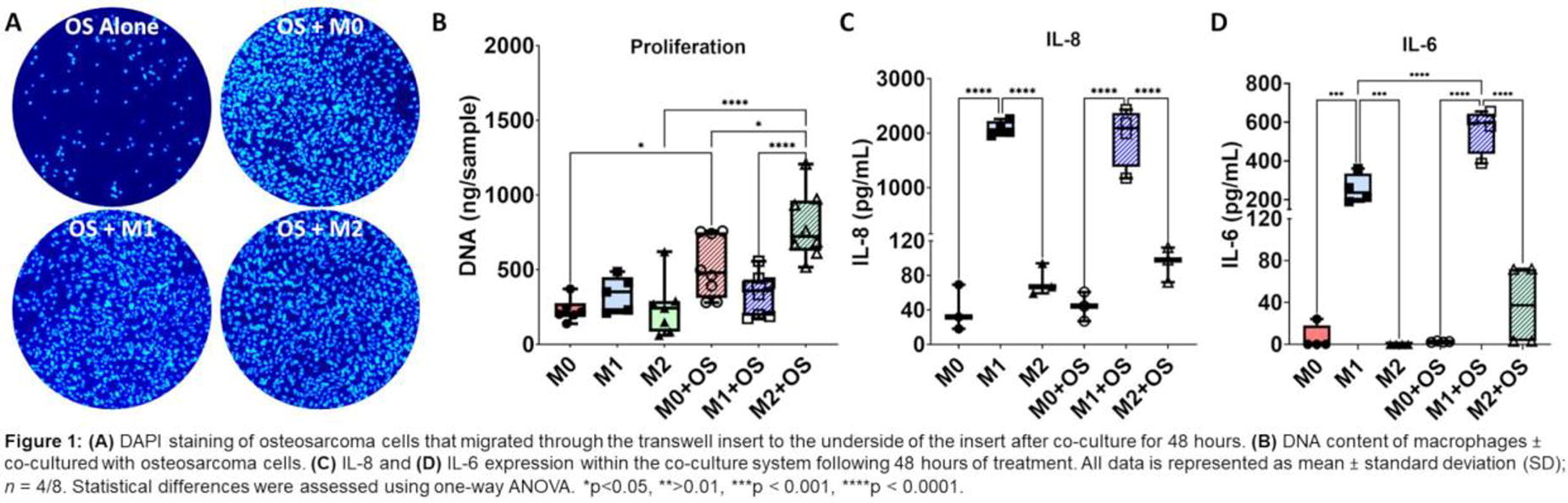
C63 - Investigating The Role Of Mutant Kras Expression In An Early Onset Lung Adenocarcinoma Model
E. Mitta, J. Searle, A. Gilmore, A. Malliri, S. Richardson, S. Cartmell
University of Manchester, Manchester, United Kingdom
*Purpose/Objectives: In recent years significant efforts are being made to improve early detection of lung cancer cases which are currently diagnosed at a late and difficult to treat stage. In this study, we aim to investigate the early transcriptional and morphological changes taking place immediately after expression of mutant KRAS, a frequently mutated oncogene in lung adenocarcinoma. To do this, we study the cells either alone or in a co-culture system which models an early stage of lung adenocarcinoma where few cancer cells are present. Experiments are run in static or dynamic conditions which mimic the breathing motion, a key feature in lung physiology that is rarely considered in cancer in vitro models, yet it is shown to affect disease progression.
*Methodology: Wild-type MLE12 cells and cells containing doxycycline-inducible KRAS G12C or G12D constructs were cultured according to ATCC guidelines. To induce mutant KRAS expression, cells were treated with 10 or 100 ng/ml doxycycline for a maximum of five days. For identification, lentiviral vectors including either a tagRFP-CAAX sequence (for parental cells) or GFP-CAAX sequence (for either mutant cells) were introduced and expressed stably. Parental and mutant KRAS-expressing cells were cultured either in a mono- or co-culture, the latter consisting of 75% parental and 25% G12C or G12D cells. To apply breathing-like tension, cells on collagen type I-coated Bioflex plates were mounted on a Flexcell tension system to apply 10% strain at 1.3 Hz frequency. Localisation of cell-cell junction markers (E-cadherin, ZO-1) was investigated by immunofluorescent staining. Protein levels of E-cadherin and vimentin were established by western blotting. Cell migration was evaluated by a wound-healing assay.
*Results: Upon expression of either mutant version of KRAS, cells assume a more spindle-like shape compared to the cobble-stone appearance of their parental counterparts. Cell-cell junction markers that appear on the cell membrane of parental MLE12 cells are absent from plasma membranes of mutant cells. Expression levels of epithelial-to-mesenchymal transition (EMT) markers E-cadherin and vimentin show opposite trends with the former being significantly downregulated and the latter upregulated in the highest treatment group compared to their untreated counterparts. Mutant cells also harbour higher migration rates compared to their parental and their untreated counterparts. In a co-culture setting, parental cells maintain their cell-cell junction properties forming an epithelial monolayer, whereas G12C and G12D cells appear growing on a different Z-plane with long protrusions evident of a migratory phenotype observed in monocultures. Upon breathing tension, all cells retain their cell-cell junction properties despite induction of mutant KRAS, and co-cultures retain the morphological characteristics of their untreated counterparts.
*Conclusion/Significance: Here, we show cells undergo rapid transformation upon mutant KRAS expression which is maintained in a co-culture setting and could have potential effects on healthy wild type cells. Short-term application of breathing tension had little effect on healthy epithelium and a potentially slowing effect on transformation of mutant cells. Longer time points are currently investigated. Future experiments will investigate the transcriptional changes of either cell populations utilising RNA sequencing and significant hits will be further validated in co-cultures.
C64
C65 - Development Of Vascular Networks In 3D In Vitro Models Of Cancer
A. R. Upadhyay, S. Galal, J. Pape, D. Bakkalci, A. Macrobert, M. Loizidou, U. Cheema
University College London, London, United Kingdom
*Purpose/Objectives: Tumour cells require angiogenesis and lymphoangiogenesis in order to disseminate and invade into other organs as the cancer develops. The metastatic process is guided by the extracellular matrix (ECM) composition such as protein density and biochemical profiles, as microenvironmental cues will also regulate the formation of vascular networks. We bioengineered compartmentalized cancer tumouroids comprised of various stromal cell populations to investigate how the ECM composition influences the formation of vascular networks.
*Methodology: Compartmentalised dense collagen I tumouroids were fabricated and cultured over 21 days, with a central mass containing cancer cells and surrounded by the tumour stroma populated with stromal cells such as cancer associated fibroblasts and endothelial cells. The 3D matrix composition also includes basement membrane proteins such as laminin [20ug/ml] and fibrin [2mg/ml]. Cancer cell populations include colorectal cell lines (HT29 or HCT116) and breast cell lines (MDA-MB321 and MCF-7).Immunofluorescence staining and imaging was conducted to determine the extent of vascular network formation in all 3D cultures, with image analysis using ImageJ. Gene expression of key markers was determined using qPCR and protein expression using ELISAs.
*Results: Vascular network formation is promoted by biophysical parameters such as ECM composition and chemical cues provided by growth factors such as FGF and VEGF. Tumour cells will generate angiogenic growth factors and the tumouroid model allows for the generation of chemotactic gradients in the 3D matrix. Endothelial cells are responsive to these gradients and fuse to form mature branched networks (Figure 1). The inherent characteristics of cancer cells such as their invasive nature influences the development of vascular networks and the introduction of cancer associated fibroblasts in the stroma will disrupt the network formation.
*Conclusion/Significance: The biomimetic 3D tumouroid model recapitulates the tumour microenvironment, which can form vascular networks. It provides a platform to investigate how cancer driven angiogenesis is influenced by the different facets of the biophysical and biochemical environments. In addition, it can provide a platform to better understand the interplay of different cell populations and their role in tumour vasculature. Increasing the complexity of 3D disease models allows for drug treatments to be tested with greater biomimicry.
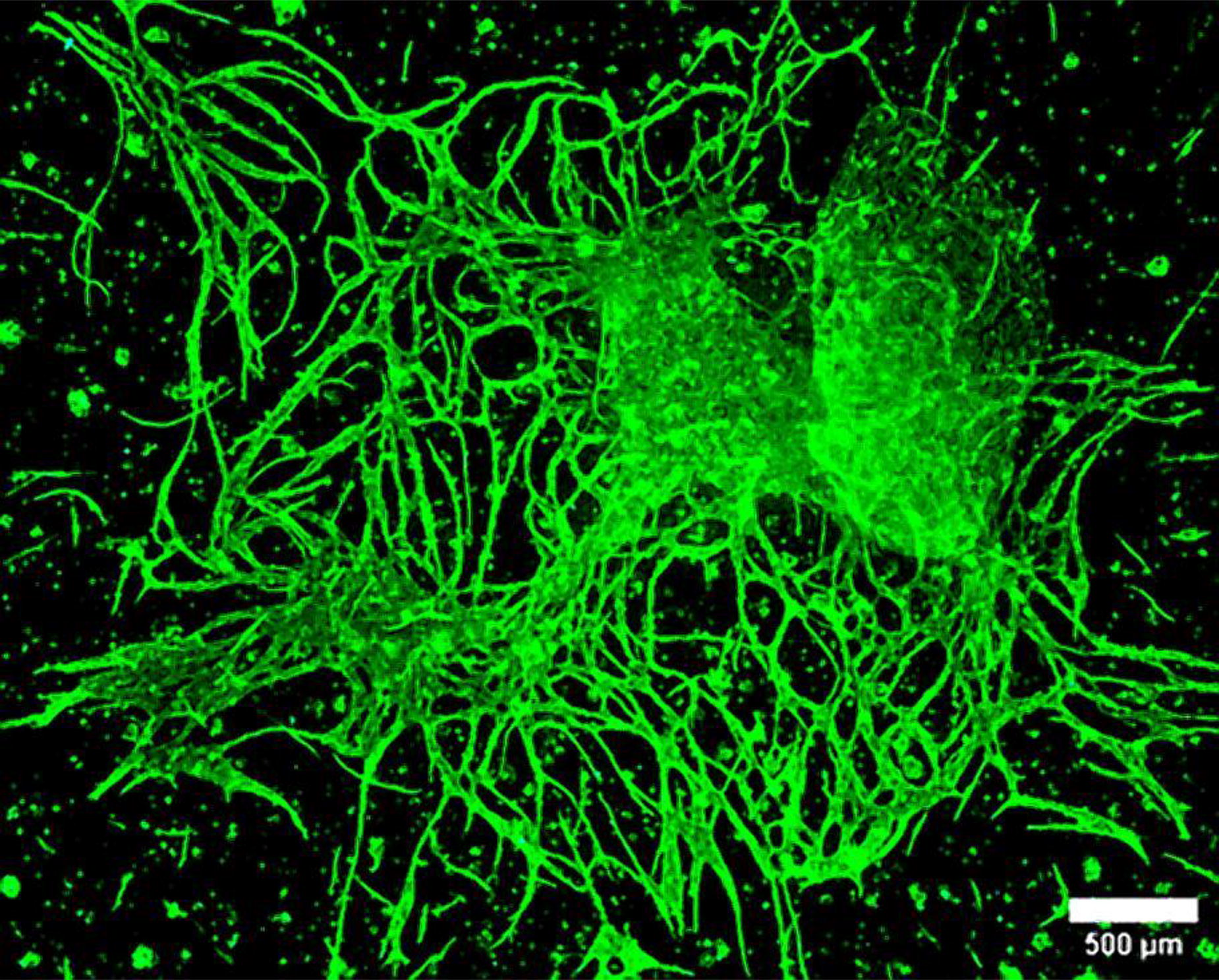
C67 - Development Of A Combined Melanoma/skin Organoid System To Study Tumor-stroma Interactions
G. Nomdedeu-Sancho, A. Gorkun, N. Mahajan, N. P. Edenhoffer, A. Atala, S. Soker
Wake Forest Institute for Regenerative Medicine, Winston-Salem, NC
*Purpose/Objectives: Overcoming intrinsic and acquired resistance to targeted and immunotherapies remains a challenge in melanoma management. The interaction between cancer cells and the tumor microenvironment (TME) -- especially with cancer-associated fibroblasts (CAFs) -- provides a tumor-supportive environment that promotes melanoma growth and immune evasion, rendering therapies ineffective. Current in vitro 3D melanoma models fail to reproduce the intricate cellularity and structure of the healthy skin around the tumor, which hinders their ability to represent the TME. Thus, the development of physiologically and architecturally accurate melanoma models is crucial to studying the interplay between cancerous and healthy cells that impacts the tumor’s response to therapy.
*Methodology: We incorporated an aggressive melanoma cell line into spherical layered skin organoids containing epidermal, dermal, and hypodermal components. This resulted in the generation of in vitro 3D combined melanoma-skin organoid systems that can potentially be used to 1) simulate melanomagenesis, epidermal and dermal invasion, 2) explore tumor-stroma interactions, 3) test therapies, and 4) identify early melanoma biomarkers.
*Results: SK-MEL-28 cells added during the formation of skin organoids quickly proliferated, penetrated the superficial layers of the epidermis, and subsequently egressed the organoids. Harnessing the observed migratory capacity of melanoma cells, we embedded the organoids in a dermis-like fibroblast-ladden collagen gel. In this setting, cancer cells rapidly invaded the gel and interacted with the fibroblasts, reproducing melanoma dermal invasion. Melanoma migration into the gel seemed to be enhanced by the presence of fibroblasts when compared to collagen-only cells, suggesting the existence of a tumor-stromal crosstalk favoring tumor invasiveness. Further analysis of CAF-specific marker expression allowed us to assess the melanoma-induced transformation of fibroblasts into CAFs to support tumor progression and invasion.
*Conclusion/Significance: We created a melanoma-skin organoid system that is able to recapitulate hallmark features of melanoma, such as epidermal and dermal invasion and tumor-stroma interactions in a realistic human three-dimensional environment. By incorporating patient-derived melanoma cells, we expect to use this system to enhance our understanding of the role of tumor microenvironment on therapy resistance and improve personalized melanoma treatment.
C69 - Patient Specific Colorectal Cancer Organoids On A Chip
E. Zanrè1,2, E. D’Angelo2,1, A. Marangio2, M. Agostini2,1, E. Cimetta1,2
1
University of Padova, Padova, Italy,
2
Fondazione Istituto di Ricerca Pediatrica Città della Speranza, Padova, Italy
*Purpose/Objectives: Colorectal cancer (CRC) is one of the leading causes of cancer related death. CRC is often refractive to conventional treatments highlighting the need for improved models bridging the gap between in vitro and in vivo drug evaluation phases. Ideal models are organoids: self-organizing 3D structures preserving in vivo-like structural and functional features. They are composed by cells growing in a 3D environment supported by biological or synthetic hydrogels resembling the native extracellular matrix (ECM). Innovative engineering approaches contribute to the development of on-chip technologies for the production, control, and analysis of organoids and their microenvironment, further helping in surpassing limitations of conventional culture techniques. We here propose a novel and easy-to-use device enabling on-chip formation, culture, and drug screening studies on CRC organoids.
*Methodology: The designed microdevice is composed of 2 layers separated by a porous membrane: the first generates 5 concentrations of a soluble agent and the other houses 5 organoids culture chambers. The mold is fabricated using soft-lithography and the device is produced via standard replica molding using polydimethylsiloxane (PDMS). Patient-derived organoids (PDOs) are seeded in the culture chambers within a patient-derived hydrogel. PDOs are obtained from CRC biopsies and the hydrogel from decellularized healthy colon biopsies. Perfusion is driven via connections to a syringe pump. Fluid dynamic and biological validations are performed to test the correct functioning of the platform. Live&Dead assays, cytofluorimetric analyses, gene expression and immunofluorescence analyses are performed on PDOs after perfusion culture and compared with both static and conventional 2D conditions.
*Results: Numerical simulations, fluid dynamic and biological validations confirm the establishment of 5 different concentration level inside the chambers and that the device is suitable for PDOs growth. Live&Dead assays demonstrate the correct viability of on-chip organoids. Gene expression analysis shows that proliferation, epithelial to mesenchymal transition, and inflammation genes are generally upregulated in the perfused microfluidic system compared to static and 2D conditions. For example, the proliferation marker KI-67 is significantly upregulated in dynamic conditions, similarly to the epithelial to mesenchymal marker VIM and the hypoxia marker HIF1A. Observations are also confirmed via immunofluorescence analysis, for example showing increased expression of proliferation markers, like KI-67, in perfused conditions. After 72 hours of drug treatment with 10 µM 5FU, samples cultured in standard static conditions had a higher sensitivity to the drug compared to on-chip PDOs, as confirmed by cytofluorimetric analysis.
*Conclusion/Significance: Biological validations show that our platform is suitable for cell culture applications and the device represents a more realistic and appropriate environment for the development of cancer organ-like structures. Drug tests reveal that standard culture conditions overestimate anti-cancer drug effects, differently from 3D models which more closely mimic the in vivo environment and the true body responses to treatment.
C72 - Osteoclast-independent Bone Invasion In Head And Neck Squamous Cell Carcinoma: A Bone-on-a-chip Model Study
M. A. Fraga1, C. M. França1, M. Sousa1, A. Athirasala1, S. Vignolo1, A. Correr2, L. Bertassoni1
1
OHSU, Portland, OR,
2
UNICAMP, Piracicaba, Brazil
*Purpose/Objectives: Head and neck squamous cell carcinomas (HNSCC) present a challenge due to their tendency to invade nearby bones. Originating from mucosal epithelial cells in regions like the oral cavity, pharynx, larynx, and sinonasal tract, these cancers exhibit bone invasion in over 50% of patients at diagnosis, categorizing them as advanced primary tumors (T4). This classification is associated with poor overall survival and high local recurrence rates. Despite a large body of work, the early mechanisms of HNSCC bone invasion are not fully understood. Morphologically, bone invasion in HNSCC is described as erosive or infiltrative. The erosive pattern, the most studied, is typically osteoclast-dependent, while the infiltrative pattern is marked by tumoral cells in deep cortical bone marrow, occurring in a mostly osteoclast-independent manner. We hypothesize that the osteoclast-independent bone invasion is driven by specific signals from bone cells and the extracellular matrix (ECM). Our overarching goal is to understand the role of the bone mineralization and osteoblast/osteocytes on the invasive behavior of HNSCC.
*Methodology: To understand the osteoclast-independent invasion mechanism, we utilized a “bone-on-a-chip” model composed of microfluidic device with a central chamber separated from two parallel channels by pillars. The chamber was filled with matrix of a mixture of collagen (2.5 mg/mL-1), fibrin, and thrombin, with or without osteoblasts (3x10⁶ cells/mL). The matrix was then mineralized for three days in a solution of CaCl2.2H2O, osteopontin, and K2HPO4.The mineralization process induced osteoblast differentiation in osteocytes. Subsequently, oral squamous cell carcinoma (OSCC) cells were seeded into a lateral channel and cultured for 3 days.
*Results: OSCC interacted with the ECM with and without osteoblast, showing migration, and morphological changes into a more fibroblastic phenotype. OSCC cells in the mineralized groups expressed more vimentin than non-mineralized, suggesting a phenotypic change compatible with epithelial-mesenchymal transition (EMT). Moreover, devices loaded with osteoblasts and osteocytes showed an increase in the proliferation and invasion of cancer cells with enhanced paracrine secretion of IL-6 and IL-8.
*Conclusion/Significance: In conclusion, bone mineralization and the presence of osteoblasts and osteocytes in the matrix are correlated with EMT and a more invasive behavior in OSCC cells. These findings are crucial for understanding the mechanisms of bone invasion by OSCC, suggesting potential therapeutic targets directed at this interaction.
C73 - Evaluation Of The Chemotactic Activity And The Invasive Potential Of The Mcf-7 Cell Line In A Decellularized Tissue Model
L. N. Pérez-Tulich, D. C. Peña-Córdova, A. J. Iza-Zurita, J. E. Valdivia- Silva
UTEC, Lima, Peru
*Purpose/Objectives: Breast cancer is the most common malignancy among women in the United States and Europe. According to MINSA in Peru, it represents the second most frequent neoplasia with an annual incidence of 28 cases per 100,000 inhabitants. In breast cancer, cell invasion is a process orchestrated by an enormous variety of complex microenvironmental stimuli, making it difficult to isolate and evaluate the effects of biochemical or biophysical signals (i.e., tumor architecture, matrix stiffness) on the progression of the disease. The invasive potential of cancer cells is generally evaluated in vitro using Matrigel and type I collagen, as a surrogate basement membrane, however, in vivo the interaction of cancer cells with the ECM is essential for invasion and metastasis. In this sense, physiologically relevant tumor models are necessary to perform cancer cell invasion studies under well-controlled conditions. Although matrigel and collagen I are commercially available and easy to use, they do not sufficiently mimic tumor niches, may lead to false negatives and false positives in drug screening, and could limit the utility of invasion models. Which is why we propose using the decellularized extracellular matrix as a biomaterial for breast cancer migration and invasion assays. We focused on evaluating the chemotactic activity and invasive potential of a MCF-7 breast cancer cell line in a decellularized tissue model.
*Methodology: The pig heart was decellularized by immersion to obtain an extracellular matrix rich in type I and IV collagen. The presence of nuclear material in the decellularized matrix was evaluated based on the concentration of DNA in a nanodrop, with the presence of nuclei in DAPI staining, and histological sections with Hematoxylin-Eosin staining, Masson’s Trichrome staining and scanning electron microscopy. To obtain the hydrogel, the matrix obtained was converted into powder and solubilized with Acetic Acid and neutralized with Sodium Hydroxide. Once the hydrogel was obtained, it was characterized using SEM and FTIR. Toxicity, cell migration, invasion and chemotaxis tests were carried out in 2D and 3D cultures.
*Results: Through SEM microscopy and histological stains, it was verified that the decellularization method used was optimal, because it preserves the collagen fibers and others present in the extracellular matrix.In the characterization of the hydrogel with FTIR we found that the sample has peaks in the amide A region, characteristic of peptides and proteins, the second amide I peak, corresponding to C=O (carbonyl) and the N-H bond (amino group) in the bond. peptide. The last peak corresponds to the vibrations of carbon-carbon (C-C) and carbon-hydrogen (C-H) bonds, characteristic peaks for the presence of collagen in the sample.The hydrogel obtained from the decellularized extracellular matrix favors cell viability, which was shown by the MTT assay.
*Conclusion/Significance: The hydrogel obtained allows migration and invasion, in 2D cultures (Transwells) and 3D models without significant differences when using matrigel as a basement membrane.Therefore, it is concluded that the hydrogel obtained from the extracellular matrix of the pig heart could be used as a model to study cell migration in breast cancer and other cancers.

C74 - 3D-bioprinted Liver Tissue Model For Testing The Activity And Cytotoxicity Of Drugs - Revolution In Preclinical Testing Of Oncological Drugs
M. Klak1,2, M. Popis1, O. Janowska1, M. Czajka2, M. Dec2, A. Papierniak-Wygladala1, K. Florys-Jankowska2, T. Dobrzanski1, A. Berman1,2, M. Wszola1,2
1
Polbionica Ltd., Warsaw, Poland,
2
Foundation of Research and Science Development, Warsaw, Poland
*Purpose/Objectives: The process of new drugs testing begins with 2D cultures, cocultures or spheroids. Then it moves on to in vivo tests on animal models. However, this does not take into account toxicity in the human tissue microenvironment. Creating 3D flow models could solve this problem. The aim of the research was to characterize the biocompatibility of the biomaterial and a preliminary assessment of its suitability for the production of bioconstructs imitating human liver tissue, as well as to assess the gene expression of the liver cancer tissue model.
*Methodology: The use of 3D-bioprinting and ink-jet technology required the development of bioinks that would be the optimal environment for the cells suspended in it. Two hydrogels were developed: (1) gelatin and hyaluronic acid, and (2) hydrogel (1) enriched with liver dECM proteins. The bioinks were mixed with hepatocytes and endothelium. 3D-bioprinted models with a flow system was cultured for 21 days. Microscopic imaging (FDA/Pi) was performed at designated time points and samples were preserved for histological analysis. The culture medium was collected to evaluate cells activity. Additionally, the expression of AFP, VEGF-A, GPC-3, NOS3, GP73, DCP2 genes was assessed.
*Results: In both variants, high cell viability was over 90%. Even distribution of cells and their ability to proliferate and form spheroids were demonstrated. Gene expression analysis showed the greatest change in tumor markers characteristic of HCC compared to controls. The expression of AFP, NOS3 and GP73 genes was observed from day one. After 10 days of breeding the model, the expression of all tested genes was already demonstrated. Individual expression levels showed significant differences.
*Conclusion/Significance: Based on the conducted research, the functionality and stability of the liver model was demonstrated. The developed technology will constitute an alternative research path in the preclinical phase for new drugs testing.
C75 - Circulating Tumor Cell Separation With Magnetic Bead And Electrochemical Analysis: Impedance Analyzer
K. Kang, C. Kim, C. Park
Jeonbuk National University, Jeonju-si, Korea, Republic of
*Purpose/Objectives: Capturing and observing cancer cells in the blood plays a crucial role in the early diagnosis of cancer and the collection of necessary cancer cells for treatment. However, due to the small number of cancer cells in the blood, capturing and analyzing these cells can be challenging. In this study, we utilized magnetic particles that can specifically bind to cancer cells to separate them in a microfluidic channel using a magnetic field. The separated cancer cells were monitored through an impedance analyzer, and the captured cells were used in a drug evaluation platform for the development of personalized medicine for patients.
*Methodology: Firstly, to capture cancer cells, we fabricated immunomagnetic beads using EpCAM. The produced immunomagnetic beads were incubated with cancer cells in the blood to prepare the sample. We created an electromagnet for magnetic particle separation, which controlled the attraction of cancer cells into a separation channel while they flowed through the microfluidic chip. Utilizing an SLA 3D printer, we manufactured the microfluidic chip. The inlet and outlet of the microfluidic chip were designed with different sizes to prevent the mixing of blood with the sheath flow. We patterned a 20-micrometer electrodes on the microfluidic chip to measure the impedance changes of individual cells flowing in the separation channel. Finally, we cultivated the captured cells to evaluate their responsiveness to various drugs.
*Results: We were able to control cancer cells using magnetic particles attached with EpCAM. The intensity of the magnetic particle attachment varied according to the type of cancer cell, with the most significant attachment observed in MCF-7 and HT-29 cells. By adjusting the strength of the electromagnet, we could control the separation of cancer cells from the blood in the microfluidic channel. The separated cancer cells could be counted and monitored using an impedance analyzer without missing the differences (10 ms) between rapidly flowing cells over the electrodes integrated inside the microfluidic channel. The captured cells were re-cultured and treated with PTX, allowing us to optimize the concentration capable of treating the cancer cells.
*Conclusion/Significance: Through this study, we were able to selectively separate and capture flowing cancer cells from the blood using a microfluidic chip and magnetic particles. In addition, by utilizing the electrodes integrated into the microfluidic chip, we were able to monitor the separated cells in real-time. This approach allows for the observation and analysis of cells that could potentially be lost during the analysis process. Furthermore, we were able to evaluate the drug responsiveness of the captured cells to determine the optimal drug concentration. By demonstrating a method that can quantitatively and qualitatively evaluate cancer cells captured from patients, we anticipate showing the potential for faster and more accurate diagnostic methods.

C76 - Engineering A Simple, Chemically Defined Basement Membrane (bm) For Epithelialization Studies
M. Mistry1,2, X. Yang1,2, A. D. Chen1, B. P. Chan3,2,1, M. Chan1,4
1
The University of Hong Kong, Hong Kong, Hong Kong,
2
Advanced Biomedical Instrumentation Centre, Hong Kong Science Park, Hong Kong, Hong Kong,
3
The Chinese University of Hong Kong, Hong Kong, Hong Kong,
4
Centre for Immunology & Infection (C2i), Hong Kong, Hong Kong
*Purpose/Objectives: In Tissue Engineering, epithelialization is a critical process for multi-layered tissues like the skin, intestine, and the airway. In the native tissue, the basement membrane provides vital cues to epithelial cells. These cell-ECM interactions are responsible for cell adhesion, proliferation, and function. Epithelialization studies mostly rely on using the ECM protein coatings to model the native basement membrane. Alternatively, some studies also use Matrigel, derived from mouse sarcoma. While Matrigel can mimic the native ECM in a more physiologically relevant manner, it is tumorigenic, contains a lot of growth factors and often varies from batch to batch. Thus, we have attempted to engineer a simple, chemically defined basement membrane that can give comparable results to that of Matrigel in epithelialization studies.
*Methodology: Two of the main components of both matrigel as well as the native ECM, are Collagen IV and Laminin. Thus, the effect of a cross-gradient of Collagen IV-Laminin was investigated on two cancerous epithelial cell lines (MCF7 and Caco2) and one healthy epithelial cell line (MDCK). Using Collagen type I as the bulk material, a cross-gradient of Collagen IV and Laminin was created by incorporating them in varying amounts. Upon gelation, the membranes were air-dried, and seeded with the cells to form the epithelial models. The initial adhesion and proliferation of epithelial cells on the different membranes were investigated through crystal violet assay and ki67 IF staining. The barrier integrity of the resultant epithelial models was analysed on a localized level through staining of junction proteins (Zo1 and ecadherin), as well as on a macroscopic functional level through TEER and Dextran.The optimized combinations of Collagen IV and Laminin were then crosslinked to form a chemically defined basement membrane (BM). Epithelial cells were seeded onto the airdried membrane, as well as on Matrigel as control. Comparative analysis was carried out between the different groups in terms of adhesion, proliferation, barrier integrity and function.
*Results: Characterization of the engineered basement membrane materials revealed their microstructure. Comparative analysis of the epithelial models formed in different ECMs, indicated that cells exhibited differential responses to the different matrices. Cell adhesion, and proliferation were improved in BM as compared to Matrigel. However the zo1 barrier integrity was similar in both the epithelial models
*Conclusion/Significance: The results of the study can contribute to the future development of a chemically defined Basement membrane for complex epithelialization studies, tumouroid models and drug studies
C78 - Vascular Dynamics In A Fabricated Tissue During In Vitro Conditioning
S. M. Moss, J. B. Hoying
Advanced Solutions Life Sciences, Manchester, NH
*Purpose/Objectives: The critical importance of a microvasculature in fabricated tissues is stressed in virtually every discussion related to regenerative tissue medicine. The extent, character, and integrity of such a fabricated microvasculature is determined by the vascular precursors, the cell types present in the tissue, and the biomechanical forces acting in the tissue. In a fabricated, living bone construct, we use isolated fragments of intact microvessels harvested from adipose tissue (haMVs) to create a vascularized periosteum-like shell around a core bone construct. The haMVs retain the native microvessel structure and all vessel-resident cells, including macrophages within the perivascular niche. In this bone construct, the hMSCs actively contract the loose collagen “shell” in vitro prior to implantation, improving graft handleability. Recognizing that tissue vascularization can be impacted by hMSC paracrine activity and the biomechanical cues associated with tissue compaction, we explored the vascularization dynamics of this outer tissue layer formed in the bone construct and separately how the number of haMV perivascular cells (CD45 and aSMA) impacts angiogenesis.
*Methodology: To study hMSC-haMV dynamics, haMVs and hMSCs were cocultured for 1-2 weeks within plugs of collagen I with various media including osteogenic media. Additionally, different lots of hMSCs, each with different cytokine release profiles, were included. Tissue contraction, hMSC number, and haMV angiogenesis were monitored over the culture time. Cultures were stopped at 50% contraction and fixed for confocal analyses (UEA-1 lectin and hMSC cell number). Additionally, this experiment was performed in the presence of demineralized bone matrix (DBM). DBM is native bone matrix from which cells, fats, and minerals have been removed. DBM is known to release BMP2 and other factors impacting both angiogenesis and MSC behavior. In a separate experiment, haMV cultures were fixed at different timepoints (days 0, 10) and the extent of angiogenesis correlated with the number of CD45+ or αSMA+ cells.
*Results: As expected, hMSCs promoted haMV angiogenesis (measured as vessel length density).Osteogenic media further enhanced haMV angiogenesis. Compaction of the tissue arrested angiogenesis in all media conditions. However, neovascular regression occurred in cultures with the osteogenic media after compaction, but not in the non-osteogenic (base) media conditions, in which haMV-derived neovasculatures persisted. In all instances, the presence of DBM increased tissue contraction. Unsurprisingly, osteogenic media increased hMSC cell number as compared to other culture media conditions. Interestingly, the extent of CD45+ cells, but not αSMA+ cells, present within the haMV walls at the beginning of the culture correlated with more angiogenesis.
*Conclusion/Significance: We show that haMVs were effective at vascularizing a tissue construct. We also show that it’s important to consider the dynamics between tissue conditioning (compaction in our case), stromal cell activities, and angiogenesis in vascularizing tissue constructs. We determined that while hMSCs promote angiogenesis, MSC-mediated tissue contraction can impair angiogenesis. Furthermore, this can be impacted by either the media used or the presence of other tissue components (DBM in our case). Thus, fabrication designs should entail strategies that consider these coordinated activities and create a culture environment that enables staging maturation that optimizes outcomes.
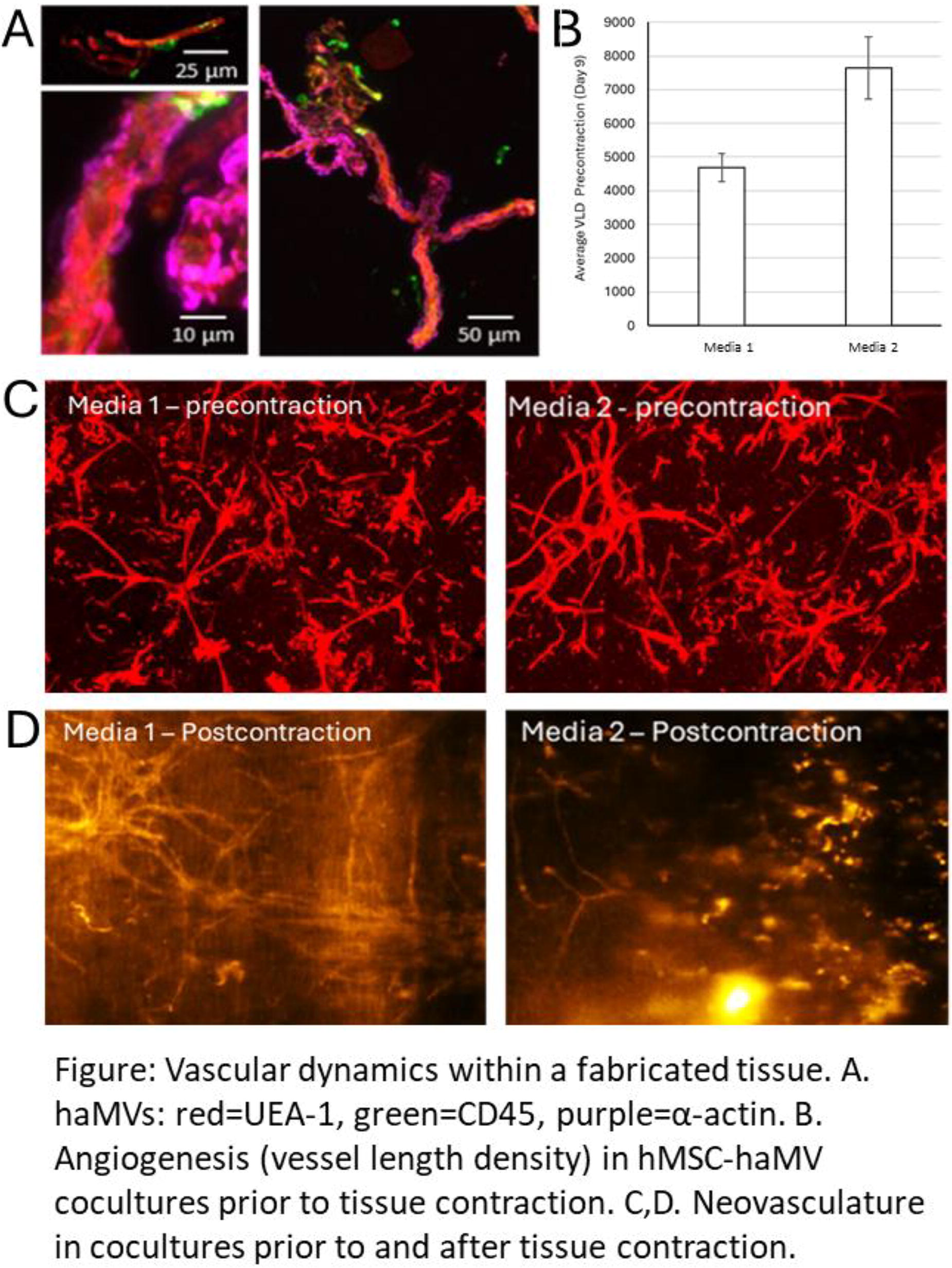
C80 - Venous Angiogenesis Of Decellularized Dermis To Create Transplantable Dermal Substitutes
S. Lau, E. Harvey, N. Makhoul, J. Barralet
McGill University, Montreal, QC, Canada
*Purpose/Objectives: Skin defects can arise from various causes, and when conventional wound healing proves inadequate, pedicle and free flaps become pivotal. These flaps can either preserve existing vasculature or undergo transfer and re-anastomosis. However, their applicability diminishes in cases involving extensive burns or skull defects, as the limited donor skin may elevate donor site morbidity or complication. Achieving transplantable vascularized dermis through arteriovenous loops (AVL) is effective but surgically demanding. This study aims to explore the vascularization potential of decellularized dermis, as a possible alternative to AVL to create a transplantable vascularized dermal tissue. A decellularized dermis was implanted around a femoral vein (DV), and a control group (DC) was implanted subcutaneously. This investigation compared the vascularization patterns of these two groups and their potential to support cell viability.
*Methodology: The decellularized dermis was prepared under chemical and enzymatic approaches involving sodium chloride and DNAse (I) solution. This study was approved by the McGill University Ethics Committee (AUP 7662). The surgeries were performed on N=10 Wister rats (350-500 grams, 4-6 months old) that were divided into two groups based on the placement of the decellularized dermis. In the DC group, incisions were made along the inguinal fold, allowing for the subcutaneous implantation of the decellularized dermis, which was then sutured under the skin. As for the DV group, dissection proceeded laterally and deepened to access the femoral vessels. Careful dissection and cauterization were performed on the femoral vein to remove any branches. Subsequently, the dissected femoral vein was enveloped with a decellularized dermis and secured in place using two sutures. The rats were monitored for 8 weeks, at which point they were sacrificed and samples were analyzed by microCT and histology to identify new vasculature.
*Results: Histological evaluation revealed a statistically significant difference in the mean number of vessels per mm². The DV group exhibited a higher mean number (20.43 ± 5.13) compared to the DC group (4.34 ± 1.72). The vascularization in the DV group was 4.71 times greater than in the DC group. Vessels in the DV group primarily originated from the inner femoral vein, extending outward, consistent with microCT imaging. In contrast, vessels in the DC group mainly grew around the DV’s edge, penetrating and developing within the dermis alongside surrounding fibrous tissue. In order to assess transplantability, DV vascularized by a central vein was excised and anastomosed with the femoral artery, and the dermal matrix was observed to bleed upon light injury with a scalpel. These findings highlight the efficacy of the DV group’s approach and its potential application in advancing vascularized tissue engineering strategies.
*Conclusion/Significance: In summary, this study confirms the effectiveness of decellularized dermis in promoting robust vascularization. Particularly in the DV group, the results substantiate the feasibility of an alternative approach to arteriovenous loops. The quantifiable difference in vessel density between DV and DC groups supports the exploration of innovative strategies for skin defect treatment. The enhanced transplantability of DV implants indicates promising clinical potential.
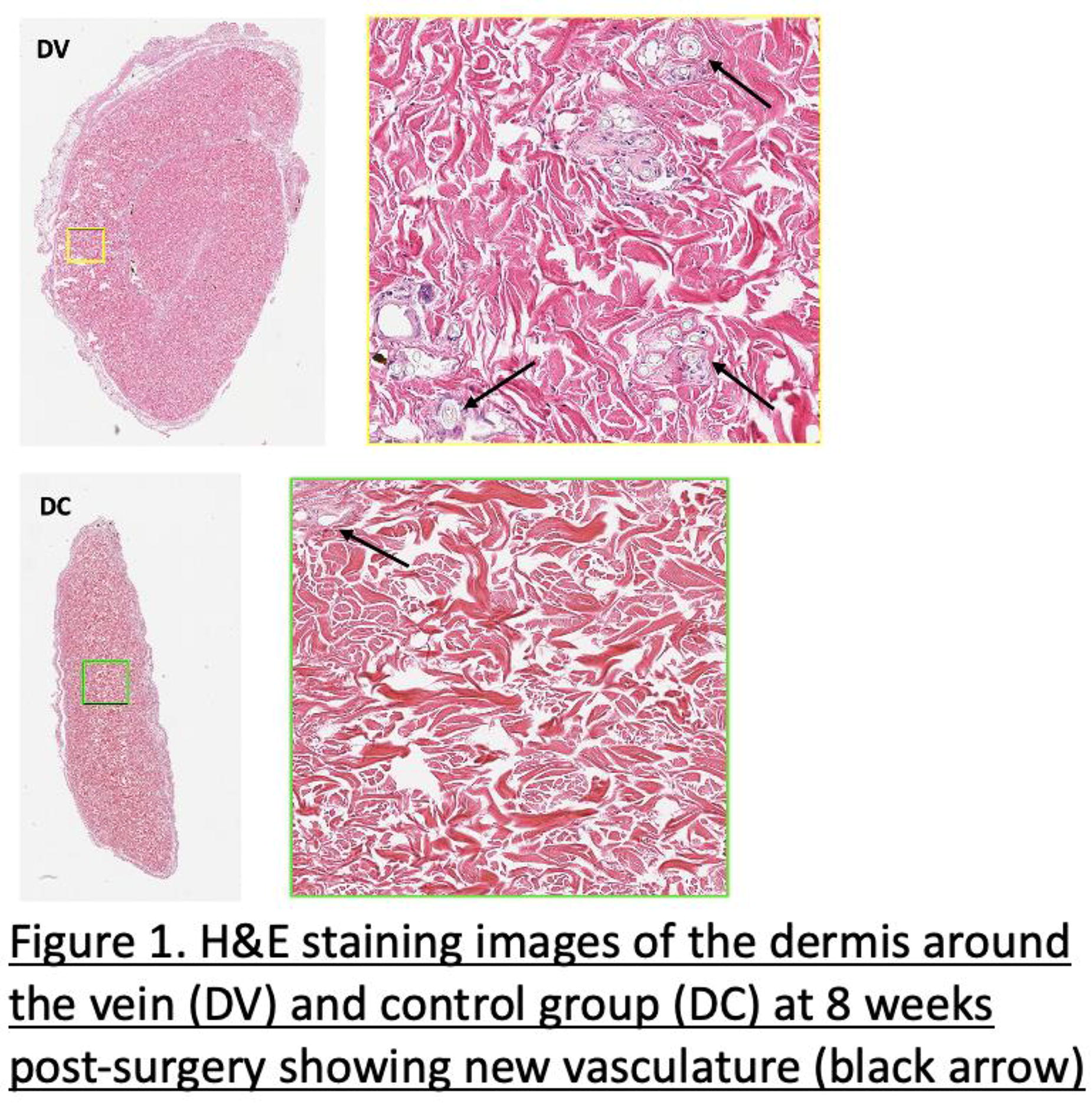
C81 - Immunoprotection, Vascularization And Enhancing Insulin Secretion Of Pseudo-islets Through Lentivirus Mediated Glp-1 Gene Therapy: All Three Features In Single Spheroid Model For Type-1 Diabetes
E. Yalcin Kaya, S. Kizilel
Koç University, Istanbul, Turkey
*Purpose/Objectives: The primary objective of this research is to enhance glycemic control through lentivirus-mediated glucagon-like peptide-1 (GLP-1) gene therapy in Beta-TC-6 cells, thereby increasing insulin secretion. This study aims to prolong the stability and functionality of injectable pseudo-islets for Type 1 diabetes. The focus is on addressing how the nanogel barrier, by providing released growth factors for vascularization, protects the co-cultured spheroid from immune attacks post-transplantation.
*Methodology: Glycemic control can be regulated through the insulinotropic effect of GLP-1. In this work, we utilize the therapeutic potential of lentivirus-mediated GLP-1 gene therapy on insulin-secreting Beta-TC-6 cells to increase insulin secretion. To mitigate the loss of the GLP-1 molecule due to mass transfer limitations, we have utilized three-dimensional spheroids. Given the short plasma half-life of endogenous GLP-1, a cluster of lentiviral transduced beta cells in a spheroid model maintains the active GLP-1 molecular pathway. The second key feature of pseudo-islets is vascularization. This involves the coculture optimization of transduced beta cells with Human Umbilical Vein Endothelial Cells (HUVEC) to ensure the formation of tubular-like structures in the transplanted spheroids, thus addressing a major challenge in tissue engineering. Our goal is for spheroids, composed of a specific cell ratio of transplanted GLP-1 transduced Beta-TC-6 and HUVEC cells, to contribute to rapid anastomosis while simultaneously providing high insulin secretion. Immunoprotection is the third critical aspect of this work, aimed at mitigating immune cell attacks post-transplantation. Therefore, we have employed growth factor-loaded gelatin-based nanogels to coat the pseudo-islets. These functional nanogels, designed to avoid internalization into cells, form a thin nano-barrier that not only prevents immune cell attacks but also facilitates the release of growth factors to the underlying endothelial layer, thus promoting rapid integration with the host vasculature.
*Results: Our results showed a 1.7-fold increase in GLP-1 secretion in the Beta-TC-6 lentiviral transduction group compared to the control. The cell ratio of transduced beta cells and HUVECs has been optimized, focusing on selecting highly viable and functional pseudo-islets. We observed the formation of tubular-like structures from the co-cultured spheroids in vitro within 4 hours in a growth factor-rich environment.
*Conclusion/Significance: This work presents a novel approach in tissue engineering by integrating immunoprotection and vascularization in a single spheroid model, consisting of endothelial cells and transduced beta cells. The study offers a promising strategy for the treatment of Type 1 diabetes by addressing several key challenges in the fields of tissue engineering and diabetes therapy. The combination of vascularization, immunoprotection, and increased insulin secretion through GLP-1 gene therapy represents a significant advancement in the development of effective diabetes treatments.
C82 - High Throughput Bioprinted Vascularized Tissue Platform To Study Environmental Toxicant-induced Vascular Dysfunction
M. Rajput, M. J. Song, D. Gerhold, M. Ferrer, E. lee
NCATS/NIH, ROCKVILLE, MD
*Purpose/Objectives: Environmental toxicants present a significant public health concern and are known to be a causative agent for vascular diseases. However, the full impact and profiling of environmental toxicants remain understudied. To understand the link between environmental toxins and their contribution to vascular disease, it is necessary to evaluate toxin-induced vascular dysfunction in a physiologically relevant human-based model. A 3D in vitro tissue engineering approach offers opportunities to study vascular dysfunction in a pathologically mimicked environment.
*Methodology: Here, we developed a 96-well format, human 3D bioprinted vascularized bed platform (h3DVas) for environmental toxicant evaluation by co-culturing primary human GFP/RFP tagged endothelial cells, fibroblasts, and pericytes. We then exposed the h3DVas model to a select panel of known environmental toxicants to measure vascular dysfunction. Using these environmental toxicant panels, we observed toxicant-dependent vascular damage by high-content imaging and quantified morphometric parameters. Furthermore, we measured cell viability and inflammatory response following titration of toxicant exposure by measuring intracellular ATP levels and secreted cytokines via Luminex assay, respectively.
*Results: By high-content imaging, we observed vascular damage under selective environmental toxicants (Sodium Arsenite, Raloxifene HCL, Pazifinib, and Axitinib). These findings were supported by changes in quantified morphometric parameters like decreased vessel length, branching index, and vessel intensity compared to mock and DMSO-treated h3DVas.The observation was further corroborated by the decreased dose-dependent cell toxicity and increased levels of select inflammatory cytokines (IL6 and INF-gamma). These findings indicated vascular inflammation and vascular damage after toxicant exposure.
*Conclusion/Significance: This data suggests that the bioprinted model can be used as a potential platform to evaluate the biological activity of environmental/chemical toxicants and their health hazard.
C84 - Fostering Antimicrobial Scaffolds For Vascular Tissue Engineering Using Low Temperature Plasma (ltp)
K. VIG1, A. Azzouz2, M. Zafaryab1
1
Alabama State University, Montgomery, AL,
2
Earlham College, Richmond, IN
*Purpose/Objectives: Bypass surgery, using the autologous vein has been one of the most effective treatments for cardiovascular diseases (CVD). More recently, tissue engineering including engineered vascular grafts to synthesize blood vessels is gaining usage. Recently engineered vascular grafts is gaining usage and Dacron/ePTFE has been employed, however, these does not work well for small diameter grafts due to intimal hyperplasia and thrombosis. The objective of this study is to develop scaffolds with antimicrobial properties to regenerate vascular tissue using low temperature plasma.
*Methodology: PTFE was treated with LTP to improve the endothelialization of graft surface. Scaffolds were also modified with Ag-PVP and the antimicrobial peptides, p753, p359 and plant derivatives CBD and Garcinol. Human umbilical vein endothelial cells (HUVEC) were plated on the developed scaffolds and cell proliferation was determined by the MTT assay. Cells attachment on scaffolds was visualized by microscopy and mRNA expressions levels of different cell markers were investigated using quantitative real-time PCR (qPCR). Antimicrobial studies were conducted using MIC and plate counts both in 2D and 3D using human cells.
*Results: XPS confirmed the introduction of oxygenated functionalities from LTP. Microscopic and MTT assays indicated increase in cell viability in LTP treated scaffolds. Gene expression studies shows enhanced expression of cell adhesion marker Integrin- α 5 gene after LTP treatment. The KB test displayed a zone of inhibition for Ag-PVP, p753 and p359 of 19mm, 14mm, and 12mm respectively. MIC values were 100 μg/mL, 12.5 μg/mL, 12.5 μg/mL, 0.2 μg/mL and 1.5μg/mL respectively for Ag-PVP, p753, p359, Garcinol and CBD. To determine toxicity of antimicrobial agents to cells, MTT assay was performed using HEK293 cells. MTT Assay exhibited that used antimicrobial agents Ag-PVP and the peptides were non-toxic to cells at 100μg/mL and 50μg/mL, respectively. Live/dead analysis and plate count of treated bacteria exhibited bacterial inhibition on develop scaffold compared to non-treated scaffold. SEM was performed to analyze the structural changes of bacteria after treatment with antimicrobial agents. Gene expression studies were conducted on RNA from bacteria treated with Ag-PVP and peptides using qRT-PCR.
*Conclusion/Significance: LTP treated scaffolds exhibited better cell proliferation and viability compared to untreated scaffolds. Antimicrobial agents Ag PVP NPs, P753, and P359 appear to be effective and safe without causing any biocidal activity against HEK 293 cells in 3D using PTFE.
C85 - Tuning Collagen-Based Hydrogels For Improved Microvascularization Of Tissue Engineering Constructs
C. F. Gomes1,2, D. B. Rodrigues1,2, D. Cruz-Moreira1,2, J. Noro1,2, S. Freitas-Ribeiro1,2, R. L. Reis1,2, R. P. Pirraco1,2
1
3B’s Research Group, I3Bs – Research Institute on Biomaterials, Biodegradables and Biomimetics, University of Minho, Headquarters of the European Institute of Excellence on Tissue Engineering and Regenerative Medicine, Guimarães, Portugal,
2
ICVS/3B’s - PT Government Associate Laboratory, Braga, Portugal
*Purpose/Objectives: Tissue engineering (TE) is an interdisciplinary field dedicated to creating functional biological substitutes for repairing damaged tissues or organs. Establishing a functional vascular network is a critical challenge in developing TE constructs, as nutrient and oxygen supply, along with the collection of metabolic waste products, are vital for the survival of engineered tissues post-transplantation. This depends, to a large extent, of the biomaterials used for scaffolding in the TE constructs. Collagen, a major component of native extracellular matrix, is believed to be an ideal biomaterial due to its exceptional biocompatibility, bioactivity and support of vascularization. The use of collagen-based hydrogels for TE has been widely studied but has been shown to be hindered by challenges such as contraction during cell culture due to cell-mediated remodeling and overall poor mechanical properties. In this study, we aimed to design and characterize collagen-based hydrogels tailored specifically for supporting the vascularization process while overcoming some of the shortcomings related to contraction and mechanical properties.
*Methodology: Bovine collagen was used to produce uncrosslinked collagen hydrogels (control), genipin-crosslinked collagen hydrogels, and glutaraldehyde (GTA)-crosslinked collagen hydrogels. For each crosslinking group, two discrete crosslinking methodologies were used, with the crosslinker applied on top of the gelified hydrogel or directly within the collagen solution for subsequent gelification. This resulted in different dynamics of collagen fibrillation. Characterization of mechanical properties like compressive strength and Young’s modulus was performed. Samples were furthermore characterized using Scanning Electron Microscopy, Differential Scanning Calorimetry, Fourier-transform Infrared Spectroscopy and Circular Dichroism. Additionally, swelling behavior and degradation kinetics were assessed. Human dermal microvascular endothelial cells (hDMECs) and human adipose-derived stem cells (hASCs) were then cultured on the hydrogels and cell morphology and cytotoxicity evaluated throughout time. Hydrogel contraction during cell culture was also assessed. To evaluate the angiogenic potential of the developed hydrogels, an in vivo experiment was conducted using a CAM model.
*Results: Our findings reveal that different crosslinking regimens of collagen-based hydrogels induced, as expected, relevant alterations in their chemical, structural, and mechanical properties. In fact, the modification of fibrillation time resulted in significant changes in the degradation rate and mechanical properties, overcoming some of the issues associated with the use of uncrosslinked collagen hydrogels. Crosslinked hydrogels, in general, displayed exceptional biocompatibility, supporting the adhesion and proliferation of hDMECs and hASCs as well or better than unmodified collagen. Importantly, preliminary results of in vivo experiments showed their suitability to support vascularization after implantation.
*Conclusion/Significance: Overall, our findings demonstrate the successful production of a range of collagen-based hydrogels specifically tailored to support vascularization after implantation. Crosslinked hydrogels presented excellent performance in in vitro and in vivo assays, indicating this hydrogel formulations as potential tools in the quest to enhance vascularization and overall tissue integration of tissue engineered constructs.
*Acknowledgements: EU Horizon 2020 research and innovation program under the ERC grant CapBed (805411).
C86 - Vascularization Of Kidney Organoids And Glomerular Cells With Self-assembled Vasculature
C. Liu, S. Landau, Q. Wu, M. Radisic
University of Toronto, Toronto, ON, Canada
*Purpose/Objectives: The human kidney plays a crucial role in maintaining electrolyte homeostasis and removing waste products, a process coordinated by several components (including glomerulus) of its functional unit nephron. The dysfunction of glomeruli and a reduction in glomerular filtration are early pathological hallmarks of chronic kidney disease (CKD). To better study kidney pathophysiology and develop therapeutics, engineered in vitro models that recapitulate the complex structure and function of the kidney and/or the glomerulus are required. Existing models such as kidney organoids lack perfusable vasculature. It remains a challenge to develop a 3D glomerular filtration barrier model with integration of glomerulus-patterned structures and underlying perfusable vasculature. This study aims to vascularize kidney cells and organoids in vitro using self-assembled vasculature for the development of high fidelity 3D vascularized kidney models.
*Methodology: Vasculature is formed by co-culturing endothelial cells (ECs) and fibroblasts in hydrogels, incuding fibrin, gelatin methacrylate (gelMA), and fibrin/gelMA mixture, in a 384-well. Green fluorescent protein (GFP)-labelled ECs are used to visualize and assess the self-assembled vascular network. Vasculature formation is optimized by varying the hydrogel compositions and the ratio of ECs and fibroblasts. The perfusable vascular network is established using a previously published 384-well plate-based platform called IFlowPlate, where the middle well containing the self-assembled vasculature via ECs and fibroblasts embedded in a fibrin gel is connected with the adjacent wells by microchannels lined with ECs. Human glomerular epithelial cells (podocytes) are then seeded onto the pre-formed vascular network and cultured until an epithelial barrier is formed. For vascularization of kidney organoids, human induced pluripotent stem cells (iPSCs) are first differentiated to intermediate mesoderm, followed by organotypic culture under air-liquid interface to induce nephrogenesis, and encapsulation with a fibrin gel containing ECs and fibroblasts.
*Results: Self-assembled vasculature is able to form in fibrin (10-20 mg/ml), gelMA (1%-2%), and their mixture at an equal ratio. Interestingly, the self-assembled vasculature in gelMA or gelMA/fibrin mixture exhibits a globular shape with vessels extending from it, which resembles the structure of a native glomerulus. While the vessels are wide and the vascular network formed in fibrin occupies the entire well, the vasculature in gelMA or gelMA/fibrin has thinner vessels and contracts overtime. Thus, polymeric particles are embedded together with the cells in gelMA during seeding to reduce the contraction. A functional vascularized glomerular model is developed and can be used for glomerular filtration studies when human podocytes form an epithelial barrier on a perfusable vascular network established on the IFlowPlate. Kidney organoids under fibrin gel encapsulation exhibit morphological changes, whereas ECs and fibroblasts in the fibrin gel self-assemble into vasculature around each organoid.
*Conclusion/Significance: Vascularization of kidney organoids and cells in a 3D configuration can afford significant improvements in the development of engineered in vitro models with kidney-specific physiological features. High fidelity 3D vascularized kidney models represent a powerful tool for studying patient-specific disease mechanisms, discovering novel therapeutic targets, and screening nephrotoxic drugs.
C87
C88 - Biofabrication Of Blood Vessels From Human Induced Pluripotent Stem Cell-derived Mesodermal Progenitor Cells
L. Dogan1, N. C. Cabezas2, J. Groll2, P. Wörsdörfer1, S. Ergün1
1
Institute of Anatomy and Cell Biology, Würzburg, Germany,
2
Department for Functional Materials in Medicine and Dentistry, Würzburg, Germany
*Purpose/Objectives: An intricate blood vessels network is essential for artificially generated tissues. However, the engineering of proper and functional vasculature using biofabrication remains a major challenge spite of recent advances in bioprinting approaches. Additionally, vascularization is needed to advance 3D in vitro tissue models, which have emerged as valuable tools for longitudinal studies on tissue development, function, and disease progression. The objective of this work is to biofabricate a hierarchical vascular system using human induced pluripotent cells (hiPSC)-derived mesodermal progenitor cells (hiMPCs). The study is based on the intrinsic morphogenesis and vasculogenesis capacity of the hiMPCs which have been already showed to retain their ability forming complex and hierarchically organized vessels. This process mimics the embryonic steps of vessel formation via vasculogenesis and resulted in formation of multi-layered vascular wall.
*Methodology: In the current study, gelatin-based bioinks were used to print hiMPCs in tubular geometry via in-gel bioprinting to generate larger (diameter of up to 3.5mm) vessel-like tubes. Cell viability and morphogenesis were evaluated using life-dead staining, H&E staining, immunofluorescence analyses, tissue clearing and electron microscopy. Gelatin derivatives were used and supplemented with collagen type 1 and sacrificial gum.Our established strategies focus on the printing tubular structure having an artificial lumen which could increase the nutrients and oxygen adsorption under steady state culturing conditions. Moreover, our strategy relies on the formation of a channel system using hiMPCs loaded-gelatine-based inks and additionally perfusion of the artificial lumen with mature endothelial cells for confluent endothelization in vitro.
*Results: When some screening tests were performed under shape-free printing conditions, cells survived, differentiated, and underwent morphogenesis resulting in small vessel-like structures. Afterwards, vascular network formation by angiogenic sprouting was observed. Likewise, under optimized shape-defined printing conditions, the gelatin-based bionks supported cell viability, proliferation, differentiation, and migration to a varying extent under steady state culturing conditions. The lumen of bioprinted tubes was lined by CD31+ endothelial cells like in the vascular intima and were circularly surrounded by SMA+ smooth muscle-like cells mimicking the vascular tunica media. Additionally, micro vessels were formed within the wall of the printed tube.
*Conclusion/Significance: We demonstrated that bioprinting of hiMPCs is suitable for the biofabrication of large as well as small/micro vascular structures. However, the success strongly depends on the composition of the bioinks. Furthermore, printing of tubular structures resembling larger vessels is possible using an in-gel printing approach. Human-induced MPCs survive this process and retain their differentiation capacity. Post-biofabrication, a proper and remodelled vasculature can be obtained using continuous perfusion after bioprinting process. Hierarchic organization of vessel with a multilayered wall structure, such as intima, media, and adventitia, is important for tissue vascularization with a proper functioning.
C90 - Placenta Derived Mesenchymal Stromal Like Cells Promote 3d Engineered Muscle Tissue Differentiation And Vessel Network Maturation
A. Tsukerman
Technion, Haifa, Israel
*Purpose/Objectives: Vascularization improves engineered tissue integration and functionality upon implantation. To promote vascularization, tissue-specific cells such as skeletal muscle cells can be co-cultured with endothelial cells and supportive cells, which self-assemble into vascularized tissue constructs. Placenta-derived mesenchymal stromal-like cells (MSCs) (PLX-PAD) were shown to stimulate angiogenesis and aid muscle tissue recovery after injury. Therefore, we aimed to study the effect of PLX-PAD cells on the vascularization and maturation of engineered skeletal muscle tissue.
*Methodology: We examined PLX-PAD effects when placed in direct contact with other cells that comprise the engineered tissue: endothelial cells, pericytes, and myoblasts co-cultured within micropores 3D scaffolds. Additionally, using the conditioned medium taken from PLX-PAD cells, grown under hypoxic conditions—known to induce angiogenic factor secretion, we tested the indirect effects of PLX-PAD cells on vascularization and differentiation of the engineered tissue. Next, the engineered tissue was implanted into an induced abdominal defect in a nude mouse.
*Results: The results revealed that the direct contact of PLX-PAD cells with the myocyte cells stimulated myocyte differentiation and the conditioned medium of PLX-PAD cells facilitated mainly the formation of a vascular network. Based on these findings, a multi-culture system of vascularized human skeletal muscle tissue containing the placental cells was developed. Implantation of this system into the hypoxic environment of mice with an induced abdominal defect promoted myocyte differentiation and tissue integration.
*Conclusion/Significance: These results indicate the potential of placenta-derived cells to play a key role in various applications in the field of tissue engineering and regenerative medicine.
C91 - Generating A Multi-scale Perfusable Neurovascular Model
M. Shuhmaher1, L. Debbi1, A. Lotan1, L. Power2, M. Yaakov1, D. Safina1, E. Meshorer3, D. Kaplan2, S. Levenberg1
1
Technion, Haifa, Israel,
2
Tufts, Medford, MA,
3
Hebrew university, Jerusalem, Israel
*Purpose/Objectives: Blood-brain barrier (BBB) is a selective gate separating the blood and the central nervous system (CNS). Composed of brain microvascular endothelial cells (BMEC), astrocytes and pericytes, the BBB in conjunction with immune cells and neurons forms the neurovascular unit (NVU), which is pivotal for proper CNS function. BBB malfunction along with other CNS components was already linked to several neurodegenerative diseases such as Alzheimer’s or Parkinson’s. However, the contribution of BBB malfunction in the development of several other neurovascular diseases is not well described. In addition, while the BBB limited permeability prevents pathological agents from penetrating the CNS, it presents a challenge for delivering drugs to the brain. Therefore, a relevant human in vitro model is needed to investigate these aspects. We aim to establish a perfusable 3D human model of multi-scale complex neurovascular system that will be used to mimic neurovascular healthy and specific neuropathological conditions to investigate drug transportation and neurovascular diseases development.
*Methodology: We utilize an integrated approach of scaffold pre-patterning and vascularization using brain microvascular endothelial cells (BMEC), pericytes and astrocytes culture to create 3D human in vitro perfusable model of the NVU.
*Results: Our results demonstrate that BMEC lining a macrochannel form an intact and patent macrovessel that responds to angiogenic cues. Tri-culture of BMEC, pericytes and astrocytes form mature microvasculature formation that express BBB markers on scaffolds with various stiffnesses. In addition, the integration of macro-and micro-vessels generates a multi-scale BBB tissue, which shows indications of endothelium maturation under flow.
*Conclusion/Significance: We fabricated a thick and perfusable human BBB model. This model may be used to investigate neurodegenerative diseases, drug delivery as well as physical injuries such as traumatic brain injury and to assist in unravelling the intricate neurovascular environment.
C92 - Collagen-based Scaffolds For Enhanced Vascularization In Tissue Engineering
D. Cruz-Moreira1,2, C. F. Gomes1,2, D. B. Rodrigues1,2, J. M. Noro1,2, R. P. Pirraco1,2
1
3B’s Research Group, I3Bs – Research Institute on Biomaterials, Biodegradables and Biomimetics, University of Minho, Headquarters of the European Institute of Excellence on Tissue Engineering and Regenerative Medicine, Guimarães, Portugal,
2
ICVS/3B’s - PT Government Associate Laboratory, Guimarães, Portugal
*Purpose/Objectives: Strategies for the successful vascularization of engineered tissues are essential to advance the field, as they address what is arguably the most significant challenge hindering the clinical application of Tissue Engineering (TE) products. Replicating in TE scaffolds the intricate native microvascular structures essential for the viability and integration of engineered tissues, is an essential step towards the development of a successful vascularization strategy. Collagen hydrogels are often used as scaffolds for TE but are plagued by issues such as poor mechanical properties and fast degradation in culture. In this work, we tested the amenability of crosslinked collagen hydrogels to support the creation of hollow channels capable of being connected to a microfluidic platform.
*Methodology: Our approach involved the use of crosslinked collagen hydrogels in order to enhance their mechanical properties while preserving collagen’s intrinsic biocompatibility. Hollow channels with different sizes were created within these hydrogels using needle puncturing, a method that allows precise control over the size and distribution of these channels. Following this, the hydrogels were placed in a microfluidic chip that allowed the perfusion of human dermal microvascular endothelial cells (HDMECs) to endothelize the channels lumens. LIVE/DEAD and metabolic assays evaluated the effect of the hydrogel formulation on cells. Hydrogels were then fixed and analysed using immunocytochemistry and confocal microscopy.
*Results: Crosslinked collagen hydrogels with endothelialized channels were successfully produced. The formulation of the hydrogel and perfusion protocol was shown not to significantly affect cell viability and survival. Confocal microscopy results show that HDMECs were homogeneously distributed throughout the surface of the lumens, creating a layer characteristic of native endothelium.
*Conclusion/Significance: The successful endothelialization of hollow tubes in crosslinked collagen hydrogels suggests that these scaffolds could be viable options to address vascularization in tissue-engineered constructs. Future studies will explore different manufacturing technologies capable of creating more complex network architectures in these particular hydrogels.
Acknowledgements: EU Horizon 2020 research and innovation program under the ERC grant CapBed (805411).
C93 - 3D BIOPRINTING OF THICK CORE-SHELL VASCULARIZED SCAFFOLDS FOR POTENTIAL CARDIAC TISSUE ENGINEERING APPLICATION
Z. Ajji, H. Savoji
University of Montreal, Montreal, QC, Canada
*Purpose/Objectives: The promise of tissue engineering in developing functional, living, three-dimensional (3D) thick structures has been limited due to the constraints of nutrient and oxygen delivery through diffusion. Although recent advancements in additive manufacturing approaches have enhanced the fidelity and complexity of 3D (bio)printed constructs, the vascularization of such scaffolds is less investigated. Here, we have leveraged the possibility of extrusion-based core/shell constructs to develop millimeter-thick scaffolds with embedded microvasculature for potential cardiac repair.
*Methodology: Composites of methacrylated gelatin (GelMA) and gelatin have been used for 3D bioprinting of perfusable thick tissue. A systematic approach was used to investigate the effect of several parameters such as material concentrations, photoinitiator concentrations, and photocuring time on the properties of 3D bioprinted constructs. Moreover, 3D bioprinting parameters were optimized so that the cell viability was not negatively affected by the printing and photo crosslinking procedures.
*Results: We observed that 3D bioprinted scaffolds have Young’s modulus close to the myocardium. It was also observed that a continuous hollow inner core could be successfully printed within the scaffolds, which upon incorporation of endothelial cells during the 3D bioprinting process, could form micro-vessels embedded in the constructs.
*Conclusion/Significance: Our results, together, demonstrate the significant potential of the proposed approach for the development of thick vascularized/perfusable tissue-engineered scaffolds suitable for soft tissue engineering including cardiac tissues.
C94 - Multiarray peptide formulation for improved endothelialization
M. Munõz1, D. Hernandez1, E. Van Dinther1, M. Ruel1,2, E. Suuronen1,2, E. Alarcon1,2
1
University of Ottawa Heart Institute, Ottawa, ON, Canada,
2
University of Ottawa, Ottawa, ON, Canada
*Purpose/Objectives: With over 17 million annual deaths, cardiovascular diseases (CVDs) remain the leading cause ofmorbidity worldwide. In Canada, CVDs alone are responsible for 1/3 of total deaths. Coronary arterybypass graft (CABG) surgery remains one of the most commonly performed surgical proceduresworldwide.
*Methodology: For a bypass of internal diameter <6mm, autologous grafts remain the clinical standard. InCABG surgery, approximately 10-15% of vessels occlude the first year, about half of the availableconduits are only effective for 5-10 years, and after 10 years of CABG surgery, of the patent grafts, 50%of them have some degree of stenosis.
*Results: Up to now, and despite decades of research, no laboratory-madevascular graft has reached the clinic as a suitable coronary bypass material, and thousands of ineligiblepatients require a better conduit for this procedure.The major failure of laboratory-made grafts iscoagulation over time, which is a process strictly regulated by the blood interaction with the endothelialcells at the surface interface. Polymer scaffolds don’t provide the biomolecule recognition sites forproper endothelialization. Decellularized scaffolds mimic the Extracellular Matrix (ECM) but to reduce animmune response after implantation, they need to be chemically crosslinked or extensively treated,losing the ideal ECM mimetic property. Polymers decorated with biomolecules, specifically peptides, canprovide the perfect mixture of mechanical strength while possessing the biomolecules recognized byendothelial cells for effective adhesion.
*Conclusion/Significance: In this work, we develop a new multiarray peptide formulation (MPF) that is representative of the mainprotein components of the ECM. In vitro models were developed to prove improved and fastendothelialization of HUVEC cells of this MPF over polymeric structures. This work shows the ability ofthis formulation to improve endothelialization in-vitro and is expected to increase the lifetime of tissuegrafts in animal models.
C95 - Performance Of Cell Counting Assays In 2D Monolayer And 3D Scaffold-Based In Vitro Brain Tumour Models
M. Vaezzadeh, A. Nadort, B. Heng, A. Guller
Macquarie University, Sydney, Australia
*Purpose/Objectives: Accurate cell counting is essential in cancer research, tissue engineering, and regenerative medicine. The transition from conventional 2D in vitro models to advanced 3D systems raises concerns about the reliability of existing cell counting protocols in these complex conditions. We identified a knowledge gap related to robust and accurate cell quantification in 3D scaffold-based in vitro cancer models. Such models include extracellular matrix, an essential component of the tumour microenvironment. Currently, the reliability of cell counting methods, mainly designed for 2D cultures, is not yet well documented in complex 3D environments, impacting the accuracy of experimental outcomes.
This study aims to evaluate the performance of four popular cell counting assays (Alamar Blue, MTT, CellTiter Glo 3D, and PicoGreen) in 2D and various scaffold-based 3D cell cultures of human glioblastoma cells to improve cell quantification reliability.
*Methodology: The study employed quantitative analysis in 2D monolayers and two types of 3D scaffold-based cultures, utilizing U251 human glioblastoma cells. Metrics such as coefficient of determination (R²), coefficients of variation (CVs), and proportionality index (PI) were used for evaluation.
The 3D scaffold-based models included one-day and multi-day cultured models combining U251 cells embedded in Matrigel® and original tissue engineering constructs (TECs) where the same cells were seeded on the surface of decellularized sheep brain scaffolds and cultured for 1-21 days.
The cell counting assays used in the study relied on different mechanisms, including quantifying the amount of DNA (PicoGreen assay) and various metabolic processes (other assays).
*Results: Performance in 2D cultures: The assays demonstrated varying degrees of accuracy and consistency in 2D cultures. For instance, the DNA assay showed a strong linear relationship with the cell numbers. In contrast, the metabolic assays (Alamar Blue, MTT, ATP) revealed some discrepancies in sensitivity to cell density changes.
Performance in 3D scaffold-based cultures: The assays’ performance varied significantly between 2D and 3D cultures. The MTT assay provided the most reliable results in 3D environments, maintaining good linearity and moderate variability.
Some specific observations: In 3D cultures, certain assays required adjustments (e.g., cell extraction) before the measurements. This was due to high background noise from the scaffold or the cells’ metabolic response to 3D culture conditions.
Statistical analysis: Through regression and correlation analysis, the study quantified the degree of variance, linearity, and correlation of each assay with the actual cell numbers. This provided a comprehensive understanding of each assay’s strengths and limitations in different culture conditions.
Comparative Performance: The study also included a comparative analysis between assays, highlighting how each assay responds differently to cell density changes and scaffold materials.
*Conclusion/Significance: This research highlights the need to adapt cell counting techniques for application in 3D scaffold-based cultures such as Matrigel-based and decellularized brain-based in vitro models of glioblastoma. Importantly, all the tested assays possess intrinsic limitations that may affect the accuracy of estimating the experimental treatment effects in 3D scaffold-based tumour models. The findings can guide researchers in choosing suitable assays for accurate cell quantification, which is crucial for advancing cancer research, tissue engineering and regenerative medicine.
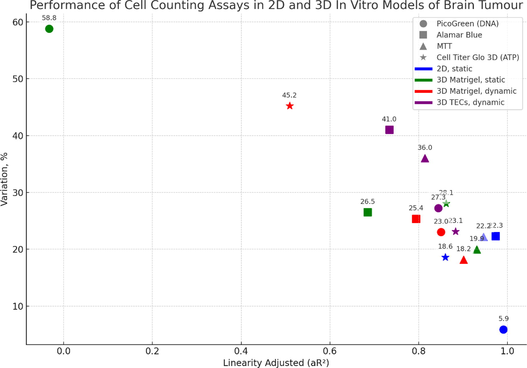
C96 - Development Of A Mathematical Model To Improve Bioreactor Efficiency
S. E. White, C. G. Hincher, D. O. Freytes
North Carolina State University, Raleigh, NC
*Purpose/Objectives: Tissue engineering increasingly uses extracellular matrix (ECM)--based biomaterials for constructing functional scaffolds that may lead to constructive remodeling of damaged tissues. Decellularized ECM stands out among these biomaterials due to its favorable wound-healing properties, the unique composition of structural proteins, and bioactive molecules that can help promote healing and tissue repair. However, the decellularization process, aimed at removing immunogenic factors such as DNA to prevent host rejection, is currently lab-specific. This variability in decellularization protocols leads to inconsistent results, imposing significant limitations on the cost and timelines of advancements in the field. To address these challenges, mathematical models can be used as predictive tools by incorporating key variables, facilitating smoother pathways for scientific discoveries. The primary objective of this study was to develop a mathematical model to describe the efficiency of the decellularization process.
*Methodology: Porcine vocal fold and supraglottic tissues were isolated and prepared for decellularization. Monitoring the process involved collecting reagent waste at regular intervals, with absorbance at 260 nm measured to approximate the released DNA content. Utilizing an exponential plateau model framework, a set of nonlinear equations described the DNA release profiles during decellularization. Furthermore, we explored the impact of critical factors such as the mass of the tissue, bioreactor volume, and time, which are essential for optimizing translating in vitro benchtop experiments for industry use.
*Results: Combining data from literature and parameter optimization methods, the exponential plateau equation reasonably fits the experimental data (R2=0.79-0.97). Employing experimental factors like time, total DNA amount, tissue mass, and bioreactor volume, we developed various mathematical models to estimate decellularization efficiency. These additional models were validated using in vitro absorbance data obtained beforehand.
*Conclusion/Significance: Continued refinement of the mathematical decellularization model and relevant in vitro experiments will optimize decellularization conditions and account for parameters such as tissue types, particle size, and additional reagents. This model is crucial for sharing insights with other tissue engineering research groups and integrating tissue engineering and regenerative medicine benchtop strategies into clinical and industrially focused applications.
C97 - Machine Learning Modelling To Understanding Biomaterial Properties
S. CAKICI, R. TUTAR
Istanbul University-Cerrahpaşa, Istanbul, Turkey
*Purpose/Objectives: Machine learning (ML) in material synthesis is an advantageous technique in many ways, such as saving time. It is observed that the use of ML is used more and more in the scientific field every day. It can predict data such as experimental results by analyzing the high data set. Therefore, costs are reduced, and time is saved1. The aim of this study is the development of a ML modeling for understanding of the effect of the amount of methacrylate anhydride (MA) used during GelMA synthesis and the effects of different degrees of methacrylation on the GelMA molecular structure and the physical properties of GelMA.
*Methodology: Gelatin was methacrylated to prepare GelMA and different amounts of MA were used. Then hydrogels were prepared by using visible light photoinitiator system. Swelling and degradation measurements were taken to observe its physical properties. The time intervals were set as 1 hour, 2 hours, 3 hours, 4 hours, 5 hours, 6 hours, 7 hours, 24 hours. The decision tree regression model, which is most suitable for ML application, was applied. Performance was measured with mean squared error values based on the estimated values and comments were made on the graphs.
*Results: Using the Python programming language, the decision tree regressor model was trained on the obtained data set results. The mean squared error method was used to observe the performance of the predicted results with the trained data. To increase the performance, hyperparameter was installed and performance was improved. The mean squared error value was measured with a hyperparameter. Graphs were created to compare the predicted results with their actual values. Graphs were created and interpreted to understand the data set.
*Conclusion/Significance: By approaching the material design with its physical properties according to four different methacrylation degrees over the biomaterial GelMA, the performance results were observed with ML models and model training. It was observed that ML models would save time and materials in material design. The model can be improved with different factors, such as increasing the data set, and it gets closer to the correct result. With this study, the importance of the model applied in biomaterial design was observed.
Keyword: Machine learning, data analysis, GelMA, methacrylic anhydride, physical properties
Acknowledgement: This study was financially supported by The Scientific and Technological Research Projects Funding, TUBITAK-2209-A/ 1919B012307651.
References: 1. Zhou, Qionghua, et al. “Property-oriented material design based on a data-driven machine learning technique.” The journal of physical chemistry letters 11.10 (2020): 3920-3927.
C99
C100 - Gain-of-function Mutations In The Mechanosensory Ion Channel TRPV4 Enhances The Response Of Synthetic Mechanogenetic Gene Circuits In Human IPSC-derived Chondrocytes
Y. Kim1,2, N. Steward1,2, A. Kim1, I. Fehle1, F. Guilak1,2
1
Washington University in St. Louis, Saint Louis, MO,
2
Shriners Hospitals for Children, Saint Louis, MO
*Purpose/Objectives: Transient receptor potential vanilloid-4 (TRPV4) is a mechanosensory ion channel that responds to a variety of physiological stimuli such as mechanical and osmotic stresses. Recently, synthetic mechanogenetic gene circuits have been developed that express IL-1 receptor antagonist (IL-1Ra) in response to physiologic loading and subsequent TRPV4 activation. By relying on mechanotransduction pathways for activation, mechanogenetics offer a promising platform for developing cell-based therapies for inflammatory conditions such as arthritis. The goal of this study was to “tune" the therapeutic output of our mechanogenetic gene circuits by altering chondrocyte mechanoresponsiveness by targeted mutations in TRPV4. We edited human iPSCs to incorporate gain-of-function mutations of TRPV4 to fabricate engineered cartilage constructs with altered response to physiological stimuli. We hypothesized that alterations in ion channel activity in mutant TRPV4 channels could alter the dynamics of the mechanogenetic gene circuit response.
*Methodology: Chondrocytes were differentiated from three hiPSC cell lines, one isogenic control (WT) and two mutant lines harboring the V620I or T89I mutation in TRPV4. Cells were lentivirally transduced with mechanogenetic gene circuits that contain either a luciferase or IL-1Ra gene downstream of an NF-κB-inducible promoter. Tissue-engineered cartilage constructs were then fabricated by encapsulating transduced cells in 2% (w/v) agarose. A luminescence plate reader was used to measure circuit responses of NF-κB-Luc constructs, and ELISA was used to measure IL-1Ra secreted by NF-κB-IL1Ra constructs over 24 hours post-stimulation.
*Results: We first studied the temporal aspects of mechanogenetic circuit activation by measuring bioluminescence from NF-κB-Luc constructs. All three cell lines responded to both pharmacologic (GSK101, a TRPV4 agonist) and physiologic (hypo-osmotic and mechanical) loading but showed different response profiles. In all concentrations of GSK101 tested, mutants showed higher peak luminescence and area under the curve (AUC) over 24 hours compared to WT (fig. A-D). Following mechanical loading, both mutants displayed weaker albeit more sustained responses, as demonstrated by lower peak luminescence but higher AUC compared to WT (fig. E). TRPV4 mutations also led to stronger mechanogenetic response from NF-κB-IL-1Ra constructs. Both mutant constructs produced significantly higher IL-1Ra compared to WT even from the vehicle-treated group (fig. G), suggesting that the mutant constructs have a higher basal circuit activation level compared to WT. Similar trends were observed from mechanical loading but without a pronounced dose-dependence (fig. H). In contrast, hypo-osmotic loading did not lead to higher IL-1Ra secretion from mutant constructs compared to WT (fig. I).
*Conclusion/Significance: Using engineered cartilage tissue constructs derived from human iPSCs, we successfully demonstrate that the mechanogenetic circuit response can be tuned by introducing mutations in TRPV4. By increasing the channel’s open probability, the two gain-of-function mutations tested in our study led to an overall stronger circuit output compared to WT. Furthermore, sustained response of mechanically-loaded mutant constructs offers a more suitable alternative to a quick “burst” response from WT for a sustained drug delivery. Combined with their nearly limitless differentiation capacity, hiPSCs with TRPV4 mutations could be a promising cell source for fabricating mechanogenetically engineered cartilage constructs that can deliver anti-inflammatory drugs locally and on demand.

C102 - Self-illuminating Hydrogel Materials For Protein Cargo Release
T. Rapp
University of Oregon, Eugene, OR
*Purpose/Objectives: Many biological processes are directed by well-defined and temporally orchestrated gradients of signaling proteins (e.g. cytokines, chemokines) expressed by local cell populations. Upon tissue damage, including that which accompanies biomaterial implantation, these signaling gradients are disturbed. Such disruption can yield negative outcomes in the local environment, critically including overactivation of the innate immune response. Re-establishment of the appropriate graded cues, particularly in the case of implanted biomaterials harboring living cells, is likely to translate to better clinical outcomes.
*Methodology: In this work, we introduce a robust synthetic strategy to establish soluble protein gradients deep within living tissue, on demand and with spatiotemporal control, via the coupling of highly photosensitive Ruthenium compounds with bioluminescent protein NanoLuc. Various strategies are employed to attach protein cargo to a polymer matrix, using a click chemistry that is translatable to both tissue-agnostic synthetic polymers such as poly(ethylene) glycol and natural polymers including hyaluronic acid.
*Results: Leveraging the high photosensitivity of Ru compounds we demonstrate rapid protein release through complex tissue via either direct illumination with tissue penetrating 550 nm light or application of the NanoLuc substrate furimazine. No loss in efficiency was observed in dose-dependent protein release between exogenous- and self-illumination.
*Conclusion/Significance: The use of bioluminescence resonant energy transfer (BRET) as an endogenous light source has the potential to address the need for on-demand drug delivery in deep tissue. In this work we have shown that BRET-based bioluminolysis is effective for the release of protein cargo from hydrogel depots, suggesting a powerful new method for on-demand drug delivery based on systemic administration of a small molecule.

C103 - Magnetic Microgels For Composite Musculoskeletal Tissue Regeneration
R. Chen1, B. Chin-Young2, W. Han2, F. Qu1
1
University of Washington, Seattle, WA,
2
Icahn School of Medicine at Mount Sinai, New York, NY
*Purpose/Objectives: The regenerative capacity of complex musculoskeletal tissues is restricted, such that healing often culminates in scarring. To interrogate the potential for mammalian limb regeneration, we and others use the murine digit tip, which regenerates after distal, but not proximal, amputation. Previous studies show that the combined delivery of bone morphogenetic protein-2 (BMP-2) and BMP-9 to proximally amputated murine digits induces skeletal elongation and synovial joint regeneration, respectively. Building upon this work, we hypothesized that stimuli-responsive, bioactive hydrogels could direct the sequential and spatial formation of bone followed by articular cartilage in the murine digit. Towards this goal, we developed magnetic microgels that can be translocated using a user-controlled magnetic field to instruct composite tissue regeneration.
*Methodology: 8-arm 40 kDa Poly(ethylene glycol)-norbonene (PEG-NB, 5% w/v), PEG-dithiol (3.8 mM), SH-PEG-Biotin (2 mM), and photoinitiator (Lithium phenyl-2,4,6-trimethylbenzoylphosphinate, 2.5 mM) were dissolved in PBS (Control). Microgel droplets were generated by agitating this mixture in a water-in-oil emulsion and photopolymerized by applying a field of light (λ: 365 nm). To generate magnetically responsive microgels, either a Low (23.3 mM) or High (221.4 mM) concentration of iron oxide (FeO2) nanoparticles was incorporated in place of SH-PEG-Biotin. Microgels in PBS were imaged with a bright field microscope and analyzed using ImageJ, where the average diameter, circularity (4π(area/perimeter2)), and opacity (0: transparent, 255: opaque) of individual microgels were quantified from binarized images. To test if microgels could respond to a magnetic field, Low FeO2 microgels were injected into a linear polydimethylsiloxane (PDMS) channel with a cylindrical Neodymium magnet (5/8” thick, 9/16” diameter, 19.5 lb pull force, D9A, K&J Magnetics) placed approximately 1 cm away. Velocity was determined by tracking the microgel centroid location over a 10-second time course. To test whether magnetic particle incorporation could be localized to the microgel surface, superparamagnetic beads coupled with streptavidin (Dynabeads, Invitrogen; 1:10) were incubated with Control microgels and assessed by bright field microscopy. Significance was assessed by 2-tailed Student’s t-test or 1-way ANOVA with Tukey’s post-hoc test (p<0.05).
*Results: Magnetically responsive PEG-NB microgels were successfully fabricated by encapsulating FeO2 nanoparticles. Microgel size, shape, and opacity depended on the FeO2 concentration. The average diameter and circularity of both Low and High FeO2 microgels were significantly less than the Control values. As expected, microgel opacity increased with FeO2 concentration. High FeO2 microgels exhibited the greatest morphometric heterogeneity, indicating that higher nanoparticle concentrations may disrupt microgel droplet formation. In the presence of a magnetic field, Low FeO2 microgels moved in a linear direction through PBS at approximately 0.2 mm/s. In contrast, microgel movement was negligible in the absence of a magnet. Similarly, Control microgels with surface-conjugated Dynabeads were magnetically responsive.
*Conclusion/Significance: Magnetic PEG-NB microgels represent a versatile biomaterial platform that may be adapted for spatially controlled drug delivery. Ultimately, this research will establish a clinically translatable strategy to promote composite musculoskeletal tissue regeneration after digit amputation.
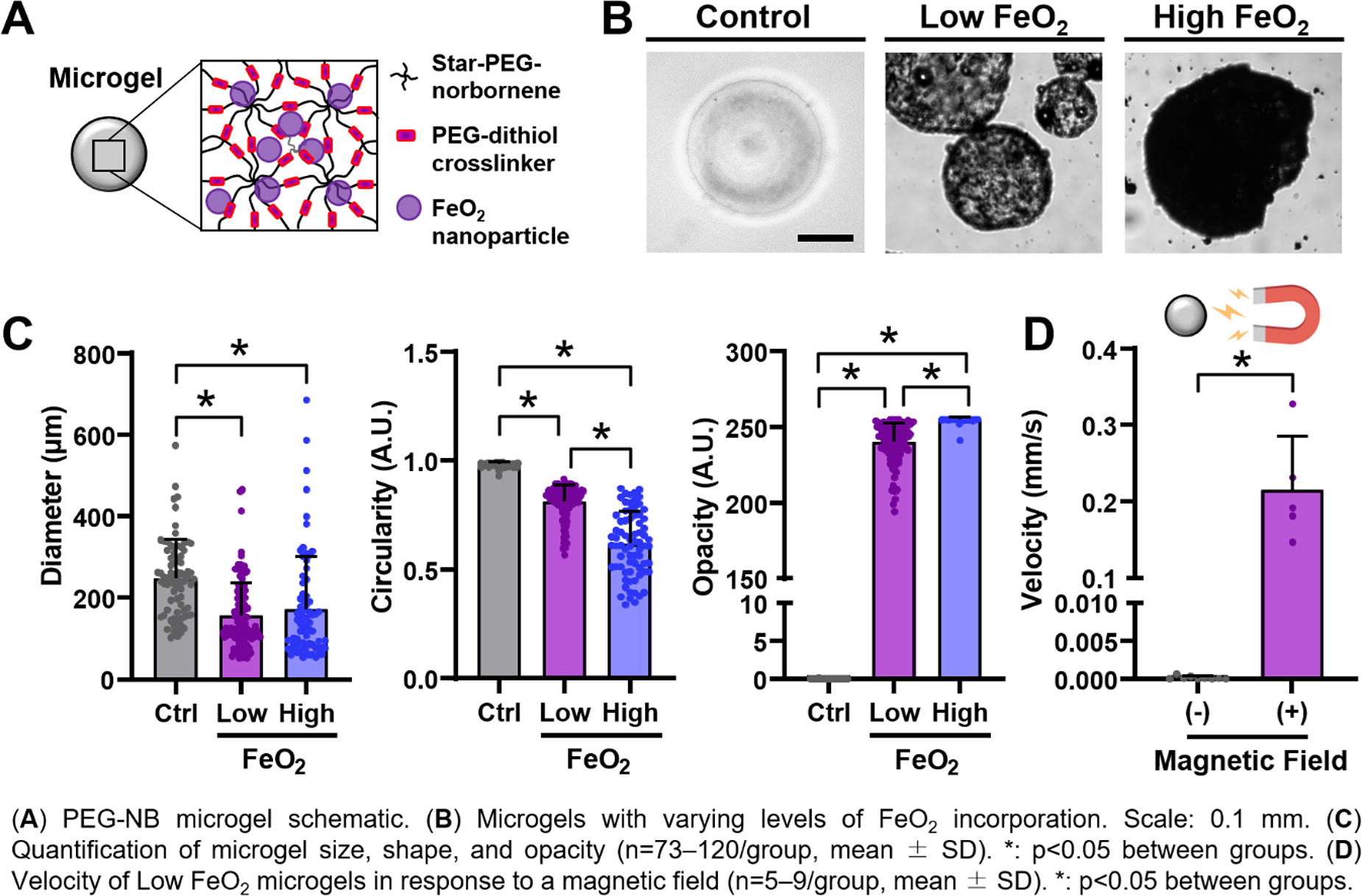
C104 - Unlocking The Potential Of Ionic Hydrogels Serving Muscle Tissue Engineering Applications
J. Van Meerssche1, J. Vekeman2, T. Verstraelen2, J. Madden3, S. Van Vlierberghe1
1
Ghent University, Gent, Belgium,
2
Ghent University, Ghent, Belgium,
3
University of British Columbia, Vancouver, BC, Canada
*Purpose/Objectives: This study seeks to connect the fields of iontronics and muscle tissue engineering. Exploring the intersection of these disciplines may offer unique perspectives regarding hydrogel applications in ionic environments and their potential effects on cellular behavior in the field of muscle regeneration. Muscle tissue engineering aims to create functional constructs for damaged tissues or organs. Research often mimics natural tissues’ properties, such as conductivity. While metallic and carbon particles enhance scaffold conductivity in hydrogels, they pose challenges including inhomogeneity, hampered additive manufacturing, and toxicity concerns. Current conductive hydrogel studies generally rely on these particles, constraining the available options of material scientists. Inspired by nature, ion transport is employed herein, rather than electron transport, for precise control of hydrogel properties during manufacturing. In the current work, hydrogels have been developed without using particles for increasing conductivity. Beyond ionic conductivity, an emerging research area explores the electricity generation of ionic hydrogels through mechanical deformation, termed the piezoionic potential. Found in all hydrogels, this property holds promise in the tissue engineering field. This work will highlight the tunability and significance of these properties in ionic hydrogels with application in muscle tissue engineering.
*Methodology: Novel RAFT copolymers were developed using neutral, cationic, and thiolated monomers, allowing independent tuning of crosslinking and charge density. More specifically, cationic photo-crosslinkable RAFT polymers were synthesized, serving as thiol crosslinkers for gelatin-norbornene to produce hybrid ionic hydrogels. Upon photo-crosslinking these RAFT polymers with gelatin-norbornene in the presence of a photo-initiator, hybrid ionic hydrogels were successfully formed. By varying the neutral:cationic:thiol ratio the crosslinking and cationic charge density can be tuned independently. The storage/loss modulus, mass swelling ratio, gel point, and gel fraction of the hydrogels were characterized.
*Results: At constant crosslinking density, the storage modulus remained unchanged, but swelling decreased significantly with increasing cationic charge density. Furthermore, by adjusting the thiol content within the RAFT polymer, the storage moduli of hybrid gelatin hydrogels can be fine-tuned within the range of 5 to 10 kPa. Conversely, modifying the cationic content in the RAFT polymer allows for changes in the mass swelling ratio, resulting in values ranging from 5.5 to 8.7 in double distilled water. All gel fractions surpassed 95%, indicating a highly efficient crosslinking process. This efficacy in crosslinking is attributed to the thiol-ene photo-crosslinking mechanism, being a step-growth process. Currently, in vitro biocompatibility assays are ongoing using the C2C12 myoblast cell line. Collaboration with Prof. John Madden from the University of British Columbia (Canada) is ongoing for the measurements of ionic conductivity and piezoionic potential. The control group comprises thiolated gelatin/gelatin-norbornene and gelatin-methacrylamide.
*Conclusion/Significance: This innovative three-component RAFT copolymer approach allows independent control of the charge and crosslinking density of the hybrid hydrogels. The materials employed in this study are anticipated to lead to new insights regarding the role of ionic conductivity and piezoionic potential in hydrogels. These insights can advance the state-of-the-art of muscle tissue engineering.

C107
C111 - Development Of Prime Editing Systems With Broad PAM Range And Improved Efficiency For Correction Of Pathogenic Mutations In Human Induced Pluripotent Stem Cells
D. H. Pham
National Tsing Hua University, Hsinchu, Taiwan
*Purpose/Objectives: Background: Prime editing technology is the first gene editing tool which enables all possible types of base pair substitutions, small insertions and deletions. Despite the editing versatility, the practical application of prime editing is still hindered by multiple challenges. Firstly, the need for a specific PAM sequence for SpCas9 recognition restricts the targeting scope of prime editing. This PAM limitation of the conventional SpCas9-based PEs poses a challenge to their applicability for correction of pathogenic mutations. Secondly, the efficiency of current prime editors remains low and varies considerably among editing types, targeting sites and cell lines leading to relatively inefficient genome editing in human iPSCs bearing pathogenic mutations.
*Methodology: Methods: We first described a next generation of prime editors using a PAM-flexible Cas9 ortholog - Sc++Cas9 derived from Streptococcus canis can expand the targeting range of prime editing. To improve the efficiency of PE in human iPSCs, we optimized the delivery modalities for the PE components using in vitro transcribed mRNA based delivery of the nCas9-RT and pegRNAs as well as studied the cell cycle dependence of prime editing using cell synchronization experiments. Finally, by combining those approaches enabled the corrections of pathogenic mutations in human iPSCs.
*Results: Results: Using the Sc++PE systems, we could successfully generate various types of mutations with up to 72.1% prime-editing activity in HEK293T. In addition, we found that mRNA-based delivery of PE components and cell cycle phase via small molecules synergistically enhanced precise prime editing frequency in both HEK293T and human iPS cells. When those approaches combined with our next generation Sc++PE systems, we successfully corrected the GJB2 c235Cdel pathogenic mutation associated with autosomal recessive non-syndromic hearing loss, with higher efficiencies compared with those using the conventional PE systems.
*Conclusion/Significance: Conclusion: Overall, our next generation prime editors hold promise for broadening the applicability of CRISPR-based prime editing technologies in the field of tissue engineering and regenerative medicine.
C112 - Development Of An Efficient And Highly Specific Prime Editing System For Targeted Correction Of GJB2-Related Hearing Loss Mutations
Q. T. Dang, Y.-H. Lin, Y.-C. Hu
National Tsing Hua University, Hsinchu, Taiwan
*Purpose/Objectives: GJB2 c.235delC and GJB2 c.109G>A mutations are among the most prevalent hearing-loss-causing mutations in Taiwan population. Next-generation genome editing technologies, such as CRISPR/Cas9 and Base editing (BE), have their inherent setbacks. While CRISPR/Cas9 induces DSB and destabilizes chromosomes, BE only converts a limited number of single base mutations. Alternatively, Prime editing (PE), provides a unique platform for DSB-free correction of these non-deamination single base and deletion mutations. However, challenges of PE include nickase-inducing off-target and suboptimal editing activity necessitating further improvements for its expanded application in clinical genetic disease restoration. Our study aims to improve PE efficiency and specificity, facilitating its transition into clinical applications.
*Methodology: We developed two additional PE variants harboring HiFiCas9 or eSpCas9 nickases with comparable activity but more precise than wild-type SpCas9. We co-transfected HEK293FT or induced pluripotent stem cell (iPSC) with HiFiPE3 or eSpPE3, harboring the corresponding nickase fused with an engineered MMLV reverse transcriptase (RT), a pegRNA encoding the desired new genetic information, and a nicking sgRNA. Subsequently, to further improve editing efficiency, we harnessed the HiFiPE5max architecture, comprising an extensively optimized HiFiPE3 and concomitant overexpression of MLH1dn. Ultimately, we also tested the impact of an engineered pegRNA (epegRNA), p53 inhibition by co-transfecting p53DD, or cell cycle arrest by nocodazole on editing efficiency. Genomic DNA of the cells was isolated 2 or 3 days post-transfection and the targeted region was PCR amplified with designed primer pair, followed by Sanger Sequencing and deep sequencing to characterize and quantify the editing events. Mismatch tolerance was assessed by replacing the spacers of the cognate pegRNA with a series of single mismatches.
*Results: Of the two variants, HiFiPE3 exhibits comparable editing efficiency with heightened specificity compared to the prototypic PE system using SpCas9. Further optimization strategies for the HiFiPE5max and epegRNA were explored to enhance editing efficiency (2.3x - 5x). The system demonstrated its ability to facilitate gene correction in iPSCs harboring GJB2 c.235delC and GJB2 c.109G>A mutations (2.6% and 10.5%, respectively), complemented by overexpressing p53DD and inducing cell cycle arrest.
*Conclusion/Significance: Altogether, these findings underscore the potential translation of HiFiPE into clinical settings, offering a promising avenue for precise and efficient gene therapy in the context of GJB2-related hearing loss.

C114 - Role Of Microrna-365a/b-3p As A Differential Biomarker Between Normal Skin And Hypertrophic Scar
S. Kwak, E. Oh, H. Kim, H. Chung
Kyungpook National University, Daegu, Korea, Republic of
*Purpose/Objectives: The clinical aspects of hypertrophic scarring vary according to personal constitution and body part. However, themechanism of hypertrophic scar(HS) formation remains unclear. MicroRNAs (miRNAs) are known to contribute to HS formation, however, their detailed role remains unknown.
*Methodology: In this study, candidate miRNAs were identified and analyzed as biomarkers of hypertrophic scarring for future clinical applications. HSfibroblasts and normal skin fibroblasts from patients were used for profiling and validation of miRNAs. An HS mouse model with xenografted human skin on nude mice was established. The miRNA expression between normal human, normal mouse, and mouse HS skin tissues was compared. Circulating miRNA expression levels in the serum of normal mice and mice with HSs were also analyzed.
*Results: Ten upregulated and twenty-one downregulated miRNAs were detected. Among these, miR-365a/b-3p and miR-16-5p were identified as candidate miRNAs with statistically significant differences; miR-365a/b-3p was significantly upregulated (p = 0.0244). In mouse studies, miR 365a/b-3p expression levels in skin tissue and serum were higher in mice with HSs than in the control group.
*Conclusion/Significance: These results indicate that miRNAs contribute to hypertrophic scarring and that miR-365a/b-3p may be considered a potential biomarker for HS formation.
C117 - Tackling The Inflammatory Burden Of Peri-implantitis - An In Vitro Evaluation Of Green Synthetized Zinc-magnesium Nanoparticles With Immunomodulatory Activity
P. S. Gomes1, V. Z. Martin1, A. Bettencourt2, M. R. Fernandes1, M. Alves3, M. Hanafy4, Z. Cui4, C. Santos5
1
Faculty of Dental Medicine, U Porto, Porto, Portugal,
2
Faculty of Pharmacy, Universidade de Lisboa, Lisboa, Portugal,
3
Instituto Superior Técnico, Universidade de Lisboa, Lisboa, Portugal,
4
College of Pharmacy, The University of Texas at Austin, Austin, TX,
5
EST Setúbal, Setúbal, Portugal
*Purpose/Objectives: Periimplantitis (PI) is a microorganism-triggered inflammatory disease that causes progressive destruction of implant-supporting bone. PI can lead to implant loss, impairing esthetics, and masticatory function of patients. Periopathogens at implant sites are the main etiologic factor in PI. Despite their ability to induce tissue damage via proteases and byproducts, tissue destruction is primarily perpetrated by the host’s inflammatory response, via leukocyte’s production of reactive oxygen species (ROS), autoantibodies against extracellular matrix components and incited osteoclastogenesis. Nonetheless, the standard therapeutics relies on mechanical cleansing and antibacterial regimens, which are not specifically devised for immunomodulation. Accordingly, the present study portrays the development and biological characterization of layered hydroxide (LH) nanoparticles as potential local adjunctive therapy for PI, targeting immunomodulation.
*Methodology: Zinc-magnesium LH nanoparticles (ZM NPs) were produced using a co-precipitation method. Rosehip (RH) extract was added into the synthesis of ZM NPs to enhance NPs’ anti-inflammatory and antioxidant properties (RH-ZM NPs). The physicochemical characterization included crystallinity assessment, composition, and morphology. Cytocompatibility and antioxidant activity were assessed in vitro using human gingival fibroblasts (HGFs), setting on cultures’ metabolic activity, morphology, and ROS production. Anti-inflammatory activity of ZM and RH-ZM NPs were evaluated through gene expression analysis of inflamed (via lipopolysaccharide exposure) 3D organotypic oral mucosal cultures. The expression of relevant pro-inflammatory genes (i.e., IL-1β, IL-6, CXCL-8, NFkB1, and TNF) was assessed by quantitative PCR (qPCR).
*Results: NPs displayed zinc hydroxynitrate as the precipitated crystalline phase, indicating a monoclinic nanolayered zinc hydroxide structure with a plate-like morphology. The presence of RH phytochemicals was confirmed by the detection of polyphenols, further resulting in lower crystallinity and thinner organizational layers of RH-ZM NPs. Both NPs were found to be cytocompatible at 1 and 10 µg/mL, while cytotoxicity was observed at 50 µg/mL. HGFs presented a stretched shape, prominent stress fibers, rounded nuclei, and slender cytoplasmic extensions when incubated with 10 µg/mL of NPs. Incubation with ZM and RH-ZM NPs did not induce ROS production in HGF cultures. Moreover, both NPs’ formulations significantly lowered the effects of H2O2 upon HGFs, maintaining ROS levels comparable to negative control. Regarding their anti-inflammatory properties over organotypic mucosal cultures, the expression of IL-6, CXCL-8 and NFkB1 genes was significantly downregulated by ZM and RH-ZM NPs at 10 µg/mL. However, RH-ZM NPs displayed a superior efficacy in downregulating all assessed pro-inflammatory genes (including IL-1β and TNF), achieving values comparable to those of LPS-untreated cultures.
*Conclusion/Significance: ZM and RH-ZM NPs were found to be cytocompatible and presented strong antioxidant activity when tested in vitro using HGF cultures. Additionally, both NPs demonstrated potent anti-inflammatory effects in an advanced organotypic mucosal model and the addition of the RH extract in the synthesis boosted the anti-inflammatory effects of the formulation. Overall, these results highlight the potential of RH-ZM NPs as a local adjunctive therapy for PI, focusing on local immunomodulation.
C118 - Production Of Particles Of PCL Containing Vitamins A And D For Bone Regeneration
N. Maurmann, T. F. Elautério, L. R. Wailer, R. P. Silveira, R. Zimmermann, P. Pranke
Hematology and Stem Cell Laboratory, Faculty of Pharmacy, Universidade Federal do Rio Grande do Sul (UFRGS), Porto Alegre, BRAZIL.
*Purpose/Objectives: Vitamin D is responsible for calcium homeostasis and the maintenance of bone metabolism balance. Vitamin A deficiency can result in delayed bone growth, reduced bone mineral density and changes in bone structure. This work aimed to produce and characterize a modified release system and stable formulation of poly(caprolactone) (PCL) particles containing vitamins A and D and to evaluate the particles’ cytotoxicity in stem cells.
*Methodology: Particles were prepared by solvent displacement using PCL, vitamins A and D in acetone with the oil phase dripped in the water phase composed of water and Tween® 80. They were characterized in terms of size and zeta potential. Cell viability was evaluated by 3-(4,5-dimethylthiazol-2-yl)-2,5-diphenyltetrazolium bromide (MTT) assays after 1 day of contact of the stem cells with the particles containing 0.4 UI/mL of vitamin D and 2.0 UI/mL of vitamin A. A white control (culture media without cells) was used and death controls with Triton X-100. The results were expressed as the mean ± standard deviation.
*Results: The PCL particles loaded with vitamins A and D presented a zeta potential of -0.143 ± 0.099 mV, a diameter of 313 ± 9 nm in the Zetasizer equipment (with a detection limit of 10 µm), and up to 500 µm by optical microscopy. Therefore, the particles showed nano- and micrometric sizes and a high level of size variation within the particle population. The zeta potential indicated a slightly negative particle charge surface, with a modest tendency to repel each other. The negative charge observed in the obtained particles can be attributed to the presence of PCL and Tween® 80. The PCL particles containing vitamins did not significantly affect stem cell viability, with values of absorbance of 0.283 ± 0.069 for cells cultivated on the tissue culture plate (control) and 0.279 ± 0.051 for cells incubated with the particles for one day (p = 0.998). Conversely, the utilization of death and white controls resulted in absorbances of 0.024 ± 0.004 and 0.076 ± 0.004, respectively, indicating statistical differences in comparison with the control and particle groups (p = 0.000). Therefore, the produced material was not cytotoxic to stem cells.
*Conclusion/Significance: The solvent displacement technique is an effective procedure to produce particles. The obtained results reveal that the particles of PCL with vitamins A/D presented a micrometric size, a slight surface negative charge and a high dispersion index. The particles with vitamins maintained the stem cells’ viability analogous with the control group. Therefore, the use of polymeric particles can be a strategy for the supplementation of retinol and cholecalciferol, incorporating this produced biomaterial into bioinks for bone tissue engineering. Acknowledgments: Office of Naval Research Global (ONRG Award N62909-21-1-2026), National Council for Technological and Scientific Development (CNPq), Financier of Studies and Projects (FINEP), National Institute of Science and Technology for Regenerative Medicine (INCT-Regenera) and Stem Cell Research Institute (IPCT).
C120 - Histological And Biomechanical Impact Of Titanium Implants Produced By Additive Manufacturing Process In Osteoporotic Animals Using Risendronate
E. C. Kalil1, K. A. Cotrin1, K. Bechara1, L. Takakura1, J. S. Souza1, r. okamoto1, J. Moura2, M. Dutra3, J. A. Shibli1
1
Guarulhos University, Guarulhos, Brazil,
2
Aracatuba UNESP, FOA-UNESP, Brazil,
3
FOA Araçatuba UNESP, Araçatuba, Brazil
*Purpose/Objectives: Osteoporosis is a metabolic disease that can lead to a decrease in bone density and a deterioration in its microarchitecture, impairing the quality of bone tissue and its repair process. Recently, risedronate has been successfully used as a first-line drug for the treatment of osteoporosis. Dental implant placement in osteoporotic subjects could be a challenge, and to increase the treatment success, different surface topographies were modified to increase the bone-to-implant contact. Recently, progress has been made using an additive manufacturing process to produce dental implants using Ti23 grade, resulting in a unique surface microstructure.This preclinical study evaluated the histological aspect and biomechanical impact of risedronate on removal torque as part of assessing the osseointegration of implants produced by additive manufacturing placed in an animal model of induced osteoporosis.
*Methodology: Female rats were divided into control group SHAM, ovariectomized rats OVX, and ovariectomized rats treated with Risendronate OVX-RIS. In each tibia of each animal, an implant produced by additive manufacturing with a 2mm diameter and 4 mm height was placed. After 28 days, the animals were euthanized. In one tibia, the removal torque test of the implant was performed by applying a counterclockwise force to break the bone-implant contact completely. In the other tibia, samples containing implants and peri-implant bone were collected for histological analysis.
*Results: The maximum removal torque values obtained were 16.84 n.cm ± 0.43, 13.94 n.cm ± 0.78, and 10.74 n.cm ± 3.14 for the SHAM, OVX, and OVX-RIS groups, respectively, with a statistically significant difference between the SHAM and OVX-RIS groups (p <0 .05).During the histological evaluation, in the SHAM group its was possible to identify an important area of bone formation, with characteristics of mature bone next to the implant threads. In the OVX group, an opposite pattern was found, and no bone formation close to the implant thread was observed. In the OVX-RIS group the presence of a more organized connective tissue, with some areas of bone formation close to the implant threads was observed
*Conclusion/Significance: The use of risedronate in the treatment of osteoporosis improved the peri-implant bone formation in osteoporotic animals. However, it did not influence the removal torque value of the implants.
C121 - A Comparative Investigation Of Chemical Decontamination Methods For In Situ Cleaning Of Dental Implant Surfaces
B. Hussain1, S. Lyngstadaas1, J. Ellingsen2, H. J. Haugen1
1
University of Oslo, Oslo, Norway,
2
University of Oslo, Oslo, Norway
*Purpose/Objectives:
Purpose/Objectives: Eradicating biofouling from implant surfaces is essential in treating peri-implant infections, as it directly addresses the microbial source for infection and inflammation around dental implants. This controlled laboratory study examines the effectiveness of the four commercially available debridement solutions (EDTA (Prefgel®), NaOCl (Perisolv®), H2O2 (Sigma-Aldrich), and Chlorhexidine (GUM® Paroex®)) in removing the acquired pellicle, preventing pellicle re-formation, and removing of a complex oral biofilm growing on a titanium implant surface, and compare the results with the effect of a novel formulation of a peroxide activated Poloxamer gel (Nubone® Clean).
*Methodology: Free chemical decontamination solutions, including a combination of Pluronic gel (NuBone Clean), have been tested for their effectiveness in cleaning titanium implant surfaces by EDX, XPS and various other surface techniques. The study also assessed chemical agents, cytotoxic and antibacterial agents in a multiple-species anaerobic biofilm model (initial colonizer Streptococcus oralis one second colonizer Physobacterium nucleatum and late colonizer Aggregate achter achtinompschetemchomitans). Its operational safety has been tested in human subjects (n=10).
*Results:The results revealed varying degrees of effectiveness among the tested decontamination solutions. Notably, all the gels, except for PrefGel®, demonstrated some level of effectiveness in cleaning the implant surfaces. NuBone®Clean stood out, showing significant effectiveness in providing a cleaner titanium surface as it was more effective in biofilm removal, showed greater inhibitory effects on pellicle formation, and resulted in cleaner titanium surfaces, as visualised by significantly reduced total carbon contamination after treatment when compared to the other test groups. Importantly, none of the chemical agents tested exhibited cytotoxic effects. The safety assessment further indicated no adverse events were associated with using these chemical decontamination solutions.
*Conclusion/Significance: Conclusion: All the tested gels had some effect on the surface cleanness except for PrefGel®. Among the tested chemical decontamination candidates, NuBone®Clean demonstrated effectiveness in providing a cleaner titanium surface. Furthermore, none of the tested chemical agents exhibited any cytotoxic effects, and the safety assessment showed no adverse events. Local, safe, and sustainable management of implant-mediated infections is the cornerstone of modern implant maintenance and peri-implantitis treatment. Today, clinicians rely on non-sustainable and aggressive solutions like antibiotics and chlorides to control these infections. Among the tested solutions, the novel, non-toxic, and sustainable NuBone®Clean formulation was superior in cleaning and protecting the implant surface. The findings are promising, and merit progress into clinical testing in combination with established mechanical cleaning procedures. However, further investigation through randomised clinical trials is necessary. By adhering to rigorous testing protocols, the development of safe and efficient chemical decontamination strategies can be advanced, benefiting patients and promoting progress in the field of implant dentistry.

C123 - Confirmation Of High Biocompatibility And Early New Bone Formation Of Synthetic Bone Graft Materials
K. Jang1, E. Yang1, J. Kim2, K. Shim1, S. Kim1, S. Kang1
1
Chonnam national university, Gwangju, Korea, Republic of,
2
Yeungnam University, Gyeongsan, Korea, Republic of
*Purpose/Objectives: Octacalcium phosphate (OCP), Ca8H2(PO4)6·5H2O, has emerged as a promising alternative to conventional bone graft materials in dentistry due to its neutral pH, biodegradability, and potential as a precursor to biological apatite crystals in teeth and bones. While previous studies have investigated the combination of OCP with water glass (Na2O.SiO2.nH2O) to enhance cell activity and bone formation, the comprehensive exploration of OCP combined with water glass (OCP-WG) is lacking. This study compared the biodegradability, and early new bone formation of synthetic bone grafts OCP and OCP-WG with xenografts, utilizing a rabbit calvarial defect model for dental bone graft evaluation.
*Methodology: Four 6 mm diameter bone defects were created on rabbit calvaria using a trephine bur and divided into four groups: a negative control group (Defect Only), deproteinized bovine bones graft group (Xenograft), and two experimental groups grafted with OCP and OCP-WG respectively. Rabbits were euthanized at 2-, 4-, and 8-weeks post bone graft implantation. X-ray radiography and micro-computed tomography (micro-CT) 3D image analysis confirmed the presence of bone graft particles and new bone formation. Histological analysis was performed with Hematoxylin and Eosin (H&E) staining and Masson trichrome staining to assess biocompatibility and new bone formation. Micro-CT and histomorphometric analysis were conducted to evaluate new bone formation quantitatively.
*Results: X-ray radiography and micro-CT 3D imaging findings demonstrated a gradual increase in new bone formation and density at the defect margins in all bone graft groups 2 and 4 weeks after the bone grafting. Quantitative analysis showed that new bone formation was significantly higher in the OCP-WG group compared to the Defect Only group at week 2 and significantly higher in the OCP group compared to the Defect Only group at week 4. However, at week 8, X-ray radiography and micro-CT 3D imaging revealed that the OCP-WG particles were less dense at the defect sites than those of the other bone graft groups, likely due to their biodegradability.
*Conclusion/Significance: This study established the high biocompatibility of both OCP and OCP-WG as synthetic bone graft materials. OCP demonstrated comparable or superior new bone formation to xenograft throughout the study. Although OCP-WG exhibited higher new bone formation in the early stages, it displayed higher biodegradability at week 8. These findings provided valuable insights into the potential applications of OCP and OCP-WG in dental bone regeneration, particularly regarding the possibility of utilizing OCP-WG’s ability to promote early new bone formation and its high biodegradability.
*Acknowledgment: This research was financially supported by the Ministry of Trade, Industry, and Energy (MOTIE), Korea, under the “Infrastructure program for industrial innovation” supervised by the Korea Institute for Advancement of Technology (KIAT). (P0025496)
C124 - Enhancing Alveolar Bone Regeneration:modulating Scaffold Properties Using Element Doped Zno Nanoparticles For Improved Cellular Dynamics And Mechanical Integrity
H. Akhtar1, F. H. Alhamoudi2, S. Naz1, H. Khalid1, A. F. Khan1, G. Reilly3
1
COMSATS University Islamabad, Lahore campus, Lahore, Pakistan,
2
Dental Technology Department, applied medical Science, King Khalid University, Abha, Saudi Arabia,
3
University of Sheffield, Sheffield, United Kingdom
*Purpose/Objectives: INTRODUCTION: Alveolar bone defects & fractures represent critical challenges in oral and maxillofacial surgery, significantly impacting both the function and aesthetics of the oral cavity. These defects can arise due to various factors, including periodontal diseases, traumatic injuries, congenital abnormalities, or as a consequence of surgical procedures such as tooth extraction or tumour resection. The impaired structural integrity of the alveolar bone not only compromises the stability of adjacent teeth but also poses obstacles for successful dental implant placement and prosthetic rehabilitation, thereby affecting patients' overall oral health and quality of life. fractures in the alveolar bone present unique challenges due to the delicate nature of the region and the requirement for precise and stable reconstruction to restore proper occlusion and function. The quest for effective regenerative solutions specifically tailored to address alveolar bone defects and fractures has led to the exploration of scaffold-based approaches. In this study, we examine the impact of doped zinc oxide (ZnO) nanoparticles on the structure, mechanical integrity, and biological performance of porous scaffolds for alveolar bone regeneration. We synthesised scaffolds using polyurethane (PU) incorporated with ZnO nanoparticles doped with strontium, cerium, copper and zirconium respectively.
*Methodology: METHODS: New composite scaffolds were synthesized using freeze extraction method. Osteoblast cell line (MC3T3-E1), Fibroblast cell line (NIH), Human embryonic mesenchymal like stem cells (hES-MP) and Bone marrow stromal cells (HTERT-Y201) were used to investigate detailed biological properties which included cell seeding efficiency, attachment, cytotoxicity, biocompatibility and differentiation studies.
*Results: Results and Discussion: the nanocomposite scaffolds replicated the porous architecture of cancellous bone with unparalleled fidelity, exhibiting a peak pore size of 500 µm. Among the doped scaffolds, cerium and copper dopants emerged as particularly efficacious in promoting cellular adhesion up to 60% and 75% respectively (fig. 1), as substantiated by metabolic activity assays.
*Conclusion/Significance: Beyond mere cell attachment, these scaffolds also promoted cellular proliferation in comparison to controls which are only PU, PU-HA and PU-HA-ZnO_2, potentially enhancing their applicability in tissue engineering. Furthermore, zirconium-doped scaffolds outperformed in mechanical assessments (tensile strength = 0.81MPa, Young’s modulus = 0.655), showcasing enhanced tensile properties and adequate flexibility—a key consideration for alveolar bone regeneration applications.

C125 - Characterizing Decellularized Murine Pulmonary Circuits Using X-ray μCt
E. A. Chadwick1, A. Zargar1, T. Suzuki2, S. Bian2, D. Romero1, C. Amon1,3, T. K. Waddell2,3, G. Karoubi1,2,4, A. Bazylak1
1
Mechanical and Industrial Engineering, University of Toronto, Toronto, ON, CANADA,
2
Toronto General Hospital Research Institute, Toronto, ON, CANADA,
3
Institute of Biomedical Engineering, University of Toronto, Toronto, ON, CANADA,
4
Department of Laboratory Medicine and Pathobiology, University of Toronto, Toronto, ON, CANADA.
*Purpose/Objectives: In this study, high-resolution 3D X-ray micro-computed tomography (μCT) was employed to analyze the structure of decellularized and recellularized murine lung vasculature, and subsequently compared to native lung vasculature. To identify the pulmonary vasculature in each lung, acquired images were processed using filtering, segmentation, and skeletonization. Morphological characteristics of the pulmonary vasculature networks in mouse lungs were assessed by measuring vessel length, diameter, tortuosity, and connectivity. Blood vessels were then categorized into generations according to their connections. The connectivity of lung vasculature networks were evaluated to examine the impact of decellularization and recellularization on the structural features of mouse pulmonary vasculature circuits.
*Methodology: Lungs from adult C57B1/6 mice (12 weeks old) were procured as per established protocols in our research group. Briefly, mice were euthanized using CO2 overdose, followed by extraction of heart and lung blocks from the thoracic cavity. Subsequently, the lungs were decellularized using a detergent-based protocol. A liquid contrast agent (Microfil MV-117, Flow Tech Inc.) was administered to the lungs via the pulmonary artery to improve the contrast of vasculature circuits for X-ray μCT imaging. Reconstructed X-ray μCT images underwent Gaussian blur refinement with a sigma value of 5 voxels as well as vesselness filtration to diminish intensity variance. This filtration aided in avoiding segmentation errors such as non-physical gaps or vessel inconsistencies. Image segmentation was conducted via two stages: thresholding using Otsu’s technique to binarize filtered images and isolating the largest connected voxel cluster. This approach ensured a continuous path throughout the vasculature. Meanwhile, the discarded clusters were carefully examined to confirm that the contrast agent has not been separated during injection, and to ensure that no vascular structures were excluded. Vasculature volumes were subsequently skeletonized, and the Vessel Network Extraction algorithm was employed to assess the morphology of each pulmonary circuit based on vessel length, diameter, tortuosity, and connectivity.
*Results: A significant decrease in vascular volume was observed in decellularized lung vascular networks, indicating an incomplete penetration of the contrast agent. The integrity of the native and decellularized lung circuits was assessed to identify the reason for this volume loss. The contrast agent permeated both arterial and venous portions of the lung circuit; however, in comparison to the native lung, the decellularized vasculature had fewer vessels per generation as well as limited connections between arterial and venous segments. Obstructions in the blood vessels and leakage through the porous extracellular matrix of vascular walls may be responsible for the reduced number of connecting vessels and vessels per generation in decellularized lungs. Furthermore, our results highlight a partial vasculature restoration particularly within the proximal regions, in the recellularized tissue.
*Conclusion/Significance: In summary, current decellularization techniques may yield lung scaffolds with inadequate connectivity of the pulmonary circuit, thus making them unsuitable for fluid flow due to subtle damage to the vasculature structure. To create functional scaffolds for recellularization, it is crucial to maintain the structural integrity of the vasculature during decellularization and remove blockages so that cells have a clear pathway for the recellularization process.
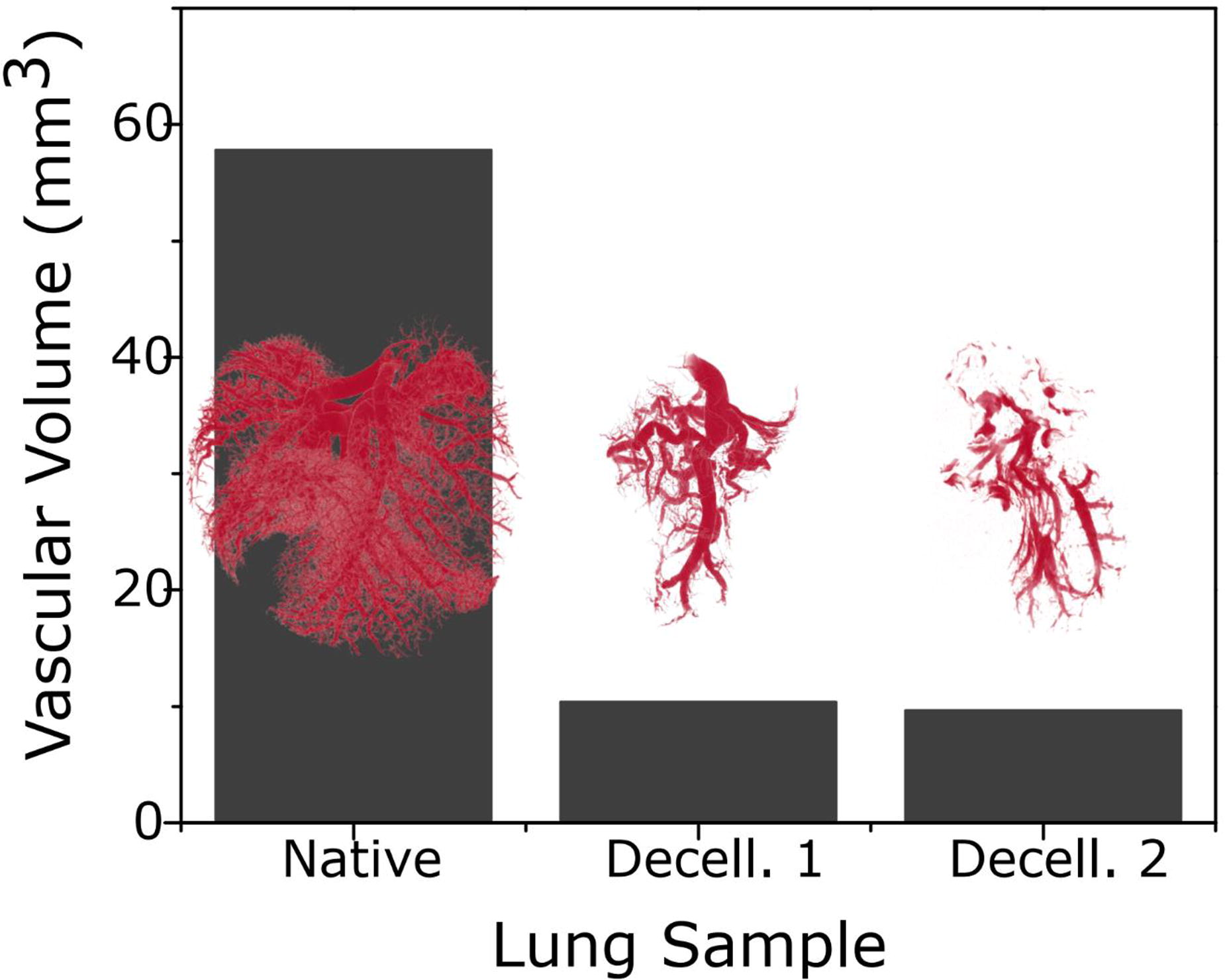
C127 - INTRAVITAL REAL-TIME IMAGING OF AVIAN EMBRYOS THROUGH AN IMAGING WINDOW INTEGRATED TO A SYNTHETIC EGGSHELL PLATFORM
C. Conci1, L. Cherubin1, M. Laganà2, C. Martinelli2, E. Jacchetti1, M. T. Raimondi1
1
Politecnico di Milano, Milano, Italy,
2
Gemma Prototipi, Longone al Segrino, Italy
*Purpose/Objectives: The chicken embryo is a valuable model for studying the effects of new drugs on the vascular system, crucial for many diseases as cancer, diabetes, and viral infections. However, conventional methods of intravital imaging require upright microscopes and invasive imaging windows, which limit accessibility of the vascular network. Then, we developed a novel platform (Eggs&Beacon) that uses a synthetic eggshell to house avian embryos and allows intravital microscopy on commercial inverted microscopes. We tested the biocompatibility and oxygen permeability of our platform, and we confirmed its suitability for supporting embryo development. We then demonstrated the feasibility of performing nonlinear microscopy (TPEF and SHG) on the platform and we quantified changes in blood vessel density and collagen production at different time-points. Our platform offered a fast and versatile way to evaluate the efficacy and safety of new pharmaceuticals or cellular models, targeting the vascular system in vivo, and this will be employable in every biological laboratory equipped with fluorescence inverted microscopes.
*Methodology: We fabricated a synthetic eggshell with a breathable membrane and a plastic casing (with a body, a cap and an imaging window) using vacuum-casting and 3D stereolithography techniques. This platform was designed to fit embryos on an inverted motorized microscope stage (Eclipse Ti-2, Nikon). This must also enable the orientation of the embryo and the exposure of its blood vessels to the microscope lens, so we could reposition the same embryo at different time-points (EID7-14). The imaging window was a fluidic device that could be imaged with a long-working distance objective (40X, NA=1.1, Nikon) with a reduced thickness (<500μm), and with the possibility to flow fluids (i.e.phantoms, water, etc.) to correct optical distortions caused by the mismatch of refractive indices.
*Results: Working prototypes of breathable membranes were realized thanks to vacuum casting and stereolithography techniques as to create the synthetic eggshell. The realized membranes had a silicone-like texture and a thickness of 250μm, which enabled the embryos to survive until EID14(n=3). The Eggs&Beacon platform let us flip the growing embryo(at EID12,14) and expose the vasculature to the microscope objective, without compromising the embryo’s viability and the platform’s sterility. We imaged the vasculature with TPEF microscopy(Z=600mm,λexc=800nm,λem<450nm), which allowed us to detect blood vessels from the imaging window and the cap. SHG(Z=600 mm,λexc=900nm,λem=450/55nm) showed us the collagen I matrix development and enabled us to measure the amount and direction of the fibers(EID12-14).
*Conclusion/Significance: We developed a disruptive device that enabled us to image a living organism, up to EID14, on inverted microscope systems and at multiple time-points. This device potentially reduces the animal replicates needed for intravital studies, while providing reliable data on the vascular growth of organisms. This device will facilitate the investigation of the organism’s systemic and microvascular response to external factors (such as drug, cellular models, etc.). Our initial tests indicated that the device is compatible with various types of inverted microscopes and objectives. We are currently working on correcting optical distortions by integrating the imaging window to improve the image quality and depth.
Acknowledgements: ERC-project BEACONSANDEGG,G.A.101053122
C129 - Molecular-sensitive Imaging To Monitor Cell And Tissue Dynamics
J. Marzi1,2, J. Alber1, P. Loskill1,2, K. Schenke-Layland1,2
1
NMI Natural and Medical Sciences Institute at the University of Tübingen, Reutlingen, Germany,
2
Eberhard Karls University, Tübingen, Germany
*Purpose/Objectives: 3D tissue models in Organ-on-a-Chip (OoC) systems enable to recapitulate (patho-) physiological and dynamic cellular processes such as metabolic response, phenotypic switching or tissue mechanobiology. To unravel the complex information provided by microphysiological tissue models, there is a high demand for the development of novel sensors and methods which allow for on-chip measurements.
*Methodology: Non-invasive imaging techniques such as Raman Microspectroscopy (RMS) and Fluorescence Lifetime Imaging Microscopy (FLIM) are promising tools for marker-independent in situ monitoring and biomarker development. Whereas FLIM is especially sensitive to metabolic changes by targeting the endogenous fluorophores NADH and FAD, RMS can access various cell and tissue structures due to their unique molecular-sensitive spectral fingerprints. Combined with advanced 3D in vitro models, non-destructive investigations of (patho-)physiological cellular processes can be obtained.
*Results: We implemented RMS and FLIM for ex vivo tissue analysis and in in vitro models, such as Organ-on-Chip platforms. It was demonstrated, that RMS enables the identification and visualization of major subcellular structures e.g., nuclei, proteins, lipids. In addition to quantitative image-based assessment, analysis of the extracted spectral information can further identify alterations in molecular composition, e.g. changes in lipid composition and oxidation were demonstrated upon culture duration or external stimulation. Moreover, FLIM enabled to investigate the metabolic balance between glycolysis and oxidative phosphorylation in tissue spheroids and was sensitive to detect early signs of apoptosis and response to drug treatments.
*Conclusion/Significance: Overall, our results showed that both, RMS and FLIM, provide real-time insights on tissue dynamics and should be further established and developed as complementary tools in tissue diagnostics and 3D in vitro culture systems. Thereby, we will enable in situ monitoring of tissue-relevant parameters, fostering clinical translation of tissue-engineered in vitro models and personalized therapies.
C130 - In Vivo Microvascular Imaging By Supramolecular MRI Contrast Agents
A. Mahara1, R. Soni1, X. Tian1, S. Saito2,1, T. Yamaoka3,1
1
National Cerebral and Cardiovascular Center Research Institute, Suita, Japan,
2
Osaka University Graduate School of Medicine, Suita, Japan,
3
Komatsu University, Komatsu, Japan
*Purpose/Objectives: The microvessel formation is closely related to various physiological events such as tissue regeneration and cancer tissue growth. However, it is hard to visualize the microvascular formation and its characteristics spatiotemporally at the whole tissue level. To realize this, we developed a supramolecular MR contrast agent that can visualize microvessel structure throughout a tissue in a single individual on MRI scan. Fluorescein and Gd-chelate conjugated 8-arm PEGs (8-arm PEG-FGd3) are synthesized as the MRA contrast agents, and we have reported that microvascular structure with the resolution of 45 μm was enhanced on MRA scan. In this presentation, we report on the properties of this probe and the mechanism of supramolecular structure formation. Furthermore, we discuss the microvessel structure formed during tumor development and the blood brain barrier (BBB) formation process using a rat brain tumor model with MRI images.
*Methodology: The 8-arm PEG conjugated with one fluorescein and three Gd-chelates (8-arm PEG-FGd3) was synthesized by the reported procedure. Briefly, fluorescein isothiocyanate and 1,4,7,10-Tetraazacyclododecane-1,4,7,10-tetraacetic acid (DOTA) mono-N-hydroxysuccinimide ester were added into the 8-arm PEG with 15 kDa of the molecular weight. The polymers were purified by a dialysis membrane. After the addition of gadolinium, 8-arm PEG-FGd3 was acquired. The products in each step were confirmed by 1H-NMR. As the control, 8-arm PEG conjugated with one fluorescein (8-arm PEG-F) was also prepared. Structural features were estimated by dynamic light scattering analysis (DLS) and atomic force microscopy (AFM), and the molecular environment of fluorescein was analyzed by fluorescence spectroscopy and 1H-NMR measurements. The brain tumor in the SD-rat was scanned by 7T-MRI with or without contrast agents.
*Results: Although the increment was not observed in the case of 8-arm PEG, the hydrodynamic radius of 8-arm PEG-FGd3 increased with increasing the concentration up to approximately 1000 nm. It was observed on the AFM scan that the 8-arm PEG-FGd3 formed a fibrous supramolecular structure when the hydrodynamic radius increased. The supramolecular formation was induced with π-stacking interaction between the fluorescein, which was characterized by induced CD signals. Analysis of the blood concentration of the agents indicated that the circulation time increased due to the supramolecular structure formation. We succeeded in quantifying the rate constant of supramolecular structure decomposition in blood using a modified two-compartment model. When this contrast agent was injected into rats with brain tumors intravenously, microvessels around the tumors were visualized with a resolution of 45 μm. Furthermore, the leakage of contrast agents into the cancer tissue was also confirmed in the images. This result indicates that BBB formation can be quantified on the scan.
*Conclusion/Significance: We revealed that supramolecular MR contrast agents visualize microvascular structure in a spatiotemporal manner.
C132 - Fluorescent Molecules -Calcein And Green Fluorescent Protein- Demonstrate Intracellular Delivery And Sustained Release Using Biocompatible Nanoparticle Emulsion
R. Negru, F. Teymour, G. Papavasiliou, M. Vaicik
Illinois Institute of Technology, Chicago, IL
*Purpose/Objectives: Delivery of nucleic acids, such as micro ribonucleic acid (mRNA) and small interfering RNA (siRNA), to the cellular cytoplasm, where RNA interference (RNAi) occurs to break down specific mRNA protein sequences and decrease that mRNA-coded protein’s expression, is one method for improving functionality in diseased cells and tissues. A carrier system is required for RNAs to enter the cell as their high molecular weight and negative charge make the cell membrane impermeable to them. Beyond this, carrier systems designed for sustained delivery overcome the challenges of repeated dosing. There is a significant need for the development of an RNAi drug carrier capable of crossing the cellular membrane, of sustained RNAi drug delivery within the cytoplasm, and of treating cells that pose challenges in transfection. The use of fluorescent molecules, such as calcein and green fluorescent protein (GFP), may help bridge the gap towards such an RNA carrier system.
*Methodology: BCNEs, synthesized using inverse-phase miniemulsion polymerization, result in the formation of poly(ethylene) glycol (PEG) hydrogel nanoparticles (NPs) dispersed in soybean oil. BCNEs with encapsulated calcein -a membrane impermeable fluorescent dye that self-quenches at high concentrations and low pH, such as in the endosome- and BCNEs with GFP -a protein of similar dimensions to siRNA- will be synthesized, defined in terms of NP size, release kinetics, and bioactivity, and delivered intracellularly. Fibroblasts will be given calcein-BCNE and monitored in real time using confocal microscopy for the observation of endosomal escape and stained for end-point lysosomes. Keratinocytes, stem cells, neuronal cells, and adipocytes (cell types that pose challenges in transfection) will receive GFP-BCNE and will be monitored and imaged in real time for five time points of interest so as to quantify sustained GFP activity in the cellular cytoplasm: (1) two hours after initial treatment, (2) 50% GFP release, (3) 100% GFP Release, (4) 50% reduction in GFP signal, and (5) 100% reduction in GFP signal. Imaging results will be correlated with complementary Fluorescence Activated Cell Sorting (FACS) analysis results.
*Results: In preliminary studies, we have observed GFP and fluorescein disodium salt (FDS) delivery via BCNE into human-derived primary adipose tissue (Figure 1), which encourages our hypotheses. Remaining calcein-BCNE and GFP-BCNE experiments are ongoing, and results will be presented at the completion of the study. We expect the nanoparticle will cross the cellular membrane of each cell type, enter the cellular cytoplasm via endosomal escape, swell in the cellular cytoplasm, release the encapsulated molecule into the cytoplasm, and demonstrate sustained fluorescence.
*Conclusion/Significance: The successful transfection of keratinocytes, neurons, adipocytes, and stem cells along with sustained delivery of the non-invasive, visual guide, GFP, delivered and released using our biocompatible nanoparticle emulsions, will provide supportive evidence for the delivery of RNAs using BCNEs towards the improvement of cell and tissue functionality in vitro and in vivo.
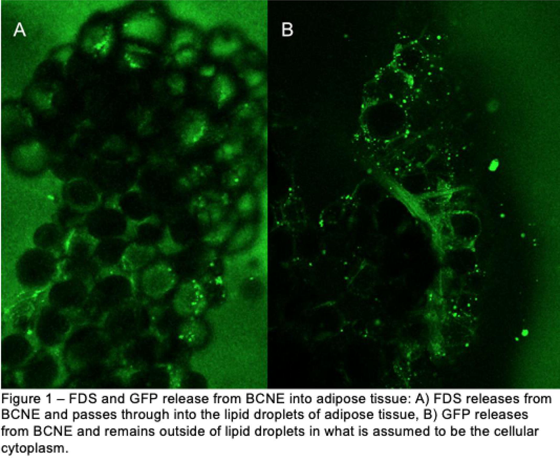
C133 - Advancing Immunocompetent Bioprinted 3D Human Tissue Models
L. P. da Silva, J. A. Macedo, S. Chaves, R. L. Reis, A. P. Marques
University of Minho, Guimarães, Portugal
*Purpose/Objectives: 3D immunocompetent tissue models are essential to study immune cell behavior in several contexts including immune-related diseases (e.g. autoimmune, allergies and cancer), wound healing and response to therapeutics. One challenge in creating these models lies in using a biomaterial that provides adequate cell support for their proper functioning while not activating immune cells, which would compromise the representation of the overall model function. Inks based on gellan gum (GG), a low immunogenicity polymer, were shown to support key functionalities of complex 3D printed tissue-like constructs. Thus, in this work, we aimed to understand whether ink’s polymer content, in the presence and absence of cell adhesive cues (cyclicRGD), affected human monocyte-derived macrophages (MDM) phenotype and their ability to respond to external stimuli.
*Methodology: Monocytes were separated from human peripheral blood mononuclear cells by CD14+ cells magnetic sorting and differentiated towards MDM with M-CSF. MDM were encapsulated in GG-based ink formulations (with/without cyclicRGD; low/high polymer content) and analyzed regarding their viability (calcein/propidium iodide), adhesion (phalloidin) and phenotype (phosphorylation multi-pathway array/luminex) with/without classical (IFN-γ + LPS) or alternative (IL-4) pathway activation.
*Results: The viability (up to 12 days) of encapsulated MDM was not affected by the tested formulation while their morphology varied from a round shape in the absence of cyclicRGD to an adhesive-like stretched form in cyclicRGD-containing formulations. Within 24 hours in the absence of cyclicRGD, polymer content promoted (p<0.05) the phosphorylation of regulators of MAPK (MSK2/MMK6) and NFkB (ATM) signaling pathways while reducing (p<0.05) the phosphorylated forms of TYK2/EGFR, ATF2, BAD/GSK3B/p27 MEK, respectively involved in the JAK/STAT, TGFb, AKT and MAPK pathways. In the presence of cyclicRGD, besides the MSK2 and ATM phosphorylated proteins, also mTOR/P70S6K (AKT), eIF2a/ATM (NFkB) and ERK (MAPK) were upregulated (p<0.05) for higher polymer content. Moreover, both pro-inflammatory (IL-1β/IL-6/IL-12p40/TNF-α) and anti-inflammatory (IL-1RA/IL-10/CCL22) cytokines were detected for all formulations. However, the levels of IL-6, TNF-α, IL-10 and IL-1RA were significantly reduced (p<0.0001) after three days. At this point, in which the effect of the testing conditions over MDM was neglectable, we tested MDM response to external stimuli. Cells responded with the release of pro- (classical) and anti-inflammatory (alternative) mediators (p<0.05), which varied with the polymer content in the absence of cRGD (p<0.001). Then, we aimed to understand how cells responded under continuous stimuli (up to 10 days with replenishing every 2-3 days) or to an additional stimulus (48 hours) after a resting period of 5 days. Cells were still able to respond to the external but a lower release of IL-6 and TNF-α was detected under continuous stimulation confirming an effect of the culture conditions and of the in vitro microenvironment on the observed response. Yet, only CXCL10 secretion was dependent on polymer amount and in the absence of cRGD confirming that the long-term MDM response will not be determined by the biomaterial used to fabricate the tissue-like model.
*Conclusion/Significance: Overall, we demonstrate that GG-based inks do not determine or condition MDM responses enabling the advance of corresponding bioprinted 3D tissue-like models rendering them immunocompetent.
C134
C136 - Investigation Of Immune Responses To Nano/microplastics Using A Genetically-engineered Human Macrophage
M. Yamamoto1, S. Hiranphinyophat1, N. Washihira1, T. Hiraoka1, M. Murakami1, A. Kishida2, S. Fujii3, T. Tanabe4, T. Kimura5
1
Tohoku University, Sendai, Japan,
2
Tokyo Medical and Dental University, Tokyo, Japan,
3
Yamagata University, Yamagata, Japan,
4
Shibaura Institute of Technology, Tokyo, Japan,
5
Tokyo Medical and Dental University, Sendai, Japan
*Purpose/Objectives: Microplastics (MPs) pollution is a global environmental problem. Potential risks have been considered regarding the effects of MPs on humans through the food chain. To understand the negative impacts of MPs on humans, it is essential to investigate the response of human cells to model plastic particles that mimic environmental MPs. Moreover, due to their small size, nanometer-sized plastic fragments have the undeniable potential to be uptaken into the body and distributed to a specific organ as seen in nanocarriers as a drug delivery system. Nanoplastics (NPs) with a small quantity and size, however, have limitations in collecting from oceans. To date, most studies on NPs have been conducted using polystyrene nanoparticles with a defined size and surface property to understand the body distribution and adverse effects in small organisms and fishes. In this study, we employed a genetically-engineered human macrophage (THP-1 cells) to investigate cellular responses to MPs and/or NPs of poly(ethylene terephthalate) (PET) and low-density polyethylene (LDPE) fragments.
*Methodology: A chemiluminescent tag HiBiT® was introduced into the downstream of interleukin-1β (IL-1β) using CRISPR/Cas9 genome editing. Several methods for oxidation and mechanical pulverization have been conducted to obtain plastic fragments. The resulting plastic fragments were evaluated in terms of the surface functional groups, size distribution, morphology, and thermal properties by Fourier Transform Infrared Spectroscopy, X-ray Photoelectron Spectroscopy, Scanning Electron Microscope, and Differential Scanning Calorimetry, respectively. The genetically-engineered THP-1 cells were polarized into M0, M1, and M2 macrophages, and the resulting macrophages were exposed to the plastic fragments at different concentrations. The immune responses to the plastic fragments were examined in terms of IL-1β secretion.
*Results: Photo-oxidation and ultrasound pulverization or accelerated oxidation reaction decreased the particle size of PET and LDPE fragments, making them irregularly shaped, and increased their crystallinity. In addition, an increase in the carboxyl group introduction ratio and negative surface potential were observed, suggesting surface oxidation. Both PET and LDPE fragments induced a higher level of IL-1β secretion on M1-polarized macrophages than non-oxidized ones.
*Conclusion/Significance: Several oxidation and mechanical pulverization techniques enable the efficient preparation of model plastic particles with different sizes, shapes, and surface and bulk properties, and are expected to provide new insights into the environmental threat of NPs and MPs on humans.
C138 - Bactericidal High Aspect Ratio Titanium Nanostructures Modulate Macrophage Responses
B. I. Eijkel, I. Apachitei, M. Klimopoulou, L. E. Fratila-Apachitei, A. A. Zadpoor
Delft University of Technology, Delft, Netherlands
*Purpose/Objectives: High aspect ratio (HAR) titanium nanopillars show promise in preventing implant-associated infections (IAIs) and antimicrobial resistance (AMR) due to their inherent bactericidal properties and the underlying contact-killing mechanisms [1]. Among these biomaterials, dry-etched titanium (DETi) HAR surfaces, generated under controlled conditions, not only possess bactericidal properties but also demonstrate enhanced osteogenic properties relative to non-treated surfaces [2, 3]. However, the effects of DETi surfaces on immune cells remain unknown. The ability to modulate the immune response, directing it towards a short-lived antibacterial effect followed by enhanced immune-mediated osteogenesis, represents a promising strategy for the development of multi-functional orthopedic biomaterials. This study first investigated the response of murine macrophages to polished titanium (pTi) and DETi surfaces. Secondly, it explored the effects of these immune responses on the antibacterial properties of DETi.
*Methodology: DETi was fabricated by inductively coupled plasma reactive ion etching (ICP-RIE) with Cl2 and Ar using specific flow rates, temperature, pressure, and etching time. The inflammatory response of unstimulated (M0) and pro-inflammatory (M1) murine macrophages (J774A.1) to both types of titanium surfaces was assessed through qPCR, and the morphological changes of the cells were examined by scanning electron microscopy (SEM) after 4, 24, and 48 hours of culture. In addition, a macrophage-bacteria co-culture model simulating perioperative IAIs was used to assess the effects of the immune response on the antibacterial properties of DETi.
*Results: The findings revealed differential responses of M0 and M1 macrophages after 48 hours of culture on both surfaces. Moreover, surface-related as well as time-related effects were identified. The findings obtained by using macrophage monocultures and macrophage-bacteria co-cultures were discussed regarding the modulatory role of the immune cells interfaced with such surfaces for generating a regenerative and antibacterial microenvironment.
*Conclusion/Significance: This study provides a unique perspective on the role of immune response induced by DETi surfaces in modulating tissue regeneration while preventing early peri-implant infections. Acknowledgements: This publication is part of the project DARTBAC (with project number NWA.1292.19.354 of the research programme NWA-ORC which is (partly) financed by the Dutch Research Council (NWO). References: [1] Higgins, S. G., Becce, M., Belessiotis‐Richards, A., Seong, H., Sero, J. E., Stevens, M. M. (2020). High‐Aspect‐Ratio Nanostructured Surfaces as Biological Metamaterials. Adv. Mater. 32, 1903862. doi: 10.1002/adma.201903862.[2] Modaresifar, K., Ganjian, M., Angeloni, L., Minneboo, M., Ghatkesar, M. K., Hagedoorn, P. L., Fratila-Apachitei, L. E., Zadpoor, A. A. (2021). On the Use of Black Ti as a Bone Substituting Biomaterial: Behind the Scenes of Dual‐Functionality. Small 17, 2100706. doi: 10.1002/smll.202100706.[3] Hasan, J., Jain, S., Chatterjee, K. (2017). Nanoscale Topography on Black Titanium Imparts Multi-biofunctional Properties for Orthopedic Applications. Sci. Rep. 7, 1-13. doi: 10.1038/srep41118.
C140
C141 - A 3D In-vitro Model Of Tissue Macrophages To Unlock Their Role In Obesity
A. Boussahel, T. Oates, A. Toye, A. Perriman
The University Of Bristol, Bristol, United Kingdom
*Purpose/Objectives: Macrophages are immune cells present in distinct phenotypes in most tissues with a range of functions. Their heterogeneity is a result of both their origins (bone marrow or embryo) and the influence of the environment in which they reside. In most tissues, macrophage subtypes have distinct roles in tissue function and disease development. For instance, in the adipose tissue they have recently emerged as key players in inflammation, the development of obesity and metabolic dysfunction. However, the mechanisms underlying interactions of adipocytes with macrophages and how these change in obesity and lead to metabolic dysfunction are not yet fully understood. This limits our ability to develop novel therapeutic strategies targeting macrophages. Currently, studying the role of macrophages in obesity is heavily reliant on the use of animal models, which have been shown to be untranslatable to human outcomes in the case of obesity and immune cells research. This is exacerbated by the lack of dedicated in-vitro tools that replace them. We aimed to develop the first in-vitro model of macrophages in the human adipose tissue that captures the origins of macrophages and their adipose tissue microenvironment-dependent specificity.
*Methodology: We developed an extracellular matrix (ECM) biomimetic hydrogel and optimised the differentiation of macrophages from different sources to model their heterogeneous origins, human monocytes isolated from blood (to model bone marrow derived macrophages) and induced pluripotent stem cells (iPSCs) (to model embryonic macrophages). We optimised the culture of macrophages in the 3D hydrogel and charcteriised their behaviour e.g. viability, macrophage markers using flow cytometry, secretions using Elisa, and polarisation using both Elisa and immunofluorescence. We also assessed functionality of macrophages by looking at motility using live cell imaging and phagocytosis. We optimised the co-culture of macrophages with adipocytes in the 3D hydrogel and fully characterised their behaviours and interactions.
*Results: The 3D ECM mimicking hydrogel supported the culture and differentiation of macrophages and adipocytes. We found macrophages tend to adopt an activated phenotype in the 3D hydrogel environment compared to the 2D culture. We were also able to modulate the macrophage phenotype and polarisation by changing the hydrogel physical properties such as stiffness and composition and this enabled the instruction of specific macrophage behaviours and phenotypes in the 3D culture system. We characterised the behaviours of macrophages in co-culture with adipocytes and their interactions using live imaging and established a baseline of co-culture mimetic to healthy lean adipose tissue. We are currently exploring methods to induce obesity changes in the in-vitro model and study how macrophages-adipocyte interactions change accordingly.
*Conclusion/Significance: We developed an in-vitro model of the human adipose tissue that captures the heterogenous nature of macrophages and enables the study of the intricate interactions of different macrophages with adipocytes. Once we simulate changes in obesity in the in-vitro model, we will be able to study how changes to macrophage-adipocyte interactions mediate obesity and metabolic dysfunction mechanisms.
C142 - In Vitro Platform To Evaluate The Efficacy Of Immunoengineered Platforms To Prevent Allorejection And Recurrence Of Autoimmunity In Islet Transplantation
I. Pasolini1, G. C. Gonzalez2, C. M. Li1, L. Teles1, A. Tomei1
1
University of Miami, Miami, FL,
2
Diabetes Research Institute, University of Miami Miller School of Medicine, Miami, FL
*Purpose/Objectives: Type 1 diabetes (T1D) is an autoimmune disease resulting in destruction of insulin-secreting beta cells. There is no cure for T1D because its pathophysiology is poorly understood. Rather, T1D is managed by insulin injections or, in a small portion of patients with severe T1D, by islet transplantation with immunosuppression. In vitro models co-culturing beta cells and immune cells can help elucidate the pathophysiology of T1D and test the efficacy of immunoengineered approaches that block islet autoimmune destruction or treat T1D through islet transplantation without immunosuppression. We developed and validated a simplified in vitro model to investigate the efficacy and mechanisms of action of islet encapsulation in preventing islet destruction by autoreactive and alloreactive immune cells. We included HLA-matched and -mismatched murine insulinoma cell clusters and beta cell autoreactive and alloreactive CD8+ cytotoxic T cells (CTLs) based on the availability of (1) mouse insulinoma NIT-1 clusters from non-obese diabetic (NOD) mice with consistent 150µm-diameter of human islets and glucose-stimulated insulin secretion (GSIS) and of (2) T cell receptor-transgenic mice (autoreactive) and alloreactive CTLs.
*Methodology: We optimized the in vitro assay to enable GSIS testing of NIT-1 clusters in a 96 well-plate with minimal numbers of clusters compared to traditional GSIS assays by varying cluster Islet Equivalents (IEQ) and well type as readout of (a) NIT-1 cluster function in the model. We also optimized a flow cytometry panel to assess NIT-1 phenotype (viability and expression of MHC-I/II, Fas, and PD-L1) as readout of (b) NIT-1 cell viability and phenotype in the model. In parallel, we optimized a flow cytometry panel to assess CTL phenotype (proliferation, activation, cytotoxicity) as readout (c) of T cell activation. We used the model to evaluate the efficacy of NIT-1 cluster encapsulation on each of the above measured outputs. We compared the phenotype of NIT-1 treated with IFNγ and/or thapsigargin. We also optimized the co-culture of NIT-1 clusters with activated CTLs at immune/NIT-1 target cell ratios of 0.5, 1, and 2 with CTLs from beta cell-autoreactive NY8.3 mice or allogeneic CTLs from C57BL/6 mice activated with anti-CD3/CD28. After co-culture, immune cells and NIT-1 clusters were separated to evaluate readouts (a), (b), and (c) and validate the model. Finally, we applied the model to evaluate these readouts when NIT-1 clusters were encapsulated in 1.2% MVG alginate (Pronova).
*Results: We found that 60 IEQ in a U-bottom 96-well-plate provides comparable GSIS results than traditional GSIS assays as readout (a) of the model. We also optimized a flow cytometry panel to assess NIT-1 viability and phenotype with IFNγ, thapsigargin, and CTL co-culture and CTL cytotoxic phenotype as readouts (b) and (c) of the model. We are currently testing the effects of NIT-1 cluster encapsulation on these three readouts and expect increased NIT-1 functionality and viability in encapsulated compared to uncoated clusters.
*Conclusion/Significance: This platform can screen different approaches to decrease autoreactive and alloreactive T cell-mediated beta cell destruction and test mechanisms in simplified settings for application in T1D prevention and treatment.
C143 - In Vitro Extracellular Matrix Hydrogel Platform For Interrogating 3D Immune Cell Responses To Organoid Targets
M. M. Samojlik, A. T. Fernandez, S. N. Patel, Y. Li, M. D. Du, E. A. Phelps, C. L. Stabler
University of Florida, Gainesville, FL
*Purpose/Objectives: To bridge the knowledge gap in our understanding of Type 1 Diabetes (T1D) immunopathogenesis, robust in vitro platforms for the co-culture of immune cells and pancreatic islets that allow for the stable spatial distribution of the cells, migration, and dynamic cell-cell interactions are needed. Herein, we describe a new extracellular matrix (ECM) hydrogel in vitro platform that permits real time interrogation of antigen-specific cell-cell interactions, as well as T cell migration through 3D imaging analysis.
*Methodology: To study the temporal changes in T cell migration upon interaction with target islets, OTI-GFP cytotoxic CD8+ T cells (CTLs) were co-cultured with mOVA (antigen-specific) or B6 (negative control) islets in ECM hydrogel. Confocal timelapse imaging was performed at 90 second intervals for 16 hours and Fiji TrackMate was used to track the migration of T cells. Outputs were additionally processed in MATLAB, where a library of migration tracking analysis programs was created for visualization, temporal analysis, and trajectory classification.
*Results: Cytotoxic T cells cultured within ECM exhibited highly migratory behaviors throughout the 3D space over time, demonstrating the capacity of the matrix to support both the distribution and migration of the cells. Furthermore, CTLs co-cultured with antigen-specific or non-antigen-specific islets exhibited significant differences in their T cell migratory patterns. Compared to the negative control group, the CTLs interacting with antigen-specific islets exhibited significantly decreased displacement and mean speed but increased mean directional change rate upon interactions with the target islets. Additionally, significant accumulation of the CTLs on the mOVA islet surface over time was observed, in contrast to the B6 islet surface. Temporal analysis of total distance traveled by the CTLs revealed that mOVA islet-interacting CTLs exhibited overall significantly lower total distance traveled than the negative control. Trajectory classification of the migration tracks, based on their mean squared displacement, revealed that CTLs responding to mOVA islets exhibited less Lévy walk (superdiffusive) and more subdiffusive migration than those responding to non-antigen-specific islets. CTLs that did not interact with the mOVA islet exhibited a higher percentage of cells migrating in a superdiffusive, Lévy walk manner than CTLs cells that interacted with the islet surface.
*Conclusion/Significance: In conclusion, this data illustrates the unique insights provided by this 3D ECM immune cell-islet co-culture platform for the study of T cell migration characteristics. Together with the library of MATLAB programs designed specifically to visualize and analyze the different aspects of T cell migration, additional quantifiable metrics related to dynamic cell-cell interactions in the context of T1D, such as the role of antigen, can be elucidated. Furthermore, our data indicates that the interactions between antigen-specific T cells and their cognate antigen targets result in significant temporal changes in general migration parameters and in the type of migration pattern exhibited, with switching from the exploratory Lévy walk to subdiffusive migration upon target interaction. Ongoing work focuses on understanding the resulting T cell mediated islet-cell killing.
C144 - Manipulating The Specialty Of Fibroblastic Reticular Cells
M. E. Heon, I. I. Torres-Vazquez, N. Martinez-Rivera, E. Rosa-Molinar
The University of Kansas, Lawrence, KS
*Purpose/Objectives: In lymph nodes, fibroblastic reticular cells (FRC) are a major part of the stromal cell network, and they have been recognized to play an important role in remodeling the lymph node and modulating the immune response. Due to their role, FRCs could be used as a basis for a tissue engineered lymph node; they could create a more realistic microenvironment for immune cells. An issue arises, however, when you consider the high heterogeneity of these cells. Instead of being a homogenous cell population present throughout each lymph node lobule, as assumed by most studies on these cells (Figure 1A), FRCs have many different subpopulations with roles specialized for the different areas of the lobule (Figure 1B). Even within these specialized areas, recent research has suggested the possibility of even more heterogeneity (Figure 1C). The challenge then is obtaining a FRC cell population that represents the high heterogeneity and finding a way to culture them to encourage the formation of specialized areas.
*Methodology: In this work, we first generated an immortalized and heterogenous population of Fibroblastic Reticular Cells. The cells were isolated from mouse inguinal lymph nodes using both chemical digestion and physical disruption techniques. Then, they were immortalized using a lentiviral vector with SV40-T. After five passages (P5), the cells were identified as FRCs by checking for a negative expression of surface immune markers CD45 and CD31 and positive expression of mucin-type transmembrane protein PDPN using flow cytometry. Expression of the adhesion molecules ICAM and VCAM was used to determine if the population was heterogenous.
*Results: Flow cytometry results indicated that the immortalized population was heterogeneous, with the backgating data clearly showing the presence of multiple clusters. Next, we identified that the way we were culturing the cells caused a slow shift in the subpopulations. The expression of CD45, CD31, PDPN, ICAM, and VCAM was checked again after 12 passages (P12) and the distribution of cells across CD31, PDPN, ICAM, and VCAM expression had shifted from the results at P5. To confirm these results, we looked at cell morphology using immunofluorescence and observed an overall change in the number of cells with each morphology. This shift in heterogeneity suggests that we can alter the subpopulations, and thus specialty, by changing their culture environment.
*Conclusion/Significance: In this work, we demonstrated our ability to alter the specialties of a heterogenous population of FRCs. All of this work was performed in two-dimensional cell culture which is neither natural to this cell type nor allows them very much control over their environment. As we transition from two-dimensional to three-dimensional cell culture, we may be able to manipulate the specialty of FRCs to better mimic the different regions of a lymph node lobule.
C145 - Instructive Hydrogel/MEW-FDM Hybrid Scaffolds For Regeneration Of Full-thickness Osteochondral Defect
J. Idaszek1, Z. Gorecka1, D. Martinez Perez1, M. Volpi1, E. Walejewska1, K. Pucia2, G. Gut2, W. Święszkowski1
1
Warsaw University of Technology, Warsaw, Poland,
2
Medical University of Warsaw, Warsaw, Poland
*Purpose/Objectives: Treatment of osteochondral defect (OCD) requires simultaneous regeneration of articular cartilage and subchondral bone. Hydrogels are a class of biomaterials which are often used for engineering of the aforementioned tissues, however, they lack sufficient mechanical properties to withstand the stress generated at the implantation site. Among various reinforcement strategies, incorporation of fibrous 3D-printed scaffolds seems to be particularly promising. The aim of this study was to combine the scaffolds fabricated by means of fused deposition modelling (FDM) and melt electrowriting (MEW) to reinforce tissue-specific, instructive hydrogels for regeneration of full thickness OCD through chondrogenic priming of human bone marrow-derived mesenchymal stem cells (hMSC).
*Methodology: 3D fibrous scaffolds for bony compartment were fabricated using PCL-based composite (β-TCP, 25 wt%) and FDM, and for cartilaginous compartment using pristine PCL and MEW. The full-thickness OCD scaffolds were fabricated by attaching the MEW compartment to the molten surface of the FDM scaffold. The bony hydrogel contained either 10 mg/ml of bone-derived decellularized extracellular matrix (dECM) or type I collagen (control) mixed with alginate (30 mg/ml) and 20x106/ml of hMSC, and was designed to induce in vivo bone formation through endochondral ossification pathway. The cartilaginous hydrogel comprised 60 mg/ml of gelatin methacrylamide (GelMA), 40 mg/ml of chondroitin sulfate amino ethyl methacrylate (CS-AEMA) and 20x106/ml of cells (hMSCs and human articular chondrocytes (4x10⁶/ml)). 50 μl of each bioink was infused into corresponding fibrous scaffolds and cultured in chondrogenic medium for 28 days. Cell performance was evaluated using live/dead staining, MTS, ALP and DNA assays. Secretion of cartilaginous matrix was assessed by staining for collagen type II and X, and quantified using 1,9-dimethylmethylene blue assay. The mechanical properties were determined using compression tests. Following the 4 weeks of chondrogenic priming, the OC constructs were implanted subcutaneously into nude mice. After 6 weeks, the scaffolds were explanted and analysed using microcomputed tomography (μCT) and histological stainings (H&E, Alcian Blue, von Kossa).
*Results: The acellular GelMA-CS-AEMA/MEW scaffolds exhibited significantly higher stiffness than the pristine hydrogel scaffolds (83±35 vs 0.9±02.2 kPa, respectively). However, stiffness of the cell-laden pristine hydrogel was at similar level to the MEW-reinforced scaffolds after 4 weeks of chondrogenic differentiation (∼120 kPa). Moreover, higher GAGs synthesis was measured in the pristine hydrogels than the GelMA-CS-AEMA/MEW scaffolds (49±11 vs 33±4 μg GAG/μg DNA, respectively). The stiffness of the acellular FDM scaffolds was 53±8 MPa. Expression of collagen type II was observed throughout the entire FDM scaffolds, while of collagen type X was localized at their peripheries. The ALP activity (marker of chondrocyte hypertrophy) was 90% higher in the dECM/FDM hydrogels than the corresponding collagen scaffolds at day 28. After 6 weeks of in vivo implantation, we measured mineral deposition in the outer regions of the bony compartment but not the cartilaginous one.
*Conclusion/Significance: The developed hybrid scaffolds containing tissue specific, instructive hydrogels supported formation of phenotypically stable hyaline cartilage and subchondral bone spatially-confined to the corresponding compartments of the scaffold. To confirm their suitability for reconstruction of full-thickness OCD, they have to be evaluated at orthotopic site.
C146
C147 - A Photocrosslinkable Granular Hydrogel System For The Repair Of Chondral Defects
A. Atwal1, T. P. Dale1, M. Snow1,2, N. R. Forsyth1,3, P. Davoodi1
1
Keele University, Stoke-on-Trent, United Kingdom,
2
Robert Jones and Agnes Hunt Hospital, Shropshire, United Kingdom,
3
University of Aberdeen, Aberdeen, United Kingdom
*Purpose/Objectives: Untreated traumatic cartilage defects pose a significant risk, potentially progressing to osteoarthritis and severe joint damage, causing debilitating pain for the patient. The global prevalence of osteoarthritis is estimated to affect 7% of the population, a figure that is only increasing with an aging demographic. Surgical interventions such as microfracture, autologous chondrocyte implantation, and mosaicplasty are costly, requiring multiple surgeries and rarely achieving complete cartilage regeneration. Recently, hydrogels have emerged as a promising avenue in cartilage tissue engineering due to their injectability and potential for minimally invasive administration, offering potential cost reductions in healthcare while simultaneously enhancing functional cartilage repair. To this end, our research group previously developed an injectable hybrid system comprising methacrylated gelatin (GelMA), hyaluronic acid (HAMA), and platelet lysate (PLMA). While this system supported extracellular matrix (ECM) formation and maintained the chondrogenic phenotype, it faced limitations. The thermal gelation of the prepolymer solution made it challenging to inject at room temperature, and the high degree of crosslinking necessary for mechanical stability hindered nutrient diffusion, cell spreading, and neotissue deposition.
*Methodology: To overcome these challenges, we designed GelMA:HAMA:PLMA microgels using a microfluidic water-in-oil emulsion technique to generate GelMA:HAMA:PLMA microparticles. Partial photocrosslinking was applied to create structurally stable hydrogel microparticles with a size of <100 µm. After thorough washing to remove residual oil, the small particle size enabled injection through small gauge needles to the defect site. Subsequent exposure to UV light facilitated interparticle crosslinking, effectively annealing the microgels together. Within the interstitial voids between particles, it was hypothesised chondrocytes could proliferate and allow for increased neotissue deposition (Figure 1). Mechanical and rheological analysis was carried out to assess the structural stability of the system in addition to biological characterisation such as live/dead staining, GAG/DNA content, histological and immunohistochemical imaging in both an in vitro and ex vivo model. These were assessed and compared to the previously developed bulk system.
*Results: The mechanical and rheological properties of the microgel system were adjustable based on particle size and polymer concentration, influencing intermolecular voids and playing a pivotal role in the rheological properties of the system. In a comparative study using an ex vivo defect model, histological and immunohistochemical staining revealed a notable increase in neotissue formation rich in type II collagen after 31 days within the interparticle voids of the microgel system compared to an equivalent bulk hydrogel with the same polymer composition. Furthermore, GAG and DNA content measured over time demonstrated increased content compared to the bulk gel system, illustrating the promising potential of this granular hydrogel system for cartilage repair.
*Conclusion/Significance: In conclusion, our granular hydrogel system, comprising a blend of GelMA, HAMA, and PLMA, emerges as a promising candidate for cartilage repair and regeneration. The system’s tunable characteristics, including variations in particle size, polymer concentration, and degree of methacrylation, offer a versatile and highly customizable approach to cartilage tissue engineering. This versatility surpasses that of traditional bulk hydrogel fillers, which, while promising, tend to exhibit slower regeneration due to the constraint on cell movement.
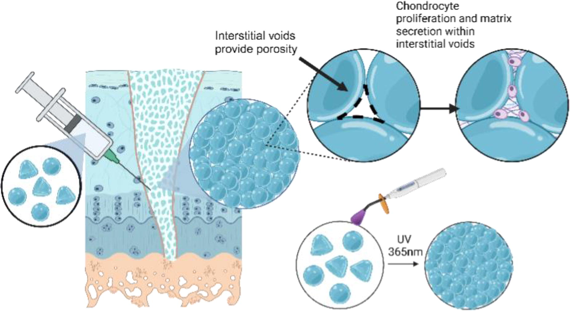
C148 - A Regenerative 3d Cartilage Model For HTP Drug Screening
A. El Haj1, H. Hajiali1, J. Cholewa-Warclaw2, A. Imere1, J. Ballard1, R. Elliott3, N. Carragher4
1
Healthcare Technology Institute, Birmingham, United Kingdom,
2
Institute of Regeneration and Repair, Edinburgh, United Kingdom,
3
Institute of Genetics and Cancer, Edinburgh,, United Kingdom,
4
Centre for HTP screening, Edinburgh, United Kingdom
*Purpose/Objectives: New drug candidates for treating Osteoarthritis are lacking and few are being tested in the pipeline. A 3D Wnt GelMA model of the chondrogenic niche has been developed which is capable of scale up to 96 and 386 wells for screening multiple drug libraries, and assess the interactive effects of bioactive compounds, such as Wnt3A. Using our new model, we have been able to assess candidate drug effects on cell proliferation, cell size, cell migration and chondrogenic differentiation markers, Sox9 and Col2 in one assay. In this presentation, we outline the use of our model to identify multiple candidates as novel repurposed drugs to treat damaged cartilage during Osteoarthritis.
*Methodology: Specifically, we have established reproducible 3D Wnt platforms utilizing the Y201 bone marrow stromal cell line with GELMA and bound surface Wnt platforms providing a multifaceted approach beyond standard 2D cell culture. We also conducted experiments to assess the variability between samples comprising cells, gels, media, and antibody staining to ensure that the variability falls below a defined threshold before proceeding to the compound screening phase. Compound screening was conducted with an in house bioactive drug library consisting of 56 compounds in three different concentrations. In addition, we employed robotic systems for various critical tasks in this phase. Robots were used for seeding cells, inserting gels, and employing 96 LEA arrays to crosslink the gels. The use of robotics significantly reduced the potential for variability, ensuring the reliability of the data obtained. Variability was assessed by analysing cellular morphology, e.g. cell area and cell roundness as key parameters, alongside migration, proliferation and expression of key chondrogenic markers.
*Results: Initially, we conducted experiments to assess the variability among samples, including cells, gels, media, and antibody staining. The primary objective was to ensure that the variability remained below a predefined threshold (CV < 20) before advancing to the drug screening phase. The variability test was successfully completed with a very low CV. The subsequent phase centred on drug screening using a bioactive drug library comprising 56 compounds in three different concentrations. In our preliminary analysis, we have identified specific small molecules capable of inducing chondrogenic cell phenotypes. Multiple compounds were identified which induce significant changes in cell morphology, migration and chondrogenic marker expression. Further validation with the selected hit compounds and additional chondrogenic markers and cartilage formation in long term culture conditions have been conducted.
*Conclusion/Significance: In conclusion, this 3D regenerative cartilage model developed offers a valuable tool for investigating cartilage biology, disease mechanisms, and for scale-up in drug screening applications. Our data support the potential of this model for cartilage tissue engineering and provide insights into cartilage regeneration mechanisms in a cartilage repair environment. Furthermore, we have validated the robustness of our model and its potential to discover novel therapeutic approaches for cartilage repair and osteoarthritis treatment. Continued optimization and validation of this model will contribute to the advancement of cartilage tissue engineering and accelerate the development of effective therapies for cartilage regeneration in response to diseases such as Osteoarthritis.
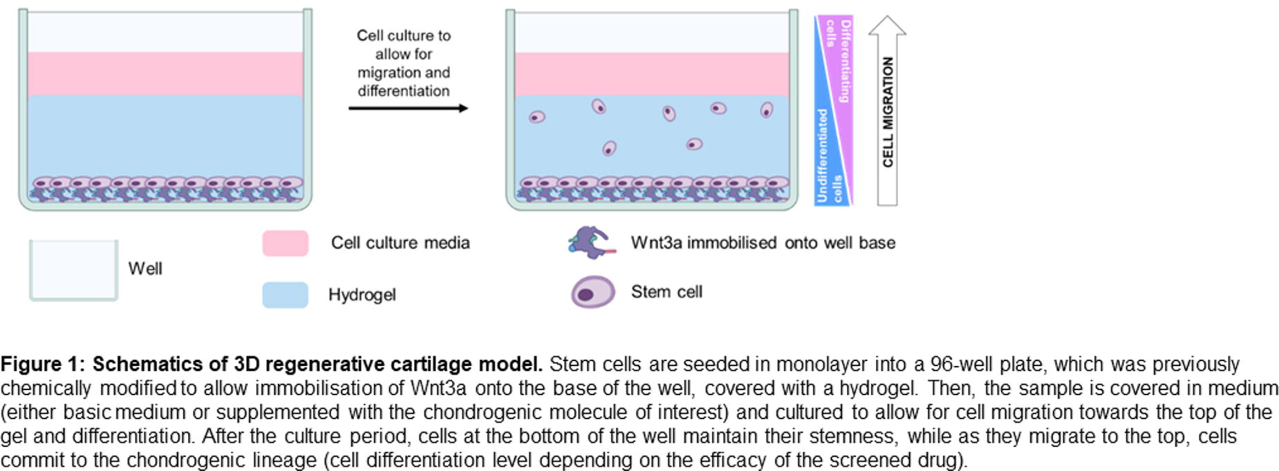
C149 - Effect Of High-stiffness Cartilage Hydrogels On Cell Viability
E. A. Kiyotake, C. Iribagiza, M. S. Detamore
University of Oklahoma, Norman, OK
*Purpose/Objectives: High-stiffness cartilage extracellular matrix-based hydrogels are highly translational with easy surgical placement, and may contain intrinsic chondroinductive components for treating cartilage injuries or degeneration. We previously fabricated pentenoate-functionalized, solubilized, devitalized cartilage hydrogels (PSDVC), which had an injectable and paste-like precursor for easy surgical placement, fast-crosslinking for enhanced material retention after placement, and high-stiffness for enabling patients to be immediately weight-bearing after surgery. Typical hydrogels used in tissue engineering are under ∼200 kPa in compressive stiffness and the effects of stiffness on the encapsulated cell viability and chondroinductivity under 200 kPa have been characterized. However, the encapsulated cell response has not been characterized for stiffer PSDVC hydrogels (up to 3 MPa). Therefore, the viability of encapsulated human bone marrow-derived mesenchymal stem cells (hBMSCs) was evaluated in three PSDVC hydrogel formulations with varying stiffnesses from ∼240 kPa up to 2,170 kPa.
*Methodology: PSDVC was synthesized from the articular cartilage of porcine knees, followed by cryogrinding to form devitalized cartilage (DVC) particles, solubilizing with pepsin to form solubilized DVC (SDVC), and functionalizing with varying amounts of 4-pentenoic anhydride to obtain different stiffnesses of PSDVC hydrogels. PSDVC hydrogels (10 wt%) were formed with LAP (photoinitiator, 2.2 mM) for testing the compressive elastic moduli and cell viability. During viability testing, hBMSCs were encapsulated (10E6 cells/mL) in the three formulations of PSDVC hydrogels and cultured in chondrogenic media (with or without TGF-β3) for 1 and 14 days. The viability was assessed with metabolic activity and DNA content.
*Results: The three PSDVC batches had compressive elastic moduli of 239 ± 26 kPa, 594 ± 62 kPa, and 2,170 ± 210 kPa. On day 1, the metabolic activity (normalized to the DNA content) of the hBMSCs in the softest hydrogel was 12 to 177-times greater than those in the medium or stiff hydrogels (p < 0.0001), and the normalized metabolic activity of the cells in the stiffest vs medium stiffest hydrogels were not significantly different. On day 14, the normalized metabolic activity of the cells in the softest hydrogel was 3 to 25-times higher than those in the medium and stiffest hydrogels (p < 0.0001). The cells in the medium stiffest hydrogel were 5.5 to 8.1 times greater than those in the stiffest hydrogel (p < 0.05).
*Conclusion/Significance: The cell viability in PSDVC hydrogels stiffer than 200 kPa were evaluated and while the stiffest hydrogel had a compressive elastic modulus in the range of native cartilage, the high stiffness (∼2 MPa) and medium stiffness (∼600 kPa) hydrogels led to the lowest cell viability of encapsulated hBMSCs after 1 and 14 days in culture. Overall, the softest ∼240 kPa PSDVC hydrogel had the highest encapsulated cell viability. The ∼240 kPa PSDVC hydrogel, which is still stiffer than typical hydrogels from the literature, is soft enough to support high cell viability and yet may be stiff enough to enable weight-bearing as a future translational cartilage hydrogel for cartilage repair.
C150 - Synovial-cartilage Organoids To Investigate Size-dependent Function Of Hyaluronic Acid
M. Hofmann, N. Shchotkina, M. Gautreaux, G. Lindberg
University of Oregon, Eugene, OR
*Purpose/Objectives: Osteoarthritis (OA) is a costly, painful disease and a leading cause of physical disability worldwide. Injectable hyaluronic acid (HA)-based viscosupplements have shown promise to treat pain and delay surgery in OA. In this context, HA has conventionally been used for its mechanical properties, providing additional lubrication and shock absorption to the joint. However, HA is also an important signaling molecule with receptors (e.g., CD44) regulating cell migration, proliferation, and angiogenesis. Interestingly, HA’s size has been suggested to determine its interaction with these receptors: high molecular weight HA is anti-inflammatory, while low molecular weight HA is pro-inflammatory. HA’s duality of function remains to be fully characterized, particularly relating to OA disease progression. To address this knowledge gap, we aim to utilize an inflammatory 3D OA model to assess the chondroprotective effect of HA with varying molecular weights.
*Methodology: Cartilage tissue was fabricated using pelleted human or bovine articular chondrocytes (250k cells) in chondrogenic media under physoxic conditions (5% O2). The cartilage tissue was cultured with inflammatory cytokines (TNF-α, IL-1β) or inflammatory T-cells (75k, CD8+) to recapitulate pre-clinical stages of OA. Varying molecular weights of HA (15 kDa, 40 kDa, 100 kDa, 500 kDa, and 1.8 MDa; 0.3 wt%) were injected to assess therapeutic effects. Biochemical assays and histological evaluations were used to quantify cartilage degradation, and Luminex multiplex proteomics were used to quantify chondrocyte and T-cell cytokine expression. Parallel plate rheometry was used to characterize the viscoelasticity of the HA solutions.
*Results: T-cell and cytokine-exposed cartilage tissue demonstrated significant glycosaminoglycan reduction, mimicking the onset of OA. 1.8 MDa HA attenuated this deteriorative effect, while 15 kDa, 40 kDa, and 100 kDa HA did not display any chondroprotective effects. This was confirmed through an increased positive stain for proteoglycans and type II collagen in the 1.8 MDa HA group. 500 kDa HA also reduced cartilage loss within inflammatory conditions but with significantly less effect as compared to 1.8MDa HA. Neither of the HA injections were observed to induce cartilage repair following cytokine-driven degradation. Within 100 kDa HA, histology revealed cell migration from the spheroid core to the periphery that was not observed in other groups. Proteomic analysis of T-cells cultured in HA demonstrated modulated cytokine expression (e.g., TNF-α, IL-17A, IL-1RA IL-7, IL-8) as compared to controls. At biologically relevant shear rates (0.5-10 Hz), 1.8 MDa HA displayed a similar viscosity (0.16-0.49 Pa*s) as native synovial fluids of OA patients.
*Conclusion/Significance: Here, we demonstrate the proficiency of the newly developed in vitro OA model in examining the interplay between cartilage tissue, inflammatory cytokines or immune cells, and HA. This work unveils the size-dependent functions of HA, providing valuable insights for guiding future therapeutic applications.
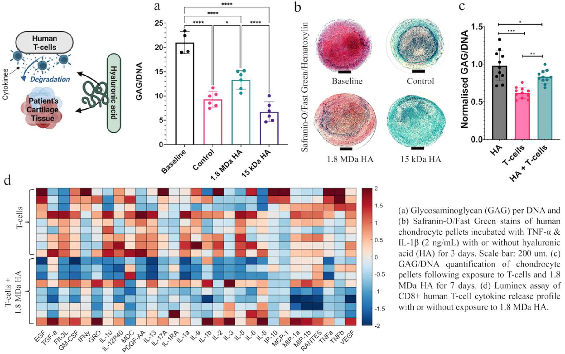
C151 - Modulating Pentenoate-functionalized Hyaluronic Acid Hydrogel Network Formation For Meniscal Fibrochondrocyte Mechanotransduction
K. Burkey, P. Mulimani, N. Sniadecki, J. Robinson
University of Washington, Seattle, WA
*Purpose/Objectives: A knee meniscus tear is a common musculoskeletal injury. The meniscus is a mechanically active tissue that provides load distribution and joint stability but is largely incapable of self-healing after injury. Current treatments fail to restore the mechanical properties of the native tissue, necessitating the development of regenerative therapies. Resident cells of the meniscus, meniscal fibrochondrocytes (MFCs), respond to biomechanical signals generated by their extracellular matrix (ECM). This MFC-ECM mechanotransduction directs cell behaviors necessary for regeneration. MFCs also exert traction forces (TFs) on their microenvironment through focal adhesions. These TFs are important components of the dynamic MFC-ECM relationship. To better understand the biomechanical cues that drive regeneration in the meniscus, in vitro models of the meniscal ECM must be developed to investigate mechanotransduction and the nature of focal adhesions and TFs. The goal of this work is to use a mechanically tunable hydrogel system to characterize MFC behavior in response to change in substrate compressive modulus.
*Methodology: Hyaluronic acid and pentenoic anhydride were esterified to obtain pentenoate-functionalized hyaluronic acid (PHA). PHA with a range of degrees of pentenoate -ene group substitution (DoS) was crosslinked into hydrogels with a range of compressive moduli (E’). Compressive modulus was defined as the slope of the stress-strain curve from 10% to 20% strain (0.005 mm/s uniaxial compressive strain rate). Human MFCs (3 male (17-18 y/o, P3-4) and 3 female (13-16 y/o, P3-4) donors) were seeded on hydrogels (15,000 cells/cm2) and cultured for 48 hours, then stained with DAPI, phalloidin to visualize actin cytoskeleton, and paxillin and vinculin to identify focal adhesions. Images were analyzed using ImageJ. To further investigate cellular TFs, substrates of known elastic modulus were modified with a layer of black dots and then seeded with MFCs (∼2500 cells/cm2). Cells were cultured for 48 hours then stained and imaged to view the deformation of the black dots pattern in response to cell TFs. Images were analyzed with a custom MATLAB code.
*Results: Compressive moduli achieved in this study covered the range of that of native tissue (50-1,000 kPa) (Figure Panel A). Modulating PHA hydrogel modulus results in control of MFC morphology (Figure Panel B). Current data suggests that female cells are more sensitive to change in substrate modulus at low DoS, but less responsive at higher DoS when compared to male cells (Figure Panels B and C). These results motivate investigation of MFC TFs, quantification of which is currently underway. Additionally, MFCs on substrates of all tested moduli express paxillin and vinculin, and further quantification of focal adhesion expression is underway. Overall, morphology results suggest that softer hydrogels promote a chondrocyte-like morphology while more rigid hydrogels promote a more fibroblast-like morphology.
*Conclusion/Significance: In conclusion, determining the mechanism of mechanotransduction in the meniscus relies on synthesizing microenvironments that mimic the mechanical properties of the native tissue. This study demonstrates that this PHA hydrogel system is suitable for further investigation of ECM mechanotransduction. Finally, this study reveals a difference in response of MFCs to substrate modulus based on sex which requires further exploration.

C152 - Neonatal And Postnatal Inspired Growth Rates Differentially Drive Collagen Maturation In Engineered Ligaments
K. Ott, J. Puetzer,1
Virginia Commonwealth University, Richmond, VA
*Purpose/Objectives: Hierarchical collagen fibers are the primary source of strength in tendons and ligaments. These fibers do not regenerate after injury, with repair, or in engineered replacements, limiting treatment options. Previously, we developed a culture system that guides anterior cruciate ligament (ACL) fibroblasts to produce aligned fibrils by 2 weeks, and native-sized fibers and fascicles by 4 and 6 weeks (Fig.1A). However, further maturation is necessary for clinical relevance. Mechanical cues, including slow growth elongation, are critical for fiber maturation during development; however, ACL growth-rates vary by more than 10-fold between embryonic and postnatal stages while there are significant changes in collagen organization, making it difficult to assess appropriate stretch rates in vitro. Our objective was to investigate whether stretching at neonatal and postnatal ACL growth-rates differentially drives hierarchical collagen fiber development in our system and their effect when applied at different degrees of collagen organization.
*Methodology: Bovine ACL fibroblasts were mixed with collagen to produce constructs at 20 mg/mL collagen and 5x10⁶ cells/mL. Constructs were then cultured in our static device to guide hierarchical fiber formation (Fig.1A). At 0, 2, or 4 weeks, once constructs had unorganized collagen, aligned fibrils, or fibers, respectively, constructs were transferred to a bioreactor and stretched at neonatal (1 mm/day) or postnatal (0.1 mm/day) ACL growth-rates for 1 week. Controls remained statically clamped and timepoints were at 1, 3, or 5 weeks. Post-culture, collagen organization was analyzed via confocal reflectance and images were analyzed with a FFT-based code to determine alignment and fiber diameter. Construct tensile properties and composition were also evaluated. Significance was determined via 2-way ANOVA with Tukey’s post-hoc.
*Results: Both stretch rates generated more aligned fibers compared to static controls at all levels of organization (Fig.1B). Neonatal stretch had larger effects on alignment in unorganized collagen, while postnatal stretch produced increased alignment and fiber diameters when applied after fibers had formed (Fig.1C). Both stretch rates induced collagen crimp formation when applied at 4 weeks, after fibers had formed (Fig.1B). Mirroring organization, changes in mechanics also depended on timing and stretch rate (Fig.1D). Neonatal stretch increased tensile modulus and ultimate strength (UTS) when applied to unorganized collagen; postnatal stretch produced similar increases when applied at higher levels of organization (Fig.1D). Stretched constructs also had increased failure strains at 5 weeks, suggesting crimp may be absorbing initial loads (Fig.1D). Neonatal stretch applied to unorganized collagen resulted in increased GAG and LOX activity, and both stretch rates decreased collagen (hypro) content at 1 week, but these effects were lost when applied at higher levels of organization (Fig.1E).
*Conclusion/Significance: Slow stretch at neonatal and postnatal growth-rates increases collagen organization, composition, and tissue mechanics. Neonatal growth-rates have the largest impact on cells in unorganized collagen, while postnatal rates largely impact cells when applied at the fibril and fiber level. This suggests cells sense and respond to stretch differentially depending on the degree of organization and matrix maturation. A better understanding of how mechanical cues regulate fiber formation will better inform rehabilitation protocols and engineered replacements.
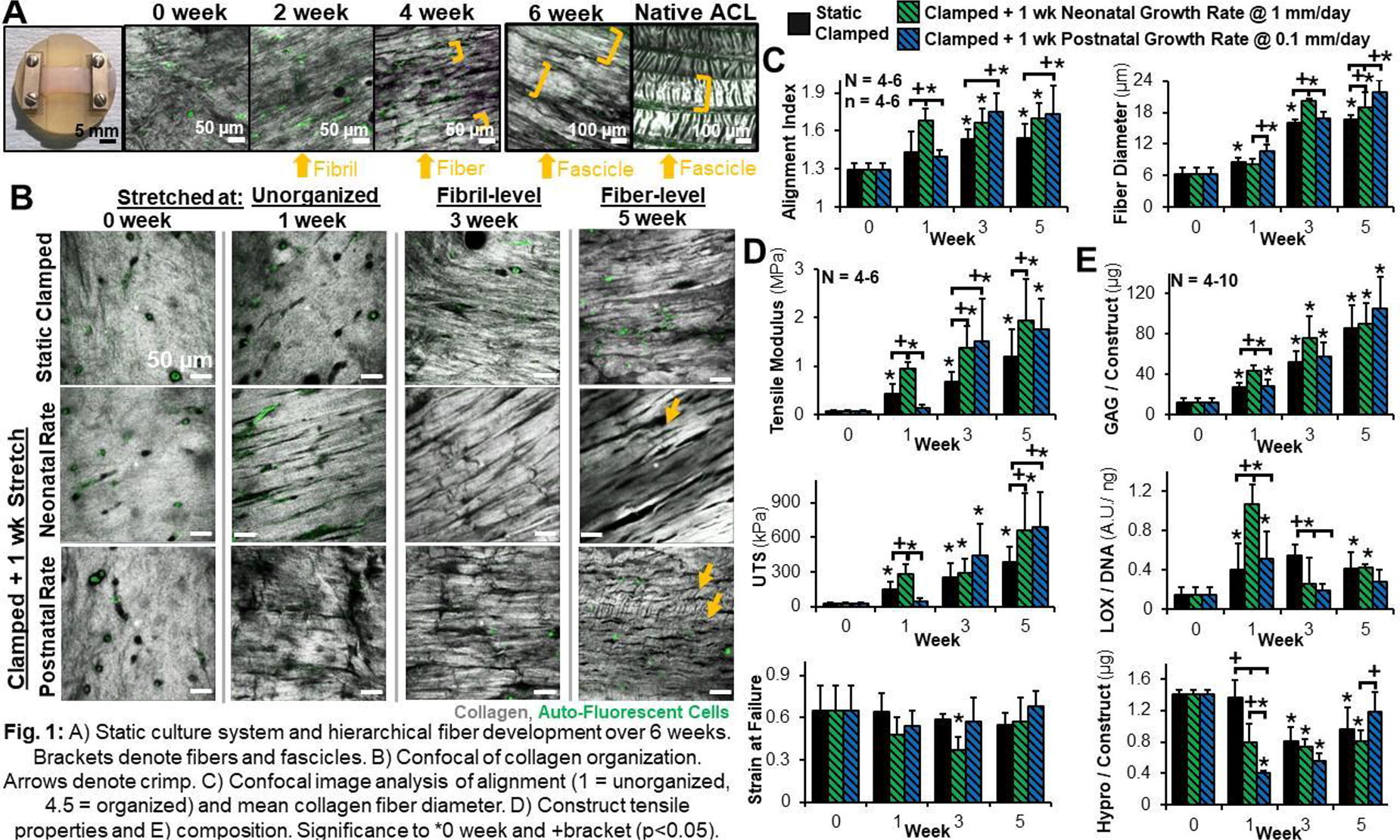
C153 - Region-specific AndMulti-material Electrospun Bundles Drive Stem Cells In The Enthesis Regeneration
A. Sensini1, F. Giacomini1, O. Stamati2, B. Lukic2, J. Villanova2, C. Peniche-Silva1, A. Di Lorenzo1, Z. Tahmasebi Birgani1, G. Tozzi3, R. Truckenmüller1, M. van Griensven1, L. Moroni1
1
MERLN Institute - Maastricht University, Maastricht, Netherlands,
2
ESRF - The European Synchrotron, Grenoble, France,
3
University of Greenwich, Chatam Maritime, United Kingdom
*Purpose/Objectives: Enthesis injuries are among the major sources of tendon injuries worldwide. Several biofabrication techniques were employed so far to mimic the enthesis tissue without satisfactory outcomes. This originates from the complex structure of the enthesis, which develops in a progressive gradient of mineralization, collagen fibrils orientation and mechanics of the extracellular matrix (ECM) from the tendon tissue to the bone. Electrospinning is a suitable technique to reproduce the ECM complexity of the enthesis. In this study, innovative electrospun bundles, made nanofibers of a blend of poly(L-lactic acid) (PLLA) and collagen type I (Coll) nanofibers, mimicking all the different regions of the enthesis, were developed and characterized.
*Methodology: An electrospinning machine with a rotating drum collector, rotating at different speeds, was used to obtain electrospun bundles of a PLLA/Coll blend. Along their length, bundles had three continuous regions mimicking the mineralized fibrocartilage (random + aligned nanofibers), the non-mineralized fibrocartilage (with a biomimetic conical shape of aligned nanofibers) and the tendon tissue (axially aligned nanofibers). To investigate the effect of mineralization, a group of scaffolds was mineralized with apatite nanocrystals at the mineralized fibrocartilage region by incubation in a modified simulated body fluid. Scaffolds’ morphology was investigated via scanning electron microscopy (SEM) and synchrotron X-ray nano-tomography (ESRF beamline ID16B) and micro-tomography (ESRF beamline ID19) at different voxel sizes (650, 480, 300 and 100 nm). Mechanical tensile tests to failure, were done at multiple physiological strain rates (0.4, 10 and 100 %/s). Human mesenchymal stromal cell (hMSCs) spheroids were cultured on the scaffolds up to 28 days using standard basic culture medium. Confocal images, SEM and histology were performed. To elucidate the multiscale mechanical behavior of scaffolds, a custom-made in-situ tensile tester (kindly provided by Henry Proudhon and Maryse Gille from MINES Paristech) was used to carry out multiscale in situ micro/nano -CT tensile tests and digital volume correlation (DVC) on the most promising scaffolds’ family (i.e. non-mineralized).
*Results: SEM and nano-CT confirmed the biomimicry of scaffolds with the natural enthesis tissue. Mechanical tests highlighted the ability of conical junctions to modulate the mechanics of the two regions, resembling the natural enthesis.F-actin staining and SEM imaging revealed a progressive colonization of the scaffold. Gene expression showed an enhanced expression of enthesis-related markers, such as collagen type I, II and tenascin-C, in cells cultured on as-spun scaffolds. In contrast, cells cultured on nano-mineralized scaffolds display ed elevated expression of Sox-9, osterix and Runx-2. Further validation through immunofluorescence staining confirmed the synthesis of enthesis-related extracellular matrix by these cells. DVC analysis revealed an auxetic mechanical behavior of the conical junction, similar to that in native enthesis.
*Conclusion/Significance: These results open the way for a new generation of biomimetic scaffolds able to guide hMSCs in the regeneration of the enthesis tissue. Moreover, the integration of micro/nano-CT and DVC allowed to elucidate, for the first time, the multiscale full-field mechanics of electrospun nanofibers during the application of a physiological load.
C154 - Enthesitis On Chip - A Model For Studying The Pharmacological Treatment Of Acute And Chronic Inflammation Of The Enthesis
F. Giacomini, H. Suk Rho, M. Eischen-Loges, Z. Tahmasebi Birgani, M. van Griensven, S. Giselbrecht, P. Habibovic, R. Truckenmuller
Maastricht University, Maastricht, Netherlands
*Purpose/Objectives: Enthesitis is a common musculoskeletal inflammatory disease characterized by the inflammation of the enthesis, the insertion site of tendons or ligaments to bones. The inflammation process originates from the fibrocartilage region of the enthesis as a consequence of mechanical stress and tissue damage. During enthesitis, waves of inflammatory cytokines propagate in the fibrocartilage region, resulting in the formation of bone in the soft tissue. A collective understanding of enthesitis in human tissues remains limited, mainly due to the lack of in vitro model systems. In this work, we aimed to establish the first in vitro model of enthesitis.
*Methodology: We first developed an enthesis-on-chip model, consisting of two stacked microfluidic chambers separated by a porous membrane. This platform allows selective differentiation of human mesenchymal stromal cells into tenocytes-like and hypertrophic chondrocytes-like cells at the opposite sides of the porous membrane. Then, by introducing an inflammatory cytokine cocktail in the chondrocyte chamber, we could recapitulate key aspects of acute and chronic enthesitis. Finally we mitigated the inflammatory state by the addition of the anti-inflammatory drug Celecoxib.
*Results: We successfully differentiated hMSCs into tenocytes-like and hypertrophic chondrocytes-like cells at the opposite sides of the porous membrane as shown by the expression of key tenogenic and hypertrophic markers. Then, by introducing an inflammatory cytokine cocktail in the chondrocyte chamber, we could recapitulate key aspects of acute and chronic enthesitis, measured as increased expression of inflammatory markers, such as IL-1ß and Mmp13. Upon inducing chronic inflammation, we could also observe hydroxyapatite deposition and enhanced expression of osteogenic markers. Finally, we were able to mitigate the inflammatory state in both acute and chronic conditions upon addition of the anti-inflammatory drug Celecoxib.
*Conclusion/Significance: In conclusion, our developed enthesis-on-chip model successfully replicated key aspects of acute and chronic enthesitis, demonstrating its potential as a valuable tool for advancing our understanding of enthesitis pathogenesis and treatment.
C156 - An Innovative Approach To Generating Implantable Tissue-engineered Muscle For The Treatment Of Volumetric Muscle Loss
C. Gabbott1, J. Tarum1, H. Degens2, Y. Wei1, J. A. Hunt1, L. Santos1
1
Nottingham Trent University, Nottingham, United Kingdom,
2
Manchester Metropolitan University, Manchester, United Kingdom
*Purpose/Objectives: Volumetric muscle loss (VML) is a debilitant condition where the skeletal muscle is injured so severely that it cannot recover its initial mass, strength or function. Implantation of tissue-engineered muscle (TEM) holds great promise towards the regeneration of muscle in VML. However, current tissue engineering approaches fail to provide an anchoring system that could be used to fix the TEM into the VML site. Other limitations include the need for more scalability and insufficient length. To overcome these limitations, we developed a pioneering cell culture insert (patent PCT/GB2023/053038) that allows the generation of implantable TEM, upscales their fabrication and controls their length. In the long term, the proposed technology is expected to improve muscle regeneration and reduce VML-related disabilities both in the civilian and military populations (Figure 1).
*Methodology: The inserts were fabricated in clear biomedical grade resin (BioMed Clear Resin, formlabs) by 3D printing (3B, formlabs). To generate the TEM, myoblasts (C2C12) were suspended in a type I collagen and Matrigel™ mixture, cast in the central channel of the insert and cultured for two days in growth medium [high glucose DMEM, 10% foetal calf serum (FBS), 1% antibiotics]. To grant TEM implantability, a non-woven biocompatible fabric was integrated at both ends of the TEM. After two days in culture, the TEM either remained in the channel (control) or was transferred to a set of pillars, allowing it to mature in suspension under static tension. At this point, the growth medium was replaced by differentiation medium (high glucose DMEM, 2% FBS, 1% antibiotics), and TEM was allowed to develop for up to 21 days. To measure functional contractility, twitch and tetanic forces were recorded using a force transducer (403A AURORA) on days 7, 14, and 21. TEM morphology was investigated by confocal microscopy at the same time points.
*Results: The non-woven fabric successfully integrated with both ends of the TEM, which allowed the growth of the muscle strips in suspension and potentially enabled fixation into a patient to regenerate VML. The functional contraction of the TEM was measured in response to electric stimulation. The tetanic forces were significantly higher in TEM cultured in suspension compared to the control. Specifically, TEM in suspension for 7 days exhibited a tetanic force of 63±8 µN vs 29±6 µN in the control (p=0.001), and at 21 days 130±20 µN vs 90±14 µN in the control (p<0.0001). Tetanic forces also increased significantly between days 7 and 21 in the TEM cultured under suspension (p<0.0001). Morphological analysis revealed the presence of aligned cells in the implantable TEM, particularly when this matured in suspension.
*Conclusion/Significance: An implantable and functional contractile TEM has been successfully developed, as evidenced by the integration of a non-woven fabric anchoring system and improved contractility particularly when this was cultured in suspension. Culturing in suspension was superior likely because it provided static tension and improved the delivery of nutrients and gas to the TEM. In the future, TEM will be generated from human cells to accelerate the translation to the clinic.

C157 - Aligned Hyaluronic Acid-Coated Polycaprolactone Melt Electrowritten Scaffolds For Muscle Repair
A. Galindo, I. Liashenko, K. L. O’Neill, S. Guaderrama, J. D. Khachatourian, P. D. Dalton, M. H. Hettiaratchi
University of Oregon, Eugene, OR
*Purpose/Objectives: Volumetric muscle loss (VML) poses a significant challenge due to the limited regenerative capacity of muscle tissue, leading to fibrotic tissue and impaired mobility. Tissue-engineered scaffolds that resemble muscle tissue can provide structured support and enhance functional muscle repair by guiding cellular alignment and differentiation. The goal of this work was to develop hyaluronic acid (HA)-coated melt electrowritten polycaprolactone scaffolds to direct cellular alignment and serve as a platform for therapeutic protein delivery. We hypothesized that coating scaffolds with an HA hydrogel would increase the attachment of skeletal myoblasts and guide cellular alignment.
*Methodology: Scaffolds were fabricated using melt electrowriting, an additive manufacturing technique that combines electrohydrodynamics and melt extrusion of polymer fibers to produce porous scaffolds. Our current scaffold design has fibers spaced 500µm in the x-direction and 250µm in the y-direction to increase the number of fibers in a single direction and promote anisotropic myoblast alignment. We evaluated fiber diameters of 10, 20, or 30 µm with a total scaffold height of approximately 200µm (Fig.1A). Scaffolds were etched in NaOH, sterilized in ethanol, and then embedded between equal volumes of modified HA polymer solutions. Following complete hydrogel crosslinking, C2C12 murine skeletal myoblasts (57,143 cells/cm2) were seeded on top of the scaffolds and allowed to attach in a small volume for 2 hours before adding high serum DMEM (10% fetal bovine serum, FBS). Cells proliferated for 24 hours before exchanging media to low serum DMEM (1% FBS) for 48 hours to promote the early phase of myoblast differentiation and myotube formation. Cells were then fixed with 4% paraformaldehyde and stained with phalloidin to label F-actin and DAPI to label nuclei for confocal microscopy.
*Results: A hydrazone-crosslinked HA hydrogel was previously optimized for muscle repair using a statistical modeling tool called Design of Experiments. This hydrogel consisted of 3.00%w/v adipic acid dihydrazide-modified HA and 2.33%w/v oxidized HA containing aldehyde functional groups. It displayed a gelation time of 3.7±1.1 minutes, compressive modulus of 62.1±15.6 Pa, and minimal mass change over 28 days, enabling stable presentation of biochemical and biophysical cues for cellular attachment and alignment. Using this hydrogel as a coating, we observed a significant increase in cellular attachment on HA-coated scaffolds compared to non-coated scaffolds; however, we observed no significant effect of varying fiber diameters (Fig.1C). We are currently investigating skeletal myoblast differentiation on the scaffolds by exposing the cells to differentiation media (DMEM containing 2% horse serum). Cells will be stained for myosin heavy chain and DAPI to evaluate the extent of differentiation.
*Conclusion/Significance: We have developed a tissue-engineered scaffold for the robust attachment and alignment of murine skeletal myoblasts with the potential to aid in muscle regeneration. In future work, we will vary the fiber spacing to improve anisotropic alignment. We have also incorporated functional groups into our HA hydrogel that will enable the tethering of peptide binders for tunable therapeutic protein delivery. Ultimately, this biomaterial could provide a novel platform to accelerate functional muscle tissue repair through the unique combination of tunable biophysical and biochemical cues.

C173 - Dual-engineered Cartilage-targeting Extracellular Vesicles Derived From Mesenchymal Stem Cells Enhance Osteoarthritis Treatment Via Mir-223/nlrp3/pyroptosis Axis: Toward A Precision Therapy
W. Liu
Peking University Shenzhen Hospital, Shenzhen, China
*Purpose/Objectives: Osteoarthritis (OA) is the most common disabling joint disease with no effective disease modifying drugs. Extracellular vesicles (EVs) released by several types of mesenchymal stem cells could promote cartilage repair and ameliorate OA pathology in animal models. The aim of our study is to develop a novel engineered EVs to promote OA therapy.
*Methodology: In this study, we demonstrated that extracellular vesicles derived from human umbilical cord mesenchymal stem cells (hUC-EVs) could maintain chondrocyte homeostasis and alleviate OA, and further revealed a novel molecular mechanism of this therapeutic effect. miR-223, which could directly bind with the 3′UTR of NLRP3 mRNA, was found to be a key miRNA for hUC-EVs to exert beneficial effects on inflammation inhibiting and cartilage protecting. For enhancing the effect on mitigating osteoarthritis, exogenous miR-223 was loaded into hUC-EVs by electroporation, and a collagen II-targeting peptide (WYRGRL) was modified onto the surface of hUC-EVs by genetic engineering to achieve a more targeted and efficient RNA delivery to the cartilage.
*Results: The dual-engineered EVs showed a maximal effect on inhibiting the NLRP3 inflammasome activation and chondrocyte pyroptosis, and offered excellent results for the treatment of OA.
*Conclusion/Significance: This study provides a novel theoretical basis and a promising therapeutic strategy for the application of engineered extracellular vesicles in OA treatment.
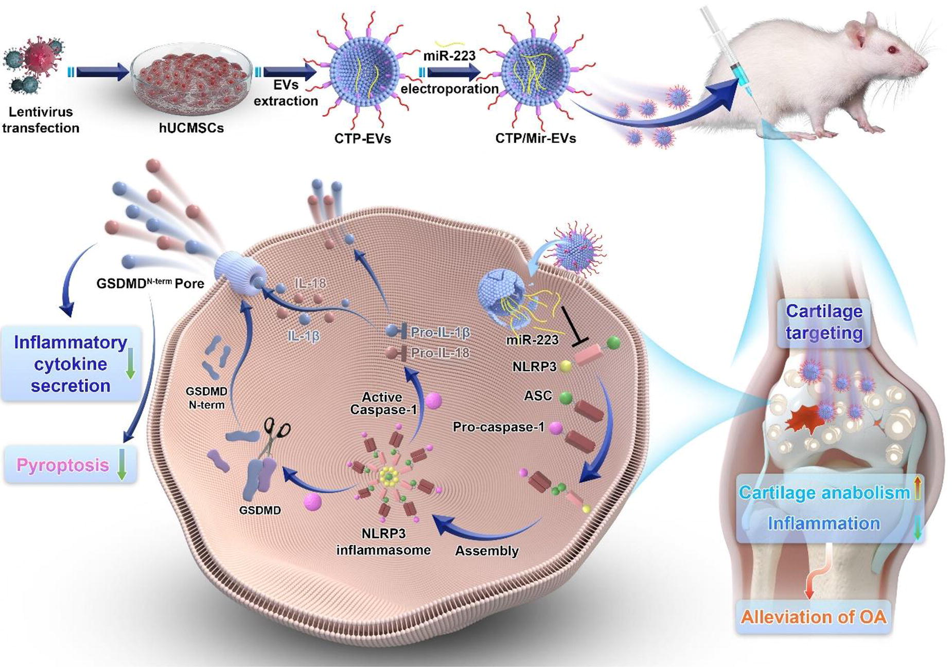
C175 - Layer-by-layer Mimicry Of Articular Cartilage
R. Cordeiro1,2,3, S. B. Gonçalves4, P. R. Fernandes4, N. Alves1,5,6, A. C. Maurício2,7,8, C. Moura1,3,9
1
Polytechnic of Leiria, Centre for Rapid and Sustainable Product Development, Marinha Grande, Portugal,
2
Veterinary Clinics Department, Abel Salazar Biomedical Sciences Institute (ICBAS), University of Porto (UP), Porto, Portugal,
3
Applied Research Institute, Polytechnic Institute of Coimbra, Coimbra, Portugal,
4
IDMEC, Instituto Superior Técnico, Universidade de Lisboa, Lisboa, Portugal,
5
Department of Mechanical Engineering, School of Technology and Management, Polytechnic of Leiria, Leiria, Portugal,
6
Associate Laboratory for Advanced Production and Intelligent Systems (ARISE), Porto, Portugal,
7
Animal Science Studies Centre (CECA), Agroenvironment, Technologies and Sciences Institute (ICETA), University of Porto (UP), Porto, Portugal,
8
Associate Laboratory for Animal and Veterinary Science (AL4AnimalS), Lisboa, Portugal,
9
Research Centre for Natural Resources Environment and Society (CERNAS), Polytechnic Institute of Coimbra, Coimbra, Portugal
*Purpose/Objectives: Musculoskeletal diseases, such as osteoarthritis, cause total loss of articular cartilage (AC), and this loss is aggravated by AC’s intrinsic incapacity for self-repair. Mimicking the complexity of this native tissue, recreating both, fibre alignment and extracellular matrix composition, is a great challenge. This challenging project aims to develop an AC substitute with region-specific customisation. A hybrid scaffold will be manufactured layer-by-layer, mimicking each of the AC zones: superficial zone (SZone), middle zone (MZone), deep zone (DZone) and calcified zone (CZone).
*Methodology: This study focuses on the development of the SZone, DZone and CZone. The SZone was manufactured through FDM 3D printing with poly(lactic-co-glycolic acid) (PLGA) (85:15), a medical-certified polymer. To mimic this zone, structurally parallel to the surface and with 90% porosity, a mismatched orthogonal scaffold with 0.9 mm height, (15% of AC), was considered. To mimic the DZone, a columnar alignment with 80% porosity was recreated. DZone was also 3D printed with PLGA by using a custom-made GCode written to manufacture a scaffold with columnar arrangement. The resulting structure is 1.8 mm in height (30% of AC). For CZone, a PLGA-hydroxyapatite (HAp) composite was hypothesised, which will result in a structure with CZone native porosity (around 70%). Incorporating HAp, a naturally present bioceramic in CZone, is intended to promote mineralised matrix production. Composites with PLGA (10% (w/v)) and 10% and 20% HAp (w/w) were tested to achieve better binding percentages through FTIR and TGA.
*Results: So far, a structure with 86.97% porosity has been found for the SZone. Pore size optimisations will still be required to achieve the desired porosity. Concerning DZone, micro-CT analysis revealed that a columnar structure with 78.43 ± 0.20% porosity was obtained. Mechanical properties at compression were performed in comparison to an orthogonal one with the same porosity (78.42 ± 0.35%). Results suggest very similar Young’s moduli; 23.61 ± 0.46 MPa for orthogonal and 22.45 ± 0.19 MPa for columnar structure, and a higher maximum elastic stress in the columnar scaffold (7.29 ± 0.36 MPa) than in the orthogonal one (6.54 ± 0.48 MPa). The Young Modulus values are higher than those reported in the literature for native DZone (6.44-7.55 MPa), thus guaranteeing that it will withstand the compressive forces exerted naturally. Biological tests should be carried out to confirm cell alignment. For CZone composite, FTIR and TGA proved that the two materials are bonded through the peak at 1170 cm-1 and that this bond represents 6.37% and 2.42% of the composite, for 10% and 20% HAp respectively. Further characterisation through SEM and NMR will be performed to detail each composite’s structure and bonding type. The most suitable composite will be extruded into a 3D printing filament to manufacture the CZone structure.
*Conclusion/Significance: Once all the AC zones have been manufactured, biological tests will have to be carried out to validate the fibre/cell structural organisation. However, the developed processes of layer-by-layer mimicry of AC have demonstrated the feasibility of producing 3D-printed structures that mimic the mechanical and functional properties of each native AC layer.
C176 - Hydrogel Tissue Sealants For Articular Cartilage Tissue Injuries
K. VERAL1, R. TUTAR2
1
Istanbul University-Cerrahpaşa, İstanbul, Turkey,
2
Istanbul University-Cerrahpaşa, Istanbul, Turkey
*Purpose/Objectives: Cartilage is a highly differentiated tissue with limited regenerative capabilities. Every year, 1,000,000 surgical interventions are performed in the United States alone for the repair of cartilage injuries1. Studies focusing on the repair of cartilage through the use of cells, scaffolds or signaling molecules, either individually or in combination, have become increasingly prevalent in clinical practice. In this study, it was aimed to prepare polyethylene glycol diacrylate (PEGDA)/ Methacrylate-modified alginate (AlgMA) tissue adhesives, which were applied in-situ to the wound area and showed dual-crosslinked gelling properties in the joint environment. In addition, their gelation status, physical, chemical and adhesion properties were examined for cartilage tissue injuries.
*Methodology: PEGDA and AlgMA were prepared by using chemical modification method to make them light active materials. After modification process, the pre-polymer solutions were prepared by selected different concentrations of AlgMA with combined with 10% (w/v) PEGDA. Visible light photoinitiators TEA, VC and Eosin Y disodium salt solutions were prepared and used to make hydrogels chemically crosslinked. In addition, physical crosslinking has facilitated by the ionic binding of bivalent cations such as Ca2+ to the carboxyl groups of glucuronic acid in the alginate chain using an aqueous CaCl2 solution.
*Results: Prepared chemically modified PEGDA and AlgMA were analysed succesfully based on structural by using spectroscopic methods like FTIR. Swelling ratios were increased with increasing AlgMA concentrations whereas degradation properties were decreased. In addition, in vitro adhesion properties were determined from 5kPa to 20kPa by adding different amounts of AlgMA to PEGDA.
*Conclusion/Significance: Biocompatible and injectable hydrogel tissue structures with adapted features for different types of cartilage defects have been developed and characterized. Physical, mechanical and adhesive properties were showed. These structures are based on hydrogel content, demonstrating biocompatibility, strong adhesion, and elasticity to meet the requirements of cartilage tissue engineering and precision medicine.
Keywords: PEGDA, AlgMA, Cartilage tissue engineering, Dual-crosslinked.
Acknowledgement: This study was financially supported by The Scientific and Technological Research Projects Funding, TUBITAK-2209-A/ 1919B012107516.
References: 1) Hangody L, Dobos J, Baló E, Pánics G, Hangody LR, Berkes I. Clinical experiences with autologous osteochondral mosaicplasty in an athletic population: a 17-year prospective multicenter study. Am J Sports Med 2010;38:1125-33.
C177 - The Molecular Regulation Of The Migration Of Dental Pulp Stem Cells-derived Chondrogenic Cells In Type I And Type Ii Collagen Hydrogels
L. Yao, N. Flynn, P. Kaphle
Wichita State University, Wichita, KS
*Purpose/Objectives: Human dental pulp stem cells (hDPSCs) are attractive stem cells for cartilage and nucleus pulposus (NP) generation. The cells are easily accessible and have demonstrated similar differentiation capability as other MSCs. The transplantation of hDPSCs-derived chondrogenic cells encapsulated in type II collagen hydrogels mimicking the native cartilage and NP tissue is potentially a novel approach to regenerating the degenerated cartilage and NP. In previous study, we reported the differentiation of hDPSCs toward chondrogenic cells and cell culture in the collagen hydrogels. The motility of transplanted cells is critical because the cells need to migrate in the hydrogels or native tissue and disperse from the grafted location. We reported the differentiated cell migration in the type I and type II collagen gels. In degenerated cartilage and disc, the expression of cytokines such as IL-1β and TNFα increased significantly compared with healthy cartilage and NP tissue. It is not clear how the chemokines affect the motility of hDPSCs-derived chondrogenic cells in the collagen hydrogels. In this study, we investigated effect of these factors on cell migration.
*Methodology: HDPSCs were differentiated into chondrogenic cells using chondrogenic medium including high-glucose DMEM supplemented with TGF-β1, ascorbate-2-phosphate, dexamethasone, sodium pyruvate, proline, and ITS premix. The hDPSCs and stem cell-derived chondrogenic cells were grown in type I and type II collagen hydrogels for 2 weeks. The migration of hDPSCs and chondrogenic cells in the hydrogel was recorded after they were cultured for 2 days or 2 weeks. The viability of stem cell-derived chondrogenic cells at day 2 and 2 weeks in the gels was tested using Alamarblue assay. The cells were treated with IL-1β and TNFα in the cell culture. The cells migration was recorded with time-lapse microscopy.
*Results: The migration velocity and distance of hDPSCs and hDPSCs-derived chondrogenic cells showed that the migration velocity of both cell types was lower in type II collagen compared with type I collagen. The migration velocity was lower after culturing in the gel for 2 weeks in comparison with the cells cultured for 2 days. We observed the cell proliferation in the collagen gels after culturing for 2 weeks with Alamarblue assay. We show the negative effect of IL-1β and TNFα on differentiated cell migration in the hydrogels.
*Conclusion/Significance: The study demonstrated the impact of chemokines on HDPSCs-derived chondrogenic cell motility in collagen hydrogels and indicated the potential application of the cells in cartilaginous tissue regeneration.
C178 - Semiconductor Embedded Fabric Improves Chondrogenic Differentiation
C. Duranti1, G. Bagni1, J. Iorio1, R. Colasurdo1, V. Devescovi1, A. Arcangeli1, G. Myers2
1
University of Florence, Florence, Italy,
2
Incrediwear Holdings, Inc., Chico, CA
*Purpose/Objectives: Osteoarthritis (OA) is a clinical state which is identified by the degeneration of articular cartilage. OA is a common condition (>500 million people affected worldwide), whose frequency is anticipated to continue to rise. The optimal treatment for OA is based on a combination of therapeutic approaches, including regenerative medicine with Adipose Derived Stem Cells (ADSCs). Germanium embedded sleeves (Incrediwear® Functional Cred40 fabric) have been shown to have positive effects on OA and are envisaged to give encouraging effects also on tissue reformation. Still, the biological mechanisms underlying this therapeutic modality have not been defined so far. We tested the hypothesis that Germanium embedded sleeves could enhance chondrogenesis through emission of infrared (IR) waves and negative air ions (NAIs).
*Methodology: We applied Incrediwear® germanium embedded sleeves to human adipose-derived stem cells (hADSCs) induced to chondrogenic differentiation in vitro. Chondrogenic markers (ACAN, SOX9, RUNX2, COL2A1, COL10A1) were quantified following 21 days of treatment.
*Results: We here provide pilot demonstration that Germanium embedded sleeves (Incrediwear®) are able to induce cartilage regeneration in vitro, inducing hADSCs chondrogenic differentiation and maturity.
*Conclusion/Significance: Our results indicate that treatment of hADSCs with Incrediwear® germanium embedded sleeves induces the modulation of some of the main genetic markers of chondrogenesis (specifically inducing healthy cartilage markers up-regulation and pathogenic cartilage markers down-regulation), as well as modifications in cell morphology indicative of chondrogenic differentiation and cartilage regeneration.
C179 - Influence Of Different Molecular Weights Of Recombinant Hyaluronic Acid In Self-healing Hydrogels On Collagen Deposition For Cartilage Tissue Engineering
M. Sundaram, C. Balavigneswaran, G. Jayaraman, V. Muthuvijayan
Indian Institute of Technology Madras, Chennai, India
*Purpose/Objectives: Injectable hydrogels with self-healing properties have emerged as a promising approach for cartilage tissue engineering, surpassing conventional brittle hydrogels. These hydrogels possess the ability to autonomously repair themselves upon damage, without relying on external stimuli.
*Methodology: In this study, we have developed self-healing hydrogels using oxidized alginate and gelatin, reinforced with different molecular weights (0.5 MDa, 1 MDa, and 2 MDa) of recombinant hyaluronic acid (HA) derived from the metabolically engineered Lactococcus lactis. The evaluation encompassed the hydrogel’s self-healing capacity, mechanical strength, and shear-thinning properties.
*Results: Our results demonstrated that the reinforcement of HA, enhanced thehydrogel's self-healing ability while maintaining suitable mechanical strength for cartilage tissue engineering. Notably, the formulation having 1 MDa HA (OAGH 1 ), exhibited better injectability and shear-thinning properties than the other HA-reinforced hydrogels. Furthermore, HA- reinforced hydrogel did not show any cytotoxicity against goat knee chondrocytes that revealed the biocompatibility of the hydrogels. Interestingly, the presence of HA influenced cell infiltration into the hydrogels, resulting in the formation of multiple layers of migrated and aggregated cells. Moreover, HA played a significant role in influencing collagen deposition by chondrocytes.
*Conclusion/Significance: Considering the combined potential of promoting cell migration and collagen deposition observed in vitro, recombinant HA-reinforced hydrogels can be successfully used for cartilage regeneration.
C181 - Silylated Biopolymer-based Hydrogels For Osteochondral Repair
L. Roberts1, C. Echalier1, E. Perrier-Groult2, D. Nöel2, S. Pellegrino3, G. Subra1
1
University of Montpellier, CNRS, ENSCM, Montpellier, France,
2
University of Montpellier, INSERM, CHU Montpellier, Montpellier, France,
3
University of Milan, Milan, Italy
*Purpose/Objectives: Osteochondral repair remains a major clinical challenge due to the complex architecture of the osteochondral tissue and the limited intrinsic regenerative properties of articular cartilage. In a tissue engineering approach, scaffolds are an important component as they provide structural support for cells and guidance for tissue development. Unfortunately, there is a lack of suitable biomaterials that can mimic both bone and cartilage properties to act as scaffolds for osteochondral repair. To address this gap our research has focused on the development of silylated biopolymer-based hydrogels.
*Methodology: Natural biopolymers, such as hyaluronic acid and gelatin1, have been functionalized with alkoxysilane groups and covalently cross-linked under biocompatible conditions using the sol-gel process2 to produce hybrid hydrogels with tailormade mechanical properties3.
*Results: These hybrid hydrogels can be formed in the presence of living cells and further functionalized with bioactive peptides4,5 to direct mesenchymal stem cell differentiation towards the chondrogenic and osteogenic lineages. Additionally, cell-laden silylated biopolymer-based hydrogel solutions can be extruded with a 3D bioprinter6,7 to recapitulate the spatial complexity of native osteochondral tissue via precise cellular deposition.
*Conclusion/Significance: Silylated biopolymer-based hydrogels are promising biomaterials for osteochondral regeneration as they can be tailored to mimic the properties of both cartilage and bone while promoting directed cell differentiation. This could lead to improvements in osteochondral defect repair and may also prove useful as an in vitro model for studying the regeneration mechanisms of articular cartilage.
C182 - Neuroimmune Axis As A Potential Therapeutic Target Of Dioscorea Opposita Thunb-derived HKUOT-S2 Protein For Accelerated Bone Regeneration
A. S. Brah, J. A. Kubi, K. M. Cheung, K. W. Yeung
Orthopaedics and Traumatology, The University of Hong Kong, Hong Kong, HONG KONG
*Purpose/Objectives: Treatment of delayed bone defects remains a global challenge to clinicians due to the increasing rate of bone non-unions. The neuroimmune axis has been shown to play a vital role in modulating the cellular and molecular mechanisms between the nervous and the immune systems to enhance bone regeneration and defect repairs. For instance, the sensory nerves release neuropeptides such as calcitonin gene-related peptide (CGRP) and prostaglandin E2 (PGE2) which induce the immune cells to trigger inflammatory responses necessary for bone defect healing. The neuroimmune axis also promotes the activities of mesenchymal stem cells (MSCs), osteoblasts, and osteoclasts to facilitate bone regeneration. Consequently, neuroimmune axis modulation has recently emerged as one of the potential therapeutic targets for accelerating delayed bone defect repairs. The development of bioactive compounds that can induce appropriate neuro-immunogenic stimuli to accelerate bone regeneration is therefore urgently needed for the treatment of delayed bone fractures. In this study, we provide intriguing findings from a newly isolated unique low-molecular-weight “HKUOT-S2” protein (32kDa) derived from Dioscorea opposita Thunb that possesses both osteogenic and neuroimmunomodulatory properties to facilitate bone defect healing.
*Methodology: HKUOT-S2 protein (32 kDa), a low molecular weight protein was successfully isolated and characterized from the protein fraction of Dioscorea opposita Thunb tubers extract. In this study, a mice bone defect model was used to assess the HKUOT-S2 protein-induced neuro-immunomodulatory bone regeneration potentials by direct subcutaneous (s.c) injection of 2.18mg/Kg HKUOT-S2 protein at the defect site three times per week for 4 weeks.
*Results: The HKUOT-S2 protein treatment enhanced the bone defect healing within 4 weeks by increasing BMD, Tb.th, Tb.N, and BV/TV compared to the sham control group. In addition, HKUOT-S2 treatment increased the expression of OCN, ALP, and RUNX2 and bone mineralization. Also, HKUOT-S2 treatment promoted M2 macrophage polarization in vitro and in vivo by increasing the expression of M2 macrophage-related genes (Arg-1, Cd206, and Mgl-1). Interestingly, the HKUOT-S2 treatment also increased the expression of neuronal markers CGRP and PGP9.5 at the bone defect sites.
*Conclusion/Significance: In conclusion, the HKUOT-S2 protein can be applied as a potent osteogenic and neuro-immunomodulatory therapeutic agent for accelerating bone regeneration and bone defect healing.
C183 - HKUOT-S2 Protein-derived Novel Peptide Suppresses Osteoporosis Through Estrogenic Modulation Of Neuro-osteogenic Axis
J. A. Kubi, A. S. Brah, K. M. Cheung, K. W. Yeung
The University of Hong Kong, Hong Kong, Hong Kong
*Purpose/Objectives: Estrogen deficiency-induced osteoporosis, predominant among the aged population, is marked by decreased bone mass, deteriorated bone architecture, and increased bone fragility. Fractures associated with osteoporosis put a huge global economic burden on health service providers, and patients due to high treatment cost. Anti-resorptive (bisphosphonates, and raloxifene) and anabolic (teriparatide) agents have been used extensively for treating osteoporosis. However, various clinical side effects such as blood clotting, increased risk of cancer development and jawbone osteonecrosis have been associated with the current anti-osteoporotic drugs including the FDA-approved raloxifene. Studies have shown that estrogenic stimuli play unique roles in regulating neuronal activities and signaling pathways to maintain bone homeostasis. The development of affordable, biocompatible bioactive agents that can harness estrogen-associated neuromodulation to prevent osteoporosis could be a breakthrough in this field. Here, we demonstrated that a novel peptide from Dioscorea opposita Thunb-derived HKUOT-S2 protein can suppress osteoporosis in ovariectomized (OVX)-mice by upregulating estrogen receptor GPR30 expression in bone-resident sensory nerves to facilitate neuro-osteogenic-mediated bone formation.
*Methodology: The HKUOT-S2 protein-derived novel peptide (1.046 kDa) was successfully synthesized. In the current study, an OVX-induced osteoporotic mice model was used to assess the osteoporosis suppression potentials of the HKUOT-S2 protein-derived peptide by targeting estrogen receptor-associated neuro-osteogenic. The experimental groups were sham control (non-OVX, PBS), OVX control (PBS), 30 µg/Kg Estradiol (E2) and 0.5 mg/Kg HKUOT-S2 protein-derived peptide. All mice were subjected to intraperitoneal injections three times per week for 4 weeks. Micro-computed (µCT) scans were done bi-weekly to assess bone growth parameters.
*Results: The HKUOT-S2 protein-derived peptide treatment suppressed osteoporosis progression by improving bone mineral density (BMD), trabecular thickness (Tb.th), and trabecular number (Tb.N) when compared to the OVX control group. The HKUOT-S2 protein-derived peptide also significantly increased skeletal expressions of osteogenic markers OCN, ALP, and RUNX2. Intriguingly, the HKUOT-S2 protein-derived peptide treatment also upregulated the co-localized expression of the GPR30 estrogen receptor and MAP2 neuronal marker on the periosteal surface of the bone to suppress osteoporosis progression.
*Conclusion/Significance: The HKUOT-S2 protein-derived peptide can upregulate the neuronal estrogen receptor GPR30 coupled with increased neuronal functions in periosteum to trigger anti-osteoporosis activity in OVX mice. The HKUOT-S2 protein-derived novel peptide can be clinically applied as estrogenic anti-osteoporotic peptide for treating osteoporosis.
Keywords: HKUOT-S2 protein-derived peptide, GPR30, neuro-osteogenic, osteoporosis.
C187 - Genomic Characterization Of Human Meniscus Tissues Reveals Distinct Clusters Of Matrix-producing Cell Subpopulations
S. P. Simmonds, J. C. Bradford, W. Tang, C. J. Watson, R. Y. Kwon, J. L. Robinson
University of Washington, Seattle, WA
*Purpose/Objectives: Meniscus injuries, the most common intra-articular knee injury, can result in severe pain and disability. Current treatments for these matrix-abundant tissues, such as arthroscopic repairs or meniscectomies, often result in post-traumatic injuries throughout surrounding tissues and increased risk of osteoarthritis. Tissue regeneration-based repair is an ideal route to treating injuries and maintaining the function of these vital connective tissues. Critical to this goal is defining functional cell subpopulations in the meniscus, as current consensus identifies meniscal fibrochondrocytes as col1/col2 co-expressing cells. Specifically, there is an urgent need to identify matrix-producing cells, analyze their potential for repairing injured tissue, and define their corresponding biomarkers’ biological functions.
To address this gap, we performed single-cell RNA sequencing (scRNAseq) on clinically relevant human meniscus tissues. Preliminary analysis of this data has revealed differential expression of genes related to matrix production and organization within distinct cell subpopulations. Several of these key biomarkers are not yet well-characterized in the meniscus and other human connective tissues. Concurrent scRNAseq analysis of zebrafish, a traceable and high-throughput genetic model system, has revealed expression of several key meniscus biomarkers in related matrix-producing tissues, such as osteoblasts and the notochord.
In this work, we:
1) utilize scRNAseq data from a robust set of meniscus tissues to determine globally conserved cell subpopulations; and
2) validate genetic expression of key biomarkers of matrix-producing meniscus subpopulations in orthologous zebrafish tissues and cultured human meniscus cells.
*Methodology: Tissues used for scRNAseq (Children’s Mercy Hospital, IRB Exemption #STUDY00000746) include control meniscus samples (n=2 male, n=2 female) acquired during partial meniscectomies and discoid meniscus samples (n=3 male, n=2 female) obtained following saucerization. NovaSeq analysis was performed at 50,000 reads per cell.
*Results: Data analysis with Seurat generated a UMAP of novel meniscus cell subpopulations whose identities have been preliminarily described (Fig 1a). Analysis of distinct cluster biomarkers shows that most cells pertain to a cluster which differentially expresses either col1 or col2. Several cluster biomarkers have been previously identified in meniscal fibrochondrocytes.
We found that several key meniscus biomarkers, including col1 and col2, are differentially expressed within zebrafish fin osteoblasts (Fig 1b) and somite tissues (Fig 1c), which were similarly analyzed.
Studies are underway to further validate the expression of previously uncharacterized biomarkers from matrix-producing subpopulations within wild-type zebrafish and cultured human meniscus cells using RNA fluorescence in situ hybridization.
*Conclusion/Significance: Our studies present a novel characterization of human meniscal fibrochondrocytes, notably identifying distinct cell subpopulations which differentially express either col1, representing fibroblast-like cells, or col2, representing chondrocyte-like cells. Identification of these and other key biomarkers within relevant zebrafish tissues and cultured human meniscus cells will support our goal of targeting matrix-producing cell subpopulations to regenerate injured meniscus tissue. Future in vivo studies will elucidate the developmental functions of previously uncharacterized genes and how they might contribute to other connective tissue environments in mutant fish populations.
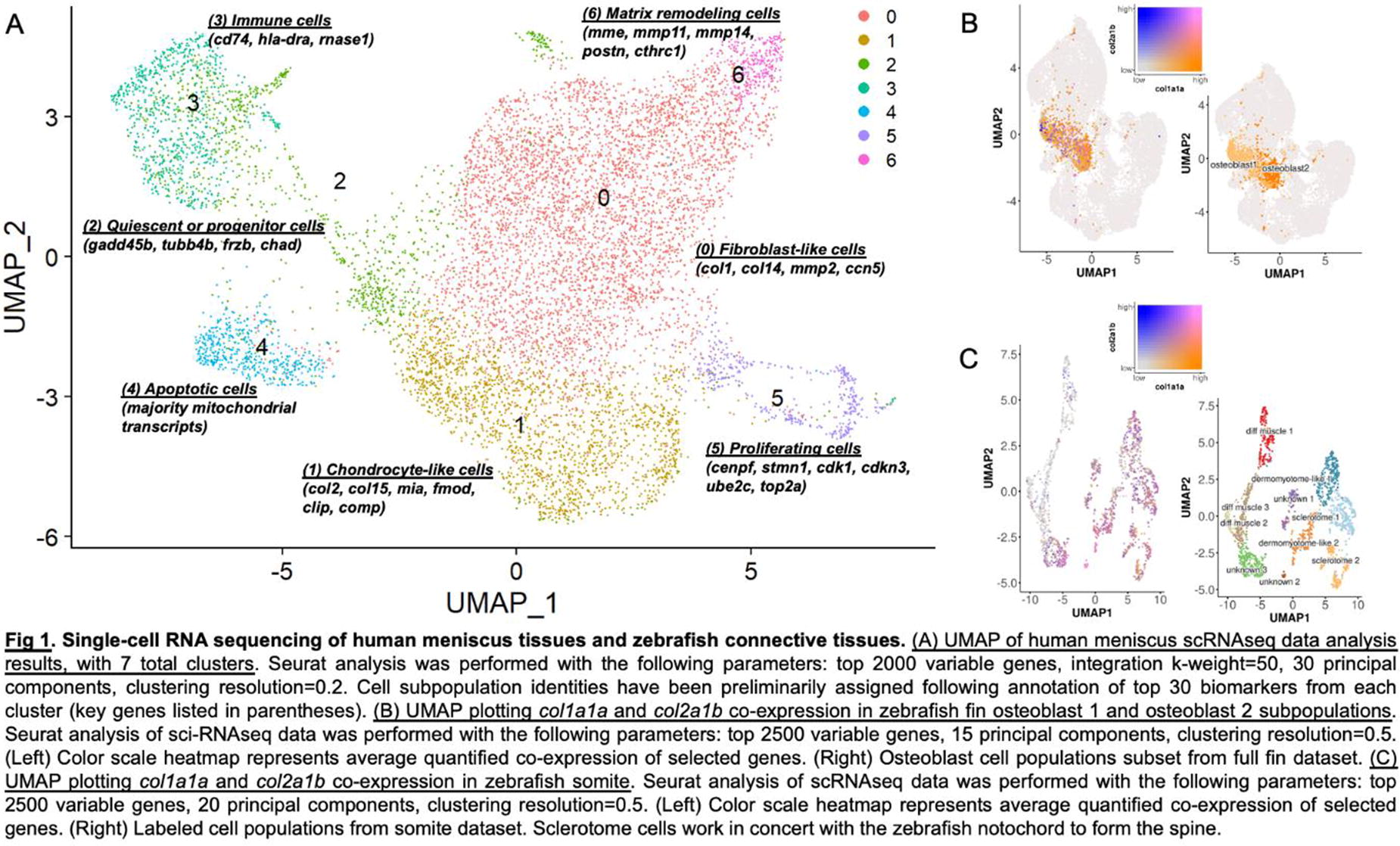
C188 - Elucidating The Regenerative Healing Capacity Of Fetal Tendons
C. D. Hart, P. Nguyen, C. K. Kuo
University of Maryland, College Park, MD
*Purpose/Objectives: Adult tendons heal as fibrotic scars with aberrant extracellular matrix and mechanical properties. Sheep fetal tendons heal scarlessly, although by unknown mechanisms. Our long-term goal is to elucidate mechanisms of fetal tendon healing to inform adult tendon regeneration strategies. We have established the chick embryo as a model to study fetal tendon development and healing. The chick embryo tendon shares significant molecular overlap with mammalian tendon biologically and functionally, develops in 21 days, and is directly accessible during development. Here, we characterized younger (YT) and older fetal tendon (OT) healing capacities using the chick embryo model, and hypothesized that YT heal more regeneratively than OT.
*Methodology: Fertilized White Leghorn chicken eggs were cultured until embryonic day (D)13-14 (younger) or D16-17 (older) for explant and in ovo studies. Embryos were sacrificed and legs harvested for explant culture. The Achilles tendon of one leg was injured. The contralateral leg was uninjured control. Legs were cultured and tendons harvested after 12h for RT-PCR, and 0 and 5 days for tensile testing. For in ovo studies, a window was cut in the eggshell and the Achilles tendon of one leg was injured through the window. After covering the window, the embryo was incubated to develop in ovo until sacrifice and tendon harvest at 12h and 24h post-injury for metabolite and protein content analysis, and on D20 for histology and histomorphometry. Tendons were injured via surgical transection in the center 25% of the midsection. N=3-6 biological replicates were needed per assay. Student’s t-tests and 2-way ANOVA with Sidak’s multiple comparisons were used to determine statistical significance.
*Results: Elastic modulus and peak stress of YT were similar to uninjured controls, but OT mechanical properties remained statistically significantly lower than controls, suggesting YT have greater healing capacity (Fig. 1a). Cell and collagen fiber density and alignment after injury appeared similar as uninjured controls in YT, but were lower density and less aligned than controls in OT (Fig. 1b). YT significantly upregulated mRNA levels of tendon markers, collagens, collagen crosslinking regulators, and pro-inflammatory mediators, whereas OT exhibited no changes in the same markers (Fig. 1c). YT and OT both had increased protein, amino acid, and TCA organic acid levels, but only YT exhibited statistically significant increases in protein and essential and non-essential amino acid levels during healing over time.
*Conclusion/Significance: Our exciting data compellingly suggest that scarless and regenerative tendon healing occur in the chick embryo, based on histological and mechanical characterizations. YT appeared similar to normal tendons after healing in this study, whereas injured adult tendons are unable to restore normal collagen fiber architecture and mechanical properties. The unique gene expression, protein, and metabolite responses by YT compared to by OT may reflect greater regenerative healing responses by YT, although this requires further investigation. Building on our exciting data, in ovo perturbation studies are underway to investigate specific mechanisms of scarless and regenerative tendon healing by YT. We anticipate these studies will lead to the development of novel therapeutics to promote regenerative adult tendon healing.
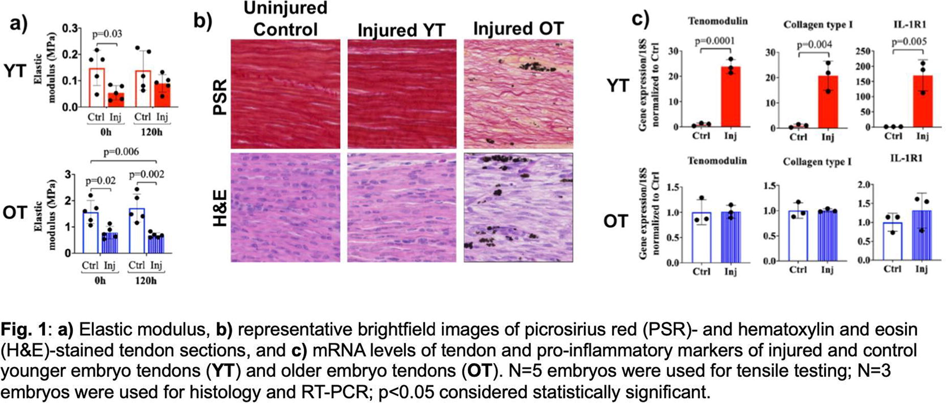
C189 - Engineering Large Anisotropic Meniscal Microtissues Via Digital Light Processing Printed Molds
A. DeFoe1, A. Dhand2, M. Obenreder1, J. A. Burdick1
1
University of Colorado Boulder, Boulder, CO,
2
University of Pennsylvania, Philadelphia, PA
*Purpose/Objectives: The menisci are highly aligned fibrocartilaginous tissues that provide load distribution, joint lubrication, and shock absorption in the knee. Meniscal injuries are common and may lead to pain and early onset of osteoarthritis if left untreated. Although meniscectomies are often used for treatment, meniscal tissue removal alters joint loading and increases contact stresses, resulting in biomechanical consequences. This motivates the development of new therapies to replace damaged meniscal tissue. Here, we leverage advances in 3D printing to fabricate large meniscal microtissue constructs that recapitulate the highly aligned structure of healthy meniscal tissue for treatment of meniscal defects.
*Methodology: To accomplish this, digital light processing (DLP) was used to fabricate molds with posts spaced at a 4:1 aspect ratio and varied dimensions (e.g., rectangular versus curved) using a poly(ethylene glycol) resin. Suspensions of meniscal fibrochondrocytes (MFCs) in FITC-labeled collagen were added to the molds and cultured for up two weeks (figure A(i)). Fluorescent images were acquired to quantify construct width via ImageJ (figure A(ii)). Selected constructs were fixed and stained for nuclei (DAPI) and actin (phalloidin), and representative Z-stacks were acquired of the construct centers and analyzed for nuclei/actin alignment using the Directionality plugin in ImageJ. In select formulations, contractility inhibitors (blebbistatin, Y-27632) were added to limit construct necking.
*Results: Using adapted protocols to form microtissues, we fabricated aligned fibrous microtissues that are larger than those previously reported. Specifically, constructs (1.6 mg/ml collagen, 0.50*106 MFCs/ml) cultured in rectangular molds of various lengths (6, 12, 24 mm) underwent visible contraction and reorganization, with ‘necking’ observed by day 7 in the largest molds (figure B(i)). Significant necking is undesirable as it yields constructs that are too thin for implantation towards repair of meniscal defects. Several approaches were investigated to mitigate necking. First, suspension formulation was tuned; higher collagen density (2.0 mg/ml collagen) and lower cell density (0.25*106 MFCs/ml) significantly decreased the magnitude of contraction in early days, but neither formulation completely prevented necking during the first 7 days (results not shown). Next, curvature was incorporated into 24 mm molds to increase initial construct volume while maintaining post spacing. The resulting constructs had decreased contraction during the first 7 days relative to constructs cultured in rectangular molds (figure B(ii)) and nuclei and actin alignment increased with culture throughout 7 days (figure C). To further limit necking, blebbistatin (myosin inhibitor, 50 ) and Y-27632 (Rho-associated kinase inhibitor, 50 ) were added to media and significantly decreased construct contraction (results not shown) during the first week of culture.
*Conclusion/Significance: Large anisotropic microtissues were fabricated with MFCs and collagen gels within DLP-printed molds. Although we encountered undesired necking using this platform, changes in mold dimensions and the addition of contractility inhibitors reduced this behavior. We have selected a 24 mm curved mold and early culture with contractility inhibitors to stabilize construct dimensions. Ongoing work is to culture the constructs for longer periods (e.g., 8 weeks) in chondrogenic media and assess extracellular matrix deposition, construct mechanical properties, and integration with meniscal tissue.
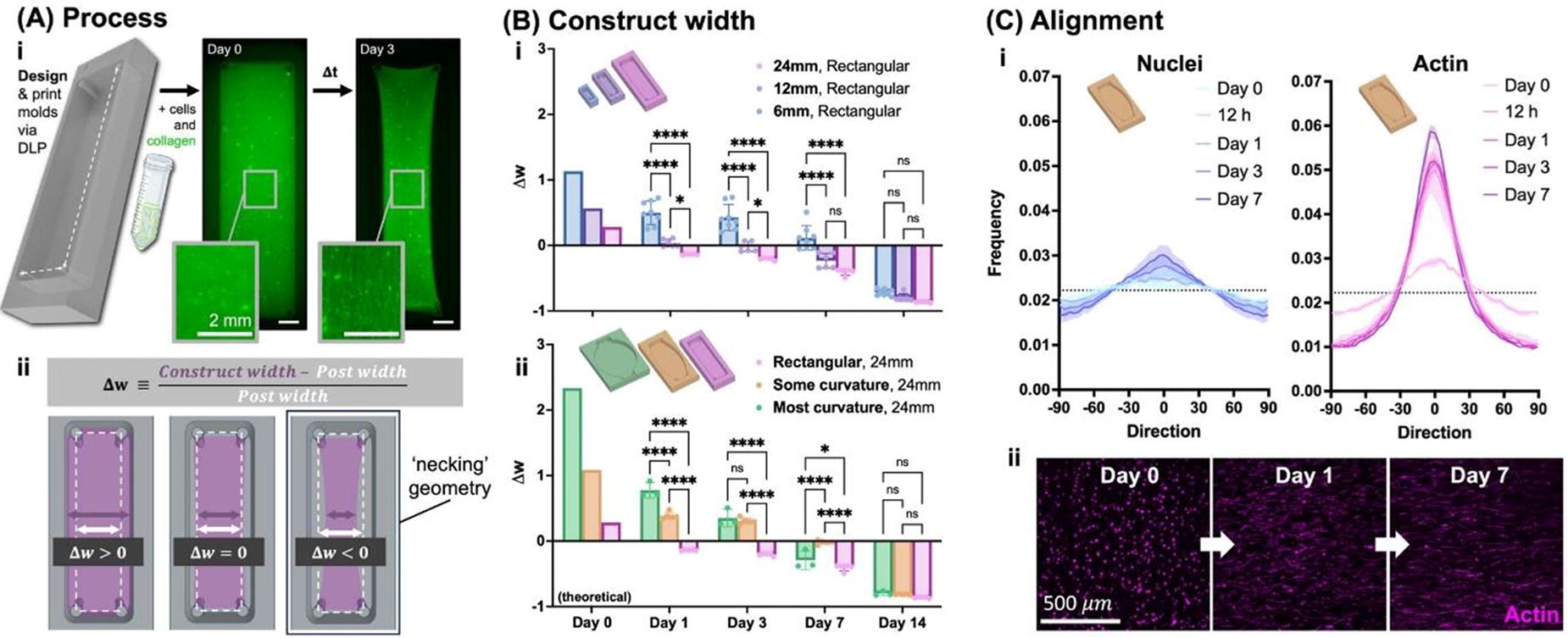
C190 - Developing An In Vitro Meniscus Injury Model Using Modified Electrospun Fibrous Scaffolds
K. Meinhold1, R. Darlington1, K. Knewtson2, J. Robinson1
1
University of Washington, Seattle, WA,
2
University of Kansas, Lawrence, KS
*Purpose/Objectives: In fibrous connective tissues like the meniscus or intervertebral disc (IVD), there is complex interplay between mechanical and chemical signaling which work together to keep the tissue healthy and aid recovery from injuries. Prior experiments in IVD tissue have shown a loss of cells’ ability to sense strain post-injury resulting in increased apoptosis and development of fibrotic phenotype. Tears to the meniscus modify both the chemical signaling and mechanical signaling of native cells by disrupting inherent tissue organization and is particularly impactful in this tissue because of its multiple levels of organization. The similarities in tissue types indicate meniscal fibrochondrocytes (MFCs) may react to injuries similarly. In vitro systems offer the ability to recapitulate these studies in controlled environments and ask specific questions about singular system modifications without confounding factors present in explant or animal models.
The objective of this study is to establish an in vitro tear model to examine the impact of applied strain on MFC morphology and fibrotic phenotype.
*Methodology: Sample Collection: Solutions of 20% w/v polycaprolactone (PCL) in chloroform (CHCl3) were spun at consistent collection conditions. Aligned mesh were collected using a 2” diameter rotating mandrel at high speed. Fiber Diameter: Mesh imaged with Phenom Pro Desktop scanning electron microscope: 1000x, 10kV accelerating voltage, backscatter detector, and micrographs analyzed with DiameterJ. Cell Morphology and Focal Adhesions: MFCs (male and female, n=2, ages 17, 18, 14, and 13, respectovely lateral meniscus) were cultured on aligned and unaligned mesh at 10,000 cells/mesh for 3 days before fixing and staining with DAPI, Paxillin, and Phalloidin (actin). Injury Model Cell Morphology: MFCs were cultured on unaligned mesh at 120,000 cells/mesh and incubated for 24 hours before testing. 10% strain was applied to mesh with and without mock injury for 1 hour using MechanoCulture J1. Samples were fixed and stained with DAPI, Paxillin, and Phalloidin or DAPI, Phalloidin, caspase-3, and αSMA.
*Results: As shown in Figure 1 A. i-iii), quantification of SEM micrographs, showed decreased fiber diameter within 3μm range and increased fiber fraction in aligned scaffolds and preliminary imaging of MFCs demonstrated increased alignment on aligned scaffolds. As shown in Figure 1 B. v) there were trends of decreased spread in strained vs. unstrained samples and injured vs. un-injured samples. Representative fluorescent images of these groups are in Figure 1 B. i-iv) and both applied strain and induced injury appear to modify paxillin signaling implying changes to the focal adhesion complex.
*Conclusion/Significance: In randomly aligned scaffolds there appear to be no significant changes to MFC morphology on application of low-level tensile strain with or without an induced injury condition. However, both spread and focal adhesion signaling is changed by introduction of strain or injury and it is expected that these trends will be more significant in aligned fibers.
This experiment will provide insight to how strain post-injury impacts apoptotic signaling and development of fibrotic phenotype in primary MFCs in aligned and unaligned scaffolds with modified mechanical characteristics and structures
.
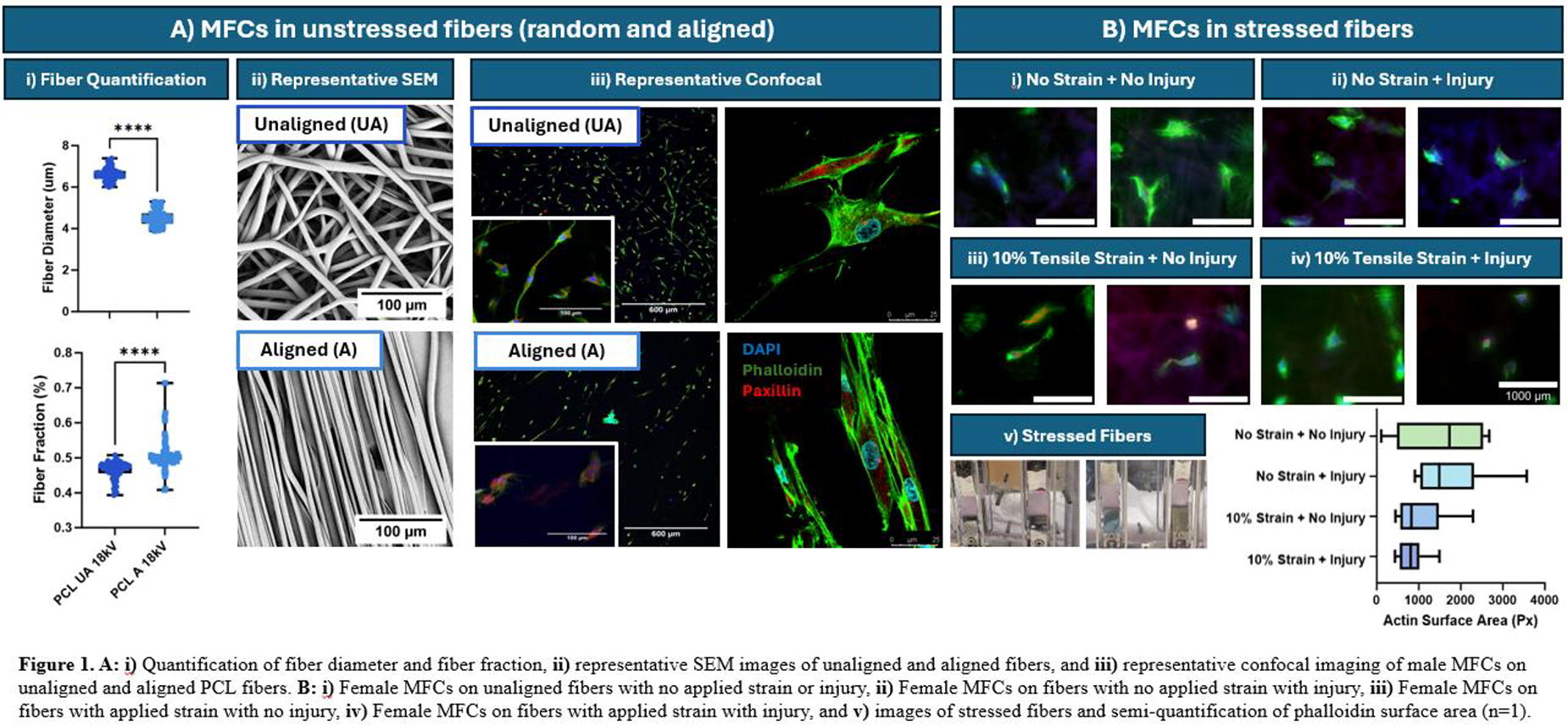
C191 - Formation Of Tenon-like Microtissues
F. Giacomini, A. Vitorino Pereira, D. Barata, Z. Tahmasebi Birgani, R. Truckenmuller
Maastricht University, Maastricht, Netherlands
*Purpose/Objectives: Tendon injuries are common among both older, less active individuals and younger, more physically engaged ones. However, tendons possess a limited self-healing capacity, often leading to the formation of scar tissue and reduced mechanical strength upon injury. Tendon tissue engineering aims to restore the natural functions of these tissues by using a combination of cells and materials. Among these, topographical cues and mechanical stimulation show promise in enhancing tendon cells phenotype and function. This study, aims to understand how microgrooved surfaces and mechanical stimulation impact tendon microtissues derived from induced pluripotent stem cells (iPSCs), aiming to create an environment for studying tendon regeneration.
*Methodology: At first, we derived tenocytes from human iPSCs by adapting a previous published protocol which recapitulates the normal progression of step-wise fate decisions in vertebrate embryos. Then, we fabricated microgrooved surfaces with different sizes of grooves and ridges onto poly(dimethylsiloxane). Tendon-derived iPSCs embedded in a 3D matrix of collagen type I were cultured on the substrates and subjected to uniaxial elongation at either physiological load or overload conditions. This regime was applied for 6h a day at 0.1 Hz for 7 days.
*Results: After 21 days of differentiation tenocytes derived from iPSCs expressed representative tenocytes markers and specific extracellular matrix-related genes, such as scleraxis, collagen type 1a2, collagen type 3a1 and decorin. The cells cultured on PDMS substrates with 3 µm grooves / 1 µm ridges surfaces showed a more pronounced alignment compared to the other conditions, measured as an increase in form factor and eccentricity. In such condition, we also measured the of expression of scleraxis. After embedding in a 3D matrix of collagen type I and applying mechanical stress, the embedded cells created cell assemblies and exhibited an aligned extracellular matrix structure.
*Conclusion/Significance: . In conclusion, we successfully differentiate iPSCs to recapitulate human tenocytes and we demonstrated that the combination of aligned microgrooved topographies and mechanical loading reinforces tissue maturation . This study sets the groundwork for investigating potential approached to improve tendon regeneration.
C192 - In Vivo Biocompatibility And Tissue Response Of Poly(glycerol Sebacate Urethane) Scaffolds Intended For Tendon Repair
A. Samourides1, Z. Xenou1, D. Kokkinidou1, K. Spanoudes2, K. Kapnisis1, A. Anayiotos1
1
Cyprus University of Technology, Limassol, Cyprus,
2
University of Nicosia, Nicosia, Cyprus
*Purpose/Objectives: Soft tissue engineering (TE) is a multidisciplinary field with the aim to regenerate or replace a dysfunctional or damaged tissue. Usually, a three-dimensional porous scaffold is produced to provide a temporary structure and mechanical support while the tissue grows and subsequently replaces it. For this reason the scaffold should be highly tunable, and exhibit properties such as interconnected porous structure, be biocompatible, biodegradable and ideally to degrade linearly at the same rate as the tissue develops. Poly(glycerol sebacate urethane) (PGSU) was recently developed and fabricated as a porous scaffold, and it was found to be angiogenic and promote tissue ingrowth in vitro, demonstrating high potential in the field of soft TE. In this study, three PGSU were investigated for in vivo for their microstructure, biocompatibility and their ability to degrade at the same rate as tissue develops into the scaffold.
*Methodology: PGSU scaffolds were fabricated with hexamethylene diisocyanate (HDI) ratios of 0.8 and 1.0 and polymer concentrations (w/v%) equal to 10% and 15%. In vivo biocompatibility of the scaffold was investigated by implanting scaffolds subcutaneously in CD1 albino mice for six weeks. Specifically, 12 mice were implanted with four scaffolds of each sample group and the rest of them with Ethilon® Nylon suture 4-0 spheres which acted as the positive control. All PGSU scaffolds were weighed and measured to calculate their degradation and density. The scaffolds were also characterized for their microstructure using scanning electron microscopy (SEM) (SEM Quanta 200, FEI, United States). Finally, to assess any inflammatory response, whole body imaging (WBI) was used after matrix metalloproteinase (MMP) administration which act as a marker of inflammation.
*Results: It was found that the previously open pore microstructure was filled with new tissue, demonstrating uniform tissue ingrowth. Despite the tissue ingrowth the mass of the scaffolds did not change, but the density increased significantly. This shows that the scaffolds became less porous (denser) while remaining at the same mass, which demonstrates in vivo biodegradation. Subsequently, as the density increases the porosity decreases, in combination with the decrease in volume and no change in mass it can be said that the scaffolds degraded by surface erosion and the tissue ingrowth into the scaffolds is similar to their rate of biodegradation. Finally, no inflammatory response was observed during the in vivo imaging which shows that the scaffolds are biocompatible and their degradation by-products do not cause any additional immune response.
*Conclusion/Significance: Large PGSU scaffolds were synthesized, fabricated and implanted subcutaneously into a mouse model for six weeks. The scaffolds demonstrated excellent integration with the surrounding tissue, and it promoted tissue ingrowth. At the same time, the scaffolds began degrading at a similar rate to the tissue development which is the ideal characteristic of a scaffold for the purpose to replace a damaged tissue. PGSU was shown in vivo to be biocompatible, biodegradable and promote tissue ingrowth at a rate that synchronizes with its degradation.
C193 - The Sheep Calcaneal Enthesis: Structure And Mechanics Of A Relevant Animal Model For Tissue Engineering
A. Sensini1, L. Raimondi2, A. Malerba3, A. Tits3, S. Blouin4, A. Di Lorenzo1, D. Ruffoni3, M. Hartmann4, L. Cristofolini2, A. Zucchelli2, M. van Griensven1, L. Moroni1
1
MERLN Institute - Maastricht University, Maastricht, Netherlands,
2
University of Bologna, Bologna, Italy,
3
University of Liège, Liège, Belgium,
4
Hanusch Hospital of OEGK and AUVA Trauma Centre Meidling, Vienna, Austria
*Purpose/Objectives: Enthesis lesions are among the most critical tendon-related injuries worldwide. At the enthesis, a complex mix between the hierarchical structure of the extracellular matrix and a progressive gradient of mineralization coexist. This produces nonlinear and region-specific gradients of mechanical properties. Due to the similarity with the human tissue, the sheep calcaneal tendon (i.e. tendon of triceps surae muscle or TTSM) is an excellent animal model. However, no works have focused on describing both the sheep TTSM structure and mechanics. In this study, we investigated the structure of sheep TTSM enthesis via scanning electron microscopy (SEM). Mechanical tensile tests coupled with digital image correlation (DIC), cyclic tests and nanoindentation (nIND) were used to analyze the strain distribution and mechanical properties of TTSM.
*Methodology: 17 right TTSM were extracted from sheep killed for alimentary purposes. For SEM, samples (n=2) were fixed and decellularized adapting a consolidated protocol. The orientation of collagen fibrils at the enthesis and tendon were computed using ImageJ. Samples (n=3) were embedded in polymethylmethacrylate (PMMA) resin and surface grinding and polishing were performed. The elastic modulus and hardness were measured using nIND (Berkovich tip, 200 nm penetration) on the mineralized part of the enthesis. Tensile tests were carried out in a testing machine equipped with a 10 kN load cell. A commercial 3D-DIC system monitored the resulting displacement by using a white speckle pattern onto samples colored with methylene blue. Images were collected with a frame rate of 10 Hz. Before the test, samples were hydrated using saline. Samples (n=6) were monotonically loaded to failure with a strain rate of 1% s-1. A zero-strain analysis was performed by acquiring two images of the unloaded samples. Another set of samples (n=6) was cyclically loaded (100 cycles) in displacement control at 5% of strain with a frequency of 1 Hz. Peaks of load were acquired for each cycle.
*Results: TTSM morphology revealed a collagen fibril orientation dependent on the region investigated (randomly oriented at the enthesis and aligned in the tendon). Tensile tests confirmed a nonlinear behavior of TTSM. The maximum principal strains measured by the DIC showed peaks up to +40% in the tendon body. The minimum principal strains showed both positive values (up to +15% at the sides of the enthesis and in the tendon tissue) and negative ones (down to -40% in the central region of the enthesis). Load peaks showed a progressive reduction during the cycles. nIND highlighted a steep gradient in the indentation modulus and hardness at the tendon-bone interface, occurring over a 20 μm width transition region.
*Conclusion/Significance: Our study elucidated the structure and mechanical behavior of the sheep TTSM, paving the way to produce biomimetic scaffolds to regenerate the enthesis tissue. A quantitative backscattered electron imaging in the same regions used for nIND will be performed, allowing to acquire 2D mineral content maps to be correlated to the elastic modulus measured.
C194 - Preparation Of Mineralized Decellularized Pericardium For Bone-ligament Tissue Application
M. Suzuki1, T. Kimura1, M. Okada2, T. Matsumoto2, N. Nakamura3, Y. Hashimoto1, A. Kishida1
1
Institute of Biomaterials and Bioengineering, Tokyo Medical and Dental University, Tokyo, Japan,
2
Department of Biomaterials, Okayama University, Okayama, Japan,
3
Department of Bioscience and Engineering, College of Systems Engineering and Science Shibaura Institute of Technology, Saitama, Japan
*Purpose/Objectives: In the human body, there are interregional tissues between hard and soft tissues such as tendon-bone, ligament-bone, and cartilage-bone. The inter-regional tissue is a complex tissue that gradually changes in cell morphology, tissue composition, and calcification from soft to hard tissue. Currently, decellularized tissue is attracting attention as one of the new biomaterials. For orthopedic surgery, the decellularized ligament and tendon are applied as alternative tissues and show good compatibility biologically and mechanically. However, there are still some problems that need to be addressed. One of the reasons is that host cells are difficult to infiltrate into them because of high-density collagen fibers. Another is that there is little knowledge of regeneration for soft-hard inter-region, when applying the decellularized tissue to the inter-region orthotopically or ectopically. The aim of the present study was to prepare a mineralized decellularized pericardium to construct a soft-hard inter-regional tissue.
*Methodology: The decellularized pericardium was prepared using the high hydrostatic pressurization (HHP) method and the surfactant method. The pericardium decellularized using the two methods was soaked in a calcium solution (CaCl2/Tris-HCl [pH 7.4], 200 mM) for 10 s at 37°C and then rinsed with Milli-Q water. The pericardium was subsequently soaked in sodium phosphate solution (Na2HPO4, 120 mM) for 10 s and then rinsed with Milli-Q water. These soaking processes constitute one mineralization cycle. The mineralized pericardium was analyzed by X-ray diffractometer (XRD), scanning electron microscope (SEM) and transmission electron microscope (TEM). To evaluate biocompatibility and bone affinity of mineralized pericardium, samples were implanted into subperiosteal pocket of the 8 weeks old rat calvaria. After 4weeks at post implantation, samples were analyzed by using histological sections stained with HE, TRAP, and Masson’s trichrome (MT).
*Results: The pericardium consisted of the bundles of aligned fibers, and the bundles were slightly disordered in the surfactant decellularized method compared to the HHP decellularization method. The surface of the mineralized pericardium was covered with calcium phosphate precipitates, which were accumulated on the surface with increasing number of soaking cycles. The inside of the HHP decellularized pericardium was mineralized uniformly, whereas the mineralization of the surfactant decellularized pericardium decreased toward the interior. To evaluate biocompatibility and bone affinity of mineralized pericardium, histological analysis was performed through HE, TRAP and MT staining. In unmodified decellularized pericardium, samples remained on the skull without defects and cell infiltration was observed. On the other hand, the osteoblast cells were aligned on the surface of mineralized pericardium and new bone was formed within it. These results suggest biocompatibility and bone affinity of the mineralized decellularized pericardium.
*Conclusion/Significance: It was founded that the decellularization method strongly affected the structure and mineralization of the decellularized pericardium. The good biocompatibility and bone affinity of the mineralized pericardium was showed. It is suggested that the mineralized pericardium prepared by alternative soaking could be a candidate material to reconstruct an alternative interregional tissue, such as ligaments and tendons, although further studies are needed.
C196 - Bioprinting Methacrylated Collagen Peptides-Xanthan Gum (COPMA-XG) Hydrogel For Ligament Tissue Engineering
H. Weng1,2, M. C. Decarli1, K. Bernaerts2, L. Moroni1
1
Maastricht University, Maastricht, Netherlands,
2
Maastricht University, Geleen, Netherlands
*Purpose/Objectives: Herein, we aim to synthesize water-soluble methacrylated collagen peptides (COPMA) and blend it with xanthan gum (XG), as a polymer thickener to increase the viscosity of COPMA, to create new bioinks for bioprinting. The biofunctionalities of human mesenchymal stem cells (hMSCs) laden COPMA-XG bioprinted hydrogel in ligament tissue engineering were investigated.
*Methodology: The water-soluble COPMA was first synthesized and COPMA-XG hydrogels with different ratios (4.3:1, 6:1, 8.3:1) were 3D printed. The best printing parameters were selected, varying speed from 25-55 mm/s and pressure from 25-45 kPa. Secondly, the mechanical properties of COPMA-XG were studied by rheology and compressive tests. Thirdly, hMSCs were mixed with hydrogel precursor and crosslinked via UV. Furthermore, the biocompatibility of bioprinted hydrogels was investigated by Cell Titer-Glo 3D assay, CyQUANT cell proliferation assay, and live/dead staining. The morphology and bioactivity of hMSCs in bioprinted construct were imaged by immunofluorescence.
*Results: The COPMA4.3-XG1 hydrogel showed the best printability with a homogenous structure and was further bioprinted with hMSCs. Besides, COPMA4.3-XG1 scaffolds showed good compressive mechanical properties with fast UV-crosslink within 3 min. From ATP assay, the COPMA-XG hydrogel encapsulated with hMSCs showed good biocompatibility in both proliferation and differentiation medium. The live/dead staining showed that hMSCs were homogeneously encapsulated in the hydrogel with high viability in an organized structure for 28 days. Besides, according to the immunostaining, the hMSCs spread throughout the 3D scaffold and secreted collagen I, showing good interaction between cells and hydrogel and potential to promote hMSCs differentiation into ligaments.
*Conclusion/Significance: Therefore, the cell-laden COPMA-XG hydrogel could be a promising system for ligament regeneration.
C198 - A Bioreactor-Based Approach For Large-Scale Culture Of Cartilage Tissues
F. Staubli1,2, D. Gawlitta1,2, R. Das3, I. Pijnenburg3, A. Rosenberg1
1
University Medical Center Utrecht, Utrecht, Netherlands,
2
Regenerative Medicine Center Utrecht, Utrecht, Netherlands,
3
Scinus Cell Expansion Netherlands BV, Bilthoven, Netherlands
*Purpose/Objectives: Endochondral bone regeneration employs a cartilaginous implant to heal bone defects, mirroring native bone formation pathways. Various cell types and tissue formats have been explored to generate such implants in vitro. However, spheroidal (micro-)tissues remain the most common format, which can be used as modular building blocks for the bottom-up fabrication of large implants. Most protocols for developing such cartilage tissues are based on static culture systems, e.g. pellet culture in 96-well plates. However, static culture systems are tedious in terms of medium exchange, time-consuming, non-scalable and difficult to automate. In contrast, dynamic bioreactor systems overcome these limitations and offer a solution for the large-scale culture of cartilage tissues. Here, we aim to use the SCINUS bioreactor system (Figure A), which has previously shown promise as a tool for up-scaled and GMP-compliant cell expansion, to chondrogenically differentiate pre-aggregated mesenchymal stromal cell (MSC) pellets.
*Methodology: Human MSCs at passage 4 were cultured in chondrogenic medium within a suspension U-bottom 96-well plate (2.5x10⁵ MSCs/well) for two days. Subsequently, the pellets were transferred to a culture bag in the bioreactor platform (operating at 180° maximum rotation angle (completely vertical position on both sides), 90°/second and 2 seconds vertical pause) for additional 19 days in chondrogenic medium. As a positive control, pellets were kept in static culture in the well plate. For both groups, 50% of the medium was changed twice a week. Cell viability (Live/Dead assay) was evaluated on days 3, 5, and 7. On day 21, chondrogenic differentiation was evaluated by Safranin O staining.
*Results: Pellets in the bioreactor were stable and did not fall apart during the whole culture period. Live/Dead assays that were performed during the first week showed comparable viability between static and dynamic groups. Chondrogenesis was confirmed in static as well as dynamic culture after 21 days. Slightly more intense Safranin O staining (proteoglycans are stained red) was observed for the static group (Figure B), as well as a thinner fibrous layer on the pellet periphery compared to the dynamic group (Figure C). In the second week of culture, some of the pellets in the bioreactor started fusing, leading to larger aggregates. However, these merged pellets also showed positive staining throughout the whole sample (Figure D-E).
*Conclusion/Significance: This study shows the feasibility of differentiating pre-aggregated MSC pellets in a bioreactor, such as the SCINUS system. Improving control over pellet fusion and reproducibility among MSC donors is currently ongoing. Furthermore, we are looking into the effect of dynamic differentiation on the cartilage matrix, especially in terms of hypertrophic differentiation. Optimization of this differentiation approach takes us a step closer to achieving large-scale and GMP-compliant culture of building blocks for endochondral bone implants. This progress is crucial for the successful clinical application of cartilage implants to treat large bone defects.

C199 - Sonic Micro-fragmentation: An Approach To Expose The Extracellular Matrix Components To Increase The Osteogenic Potential Of Allograft Human Decellularized Bone Tissue
A. Bucciarelli1, L. Gambari1, M. Petretta2, L. Vivarelli1, D. Dallari1, B. Grigolo1, D. Maniglio3, F. Grassi1
1
IRCCS Istituto Ortopedico Rizzoli, Bologna, Italy,
2
RegenHU, Villaz-St-Pierre, Switzerland,
3
Univeristy of Trento, Trento, Italy
*Purpose/Objectives: Reconstructing bone defects is still a challenge in orthopedic surgery. The gold standard is the autograft implant, in which the transplanted bone tissue is harvested directly from the patient. For this reason, autografted bone carrying the patient's own cells is considered a “living” material with osteogenic, osteoinductive and osteoconductive properties. However, there are some cases (patients with bone diseases or particularly large defects) in which autologous bone grafting is not possible. In these cases, implantation of decellularized bone from donors is the first choice. This allograft material consists of inactive bone that is primarily osteoconductive and weakly osteoinductive. The primary purpose of this work was to enhance the biological response of human decellularized allograft bone fragments used in clinical allograft implants through an additional ultrasound step. The second purpose was accurate and robust control of the effect of the ultrasound process on the bone fragments properties, which is fundamental to develop new products for the clinical use.
*Methodology: Bone fragments were produced from cortical human bone harvested from cadavers in Rizzoli Musculoskeletal Tissue Bank. The fragments were fragmented with a ball miller, sieved, treated to remove cells and finally processed with a probe ultrasonicator. A Response Surface Method (RSM) was used to develop empirical model relating the sonication power, the process time, and the duty cycle to the properties of the bone particles produced. The morphology was studied by Secondary Electron Microscopy (SEM) and Confocal Microscopy. The particle features (dimension and morphology) were extracted by a deep-learning image segmentation. The particles were embedded in a silk hydrogel matrix (to allows their eventual use in clinic) and tested in-vitro for 14 days using Adipose Derived Stromal Cells.
*Results: The treatment was found to be effective in reducing particle dimensions and exposing the organic components. The bone particles exploded during the ultrasound treatment step and released their internal organic phase. This was confirmed by both SEM (Figure A) and confocal imaging. By computing a correlation matrix and the corresponding p-values (Figure B), we demonstrated a significant inverse correlation among the osteoinductivity and the particle’s mean area (and equivalent volume) and, consequently, a direct correlation among the osteinductivity and the total exposed surface. The organic content was probably the reason for the improved osteoinductivity. The RMS model of the sonication process revealed a general decrease in particle dimensions with increasing ultrasonic power and processing time, while the influence of duty cycle was not significant. The extrapolated empirical equations allowed us to control the mean particle dimensions. Finally, we demonstrated the possibility to treat a larger amount of bone particles.
*Conclusion/Significance: The ultrasonication process to fragment decellularized bone particles was proved to be effective in improving the osteoinduction. This may increase the number of treatable patients with the same amount of raw bone tissue. Furthermore, the process here reported can be easily implemented in any Bone Tissue Bank to produce large amount of material because is fast, economical, and scalable to large production due to the availability of the technology at industrial level.
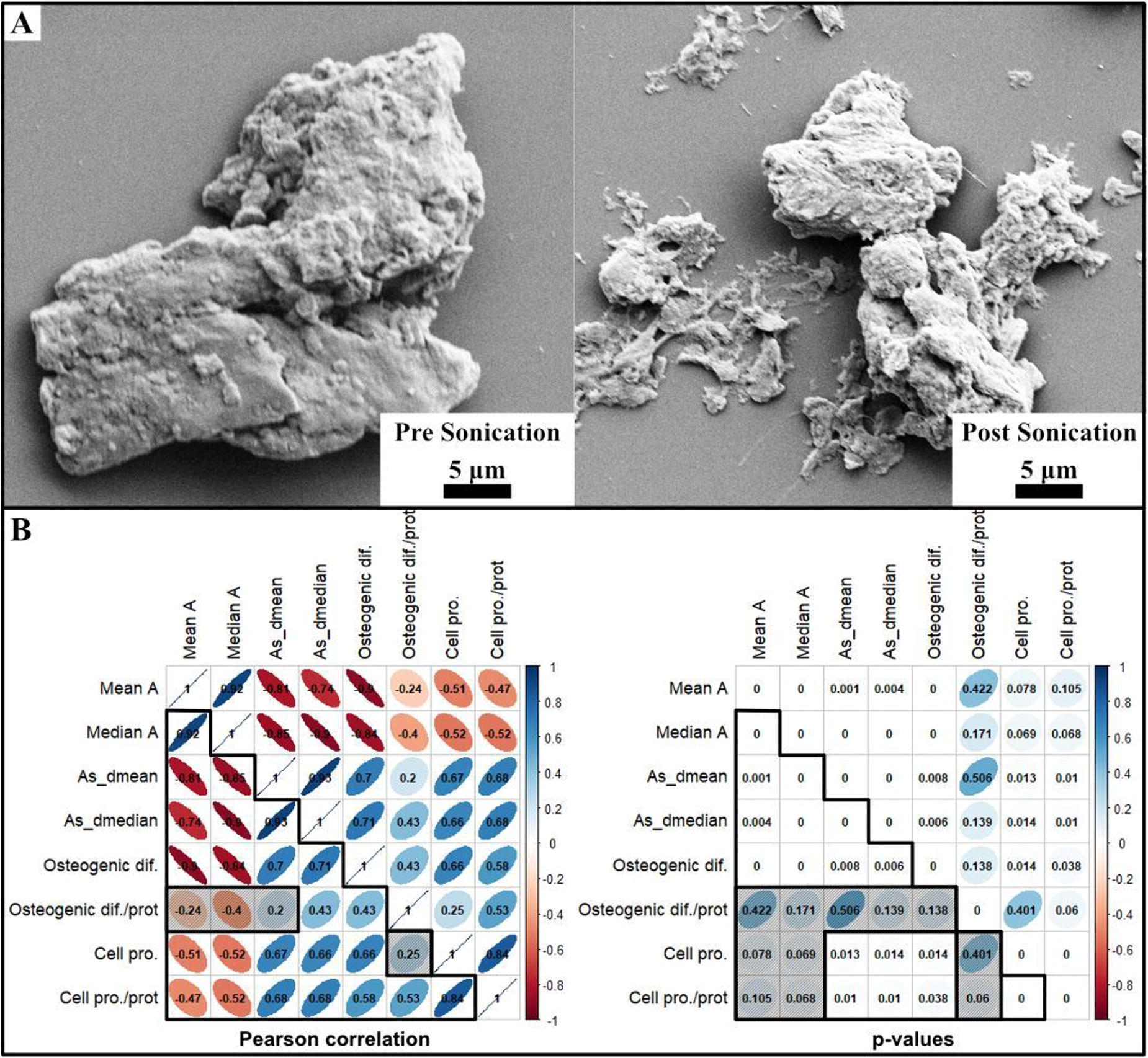
C200 - Innervation Of The Bioengineered Skeletal Muscle Construct In A Pre-clinical Muscle-nerve Injury Model
J. Kim, V. Mashanov, E. Billman, A. Poerio, Y. Ju, I. Kim, S. Lee, B. Vaughan, J. Jackson, A. Atala, J. Yoo
Wake Forest Institute for Regenerative Medicine, WINSTON-SALEM, NC
*Purpose/Objectives: Volumetric muscle loss is a debilitating condition, requiring timely repair with proper innervation to restore function. To address this critical issue, we developed a microsphere-loaded neurotrophic factor (NF) delivery system involving poly(lactic-co-glycolic acid) (PLGA) microspheres loaded with ciliary neurotrophic factor (CNTF) and glial cell line-derived neurotrophic factor (GDNF). Implantation of bioprinted muscle constructs with NF-loaded microspheres in an ectopic transposed nerve model enhanced in vivo innervation of skeletal muscle implants. This study aims to determine the capacity of the NF delivery system to facilitate muscle recovery in complex nerve-muscle injuries.
*Methodology: The nerve-muscle injury model was created by removing approximately 30% of the tibialis anterior muscle and resecting the distal end of the common peroneal nerve in athymic male rats. Two bioprinted skeletal muscle constructs containing human muscle progenitor cells (hMPCs) were implanted into the muscle defect. The severed host nerve was directed to the implants via a silicone conduit. Three experimental cohorts were used: the Controlcohort with no extrinsic NFs, the Free NFs cohort with the NFs freely dissolved in the hydrogel matrix of the implant, and the NF-loaded Microspheres cohort containing the matching NF load in PLGA microspheres. The levels of implant innervation and functional maturation were assessed using histological and immunohistochemical assays.
*Results: The nerve successfully regenerated through the conduit, and the sprouted neurites integrated into the muscle implant in all cohorts. At 12 weeks post-implantation, the neurite sprouting in the NF-loaded Microsphere cohort was 67% higher than the Control cohort and 37% higher than the Free NF cohort. The growing neurites formed neuromuscular junctions with the muscle cells within the implant. Approximately 60% of the implanted hMPCs maintained their muscle phenotype with adequate vascularization.
*Conclusion/Significance: This study validates a clinically relevant strategy that facilitates the innervation of bioengineered skeletal muscle implants, improving functional maturation and physiological control of the grafted muscle cells in a pre-clinical muscle-nerve injury model.
C201 - Impact Of Human Recombinant Irisin On Tissue-engineered Skeletal Muscle Structure And Function
M. H. Nguyen, E. Y. Su, C. S. Kennedy, O. M. Wroblewski, D. H. Hwang, L. M. Larkin
University of Michigan, Ann Arbor, MI
*Purpose/Objectives: Tissue engineering of exogenous skeletal muscle units (SMUs) through isolation of muscle satellite cells from muscle biopsies is a potential treatment for acute volumetric muscle loss (VML), which is a 30% or more loss of skeletal muscle tissue. A current issue with this treatment is the limited capacity for muscle stem cell (satellite cell) expansion in cell culture, resulting in a decreased ability to obtain enough cells to fabricate SMUs of appropriate size and structural quality that produce native levels of contractile force. This issue is a possible target for remediation by human recombinant irisin, which has been shown to increase myoblast proliferation and decrease the proportion of fibroblasts in the cocultures. Thus, this study determined the impact of human recombinant irisin on the proliferation, differentiation, and creation of three-dimensional (3D) SMUs from rat muscle cell isolate.
*Methodology: Rat muscle satellite cells were cultured without irisin (control) or with 50, 100, or 250 ng/mL of irisin supplementation. Light microscopy was used to analyze myotube formation with particular focus placed on the diameter and density of the monotubes during growth of the 3D SMU. Following the formation of 3D constructs, SMUs underwent measurement of maximum tetanic force to analyze contractile function, as well as immunohistochemical staining, to characterize muscle structure.
*Results: At 14 days post seeding, when treated with irisin, there was no significant change in the total number of nuclei or myogenin positive cells in culture, but both the 100 and 250 ng/mL irisin groups showed significantly higher numbers of MyoD positive cells than the control (p = 0.0018, p = 0.0001). In conjunction, there was an upward trend in myotube diameters between control and the 100 and 250 ng/mL irisin experimental groups (p = 0.0022). The 250 ng/mL group had significantly larger diameter than the 50 ng/mL and control groups (p = 0.0048, p = 0.0035). There was no significant difference between any of the other experimental groups. Further, there was a significant decrease in fusion index between the control and the 100 and 250 ng/mL groups (p = 0.0059, p = 0.0029). However, there was not a significant difference in maximum tetanic force between control and irisin experimental groups (p = 0.6580) or SMU cross-sectional area (p = 0.9701). Addition of irisin had a significant impact on normalized force with the 50 ng/mL group showing significantly higher force production in comparison to the control (p = 0.0038).
*Conclusion/Significance: The results suggest that the addition of human recombinant irisin led to significant improvements in myotube diameter and myoblast number. Furthermore, the addition of irisin to cell culture media resulted in a faster time to delamination for high concentration groups, suggesting that irisin has a positive impact on muscle cell proliferation. While the fusion index of muscle cells did not increase with the supplementation of irisin, there was a clear increase in total myoblasts which may lead to an increased number of fused myoblasts if allowed greater time to mature. In conclusion, supplementation of human recombinant irisin promotes the proliferation and differentiation of myotubes and has the potential for impacting contractile force production in scaffold-free tissue-engineered skeletal muscle.

C204 - GDNF-infused Biosponges For Improved Muscle Reinnervation Following Volumetric Muscle Loss
J. Tadiwala1, C. Tobo1, M. Ridolfo1, M. Wood2, K. Garg1
1
Saint Louis University, Saint Louis, MO,
2
Washington University in St. Louis School of Medicine, Saint Louis, MO
*Purpose/Objectives: Skeletal muscle tissue has a limited capability to regenerate after volumetric muscle loss (VML), a traumatic injury defined as the loss of >20% of a muscle’s mass [1]. VML directly reduces the number of myofibers and causes axotomy of nerves, resulting in reduced muscle function and neuromuscular junctions (NMJs). Our lab has created biosponge scaffolds composed of gelatin, collagen, and laminin-111, that have improved muscle mass, cross-sectional area, and myofiber number following VML. However, improvements in NMJ quantity and muscle function were not observed. Glial cell line-derived neurotrophic factor (GDNF) is a growth factor that enhances motor unit survival and neurite outgrowth [2]. Therefore, the objective of this study was to increase NMJ quantity, and enhance muscle function by incorporating GDNF into the biosponge structure.
*Methodology: Motor neurons (NSC-34) were cultured on 100 µg/ml ECM-coated (collagen, gelatin, HA, and laminin) 48-well plate at a density of 20,000 cells/well (n=4/group) and cultured for 3 days. In another experiment, NSC-34 cells (20,000 cells/well) were incubated with GDNF (100 ng/ml) for 4 days. Brightfield images were taken at 20x magnification from every group and neurite length was determined in ImageJ. Biosponges were prepared as previously described [3]. After lyophilization, the sponges (6 mm x 5 mm cylinders) were disinfected using ethanol and UV light. The sponges were then placed in a desiccator at 4° C overnight to thoroughly dry them. Subsequently, we electrostatically coupled GDNF (1000 ng/mL) with negatively charged gelatin nanoparticles (5mg/mL). This GNP/GDNF solution was then used to rehydrate the biosponges prior to surgery. To create the VML injury in male Lewis rats, 20% of muscle mass was removed from the tibialis anterior using a 6 mm biopsy punch. The VML defect was either left untreated or was treated with GDNF loaded biosponges. An uninjured muscle was used as the control.
*Results: Our in vitro results showed that 100 ng/ml GDNF added to motor neuron cultures significantly increased the percentage of neurite extensions in the size range of 30-45 microns. In vitro results also showed that motor neurons cultured on gelatin resulted in a significantly higher percentage of neurite extensions in the size range of 50-100 microns compared to collagen, laminin, HA, and tissue culture plastic (TCP). As expected, confocal imaging analysis showed that VML injured muscles frequently showed partial or no overlap between pre- and post-synaptic structures (i.e., AchRs and Synaptophysin/neurofilament), relative to uninjured muscles. The analysis of VML injured muscles treated with GDNF loaded biosponge is currently ongoing.
*Conclusion/Significance: Our in vitro studies show that gelatin is an excellent substate for motor neuron differentiation and neurite growth, which provides the rationale for using gelatin based biosponges for VML treatment. Additionally, GDNF supplementation resulted in longer neurite extension in vitro. Based on these results, we incorporated GDNF into the biosponges for accelerated reinnervation of VML injured muscles. Ongoing studies will determine the extent to which GDNF-loaded biosponges enhance reinnervation and functional muscle regeneration following VML.
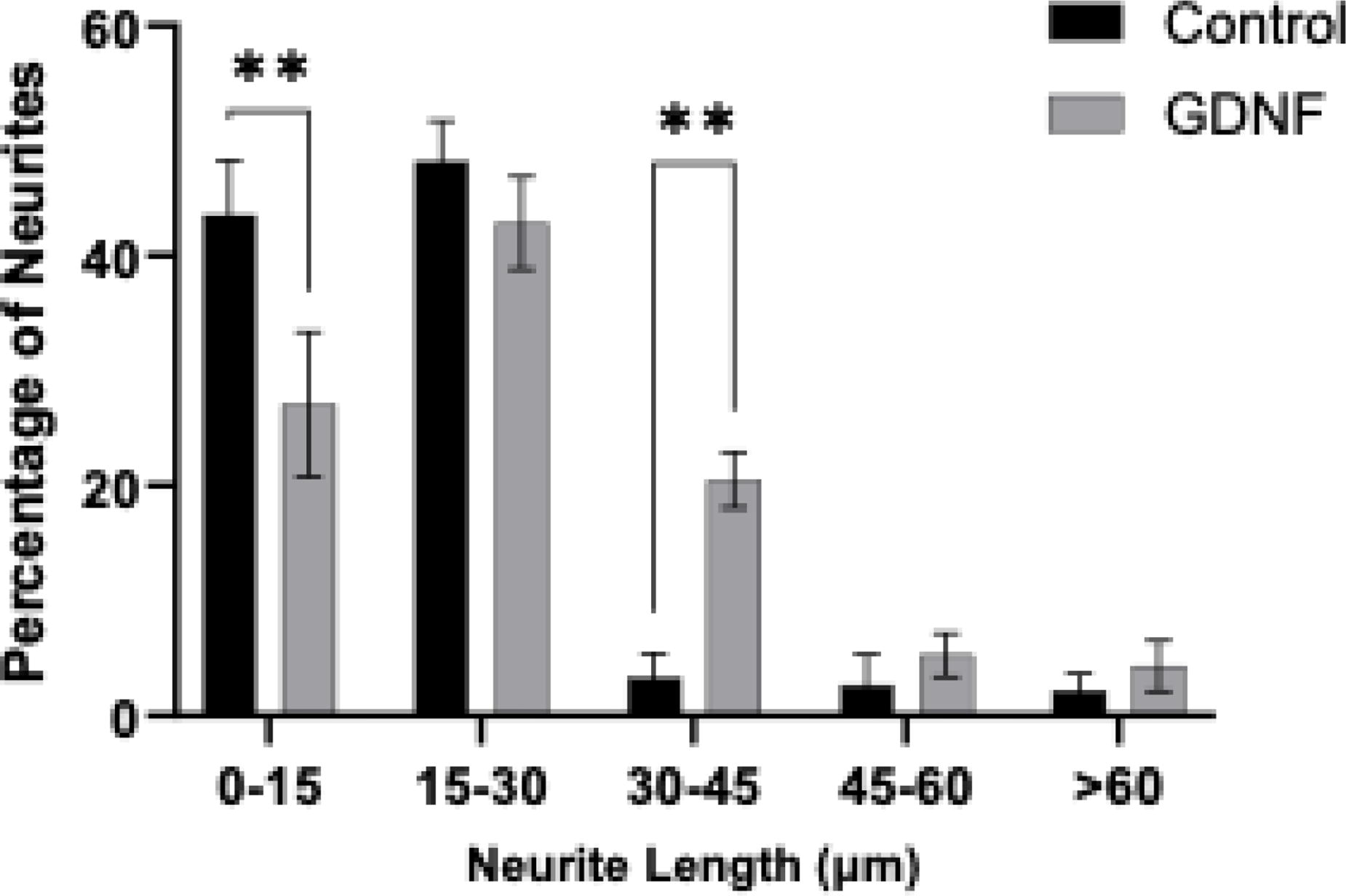
C209 - FRESH 3D Bioprinting Of Decellularized ECM Scaffolds For Volumetric Muscle Loss
A. Behre1, C. Gatti1, M. L. Arral1, S. A. Gershanok1, A. Kang1, R. Jin1, K. A. Whitehead1, T. Cohen-Karni1, D. J. Weber1, S. F. Badylak2, A. W. Feinberg1
1
Carnegie Mellon University, Pittsburgh, PA,
2
McGowan Institute for Regenerative Medicine, Pittsburgh, PA
*Purpose/Objectives: Volumetric muscle loss (VML) due to trauma or disease is a severe condition characterized by the loss of >20% of muscle mass and impaired contractile function. VML overwhelms the innate regenerative capacity of skeletal muscles, leading to chronic injuries with permanent deficits. The current gold-standard treatment is autologous muscle grafts, but these suffer from limited graft efficacy, and challenges with donor site availability and morbidity. Regenerative medicine approaches using decellularized extracellular matrix (dECM) scaffolds hold promise for in vivo muscle regeneration by providing myogenic biochemical cues and have progressed to human clinical trials. However, these dECM scaffolds are commonly implanted as powders or thin sheets that are unable to effectively fill the VML space and provide structural cues. Given the crucial role of myofiber alignment in the functional contraction of skeletal muscle, an improved solution would provide the necessary biochemical cues present in dECM together with physical guidance cues to recreate the 3D muscle tissue architecture. Here, we employ freeform reversible embedding of suspended hydrogels (FRESH) 3D bioprinting to fabricate biomimetic scaffolds from dECM hydrogels. These scaffolds are designed to restore native skeletal muscle 3D architecture by modulating the immune response and guiding myofiber alignment.
*Methodology: For the VML in vivo model, a surgical resection ∼20% of the tibialis anterior (TA) muscle in Sprague-Dawley rats was created, under approval from the CMU IACUC. Scaffolds designed to fill wound were aseptically FRESH 3D bioprinted and lightly crosslinked. Bioprinted dECM scaffolds were then implanted into the defect area and secured in place using fibrin glue. To address the intrinsically limited printability of the dECM, collagen type 1 (Col1) was incorporated into the bioink to enhance the handleability of the resulting implantable scaffolds. Rodents received implants of five different bioink compositions, varying the combination of dECM and Col1. Short-term studies (day 5) evaluated the impact of bioink composition and scaffold architecture on macrophage polarization and satellite cell activation through flow cytometry and histological analysis.
*Results: We have successfully built, implanted, and immobilized 3D bioprinting dECM scaffolds that emulate muscle architecture within the TA muscle of rodents. While scaffolds with dECM alone encounter challenges in maintaining their shape post-implantation, the incorporation of collagen into the bioinks enables the scaffold to retain its structure. At day 5 post-implantation, scaffolds with added Col1 are present with distinct fiber orientation still evident. Preliminary flow data suggests that the addition of ECM scaffolds increases the recruitment of macrophages to the wound site and delays the macrophage shift from M1 to M2. This, in turn, facilitates enhanced activation of satellite cells at the wound site.
*Conclusion/Significance: This dECM scaffold has been implanted into over 50 rodents and studies are ongoing. In vivo data is currently being collected to assess the scaffolds potential for guiding skeletal muscle regeneration. Long-term studies up to 56 days will utilize a functional testing platform and histological analysis to assess muscle function recovery, fiber alignment, myofiber diameter, scar tissue formation, and revascularization/reinnervation. If successful, this could improve patient morbidity after severe musculoskeletal injuries.
C210 - Muscle Fiber Fragments For Restoration Of Muscle Tissue Function
J. Kim, I. Ko, E. Yoo, S. Park, B.-K. Lee, J. Yoo, A. Atala
Wake Forest Institute for Regenerative Medicine, WINSTON-SALEM, NC
*Purpose/Objectives: Treatment of muscle function loss due to traumatic injury, congenital deformity, or tumor ablation is clinically challenging. The current treatment standard is the grafting of autologous muscle flaps; however, significant donor site morbidity and graft tissue availability remain problematic. Muscle fiber therapy has been attempted to treat muscle injury by transplanting single fibers into the defect site. However, irregularly organized long fibers resulted in low survivability due to delay in vascular and neural integration, thus limiting the therapeutic efficacy. We developed a novel method that produces uniformly sized native muscle fiber fragments (MFFs) for muscle transplantation. In this study, we applied autologous MFFs to restore injured muscle anatomy and function.
*Methodology: We developed an MFF processing method that produces uniformly sized fragments (approximately 100 μm in width and length) with intact muscle cells on the fiber surface. To test the therapeutic effects of the MFF technology, we created several rodent muscle injury models, including 1) a muscle atrophy model using toxin treatment, 2) a volumetric muscle defect model by surgical ablation, and 3) urinary incontinence (UI) model by damaging the external sphincter of the urethra. The effectiveness of the MFF therapy was determined by the structural and functional recovery of muscle tissues in these models.
*Results: The processed MFFs have a dimension of approximately 100 μm and contain living muscle cells on extracellular matrices (ECM). In preclinical animal studies using muscle atrophy, volumetric defect, and urinary incontinence models, histological and functional analyses confirmed that the transplanted MFFs into the injury sites were able to effectively integrate with host muscle tissue, vascular and neural systems, which resulted in significant improvement of muscle function and mass.
*Conclusion/Significance: These results indicate that the MFF technology platform is a promising therapeutic option for restoring muscle function in several muscle defect models. Our study results strongly suggest that as a point-of-care treatment, the MFF technology platform can potentially restore muscle anatomy and function, enhancing overall patient functionality and quality of life.
C211
C212 - Skeletal Muscle Bioreactor For Tissue Engineering Research
T. Lloyd
Walla Walla University, College Place, WA
*Purpose/Objectives: Victims of accidents are inflicted with a variety of injuries, many of which involve the loss of skeletal muscle, a dynamic tissue responsible for providing structure and mobility. Muscle loss beyond its natural regenerative properties is called volumetric muscle loss (VML) and can lead to disablement, disfigurement, and amputation. While autologous grafts are used to mitigate damage, victims will lose function from the autologous extraction site. Engineering skeletal muscle replacements should eliminate this issue; however, tissue engineering better solutions for VML treatment can only be furthered by tissue-specific bioreactors that help mimic the stresses and blood flow of the body in a controlled environment. While there are many different types of bioreactors including spinner flasks, rotating vessels, and perfusion systems, tissue-specific bioreactors that apply perfusion and additional stimuli are of increasing interest, especially for muscle tissue engineering.
*Methodology: A novel skeletal muscle bioreactor was prototyped that applies mechanical loading and perfusion of media directly to the muscle scaffolds. Muscle scaffolds with a width of 12mm are clipped into place, one side to the vessel wall and the other side to a cylindrical drum submerged in the media. A stepper motor oscillates the cylindrical drum applying up to 10% strain on scaffolds according to similar studies loading skeletal muscle tissue. Using a peristaltic pump, media is transferred from the vessel to the space inside the cylindrical drum before perfusing the matrix through vents in the drum.
*Results: The bioreactor is designed to be cost-effective, sterilizable for multiple runs, and accommodates eight scaffolds each run. The scaffolds may be easily removed from the vessel without damaging the tissue. The bioreactor vessel is 240 x 180 x 120mm and may fit in a large incubator for temperature control. The vessel may be modified to accommodate additional sensors and is designed to retain the skeletal muscles' striated structure while sustaining and promoting healthy cell development.
*Conclusion/Significance: This novel device is designed to provide both perfusion and mechanical loading to seeded matrices to better mimic in vivo conditions. As advances are made in tissue engineering and new treatment methods are explored, tissue-specific bioreactors are expected to become more common to support the unique physiological requirements of different tissue grafts and therapies.

C213 - COAXIAL BIOPRINTING OF VASCULAR JUNCTION-CONSTRUCTED SKELETAL TISSUE
A. Tahmasebifar1, Z. Yildirim1, C. Ozdemir2, E. Balikci1, E. Baran1, B. Yilmaz1
1
University of Health Sciences Turkey, Istanbul, Turkey,
2
Yildiz Technical University, Istanbul, Turkey
*Purpose/Objectives: Skeletal muscle has the ability to regenerate after minor damage, but significant volumetric muscle loss results in permanent functional impairment. A recent analysis of Global Burden of Disease (GBD) data shows that approximately 1.71 billion people worldwide have musculoskeletal disorders [1]. Skeletal muscle structures developed through tissue engineering approaches promise the potential to revolutionize the treatment of major muscle injuries through the regeneration of functional skeletal muscle. On the other hand, engineered tissues require a well-established vascular network to facilitate cellular respiration. In the tissue engineering paradigm, the angiogenic process unfolds gradually, subjecting the tissue to hypoxia and insufficient nutrient supply during the maturation of functional blood vessels derived from an engineered construct implanted in the host [2-3]. To overcome all the aforementioned challenges, coaxial bioprinting is a method that enables the production of complex tissue structures with a controlled accumulation of biomaterials [4]. With the scope of that, we report the development of a biodegradable coaxial core/shell collagen methacrylate (ColMA) scaffold for mimicking skeletal tissue with a vascular lumen.
*Methodology: In the study, we utilized our proprietary insulation biomaterial, marine collagen, extensively characterized in prior studies. Methacrylated collagen bio-inks were subjected to cross-linking through exposure to visible light. The incorporation of HUVEC and Myoblast cells (C2C12) into the bio-ink and their co-culture facilitated the establishment of a vascular network, mimicking the intricacies of muscle structure. In addition to rheological analysis, cell viability, and proliferation potential were evaluated and analyzed after 7 and 14 days of culture.
*Results: The results obtained from the DAPI/Actin and Live/Dead assays confirmed cell viability to be higher than 80%. In the degradation study performed after coaxial bioprinting, tissue formation was preserved. Finally, conducted with a fluorescent microscope, the presence of lumens has been demonstrated through endothelial cell markers.
*Conclusion/Significance: Our findings demonstrate that bio-inks crosslinked with visible light using marine collagen can be effectively employed in coaxial bioprinting and hold promise in the field of musculoskeletal tissue engineering.
C214 - Contractility And Myokine Secretion Of Tissue-engineered Skeletal Muscle By Electrical Stimulation
T. FUJISATO, K. SASAKI, T. NAKAMURA
Osaka Institute of Technology, Osaka, Japan
*Purpose/Objectives: Skeletal muscles are mainly responsible for maintaining and changing body posture and locomotion. Besides that, they produce various cytokines or peptides, called myokines, such as interleukin 6 (IL-6), brain-derived neurotrophic factor (BDNF), leukemia inhibitory factor (LIF), irisin, and secreted protein acidic and rich in cysteine (SPARC). All of them are associated with metabolic changes, tissue regeneration and repair, and maintenance of healthy bodily functioning. In this study, the tissue-engineered skeletal muscle (TEM) was cultured under various electrical stimulations and examined its contractility and myokine secretion.
*Methodology: Two artificial tendons were introduced at both ends of our engineered muscle. The two tendons were hold with stainless steel pins placed 12 mm apart before cell seeding. The C2C12 myoblasts embedded in collagen gel was added between and on the surface of the two tendons. After gelation of the cell suspension, the constructs were cultured in growth medium of high-glucose DMEM with 10% FBS for 2 days and then in differentiation medium of high-glucose DMEM with 7% horse serum for 2 weeks, followed by electrical stimulation (ES) culture with 1 to 12 hours per day for 1 week. The electrode holding jig was fabricated by 3D printer and Pt electrodes were attached on the jig to be placed both sides of TEM. The jig and wirings were placed at a position where they did not touch the culture medium. After the ES culture, the contractile force was measured by strain transducer and the amount of IL-6 in culture medium was measured using the IL-6 EIA Assay Kit.
*Results: The isometric twitch force of the TEM cultured with ES of 5V, 2msec, and 0.5Hz was remarkably higher than that of without stimulation. In addition, the amount of IL-6 secreted with ES was larger than that of without stimulation. They were affected by duration of ES par day.
*Conclusion/Significance: These results suggest that the TEM may be applied to physical exercise model in vitro.
C216 - Nerve Tissue Model Development On A Micropatterned Surface
D. G. Tamay1, D. Arslantunali-Sahin1, S. Isik2, G. E. Kole2, D. Yucel2, N. Hasirci1, V. Hasirci1,2
1
Middle East Technical University, Ankara, Turkey,
2
Acibadem Mehmet Ali Aydinlar University, Istanbul, Turkey
*Purpose/Objectives: Microtopographical elements are often utilized in materials design to mimic the microarchitecture of native tissues and modulate cell behavior. This study aims to achieve separation of neural cell body and axons via different microtopographical elements, in an effort to model peripheral nerve tissue. Microgrooves and netrin-1 are used to achieve guided anisotropic elongation of axons on a micropatterned PDMS surface. The model is expected to be used as a platform for nerve damage and regeneration studies.
*Methodology: Micropatterned surfaces (MPS) are produced via soft lithography method and have 3 distinct regions: A: An unpatterned area for cell seeding, B: An area with micropillars for cell body-axon separation, and C: An area with microgrooves for anisotropic axon elongation. PC12 and Schwann cells (SCs) were seeded on the surfaces, to conduct cell-surface interaction studies. To observe axon damage and healing processes on the MPS PC12 cells were damaged with a microtome blade.
*Results: Surface features (width of microgrooves, dispersion and organization of micropillars) and culture conditions (serum and growth factors amount in media) most suitable for cell body-axon separation, axon guidance, and extension were determined by examining PC12 cell behavior on different surface designs and media compositions. Axon length reached up to 250 µm when PC12 cells were co-cultured with SCs, in the presence of Netrin-1 and using culture media with 90% reduced serum and 50 ng/mL NGF. SCs were observed to form clusters around neural cell protrusions, similar to the myelinating SCs in native peripheral nerve (Figure 1a, b). Preliminary axon damage and regeneration studies showed that PC12 cells started forming growth cones 5 days after transection (Figure 1c), and reached pre-damage axon length on day 7 (Figure 2d, e). Maintenance of viability, growth cone formation and axon re-extention after transection indicate that nerve cell damage and healing processes can be examined with this model.
*Conclusion/Significance: The micropatterned surface and peripheral nerve model developed in this study shows promise for the monitoring of damage and recovery processes of transected neurons. The model has potential to be developed into a research platform for testing the effects of novel drugs or biological agents on nerve regeneration.

C217 - Injectable GelMA Hydrogels For Axonal Regeneration Following Spinal Cord Injury
I. Liubchak, A. Pieters, T. Bennet, T. M. Caffrey, K. C. Cheung
University of British Columbia, Vancouver, BC, Canada
*Purpose/Objectives: Injuries to the spinal cord (SCI) result in the partial or complete loss of physiological, motor and sensory functions below the level of injury. The microenvironment at the SCI site strongly inhibits the repair process due to glial scarring and inflammation. However, injured axons originating from the central nervous system have the potential to regenerate when provided with a permissive environment for regrowth. We aim to design a hydrogel biomaterial that could be applied at the SCI site via minimally invasive needle injection and form a soft supportive structure for axonal growth. Additionally, it serves as a delivery vehicle for bioactive molecules that stimulate axonal repair and inhibit the formation of a glial scar, and physical (polymer microstructures) cues that guide axonal growth across the lesion. The polymeric, rod-like microstructures will be aligned in a magnetic field after injection, and thus controllable, triggered crosslinking of the hydrogel is needed. In comparison to UV crosslinking photoinitiator systems, visible light-induced crosslinking conditions are expected to have reduced phototoxicity, and the longer wavelengths will have deeper penetration into the hydrogel.
*Methodology: Gelatin Methacryloyl (GelMA) was selected for the development of an injectable biomaterial. We are using a formulation containing GelMA, ruthenium photoinitiator and sodium persulfate oxidizer. After activation under blue light wavelengths, the mixture quickly crosslinks into a soft, highly hydrophilic polymer network. GelMA naturally contains cell adhesive arginine-glycine- aspartate (RGD) motifs and has excellent tunable mechanical properties. The crosslinking density and crosslinking efficiency of GelMA formulations containing different polymer and photoinitiator concentrations were examined through mechanical testing, sol fraction measurements, and swelling experiments. Following the optimization of hydrogel formulations using the Design of Experiments approach, the encapsulation of neurons in selected hydrogel formulations was performed in a microfabricated, 3D SCI-on-a-chip model (Figure 1 (a)). The orientation of axonal growth in the GelMA hydrogel was evaluated using fluorescence microscopy.
*Results: GelMA hydrogels have had no toxic effect on the neuroblastoma and primary neuron cell cultures and have supported the growth of neurites from neurons encapsulated in the hydrogels. SCI-on-a-chip microdevice is also used to study the regrowth of axons in GelMA following their transection (Figure 1 (b)) and serves as an in vitro platform to evaluate and select hydrogels for injection experiments in the in vivo models.
*Conclusion/Significance: In this work, we have designed and applied the photocrosslinkable GelMA hydrogel formulation for the encapsulation of primary neurons and successfully demonstrated axonal growth in vitro. Currently, there are no treatments capable of restoring the structural and functional integrity of the damaged spinal cord. GelMA hydrogel is a promising material for the in vivo application for the regeneration of the spinal cord, ultimately improving the quality of life in individuals living with SCI, helping them regain their mobility and physiological functions.

C219 - Niura: A System For In-Ear Silicone Electrodes Inside Wireless Earbuds To Screen Neural Brain Abnormalities
R. Ahmed, W. Yu, A. Das, S. Huda, P. Patel, S. Nasrin, G. Chang, M. Rahim, A. Khan, A. Singh, V. Kolli, A. Chowdhury
Niura Corporation, Plainview, NY
*Purpose/Objectives: The limited accessibility of brain health monitoring in the United States hinders the detection of neurological conditions. Electroencephalograms (EEG), despite being non-invasive, are impractical for widespread usage due to the their high cost and complex user interface. Niura addresses these challenges head-on. Backed by six provisional patents, this cost-effective, easy-to-use device integrates advanced conductive silicone electrode technology into an earpiece. Niura paves the way for regular, accessible brain health monitoring, making early screening for neurological conditions feasible for all.
*Methodology: A four-layer circuit and conductive silicone electrode technology have been embedded in an earbud to monitor neural oscillation waves. The main processor, nRF5340, operates in high voltage mode, powered by the battery or the 5V input power. A power section, in which the power multiplexer, TPS2113ADRBR, is used to switch between battery and charger power and avoids digital noise coupling into analog power. The primary EEG device, ADS1291IPBS, is powered by 2 LDO outputs that communicate through SPI with the nRF5340. An audio amplifier, Texas Instruments TLV320DAC3100, is powered by the digital power input, while analog power is generated internally by an integrated LDO.
EEG electrodes set in the earbud tips are positioned in the ear canal to contact the skin covering the skull's temporal bone. A low or high-power, low-noise printed circuit board (PCB) with an analog front-end (AFE) measures brain electrical activity. AI interprets collected data and can transmit it to other devices through wired or wireless technology. A fast fourier transform identified phase, amplitude, and frequency from sine waves. The assembled device provides a pre-screening method for neurological conditions such as epilepsy and aneurysms.
*Results: After 40 trials using diverse formulas, integrating graphite, carbon fiber into a two-part silicone electrode produced conductive rubber earbud tips capturing EEG data in the ear. This novel silicone formula has very high conductivity, while presenting low resistivity of around 5-6 ohms. Thus, Niura's proprietary EEG electrode system offers seamless implementation into existing in-ear audio systems through patent-pending conductive earbud tips suitable for use in inpatient, outpatient, and intraoperative settings. Furthermore, live monitoring during a patient’s most natural state improves EEG accuracy compared to a clinical resting position.
*Conclusion/Significance: Niura’s in-ear EEG technology pioneers neurological health monitoring, enabling real-time detection of neurological issues via live data - a groundbreaking advancement. Critical parts of this study highlight the technology developed that allows users to monitor their brain waves with the new silicone formula using a much more compact circuit. Its compact design, user-friendly experience, and HIPAA compliance makes it promising for widespread adoption. Future studies should investigate its efficacy with diverse patient populations, comparing it with medical-grade EEG devices, and exploring its long-term monitoring capabilities. Additionally, integrating machine learning algorithms for personalizing audio content, optimizing comfort, and enhancing usability would amplify its impact in modern healthcare. Embracing these recommendations will revolutionize neurological monitoring and ultimately elevate patient care standards.
C220 - Chitin Nerve Conduits With Three-dimensional Spheroids Of Mesenchymal Stem Cells From SD Rats Promote Peripheral Nerve Regeneration
J. Haoran, Z. Peixun, M. Zhang, L. Ci
Peking University People's Hospital, Beijing, China
*Purpose/Objectives: Peripheral nerve injury (PNI) is an unresolved medical problem with limited therapeutic effects. Epineurium neurorrhaphy is an important method for treating PNI in clinical application, but it is accompanied by inevitable complications such as the misconnection of nerve fibers and neuroma formation. Conduits small gap tubulization has been proved to be an effective suture method to replace the epineurium neurorrhaphy. In this study, we demonstrated a method for constructing peripheral nerve conduits based on the principle of chitosan acetylation. In addition, the micromorphology , mechanical properties and biocompatibility of the chitin nerve conduits formed by chitosan acetylation were further tested. The results showed chitin was a high-quality biological material for constructing nerve conduits. Previous reports have demonstrated that mesenchymal stem cells culture as spheroids can improve the therapeutic potential. In the present study , we used a hanging drop protocol to prepare bone marrow mesenchymal stem cell (BMSCs) spheroids. Meanwhile, spherical stem cells could express higher stemness-related genes. In the PNI rat model with small gap tubulization, BMSCs spheres exhibited a higher ability to improve sciatic nerve regeneration than BMSCs suspension. Chitin nerve conduits with BMSCs spheroids provide a promising therapy option for peripheral nerve regeneration.
*Methodology: First, we performed the isolation, purification and identification of rat BMSCs. Then, we grow the stem cells in three-dimensional spherical shape by suspension drop culture technique. Subsequently, the cytoskeleton and nuclei were labeled with immunofluorescence and observed under a laser scanning confocal microscope. qRT-PCR was used to detect the expression of Nanog, Sox2, and POU5F1genes in BMSCs cultured by two-dimensional planar or three-dimensional stereotaxic culture, and it is important to note that the number of cells was equal between the compared groups. Finally, this chitosan neurorepair conduits loaded with three-dimensional cultured stem cells was implanted into rats, a model of peripheral nerve dissection, to observe the effect of peripheral nerve repair.
*Results: In vitro, the expression of Nanog, Sox2, and POU5F1 genes was increased in bone marrow MSCs cultured in three dimensions, suggesting a higher potential for division and differentiation of stem cells. In vivo, the 3D BMSCs group showed higher SFI, lower neurophysiological CMAP latency, and higher CMAP amplitude than the 2D BMSCs and control group. Moreover, the number, diameter and thickness of remyelination sheath in the experimental group were better than those in the control group. Overall, the experimental group had better functional recovery.
*Conclusion/Significance: Chitosan nerve conduits loaded with three-dimensional spherical bone marrow mesenchymal stem cells have shown good results in repairing peripheral nerve injury.
C221 - Development Of An Alginate/ CNTs Nanocomposite Bioink For Neural Applications
E. Capuana, F. Lopresti, C. Di Marco, V. La Carrubba
University of Palermo, Palermo, Italy
*Purpose/Objectives: In the field of neural tissue engineering, one of the main purposes is creating electrically conductive scaffolds with customized properties. This study aims to develop a nanocomposite bioink by combining alginate and carbon nanotubes (CNTs); it also addresses the challenges of CNT dispersion due to its highly hydrophobic nature, leading to their agglomeration in aqueous environments [1].
*Methodology: To achieve effective dispersion of CNTs, ultrasonic frequencies (40 kHZ) and Pluronic at a concentration of 0.5% w/v in water were used. Three different concentrations of CNTs (0.005%, 0.01%, and 0.02% w/v) were added to the solvent system while keeping the alginate polymer at 5% w/v (Fig. 1Ai). At a concentration of 0.5% w/v, the Pluronic could effectively reduce the surface tension of water thanks to its surfactant properties [2]. Choosing the right concentration of Pluronic is crucial because higher concentrations could potentially affect the rheological and electrical characteristics of the bioink as well as its cytotoxicity in undesirable ways [3]. To obtain the hydrogel, CaCl2/water solution at a concentration of 2% w/V was used as crosslinking agent (Fig. 1Aii). Scanning electron microscope (SEM), rheological analysis and electrochemical impedance spectroscopy were performed on the Alginate/CNTs dispersions or on the crosslinked systems.
*Results: First, SEM analysis, performed on the freeze-dried and gold-coated gels, showed the absence of nanotube agglomerates in the structure, confirming the effectiveness of the dispersion process (Fig. 1Bi, ii). Rheological analysis showed that the behavior of the bioink changed depending on the concentration of CNTs, providing insights into the printability of the bioink. Interestingly, analyzing the obtained flow curves (Fig. 1C), the concentration of 0.01% w/v of CNT showed higher complex viscosity at low angular frequency. Furthermore, this concentration exhibited solid-like behavior at low frequencies, which is advantageous for bioprinting. Electrochemical impedance spectroscopy (Fig. 1D) on crosslinked systems showed that pure alginate and the 0.005% concentration had equivalent conductivity values. This equivalence between the measured conductivities was observed for concentrations of 0.01% and 0.02%, with a value of 2 mS/cm, similar to the physiological neural conductivity [4].
*Conclusion/Significance: In this study, a nanocomposite based on alginate with well-dispersed CNTs at three different concentrations was successfully achieved. The distinctive combination of electrical conductivity and adequate viscosity of the bioink is promising for neural tissue engineering applications. The ability to reproduce neural conductivity levels within the bioink opens up new possibilities for the development of bioelectronic interfaces that closely mimic the properties of native neural tissue. The goal is to maximize the convergence of conductivity and functional structure of bioprinted neural constructs as potential for facilitating seamless communication within neural networks. Future efforts will focus on actual molding and conducting in vitro studies to evaluate biocompatibility and potential for neural differentiation.
Acknowledgements: The research was supported by the Italian Ministry of Health in the frame of the project “PJ MIN SALUTE METODI SOSTITUTIVI 2022”, by Complementary Operational Programme (POC) 2014/2020 of the Sicily Region, and by “SiciliAn MicronanOTecH Research And Innovation CEnter ”SAMOTHRACE" (MUR, PNRR-M4C2, ECS_00000022).
References: [1]doi.org/10.1038/s41598-020-59463-9; [2]doi.org/10.1016/J.MTCHEM.2021.100617; [3]doi.org/10.3390/PHARMACEUTICS14122837; [4]doi.org/10.1002/ADFM.202104441.

C222 - Effect Of Odorants As Chemoattractant For Non-olfactory Neurons
L. Won, S. Sakiyama-Elbert
University of Washington, Seattle, WA
*Purpose/Objectives: After facial nerve injury, a lack of guidance cues can result in misconnection of regenerating axons. This can result in facial synkinesis, unintentional muscle movement (e.g. mouth twitching) paired with an intended muscle movement (blinking). Although physical therapy can help, not all patients fully recover, resulting in both physical and psychological struggles. Additionally, although growth factors may help promote regeneration of injured facial nerves, their use is contraindicated post-tumor resection, which is a common cause of synkinesis. By guiding regenerating axons to their correct target of innervation, the severity of synkinesis can be decreased. The goal of this project is to test the ability of odorants to direct axon growth from motor neurons, which express odorant receptors, and preferentially direct growth toward specific targets.
*Methodology: A selectable transgenic mouse embryonic stem cell line (puromycin resistance under the control of the Hb9 promoter) was used to generate enriched motor neurons. Puromycin was added after 6 days of induction to enrich for motor neurons. After 1 day of selection, they were aggregated via AggreWells for 2 days, then seeded into plates and treated with different odorants or growth factors (BDNF, GDNF, NT3 each at 10 ng/mL - positive control) for another 2 days. Neurite outgrowth was visualized with a Nikon Eclipse Ti2 widefield microscope (using 10X objective) and quantified by measuring the average radius of outgrowth from the aggregates using ImageJ. Then the results were normalized to the positive control for each experiment. An ANOVA was run with Scheffe’s post hoc test to test for statistical significance.
*Results: Cells treated with odorants (acetophenone, citric acid, and decanoic acid) without growth factors in the media exhibited neural growth that was similar to the positive controls, which were only treated with growth factors (Figure 1). When the results were normalized to the neurite length of the positive control, whose average neurite length was around 457 µm, the mean neurite length of the cells without growth factors (or odorants - negative control) were 74% as long as the positive control (p<0.001). Meanwhile, the average lengths of acetophenone, citric acid, and decanoic acid were 88%, 85%, and 85% of the positive control, respectively.
*Conclusion/Significance: Single odorants can promote neural growth similarly to low doses of growth factors. Future studies will include co-treating cells with growth factors and odorants, as that would help determine whether the odorants and growth factors influence neurite growth through the same or two different pathways. If there is a synergistic effect between the two, odorants may be a growth factor-independent method of influencing cell behavior.

Figure 1: Neurite lengths of neurons when treated with odorants without growth factors Mean motor neuron neurite outgrowth from aggregates were not different with acetophenone, citric acid, decanoic acid compared to the cells treated with growth factors. Neurite length from each aggregate was normalized to the average lengths of cells treated with growth factors alone, which is ∼ 457 µm on average. (*** p-value < 0.001, ‘ ’ ns)
C223 - Formation Of Adhesive And Conductive Interface Based And Bioelectronics Application
H. Woo, K.-i. Song, H. Cha
Pohang university of Science and Technology, Pohang, Korea, Republic of
*Purpose/Objectives: There are increasing demands for conductive materials in biophysical environments, especially for bioelectronics device applications, including neural and cardiac stimulation and recording, electroceuticals, and neural prostheses. To overcome the mismatch caused by the difference in physical properties and charge carriers between conductive materials and biological tissues, there have been lots of trials to bridge the interfaces.
*Methodology: In the present work, we newly developed double-layered biocompatible nanoparticles with a eutectic gallium-indium alloy (EGaIn) core-shell. EGaIn, which is used as a core, is in liquid phase at body temperature for its low melting point, is far less toxic, and possesses a negligible vapor pressure. The coating layer, hyaluronic acid (HA), is a widely used biocompatible and biodegradable polysaccharide. Bulk EGaIn is dispersed evenly via ultrasonic in solution, which contains HA molecules whose net charge is negative in neutral pH conditions, forming single layered nanoparticles at the same time. After that, bioengineered mussel adhesive protein (MAP), which contains rich DOPA residues playing a critical role in underwater adhesive and cohesive effect in mussel byssus and abundant lysine residues making protein positive net charge, form dense coacervate with HA-coated nanoparticles making conductive and adhesive liquid phase formation (Conductive Bioglue, CBG).
*Results: The thickness of the coating has been regulated through the concentration of HA solution in sonication process. CBG was formed in dense liquid phase and crosslinked via electrical stimulation. Crosslinked CBG (e-CBG) showed proper electrical, mechanical property and high adhesiveness between various substrates.
*Conclusion/Significance: CBG can be applied as the interface between electrodes and tissues by utilizing various residues in MAP, showing applicability for bioelectronics.
C224 - Neural Differentiation And Spinal Cord Organoid Generation From Induced Pluripotent Stem Cells (ipscs) For Als Modelling And Inflammatory Screening
J. MA, H. CUI
HEBEI MEDICAL UNIVERSITY, SHIJIAZHUANG, China
*Purpose/Objectives: C9orf72 genetic mutation is the most common genetic cause of ALS/FTD accompanied by abnormal protein insufficiency. Induced pluripotent stem cell (iPSC)-derived two-dimensional (2D) and three-dimensional (3D) cultures are providing new approaches. Therefore, this study established neuronal cell types and generated spinal cord organoids (SCOs) derived from C9orf72 knockdown human iPSCs to model ALS disease and screen the unrevealed phenotype.
*Methodology: Wild-type (WT) iPSC lines from three healthy donor fibroblasts were established, and pluripotency and differentiation ability were identified by RT-PCR, immunofluorescence and flow cytometry. After infection by the lentivirus with C9orf72-targeting shRNA, stable C9-knockdown iPSC colonies were selected and differentiated into astrocytes, motor neurons and SCOs. Finally, we analyzed the extracted RNA-seq data of human C9 mutant/knockout iPSC-derived motor neurons and astrocytes from the GEO database and the inflammatory regulation-related genes in function and pathways. The expression of inflammatory factors was measured by qRT-PCR.
*Results: The results showed that both WT-iPSCs and edited C9-iPSCs maintained a similar ability to differentiate into the three germ layers, astrocytes and motor neurons, forming SCOs in a 3D culture system. The constructed C9-SCOs have features of spinal cord development and multiple neuronal cell types, including sensory neurons, motor neurons, and other neurons. Based on the bioinformatics analysis, proinflammatory factors were confirmed to be upregulated in C9-iPSC-derived 2D cells and 3D cultured SCOs.
*Conclusion/Significance: The above differentiated models exhibited low C9orf72 expression and the pathological characteristics of ALS, especially neuroinflammation.
C225 - An Affinity-based Release Strategy To Deliver Insulin-like Growth Factor 1 For Retinal Degeneration
S. P. Lu1, J. M. Labriola1, H. K. Chin1, L. C. Comanita2, V. A. Wallace2, M. S. Shoichet1
1
University of Toronto, Toronto, ON, Canada,
2
Toronto Western Hospital, Toronto, ON, Canada
*Purpose/Objectives: Retinal degenerative disorders, such as age-related macular degeneration and retinitis pigmentosa, vary widely in their underlying cause and pathophysiology. Their common characteristic is progressive degeneration of the light-sensing photoreceptors, often coupled with chronic inflammation, which ultimately leads to blindness. Insulin-like growth factor 1 (IGF-1) is a pleiotropic hormone that exerts neuroprotective and anti-inflammatory effects within the central nervous system, including the retina. As such, IGF-1 is a promising protein therapeutic for the treatment of retinal degenerative diseases. However, there are barriers to its clinical translation including poor pharmacokinetics and tissue penetration, as well as off-target effects. Localized delivery of IGF-1 using hydrogels is one method of overcoming these challenges. We have previously developed a biocompatible hydrogel with properties comparable to the vitreous humour, making it ideal for injection into the eye. The objective of this project is to engineer an affinity-based release formulation with this hydrogel to deliver IGF-1 to the retina. We aim to determine the efficacy of this controlled release system in preventing photoreceptor degeneration and preserving photoreceptor function in a mouse model of retinal degeneration.
*Methodology: Ketone-modified hyaluronic acid (HAK) polymers were immobilized with weak binding peptide (WBP), which has low affinity for SH3 protein domains, and covalently crosslinked using oxime chemistry to form a hydrogel. HAK-WBP acts as the delivery vehicle for the affinity-based release of an SH3-IGF-1 fusion protein. WBP was synthesized using a peptide synthesizer, and SH3-IGF-1 was expressed and extracted from E. coli. The rd10 mouse was used to evaluate the efficacy of this delivery strategy. SH3-IGF-1 was loaded into HAK-WBP hydrogel, mixed with a PEG crosslinker, and injected intravitreally into the mouse eye at post-natal day 22 (P22). A week later, the eyes were enucleated, and immunohistochemistry was used to assess photoreceptor morphology and preservation of the photoreceptor layer.
*Results: In vitro release experiments demonstrated the sustained release of SH3-IGF-1 from HAK-WBP over a period of 28 days. Preliminary results of the in vivo studies show preservation of the photoreceptor cell layer in the treatment group (SH3-IGF-1 delivered by HAK-WBP) compared to mice treated with SH3-IGF1 in saline, or saline controls.
*Conclusion/Significance: The development of an affinity-based release system providing sustained delivery of SH3-IGF-1 to the retina can confer neuroprotection on endogenous photoreceptors, mediate retinal cell apoptosis and inflammation, and could be a preventative measure of severe retinal damage. Furthermore, sustained release can reduce the number of invasive injections required, paving the way for future protein therapeutic delivery strategies to prolong photoreceptor cell survival and ultimately preserve retinal cell function.
C227 - Device-free Isolation Of Photoreceptors From Patient Stem Cell Derived Retinal Organoids
N. E. Stone, L. R. Bohrer, N. K. Mullin, R. F. Mullins, E. M. Stone, B. A. Tucker
University of Iowa, Iowa City, IA
*Purpose/Objectives: Autologous cell replacement shows great promise for the treatment of inherited retinal degeneration. While mature differentiation protocols exist to produce retinal organoids from patient-derived stem cells, not all of the cell types present in these organoids are desirable for transplant. To increase the potency of future cell therapies, methods for isolating photoreceptors from dissociated retinal organoids are needed. In this work, we show how partial dissociation can be used to exploit the spatial organization of retinal organoids to produce highly pure photoreceptor populations without the use of specialized sorting devices or reagents such as antibodies.
*Methodology: Retinal organoids were generated using a modified form of the 3D differentiation protocol developed by Meyer et. al as we have described previously. For flow cytometry experiments, organoids were dissociated using papain for 30 minutes or for 90 minutes followed by trituration. For scRNAseq experiments, liberated cells were removed from dissociating organoids at 20 minute intervals. At the conclusion of the experiment, remaining tissue was fully dissociated. A control 60 minute full dissociation was performed in parallel. Photoreceptor purity was assessed via flow cytometry and scRNAseq.
*Results: Figure 1A-B demonstrates that photoreceptors desired for transplant are typically found in the outer layers of retinal organoids, suggesting that partial dissociation could selectively release these cells. Figure 1C-D shows flow cytometry results indicating an increase in purity of CD133+ cells from 21.1% to 91.7% when cells were partially dissociated as compared to full dissociation. Figure 1E confirms these results, showing a maximum purity of 96% of CRX+ cells as compared to 66% pure for traditional dissociation.
*Conclusion/Significance: By timing the dissociation of retinal organoids, highly pure photoreceptor populations suitable for cell replacement therapies can be obtained under cGMP without the use of sorting reagents or equipment.

C228 - Cytocompability Properties Of The Ovine Tendon Collagen Type- 1 (otc-1) Hydrogel For Corneal Transplant Bio-scaffold
W. Wan Abdul Halim1, N. Ra'oh1, F. Mh Busra1, R. Che Man1, M. Buyong2, N. Hwei1, N. Abd Ghafar1
1
Universiti Kebangsaan Malaysia, Cheras, Malaysia,
2
Universiti Kebangsaan Malaysia, Bangi, Malaysia
*Purpose/Objectives: The shortage of donor cornea is a significant drawback for corneal transplant success. Biocompatible ovine tendon collagen type-1 (OTC-1) hydrogel may be utilised as non-permanent bio-scaffold for corneal graft. The study aims to assess the biocompatibility of the OTC-1 hydrogel for corneal transplantation purposes.
*Methodology: The collagen fibrils of OTC-1 were extracted and aligned by labratory rocker. The biomatrix were divided and underwent a cross-linking process with different cross-linker agents which are Genipin, Quercetin, Genipin- Quercetin and EDC. Corneal epithelial cells (CEC) are seeded on the biomatrix and assessed for its cytocompatibility properties. The parameters evaluated are corneal epithelial cell attachment, live and dead assay, immunocytochemistry (ICC), 3-(4,5-dimethylthiazol-2-yl)-2,5-diphenyl-2H-tetrazolium bromide (MTT) assay, Hematoxylin-Eosin staining and immunofluorescence (IF) staining.
*Results: The result showed that the genipin and double crosslinked group has high cell attachment and cell viability (MTT) compared to the other groups. The biocompatibility is present both groups where the cells numbers maintained and do not proliferate after 1, 3, 7 and 14 days. Result from ICC showed CK3 expression which proves not alteration or differentiation of the cell cultivated on all groups of OTC-1 hydrogels. Presence of CECs and absent of morphological changes on SEM, hemotoxylin-eosin staining, and IF were noted in all groups.
*Conclusion/Significance: All the OTC-1 hydrogels support CEC regeneration and are suitable for future corneal substitutes.
C230 - Revolutionizing In Vitro Intestinal Models Through A Multi-compartmental Organ-on-chip Approach
S. Scaglione1, E. Palama2, G. Borka2, M. Aiello1
1
CNR, Genova, Italy,
2
React4life, Genova, Italy
*Purpose/Objectives: Human intestinal cell models play a crucial role in studying various aspects of gastrointestinal biology and diseases: they are essential for investigating host-pathogen interactions, evaluating drug absorption and metabolism, understanding the mechanisms behind the inflammatory bowel diseases, and exploring the effects of different treatments on the gut epithelium. Furthermore, intestinal mucus, lining the luminal surface, is crucial in defending the body against external insults and maintaining gut homeostasis. However, current in vitro models often lack representation of these key elements, limiting their applicability. Leveraging human intestinal cell lines—particularly Caco-2 and HT-29—as models of the epithelial barrier, our study aims to address this gap. We propose a co-culture approach within a multi-compartmental organ-on-chip system to assess the efficacy of HT-29 cells in mucin secretion, barrier function over time, and their influence on bacterial adhesion.
*Methodology: By employing the cutting-edge OOC technologies, we have developed an in vitro model of human intestinal tissue where Caco-2 and HT-29 cells were cultured under different ratio (from 100:0 to 75:25, upon physiological range) in fluid dynamic conditions resembling the bloodstream in the basal compartment. Trans-epithelial electrical resistance (TEER) measurements were carried out at different time points to assess the proper functional barrier of the intestinal tissue. Alcian blue staining and specific immunofluorescence were used to highlight the mucus production and expression of differentiation-specific markers, respectively. After model establishment, different concentrations of Escherichia coli (104, 105, 106) were applied on the apical surface, under fluid dynamic conditions, to recapitulate the intestinal lumen niche Colony-forming unit counting from samples collected from both apical and basal compartments were performed at different time-points to assess bacterial presence, while scanning electronic microscopy provided insights into cell-bacteria interactions.
*Results: The dynamic culture conditions enabled to significantly reduce cell maturation time, obtaining a differentiated intestinal layer within 7 days, compared to 21 days of static culture. The HT29-CaCo2 75:25 ratio exhibited significantly higher alcian blue positive staining, compatible with the highest concentration of mucus-secreting cells. On the other side, 90:10 ratio showed sensitively higher TEER values. Interestingly, the 86:14 ratio revealed intermediate features, showing a high mucus production, still maintaining good TEER values, presenting as the optimal compromise.Finally, bacterial adhesion was also investigated by co-culturing intestinal cells and E.coli. The 1x10⁵ concentration enable maintaining the barrier integrity over time, reaching a plateau in bacterial growth within 2 days. Interestingly, while dynamic condition showed a level of cell-bacteria interaction with intestinal layer structure maintenance, bacteria in static culture were heaped on the top of gut cell layer, suffocating the intestinal barrier. We also successfully demonstrated that this multi-compartimental OOC configuration allows the sampling of culture media from different physiological niches (gut lumen and bloodstream), enabling to investigate the pathogen passage through the intestinal barrier, which appears where leaky gut occurs.
*Conclusion/Significance: In conclusion, the combination of the OOC platform with human coculture intestinal models represent a highly innovative and reliable in vitro model to study probiotics/drug’s efficacy in vitro, host-pathogen interaction and gut diseases conditions such as leaky gut.

C231 - A Vascular Heart-on-chip Model For Accelerated Aging In Space Environment
A. Dobos1, J. Van Hoorick1, A. Arslan1, M. Ferreira2, H. Stenuit2, M. Reniers3, S. Baatout4, K. Tabury4
1
BIO INX, Ghent, Belgium,
2
Space Applications Services, Sint-Stevens-Woluwe, Belgium,
3
QbD, Wilrijk, Belgium,
4
SCK CEN, Mol, Belgium
*Purpose/Objectives: Cardiac ageing induces functional changes in the heart that increase the risk of cardiovascular diseases (CVDs). However, the causes of age-related CVDs are still poorly understood. As of today, no adequate human-derived model exists for studying cardiac ageing. The present research focuses on the development of a human-derived vascularized heart-on-chip model for testing the influence of cardiac ageing in space, where ageing processes are significantly accelerated. The real-time monitoring of the chips will allow the study of cell behavior in this unique environment with micro-gravity and increased radiation.
*Methodology: Human iPSC-derived cardiomyocyte spheroids were encapsulated in a commercial bioink HYDROBIO INX U200 (BIO INX) and printed into microstructures with pre-defined channels (Figure 1a) on an UPNANO NanoOne multiphoton lithography system (5x objective, 750 mm s-1 writing speed and 150 mW laser power). After printing, the samples were cultured up to 6 weeks. The viability of the cells was assessed by live-dead staining using laser scanning microscopy and their metabolic activity was evaluated via Presto Blue assay. As controls, non-encapsulated spheroids were used.
*Results: 3D printed constructs with pre-defined channels for sufficient perfusion were successfully printed in the presence of spheroids. The printed vascular channel template directed the alignment of endothelial cells to accelerate the vascular network formation (Figure 1b). After printing, the cardiomyocyte spheroids showed high cell viability and maintained the beating over the course of 6 weeks.
*Conclusion/Significance: This study is scheduled for a mission to the International Space Station in 2025, where human-derived heart on-chip models will be exposed to microgravity and space radiation. The resulting changes will be evaluated and compared to ground controls in view of developing a model of cardiac ageing. The development of a new model of cardiac ageing may lead to improved therapeutic strategies for personalized medicine on Earth and in space.
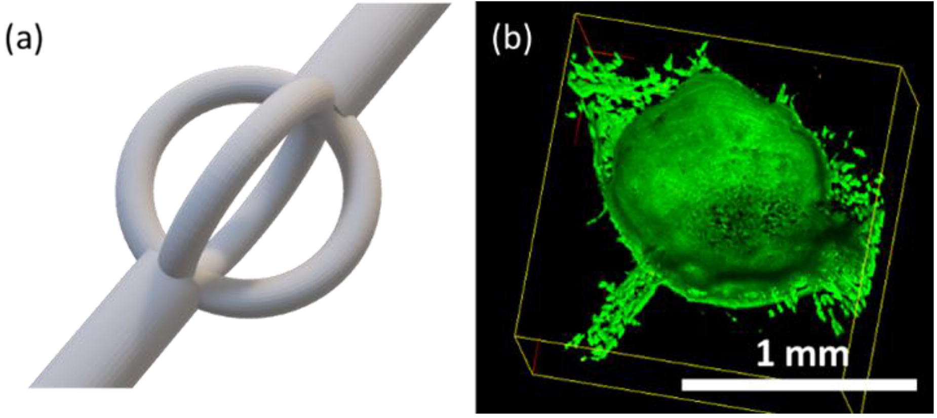
C232 - Designing An Artificial Vascular Network To Supply Larger Scale Tissue Models
P. V. Srinivasula1, S. Hardt1, U. A. Nuber2
1
Institute for Nano- and Microfluidics, TU Darmstadt, Darmstadt, Germany,
2
Stem Cell and Developmental Biology, TU Darmstadt, Darmstadt, Germany
*Purpose/Objectives: Three-dimensional tissue models generated in the laboratory, including larger organoids, that recapitulate structures and functional units of natural human tissues provide a versatile platform for drug development and toxicology assessment, and hold great promise for regenerative medicine. However, the lack of a vasculature to supply cells with oxygen and nutrients and to remove metabolic end products represents a critical deficit of many human tissue models generated in the laboratory.
*Methodology: One strategy to overcome this deficit is the integration of an artificial network made of synthetic materials. However, since the materials of the fluidic network channels and the fluids (cell culture media instead of blood) differ from the in vivo situation, merely mimicking morphologies of a natural vasculature does not ensure a sufficient transport of metabolic species required for in- vivo -like cellular activities in the tissue model. Rather than that, the design of such an artificial network shall be derived systematically for an optimal balance between biological requirements and the physical and engineering constraints of the materials and methods employed in its fabrication.
*Results: In this work, we conduct rigorous mathematical modeling of a 3D tissue model consisting of a cell-hydrogel matrix and an aqueous network to transport metabolic species between the cells and the external environment, as shown in figure 1A. The results of the computational study feed into a four-step procedure (figure 1B) to derive an optimal design based on the type of tissue, engineering materials, as well as the fabrication and operational parameters of the supply network. Firstly, such diverse biological and engineering aspects shall be considered to propose a conceptual design, such as the cubic network geometry of the supply system shown in figure 1A. Secondly, the geometric and operational parameters shall be quantified to avoid cellular hypoxia in the tissue model. The third step is to modify the parameters to ensure that the metabolic activity in the tissue model is similar to in vivo conditions. Finally, the necessary design changes and margins shall be included to optimize the fabrication and resilient operation, considering the permeability of the fluidic channel walls and the probability of gas bubble formation in the channels.
*Conclusion/Significance: Through these results, we demonstrate that a supply network that can be fabricated via 3D printing is sufficient to meet to the metabolic requirements of a several centimeters large tissue model. The design procedure introduced here is formulated in such a way that it is applicable to a variety of tissue types and materials.

C233 - Using Natural Killer Cells In 3D Melanoma Organ-on-a-Chip Platform To Evaluate Immunotherapy
S. Chakraborty1, A. Skardal1, A. Vilgelm1, A. Davies2, V. Bharti3
1
The Ohio State University, Columbus, OH,
2
Oregon Health and Science University, Columbus, OH,
3
Vanderbilt University, Belleville, MI
*Purpose/Objectives: In invasive and metastatic tumors, traditional therapeutic modalities including radiation, chemotherapies, stem cell therapy, etc. often exhibit poor efficacy. Despite positive outcomes-effectiveness and durability in subsets of patients-of techniques using immune checkpoint inhibitors and therapeutic immune cells, (e.g. Chimeric Antigen Receptor T cells or CAR-T), many tumors are resistant to these therapies as a result of immune suppression and are characterized as ‘cold’ or ‘immune deserts3’. Also, deficiency of tumor antigens or high variability in the few present, make T cell directed approaches like Chimeric Antigen Receptor T cells (CAR-T) and Immune Checkpoint Blockade (ICB), ineffective. Thus, using another lymphocyte population, natural killer (NK) cells, in cell-mediated immune therapy can be advantageous as these cells target other cells such as most cancer cells that lose their MHC class I expression.Here, we will us natural killer (NK) cells in a 3D microfluidic device-a
N
atural
K
iller cell-containing
T
umor-
o
n-a-
C
hip (NK-TOC)-to effectively assess immunotherapy. We aim to do this via studying homing and migration patterns of chemokine-driven NK cells towards tumors (cells).
*Methodology: Organoids are constructed using a methcrylated collagen-thiolated hyaluronic acid-based hydrogel and highly metastatic A-375 melanoma cells. The tumor cells are treated with small molecule kinase inhibitor drugs alisertib (ALS) and abemaciclib (ABE) for 5 days which render tumor cells senescent. Once in this state, the tumor cells secrete a plethora of chemokines like CCL5 and CXCL10 which we hypothesize attract the NK cells towards the tumor cells and thus enhance their cytotoxic functions. The NK-TOC platform is designed with specific chambers where the organoids can be injected using a small gauge needle. Labeled-NK-92 cells are infused through the chambers via a peristaltic pump on day 5. This enables tracking of NK-cell migration as well as challenges NK cells to leave their natural path of circulation and move towards the organoids to carry out their effector function. All imaging and viability-based output metrices are measured on day 6.
*Results: True to our hypothesis, we observed not only greater NK cell migration towards drug treated organoids but also greater secretion of these chemokines, particularly CCL5 and CXCL10 in the drug-treated samples than those without drug.
*Conclusion/Significance: Thus, chemokine-fueled NK cell migration can boost effector function and augment existing chemotherapies. The next steps of this study will include stromal components like fibroblasts, which are abundantly found in the tumor microenvironment, in this system to create a more complex and realistic environment and evaluate NK-cell efficacy.
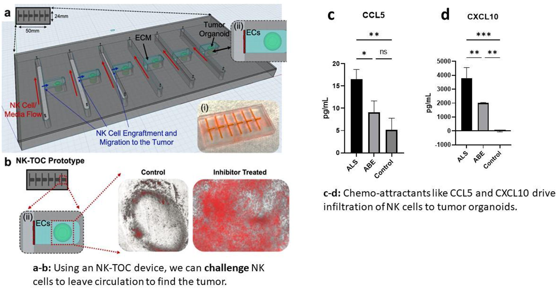
C234 - The Glomerular Filtration Barrier: Fabrication And Testing Of A 3D In-vitro Model
C. Mussoni1, A. Rederer2, J. Müller-Deile2, T. Ahmad1
1
University of Würzburg, Würzburg, Germany,
2
Erlangen University Hospital, Erlangen, Germany
*Purpose/Objectives: The kidney is a very dynamic organ that operates the key function of filtering waste from the blood in 2 million glomeruli. The glomerulus is the complex initial part of the nephron, and the core functional component is the filtration barrier: a triple layer of highly specialized cells, podocytes, the basement membrane, and glomerular endothelial cells (GEC). Failures in this organ, especially in the integrity of the filtration barrier, originate many kidney diseases. It is, therefore, interesting to study in more details the mechanisms and structures of the glomerulus. The challenge lies in replicating a functional in vitro model that addresses the needs of these cells and can be versatile to study healthy and diseased tissues. So far, only a few studies have focused on tailoring the properties of the artificial membrane to improve the bio-mimesis of a glomerulus model. We hypothesize using a biomimetic membrane to support the co-culture of glomerular cells, tested by replicating physiological conditions in a custom- built bioreactor.
*Methodology: The aligned nanofibers of Poly-L-Lactic Acid were fabricated through electrospinning, and then functionalized with polydopamine-assisted gelatin immobilization. The surface characterization of the membrane was analyzed using Scanning Electron Microscopy and Raman spectroscopy. To measure the dopamine coating, gelatin immobilization, and gelatin release over three weeks, a micro-BCA assay was conducted. First, human GECs and podocytes were seeded on each side of the membrane to determine cell adhesion in static condition. Later, we 3D printed a millifluidic bioreactor using a Prusa DLP Printer, assembled the membranes in it, and cultured cells on either side of the membrane. To prevent hypoxia, a PDMS window is present on the reactor; flow simulations were run to optimize the structure. We then perfused media separately on either channel with a rate of 5μL/min for GECs and 1 μL/min for podocytes. Furthermore, albumin was used as a model protein to evaluate the membrane's function.
*Results: The membrane presented an average thickness of 10 μm and fibers an average diameter of 0.5 μm and are aligned at 86%. Porosity is 40%. Raman spectroscopy shows the typical peaks of the amide bond of gelatin in addition to the PLLA peaks. After 24 hours, 80 μg/cm2 of gelatin is present on the membrane. After 21 days 50% of the gelatin was released in PBS under static conditions. Cells present an elongated morphology, spread, attach, and form a tight monolayer. Thereafter, we analyze the high viability of cells under flow. The SEM images revealed that both cell types remained attached on each side after 5-days of flow. Further, we observed the high viability of cells during their culture type. The Actin staining revealed that cell morphology was significantly affected under dynamic culture conditions and cell-typical structures are present.
*Conclusion/Significance: This in vitro model of the glomerular filtration barrier is based on a biomimetic membrane, to address the needs of the specialized glomerular cells to recapitulate the functionality under dynamic conditions. Further studies foresee the optimization of the tissue maturation to replicate diseased tissue models.

C235 - Modeling Airborne Hazard Exposures Using An Airway Organ Tissue Equivalent To Elucidate Common Pathophysiological Mechanisms And Medical Countermeasure Targets
J. Blackburn1, D. E. Shankle1, S. Albertson1, P. Clapp1, C. Langefeld1, H. C. Ainsworth1, M. N. Gurcan1, M. Rezapour1, Y. Kim2, A. Atala1, S. V. Murphy1
1
Wake Forest University, Winston-Salem, NC,
2
US Environmental Protection Agency, Durham, NC
*Purpose/Objectives: Burn pits are frequently used in war zones to eliminate waste on military bases. However, burning waste generates toxic airborne hazards and puts personnel and others in the area at risk for developing respiratory illness. There is an unmet need to understand the link between exposure to burn pits and development of respiratory injury and illness. Our group has developed a 3D airway organ tissue equivalent (OTE) that replicates the composition and complexity of the human airway and is compatible with air liquid interface (ALI) exposures. There is little known about the toxicity of burn pit condensate and adverse health effects, so this project aims to understand effects and mechanisms of disease following burn pit exposure of specific materials using the 3D airway OTE platform.
*Methodology: Burn pit material has previously been highly characterized to identify the chemical composition of inhaled smoke from burn pits. From the composition data, four material groups have been identified as most common: plywood, cardboard, plastic, and mixtures of all three materials. Each of these materials is created through a highly controlled furnace system that achieves steady-state flaming conditions for the collection process. The airway OTEs are developed for 28 days to maturity and exposed apically to each burn pit condensate material. Burn pit condensate dosing conditions were first established to replicate the chemical, biological, and physiological pulmonary responses of the human airway following burn pit exposure.
*Results: The OTE biological response was evaluated through measurements of transepithelial resistance, cilia beat frequency, viability, cytotoxicity, oxidative stress, and gene expression changes. Given the composition difference of each material type, the measured results expressed the effects of different burn pit conditions. With the data collected, correlative bioinformatic tools will be applied to characterize the biological impact of different burn pit smoke emissions and to identify emission components associated with toxicity outcomes.
*Conclusion/Significance: The results of this project will provide a better understanding of the toxicity effects of each of the condensate material types as well as the biological and functional impacts over time. This work represents an important and impactful finding of how burn pit exposure affects airways.
C236 - Characterization Of A 3D Microfluidic Blood-brain Barrier Model; Application To Study Immune Cells Transmigration Across The BBB In A Dynamic System
A. A. El-Taibany
Wake Forest Institute for Regenerative Medicine, Winston Salem, NC
*Purpose/Objectives: Microfluidics and shear stress have been reported to affect the expression of various Brain Microvascular endothelial cells (BMVECs) genes related to important physical and functional barrier properties. Therefore, blood flow acts in conjunction with the CNS microenvironment to develop specific BBB properties. Microfluidic flow also may affect the expression of cell adhesion molecules and affect the interaction of immune cells on the surface of the BBB.
*Methodology: 3D human BBB organoids containing an outer layer of human brain microvascular endothelial cells and pericytes and a core of neuroglial cells were allowed to form by Self-assembly in ultra-low attachment plates (ULA). Organoids were exposed to microfluidic flow (2 ul and 10 ul.min-1) for 48 hours using Ibidi µ-Slide Spheroid Perfusion chips. Organoids’ viability, integrity, surface area, and BBB permeability to dextrans were examined and compared to organoids grown in static ULA plates. BBB tight junction and cell adhesion molecule expression were compared between the dynamic and static systems using TaqMan qPCR assay. Transendothelial transmigration (TEM) of PBMCs was tested at a flow rate of 2 ul.min-1 and 10 ul.min-1. and compared to TEM in static organoids. Characterization of different PBMCs subpopulations after transmigration was conducted using flow cytometry.
*Results: Organoids exposed to microfluidic conditions demonstrated better integrity, and improved viability compared to their ULA counterparts. Permeability to dextrans was not significantly changed between the two groups. Expression of some tight junction molecules was significantly increased in microfluidic chips and the expression of all cell adhesion molecules was significantly decreased in microfluidic systems. TEM of PBMCs was significantly lower in organoids under microfluidic flow compared to their ULA counterparts. The percent of CD19+ B cells and CD3+ cells were comparable in all conditions with natural killer cells and plasma cells showing significantly lower TEM.
*Conclusion/Significance: Microfluidic conditions and dynamic cell culture improved the viability and integrity of the 3D BBB model. Furthermore, it decreased the expression of cell adhesion molecules, which reflects the quiescence state of normal BBB that limits the migration of immune cells under normal conditions. Microfluidic BBB models provide a better physiological platform for testing drugs, developing therapeutics for CNS disorders, and studying the interactions of the immune cells at the BBB interface.
C237 - Optimizing T-cell Dynamics Within A Microfluidic Device
S. A. Jackson, A. Bradley, M. Samojlik, A. Fernandez, C. Stabler
University of Florida, Gainesville, FL
*Purpose/Objectives: Microphysiological systems (MPS) are advancing toward immunocompetent models that include cellular immunological components. A key challenge with the integration of immune cells into MPS is the development of methods for supporting the temporal recirculation of cells without altering their phenotype. Current limitations include pump mechanical forces, variable system shear, and settling cell suspensions. To combat these issues, we integrated new design components of shear-thinning polymers and variable agitation methods into an MPS. The efficacy of these approaches was explored using T-cells.
*Methodology: Viable Jurkat T-cells were suspended in media with a titration of methyl cellulose (MC). The effects of MC on statically cultured Jurkat viability and proliferation were assessed via flow cytometry. For dynamic experiments, cell suspension and viability were assessed via trypan blue exclusion counts of a 30min collection from pump line outflow over a course of up to 72hrs. Similarly, the combined effects of the peristaltic pump, spinner flask agitation, and MC inclusion were assessed. Confocal imaging was used to give insight to T-cell migratory ability and system accumulation.
*Results: To address challenges associated with settling of the cells within the reservoir, the viscosity of the media was altered using methyl cellulose (MC). Screening of cellular impacts over the course of 3 days found that concentrations of MC up to 0.5% had no significant impact on the viability or proliferation of Jurkat T-cells. However, MC media supplementation did not overcome the effects of settling within the first 24hrs of circulating, as no significant benefits were measured for T-cells circulated using media or media plus MC. Agitation via spinner flask without MC, however, led to significant improvements in cell count over time and had no negative impacts on viability. Finally, while no deleterious effects of MC supplementation on T-cell viability was observed for static cultures, the combination of MC with spinner flask agitation was associated with a steady decrease in Jurkat viability.
*Conclusion/Significance: These are the first steps towards the development of recirculating T-cell flow in an MPS system. 0.5% MC supplementation improves cell suspension over the course of 72 hours when compared to 0% MC group. MC addition had significant impacts on viability, even with additional agitation, making it a less viable strategy for long-term immunocompetent MPS studies. Agitation via spinner flask led to significant improvements in cell suspension over the same period, while also having no significant negative effects on T-cell viability. Cell stock agitation appears to be a more promising avenue for the development of recirculating T-cell MPS systems. This work to increase the length of time that T-cells can be maintained in suspension allows for more complex questions about in vitro cancer and autoimmune disease models to be explored. Future work centers around testing alternative MC and angular frequency parameters with Jurkats and primary activated Human CD8+ T cells, beginning the development of a type I diabetes pathogenesis MPS.
C238 - In Vitro Approaches For The Gut Microbiota-Human Crosstalk Investigation
C. Daddi, M. Calvigioni, F. Biagini, E. Ghelardi, C. De Maria, G. Vozzi
University of Pisa, Pisa, Italy
*Purpose/Objectives: The human gut microbiota (HGM) is a heterogeneous microflora that resides in the gastrointestinal tract and has a significant impact on human pathophysiology. Indeed, it establishes a symbiotic interaction with the host, and abnormal changes in its composition (i.e., dysbiosis) are associated with a plethora of disorders and diseases. Among the common HGM study approaches, the analyses of human and animal feces present reliability issues, and results from in vivo animal models have a limited translational value. In vitro approaches represent powerful tools to model and study complex microbial communities, but biomimicry remains an open challenge. Within this framework, novel in vitro culture devices could overcome the bottleneck in this research area.
*Methodology: The first step involved the establishment of a biomimetic 3D HGM in vitro model: biomaterial-based electrospun structures can be used as scaffolds for the HGM in vitro culture (Biagini F. et al., Bioengineering, 2022). Their efficiency in preserving microbial biodiversity while supporting microbial proliferation and biofilm formation was assessed through static in vitro cultures of HGM derived from healthy human feces. Electrospun gelatin scaffolds promoted the generation of stable and long-lasting biofilms and maintained the microbial community’s composition during culture. However, the static culture approach couldn’t reproduce the features of the HGM’s physiological environment, therefore lacking biomimicry. This can be overcome with dynamic culture bioreactors to allow the continuous perfusion of nutrients and the removal of waste products while generating relevant environmental conditions (e.g., anaerobiosis) (Biagini, F. et al., Bio-Design and Manufacturing, 2023). Both chemical and mechanical stimuli were demonstrated to affect microbial adhesion and proliferation. Moreover, systems reproducing the typical intestinal peristalsis were fabricated and integrated into such devices, with the perspective of replicating fundamental environmental features and stimuli (Kim, H. J. et al., Lab on a Chip, 2012).
*Results: In this context, we have proposed shear stress-controlled dynamic culture systems that ensured the development of homogeneous microbial density patterns and improved the viability and stability of the cultured HGM sample, and preliminary results seem to suggest the promotion of biofilm formation over time. The integration of a sensing system, with reduced dimensional impact and risk of sample contamination, allowed us to continuously monitor relevant environmental parameters (e.g., dissolved oxygen, pH) and set the basis for the implementation of their active control.
*Conclusion/Significance: The ultimate effort envisions the combination of these HGM in vitro culture devices with advanced systems for the in vitro co-culture of HGM and human eukaryotic cells. Specifically, the integration of the developed systems in multi-organ platforms, replicating other organs in vitro, will allow the investigation of the complex interaction between the HGM and other anatomical districts. This will shed light on the impact of the microbiota on the host’s health and dysbiosis-related diseases’ onset, progression, and treatment.
C239 - Integrating Microfluidics And Electrospun Scaffold In Breast Cancer Tumor-on-a-chip (ToC) Models: Design, Optimization, And Computational Insights
V. La Carrubba, E. Capuana, M. Testa, F. Lopresti
University of Palermo, Palermo, Italy
*Purpose/Objectives: Breast cancer, a prevalent malignancy among women, demands innovative approaches for personalized therapy and drug screening. Tumor-on-a-Chip (ToC) models hold promise for high-throughput analysis of anticancer drugs and biological factors [1]. This work outlines the design, process parameter optimization, and computational fluid dynamic (CFD) analysis of a ToC coupled with an electrospun scaffold.
*Methodology: The design of the ToC was created using Autodesk Fusion 360. It comprises a connector, a ToC device, and a removable base for support. The ToC has two hexagonal culture chambers with a biopolymer septum, which enables different cell and media conveyance. COMSOL software was used for CFD simulations to optimize flow dynamics in the chip with and electrospun membrane integrated into the device. At first, a uniform porous solid was introduced between the two chambers to model the membrane. The chip's two chambers were conceptualized with laminar flow, while the membrane was treated as a porous matrix governed by Darcy's law [2]. Two flow configurations were compared: a flow rate of 42 µl/hr was used in both chambers for the first analysis; in the second configuration, one chamber had a flow rate of 42 µl/hr, and the other had 420 µl/hr. The second CFD analysis was conducted to evaluate the shear stress that cells experience during fluid flow in the microfluidic system. The study used fluid-dynamic simulations at the microscopic scale, building on previous results. Fusion360 software was used to create a 3D structure by overlaying tubular structures onto a scanning electon microscopy (SEM) image of an electrospun membrane.
*Results: The final configuration was a 70 µm combined structure comprising two 13 µm-thick membranes (CAD-drawn) coupled to a uniform porous solid, with the fibrous matrices in direct contact with the upper and lower chamber fluid volumes [3]. The macroscopic simulation results provided velocity and pressure values at a distance of 100 µm from the membrane, then applied as boundary conditions in the microscale system. The study revealed the flow direction as the inlet flow rate changes in the two chambers. The shear stress profile along the membrane fibers was in line with thresholds for cellular integrity, set at 3 Pa for breast cancer cells [4].
*Conclusion/Significance: The innovation of this study lies in its seamless integration of microfluidics and electrospun scaffolds, which provides insights into macroscopic and microscopic interactions. The outcomes establish a foundation for exploring diverse operational and geometric conditions, offering promising avenues for advancing research on breast cancer and therapeutic strategies through a better understanding of cellular dynamics within Breast ToC models.
Acknowledgements: The research was supported by Complementary Operational Programme (POC) 2014/2020 of the Sicily Region and by “SiciliAn MicronanOTecH Research And Innovation CEnter ”SAMOTHRACE" (MUR, PNRR-M4C2, ECS_00000022).
References: [1] doi.org/10.3390/MA12182945; [2] doi.org/10.1002/jbm.a.31556; [3] doi.org/10.1007/s10409-020-00953-4; [4] doi.org/10.1038/s41698-020-0121-2.
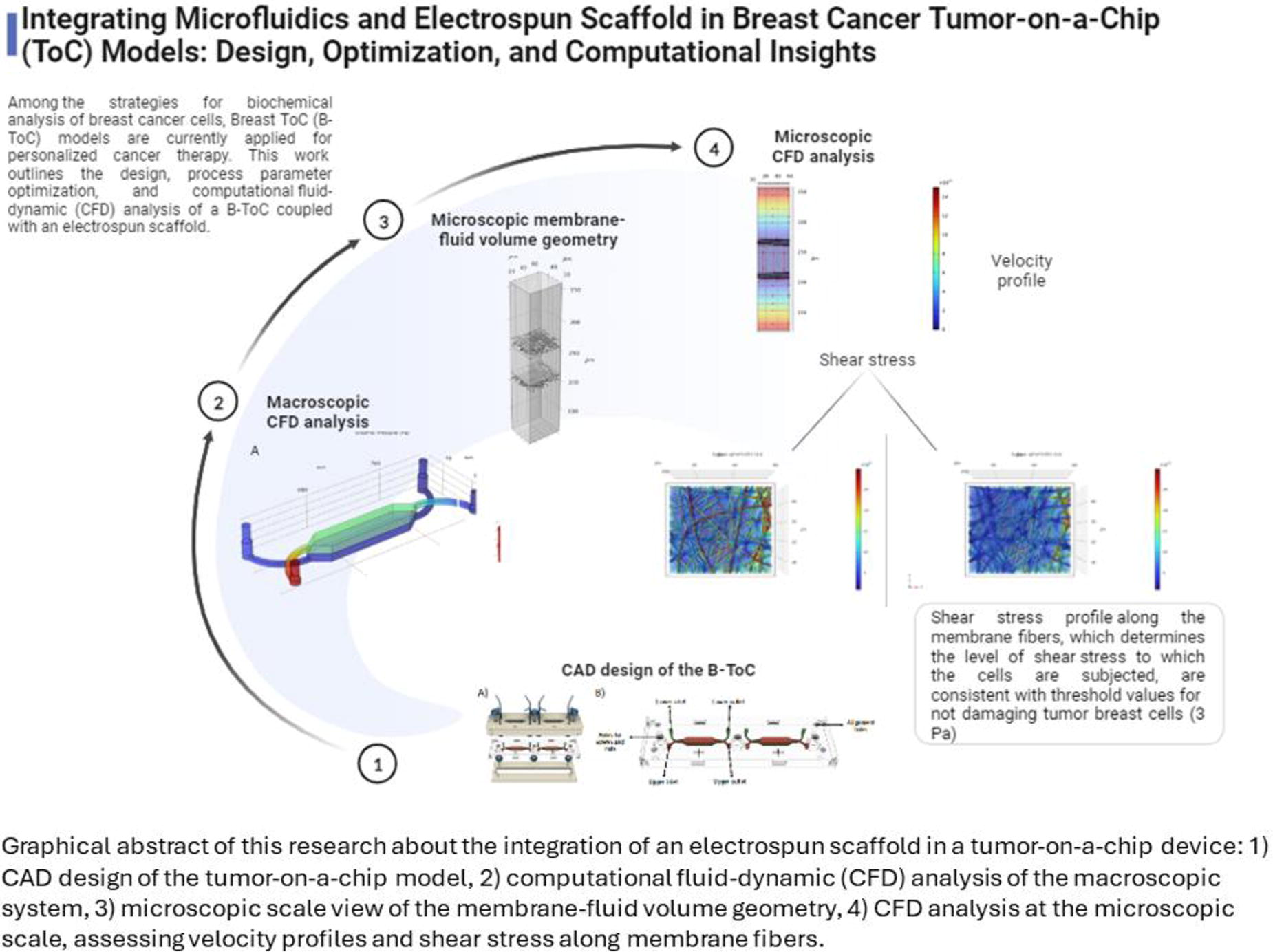
C240 - Fibercyte Template Technology: User- And Cell-friendly Channel Formation For Organ-on-chip Models
A. Seijas Gamardo1, I. Beeren1, V. de la Rosa2, R. Hoogenboom2, L. Moroni1, P. Wieringa1
1
Maastricht University, Maastricht, Netherlands,
2
University of Gent, Gent, Belgium
*Purpose/Objectives: Over 90% of drugs, identified in the preclinical pipeline, fail in the human trials. These results are typically explained by the lack of predictive power of animal models. Moreover, in addition to ethical considerations concerning unnecessary animal usage, the high failure rate results in loss of investments. Human organ-on-chip (OoC) models have been developed to resemble the physiological environment more closely. In many tissues such as cancer, lungs, or the gut, cellularized channel structures play a pivotal role in tissue function, such as microvasculature that provides oxygen and nutrients. However, the incorporation of functional, perfusable channels in OoCs has been challenging, especially in the lower micron scale. Here, we introduce the FiberCyte templating technology as a simple strategy to rapidly generate an interconnected fiber network, with channels ranging from micron to millimeter scale, and validate the robustness and versatility of template production and OoC application.
*Methodology: A range of channel templates were created by processing OXA, a family of poly(2-oxazoline) copolymers, which possess a tunable lower critical solution temperature (LCST). Using several manufacturing techniques such as melt extrusion, melt electrowriting, and laser ablation, we generated various template designs, from monochannel to branched channel structures. Template structures were assessed via light and scanning electron microscopy. We also established strategies to integrate templates into the manufacturing process of different OoC platforms. Within the OoC device, the template was embedded by introducing a variety of commonly used hydrogels, with and without cells (e.g. fibroblasts, endothelial cells). The OoC was then placed in the fridge for 30 minutes. We evaluated the formation of a channel network using microscopic methods, assessed perfusion, and evaluated cellular responses.
*Results: A robust library of polyoxazoline was developed displaying a LCST, enabling on-demand dissolution by lowering the temperature from 37°C to 4-20 °C. The speed of solubilization can be tuned depending on the copolymer composition. Subsequently, the manufacturing parameters were optimized to yield templates with a broad range of dimensions (5-200 μm) with a high degree of reproducibility. Templates were successfully integrated into OoC platforms and exhibited stable embedding within several types of hydrogels (e.g. PEGDA, alginate, collagen, and matrigel). After dissolution of the template, microscopic images as well as the perfusion of fluorescent beads confirmed the formation of an open channel in the hydrogel. Finally, cell viability was confirmed via fluorescent imaging, validating overall biocompatibility of the polymer as well as the mild dissolution procedure . Moreover, these results indicated that the fibrous template could be pre-seeded with any cell type of interest.
*Conclusion/Significance: Taken together, we successfully fabricated 3D templates of interconnected channels, ranging from micro to millimeter scale, which can be removed under benign temperature conditions. We envision making these templates broadly available for integration into new and existing OoC models to enhance the mimicry of the physiological tissue environment, paving a way forward to further enhance the predictive power of OoC models. In addition, the template supported the adhesion of various cell types, indicating potential to pre-form cellularized channel structures.
C243 - Comparison Of Adipocyte Spheroids From Human Stromal Vascular Fraction And Human Adipose Stem/stromal Cells In Adipogenic And Vasculogenic Culture Conditions
A. Yrjänäinen1, M. Juntunen1, N. Lostedt1, M. Kuuskeri2, S. Miettinen1
1
Tampere University, Tampere, Finland,
2
Tampere University Hospital, Tampere, Finland
*Purpose/Objectives: Due to increasing prevalence of obesity, there is a need for studying human adipose tissue physiology and pathophysiology in terms of weight gain and metabolic functionality. Excessive energy intake leads to adipogenesis, i.e. expansion and differentiation of adipose stem/stromal cells (hASCs) to mature adipocytes. Though the process of lipid accumulation has been widely studied in traditional 2D monocultures, the lipid droplets remain small and multilocular differing from mature, unilocular morphology (1). In addition, as adipose tissue consists of multiple different cell types and structures e.g. vascular network affecting the tissue functionality, the multicellular nature of adipose tissue needs to be implemented into the study models as well (1). Here, we aim to generate unilocular adipocyte spheroids of hASCs or cell population of stromal vascular fraction (SVF) to compare their adipogenic potential and differences in their metabolic functions to mimic the process of adipogenesis and potential angiogenesis in SVF spheroids.
*Methodology: hASCs and SVF cells were isolated from human adipose tissue samples (n=3). Two types of spheroids were plated; 10 000 hASCs or SVF cells were plated on low attachment 96-well plate in different culture conditions: basic medium (BM), adipogenic medium (AM), endothelial medium (EM), or AM+EM combination medium. Spheroid size was measured during 28-day culture. Formation of lipid droplets within spheroids was studied with confocal microscopy. Finally, adipogenic and angiogenic gene expression, secretion of adiponectin and lactate hydrogenase (LDH) were measured.
*Results: SVF cells and ASCs (Fig. 1) formed spheroids in all studied conditions. Largest spheroids were formed with AM+EM medium in both spheroid types. Also, lipid droplets were observed to form in AM conditions on both spheroid types. hASC spheroids in AM started to float before 21 days of culture indicating notable lipid droplet accumulation. Adipogenic gene expression was highest with AM cultured spheroids with both SVF cells and hASCs in line with the observed, increased lipid droplet formation. Interestingly, EM or AM+EM medium did not increase expression of angiogenic genes or support the formation of vascular structures. Thus, further optimization is needed for maintaining angiogenic potential in SVF spheroids. Highest secretion of LDH was observed with largest spheroids in AM+EM medium and highest concentration of adiponectin in AM and AM+EM medium in both spheroid types.
*Conclusion/Significance: To summarize, both spheroid types showed strong adipocyte spheroid formation capacity and notable differentiation towards adipocytes in adipogenic culture conditions. Both spheroid types will be cultured in microfluidic chips to establish a 3D adipose tissue in vitro model and further compared in terms of vasculogenic potency.

(1) Ioannidou et al. 2022 DOI:10.1113/JP281445
C245 - Development Of A Liver-on-chip Model To Simulate Liver Inflammatory Response
D. Chawla1,2, C. Pang1, H. Chan1,2, R. S. Tuan1,2
1
The Chinese University of Hong Kong, Hong Kong, Hong Kong,
2
Institute of Tissue Engineering and Regenerative Medicine, The Chinese University of Hong Kong, Hong Kong, Hong Kong
*Purpose/Objectives: Inflammation is a common response observed in various liver diseases and is usually managed by immunosuppressive therapies. Uncontrolled inflammation can lead to irreversible tissue damage and liver failure that may require liver transplantation. Since human cell-based in vitro models can potentially mimic our biology more accurately than animal models, organ-on-a-chip platforms developed using cell and organoid culture have recently been employed to investigate disease mechanisms and perform drug testing.
*Methodology: In this project, HepG2 cells and human embryonic stem cells (hESC) that were differentiated into hepatocyte-like cells (HLCs) were cultured in 2D cell culture and as 3D spheroids using various techniques for modelling liver inflammatory conditions and screening potential therapeutic agents such as drugs and stem cell-derived products. To simulate inflammation, cultures were exposed to conditioned medium derived from human monocyte cells (ThP-1 cell line) polarized into pro-inflammatory macrophages using phorbol 12-myristate 13-acetate (PMA) and treated with lipopolysaccharide (LPS) to obtain conditioned medium.
*Results: We showed that hESCs successfully differentiated into HLCs and exhibited hepatic markers in 2D, similar to cultured HepG2 cells, and formed hepatic spheroids in 3D culture systems. Treatment with a macrophage conditioned medium, shown to contain cytokines such as IL-6, IL-1β, TNF-α, was used to simulate inflammatory response. After treatment, HepG2 cells and the HLCs cultured in both 2D and 3D showed a significant drop in cell number and albumin production, as measured by CCK8 assay and ELISA, respectively, indicating detrimental impact on the cultured cells. These results demonstrated preliminarily the feasibility of using a biological, human cell-based system to simulate the response of liver cells to inflammatory cytokines.
*Conclusion/Significance: This work has laid the foundation for the development of more advanced hepatocyte cultures such as 3D organoid and organ-on-chip models to simulate inflammation in the liver, investigate disease mechanisms, and screen potential therapeutic agents. It would be of interest to investigate possible co-culturing with other liver cell types and integration of multiple organ-on-chips with biosensing monitoring and perfusion systems to model multi-organ diseases in the future.
C246 - Characterization And Evaluation Of Biomaterials For An Organ-on-chip Device For The Cardiovascular System
J. Ortiz-Ortiz, G. Carbajal-De-la-Torre, N. N. Zurita-Méndez, M. A. Espinosa-Medina, H. Aguilar-Rodriguez
Universidad Michoacana de San Nicolas de Hidalgo, Morelia, Mexico
*Purpose/Objectives: An Organ-on-Chip device is a microfluidic cell culture device created using microchip manufacturing methods that contains continuous perfusion chambers inhabited by living cells arranged to simulate physiology at the tissue and organ level. These systems can incorporate physical forces, including physiologically relevant levels of fluid shear stress, cyclic tension, and mechanical compression, allowing the analysis of organ-specific responses to physical and biochemical perturbations. In this work, the mechanical and biochemical characteristics of the biomaterials involved in the design of an Organ-on-Chip device that is intended to be used with cells of the cardiovascular system are evaluated.
*Methodology: Mechanical evaluations are carried out on Polyurethane-Polylactic-acid based scaffolds for the cardiovascular system in two different ways, compression until permanent deformation, to determine mechanical properties, and on the other hand, cyclic tests with a low rate of deformation with the objective of evaluating the response of the material to repetitive loads. The tests were carried out in universal test machine Univert from CellScale at room temperature. Characterizations by XRD, FTIR and SEM were carried out in order to analyze the microstructure, morphology, and composition of each scaffold; the evaluation of bioactivity and biodegradation was performed in simulated body fluid (SBF) and phosphate buffered solution (PBS) according to the ISO 10993-13:2010.
*Results: Physicochemical evaluations of Polyurethane-Polylactic-acid scaffolds were obtained through XDR, FTIR, and SEM characterizations from which the morphological characteristics and composition are observed, in addition to the stress-strain curves and mechanical properties through mechanical tests.
*Conclusion/Significance: With the results of the mechanical evaluation, the physicochemical characteristics and the evaluation of bioactivity and biodegradation, useful and interesting results are obtained for the materials that can be used as an extracellular matrix in an Organ-on-Chip device for cardiovascular tissue.
C247 - Microvascular Endothelial Cells Are More Prone To Preservation Injury Than Macrovascular Endothelial Cells
B. W. Ellis, I. Filz von Reiterdank, H. Chen, A. T. Dinicu, K. Uygun, B. E. Uygun
Massachusetts General Hospital, Boston, MA
*Purpose/Objectives: Chronic organ disease has been a growing global burden, accounting for 730,000 deaths yearly in the U.S. alone. The situation is even more dire at the global scale with over 90% of the demand for organ transplantation not being met. A key technical limitation for increasing organ availability is cold ischemia-reperfusion injury, which occurs during preservation of the organ in static cold storage (SCS) and subsequent warm recovery phase resulting in edema and loss of organ viability. Literature indicates endothelial cells (EC) are particularly susceptible to ischemia and microvascular ECs (MVEC) especially are of key interest in transplantation from the perspective of hypoxia and rejection. However, research targeting the endothelium, let alone the microvasculature, in the context of organ transplantation is nearly nonexistent. As such, there remains a significant gap in our understanding of the effect of preservation on MVEC, and consequently a lack of improvements in organ preservation media and technologies. The overall objective of this study was to develop an in vitro model to elucidate the effects of SCS preservation on MVEC in the context of transplantation. This will for the first time, allow the quantification of preservation injury dynamics on MVEC and how this differs from macrovascular ECs (MACECs), where preservation injury is not as drastic. Consequently, this study will enable improving the viability of endothelium over extended storage duration allowing for the development of novel methods to improve organ preservation.
*Methodology: MVECs and MACECs were grown to confluency in standard culture conditions. For permeability experiments, cells were seeded on transwell inserts. Cells were stored in storage solution at 4°C for up to 3 days to mimic clinical SCS damage. Afterwards, cells were recovered for 1 day. Following recovery, membrane integrity (Live/Dead), metabolic activity (PrestoBlue), cascade expression (Caspase-Glo), and endothelial permeability were measured. For permeability, following storage, cells were returned to standard culture conditions with the addition of FITC conjugated dextran to the media in the transwell insert and incubated for 30 min. Afterwards, the endothelial layer permeability was determined by measuring the fluorescence of the media below the transwell insert.
*Results: We observed that preservation conditions have a detrimental effect on cellular health (Fig. 1A), metabolic state (Fig. 1B), and apoptosis (Fig. 1C) with the MVECs being more adversely affected than MACECs. Furthermore, as shown in Fig. 1D, we observed that MVECs also have a drastic increase in permeability following SCS.
*Conclusion/Significance: The results from this study demonstrate that the loss of viability and edema observed during cold storage at the total organ level is correlated with microvascular health and functionality. Additionally, this study indicates both barrier function and apoptosis as potential therapeutic targets to increase organ preservation, which will be the focus of future studies.
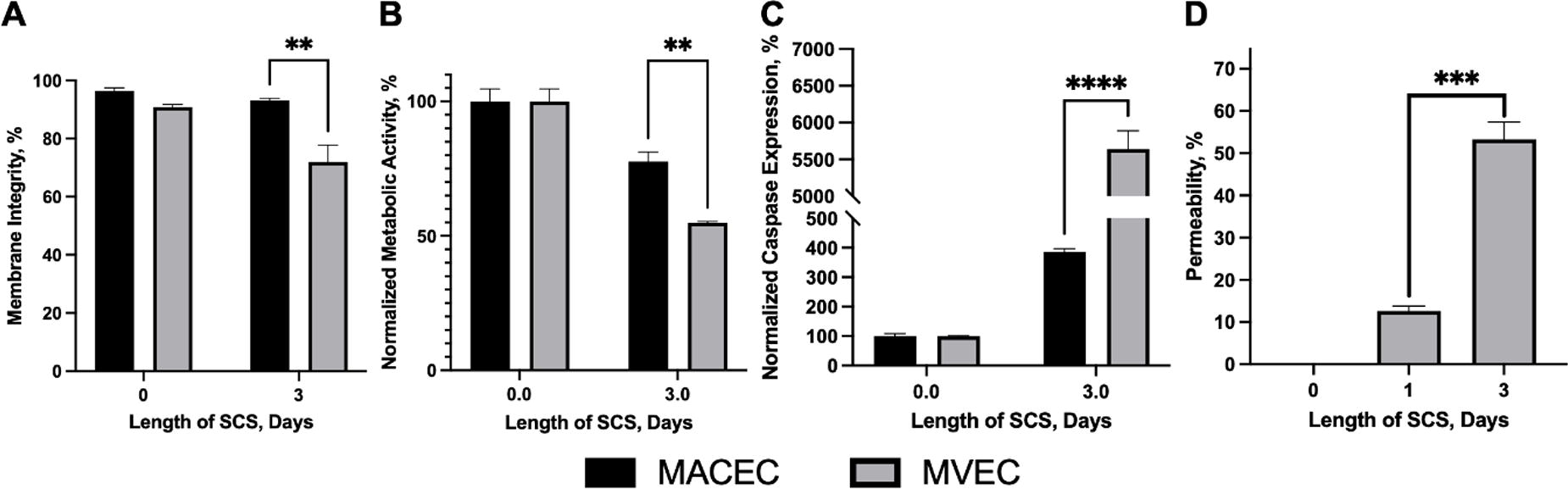
Fig. 1: Effect of SCS on MVEC and MACECs: SCS had a drastic effect on EC viability (A), metabolic activity (B), caspase expression (C), and permeability (D) with MVECs being more negatively affected than their macrovascular counterparts. **=p<0.01 and ***=p<0.001.
C248 - Stepwise Development Of Functional Airway Epithelial Cells Using Human Tonsil-derived Mesenchymal Stem Cells
S. Jung, H. Kim, H. Kim, S. Kim
College of Medicine, Ewha Womans University, Seoul, Korea, Republic of
*Purpose/Objectives: Airway defects are commonly encountered in clinical field. The main cause of defect is mostly tumor invasion or the trauma and also genetic anomaly. Morphological reconstruction could be done using many materials, including endotracheal tube materials, autologous ribs, and many tissue-engineered scaffolds. However, functional reconstruction of epithelium which should have cilia and mucus secretion function is challenging. In previous study, we developed acellular respiratory mucosal matrix using porcine tracheal mucosal using decellulrization which is enriched in site-specific extracellular matrix. For the next step, we tried to apply the respiratory epithelial cells, although autologous epithelial cells or progenitor cells exist, they are not suitable candidates for regeneration due to their difficult properties for expansion and differentiation in vitro. We developed an alternative for respiratory epithelial regeneration by using human tonsil-derived mesenchymal stem cells (TMSCs) to induce epithelial cells through stepwise differentiation.
*Methodology: TMSCs were isolated from human tonsillar tissues of patients undergoing tonsillectomy and differentiated into airway epithelial cells following the human embryonic developmental process. To generate airway epithelial cells, TMSCs were exposed to various chemical agents or protein combinations during four steps. We found that TMSCs can be induced into the definitive endoderm (DE) with a low concentration of activin A, which activator of the Nodal/ TGF-β signaling pathway. Next, a combination of growth factors regulating BMP, TGF-β, and WNT signaling induces the differentiation of DE-induced TMSCs into anterior foregut endoderm. An environment rich in BMPs, WNT, and FGFs differentiated TMSCs into lung progenitor cells. In the final step, TMSCs-derived airway epithelial cells generated through air-liquid interface culture.
*Results: The effects of activin A and serum concentration on DE differentiation were examined to quantifying the DE-specific gene expression (SOX17, CXCR4, and GSC) using qRT-PCR analysis. TMSCs-derived DE cells were differentiated by inhibiting BMP, TGF-β, and WNT signaling. qRT-PCR analysis revealed that the expression of anterior foregut-specific markers (PAX9, SOX2, and GATA3). Expression of NKX2-1, a key regulator of airway epithelial cell generation, was increased when the cells were cultured in environment rich in BMPs, WNT, and FGFs compared to control medium. Expression of keratin 5 (KRT5), a basal cell marker of airway epithelium, is also increased. TMSCs-derived airway progenitor cells were differentiated under air-liquid interface culture to facilitate physiological relevance. Immunofluorescence analysis showed significant expression of the ciliated cell marker, FOXJ1.
*Conclusion/Significance: Our results demonstrate that TMSCs-derived airway epithelial cells can be generated by stepwise differentiation and represent a potential alternative for treating functional recovery of respiratory defects.
C249 - ENGINEERING AN ARTIFICIAL ESOPHAGUS USING PATIENT-DERIVED INDUCED PLURIPOTENT STEM CELLS
A. Nemati, H. Savoji
University of Montreal, Montreal, QC, Canada
*Purpose/Objectives: Esophageal atresia in children and esophageal carcinoma in adults are severe diseases that may require esophageal replacement. Due to the lack of regenerative ability of the esophagus, continuity can only be restored by transposing the stomach or other intestinal segments into the thorax, which is challenging and associated with serious complications affecting patients’ quality of life. Therefore, combining patient-derived induced pluripotent stem cells (iPSCs) and tissue engineering approaches to create an artificial esophagus is a promising option to restore the loss of native tissue and overcome the limitations of conventional treatment. Here, we design an artificial esophagus mimicking the structural and mechanical characteristics of the native human esophagus. This artificial scaffold could allow the seeded cells to proliferate and release extracellular matrix.
*Methodology: We used extrusion-based 3-dimensional printing and melt electro-writing as novel fabrication methods to create an artificial esophageal construct using FDA-approved polymers, polycaprolactone (PCL), and poly(lactic-co-glycolic) acid (PLGA). Due to its highly tunable mechanical properties, PCL provides suitable structural integrity, whereas PLGA enhances the bioactivity and degradation rate of the scaffold following cell seeding. Pluronic F-127 was used as a sacrificial material to create pores and surface modification on PCL and PLGA polymer constructs to enhance cell attachment, growth, and proliferation. A tensile test was carried out to evaluate the mechanical properties of the printed tubes and compare the results to the native human esophagus. The structural integrity of these tubes was also assessed using a scanning electron microscope. Furthermore, iPSCs were seeded on vitronectin-coated tubes to determine the biocompatibility of the polymers to iPSCs.
*Results: The tubular scaffolds were fabricated using 3D printing and melt electrowetting technologies. The scaffolds were investigated to characterize their physico-chemical, morphological, mechanical, and biocompatibility characteristics. We have also optimized parameters, such as the polymer composition, cell seeding density, growth, and proliferation to achieve the optimal engineered esophageal tubular scaffolds. We observed that the iPSCs survive, maintain, and differentiate on the scaffolds known for their highly tunable characteristics, biocompatibility, biodegradability, and being non-toxic.
*Conclusion/Significance: This approach allows for the generation of patient-specific esophageal constructs. The urgent need for an efficient treatment for pediatric and adult populations because of congenital or acquired esophageal diseases requires developing a novel patient-specific engineered implant to improve the quality of life for the patients.

C251 - Xenogenic Application Of Human Placenta-derived Mesenchymal Stromal Cells In A Porcine Large Animal Model Of Cell Therapy
W. K. Aicher
University of Tübingen, Tübingen, Germany
*Purpose/Objectives: Mesenchymal stromal cells (MSCs) were considered as regenerative agents in many conditions. In different animal models of injuries or diseases, cell therapy with MSCs was successful. However, the translation in the treatment of human patients was not always achieved. We, therefore, explored in a proof-of-principle study, if human placenta-derived mesenchymal stromal cells (hpMSCs) can be injected into the vascularized tissue of pigs without being rejected immediately nor causing major inflammation. This study was designed as a proof-of-principle study to develop a xenogenic large animal model.
*Methodology: Male hpMSCs were expanded and characterized in detail by flow cytometry (FC), reverse transcription quantitative of mRNA and polymerase chain reaction (RT-qPCR). 1x10^6 hpMSCs each were injected into the urethral sphincter muscle of two female pigs. The reaction of the pig to the xenogenic injection was monitored by measuring body temperature, determination of inflammatory markers IL-1, CRP, and haptoglobin in blood, and histology to determine the infiltration of inflammatory cells to the area of hpMSC injection. Human DNA of male hpMSCs was detected by PCR of the AMELY- or SRY- genes in DNA extracted from cryosections of the injection site. Pigs w/o cell injections served as controls.
*Results: The hpMSCs were tolerated well. The indicators of systemic inflammation IL-1, CRP, and haptoglobine were not elevated in the blood of treated animals when compared to the controls. Male human DNA was detected in urethral cryosections seven days after the xenogenic injection of hpMSCs proving the success of the applications.
*Conclusion/Significance: The therapeutic potential of hpMSCs can be explored in a porcine large animal model of injury or disease. Using large animals and hpMSCs may facilitate surgical challenges of cell therapies associated with smaller models such as rodents.
C254 - Cartilage Tissue Models Shift From Viscoelastic To Elastic Properties In Culture
M. A. Boos1, S. R. Lamandé2, K. S. Stok1
1
The University of Melboune, Parkville, Australia,
2
Murdoch Children's Research Institute, Parkville, Australia
*Purpose/Objectives: Cartilage is a slow-remodelling tissue and has limited healing capacity. This has led to decades of trying to replicate cartilage tissue and engineer replacements. To this date achieving full functional and mechanical integrity of these biomaterials has proven difficult as cartilage tissue has distinct mechanical properties. Due to its biphasic nature, cartilage shows a viscoelastic behaviour under load driven partly by the swelling properties of the glycosaminoglycans (GAGs). Nevertheless, usually only the apparent stiffness is reported when analysing cartilage tissue engineered samples. However, the Young’s modulus does not give sufficient information due to the biphasic heterogeneous complexity of cartilage tissue. Therefore, the aims of this study were to investigate the influence of hydrogels with different GAG content on the viscoelastic properties over time.
*Methodology: Bovine chondrocytes were encapsulated in allylated gelatin (gelAGE) hydrogels containing thiolated chondroitin sulphate (CSSH) and were cultured over 35 days and the change in mechanical properties were assessed.
Briefly, gelatin was functionalised to contain alkene groups. Chondroitin sulphate was thiolated to contain a single thiol group at the non-reducing end. Thiolation of the CS was confirmed with an Ellman assay. Cells were isolated from articular cartilage of bovine carpal joints. The cartilage was minced, digested, filtered and the isolated cells were resuspended in PBS and added to the respective hydrogel solution based on the different groups at a final density of 3 x 10⁶ cells/ml. Hydrogels were cultured for 35 days.
Six samples (Ø 5mm, ∼ 1-2 mm thick) of each hydrogel group were placed in custom-made stainless-steel wells and immersed in PBS. A stress relaxation test was performed, as previously described by Nimeskern et al. [3] using a material testing machine (Zwick Z005, Ulm, Germany) with a stainless-steel indenter (Ø0.9 mm). Firstly, a preload of 3 mN was applied to the sample to determine its surface and measure sample thickness. After 5 minutes, 3 successive strain steps were applied in 5% increments of the original sample thickness. Each step was held for 20 min to allow the sample to relax. For every sample, an instantaneous modulus, equilibrium modulus, maximum stress, relaxation half-life time, and amplitude ratio were determined.
*Results: All samples showed a shift from viscoelastic properties towards more elastic properties during 35 days of culture (Figure 1). However, there were no significant differences in stiffness observed between the groups. The measured thicknesses of all samples showed no significant difference, ruling out influence of sample thickness. There were no significant differences between all groups on day 1 and day 35 for any measure. All groups showed increased instantaneous modulus, equilibrium modulus, and maximum stress at day 35 compared to day 1 indicating ECM production in the samples.
*Conclusion/Significance: These findings show a distinct change in viscoelastic properties of tissue engineered samples over time, while not showing a difference in stiffness between the different groups. The changes in mechanical properties are likely due to matrix produced by the cells. This highlights the importance of thorough mechanical evaluation beyond stiffness measurement to investigate maturing tissue engineered samples.
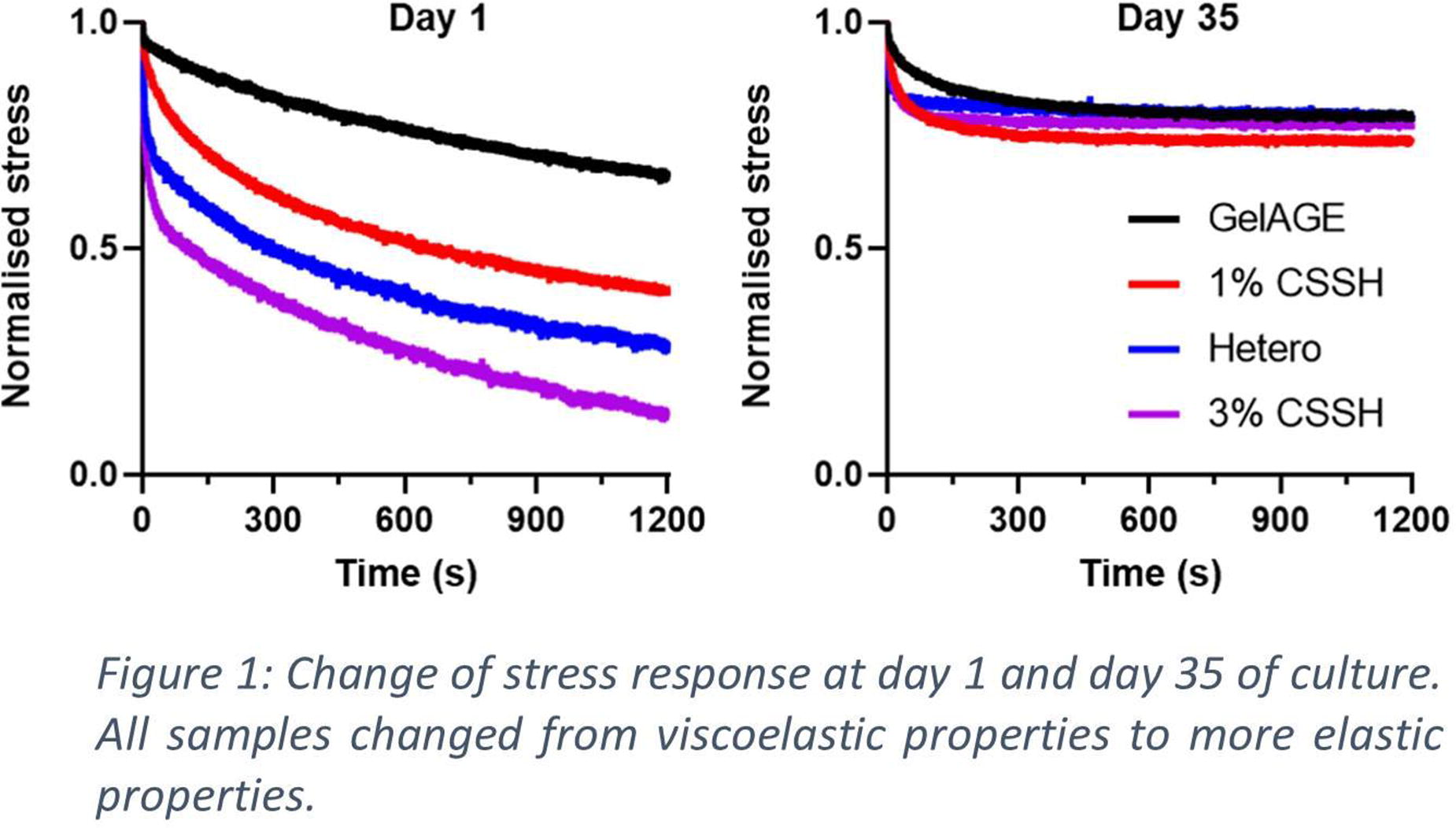
C255
C256
C257
C258
C259 - Bioengineered Granular Bioink For Development Of Porous Scaffolds
T. Seah1,2, J. Zhang1, J. Tay1, W. Wong1, C. W. Beh1
1
Agency for Science, Technology and Research (A*STAR), Singapore, Singapore,
2
National University of Singapore, Singapore, Singapore
*Purpose/Objectives: 3D bioprinting involves layer-by-layer construction of intricate structures which allows the precise deposition of biomaterials and cells to create scaffolds with physiologically relevant geometries. However, the absence of vasculature constrains the size and scalability of 3D tissue-engineered structures. Thick scaffolds relying solely on diffusion can only support cells within a few hundred microns away from the liquid-scaffold interface, thus significantly limiting the potential thickness of bioengineered tissues. In this study, we present the development of a granular hydrogel bioink designed to fabricate a highly porous scaffold that improves the mass transport of oxygen and nutrients to the embedded cells.
*Methodology: This work introduces a facile method of preparing a granular hydrogel bioink by using mechanical shearing (Fig 1a). The resulting granular bioink can be 3D bioprinted with commercially available bioprinters and photocrosslinked to yield a highly porous scaffold structure. The developed granular scaffold is composed of gelMA microparticles held together by interparticle covalent bonds creating void spaces, which contributes to the development of scaffold porosity (Fig 1b).
*Results: The granular gelMA bioink can be used to create a highly porous scaffold structure by casting, pipetting, or extrusion bioprinting. The interconnected void spaces between the microparticles make up approximately 35 % of the overall scaffold volume, and the average pore diameter is 150 µm. The granular gelMA bioink demonstrates shear-thinning and self-healing behaviour, which enables the bioink to be bioprinted and retain its mechanical stability after extrusion from the nozzle to ensure that the interparticle spaces are retained. These parameters can be tailored to specific tissue engineering needs by altering the degree of crosslinking. Additionally, due to the porous nature of the granular scaffold, media can penetrate through the entire scaffold easily, enabling cells to be seeded effectively into the scaffold. When Human Umbilical Vein Endothelial cells (HUVEC) suspended in medium are pipetted on the surface of the scaffold, they pass through the scaffold’s pores, coating the surfaces of the gelMA particles with the adherent cells (Fig 1c). Furthermore, the interparticle spaces enable larger structures like spheroids to be embedded into the granular bioink to be bioprinted. We demonstrated that mouse pancreatic cell MIN6 spheroids can be embedded into the scaffold structure (Fig 1d).
*Conclusion/Significance: We demonstrated the development of a granular hydrogel bioink that can be bioprinted and pipetted. The resulting scaffold structure is highly porous, which improves the mass transport of oxygen and nutrients to the embedded cells. The granular bioink’s suitability for bioprinting was established with rheological studies. Additionally, the two different methods of embedding cells in the scaffold were demonstrated. This approach offers a promising strategy to create complex 3D tissues with enhanced cell infiltration and functionality for tissue engineering and regenerative medicine applications.

C260 - A 3D Micro-structured Millifluidic Model Of Vascularized Tissue Regeneration In Vitro
C. Martinelli1, S. Milone1, T. Baldissera1, C. Conci1, A. Bocconi1, G. Chirico2, G. Cerullo1, R. Osellame1, E. Jacchetti1, M. T. Raimondi1
1
Politecnico di Milano, Milan, Italy,
2
University of Milano Bicocca, Milan, Italy
*Purpose/Objectives: Implantation of biomaterials is becoming an increasingly feasible approach in tissue engineering and mimicking in vitro the physiological responses triggered by this procedure is a crucial prerequisite for advancing to clinical application. Employing tridimensional (3D) cell culture systems alongside computational investigations offers a means to simulate the dynamics of living tissues. Through the integration of computational fluid dynamics (CFD) analyses and the use of a millifluidic optically accessible bioreactor (MOAB), designed for the perfusion of cell cultures within tridimensional micro scaffolds, we developed a rapid and reliable method for modeling vascularized tissue regeneration in vitro.
*Methodology: We fabricated 3D micro scaffolds consisting of an array composed of two rows of eight micro scaffolds, each of 500x500x40 μm, by 2-photon polymerization of the SZ2080 biocompatible photosensitive resin. Each 3D micro scaffold is characterized by pores of 50x50x20 μm3. We investigated the proliferation rates of co-cultured fibroblasts and endothelial cells in the presence of micro scaffolds and on flat glass slides. The optimal parameters for performing perfused cell culture and the shear stress values acting on cells cultured in the micro scaffolds’ array, together with the evaluation of cellular oxygen consumption along the bioreactor’s chamber, were set up by CFD. Confocal laser scanning microscopy (CLSM) was performed to follow 3D cell organization for up to 13 days in static and perfused conditions and upon stimulation with specific growth factors (VEGF and TGF-β1).
*Results: The optimal flow rate (10 μl/min) for achieving a maximum wall shear stress value of 0.8 mPa above the micro scaffolds was established by CFD analyses. In these conditions, the oxygen consumption inside the micro scaffolds’ array underwent a minimal decrease (of approximately 1.5 %) which did not influence cell survival and proliferation. Our device supports the formation of vascularized tissue and CLSM imaging showed that the presence of micro scaffolds in the bioreactor does not affect cell viability and promotes cell proliferation as compared to the static conditions. Moreover, a significant increase in collagen I deposition and CD31 expression, together with the reorganization of endothelial cells, were observed in long-term cell cultures (13 days) upon administration of growth factors and in the presence of the 3D micro scaffolds.
*Conclusion/Significance: CFD analyses enabled the assessment of the best parameters for cell culture under perfused conditions. Co-cultures of fibroblasts and endothelial cells proliferated in the presence of the micro scaffolds and inside the bioreactor’s chambers, showing a tridimensional re-organization upon growth factors stimulation. Our 3D micro-structured millifluidic system was demonstrated to be efficient as a model for recapitulating vascularized tissue regeneration in vitro. In future experiments, cellular uptake, and release kinetics of VEGF and TGF-β1 will be determined by biochemical assays in vitro and CFD analyses in silico.
Acknowledgements: European Commission (EU, FET-OPEN project IN2SIGHT, G.A. 964481); European Research Council (ERC, project BEACONSANDEGG, G.A. 101053122). Views and opinions expressed are those of the authors only, do not necessarily reflect those of the EU or the ERC. Neither the EU nor the granting authority can be held responsible for them.
C261 - Elaboration And Osteoregenerative Properties Of 3d Composite Scaffolds Based On Plga Modified With Metal Organic Frameworks (mofs) Particles.
C. Covarrubias1, D. Peña1, D. vargas1, A. Palomino2, M. Neira2, P. Capetillo2, A. von Marttens2, S. Acuña2, B. Le Monnier3, J. Estay2, M. Tsapatsis3, E. Whitehead4, W. Grayson4
1
University of Chile, Santiago de Chile (Metropolitana, Chile), Chile,
2
University of Chile, Universidad de Chile, Chile,
3
Johns Hopkins University, Baltimore, MD,
4
Johns Hopkins University, Johns Hopkins university, MD
*Purpose/Objectives: 3D printer technology is offering novel opportunities for the custom-made fabrication of bone regenerative biomaterials. However, available 3D printer filaments as those of Poly(lactic-co-glycolic) acid (PLGA) lacking osteoinductive properties. PLGA is a polymer with several FDA approbations and exhibit degradability times that match-well for its use as scaffold in maxillary bone reconstructions. In a previous work, we explored the synthesis of metal organic frameworks (MOFs) particles cytocompatible and with the ability to mineralize bone—like apatite and to stimulate the osteogenic cell differentiation. So, the aim of this study is to produce PLGA filaments loaded with MOFs particles to elaborate 3D scaffold and assess their in vitro and in vivo osteogenic properties.
*Methodology: MOFs were synthesized using 1,4-benzendicarboxylic acid (BDC) as organic linker bonded to calcium cations (MOF). The MOFs particles were subsequently surface modified with both Ca2+/Li+ (CaLi/MOF) and Ca2+ ions (Ca/MOF) by impregnation with Ca(NO3)2 and LiNO3 solutions. PLGA 85:15 was used to elaborate 1.75 mm composite filaments loaded with the 5, 10, 20, and 40 % of MOFs particles. Filaments were produced with an extruder at 80 ºC and then used to print cylindrical scaffolds of 4 mm diameter, 3.2 mm high and with cells of 0.6 mm. Structural characterization of the biomaterials was performed by FTIR spectroscopy and SEM/EDX microscopy. The viability of human gingiva-derived mesenchymal stem cells (GMSCs) incubated with the scaffolds was assessed with the MTS assay and osteogenic cell differentiation was measured by the alkaline phosphatase (ALP) activity. Bone tissue regenerative properties were assessed using a tibial critical-size bone defect (4 mm) in Sprague Dawley rats. Tissue formation was assessed using cone beam computed tomography (CBCT), back-scattering scanning electron microscopy (BS-SEM), and toluidine blue stain.
*Results: Fig. 1. shows the composite PLGA filaments loaded with MOFs. SEM microscopy confirms the presence of the MOFs particles homogeneously embedded into the PLGA polymer matrix. In addition, the FTIR spectrum of the composite filament presents bands around 3294 and 3485 cm-1 associated to the structure of the MOF particles. The elaborated filaments enabled printing a scaffold design for biological testing. The MOF/PLGA based scaffolds exhibited good cytocompatibility after 14 days of incubation with GMSCs (Fig. 1g) and were able to stimulate their osteogenic differentiation toward mineralizing lineage as observed the increasing of the ALP activity. The CBTC analysis (Fig. 1h) of the bone defects treated with the composite scaffolds loaded with 20% MOF showed that these biomaterials promote a higher bone tissue formation as compared to the neat PLGA scaffold. A quantitative analysis demonstrated that the mineral bone density in the defect area increased as follows: Control < PLGA < CaLi/MOF < Li/MOF. These results indicate that the incorporation of lithium-rich MOFs particles into PLGA significantly increases its bone tissue regenerative properties.
*Conclusion/Significance: The incorporation of MOF particles into PLGA enables the production of filaments and 3D PLGA scaffolds with enhanced osteoinductive properties in vitro and bone regenerative capacity in vivo. Particularly, the MOFs modified with lithium cations impart higher osteopromoter properties.

C262
C263 - The Induction Of Calvarial Bone Formation By 3d-printed Plga Microsphere Scaffolds
R. M. Klar, J. Cox, N. Raja, S. Lohfeld
University of Missouri-Kansas City, Kansas City, MO
*Purpose/Objectives: Polymeric biodegradable microspheres are readily utilized to support tissue healing by targeted drug delivery. The use of scaffolds with microspheres would enable precision health therapies by delivering biomolecules in an optimized spatiotemporal manner to maximize therapeutic effectiveness at the tissue damage site, while also providing microporosity and stiffness for the improved tissue regeneration in load bearing locations. While scaffolds 3D printed from polymer filaments with embedded microspheres as well as bulk microsphere scaffolds have been investigated for regenerative medicine and tissue engineering, 3D printed scaffolds consisting only of microspheres and having an optimized architecture that includes micro- and macroporosity have not been assessed due to challenges with their production. We have overcome these challenges with our recently established
3D
-
M
ulti
C
omposite
M
icrosphere-
A
daptive
P
rinting (3D-McMap) method.
The objective of this study were thus to determine the effectiveness of 3D-printed poly(lactic-co-glycolic acid) (PLGA) microsphere scaffolds, varying in microsphere sizes, on the induction of bone formation when implanted in the calvarial murine regeneration model.
*Methodology: 5 mm diameter x 2mm height scaffolds were printed from 50, 100, 200 and 300um PLGA microspheres with a 0o/90o layer pattern, with 0.97mm layer height, 1mm strut width, 1.7mm strut distance, and a pore diameter of 0.7mm. The scaffolds were implanted into critical sized calvarial defects in a murine model. Scaffolds that were extrusion printed from pure PLGA with the same design parameters acted as controls. During and after a 60-day implantation period, quality and quantity of de novo bone formation was assessed via uCT and histomorphometry analysis.
*Results: Histology and uCT analysis (Figure 1) showed that by day 60 the PLGA scaffolds had been resorbed by the surrounding epicranial muscle tissue. New epithelial/connective tissue formation and abundant new capillary formation was observed and rudimentary naturally occurring bone formation near the periphery of the calvarial defect was detected. The control and 50 um microsphere scaffolds were lacking new osteogenesis. However, significant bone formation by induction was detected within the medius region of the 100, 200, and particularly 300um microsphere scaffolds, which came out as approximately 2.5-3%, 2.5-4%, and 8-10% of the defect area, respectively. Figure 1 shows the extent of new bone formation in the defect area 60 days after implantation of a 300um microsphere scaffold.
*Conclusion/Significance: Our results show that PLGA microsphere-based scaffolds support new bone formation better than scaffolds made from extruded PLGA. Larger microsphere lead to larger micropores, which appear to be beneficial for inducing bone formation. Along with other parameters, a defined macroporosity in a scaffold can better facilitate the formation of the desired tissue. In addition, microporous scaffolds allow for better nutrient flow and removal of metabolic waste and have shown to support vascularization and angiogenesis. Based on these findings we will develop 3D printed microsphere-based scaffolds with macro- and microarchitectures targeting different tissue types. 3D printing with microspheres, utilizing our 3D-McMap method, allows us to produce multigradient scaffolds with high spatiotemporal control over the release of biomolecules encapsulated in those microspheres, potentially leading to superior tissue regeneration.

C264 - The Potential Of Novel L-alanine Based Poly(ester Amide)s For 3D Printing And Tissue Engineering Applications
P. Santos1,2, S. M. Richardson1, J. F. J. Coelho2, A. C. Serra2, M. A. N. Domingos1, A. C. Fonseca2
1
University of Manchester, Manchester, United Kingdom,
2
University of Coimbra, Coimbra, Portugal
*Purpose/Objectives: Synthetic polymers, and in particular polyesters, are often used in 3D printing for Tissue Engineering (TE) because of their biocompatibility, biodegradability and, more importantly, their good thermomechanical stability. However, issues associated with their poor bioactivity, acidic degradation by-products and hydrophobic nature still hinder a more widespread use. Combining the biocompatibility and biodegradability of polyesters with the superior mechanical properties of polyamides, α-amino acids based poly(ester amide)s (AAA-PEAs) have emerged as promising alternatives to traditional polyesters. The AAA-PEAs can be obtained with different structures, enabling fine-tuning of physicochemical properties (e.g., stiffness, degradability). The presence of α-amino acids allows for enhanced cell-polymer interactions, cytocompatibility, biodegradability and functionalization with pendant reactive groups. Despite the advantages, thermal processing of AAA-PEAs remains challenging and relatively unexplored. In this work, novel AAA-PEAs based on L-alanine (PEA-ala), L-alanine/glycine (PEA-ala-gly(75:25)) and L-alanine/glycine/jeffamine (PEA-ala-gly-jeff (50:25:25)) are investigated as polymeric inks for 3D printing. Physical, chemical, thermal and biological characterization of newly synthetized PEAs was conducted and compared against commercially available polycaprolactone (PCL). Similarly, the printability of PEAs was assessed and compared to PCL using an extrusion-based system. Our data confirm the superior performance of PEAs and suggest their introduction in the field TE as printable ink materials.
*Methodology: The AAA-PEAs were prepared by solution polycondensation and subjected to hot-pressing to obtain films. For comparison purposes, poly(ε-caprolacone) (PCL) films were also prepared. Both materials were characterized for their swelling capacity (SC), water contact angle (WCA) and in vitro degradation. The latter was conducted at 37 ºC in either PBS solution alone (hydrolytic) or in the presence of α-chymotrypsin (enzymatic). LIVE/DEADTM staining was used to study the in vitro biocompatibility of the materials seeded with hBM-MSCs over a period of 7 days. Finally, AAA-PEAs and PCL materials were processed into 3D scaffolds using an extrusion-based 3D printer (3D Discovery, regenHU) and their morphology assessed with Scanning Electron Microscopy (SEM).
*Results: The molecular weight of the AA-PEAs ranged from 60kDa to 70kDa, with D≈1.50. The Tg of the PEAs was approx.15 oC except for those with jeff (Tg -14 oC). WCA and SC values revealed a more hydrophilic nature of the AAA-PEAs compared to PCL, with PEA-ala-gly-jeff being the most hydrophilic (WCA = 35.8° and SC=15%). The addition of jeff also led to an increase in mass loss of the PEAs from 2% to almost 6% over 4 weeks in PBS. This value increased to 15-17% in enzymatic degradation tests. LIVE-DEADTM tests showed that hBM-MSCs remain viable over 7 days of in vitro culture suggesting the absence of any significant cytotoxic effects induced by the materials. SEM micrographs reveal the generation of 3D scaffolds with well-defined internal/external geometries and precise dimensions and according to the CAD model, confirming the good processability of AAA-PEAs.
*Conclusion/Significance: Combining superior biocompatibility and mechanical properties with good thermal processability, our newly synthetized AAA-PEAs hold great potential as polymeric inks for 3D printing of scaffolds in TE. Furthermore, preliminary studies from our group suggest that PEAs could support the osteogenic and adipogenic differentiation of hBM-MSCs.
C266 - A Novel Scaffold Platform Technology For Guided Tissue Regeneration And Cell Delivery
S. Cohen, I. Attar
BioChange, Yokneam Iliit, ISRAEL
*Purpose/Objectives: Degeneration and loss of tissue function is a significant medical issue, nowadays receiving more attention with an increasing number of studies focused on regenerative therapies. Many of these resolutions rely on cell delivery into the tissue, yet the efficiency of these treatments is far from optimal. The objective of this study is to present a breakthrough scaffold technology for guided tissue regeneration and cell delivery, based on enzymatically-crosslinked-gelatin foam particles (FPs) as a platform for supporting cell growth and survival for tissue repair.
*Methodology: The FPs were characterized for their mechanical properties and tested for their performance, both in vitro and in vivo, as a cell delivery system.
*Results: The results showed increased survival of cells cultured with the FPs. Moreover, in an in vivo efficacy study, the scaffold was examined as a cell's delivery platform. The study revealed higher viability and prolonged survival of cells delivered with scaffold, compared to cells injected with medium into the dermal tissue. The FPs were also examined for their ability to stimulate autologous tissue cells to regenerate. In this study, the FPs were injected into the subcutaneous skin tissue, which resulted in a new self-collagen generation, with supporting vascularization, indicating the ongoing regeneration process of the skin tissue. Synchronized with the tissue repair process, the FPs were completely degraded within a few months after implantation.
*Conclusion/Significance: This all demonstrates that the presented crosslinked gelatin foam is an optimal scaffold platform technology bearing unique mechanical properties, showing superiority in the stimulation and support of skin tissue regeneration.
C267
C268 - Sprayable Proteinic Adhesive Microgel-based Immunosuppressive Drug Delivery System With Topical Organ Surface Coating For Xenotransplantation
S. Lee1, H. Woo1, H. Lee1, G. Choi1, K. Joo2, H. Cha1
1
Pohang University of Science and Technology, Pohang, Korea, Republic of,
2
Ewha Womans University, Seoul, Korea, Republic of
*Purpose/Objectives: Transplantation of tissues and organs is an effective way to regenerate organs damaged by aging or accidents. However, transplanted organs are recognized as foreign infectious agents by the body's immunogenicity, leading to systemic immune rejection, which has been considered the biggest hurdle. Therefore, there is a need for effective immunosuppressive methods without side effects for organ transplantation. However, current immunosuppression therapies are administered via oral or intravenous (IV) injection route, which has limitations such as systemic toxic side effects. To overcome these limitations, we propose a novel localized drug delivery system designed to minimize systemic side effects and enhance therapeutic efficacy for organ transplantation.
*Methodology: Our system employs sprayable adhesive microgels fabricated with bioengineered mussel adhesion protein, which has biocompatibility and underwater adhesive properties.
*Results: This system allows direct coating to the transplant organ surface and forms a localized immunosuppressive environment. We evaluated this system using a rat skin xenograft model, demonstrating its ability to induce immunosuppression and reduce inflammation at the transplanted site effectively.
*Conclusion/Significance: This approach can be an efficient method of immunosuppression, thereby reducing the risk of systemic side effects associated with conventional immunosuppressive therapies.
C269
C270 - Evaluating Ultrasound Augmented Healing Potential Of Autologous Growth Factor Loaded 3d Printed Photopolymerized Silk-based Constructs For Meniscus Tears
A. Bandyopadhyay, B. Bhar, B. B. Mandal
Indian Institute of Technology Guwahati, Guwahati, India
*Purpose/Objectives: Avascular regions of meniscus are deficient in healing and often meniscal tears lead to osteoarthritis if left untreated. The conventional clinical implants are deficient in healing due to low bioactivity while being fabricated with incongruent morphology, sizes and mechanical attributes. Here we set out to 3D print autologous growth factor loaded silk-based photopolymerized meniscus constructs for determining their suitability in meniscus regeneration. Further, we aimed to evaluate the effect of augmentation with therapeutic low intensity ultrasound for potential influence over the regeneration of meniscus in an in vitro setting.
*Methodology: We formulated a photo-crosslinking silk-based biomaterial ink loaded with varying amounts of patient-derived growth factors. The biomaterial-inks were screened for their shear thinning, printability and self-standing behavior. Further, constructs were 3D printed and evaluated for their swelling, degradation (>5 weeks), growth factor release (>5 weeks) and cyclic mechanical compliance (3 weeks) profile. Biologically, the constructs were screened using human neonatal stem cells for cellular proliferation, meniscus specific differentiation and matrix deposition (biochemical and immuno-staining) profiles and in vitro and in vivo immuno-compatibility. The stem cell seeded 3D printed constructs were further evaluated for their accelerated maturation in vitro using various ultrasound intensities. The various pathways responsible for mechanical stimulation-based response and maturation were studied and the optimal intensity was identified.
*Results: The biomaterial-inks possessed apt rheological properties for 3D printing self-standing structures. The 3D printed constructs were found to uptake saline rapidly with minimal shape distortion, degrade sustainably over >6 weeks, release loaded autologous growth factors in a sustained manner over >5 weeks and maintained mechanical integrity over >40000 cycles of hyper-physiological loading in 3 weeks. Further, biological evaluation revealed stem cell proliferation, fibro-chondrogenic differentiation and excellent in vitro and in vivo immuno-compatibility. Further, ultrasound-based stimulation resulted in an accelerated maturation at 200 mW/cm2 when applied for 20 min/day for 3 weeks. The various in vitro evaluation (biochemical, cell proliferation, gene expression and immunostaining) and in silico (gene-gene and protein-protein interaction) analysis corroborated the accelerated fibro-chondrogenic differentiation and the augmentation produced by ultrasound in the meniscus-specific matrix deposition on the constructs. The anti-inflammatory effects were found to be enhanced by the therapeutic ultrasound as compared with the static conditions.
*Conclusion/Significance: We found that the use of a combinatorial approach of 3D printed autologous growth factor encapsulated constructs augmented with therapeutic ultrasound stimulation could enhance fibro-chondrogenic differentiation and significantly improve matrix deposition. We envisage that this combinatorial approach could be a key improvement in treatment of meniscal tears in a patient-specific clinical setting in the future.
C271 - The Generation Of Silver Nanoparticles As An Effective Antibacterial Agent For Use In Auricular Implants
J. T. Lynch, J. Gough, A. J. McBain, C. Angulo-Pineda, S. Cartmell
University of Manchester, Manchester, United Kingdom
*Purpose/Objectives: Patients experiencing treatment for congenital birth defects are often young, with treatment beginning at approximately 4 years old, and suffer with associated complications such as aural atresia, a difficulty determining sound direction, alongside significant social complications; these patients may rely on casted, non-degradable implants as a treatment for a deformity. These implants may suffer from infections, poor biocompatibility, and rejection; as such, research and development of a functional auricular implant is required to fully address a patient's needs. Current literature suggests that infections occur shortly after initial implantation, with further infections months into postoperative care requiring significant intervention such as incisions into the reconstructed ear and antibiotic washing alongside a course of oral antibiotics, which may have a profound impact on the patient due to their age and the scope of intervention required.
*Methodology: Silver nanoparticles (AgNPs) have a proven antibacterial effect as evidenced in current literature and as such were selected as an antibacterial agent of interest. AgNPs were generated through an in-situ reaction between dopamine hydrochloride and silver nitrate utilising the properties of dopamine as a bifunctional molecule to reduce positive silver ions from silver nitrate into AgNPs in a monodispersed solution. AgNPs were then attached to the surface of a 3D-printed PCL disc through submersion and attachment was verified using EDX/SEM analysis.
AgNP activity was assessed through a disc-based assay in which AgNPs/PCL discs were embedded in agarose and S. epidermidis, S. aureus and E. coli were cultured in direct contact with the discs due to their identification in literature as a common cause of auricular implant infections. AgNP/PCL discs were mounted on Mueller-Hinton agar plates and each bacterial strain was streaked onto the plate, ensuring the bacteria is in direct contact with the AgNP discs with the resulting exclusion zone mapped after 24 hours of incubation.
Cytotoxicity was assessed in accordance with ISO standard 10993-1 using L929 mouse fibroblast cells. L929’s was seeded directly onto the surface of AgNPs/PCL discs and allowed to culture for 72 hours before the MTT assay was performed to determine cell viability.
*Results: All bacterial strains tested exhibited a high level of susceptibility to the antibacterial properties of the AgNPs; inhibition zones were identified with each bacterial strain tested across varying initial bacteria concentrations, inhibition zones were also demonstrated when multiple bacterial strains were plated onto a single agarose plate.
Gathered cytotoxicity data indicates that in the presence of AgNPs produced through dopamine reduction L929 suffered an approximate 1.92% level of cell death in comparison with the control cells.
*Conclusion/Significance: Data indicate that a surface coating of dopamine produced AgNPs would have favourable properties to prevent infection in auricular implants, further research must be undertaken to verify the data produced in an in vivo setting. However, this project has produced reliable data which serves as an effective foundation to bridge the gap in current research into functional, next-generation auricular implants.
C272 - Development Of 3d Printed Bgs-7/pcl Composite Scaffolds For Craniofacial Bone Reconstruction
J.-O. Jeong1, Y.-W. Moon2, D.-R. Song2, C. Hong2, Y. Ju1, J. Yoo1, A. Atala1, S. Lee1
1
WFIRM, Winston-Salem, NC,
2
CGBio, Seongnam-si, Korea, Republic of
*Purpose/Objectives: Craniofacial defects may result from congenital factors, oncological bone resection, trauma, or disease, significantly affecting quality of life and necessitating craniofacial reconstruction. The intricate anatomy and physiological functions of the face and head pose substantial challenges for reconstructive procedures. To address these challenges, we have focused on leveraging 3D printing technology to enhance the limitations associated with complex structures. Previous studies have demonstrated that one of the bioglass ceramics, BGS-7, induces higher levels of osteoblast differentiation in human mesenchymal stem cells (MSCs) compared to commonly used osteoconductive materials like hydroxyapatite (HA). The primary objective of this study is to investigate the augmentation of bone formation at the graft site through the application of a personalized and osteoconductive BGS-7-based scaffold, aiming for effective craniofacial reconstruction.
*Methodology: Various ratios of bioactive glass-ceramic (BGS-7) and poly(ε-caprolactone) (PCL) were explored, with ratios of 2:8, 4:6, 4.5:5.5, and 5:5. The procedure commenced with the complete dissolution of PCL in acetone, followed by the addition of BGS-7. BGS-7/PCL composite scaffolds were fabricated using a 3D printing system (LUCA Factory, CGBio, South Korea), incorporating different ratios of BGS-7 and PCL, with nozzle scales set at 300 μm. The fabrication process involved the use of distinct parameters for each ratio variation. The investigation covered a comprehensive analysis of scaffold morphology and element composition through SEM/EDS. Additionally, assessments were conducted for compressive strength, tensile strength, 3-point bending strength, degradation, and cell viability. Furthermore, a mandibular bone structure composed of BGS-7/PCL in a 5:5 ratio was prepared for further detailed evaluation.
*Results: Various ratios of the prepared BGS-7/PCL composites were employed to 3D print lattice structures with 50% porosity, utilizing a 300-μm nozzle. Uniform structures were observed, and a significant increase in BGS-7 and elements (silicon, phosphate, and calcium) was noted with higher BGS-7 ratios. The compressive strength at 25% compressive strain for BGS-7/PCL (5:5) was 1.3 times greater than BGS-7/PCL (2:8), demonstrating an increase with higher BGS-7 content. The tensile stress increased due to the brittle nature of BGS-7, but the elongation of the 5:5 ratios decreased by 2 times compared to the 2:8 ratios. Additionally, the 3-point bending strength increased with an elevated BGS-7 ratio. Degradation behavior was assessed using 3% hydrogen peroxide for PCL and citric acid (pH 3) for BGS-7. The degradation of PCL and BGS-7 was measured over time. Both PCL and BGS-7 exhibited biocompatibility, as evidenced by non-toxicity in cell viability tests.
*Conclusion/Significance: We achieved successful fabrication of composite scaffolds using 3D printing, focusing on high-content BGS-7/PCL (5:5) ratios. To assess the clinical feasibility of manufacturing human-scaled bone structures, we also examined various shapes with thicknesses of 1.0 mm or less, employing the BGS-7/PCL composite. This innovative approach is designed to tackle challenges associated with the use of high-content BGS-7, offering a promising avenue for advancements in craniofacial bone reconstruction procedures. This study was supported by CGBio Research-Sponsored Program.
C274 - Use Of The Masquelet Induced Membrane Technique In A Personalized Medicine Approach: Evaluation Of Silicone Spacers And Statistical Analysis Of Rat Femurs
J. VENANT1, M. Geoffroy2, M. Oger1, L. Begot1, J.-M. Collombet1, D. Lutomski3, M. Durand1
1
French Armed Forces Biomedical Research Institute, BRETIGNY SUR ORGE, France,
2
Arts et Métiers Institute of Technology, Paris, France,
3
Sorbonne Paris Nord University, Bobigny, France
*Purpose/Objectives: The two-stage Masquelet induced membrane technique (IMT) consists of PMMA cement spacer-driven membrane induction followed by an autologous cancellous bone implantation in this membrane to promote large bone defect repairs. Despite the huge efficiency of this technique, some clinical needs remained to be addressed by developing a personalized medicine strategy.
*Methodology: To this end, our approach would consist in replacing the PMMA spacer by a customized silicone spacer designed by a defect imaging-driven 3D printing technology. To achieve this goal, two research axes were presented herein.
*Results: In one hand, we used an IMT rat model to validate osteogenic properties of induced membranes (IMs) generated by silicone spacers. Our results suggested that there was no difference in the inflammation produced by PMMA or silicone spacers. Both PMMA and silicone-IMs displayed similar histological properties even if silicone-IMs appeared to be thinner and more fragile than PMMA-IMs. Regarding proteomic analysis, our data indicated that PMMA-IMs overexpressed some bone metabolism proteins as compared to silicone-IMs.
On the other hand, 3D printing of silicone spacer required to predict the initial shape of a damaged rat femur. Therefore, we developed an optimized strategy of imaging processing and segmentation to allow statistical bone rat morphology analysis. We first performed in vivo microtomography to create a 46 rat database. Then, we improved an in-house method as an image pre-processing analysis for subsequent Principal Component Analysis (PCA) using Matlab software. At present, we have already defined the average shape of a standard undamaged femur and the components that statistically describe it. Next, we will project the damaged femurs into the PCA of healthy femurs using anatomical landmarks in order to study their common parts.
*Conclusion/Significance: Altogether, our results highlighted the interest of silicone as an alternative biomaterial for PMMA replacement in the IMT.Regarding the 2nd axis, we will finally be able to predict missing bone segments using Matlab's State Space Model (SSM) tool for subsequent 3D printing of personalized silicone spacers adapted to the bone defect morphology.
C275 - Patient-specific Bioartificial Glenoid Labrum Based On 3d Bioprinting Technology
F. T. da Silva1, T. D. Stocco2
1
State University of Campinas, Campinas, Brazil,
2
Brazil University, São Paulo, Brazil
*Purpose/Objectives: The glenoid labrum, crucial for shoulder stability, often sustains injuries leading to significant clinical complications. Despite existing treatments, high recurrence rates of instability and/or early glenohumeral osteoarthritis persist, emphasizing the urgent need for innovative therapeutic strategies. In this context, this study aims to develop patient-specific regenerative implants for the glenoid labrum, employing a methodology that combines 3D bioprinting with digital 3D reconstruction from magnetic resonance imaging (MRI) data.
*Methodology: Initially, a 3D digital model of the glenoid labrum was generated (InVesalius 3.1 software, CTI Renato Archer), based on volumetric MRI images of the human shoulder acquired from an electronic database. Subsequently, the created STL model underwent a slicing process (Prusa Slicer 2.6.1 software), setting the bioprinting parameters and producing a G-code file. The final phase involved bioprinting the generated 3D model, which for reproducibility analysis, was performed in triplicate. We used a Gelatin Methacryloyl (GelMA)-based bioink and LAP photoinitiator (5 mg/ml) in an extrusion-based bioprinter (BioEnder Thermo, BioEdTech) with ultraviolet light photocuring. Printing parameters included: GelMA concentration of 80 mg.ml-1, 0.3 mm layer height, 10 mm.s-1 printing speed, 22°C printing head temperature, 0.6 mm nozzle diameter. Morphological analysis was initially conducted through measurements of the external XYZ dimensions of the printed structures. Then, comparative analysis was performed using a digital 3D model reconstructed from micro-computed tomography (micro-CT) images of the structures. The initial digital model was established as the reference. After alignment, the morphological similarity between the models was assessed through 3D deviation analysis (nominal-actual comparison) and total volume (ZEISS Inspect 5.1 software). The One-Sample T-Test determined the conformity of printed structures to the original digital model, while the Coefficient of Variation (CV) evaluated print reproducibility.
*Results: The proposed methodology enabled a patient-specific 3D bioprinting of the glenoid labrum. InVesalius, a free software, proved effective for precise 3D modeling, although it requires expert anatomical knowledge for the accurate identification of the structures. The GelMA-based bioink preserved the original morphology, showing a resemblance between the initially generated 3D digital model and the printed structure. The 3D deviation and total volume analysis demonstrated high fidelity between the original digital model and micro-CT generated models, with accuracy exceeding 85% (no statistically significant difference, p>0.05), as well as good reproducibility (CV of 12% among the bioprinted structures).
*Conclusion/Significance: This work represents a significant advancement in tissue engineering, demonstrating the feasibility of manufacturing patient-specific glenoid labrum implants through a 3D bioprinting methodology based on MRI data. This approach suggests a promising alternative to conventional treatments, potentially enhancing patient outcomes in the management of glenoid labrum lesions.

C276 - Modified Procedure For Extraction Of Silk Fibroin With Consistent Physical Properties
K. Huse, L. Brunmaier, A. Clair, L. Merriam, T. Ozdemir, V. Udedike, E. McConnel, T. Walker, K. Donovan
South Dakota School of Mines and Technology, Rapid City, SD
*Purpose/Objectives: Silk fibroin (SF) is a versatile biomaterial because of its physical and chemical properties. It can be used in many different forms and applications, but issues exist in reproducibility. The steps required for extracting silk fibroin and the inherent variability in the raw cocoon can cause the final solution to vary in its concentration and volume. The inhomogeneity of the resulting solution can be attributed to conformation changes in the solute (silk) when in the commonly used solvent (water), making characterization and processing of the material inconsistent. The initial processing of a material greatly impacts the structure, properties, and performance of the final product. This relationship can be mapped through advanced characterizations of each step in the protocol. The goal of this research is to specifically control and characterize the resulting product at each step of SF extraction from bombyx mori cocoons to relate the physical properties of our SF solution and dip coated SF tubes.
*Methodology: Starting with the raw cocoon, the degumming of the raw cocoons aims to completely separate sericin from the SF to reduce the immune response for biological applications of the material. Alternatives to the solubilization with lithium bromide that is traditionally used are also investigated with the goal of establishing a stable solute/solvent solution. Conformation changes in the SF as it is processed are recorded using GPC-MALS and are related to mechanical properties from measurements of amplitude oscillatory shear via torsional rheology and extensional rheology via DOS (dripping on substrate).
*Results: Relationships are then made between the physical properties of the resulting SF solution and the mechanical properties of the SF dip coated tube.
*Conclusion/Significance: Through this research, the processing of the raw cocoon can be correlated to the physical properties of the raw SF solution to have a consistent, predictable output.
C277 - Tuning The Properties Of Collagen Hydrogels Through Poly(ethylene Glycol) Derivatives As Crosslinkers
J. Noro1,2, D. B. Rodrigues1,2, C. F. Gomes1,2, R. L. Reis1,2, R. P. Pirraco1,2
1
University of Minho, Guimarães, Portugal,
2
ICVS/3B’s - PT Government Associate Laboratory, Braga/Guimarães, Portugal
*Purpose/Objectives: Collagen-derived biomaterials are attractive constructs for tissue engineering and regenerative medicine (TERM) applications, foreseeing the repair or regeneration of damaged tissues or organs. Collagen hydrogels, in particular, hold great promise due to their remarkable biocompatibility and similarity to the natural extracellular matrix. However, they present drawbacks associated with their rapid degradation in biological media and poor mechanical properties. This has been addressed by the use of chemical crosslinking strategies that often result in increased toxicity for cells. With these challenges in mind, our goal was to tackle the aforementioned issues, developing mechanically stable collagen hydrogels by leveraging poly(ethylene glycol) (PEG) derivatives as the crosslinker agent to yield non-toxic modification of collagen.
*Methodology: Several compounds were synthesized, using distinct molecular weights of PEG (400, 600, 1000, 2000, 3350, and 4000 Da). Diverse functional groups to produce the hydrogels were investigated (N,N’- disuccinimidyl carbonate, N-hydroxysuccimide, among others), and their impact on the scaffold properties was evaluated in terms of rheology, mechanical properties, thermostability (DSC), morphology (SEM), and structural analysis (CD and FTIR). The biocompatibility of the produced hydrogels was also evaluated by culturing human adipose stromal cells (hASC) and human dermal microvascular endothelial cells (hDMECs) on the hydrogels.
*Results: Our findings show that we were able to produce a range of collagen-based hydrogels, with improved mechanical properties and biocompatibility at least as good as unmodified collagen. Depending on the size of the PEG used, different properties can be obtained.
*Conclusion/Significance: Promising collagen-hydrogels were produced, where the size of the polymer used played a major role in the acquired properties of the scaffolds. Thus, we may conclude that the molecular weight of the polymer can tune the hydrogels’ properties.
Acknowledgements: This work was financed by the EU Horizon 2020 research and innovation programme under the ERC grant CapBed (805411).
C278 - RHEOLOGICAL PROPERTIES THAT DETERMINE THE PROCESSABILITY OF SODIUM ALGINATE AND THE CORRESPONDING MECHANICAL CHARACTERIZATION
L. Brunmaier, K. Huse, K. Donovan, T. Walker
South Dakota School of Mines & Technology, Rapid City, SD
*Purpose/Objectives:
Alginate is a naturally derived polysaccharide that is extracted from algae, and it is commonlyused for biomaterial applications such as hydrogels, sponges, and microcapsules. However,the rheological characteristics of the solutions of sodium alginate can determine how successfula particular method of processing will be.
*Methodology:
To better provide a description of the molecularlevel, we utilize measurements of zero-shear viscosity of various concentrations to identifythe critical overlap concentration (c; *) of each alginate from different sources. This measurementallows us to determine molecular dynamics for solutions at variable concentrations. Weuse dynamic measurements of small-amplitude oscillatory shear to extract relevant rheologicalcharacteristics, such as viscosity, viscous modulus, and elastic modulus. Processing ofsodium alginate to form a tube is completed by dip-coating or co-extrusion. Dip-coating iscompleted by dipping a mandrel into a solution of sodium alginate and then crosslinking thealginate by dipping the coated mandrel into a solution of calcium chloride. Further, we haveengineered a novel extrusion device that utilizes co-flow of solutions of calcium chloride andthe polymer to extrude thin-walled polymer tubes. All of the tubes are collected for mechanicalcharacterization, which includes measurements of compliance and burst pressure.
*Results:
We found that molecular variability exists among different sources of alginate, resulting in significant variations in rheology and processability of the polymer solutions. A relationship can be drawn between concentration relative to c; *, the rheological characteristics, the successful extrusion of a polymer tube, and the resulting mechanics.
*Conclusion/Significance:
These characterization methods call the construction of phase diagrams to direct the development of solutions of natural polymers that are based on the desired processing method and resulting mechanics. Thus, the overall goodof this work is to eliminate process-structure-property-performance relationships for alginate through advanced characterization.
C279 - The Material Tuning Of Annealing Pva Hydrogels For The Application Of Tmj Disc Replacement
H. Mahmoud1, C. M. Puttlitz2, K. M. Labus1
1
Colorado State University, Fort Colliins, CO,
2
Colorado State University, Fort Collins, CO
*Purpose/Objectives: Temporomandibular joint (TMJ) disorders involving degeneration of the interpositional disc that fail to respond to conservative treatments are often treated with disc resection. However, disc resection fails to reduce pain in 25% of patients, and the procedure does not fully address the underlying causes of the joint degeneration. The purpose of this work was to assess Poly(vinyl) alcohol (PVA) as a material for TMJ disc replacement and evaluate the effects of several synthesis parameters on the mechanical properties of annealed PVA hydrogels. Therefore, this study utilized a full factorial design to test the interactive effects of annealing temperature, MW, and PPF.
*Methodology: PVA (MW of 89,000-98,000 and 146,000-186,000, 99+% hydrolyzed) was mixed with deionized water (DI) in an enclosed beaker for a minimum of two hours at 98°C while stirred. Hydrogel sheets of 10% and 20% PPF by weight were synthesized of each of the two molecular weights of PVA. The hydrogels were annealed for an hour at a temperature of either 120°C, 135°C, 150°C, or 165°C and rehydrated in DI water, resulting in a total of 16 groups. Tensile tests to failure were conducted on n = 5 dog-bone shaped specimens of each hydrogel variant. Unconfined compression tests were executed on 2 mm diameter biopsy punch samples of the PVA hydrogel sheets (n = 5 per group). The tangent moduli at stretch values between 1.00 to 0.96 and 0.60 to 0.54 were determined using localized linear regression. PVA hydrogel results were compared to ovine TMJ disc tissue tested in identical conditions. Results were analyzed via two-way ANOVAs with Tukey's post-hoc comparisons. ANOVAs compared MW and annealing temperature interactions as well as PPF and annealing temperature interactions.
*Results: The full factorial experimental design and statistical analysis revealed no significant interaction effects between synthesis parameters (annealing temperature, MW, and PPF). Therefore, the main effects of each parameter could be considered individually. Increasing the annealing temperature resulted in significant increases in tensile strength, tensile modulus, and compressive moduli (Figure 1). The tensile strength of the 145 kDa MW gels was significantly greater than the 89 kDa gels, but the 89 kDa gels exhibited a greater tensile modulus and similar compressive moduli. 10% PPF gels demonstrated significantly lower tensile and compressive moduli compared to 20% gels, but they exhibited similar tensile strength. The material properties of the PVA hydrogels were similar to TMJ disc in compression and in tension in the transverse direction. However, the tensile properties did not match those of the TMJ disc in the fiber direction (Figure 1).
*Conclusion/Significance: The hydrogel material properties are tunable by controlling annealing temperature, molecular weight, and PPF. The 10% PPF, 145 MW gel annealed at a 165°C exhibited a high tensile strength and a low compressive modulus, suggesting that this hydrogel variant is most similar to the compressive softness and high tensile strength of the TMJ disc. However, these annealed PVA hydrogels may need additional reinforcement to match the fiber-direction properties of the native TMJ disc.

C280 - Advanced Recycling And Sustainability Of Human Hair Waste
R. TUTAR
Istanbul University-Cerrahpaşa, Istanbul, Turkey
*Purpose/Objectives: With the development of societies, the emphasis on utilizing renewable resources has become a shared goal for both academia and industry. Human hair has become a focal point of interest for researchers, serving as a rich source of keratin. Since 2010, global hair production has increased by approximately 10,000 tons per year. However, the current effective utilization rate of hair remains very low. In this study, keratin is extracted from the human hair and repurposed, transforming hair into a valuable resource that can be used for creating biodegradable materials and hair extensions.
*Methodology: Various methodologies for extracting keratin from human hair have predominantly focused on cleaving disulfide linkages in the keratin protein backbone to enhance its aqueous solubility. A novel approach is employed for extracting keratin from human hair.
*Results: Biodegradable materials with enhanced mechanical properties were prepared by upcycling keratin biowaste obtained from human hair waste. This was achieved by combining it with different polymers, crosslinkers, and plasticizers.
*Conclusion/Significance: Keratin, derived through upcycling waste hair, exhibits low molecular weight, water solubility, and retains the chemical and biological properties of natural protein. It is natural, biocompatible, and biologically effective. New-generation products have been successfully developed by utilizing human hair keratin in diverse technological fields.
Acknowledgement: This study was financially supported by The Scientific and Technological Research Projects Funding, TUBITAK-1001/121M509.
References:1.Tutar R., Koken S.Y., Tuncaboylu D.C.,Çelebi-Saltik B., Özeroğlu C., “In Situ Formation of Biocompatible and Ductile Protein-Based Hydrogels by Michael Addition Reaction and Visible Light Crosslinking”, RSC_New Journal of Chemistry, 2023, 47, 10759-10769, DOI: 10.1039/D3NJ01230A.Keywords: Human Hair Keratin, Sustainable Resources.
C281 - Mechanically Enhanced Tissue-engineered Cartilage Regeneration And Exploratory Clinical Translational Research
Z. Ci1,2, G. Zhou3,2
1
Tongji University, Shanghai, China,
2
Shanghai Key Laboratory of Tissue Engineering, Shanghai, China,
3
Shanghai Jiaotong Univerity, Shanghai, China
*Purpose/Objectives: Cartilage defects are common in clinical practice and difficult to treat. At present, autologous cartilage transplantation is still the first choice, but the source of autologous cartilage is limited and the injury is irreversible. Tissue engineering provides a new approach to this challenge. Decalcified bone matrix (DBM) is a natural scaffold material, but due to its large pore size, it is difficult to be directly used for cartilage regeneration and repair.
*Methodology: The engineered cartilage gel (ECG) was prepared and seeded on the DBM scaffold; DBM-gelatin composite scaffold was prepared and seeded cells (chondrocytes/BMSCs) to regenerate cartilage in vitro; And the subcutaneous cartilage regeneration and osteochondral defect regenerative repair were successfully achieved; Subsequently, it was successfully applied to the clinical treatment of a variety of osteochondral deformities/defects.
*Results: Successfully prepared ECG with good flow and adhesion ability, and combined with DBM to subcutaneously regenerate cartilage in large animals; After precise modification of DBM pore size with gelatin, the pore structure and cell adhesion rate were effectively improved, and successfully achieved subcutaneous mechanically enhanced cartilage regeneration and osteochondral defect repair; Exploratory clinical translational research of mechanically enhanced cartilage regeneration technology has been carried out in many high-level hospitals.
*Conclusion/Significance: Successfully established mechanically enhanced tissue-engineered cartilage regeneration technology and launched exploratory clinical translational research, providing ideal tissue regeneration grafts for patients with various high-strength mechanical demands for cartilage and osteochondral defects.
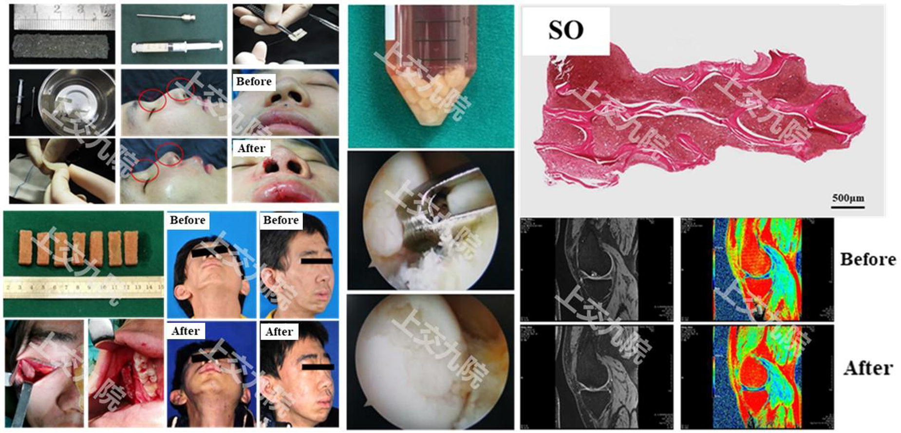
C282 - Fabricating Collagen Microblocks For Cell Instruction In Bottom-up Microtissue Engineering
E. Güben Kaçmaz, K. Song, P. Habibovic, R. Truckenmüller, N. Tahmasebi Birgani
MERLN Institute, Maastricht University Faculty of Health, Medicine and Life Science, Maastricht, NETHERLANDS
*Purpose/Objectives: In bottom-up engineering of modular tissues, microparticles can serve as scaffolding and matrix-mimicking building blocks1 to enable the production of microtissues. Additionally, they can direct cell differentiation by delivering both soluble and insoluble instructional signals for cells2. In this context, microparticle geometry, particularly when presenting anisotropy, stands out as one of the parameters that instructs cells3, which have previously been shown to respond to micro-scale topographical cues4. In order to fully investigate the impact of different microparticle geometries on the process of bone formation, we refined the process to generate microparticles using collagen, which is the main protein found in the bone matrix.
*Methodology: Polydimethylsiloxane (PDMS) templates, featuring micromolds with various shapes, were replicated from a photolithographic SU-8 master. The micromolds were filled with 8 mg/ml collagen methacrylate (CMA) solution containing 1% wt lithium phenyl(2,4,6-trimethylbenzoyl)phosphinate (LAP), and 1% v/v 1 mg/ml fluorescent dextran (for visualization), which was then incubated at 37 °C for 1 hour and crosslinked under UV light at 405 nm. A poly (vinyl alcohol) (PVA) film was cast over the micromolds and then peeled off to demold, and dissolved in water to release the crosslinked microparticles. The replication fidelity was inspected with fluorescent microscopy and the microstructure of the microparticles were characterized with scanning electron microscopy.
*Results: Micromolds contracted onto the PDMS templates could be efficiently filled with CMA. After crosslinking, CMA microparticles were removed from the micromolds using the PVA film without compromising their outer geometry (Figure 1a). The dissolution of PVA allowed the release and consequent collection of free-standing shape-specific CMA microparticles, which were stable in an aqueous environment (Figure 1b). The fidelity of the microparticle replication method was comparable to other precision microparticle fabrication methods1.
*Conclusion/Significance: Here, we presented the precision fabrication of free-standing CMA microparticles. Next, we will generate a library of microparticles with diverse geometries and study their cell instructive capacity in bottom-up engineered microtissues.
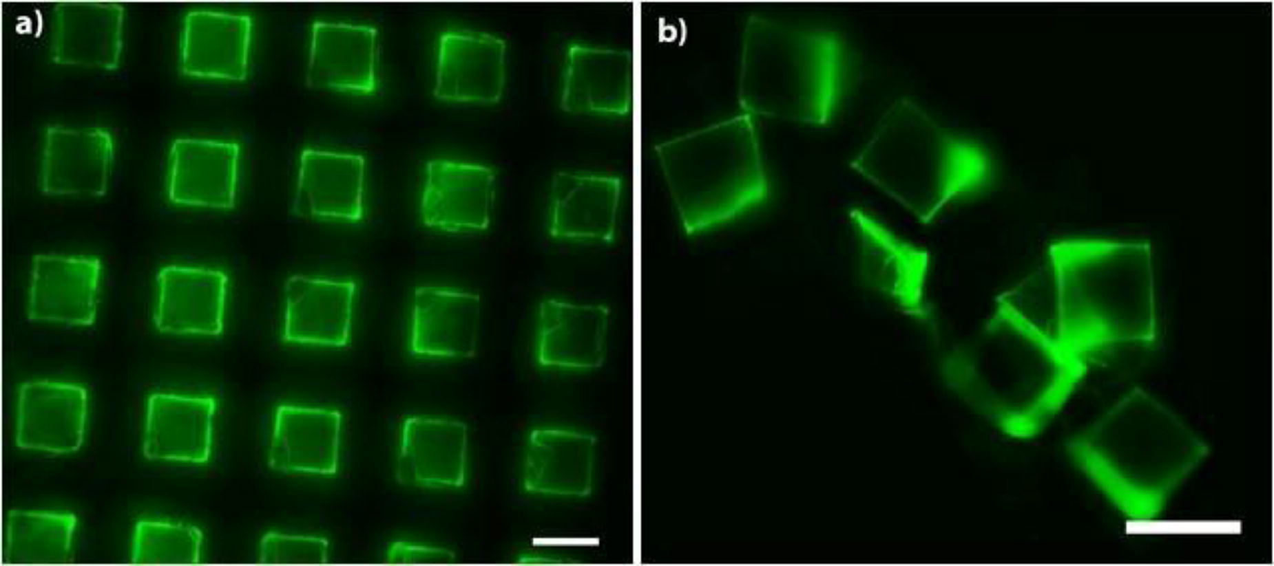
C283 - 3D Printing Technology For Bone Tissue Engineering: Biomimetic Hydroxyapatites As Osteoinductive Ingredient For Nanocomposite Inks Design
m. sandri, E. Campodoni
CNR, Faenza (RA), Italy
*Purpose/Objectives: Due to the increasing needs for 3D constructs for tissue engineering (TE) approaches, the 3D-printing process has attracted even more interest to better fulfil the TE demands and to improve the model fidelity in reproducing the complexity of biological architectures. In the common hope, 3D-printed tissues will overcome many of the shortcomings of traditional tissue engineering ensuring precise control over structural design, cells response, and cell-laden in 3D-constructs. However, advancement of bio-printing technology is limited by the availability of materials facilitating bio-printing process as well as cell viability and providing of tissue-specific cues.
*Methodology: In this contest, this research addresses the challenging task of formulating nanocomposite bioinks for the development of bone tissue substitutes capable of promoting regeneration processes and as biomatrices for the design of 3D-in-vitro models aimed at the study of biological mechanisms related to pathologies and the discovery of precision pharmacological therapies. Nanocomposite inks have caught our attention because the incorporation of nanoscale materials, has allowed for significant tunability of the mechanical, rheological, structural, and biological properties of inks and printed constructs. The modulation of their configurations in terms of composition, surface chemistry, shape, size, concentration, make nanoparticles of high interest as bioink additives for the improved design of bioactive constructs for tissue engineering.
*Results: Biomimetic synthesis approaches were exploited to engineer hydroxyapatite nanoparticles (nHAs) functioning as bioactive ingredient into printable inks and bioinks for bone tissue engineering applications and the effect of their incorporation in different polymeric matrices was investigated. Two engineered nHAs were used to prepare different ink formulations, one incorporating mesenchymal stem cells in a nanocomposite bioink composed of methacrylate-gelatin, photo-polymerizable with Vis-light, and enriched with Mg-CO3-doped-nHAs to evaluate their effects on the bio-printing process and outcomes on cells. The second, an acellularized-inks whose rheology was modulated to avoid crosslinking steps, allows to obtain freeze-dried scaffold highlighting a multi-dimensional porous structure, stable for prolonged time and suitable for TE in perfusion bioreactors for long time points.
*Conclusion/Significance: This approach enables bio-printing of suitable composite bio-constructs highly mimetic of the bone ECM. In both studies nHAs demonstrate to enhance the printability and stability of the structures and to be a strategic tool for the development of printable bioactive inks, allowing a good model fidelity in the engineering of cells-loaded or non-cellularised 3D-biomatrices.

C285 - Doubly Coated Dopamine/nano-hydroxyapatite Electrospun Membranes With Osteogenic/odontogenic Tissue Regeneration Properties
K. Loukelis1, A. Crespo Aranda1, F. Machla2, A. Bakopoulou2, M. Chatzinikolaidou1,3
1
University of Crete, Heraklio, Greece,
2
Aristotle University of Thessaloniki, Thessaloniki, Greece,
3
FORTH, Heraklion, Greece
*Purpose/Objectives: Dopamine (DA) is a molecule that has recently gathered much attention as coating agent for the biofunctionalization of tissue engineering scaffolds. DA spontaneously self-polymerizes into polydopamine (PDOPA) in alkaline environment. PDOPA enhances cell adhesion and antibacterial properties due to reactive oxygen species (ROS) as a result of oxidation of its catechol hydroxyl groups. Nanohydroxyapatite (nanoHA) together with collagen are main components of native bone and dental tissue and is known for its osteoinductive capabilities. The purpose of this work is to take advantage of the biological properties of the aforementioned materials, by fabricating flexible electrospun nanofibrous membranes and then coat them with PDOPA and nanoHA for enhanced dental tissue regeneration and antibacterial activity. The base electrospun scaffolds comprise gelatin, a highly sought biomaterial for tissue engineering as it displays great cell adhesion properties and polyvinyl alcohol (PVA), which is known for its electrospinning capacity and its biocompatible character. Their biological response was evaluated in the presence of dental pulp stem cells (DPSCs), including cell viability, cell adhesion, and osteogenic/odontogenic response.
*Methodology: Electrospun scaffolds were prepared from 5% w/v PVA and 5 % w/v gelatin solutions when mixed in a 1:1 volume ratio, with the electrospinning process being performed at 19 kV voltage, 0.4 ml/h speed and 15 cm distance between the tip and the cathode. The membranes were coated with PDOPA and nanoHA by immersion into (i) 2 mg/ml dopamine solution at pH 8.5, (ii) 1% w/v nanoHA suspension, and (iii) a combination of (i) and (ii). The membranes were physicochemically characterized, including Fourier transform infrared spectroscopy (FTIR) and X-ray photoelectron spectroscopy (XPS), mechanically analyzed and seeded with DPSCs. Cell adhesion, morphology, viability, proliferation over time in culture have been assessed. The osteogenic/odontogenic differentiation potential was evaluated through alkaline phosphatase activity, calcium mineralization and collagen production, as well as osteogenic/odontogenic gene expression.
*Results: FTIR and XPS determined the fibers consistency and the presence of the PDOPA/nanoHA particles. Mechanical analysis of the membranes indicated tensile break point values exceeding 10 MPa. Excellent adhesion of DPSCs on the membranes and increased proliferation over time have been observed. The osteogenic/odontogenic differentiation potential of the doubly coated membranes exhibited the highest response, with the highest values of alkaline phosphatase activity, secreted calcium and collagen, as well as via the upregulation of various related gene expression markers. SEM images with DPSCs laden electrospun mats showed great cell adhesion at day 2. The combined presence of PDOPA and nanoHA significantly enhanced osteogenic/odontogenic response. As a next step, we aim to evaluate the antibacterial activity of the PDOPA/nanoHA composite membranes against Gram positive and Gram negative bacteria.
*Conclusion/Significance: We successfully fabricated gelatin/PVA electrospun nanofibrous membranes and then through coating, we obtained biofuntionalized PDOPA/nanoHA scaffolds. All samples showed excellent cell viability, with the presence of PDOPA showing enhanced response.
C286 - Effect Of Thermally Aged Polymeric Scaffolds On Cell Behavior
B. DIALLO, S. CHANGOTADE, D. LUTOMSKI, G. ROHMAN
Université Sorbonne Paris Nord, Unité de Recherche en Ingénierie Tissulaire, URIT, UR, F-93430, Villetaneuse, France
*Purpose/Objectives: In tissue engineering applications, studies generally focus on the rate of polymeric scaffold degradation in an aqueous or cell-medium environment. Beside the fact that it is known that cells regulate their shape, proliferation and differentiation pathway depending on the rigidity of the adhesion support (Guo et al., 2021; Bai et al., 2020; Chen et al., 2020; Sridharan et al., 2019), no study has looked at the impact of thermal aging of materials (during storage for example) on the mechanical properties and the resulting cellular behavior.Our research unit has developed a poly(ester-urethane) elasomeric scaffod (Changotade et al., 2015) and we have recently observed that, after being stored at room temperature for 4 years, fibroblast cells still adhere and proliferate significantly on the aged scaffold, but stay round all over the pore surface (Figure 1). This variation of cell response was attributed to the alteration of scaffold mechanical properties, in particular its ability to resist permanent deformation under a given deflection and to return to its original thickness. The aim of this study is to explore how thermally accelerated aging of a poly(ester-urethane) scaffold impact on its mechanical properties and influence cellular behavior.
*Methodology: Poly(ester-urethane) scaffold were thermally aged in an oven at 90°C for periods of time varying from 1 to 30 days. They were characterized by microscopy to check morphology, porosity and pore size ; through the evaluation of the compression set and by Brillouin microscopy to assess the global and local change in mechanical properties and by Raman spectroscopy to check the chemical structure of the biomaterials.For in vitro study, fibroblast adhesion (at 3 hours) and proliferation (from 1 to 7 days) were evaluated through cell counting ; cell morphology was assessed by 3D microscopy and scanning electron microscopy and cell spreading was quantified by image analysis.
*Results: The results are currently being acquired.
*Conclusion/Significance: Storage time must not be neglected for the success of polymer scaffolds in tissue engineering. The results of this study will highlight the importance of mechanical properties on cell behavior.

Figure 1: Image of Hemalum coloration of fibroblastic cells after 30 days of culture on poly(ester-urethane) scaffold.
References: Bai M, Cai L, Li X, Ye L, Xie J. J Biomed Mater Res B Appl Biomater. 2020;108(6):2426-2440./ Changotade S, Radu Bostan G, Consalus A, Poirier F, Peltzer J, Lataillade JJ, Lutomski D, Rohman G. Stem Cells Int. 2015;2015:283796./ Chen J, Backman LJ, Zhang W, Ling C, Danielson P. ACS Biomater Sci Eng. 2020;6(9):5162-5171./ Guo Y, Mei F, Huang Y, Ma S, Wei Y, Zhang X, Xu M, He Y, Heng BC, Chen L, Deng X. Bioact Mater. 2021;7:364-376./ Sridharan R, Cavanagh B, Cameron AR, Kelly DJ, O’Brien FJ. Acta Biomater. 2019;89:47-59.
C287 - Expanding The Utility Of Electrospun Medical-grade Scaffolds Using Co-polyesters
C. J. Culbreath, S. McCullen, T. Mefford
Clemson University, Clemson, SC
*Purpose/Objectives: Currently, standard resorbable polyesters, such as poly-L-Lactide and polydioxanone have material limitations in their mechanical properties for use as tissue engineering scaffolds into clinical use. To overcome this, a series of novel materials from Poly-Med, Inc. were provided for electrospinning a range of samples. Each material was a medical grade aliphatic polyester that leveraged a range of monomer and co-polymer structures to provide unique mechanical properties and sample performance. Electrospinning (ES) produces nonwoven structures that mimic the extracellular matrix (ECM) topography and size scale at the nano to microscale, but current medical grade materials have not fully satisfied the mechanical property requirements of mimicking tissue properties such as the skin modulus range (0.1 - 140 MPa). The lack of material options for ES scaffold structures has also been concluded in literature to be the gap preventing viable use of ES technology for tissue engineering. Substrate stiffness has been observed to impact cellular regeneration and differentiation, indicating the critical nature of mechanical properties with respect to the healing process. Bioresorbable materials that can be electrospun into nonwoven scaffolds with tissue targeted mechanical properties would fill this gap.
*Methodology: Each material required optimization of electrospinning parameters associated with the process and process environment in custom enclosures manufactured for this purpose. Orthogonal designs for the optimization experiments were performed after manual ranging studies determined process boundaries. The medical grade materials were characterized dimensionally by thickness and basis weight, then by tensile strain to failure.
*Results: Macro dimensional testing allowed intrinsic tensile results to be presented, and micrograph analysis confirmed the target nanostructures were produced. Additional confirmation of cytotoxicity, residuals content, and identity testing bolstered confidence in the medical usefulness of the scaffolds. An Ashby Plot (Figure 1) was produced with the tensile results to allow observations of the produced property range.
*Conclusion/Significance: Manipulation of the co-polymer structures provided modulated strength and modulus properties. Co-spinning the materials would further allow optimization for targeted applications. The range of skin modulus (0.1 - 140 MPa) was well represented by the results, and biomimicking potential was confirmed. By electrospinning novel bioresorbable polymers, medical grade scaffolds were produced that not only mimic biological structure and mechanical properties but offered a modular platform to target a range of tissues and clinical indications. These scaffolds should replace acellular dermal matrix (ADM) in a multitude of uses and medical indications. Non-load bearing surgical reinforcement and wound healing applications would be ideal applications of the property range observed and potential for targeted scaffold development.

C288
C290 - Injectable And Biodegradable Peg-based Hydrogel For Cardiac Tissue Engineering
I. Hamouda1, A. Zoso1, E. Choppe2, E. Nicol2, V. Chiono1
1
Politecnico di Torino, Torino, Italy,
2
Le Mans Université, Le Mans, France
*Purpose/Objectives: Ischemic heart disease is caused by cardiomyocyte death under hypoxic conditions, followed by the formation of a dysfunctional fibrotic scar, populated by activated fibroblasts. The presence of a high amount of reactive oxygen species (ROS) post-injury may play a role in affecting cardiomyocyte metabolism and viability, leading to cell apoptosis. Based on recent findings, ROS-releasing hydrogels at proper dose could be exploited to train cardiomyocytes, enhancing their cardioprotection mechanisms. In this work, in vivo-like ROS were generated in water-based polymer solutions by using atmospheric pressure plasma jet (APPJ) devices and were exploited as vehicles for the delivery of ROS. Photo-cross-linkable poly(ethylene)glycol triblock copolymer (tPEO)-based hydrogels are excellent candidates for tissue engineering (TE) applications due to their tunable mechanical properties and versatile morphologies. Moreover, we already demonstrated that APPJ treatment of tPEO hydrogel enhanced final modulus with minor degradation of the polymer backbone chemistry at high concentrations. Although tPEO hydrogels present suitable mechanical properties for cardiac TE, their non-biodegradability limits the design of clinically injectable biomimetic materials. In this work, we aim to synthesize a biodegradable tPEO (btPEO) polymer, for the development of injectable, biodegradable and cell-adhesive hydrogels in combination with ROS as a minimally invasive therapy enhancing cardio-protection.
*Methodology: We first investigated the ROS dose concentration on the cell viability (resazurin assays, at days 1, 3 and 7) and gene expression (α-SMA, at day 7) of human adult cardiac fibroblasts (HCFs) post APPJ-activated cell culture media exposure during 24h. In a second step, we synthetized tPEO polymer using atom transfer radical polymerization chemistry and btPEO was obtained by introducing lactic ester groups to the initial tPEO. Then, ROS-loaded hydrogels were prepared by treating tPEO solution (up to 10 min) prior mixing with btPEO and gelatin or gelatin methacryloyl (GelMa) with proper ratio, concentration and crosslinking degree to approach the stiffness of healthy cardiac tissues (target stiffness of around 1-10 kPa). Afterwards, ROS-loaded hydrogels were used for HCFs encapsulation and culture by assessing their viability (live/dead assays) and proliferation (resazurin assay) as a function of ROS dose.
*Results: We discriminate a link between APPJ treatment time, ROS generation and the HCFs biological effects. An inferior ROS dose limit concentration (around max 80 μM H2O2) by applying a short APPJ treatment time (< 1 min duration) provides cardio-protection of HCFs. At higher APPJ treatment time (from 1 min and concentration higher than100 μM H2O2), HCFs started to be activated into myofibroblasts compared to control conditions after 7 days of culture. ROS stability in media was monitored and could be stored in water-based solutions (over 7 days at 4°C). However, a fast and efficient release within 30 min from the hydrogels was obtained once immersed in media, with minor alteration of the physicochemical properties. Hydrogels were stable in physiological and cell culture media at 37°C for up to 1 month and biodegradability increased by increasing btPEO content.
*Conclusion/Significance: In this work we developed an injectable and biodegradable PEO-based hydrogel, in combination with ROS from APPJ as a potential solution in cardiac tissue regeneration.

C291 - Macroporous Alginate Foam Scaffolds For 3D Cell Growth
M. L. Dreibelbis1, S. Cassel2, S. Robart1
1
IFF, Midland, MI,
2
IFF, Wilmington, DE
*Purpose/Objectives: Alginate is a versatile biomaterial to engineer cell culture platforms for disease modeling and tissue engineering. Alginate is bioinert and biocompatible, and with the addition of divalent cations, can form hydrogel structures for use as 3D cell culture scaffolds, drug delivery platforms, and implants. Further, the addition of moieties can be employed to improve scaffold mechanical properties (e.g., modulus, viscoelasticity) and introduce biological cues (e.g., peptides) for cell-matrix interactions. These scaffold properties can be chosen to direct proliferation, morphology, and differentiation of various cell types.
To ensure consistent cell encapsulation in a defined alginate network, an alginate-based porous foam scaffold was developed which forms a hydrogel culture when hydrated with a suspension of cells in alginate solution. The dry foam design provides a uniform base scaffold, where the molecular weight, viscosity, and functionalization of the alginate in the hydrating solution can be modulated to support optimization for the culture of various cell types. Further, these hydrogels easily dissolve for facile harvesting of cell structures (e.g., spheroids), improving downstream processing and/or biological assessment.
*Methodology: Alginate-based foam scaffolds were produced through mechanical aeration and slow internal gelling followed by casting and drying. Foam properties were assessed by texture analysis, thermogravimetric analysis, and microscopy. To investigate the influence of alginate concentration and cell density on cell response, NIH3T3 and HT-29 cells were cultured at densities of 104 to 105 cells per construct (20 µL) and polymer concentrations of 0.25 to 1 weight percent, with or without GRGDSP integrin binding peptide moieties (‘peptide-coupled alginate’ vs ‘control’). Cells were cultured in constructs for up to 14 days, where morphological changes were assessed by microscopy, and metabolic activity was measured using AlamarBlue (Thermo).
*Results: When characterizing the dry foam scaffolds, pore sizes were observed from 100 to 700 μm and when hydrated, Young’s modulus was in the range of 1 to 5 kPa. HT-29s cultured in foams formed spheroids after 7 days in all conditions tested, where inclusion of peptide-coupled alginate seemed to increase spheroid formation. NIH3T3s cultured in foams with peptide-coupled alginate showed higher metabolic activity than controls when seeded at the same weight percent alginate over 14 days. NIH3T3 proliferation and spheroid formation were observed across a range of cell density and alginate concentrations, where both lower alginate density and lower cell seeding density encouraged NIH3T3 proliferation and reorganization into larger cell structures. Larger structures with elongated cell morphology were seen in peptide-coupled alginate conditions, where cell structures colocalized with foam scaffold pores.
*Conclusion/Significance: Alginate-based foam scaffolds can achieve consistent encapsulation of cells in a defined alginate network. Modulation of alginate concentration and peptide moieties influences cell response in these networks, where tailoring these properties can aid efforts in tissue engineering and therapeutic screening.
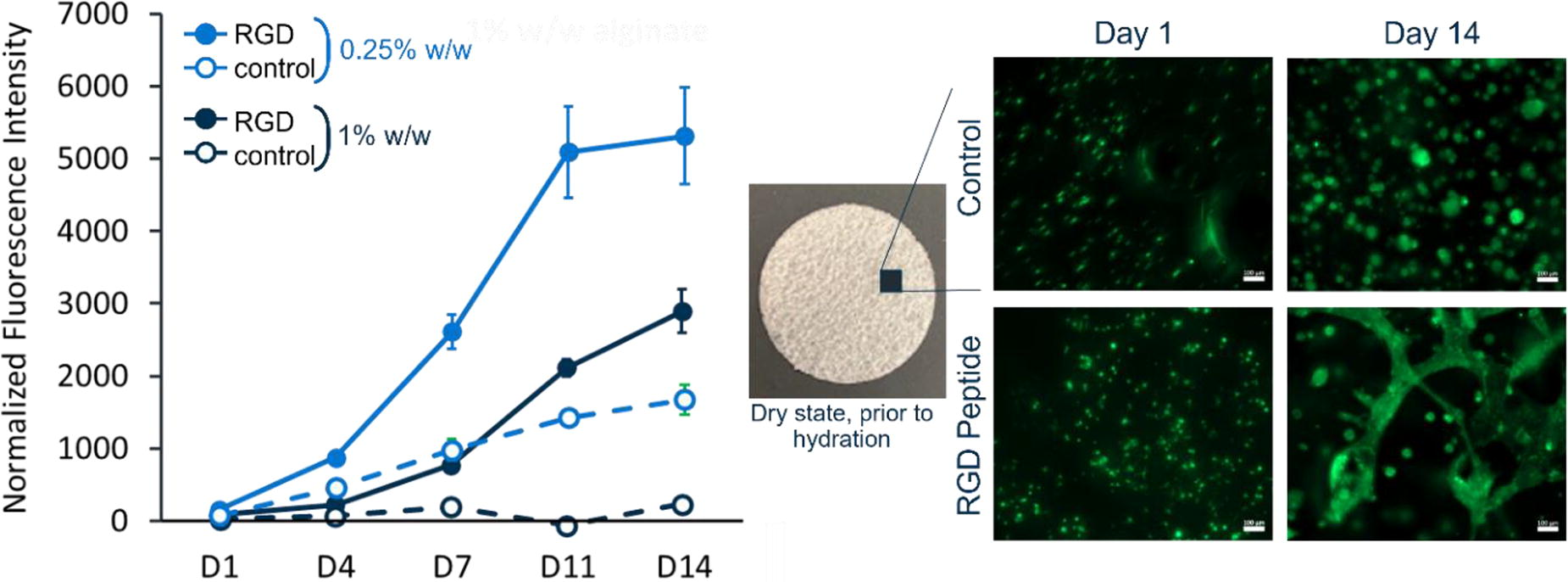
C293 - How Can Advanced Human In Vitro White Adipose Tissue (wat) Models Be Constructed? - Comparison Of 2d And Different 3D Setups
F. Albrecht1, A.-K. Schick2, A. Klatt1, F. F. Schmidt1, S. Nellinger1, P. Kluger1
1
Reutlingen University, Reutlingen, Germany,
2
Esslingen University, Esslingen, Germany
*Purpose/Objectives: Adipose tissue secretes hundreds of important cytokines that influence crucial body mechanisms like food intake, blood pressure, and immune system activation. An altered state of the cells (e.g., overweight) within adipose tissue leads to an altered cytokine profile, which in turn affects the body's homeostasis and leads to the development of severe diseases like diabetes or cardiovascular disorders. The wide-ranging secretory activity of adipose tissue underlines the importance of reliable human-based in vitro models for research purposes. Therefore, ASCs seeded on tissue culture polystyrol (TCPS) and four different 3D in vitro models based on adipose-derived stem cells (ASCs) and mature adipocytes (ACs) were established, and the cell state during adipogenesis and maintenance was analyzed.
*Methodology: Human primary adipose-derived stem cells (ASCs), adipocytes, and lobules were isolated from skin biopsies. ASC were cultured as a monolayer, formed into a spheroid (10.000 cells), or encapsulated in 1% gellan gum (GG) hydrogels, differentiated (two weeks), matured (varying), and finally, for three more weeks maintained. Adipocytes were combined with 0.5% GG to achieve cellular hydrogels, lobules were used unprocessed directly after isolation, and both were maintained for three weeks. Prior to RT-qPCR, RNA was isolated with TRIzol™-phenol precipitation and transcribed into cDNA.
*Results: The 3D models encompass ASC spheroids, ASCs in 1 % gellan gum (GG)-hydrogels, ACs in 0.5 % GG-hydrogels, and ex situ adipose lobule. During adipogenesis, ASCs in all model types accumulated lipids (lipid staining) and changed their morphology (cytoskeleton visualization) to multivacuolar cells. The successful differentiation was also demonstrated by the upregulation of PPARγ and CEBPα. By investigating individual development after the differentiation of the models, ASCs behaved differently. Within the TCPS culture, cells still exhibited a multivacuolar morphology, ASC in the GG-hydrogels, the cells were univacuolar, and within the spheroids, the ASCs partly showed an univacuolar morphology. For the models with ACs, ACs in the hydrogels showed signs of dedifferentiation during analysis, but the lobule had stable expressions during the time. All these observations were supported by gene analysis of various adipocyte-specific genes, leptin secretion, and intracellular lipid amount.
*Conclusion/Significance: In this work, we succeeded in establishing various tissue models from different primary cells from human adipose tissue. Comparison has shown that, depending on the cell type and model, the functional maximum is at another point in time, and thus, the comparability to the native state varies. This suggests that the models should be chosen according to the research question and the intended purpose.
C294 - Development Of Hydrogel Scaffolds With Tunable Mechanical Properties For Potential Applications In Peripheral Nerve Regeneration
K. Schmitz1, T. Larson1, X. Wu2, G. Ao1, M. Jack3, L. Ning1
1
Cleveland State University, Cleveland, OH,
2
Cleveland Clinic Lerner Research Institute, Cleveland, OH,
3
Cleveland Clinic, Cleveland, OH
*Purpose/Objectives: Recovery after large-gap peripheral nerve injuries remains a challenging issue1. Although various nerve scaffolds have been engineered, promotion of precise nerve regeneration requires fabrication of highly tunable scaffolds that mimic the mechanical properties of peripheral nerve environments2. Recent studies have demonstrated that cell functions can be significantly affected by both mechanical stiffness and the viscoelasticity of scaffolds3. However, effective regulation of stiffness and viscoelasticity and quantification of their influence on cells remains unachieved. The first objective of this study is to determine how the stiffness and viscoelasticity of nerve scaffolds can be regulated through biomaterial synthesis. The second goal is to develop 3D bioprinting methods to manufacture multiple-channel scaffolds with optimized structural fidelity and tunable mechanical properties.
*Methodology: GelMA based hydrogels with single-wall carbon nanotubes (SWCNTs) and hyaluronic acid (HA) were synthesized. Samples, including gelMA (6%, 8%, 10%, and 12% w/v), gelMA-SWCNTs (6 sample groups including 6% and 10% gelMA with 0.05%, 0.1%, and 0.2% SWCNTs w/v), and gelMA-HA (10% gelMA with 0.1%, 0.2%, and 0.5% HA w/v) were cast in molds and cured via UV exposure for one minute at 10 mW/cm2. Mechanical stiffness and viscoelasticity of each sample were investigated using static compression/tension, stress relaxation, and rheological tests. Embedded bioprinting methods were employed to fabricate nerve scaffolds with multiple channels (12mm x 4mm x 4mm with two channels, each with a 1mm diameter) using the prepared hydrogel precursors, and the structural fidelity was quantified. Schwann cells (RSC96) will be encapsulated in bioprinted scaffolds with tunable mechanical properties; viability, proliferation and other cell functions will then be evaluated.
*Results: Rheological tests confirmed that mechanical properties increased with the concentration of gelMA. The storage moduli of 6%, 8%, 10%, and 12% (w/v) gel-MA were about 1.5, 3.4, 6.8, and 8.9 kPa, respectively. Stress-relaxation of gelMA samples and comprehensive mechanical properties of gelMA-SWCNTs and gelMA-HA are still being investigated. While the dimensions of 3D printed scaffolds closely matched the CAD model, channels were not perfectly intact in every print; therefore, bioprinting parameters must be adjusted to optimize printing fidelity.
*Conclusion/Significance: Preliminary results suggest that gelMA-based hydrogels have great potential for the fabrication of peripheral nerve scaffolds. Tunability has been confirmed through multiple rheological and mechanical tests, indicating that this material can be modified to fit specific properties of desired tissues. The adaptability of this composite hydrogel provides an excellent platform for mimicking the extracellular matrix, which has proven to be critical for cell growth in previous studies. Fabrication of a consistent printable scaffold with properties resembling nerve tissue would greatly improve treatment options for patients with peripheral nerve damage by reducing the need for autografts.
C295
C296 - Regenerative Efficacy Of Whole Kidney Scaffold Decellularized By Supercritical Carbon Dioxide Followed By Direct In Vivo Implantation
Y.-C. Fu1, S. Periasamy2, K.-C. Yen2, Y.-J. Chen2, D.-J. Hsieh2
1
Department of Orthopedic Surgery, Kaohsiung Medical University Hospital, Kaohsiung Medical University, Kaohsiung 80708, Taiwan, Kaohsiung, Taiwan,
2
ACRO Biomedical Co., Ltd., Kaohsiung, Taiwan
*Purpose/Objectives: Chronic kidney disease (CKD) is a foremost cause of mortality and morbidity worldwide, affecting between 8% and 16% of the global adult population. The incidence of CKD is on the rise, and existing therapeutic choices remain limited. Recent advances in tissue engineering and regenerative medicine have presented new hope for the treatment of kidney diseases. The use of decellularized extracellular matrix scaffolds to help repair damaged tissue has emerged as a promising approach in the field of renal regeneration.
*Methodology: In the present study, we decellularized rabbit kidneys using supercritical carbon dioxide (SCCO2) and directly carried out orthotropic transplantation in a rabbit, the regenerated kidney was evaluated after 4 weeks of regeneration in vivo.
*Results: Decellularized kidneys were characterized by hematoxylin and eosin and 4,6-diamidino-2-phenylindole (DAPI) staining and DNA quantification that depicted complete removal of cells by SCCO2. The regenerated kidney was assessed by computerized tomography, ultrasonography and nuclear scan. The expression of PAX2 and WT1 had a modulating expression in the transplanted kidney, demonstrating regeneration. In the transplanted kidney, the stem cell marker CD34 was found to be expressed, indicating the recruitment of stem cells leading to regeneration. Pre-tubular aggregate and renal vesicle marker PAX8 were found to express in the transplanted kidney, indicating the formation of the functional unit of the kidney leading to regeneration. The regenerated kidney nuclear renal scan showed urine formation around the kidney, indicating a partial renal function.
*Conclusion/Significance: In the current investigation, we demonstrated that the SCCO2 decellularized kidney did not show immune-related rejection. We have established that SCCO2 decellularized kidneys can be regenerated in vivo with partial biological function.
C297 - The Production Of Engineered Stromal Tissue Using Stabilized Ascorbic Acid
D. Brownell1, C. Ceneparo1, L. Carignan1, M. Labroy1, S. Chabaud1, F. Bordeleau1, L. Gibot2, S. Bolduc1
1
Université Laval, Québec, QC, Canada,
2
CNRS, IMRCP, Paul Sabatier University, Toulouse, France
*Purpose/Objectives: Ascorbic acid is a critical building block of collagen. Its deficiency is associated with impaired extracellular matrix deposition. Thus, sodium ascorbate (AA) is often added to culture media in tissue engineering applications to stimulate the production and maturation of an endogenous extracellular matrix by the fibroblasts. Production of various tissues by the self-assembly technique relies on the ability of stromal cells to produce their extracellular matrix. The use of AA is therefore crucial. However, AA is unstable in aqueous solution, even when stored at 4°C, meaning that fresh solutions must be made at every medium change. This poses a challenge to produce engineered tissues as it increases cost and manipulation time. A more stable form of ascorbate, 2-Phospho-L-Ascorbate (2PAA), allows the formation of connective tissue-like structures in fibroblast cultures. It remains unclear if 2PAA constitutes an adequate substitute to sodium ascorbate. In this context, we evaluated the physico-chemical properties of AA or 2PAA solutions and how 2PAA impact the properties of dermis, bladder and vaginal tissues reconstructed using the self-assembly technique.
*Methodology: Sodium ascorbate (AA) and 2-phospho-L-ascorbate (2PAA) stability was assessed for a working dilution (50µg/ml) under storage conditions (4°C or -80°C) as well as under culture conditions (37°C humidified 5% CO2 incubator). Solution samples were taken at different time points and measurements were performed on a UV-vis scanning spectrophotometer. Engineered dermis, bladder and vaginal tissues were produced using primary tissue-specific fibroblasts. Cells were seeded in 6-well plates with a paper anchor and weight. Cells were cultured with 50µg/ml AA or 2PAA. After 4 weeks, 3 stromal sheets were stacked and fused for 4 days. Mechanical characterization was performed on an Instron Electropuls®E1000. Stroma organization was assessed with histological staining. Enzymatic activity of matrix metalloproteinases (MMPs) was assessed. Finally, a price analysis was performed.
*Results: Direct UV-Vis spectrophotometry showed a maximum of absorption around 260 nm for both freshly prepared AA and 2PAA solutions. After 15 to 30 days at 4°C, the AA solution underwent yellowing along with a shift of the maximum absorption peak, indicating degradation. To a lower extent, this was also observed for the AA solution at -80°C. No degradation was observed for the 2PAA solution. Over 72h at 37°C (culture conditions), even faster change in the color and spectrum of AA was observed while 2PAA remained stable.We then tested if tissues produced using 2PAA were equivalent to those obtained with AA. Notably, tissues produced with 2PAA had comparable thicknesses and equivalent mechanical properties compared to those produced with AA. MMP activity revealed no significant difference between AA and 2PAA, and collagen content and organization were found to be similar. A price comparison also revealed that 2PAA ($0.51 /cm2) might be particularly advantageous compared to freshly made AA ($4.79/cm2).
*Conclusion/Significance: Overall, our results demonstrate the possibility of substituting sodium ascorbate with 2-Phospho-L-Ascorbate for tissue engineering using the the self-assembly approach. This protocol would significantly reduce the costs associated with tissue production while lowering required hands-on time. These are key factors for any scaling activity.
C298 - Computational Analysis And Characterization Of Pla And Abs Scaffolds Produced Via Additive Manufacturing For Potential Hard Tissue Engineering
M. Rahatuzzaman, M. Mahmud, M. E. Hoque
Military Institute of Science and Technology (MIST), Dhaka, Bangladesh
*Purpose/Objectives: Rapid prototyping is an advanced manufacturing technology for fabricating tissue engineering scaffolds. Bone scaffolds possess a high level of porosity to promote cell attachment, spreading, and differentiation considering appropriate biomechanical properties. Optimization of various requirements and parameters often involves extensive experimental work. This study aims to design and simulate 3D-printed polylactic acid (PLA) and acrylonitrile butadiene styrene (ABS) scaffolds with two lay-down patterns potentially for bone tissue engineering and also, distinguishing the mechanical properties.
*Methodology: The scaffolds were designed using computer-aided design (CAD) software, and fabricated using a fused deposition modeling (FDM) 3D printer. Two lay-down patterns were used: 0/60° and 0/90° in fabricating the scaffolds. The scaffold morphology was observed under the scanning electron microscope (SEM) to evaluate the scaffold’s printing precision. The porosity of the scaffolds was measured using both theoretical and liquid displacement methods. The mechanical properties of the scaffolds were assessed using compression testing and finite element analysis (FEA).
*Results: SEM images showed that the 3D-printed scaffolds had good printing accuracy with filament uniformity and pore regularity. The porosity measurement had consistency between theoretical estimation and experimental values. The PLA scaffolds had a higher compressive strength and Young's modulus than the ABS scaffolds. The 0/90° lay-down pattern also resulted in higher mechanical properties than the 0/60° lay-down pattern.
*Conclusion/Significance: The preliminary results suggest that in terms of mechanical properties, the PLA scaffold with the 0/90° lay-down pattern has higher promise for bone tissue engineering than the ABS scaffold. However, there is a compromise between the porosity and mechanical strength of the scaffolds, which requires optimization when designing based on its targeted application. Besides, in vitro and in vivo biological evaluations are also essential to determine the suitability of the scaffolds that are in progress.
C300 - Influence Of Gelma-Chitosan Composite On In Vitro Fibroblast Cell Proliferation
K. Agarwal, B. Ghosh
UiT The Arctic University of Norway, Tromsø, Norway
*Purpose/Objectives: Protein-based hydrogels, notably GelMA, are widely utilized in 3D in vitro cell cultures to replicate the intricate 3D conditions of the human body, providing functional adhesion crucial for an authentic cellular environment. However, recognizing that tissues encompass both proteins and carbohydrates, which contribute vital structural support to cells. We investigated the influence of carbohydrate-protein composites on cell behavior.
*Methodology: In our research, GelMA served as the protein component, while nanofibers, derived from a blend of chitosan and polyethylene oxide, constituted the carbohydrate part. Utilizing electrospinning, we created nanofibers as a supportive framework. GelMA (2%) was combined with varying concentrations of carbohydrate nanofibers (0.1%, 0.5%, and 1%), with a control using only 2% GelMA. The resulting GelMA-nanofiber matrices were UV crosslinked and solidified. Subsequently, mouse embryonic fibroblasts were cultured on these matrices for 48 hours under standard conditions (DMEM media, 37°C, and 5% CO2).
*Results: Upon examination after the 48-hour incubation period, a notable increase in cell proliferation was observed with the addition of carbohydrate nanofibers. The most significant proliferation was evident in the 2% GelMA combined with 0.5% nanofibers. Interestingly, at lower concentrations of nanofibers (0.1%), cells exhibited elongation with prominent podial structures compared to the control. However, there was no significant difference in cell proliferation between matrices containing 0.5% and 1% nanofibers blended with GelMA.
*Conclusion/Significance: The study's findings lead to the conclusion that the incorporation of carbohydrates enhances cell functions by promoting proliferation, possibly attributed to the increased structural support provided by carbohydrates to the soft GelMA hydrogel. Additionally, the high surface-area-to-volume ratio and porosity of nanofibers could facilitate efficient permeation of oxygen and water, contributing to the observed improvements in cell behavior. This research holds promise for wound dressings and scaffolds, where protein-carbohydrate composites may speed up healing. The study provides insights into the synergistic effects of protein and carbohydrate components in creating biomimetic environments for cell cultures, opening avenues for innovative approaches in tissue engineering and regenerative medicine.
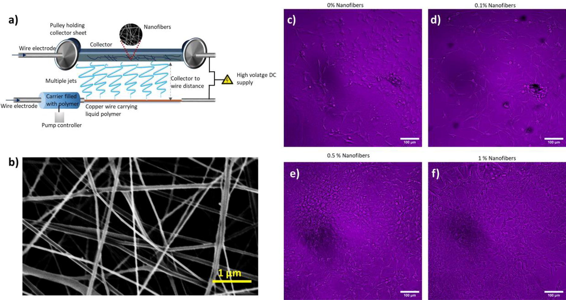
C301 - Development Of DECM-enriched Microspheres Mimicking Pancreatic Islets For Clinical Application
M. Klak1,2, O. Janowska1, K. Wozniak1, A. Papierniak-Wygladała1, S. Domanski1, D. Ujazdowska2, A. Gornik1, A. Berman1,2, M. Wszola1,2
1
Polbionica Ltd., Warsaw, Poland,
2
Foundation of Research and Science Development, Warsaw, Poland
*Purpose/Objectives: The development of organ and tissue bioprinting is experiencing significant progress. The use of 3D-bioprinted constructs is widely studied in the context of clinical research and its practical application in modern medicine. The aim of the work was to develop a method enabling bioprinting of bionic sferoids of pancreatic islets and the selection of biomaterials for bioprinting of a fully-vascularized bionic pancreas.
*Methodology: Pancreatic islet cells and bioink based on dECM as well as methacrylated biomaterials were used in the research. Cell functionality and viability tests were performed on the constructs, as well as histological analysis assessing their structure. Additionally, hemocompatibility tests were carried out to eliminate biomaterials with a high degree of coagulation. For this purpose, the level of clotting was assessed during the first 30 minutes after contact with whole blood.
*Results: Biomaterials showed significant differences in hemocompatible properties. The highest degree of hemolysis was demonstrated in dECM-based bioinks. HAMA and GELMA showed the level of 80% of hemolysis. In turn, it was presented that the use of dECM-based bioink for the production of pancreatic spheroids allows for the creation of stable 3D structures. Spheroids with dECM showed over 30% higher functionality from day 7 compared to the control group. Furthermore, at day 21, the significant viability advantage of the bionic spheroids continued. As it turned out, the concentration of dECM in the final bioink was also important for the cells. Based on histological analyzes and GSIS test results, it was shown that whole-organ bioprinting is most optimal at dECM concentrations ranging from 5 to 8%.
*Conclusion/Significance: dECM in 3D conformation of cells within bioprinted islets is a key component for maintaining the proper functionality of insulin-secreting cells. Additionally, the developed composition of the bioink enables the creation of a fully vascularized bionic pancreas.
C302
C303 - Jellyfish Collagen Filled Titanium Matrix For Large Bone DefectRepair
S. Nath Varma, C. Liu
UCL, Stanmore, United Kingdom
*Purpose/Objectives: Large bone defects can be caused by trauma, disease, surgery or tumour resection where the bone is unable to heal as normal due to the size of defect or fracture.. Autografts and allografts (with/ without non-biological materials, such as metallic fixtures) are the current gold standard but these treatment methods come with various problems from failure rates and limited supply to donor site morbidity and immune rejection.
To understand and resolve this clinical problem, regenerative medicine approaches such as tissue engineering and biomaterial use, have been widely studied and practised. A more recent type of collagen that has emerged is jellyfish collagen (Jellagen), which is collagen type 0, non-cytotoxic and biocompatible. Titanium (Ti) and its alloys are clinically established in bone implants. In this project, we are investigating to what extent a hybrid jellyfish collagen - Ti scaffold can provide a natural 3D microenvironment for bone formation.
*Methodology: Ti (Ti-6Al-4V) matrices were infilled with jellyfish collagen and freeze-dried. Either SAOS-2 cells or human osteoblast cells (hOBs) were seeded per scaffold for the following time-points: Day 1, 3, 7, 14, 21. Established quantitative and qualitative methods such as Live/Dead® Viability/Cytotoxicity Kit, PrestoBlue Viability Assay, scanning electron microscope (SEM) and immunostaining (IF) (DAPI, Vinculin and Osteopontin) have been used to determine whether the proposed hybrid scaffold supports osteoblast viability, proliferation and differentiation.
*Results: The viability assay showed that the hybrid scaffolds maintain cell growth throughout the time-points. Cell proliferation was seen through the Live/Dead images, as seen in Figure 1.The SEM images show the different surface morphologies of the samples and the high number of cells present in them. They also show that bone cells have cell-to-material interactions and cell-to-cell interactions across 14 days. The IF images show the bone cell morphology and attachment to the Ti and collagen materials, and interface. All of these results indicate that human bone cells positively respond to the biomaterials over a long period of time.
*Conclusion/Significance: The viability results show that the hybrid scaffold supports proliferating bone cells long term, further confirmed by Live/Dead images also showing viable cells throughout the scaffolds. The SEM images show the cell morphology and strong attachment to the different materials. This is also seen in the IF images where cells are present on and around the Ti matrix, around the freeze-dried collagen pores and in the collagen-Ti interface, which is of great interest. With these promising results, the next step would be to assess human osteoblast cell bone formation up to 28 days in the hybrid jellyfish collagen - Ti scaffold.

C304 - Development Of A Cryopreservation Technology For Cellularized Engineered Tissue Constructs
Y. Ju, S. Albertson, J. Scott, E. Williard, A. Atala, J. Yoo
Wake Forest Institute for Regenerative Medicine, Winston-Salem, NC
*Purpose/Objectives: Currently, tissue constructs are manufactured just before treatment, which is a limitation in personalized therapy using autologous cells. While cells can be frozen and stored for a defined period until ready to use, storing the tissue construct seeded with cells has not been tried previously. The ability to cryopreserve and store a ready-to-use cell-seeded tissue construct for a defined time until treatment will provide enormous flexibility in treating patients. In this study, we aimed to develop a safe and reliable method to store tissue constructs that maintain the viability of seeded cells.
*Methodology: A baseline cell viability of the post-thawed cryopreserved cell-seeded scaffold constructs was obtained using 10% dimethyl sulfoxide (DMSO), a common cryoprotectant. Tissue constructs were generated by seeding placental stem cells, urothelial and muscle cells on PLGA coated PGA polymer mesh. To reduce cell loss during the washing process after thawing, we adopted a cell-encapsulation system using collagen gel for cell seeding.
*Results: The cell viability of cell-seeded scaffold constructs was maintained at over 50% after cryopreservation. However, the collagen gel encapsulated tissue constructs retained the seeded cells within the scaffold (∼70%), reducing cell loss during the washing process and enhancing cryopreservation efficacy. The cell-encapsulated collagen gel groups (2.5 mg/mL) showed higher cell viability without morphological/phenotypical changes than the control (no collagen gel) group.
*Conclusion/Significance: Establishing a system to safely store cell-seeded tissue constructs until treatment is an enormous advancement in tissue engineering, providing flexibility in the treatment and allowing the shipping of tissue constructs globally.
C305 - Regenerative Efficacy Of Supercritical Carbon Dioxide Decellularized Nerve By Direct Xenotransplantation
Y.-Y. Hsueh1, M.-W. Hsu1, K.-C. Yen2, S. Periasamy2, D.-J. Hsieh2
1
Division of Plastic and Reconstructive Surgery, Department of Surgery, National Cheng Kung University Hospital, College of Medicine, Tainan, Taiwan, Tainan, Taiwan,
2
ACRO Biomedical Co., Ltd., Kaohsiung, Taiwan
*Purpose/Objectives: Peripheral nerve injury (PNI) is a common clinical condition. About 1 million PNIs arise worldwide each year. PNI is a grave monetary problem to society and leads to the loss of limb functions, which significantly affects patient quality of life. The “gold standard” for the treatment of nerve defects is autologous nerve transplantation. However, it has numerous limitations, including nerve size differences, secondary dysfunction in the donor area, risk of neuroma formation and inadequate donor sources.
*Methodology: In the present study, we decellularized porcine nerve using supercritical carbon dioxide (SCCO2) and subsequently xenogenic transplantation to a rat.
*Results: Decellularized nerves (DN) were stained by hematoxylin eosin and 4,6-diamidino-2-phenylindole (DAPI) staining and DNA depicted complete removal of cells by SCCO2. The DN was found to consist of type I collagen with α1 and α1 bands with a collagen concentration of more than 200µg per milligram of tissue and most importantly DN proved to be non-toxic in the 3T3 cell line. Animal studies revealed sciatic function index has an improvement of more than 40 points in DN-transplanted rats. Von Frey tests for paw withdrawal thresholds indicated DN-transplanted rats are relatively better than the control animal. Neuroelectrophysiological function tests showed no difference between DN-transplanted and control rats. The sciatic nerve regeneration test after 12 weeks depicted the remyelination rate of the DN graft was 73.63%, statistically significantly different to the control rat.
*Conclusion/Significance: In the present study, DN graft was regenerated to depict the motor function, sensory function and nerve regeneration. Therefore, we believe that the porcine DN graft may be the most promising graft for repairing human nerve defects.
C306 - Production Of A Composite Electrospun Scaffold Of Poly(caprolactone)/bioactive Ceramic And In Vitro Tests
N. Maurmann1, A. L. Kern1, R. P. Silveira1, R. L. Siqueira2, P. Pranke1,3
1
Universidade Federal do Rio Grande do Sul (UFRGS), Porto Alegre, Brazil,
2
Independent researcher, Itabira, Brazil,
3
Stem Cell Research Institute (Instituto de Pesquisa com Células-tronco), Porto Alegre, Brazil
*Purpose/Objectives: Bioceramics are synthetic materials used for the regeneration of both hard and soft tissue. Scaffolds manufactured through electrospinning, generally with polymers, offer a supportive framework for cellular attachment, proliferation, and tissue regeneration. This work aimed to standardize a polymeric solution with polycaprolactone (PCL) and bioceramic (BC) for the purpose of producing scaffolds through electrospinning to be tested with stem cells.
*Methodology: Scaffolds were produced by electrospinning with PCL and bioceramic. Solutions were prepared in the concentrations of 15% PCL and 1% BC in acetone: chloroform (1:1). The parameters were optimized with an injection rate of 0.5 ml/h, a distance of 15 cm between the needle tip and the collector plate and a supply voltage adjusted to 24.5 kV. The scaffold's morphological characterization was performed by evaluating material shrinkage with ImageJ and optical microscopy. Cytotoxicity was analyzed by the lactate dehydrogenase (LDH) assay, and cell viability was evaluated by the 3-(4,5-dimethylthiazol-2-yl)-2,5-diphenyltetrazolium bromide (MTT) assay after one day of cultivation with stem cells. The treatment of the cells with 1% Triton X-100 was used as a control for cell death, and the cells cultured directly on the tissue culture plate (TCP) were used as a negative control.
*Results: The scaffolds were produced with PCL and BC, with long and continuous fibers without beads, indicating that the optimization of the parameters was appropriate. The area of the materials did not suffer alterations after 2 days in the incubator, which could occur due to the electrospinning process. The scaffolds did not exhibit cytotoxicity. The average ± standard deviation (SD) concentration of LDH released after 1 day of cultivation was 97 ± 7, 96 ± 2 and 214 ± 9 U/L at TCP, scaffold, and death control, respectively, with significant statistical differences in the LDH leakage between the death control and the other groups (Ps = 0.000). In the scaffold of PCL/BC, cell viability was significantly increased in comparison with the control (TCP). The absorbance and SD values of MTT were 0.22 ± 0.01 for the TCP group, 0.34 ± 0.08 for the scaffolds (P = 0.033), and 0.05 ± 0.02 for the death control (P = 0.003).
*Conclusion/Significance: This study successfully developed electrospun PCL/BC scaffolds that are structurally stable and non-cytotoxic. The enhanced cell viability suggests the potential of these scaffolds for tissue engineering applications. It can be concluded that the utilization of bioceramic in PCL scaffolds improved the viability of the stem cells, being, therefore, a biomaterial with potential use in skin and bone regeneration. Financial support was provided by the Office of Naval Research Global (ONRG Award N62909-21-1-2026), National Council for Technological and Scientific Development (CNPq), Financier of Studies and Projects (FINEP), National Institute of Science and Technology for Regenerative Medicine (INCT-Regenera) and Stem Cell Research Institute (IPCT).
C309 - A Comparative Study Of The Effects Of Different Crosslinking Methods On The Physicochemical Properties Of Collagen Multifilament Bundles
R. Rasouli, H. Yaghoobi, J. Frampton
Dalhousie University, Halifax, NS, Canada
*Purpose/Objectives: Collagen-based scaffolds are promising materials for tissue engineering. Crosslinking is usually required to improve the mechanical properties and stability of collagen-based scaffolds. Introducing exogenous crosslinking into collagen may however affect the collagen structure. Since the architecture of collagen affects its functionality, it is important to study the effect of crosslinking methods to choose an ideal crosslinking method and condition to preserve the collagen structure and achieve the desired properties for tissue engineering applications.
*Methodology: This study compares different chemical and physical crosslinking techniques on contact-drawn collagen multifilament bundles as a collagen-based scaffold model to recapitulate the structure of a tendon. We investigated the range of conditions for different crosslinking methods to tune and improve the physicochemical properties of collagen-based scaffold without disturbing the collagen molecular structure for the desired tissue.
*Results: The presence of collagen and the formation of well-ordered collagen structures were confirmed by attenuated total reflectance Fourier-transform infrared spectromicroscopy (AT-FTIR) and X-ray diffraction (WAXS). The ultimate tensile strength (UTS) and Young’s modulus significantly improved for all crosslinked scaffolds compared to non-crosslinked scaffolds.
*Conclusion/Significance: Our finding showed it is possible to tune the mechanical properties of the tendon-like multifilament structures within the range of native tendons without interfering with collagen molecule architecture.
C310 - Polycaprolactone As A Scaffold & Implant Material - Standard Melting Enthalpy Of Polycaprolactone Revisited
T. Ayyachi1, B. Dargaville1,2, N. Bock2,3,4, D. W. Hutmacher1,2,4
1
School of Mechanical, Medical and Process Engineering, Faculty of Engineering, Queensland University of Technology, Brisbane, AUSTRALIA,
2
Max Planck Queensland Centre for the Materials Science of Extracellular Matrices, Queensland University of Technology, Brisbane, AUSTRALIA,
3
Centre for Biomedical Technologies, School of Biomedical Sciences, Faculty of Health, and Translational Research Institute (TRI), Queensland University of Technology, Brisbane, AUSTRALIA,
4
Australian Research Council Training Centre for Multiscale 3D Imaging, Modelling, and Manufacturing, Queensland University of Technology, Brisbane, AUSTRALIA.
*Purpose/Objectives: Polycaprolactone (PCL) is a widely used biodegradable polymer in tissue engineering and regenerative medicine (TE&RM) as scaffolds, membranes, and other types of implants. Its semi-crystalline nature and the dynamic changes during processing, in vitro and in vivo use are crucial for TE&RM applications. For example, understanding PCL's original crystallinity and how it changes during use is important from both a polymer and biomaterials science point of view e.g., crystallinity influences mechanical performance, thermal properties, degradation, toxicity, and ultimately in vitro and in vivo biocompatibility. Conventionally derived from thermal measurements, crystallinity (Xc) is calculated as the observed enthalpy of melting relative to a polymer-specific standard melting enthalpy of hypothetically 100% crystalline polymer (ΔHm0). Ideally, determining ΔHm0 involves processing a polymer to 100% crystallinity and measuring its enthalpy. However, due to thermodynamic and kinetic limitations, achieving 100% crystallinity is impractical. Moreover, ΔHm0 should be unique for a polymer and obtained through reproducible methods, agreed upon by the scientific community and published in standard reference source; but such settings are rare. For PCL, reported ΔHm,PCL0 values range from 135.43 J/g to 191.1 J/g, leading to variations in calculated Xc values. A commonly used value is 139.5 J/g, but this may result in Xc values above 100%, suggesting the need for a higher ΔHm,PCL0. Additionally, some reported ΔHm,PCL0 values were obtained through outdated methods like adiabatic vacuum calorimetry, and colligative methods such as the depression of melting point of polymer-diluent mixture, and density calculations, impacting accuracy.
*Methodology: In this work we aimed to obtain a range of Xc, from 0% to greater than 90%, from the melt of a PCL sample of a given molecular mass. Due to the rapidly crystallizing nature of PCL, it is impossible to obtain the complete range; usually, Xc ranging only from 50% to 60% (semicrystalline PCL) are reported when differential scanning calorimetry is used. However, in our work, by employing a recently developed technique called fast scanning chip calorimetry (FSC), a broad range of Xc from 0% (completely amorphous PCL) to 55% (semicrystalline PCL) has been obtained by employing appropriate cooling rates.
*Results: From the FSC curves, the melting enthalpy, and the corresponding change in heat capacity at Tg were calculated and plotted, resulting in the linear relationship shown in Figure 1. The extrapolation of the line to zero change in heat capacity at the Tg yields a y-intercept equivalent to ΔHm,PCL0 of 173.6 J/g.
*Conclusion/Significance: This work represents an original study that will pave the way to establish the precise value of ΔHm,PCL0. It is envisaged that this will support future research using PCL and its crystallinity as well as allow better cross-comparison of studies and polymer samples from different manufacturers and research groups. From a bigger picture point of view, this method of employing FSC to determine ΔHm0 can be extended to all crystallizable polymeric biomaterials; given the effect of crystallinity on other important biomaterial properties, the effort will accelerate the translation of biomaterials from bench to bedside.

C311 - Development Of A Cardiovascular Scaffold From Decellularized Human Placental Membrane
B. Uzunkaya1, T. Ozurun2, M. Demirci2, O. Cibik3, H. Kenar2, V. N. Hasirci2
1
Rice University, Houston, TX,
2
Acibadem Mehmet Ali Aydinlar University, Istanbul, Turkey,
3
Yildiz Teknik University, Istanbul, Turkey
*Purpose/Objectives: The utilization of the amniotic membrane, the outermost layer of the placenta, has garnered significant interest in regenerative medicine due to its easy accessibility as a medical waste after birth and its high biocompatibility. This research pivots on the novel application of the full-thickness placental membrane, including the chorionic and amniotic layers, in cardiac repair and tissue engineering. Addressing the inherent limitations of the amniotic membrane's thin structure, which compromises its suitability for cardiovascular use, our study explores the mechanical strength and biocompatibility of the chorioamniotic placental membrane. Through decellularization of the placental membrane, we aimed to augment biocompatibility and its use as a scaffold, thereby opening new avenues in cardiovascular tissue engineering. This presentation outlines our approach and preliminary findings in this endeavor, showcasing the potential of the placental membrane as a versatile scaffold in cardiac regenerative therapy.
*Methodology: Our decellularization protocol for the placental membrane involved a tailored combination of physical and chemical methods. The effectiveness of decellularization was determined through nuclear staining and DNA content analysis. Additionally, we evaluated the membrane's mechanical strength using tensile tests, and its hemocompatibility was assessed via a platelet adhesion test. To further understand its potential in regenerative medicine, we examined the cytocompatibility of the placental membrane by seeding it with endothelial cells, cardiomyocytes, and fibroblasts.
*Results: The effectiveness of the decellularization process was demonstrated by nuclear staining and measuring the DNA content per milligram in the decellularized placental membrane. There were notable changes in the mechanical properties. Observations of platelet adhesion on the decellularized membrane indicated potential issues with hemocompatibility. Additionally, cell adhesion and viability were observed post-seeding in fibroblast, endothelial, and cardiomyocyte cultures.
*Conclusion/Significance: Our key findings reveal the placental membrane's potential in cardiac tissue engineering, albeit with a need for further enhancements. This study introduces an innovative method for decellularizing and modifying the human placental membrane to serve as a scaffold, also assessing its applicability as a cardiovascular graft.
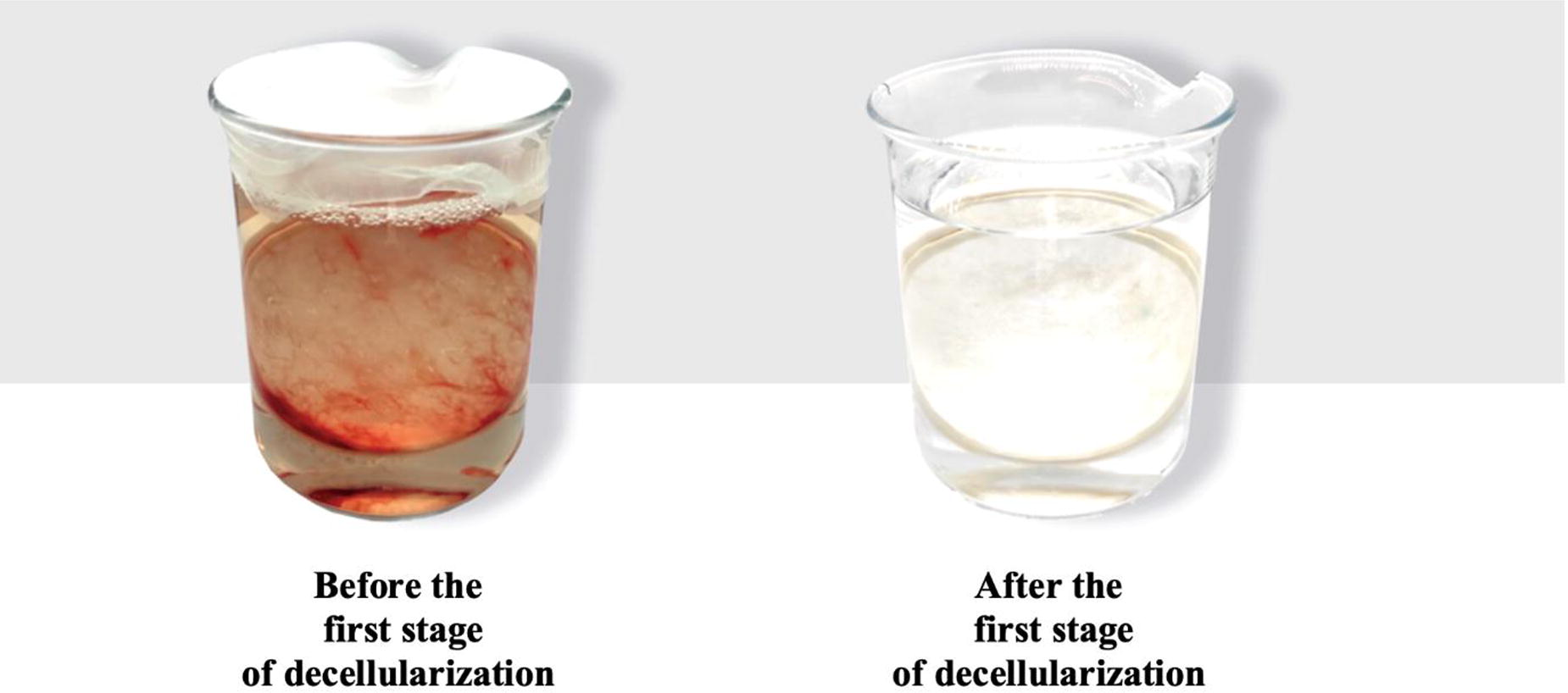
C313 - Developing An Angiogenic Periosteum Substitute To Improve Delayed Bone Healing
C. S. Ryan, G. Reilly, F. Claeyssens
University of Sheffield, Sheffield, United Kingdom
*Purpose/Objectives: The management of delayed bone healing injuries, namely critical size defects and non-union fractures, represents a substantial clinical burden. Recently, the use of free vascularized periosteal flaps to treat bone defects has gained some attention, and involves taking periosteum from a donor site, typically the medial femoral condyle, and wrapping it around the defect site. Despite having excellent union rates reported, the technique is still largely unused due to its technical difficulty and issues related to harvesting the periosteum including donor site morbidity and knee instability. Further to this, when applied to critical size defects the technique is limited by the amount of periosteum that is available to be harvested. The present work studies how a synthetic periosteum substitute could be developed to be used in place of the periosteal flap to provide a tailorable solution with fewer associated complications.
*Methodology: The periosteum substitute will be made from synthetic and easy-to-handle polymers and will have osteogenic and angiogenic properties to accelerate bone regeneration by mimicking the function of the periosteum. It is our intention that this will be used in conjunction with existing surgical procedures and in addition to void fillers to stimulate the natural healing process, in a similar manner to how the periosteal flap is currently used. To date, we have investigated the manufacture of scaffolds with multiscale porosity to promote angiogenesis-driven bone formation using polycaprolactone (PCL) and poly(glycerol sebacate) (PGS) polymerised high internal phase emulsions (polyHIPEs). The periosteal substitute material needs to be highly porous to encourage endogenous cell migration and permit molecular crosstalk and nutrient transfusion. These polyHIPE scaffolds were manufactured via emulsion templating and mesenchymal stem cells, osteoblasts, and fibroblasts were cultured onto them to assess their biological properties. Osteogenic and angiogenic potential was then furthered with two different methods: decoration with extracellular matrix (ECM) and coating with an angiogenic factor, 2-deoxy-D-ribose.
*Results: PCL scaffolds alone were too brittle when assessed for their potential to wrap around a defect, hence blends with PGS were tested to find a material that had suitable properties. The 50:50 PCL:PGS polyHIPE was found to be flexible and suturable (elastic modulus = 0.73 MPa, ultimate tensile strength = 0.47 MPa), hence this was chosen to move forward with. Scanning electron microscopy (SEM) revealed that the 50:50 PCL:PGS polyHIPE scaffolds also had the best interconnected porous structure, with both macroporosity (>200μm) and microporosity (<10μm) present, making them ideal for cell ingrowth and nutrient transfer. When tested for biological activity, it was found that cells attached and experienced an 85% increase in cell metabolic activity over eight days demonstrating proliferation. Measured collagen deposition on scaffolds after 28 days was 132% higher than on 2D controls.
*Conclusion/Significance: Overall this study has so far demonstrated that PCL and PGS polyHIPEs provide a promising approach to producing an angiogenic periosteum substitute for delayed healing bone injuries. Further analysis of the two methods to improve osteogenic and angiogenic potential will be completed using the chick chorioallantoic membrane (CAM) assay and an ex-ovo bone defect model.
C317 - Effect Of Different Concentrations Of Gelatin In Calcium Alginate For The Formulation Of Gel For Tissue Regeneration
R. P. Silveira1, N. Maurmann1, J. Girón2, M. A. Sabino G.2,3, P. Pranke1,4
1
Universidade Federal do Rio Grande do Sul (UFRGS), Porto Alegre, Brazil,
2
Renato Archer Information Technology Center (Centro de Tecnologia da Informação Renato Archer), Campinas, Brazil,
3
Simón Bolívar University, Sartenejas, Venezuela, Bolivarian Republic of,
4
Stem Cell Research Institute (Instituto de Pesquisa com Células-tronco), Porto Alegre, Brazil
*Purpose/Objectives: Alginate is a polysaccharide used for hydrogel preparations for various tissue engineering applications, especially bioink formulations. Gelatin is a collagen derivative commonly used in 3D Bioprinting due to its biocompatible and biodegradable capabilities and its ability to be easily modified to mimic the extracellular matrix environment. The blend of alginate and gelatin offers a promising strategy for creating scaffolds for growth in bone tissue engineering. In this study, different concentrations of gelatin incorporated in alginate hydrogel cross-linked with calcium were morphologically characterized and evaluated for their impact on cell viability.
*Methodology: A solution of sodium alginate was tested of 6% m/v mixture with gelatin at 0, 1, 5, 10, or 20% m/ v. The alginate was cross-linked with calcium chloride, forming a hydrogel of calcium alginate containing gelatin. The hydrogels were lyophilized, cross-sectioned and analyzed by scanning electron microscopy (SEM). The in vitro culture model employed RAW strain macrophages, which were mixed with 100µl of the hydrogel. After 7 days, cell viability was assessed using the MTT assay. The statistical analysis of viability was determined by ANOVA followed by Tukey.
*Results: SEM showed hydrogel-porous structures with very thin and well-defined walls. The addition of gelatin (1 and 10%) contributed to the formation of structures with open pores of greater diameter and more rounded when compared to pure alginate, which has elongated pores. On the other hand, 5% of gelatin showed heterogeneous pores (open and closed) and 20% of gelatin produced a spongy structure with mostly closed pores and a homogeneous distribution that did not show interconnectivity. Alginate without gelatin showed pore size between 50-150µm; 1% gelatin, 80-180μm; 5%, open porous <100μm; 10%, 100-250μm, and 20% few open pores had a size <50-80μm. The size of the pores of a gel and the distribution and interconnection between them are important in the absorption of liquids, and consequently, they favor tissue regeneration through the transport of nutrients, fluids and vascularization. The viability results exhibited a statistically significant difference among the tested groups (p = 0.009). The mean absorbance values and their corresponding standard deviations were for 0, 1%, 5%, 10% and 20% gelatin: 0.045±0.009, 0.079±0.021, 0.067±0.035, 0.039±0.013 and 0.024±0.006, respectively. Notably, the group with 1% gelatin demonstrated the highest cell interconnectivity, while the group with 20% gelatin showed lower cell viability, showing the importance of the interconnectivity of the pores.
*Conclusion/Significance: The different concentrations of gelatin in alginate tested provided a 3D microenvironment that served as a platform for replicating the structure and function of organs and tissue in the human body. The lower porosity of the scaffolds with 20% gelatin presented a disadvantage for biomedical or pharmacological applications and decreased cell viability. Specifically, the gel alginate 6%/gelatin 1% demonstrated the most favorable outcomes for cell viability and can be tested as a bioink in 3D bioprinting for bone and skin tissue engineering. Financial support: Office of Naval Research Global (ONRG Award N62909-21-1-2026), CNPq, FINEP, INCT-Regenera and Stem Cell Research Institute (IPCT). Sabino thanks FAPESP/grant #2021/13949-5 (CTI visitor researcher).
C318 - Screening Of Printable Biomaterials For Use In Neural Tissue Engineering. An In Vitro And In Vivo Study
M. Etayo-Escanilla1, S. Micó2, P. Ávila-Fernández1, J. M. Baena2,3, N. Campillo2,3, V. Carriel1, Ó. D. García-García1
1
University of Granada, Granada, Spain,
2
REGEMAT 3D S.L., Granada, Spain,
3
BRECA Health Care S.L., Granada, Spain
*Purpose/Objectives: 3D bioprinting is a powerful tool that has the capacity to produce complex tissue-like scaffolds with precise spatial distribution and anatomical architecture [1]. In the context of neural tissue engineering, this technique has the potential to create biomimetic replacements for traumatic neural injuries, which represent a leading cause of morbidity and disability in industrialized countries [2]. However, for this purpose, it is important to use biomaterials with an optimal biocompatibility and appropriate mechanical properties [3]. Taking these into account, the aim of this study was to perform a screening of the most suitable biomaterials to generate, by 3D bioprinting techniques, scaffolds with potential applications in neural tissue engineering.
*Methodology: We used filaments of polylactic acid (PLA), polycaprolactone (PCL), and Filaflex (FF), along with pellets of Flexdym (FD), and a methacrylate gelatin hydrogel (GelMA) to generate 3D printed scaffolds (REG4LIFE bioprinter). The mechanical properties of these different scaffolds were evaluated by uniaxial tensile testing to fracture (Instron, Model 5943). In the in vitro study, human neural cells from the neuroblastoma cell line (SK-N-AS) were seeded with each scaffold and cell viability and metabolic activity was determined after 72 hours and 7 days of culture (Live/Dead and WST-1). For the in vivo study, 5 mm diameter scaffolds of each biomaterial were subdermally implanted in Lewis rats (n=5 each). After 10 days, collected samples were subjected to histological analyses by hematoxylin/eosin, picrosirius staining, and CD45 immunohistochemistry.
*Results: Thermoplastic materials (PLA, PCL, FF and FD) revealed superior printing results in terms of resolution and shape-fidelity, especially PLA, FF and FD. In terms of mechanical characterization, PLA exhibited the highest stiffness, while FD demonstrated higher elasticity. Regarding the in vitro results, after 72 hours of cell culture it appears to be a similar number of viable cells in all groups, although the GelMA and, in a lesser extent, FD groups showed a greater increase in the number of viable cells after 7 days of cell culture. However, GelMA scaffolds showed minimal cell presence, possibly due to extensive crosslinking affecting natural cell adhesion sites, impacting their biocompatibility. In the in vivo analyses, all implanted groups displayed connective tissue encapsulation. When the extracellular matrix was analyzed, it confirmed a high fibrotic response around implanted scaffolds without evident differences. Finally, the CD45 antibody revealed the presence of inflammatory cells at the periphery of connective capsule, being them less numerous in the case of PLA.
*Conclusion/Significance: Future studies aim to combine these scaffolds, especially PLA, with other natural biomaterials and cells for use in the generation of novel customized and functional conduits for peripheral nerve repair. Furthermore, in vivo nerve repair studies are still needed to validate these novel biomaterials approaches in neural engineering field.
Fundings: This work was supported by the Spanish Grants FIS PI20/318, PI23/0337 and CPP2021-009070. N. Campillo is granted by PTQ2019-010731. [1] Hsieh FY. et al. Organogenesis, 2015;11(4):153-8 [2] Robinson LR. Muscle Nerve, 2022;66(6):661-70 [3] García-García OD. Et al. Front Bioeng Biotechnol, 2023;11:1162684.
C320 - Tailored Gradient Multiphasic Nanofiber Scaffolds For Advanced Tissue Regeneration: Design, Fabrication, And Promising Outcomes
S. Shahriar
University of Nebraska Medical Center, Omaha, NE
*Purpose/Objectives: This study aims to develop multiphasic 3D nanofiber scaffolds with tailored gradient features for enhanced tissue regeneration applications. The primary focus is creating scaffolds beyond traditional approaches' limitations, offering precise control over fiber alignment, pore size, and regional structure. The purpose is to address the challenges associated with tissue defects and the limitations of current regenerative strategies by introducing innovative multiphasic scaffolds.
*Methodology: The methodology involves the transformation of 2D layered fiber mats into 3D multiphasic scaffolds using a 'solids-of-revolution' inspired gas-foaming expansion technology. By manipulating nanofiber mat layers and Pluronic F127 concentrations, the study customizes pore size and fiber alignment within different scaffold regions. The cellular response to these multiphasic scaffolds is assessed, focusing on the influence of pore size and fiber alignment. In vivo subcutaneous implantation in rats is conducted to evaluate cell infiltration, tissue formation, collagen deposition, and vessel formation within the scaffolds.
*Results: The multiphasic scaffolds exhibit substantial cell infiltration, neo-tissue formation, collagen deposition, and new vessel formation, surpassing the capabilities of traditional nanofiber mats. The histological examination emphasizes the importance of optimizing pore size and fiber alignment for promoting cell infiltration and tissue regeneration. The alternating arrangement of radially aligned scaffolds (RAS) and vertically aligned scaffolds (VAS) within the multiphasic scaffold enhances cell migration bidirectionally, offering promising results for tissue interfaces characterized by variations in structural organization.
*Conclusion/Significance: This work introduces a novel approach to scaffold design, addressing the limitations of current tissue regeneration strategies. The multiphasic scaffolds with customized gradient features present exciting opportunities for tissue modeling, studying tissue-tissue interactions, interface tissue engineering, and high-throughput screening for optimized tissue regeneration. The potential to mimic natural tissue interfaces, promote cell penetration, neovascularization, and stimulate extracellular matrix production signifies a significant advancement in regenerative medicine.
C321 - Demineralization Study Of Tilapia (oreochromis Niloticus) Bones Through Time-dependent Hydrochloric Acid Treatment For Extracellular Matrix Extraction
R. Bual
MSU-Iligan Institute of Technology, Iligan City, Philippines
*Purpose/Objectives: Tilapia (Oreochromis niloticus) is a widely cultivated fish in tropical and subtropical regions such as the Philippines, generating substantial waste during processing, including bones that are a valuable source of extracellular matrix (ECM). However, the extraction of ECM from fish bones requires an essential step of demineralization. This study aimed to assess the efficiency of tilapia bone demineralization using hydrochloric acid (HCl) at different time durations.
*Methodology: Specifically, the demineralization was carried out at room temperature using 0.5 N HCl. The demineralization process involved a solvent-to-mass ratio of 25:1 (mL of HCl: g of tilapia bones) and a stirring speed of 300 rpm using a magnetic stirrer. The demineralization times used were 5 min, 10 min, 30 min, 1 h, 6 h, 12 h, and 24 h. By evaluating the residual calcium concentration, reaction kinetics, protein content, and extracellular matrix (ECM) integrity through histological analysis, composition assessment, and thermal analysis, the effectiveness of the process was determined.
*Results: Results revealed that after 1 h of demineralization, the calcium and protein contents were 1.10 ± 0.12% and 88.7 ± 0.58 μg/mL, respectively. The study found that after 6 h, the calcium content was almost completely removed, but the protein content was only 51.7 ± 1.52 μg/mL compared to 109.0 ± 1.0 μg/mL in native bone tissue. Additionally, the demineralization reaction followed second-order kinetics with an R2 value of 0.9964. Histological analysis using H&E staining revealed a gradual disappearance of the basophilic components and the emergence of lacunae, which can be attributed to decellularization and mineral content removal, respectively. As a result, organic components such as collagen remained in the bone samples. ATR-FTIR analysis showed that all demineralized bone samples retained collagen type I markers, including amides I, II, and III, amides A and B, and symmetric and antisymmetric CH2 bands.
*Conclusion/Significance: These findings provide a route for developing an effective demineralization protocol to extract high-quality ECM from fish bones, which could have important nutraceutical and biomedical applications.
C323 - Mimicking Urinary Tract Infections Caused By Uropathogenic Escherichia Coli Using A Collagen-based Tissue Engineering Model.
F.-A. Pellerin1, É. Dufresne1, S. Chabaud1, H. O. Orabi1,2, S. Bolduc1,3
1
Centre de recherche en organogénèse expérimentale/LOEX, Québec, QC, Canada,
2
University of Assiut, Quebec, QC, Canada,
3
Laval University, Quebec, QC, Canada
*Purpose/Objectives: Throughout a lifetime, 60% of women will have a urinary tract infection (UTI), and uropathogenic Escherichia coli (UPEC) will be isolated in >85% of these cases. Among UTI-affected patients, 20 to 30% will be exposed to recurrences(rUTI), leading to higher social and economic costs for the community. Indeed, some bacteria can invade uroepithelial cells, which provide isolation and protection through an F-actin network. This intra bacterial community (IBC) allows bacteria to divide while being protected from antibiotics and the immune system. Bacteria can also invade deeper in the uroepithelial cells to create a quiescent intracellular reservoir (QIR). Antibiotics are not constantly effective, as demonstrated by rUTI, and their use can lead to significant complications such as antibiotic resistance. Therefore, new strategies are required to prevent rUTI. Due to the low average rate of successful translation of 2D cell culture and in vivo animal models to clinical trials, new models, such as those produced by tissue engineering, are required. We have developed a 3D model of bladder mucosa using organ-specific cells and collagen gels.
*Methodology: Collagen gels populated with bladder mesenchymal cells created a stromal compartment. Bilayer urologic tissues were obtained by the addition of urothelial cells on the top of the constructs. After a week of horizontal expansion in submerged conditions, tissues were raised at the air/liquid interface for three weeks. The epithelium quality was evaluated, and tissues were infected with control BL21 E. coli or UPEC UTI-89 expressing GFP. They were infected for 6 h before being rinsed and incubated for three additional weeks with antibiotics. Chitosan was then used to induce a resurgence of UPEC and mimic rUTIs. The chitosan was either used in combination with antibiotics or alone to assess the effect of the combination on bacteria survival following forced resurgence.
*Results: After a 6 h infection period, we detected the presence of IBC in our tissue when UTI- 89 was used, whereas IBC was not seen with BL21. After UPEC infection, QIR was detected until three weeks of culture with antibiotics. After removing the upper urothelial layer using chitosan, UPEC resurged and infected the epithelial cells, except in conditions where antibiotic treatment was continued. Confocal microscopy and immunofluorescence microscopy were used to visualize the tissues and pinpoint bacteria location.
*Conclusion/Significance: The presence of IBC is a rare occurrence. Therefore, we continue to optimize the infection parameters to increase it because the QIR number depends on the number of IBCs. Nevertheless, our model is unique, mimicking the different phases of the UTI cycle in a human context, and we have also theorized and demonstrated the efficacy of a treatment for rUTIs with the help of chitosan used in combination with antibiotics. This approach remains experimental and needs to be perfected.
C325 - Osteochondral Regeneration Core Technology And Its Application In The Repair And Reconstruction Of Maxillofacial Defects And Deformities
G. Zhou
National Tissue Engineering Center of China, Shanghai, China
*Purpose/Objectives: Maxillofacial tissue defects and deformities are extremely common clinical diseases, which seriously affect the physiological function, appearance and psychological health of patients. How to obtain sufficient number of living autologous osteochondral grafts with controlled morphology and strength is the key to the success of the treatment, which is also an internationally recognised clinical problem.
*Methodology: 1) We have integrated a variety of innovative strategies and control technologies, such as large-scale seed cell expansion and directed induced differentiation, natural biodegradable material biomimetic preparation, precise simulation of the microenvironment of osteochondral regeneration, pathological microenvironmental remediation, and immuno-isolation of semi-permeable electrospun membranes, etc., and comprehensively solved the internationally recognised bottleneck and key scientific issues, such as the source of seed cells, scaffolding material inflammatory reaction, and the stability of ectopic osteochondral regeneration, and realised the stable regeneration of in vitro and in vivo ex vivo and in situ/allogeneic osteochondral tissues. 2) Our team integrated laser scanning, computer-aided design, steel-concrete design, bio-3D printing, bioreactor, in vitro directional induced culture and other series of independent innovative technologies, and successfully established the core technology of in vitro construction of multiple morphologies, high-strength living osteochondral bone, and confirmed the feasibility, validity and long-term stability of in vitro-constructed osteochondral grafts regeneration for repairing osteochondral defects in large animal models. 3) Based on the stem cell clinical treatment management standard, we has set up a high-standard GMP production workshop and quality control platform for the preparation of living tissues, obtained the safety certification of seed cells and scaffolding materials from the China National Institute of Inspection and Quarantine (CNIQ), and established a standardised production quality management system with aseptic control for the whole process, thus realising the technological breakthroughs and innovations in the industrialisation of the clinical-grade preparation of living tissues. Based on this, we have integrated skin expansion, microsurgery and other repair and reconstruction technologies, and successfully achieved breakthroughs in the clinical transformation of a number of biomass and cartilage regeneration technologies.
*Results: 1) Key scientific issues and international bottlenecks such as seed cell source, inflammatory reaction of scaffold material, and stability of ectopic tissue regeneration have been solved, and stable regeneration of living osteochondral bone in vivo and ex vivo has been realised.2) We have overcome the application technical problems such as morphology and strength control, in-vitro biopsy preparation, and the effectiveness of preclinical large-animal models, and have established an internationally leading core technology system for osteochondral regeneration.3) Created an industrial transformation platform for clinical-grade living tissue preparation, and achieved a number of breakthroughs in the clinical transformation of living osteochondral regeneration technology.
*Conclusion/Significance: Over the past 25 years since 1999, the team has been deeply engaged in the core technology of osteochondral regeneration and its clinical translational research, and successfully created a new treatment model of regenerative reconstruction of living tissue.
C326
C327 - Integrating in vitro and ex vivo approaches for studying muscle metabolism in polycysticovary syndrome (PCOS)
Z. Farahbakhsh1, L. Cussen1,2, T. McDonnell1,2, C. Miller1,2, A. J. Roe1, T. Ni Chonghaile1, M. W. O’Reilly1,2, M. McIlroy1
1
Royal College of Surgeons in Ireland, Dublin, Ireland,
2
University of Medicine and Health Sciences, Dublin, Ireland
*Purpose/Objectives: Polycystic ovary syndrome (PCOS) is a prevalent metabolic disorder affecting approximately 10% of women worldwide. There is an established link between elevated androgen levels and disruptions in skeletal muscle (SkM) energy metabolism. Delineating the mechanisticimpact of androgen excess on SkM energy metabolism may provide insights into the origins of metabolic disease in PCOS. Our study presents an integrative approach, by creating a 3D bioprinting model of muscle in vitro and an innovative live muscle biopsy system ex vivo. In our study the ex vivo muscle biopsy data serves as a crucial validation tool for our 3D in vitro models. The overarching aim of our study is a thorough comprehension of the mitochondrial network and lipid accumulation dynamics within the muscle tissue. By doing so, we aim to propel advancements in the development of targeted interventions to mitigate metabolic risks specifically in affected women, ultimately contributing to an enhancement in their quality of life.
*Methodology: Muscle biopsies were obtained from both healthy controls and women with Polycystic Ovary Syndrome (PCOS) as part of the FEMAIL study (REC ref no. 20/49) and the DIMOXIS study (REC ref no. 21/34). Cells were harvested from these biopsies and differentiated intomyocytes. 3D cell culture models were created using a non-contact drop-on-demand 3D bio-printer (Inventia Life Sciences). The bio-printed constructs were then treated with 11-ketotestosterone, R1881, and androstenedione, for a duration of 24 days. In the ex vivo model, muscle biopsies were maintained in a 10% penicillin-streptomycin supplemented Hank's Balanced Salt Solution for a duration of 12 hours. Subsequently, the tissue samples underwent a 2-hour staining process using 300 nm PKmito Deep Red for mitochondrialmembrane visualization and 0.1% PicoGreen (v/v) in DMEM/F12 for DNA staining. Following the staining, stimulated emission depletion (STED) microscopy, was employed.
*Results: Our observations in in vitro patient-derived 3D-bioprinted models unveiled a distinct maturation in both morphology and structure of muscle tissue, presenting a marked contrast to the more simplistic development observed in 2D models. On the other hand, the preliminary findings from our androgen ligand interventions revealed distinct morphological changes in the myocytes treated with various androgens in 3D. In the ex vivo model, our innovative method allowed detailed analysis of live muscle biopsies from healthy controls and PCOS women. Using STED microscopy, we focused on mitochondrial networks in live muscle tissues. This refined methodology enabled exploration of muscle mitochondrial morphology and network at the cellular level while maintaining tissue viability, offering valuable insights into dynamic processes within muscle tissue in vivo.
*Conclusion/Significance: Understanding the complex interplay between androgen excess, SkM function, and lipid dynamics in PCOS is crucial for developing targeted therapeutic approaches. The synergy between in vitro and ex vivo models enhances the translational relevance of our research. Byvalidating in vitro findings in a more complex ex vivo system, we aim to strengthen the robustness of our conclusions. Utilising this approach in muscle from women with PCOS will provide deeper understanding of the mechanisms contributing to metabolic dysfunction in PCOS, potentially leading to novel treatments.
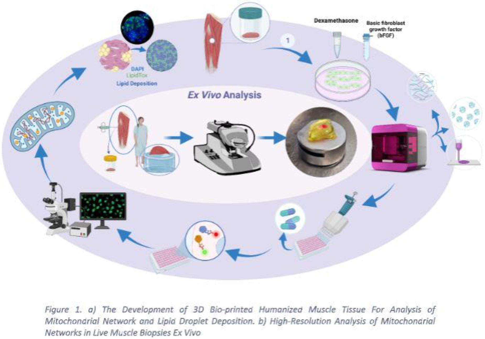
C328 - Isolation Of Matrix-bound Nanovesicles From Porcine Vaginal Extracellular Matrix
H. S. Baghaki, M. Egnot, M. Therriault, P. Moalli, A. Artsen
Magee Women's Research Institute, Pittsburgh, PA
*Purpose/Objectives: Polypropylene (PP) mesh is used in urogynecologic surgery to improve durability and reduce the invasiveness of surgical treatment of pelvic organ prolapse and stress urinary incontinence. However, PP mesh is associated with a strong pro-inflammatory and fibrotic response and the resulting high clinical complication rate precludes use for vaginal prolapse repair. Extracellular matrix (ECM) based therapies are a promising potential therapeutic to improve the foreign body response after mesh implantation or vaginal insult. Indeed, ECMs constitute a resource of proteins, growth factors, and exosomes that encourage tissue regeneration, wound healing, and immune regulation. Heterologous extracellular matrix tissue sources from porcine small intestine submucosa (SIS) and porcine urinary bladder matrix (UBM) have been used to improve vaginal wound healing after implantation of PP mesh or after vaginal insults such as childbirth injury or disruption of pelvic support. However, this has resulted in only partial improvement in healing. This may be due to differences in ECM composition by tissue source. Porcine vaginal ECM (vECM) has recently been developed as a homologous source, however, its regenerative and immunomodulatory properties have not yet been characterized. Matrix-bound vesicles (MBV) are an important regenerative element of the ECM, shown to produce the regenerative and anti-inflammatory properties associated with SIS and UBM ECM when isolated and administered alone. MBV contents have also been shown to vary based on tissue source. Here we aim to isolate MBVs from vECM to allow for characterization.
*Methodology: The porcine vagina was harvested from 6-month-old nulliparous pigs immediately after euthanasia. Decellularization was performed via serial washing in detergent and delipidating agents as previously described. vECM powder was solubilized by digesting with collagenase overnight with constant agitation. Nanoparticles were isolated in duplicate using sequential centrifugation at 500 g, 2500 g, and 10,000 g to separate and remove insoluble collagen fibrils and other non-soluble remnants followed by ultracentrifugation method at 100.000 g at 4°C for 2 h. The recovered pellet was resuspended with PBS. Nanoparticle size distribution was determined using Nanoparticle Tracking Analysis (NanoSight LM10, Malvern Panalytical) at a dilution of 1:100.
*Results: Qiagen DNEasy DNA extraction followed by fluorescent Picogreen dsDNA assay demonstrated an average 75.8% reduction of dsDNA after decellularization. NTA analysis demonstrated recovery of nanoparticles with a concentration of 1.25e+10 +/- 1.44e+09 nanoparticles/ml. Mean size of particles was 95.8 +/- 6.8 nm and the most frequent particle size was 103.5 nm, consistent with previously described MBV size, which is between 30 nm and 150 nm.
*Conclusion/Significance: Characterizing and isolating beneficial components of the ECM is crucial to determining vECM’s place in restoring tissue integrity and minimizing deleterious foreign body response secondary to PP mesh. Here nanoparticles were isolated from decellularized vECM and will be further characterized for surface markers and protein, cytokine, and miRNA content.
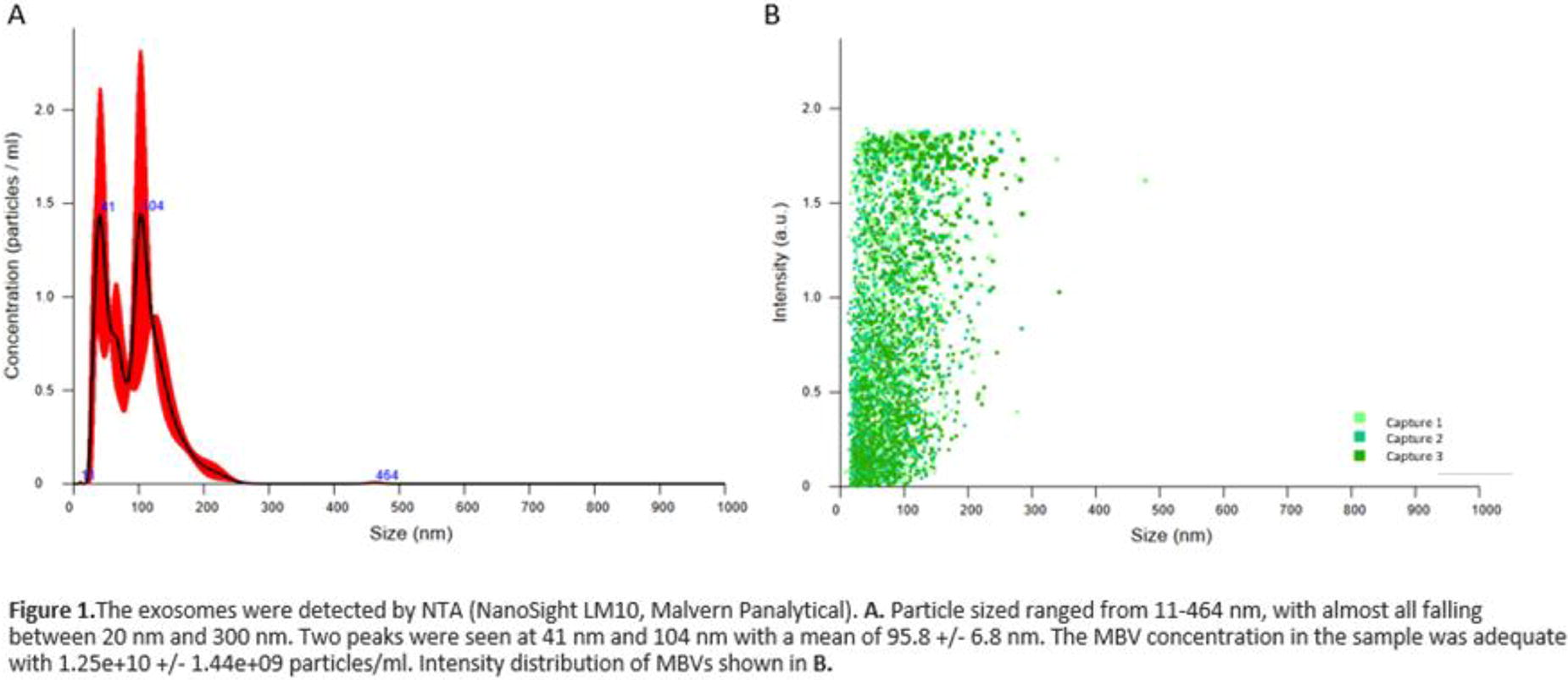
C329 - Constructing Biomimetic Uterine Tissue To Model Maternal Aging
S. E. White, A. Simpson, A. C. Brown, D. O. Freytes
North Carolina State University, Raleigh, NC
*Purpose/Objectives: Advanced maternal age is correlated with diminished fertility and heightened risks of adverse pregnancy outcomes, including low fetal weight, gestational diabetes, obstetric injuries, and uterine ruptures. The underlying causes of this increased risk remain unknown. Changes in the extracellular matrix (ECM), however, may contribute. Further, the field of tissue engineering is becoming increasingly reliant on ECM-derived biomaterials, with decellularized ECM emerging as a standout candidate due to its favorable wound-healing properties and ability to guide biological and biomechanical processes. In response to the growing prevalence of aging effects in the clinic, there is a pressing need to develop biomaterial constructs that mimic both young and aged tissue. Therefore, this study aimed to construct biomimetic tissue derived from native and decellularized extracellular matrix uterine samples from young and aged rodents.
*Methodology: Nulliparous Sprague-Dawley rats at estrus were aged two months and 11 months and euthanized via CO2 inhalation (IACUC approved). Post-euthanasia, the uterine tissue was dissected, with half of the samples randomly allocated to undergo decellularization. Biomimetic constructs were prepared using either native intact tissue or decellularized uterine tissue. The macroscale mechanical properties of the uterine constructs were determined via uniaxial tensile testing. Microscale mechanical properties were measured using atomic force microscopy (AFM), and material architecture was examined through confocal microscopy and scanning electron microscopy (SEM).
*Results: Hydrogels derived from native and decellularized uterine constructs from young and aged samples were successfully created. AFM protocols revealed that aged uterine tissue exhibited significantly greater stiffness than young uterine samples.
*Conclusion/Significance: As extracellular matrix-derived products are becoming more prevalent in tissue engineering applications, studies such as those presented herein provide a critical first step in understanding the impact of age on the uterus. Further, an enhanced understanding of uterine aging will facilitate the development of therapeutic or clinical practices that are geared toward those at greater risk of adverse pregnancy outcomes.
C331 - Donor-dependent Differences In The Characteristics Of Cell Preparations From Human Adipose Tissue
O. Hahn, P.-K. Ficht, K. Sridharan, O. Waheed, K. Peters
University Medical Center Rostock, Rostock, Germany
*Purpose/Objectives: Adult stem cells are one of the most researched topics in regenerative medicine. Adult stem cells show little or no signs of cell differentiation, can proliferate and differentiate into different cell types and are capable of self-renewal. Adipose tissue is a recognized source of adult stem cells (adipose tissue-derived mesenchymal stem cells/adMSC). During the isolation of adMSC, the so-called stromal vascular fraction (SVF: a heterogeneous cell fraction after liposuction and enzymatic digestion) is produced. In addition to adMSC, the SVF also contains progenitor cells from the endothelial and hematopoietic lineage and has a similar therapeutic potential to adMSC. Both SVF and adMSC appear to have immunomodulatory effects via the secretion of growth factors and anti-inflammatory cytokines. However, the therapeutic approaches with SVF or adMSC are probably also hampered by the fact that the characteristics of the tissue donors (gender, age, body mass index, collection site) influence the yield and the cell properties. In our studies, we therefore quantified the donor characteristics, the isolation yield and certain cell parameters in order to improve the predictability of the use of therapeutic cells.
*Methodology: The selected parameters were cell number, viability, and cell size. The data analysis (n = 120) included donor gender, donor age with a median of 44.5 years, donor BMI and collection site. In a second step, a smaller group (n = 8) was analyzed by flow cytometry to characterize the composition of the cell population in the SVF, comparing the following three cell populations between individuals to investigate inter-individual variability: endothelial (progenitor) cells, pericytes and adMSC.
*Results: The gender of the donors in our studies was distributed unequally (94 % of all donors were female, 4 % were male, 2 % not specified). Approximately 27% of the donor cohort was categorized as normal weight, so over 70% of donations were from overweight donors. In addition, donor age was found to affect cell viability, but not cell number and size. Interestingly, the viability of adMSC from older donors (over 44.5 years) was not affected (statistically even significantly higher). On the other hand, donor BMI showed statistically significant effects on cell number and size, but not on cell viability. The cell number and size of overweight (25 < BMI < 30) and obese (BMI > 30) donors was significantly increased compared to normal weight donors. In addition, the collection site also appears to have a significant influence.
*Conclusion/Significance: These data clearly show that donor characteristics can have a significant impact on the cell yield and the viability of SVF and adMSC. Therefore, the characteristics of the donors should be clearly identified in order to increase the predictability of isolation yield and cell behavior and to define discrete cell populations for personalized therapy.This work was funded by the Rostock University Medical Center and the European Union (EFRE project-No. TBI-V-1-141-VBW-116).
C332 - 3D Bioprinting Of Stem Cell Encapsulated In Decellularized Hydrogel For Burn Healing Applications
F. Chogan1, Y. Chen2, S. Haykal1,3, M. Jeschke1,2
1
Institute of Medical Science, Temerty Faculty of Medicine, University of Toronto, Toronto, ON, Canada,
2
Department of Surgery, Faculty of Health Sciences, McMaster University, Hamilton, ON, Canada,
3
Latner Thoracic Research Laboratories, Division of Thoracic Surgery, Toronto General Hospital Research Institute, University Health Network, Toronto, Canada, Toronto, ON, Canada
*Purpose/Objectives: Burn injuries, ranking as the fourth most common and among the deadliest worldwide, present a formidable challenge in healthcare. The gold standard for burn care, autograft, faces limitations due to donor scarcity. Recent advancements in tissue engineered scaffolds, particularly in 3D bioprinting, reshape burn treatment clinical trials. However, the challenge lies in finding biomaterials that mimic the extracellular matrix (ECM). While decellularized ECM (dECM) replicates native complexity, it lacks sufficient mechanical characteristics and may pose infection risks. Moreover, an optimal cell source is crucial for achieving ideal wound healing outcomes. Therefore, the synergy of innovative biomaterials, 3D bioprinting, and cell sources is key to advancing burn treatments. This study introduces a novel multifunctional 3D bioprinted skin substitute composed of porcine skin dECM and a biodegradable polycaprolactone/ polyethylene glycol/ silver nanoparticles (PCL/ PEG/ Ag NPs) for enhanced burn wound healing. A bioink of dECM preserves essential nutrients and promotes cellular growth, proliferation, and migration, while a temporary PCL/ PEG/ Ag NPs layer improves mechanical properties, minimizes moisture loss, and mitigates the risk of infection by serving as a protective barrier against bacteria.
*Methodology: Utilizing multi-response Taguchi's Design of Experiments (DoE), we optimized the 3D bioprinting of PCL/PEG/Ag NPs, focusing on crucial printability factors (extrudability, uniformity factor, pore factor, and integrity). Porcine skin was decellularized for dECM-based bioink, encapsulating human burn-derived mesenchymal stem cells (HBDMSCs) for successful 3D bioprinting. Scanning electron microscope (SEM), mechanical, rheological, water contact angle, biodegradation, water uptake, and antibacterial tests were conducted to examine the mechanical, physicochemical, and antibacterial properties of the 3D bioprinted skin substitute. Cytotoxicity, biocompatibility, cell proliferation, and cell-matrix behaviour were also assessed, ensuring the construct's suitability for burn treatment applications.
*Results: 3D bioprinting parameters have been optimized, resulting in a highly uniform and porous 3D bioprinted skin substitute. Histological and biochemical analyses confirmed the absence of cell content in the dECM matrix, and the decellularization process effectively preserved collagen, elastin, and glycosaminoglycan contents. The skin substitute's remarkable water absorption capability (798 ±126%) can facilitate absorbing the exudates from burn wounds, while its hydrophilic surface enables superior cell attachment and growth. Mechanical assessments indicate that incorporating 3D bioprinted polymeric layers as support for 3D printed dECM significantly enhances ultimate tensile strength (25.8±3.5;MPa) and elongation at break (98.6±10.4%). 3D bioprinted skin substitutes exhibit potent antibacterial activity against Pseudomonas aeruginosa and Staphylococcus aureus bacteria. In a 15-day in vitro cell culture, HBDMSCs encapsulated in the 3D bioprinted dECM demonstrate high cell viability and homogeneous dispersion of live cells throughout the biofabricated construct. The HBDMSCs were effectively proliferated and migrated within the skin substitute, which highlights the capability of the skin substitute to act as a carrier, effectively delivering HBDMSCs to the wound bed.
*Conclusion/Significance: This study highlights the potential of 3D bioprinting in crafting a stem cell-laden skin substitute with decellularized ECM-based hydrogel and biodegradable polymeric composites. The findings suggest that 3D bioprinted artificial skin could serve as a viable skin substitute for treating burns and facilitating autologous skin transplantation.
C333 - A Universal Biofunctionalized Ink For Skin Tissue Engineering
L. P. da Silva, J. C. Pires, S. Freitas-Ribeiro, R. P. Pirraco, R. L. Reis, A. P. Marques
University of Minho, Guimarães, Portugal
*Purpose/Objectives: 3D bioprinting technology has provided the unique opportunity to build complex multilineage engineered tissues. However, advances have been hampered by the incessant need of inks suitable for different cell types with intrinsic phenotypic demands. Herein, we aimed to develop a universal versatile ink designed to house multilineage cells providing native environment-like cues for developing regenerative pre-vascularized tissue-like constructs and complex skin models.
*Methodology: Different cRGD-biofunctionalized gellan gum-based inks were developed by tailoring polymer content and further evaluated regarding their rheological properties and printability using an extrusion-based printer. Printed constructs were prepared with human adipose-derived mesenchymal stem cells (HASC), human dermal fibroblasts (HDFB), human microvascular dermal endothelial cells (HDMEC) and human pre-adipocytes (HpAD) and mechanical stability was assessed up to 21 days. Cell morphology (phalloidin) and viability (calcein/propidium iodide) was analyzed up to 7 days. Pre-vascularized 3D constructs comprising up to 5 layers of HASC- or HDFB/HDMEC-laden bioinks, and a tri-layer skin model with 2 layers of HpAD- plus 2 layers of HDF/HDMEC-laden bioinks, were printed. Human keratinocytes (HKC) were then seeded on top of the latter. Immunohistochemistry and secretome analyses using Luminex were done after 21 days of maturation. A Chick Chorioallantoic Membrane assay was used to histologically assess the performance of pre-vascularized constructs 4 days post-implantation.
*Results: Inks presented non-Newtonian shear-thinning behavior and those with higher polymer presented higher shear viscosity. Temperature affected the rheological properties of the inks, but all ink formulations were printable within the 20-37°C range. Printed cell constructs were mechanically stable in long-term cultures. Cell adhesion was positively impacted by the polymer content; the ink with higher polymer amount supported the adhesion of all multilineage cells being selected as the universal ink for further printing. Among the different cell types, HDMEC/HASC were the most sensitive to the printing process (viability 57-62% vs 86-87% for HpAD/HDF) but cell viability was recovered one-week post-printing. The applicability of the selected formulation in the fabrication of pre-vascularized tissues was confirmed with the formation of a CD31+ capillary-like network with some lumen and vascular sprouts after 7 days of culture, within a HASC- or HDFB-derived matrix rich in collagen, fibronectin and laminin. Moreover, the angiogenic nature of the HASC-based pre-vascularized construct was demonstrated by its secretome - downregulation of bFGF, PDGF-BB and IL-6 and upregulation of HGF, PIGF, Angiopoietin-1 and MMP-2 from day 7 on - and its integration within the chick tissue. Recruitment of CD146+ cells into the neighboring of the implanted constructs and the absence of inflammation (undetectable KUL01+ cells) further suggest their regenerative potential. We further demonstrated the ability to print complex skin models that combined the pre-vascularized dermal-like layer with a stratified epidermis (K14+/K10) and a Nile Red+ lipidic hypodermis-like layer. After maturation (21 days), their angiogenic and adipogenic secretomes were comparable to those from human ex vivo skin.
*Conclusion/Significance: Altogether, we were able to develop a universal ink that enables the fabrication of complex tissue substitutes or stem cell-based strategies opening frontiers for a myriad of applications in the context of cutaneous wound healing.
C336 - Lipid Metabolism Alterations In A Tissue-Engineered Skin Model
A. Tremblay1,2, R. Pouliot1,2
1
Université Laval, Quebec, QC, Canada,
2
Centre de recherche du CHU de Québec-Université Laval, Quebec, QC, Canada
*Purpose/Objectives: Percutaneous absorption studies of in vitro models are still a challenge in terms of comparison with native human skin (NHS). In fact, 3D skin models are more permeable to testosterone than NHS. However, it was shown that polyunsaturated fatty acid (PUFA)-deficient culture media could be the cause for this altered skin barrier. Omega-3 PUFAs, such as alpha-linolenic acid and docosahexaenoic acid, were used to optimize the skin barrier function. In fact, their addition to the culture media was shown to improve testosterone permeability results in a similar manner. However, the addition of linoleic acid (LA), the most abundant PUFA in NHS and the lipid responsible for the formation of a functional skin barrier, did not affect permeability at all compared with unsupplemented conditions. With the objective of reducing the permeability of the self-assembled reconstructed skin, the lipid metabolism of the model was further investigated. Monoacylglycerols, especially 1/2-linoleoyl glycerol (1/2-LG), were found at higher levels in skin substitutes compared with NHS. These levels were even higher in LA-supplemented substitutes despite the overall LA deficiency.
With regards to these previous observations, we hypothesized that the skin barrier is less efficient in the model because of an accumulation of monoacylglycerols containing a LA moiety (1/2-LG). The purpose of this study was to investigate further the influence of 2-LG on the lipid metabolism and the permeability of the skin model to testosterone.
*Methodology: To achieve this, 2-LG supplemented skin substitutes were produced in parallel to unsupplemented substitutes or LA supplemented substitutes. The concentration of 2-LG and LA in their respective culture media was 10 μM. The testosterone permeability assay was performed on a Franz cell diffusion system and testosterone was quantified in samples using ultra-performance liquid chromatography. The fatty acids contained in epidermal phospholipids of skin substitutes were analyzed by gas chromatography coupled with mass spectrometry and the lipid mediators were quantified using liquid chromatography-tandem mass spectrometry. Indirect immunofluorescence and western blot analyses were conducted for tight junction proteins expression and enzymes involved in the metabolism of monoacylglycerols.
*Results: Percutaneous absorption of testosterone was similar in unsupplemented and 2-LG-supplemented skin substitutes. Levels of 1/2-LG in LA supplemented skin substitutes appeared higher compared with those supplemented directly with 2-LG. As for the levels of free LA, the levels seem to be similar for 2-LG or LA supplemented substitutes. As for the lipid profile, the phospholipid fraction contains higher proportions of LA for substitutes supplemented with 2-LG compared with those supplemented with LA. These results indicate that a supplementation with exogenous 2-LG could lead to either higher monoacylglycerol lipase activity or lower incorporation of LA in monoacylglycerols via the diacylglycerol pathway.
*Conclusion/Significance: In conclusion, these results show that the accumulation of 2-LG in the epidermis of skin substitutes could be one of the factors that prevent the adequate incorporation of LA into epidermal phospholipids of the skin model.
C337 - Alignate And Chitosan Modified Gelatin Hydrogel Microbeads For Delivery Of E. Coli Phages
F. Moghtader1, S. Solakoglu2, E. Piskin3
1
Ti/PHAGE San.Tic. Ltd. Sti, Teknopark Istanbul, Istanbul, Turkey,
2
General Manager Feyz Farm Company, Bursa, Turkey,
3
NanoBMT: Nanobiyomedtek Biyomedikal ve Biyoteknoloji San.Tic., Ltd. Sti., Istanbul/Mugla, Turkey
*Purpose/Objectives: Bacterial infections are among the most significant health problems/concerns worldwide. A very critical concern is the rapidly increasing number of antibiotic-resistant bacteria, which requires much more effective counter measures. As the Nature’s antibacterial entities, bacteriophages (shortly (“phages”) are very important alternatives to antibiotics, having much superior features over antiobiotics. Development of phage carrying/controlled releasing formulations is still challenging for protection of their activities in preparation, storage and in use and bringing them to more user-friendly forms by considering their application area/site/conditions.
*Methodology: Here, we prepared gelatin hydrogel microbeads by a two step process. For modification, sodium alginate was included within the initial recipes and these composite microbeads were further coated with chitosan. The target bacteria Escherichia coli (E.coli) and its specific phage (T4) were obtained from bacterial culture collections and propagated. Phages were loaded within the microbeads with a simple method. The phage release characteristics were investigated comparatively and demostrated here.
*Results: High release rates were obtained from the gelatin microbeads. It was possible to reduce the phage release rate by using sodium alginate in the recipe and by chitosan coating. Using these gelatin based microbeads as phage carriers matrices - especially in lyophilized forms - improved significantly the phage stability even at room temperature.
Conclusions: It was concluded that phage release from gelatin hydrogel microbeads could be further controlled by alginate and chitosan modifications and user-friendly lyophilized phage formulations could be produced with much longer shelflife.
*Conclusion/Significance: : It was concluded that phage release from gelatin hydrogel microbeads could be further controlled by alginate and chitosan modifications and user-friendly lyophilized phage formulations could be produced with much longer shelflife.

C339 - Human Multipotent Adult Stem Cells Regenerate Skin In Rats
P. A. Lucas
New York Medical College, Valhalla, NY
*Purpose/Objectives: Full thickness skin wounds can often result from thermal trauma, cancer, shear pressure after prolonged immobilization, and bacterial skin infections. Treatment can range from serial dressing changes to partial or full thickness skin grafts, but results in contracted fibrotic scar, which retains few of the functions of normal skin. There is a clear need for treatment which can be more cost efficient and offer regeneration. Multipotent adult stem cells (MASCs) have properties which make them useful for tissue regeneration: 1) an apparently unlimited proliferation potential in vitro, 2) the ability to differentiate into phenotypes of all 3 dermal lineages, 3) ability to respond to local signals to differentiate to the tissues at the site and 4) do not elicit an immune rejection response. We hypothesize that human MASCs in a biodegradable biocompatible matrix will regenerate skin in full thickness skin defect in adult rats.
*Methodology: Human MASCs were isolated and cultured from human foreskin. The MASCs were seeded into 3 cm diameter polyglycolic acid (PGA) felt at 60 x 106 cells and cultured for 1 week to ensure adherence. Two cm diameter full thickness skin defects were created superior to the spinotrapezius muscle in retired female breeder Sprague-Dawley rats, one on each side of the spine to the level of the underlying musculature. Each skin defect was assigned to one of three groups with 10 defects/group: empty defect, PGA alone or PGA seeded with human MASCs. Wounds underwent gross examination, wound size measurement and photography at post op and every subsequent 7 days until the animals were euthanized at the end of 8 weeks post-op. The tissue from the skin defect was dissected and processed for histology. Human MASCs were identified with antibodies specific to human gamma actin and phenotypes were identified with antibodies specific to those phenotypes.
*Results: Empty defects and defects treated with PGA alone exhibited scar tissue formation under gross and histological examination; they did not exhibit any cells positive to human protein. Defects treated human MASCs displayed what appeared to be regeneration complete with hair under gross examination. There was no immune rejection response to any treatment group. Immunohistochemistry indicated that the MASCs differentiated into 7 phenotypes: keratinocytes, hair follicle cells, gland cells, endothelial cells, smooth muscle cells and fibroblasts. MASCs formed vasculature with red blood cells within the vessels. All cell types were positive for both human gamma actin and the specific phenotype marker.
*Conclusion/Significance: These results demonstrate that treatment of a full thickness wound with human MASCs resulted in tissue regeneration without scar formation or immune rejection. Human MASCs differentiated to several phenotypes in the defect to regenerate the skin including the secondary structures of hair follicles and sweat glands. The MASCs also made new vasculature in the defect. MASCs differentiated into cells of both mesodermal and ectodermal origin. Allogenic MASCs could potentially be used to completely regenerate skin in burn injuries or decubitus ulcer.
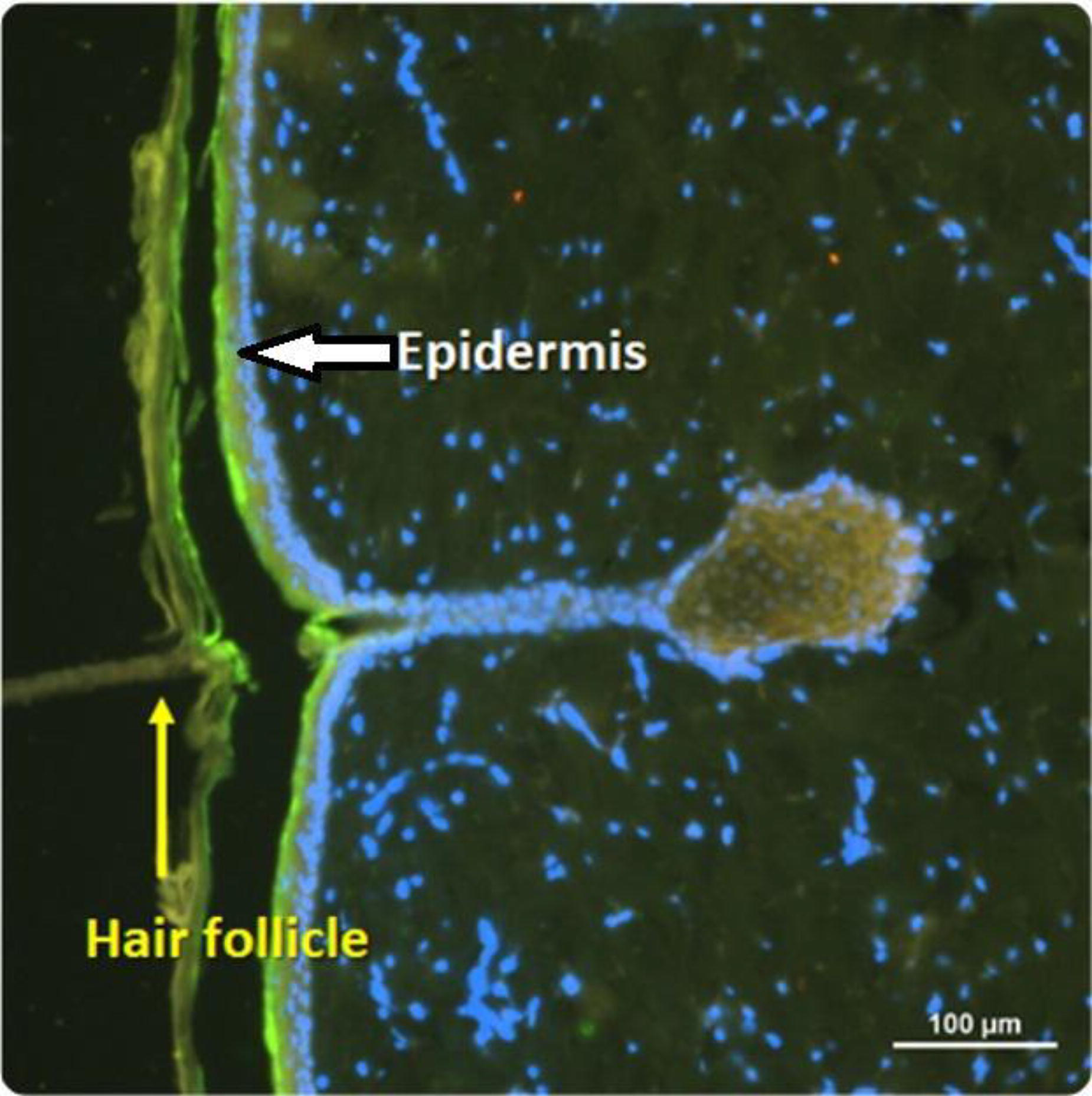
C341 - Thermoresponsive Injectable Composite Hydrogel For Repairing Contraction Suppressed Full-thickness Skin Wounds
S. Khan, A. Banu Shajahan, M. Mamachan, A. Subash, M. Karikalan, O. Vinodhkumar, A. Pawde, S. Maiti, -.-. Amarpal
ICAR-Indian Veterinary Research Institute, Bareilly, India
*Purpose/Objectives: To assess the therapeutic efficacy of Pluronic F127 hydrogel (PF127) incorporating adipose-derived stromal vascular fraction (Adipose-SVF), mesenchymal stem cells (Adipose-MSC), and conditioned media (Adipose-CM) in accelerating the healing of contraction-suppressed full-thickness wounds in a rabbit model.
*Methodology: The experimental study was conducted on twenty-four healthy adult New Zealand white rabbits divided into four groups. The treatment was given once weekly as follows (three doses): Control (PF127), Treatment 1 (Adipose-SVF + PF127), Treatment 2 (Adipose-MSC + PF127), and Treatment 3 (Adipose-CM + PF127). A transparent adhesive elastic bandage was used to suppress wound contraction, thereby prolonging the wound healing time of full-thickness dorsal skin wounds in rabbits. The healing potential of different adipose-derived cell-based and cell-free therapeutics was evaluated based on percentage wound healing, epithelialization period, epidermal thickness, scar evaluation, histopathology analysis, histochemical evaluation, immunohistochemistry (collagen type I), and hydroxyproline assay by comparing with the control group.
*Results: Collagen density analysis using different staining methods, immunohistochemistry, and hydroxyproline assay consistently showed that delivering Adipose-MSCs and Adipose-CM in PF127 hydrogel enhanced epithelialization, collagen production and organization, contributing to improved tissue strength and quality. Even though allogeneic Adipose-SVF promotes wound healing in contraction-suppressed full-thickness wound model in rabbits, it has a lower potential than Adipose-MSC and Adipose-CM when applied topically using PF127 hydrogel. Immunohistochemistry for collagen type I quantification revealed that groups receiving Adipose-MSC and Adipose-CM treatments displayed higher collagen type I densities, indicating improved tissue remodelling and maturation. Even though wounds treated with Adipose-MSCs outperformed Adipose-CM, a significant difference was not observed in the quality of healing, indicating almost similar therapeutic potential.
*Conclusion/Significance: Adipose-MSCs have low immunogenicity and can be used in the allogeneic mode without triggering significant immune responses. Adipose-CM, being cell-free, has minimal antigenic potential and is less likely to cause immune reactions, contributing to its wound-healing potential. The findings indicate that topical application of Adipose-MSCs and Adipose-CM loaded PF127 hydrogel promoted collagen production, tissue organization, and epidermal regeneration, ultimately improving the overall healing outcomes. The contraction suppressed full-thickness wound model developed in this study effectively extended the duration of wound healing, inhibited contraction, enhanced collagen deposition, and promoted re-epithelialization, indicating potential use in translational research.

C343 - Provisional Matrix Tissue Scaffold Technology For Rapid Integration And Scarless Reconstruction
J. F. Dye1, N. M. Jalil1, A. Keirouz1, F. V. Lali1, L. Nguyen2
1
Oxartis Ltd, Oxford, UNITED KINGDOM,
2
Biomaterials, Eastman Dental Institute, London, UNITED KINGDOM.
*Purpose/Objectives: There is a clinical unmet need for an off-the-shelf artificial skin for full-thickness skin-loss wound reconstruction which integrates without wound infection and results in a scarless outcome. Our goal is to introduce a new artificial skin modality for reconstruction of skin-loss wounds. This concept is an off-the-shelf biological product, to accelerate skin reconstruction with scarless healing by targeting vascularisation and tissue regeneration. Previous work established a biomaterial composition and general structure which supports rapid ingrowth and differentiation of blood capillaries and fibrocytes from the wound bed. It was also found that the nanoscale fibre mesh and microscale porosity is essential for scaffold integration. Porcine in vivo studies showed such scaffold are cellularised rapildly, and result in histologically non-scarred reconstruction. To fabricate scaffolds consistently for clinical use, we then developed a technology for forming 3-dimensional surgically handleable material from these compositions. Subcutaneous evaluation in a rat model showed histomorphometric differences between different formulations, suggestive of differences in the fibrotic or regenerative pattern of final outcome.
The specific aim was to evaluate the properties of scaffolds produced by the new method, ready for further pre-clinical evaluation and clinical quality (GMP) manufacture.
*Methodology: Protein polymer scaffolds from fibrin alone or together with hyaluronan, gelatin, chitosan, or PVA, were manufactured by the method of emulsion templating, to create 3-dimensional porous material, stabilised by chemical cross-linking and freeze-drying. Resultant structures were examined by Scanning electron microscopy, internal surface area by N2-adsorption isothermography, and mechanical properties by DMA. Proteolytic stability by trypsin degradation rate. Biocompatibility in vitro was investigated using human dermal fibroblasts, obtained with ethical consent.
*Results: Scaffold pore diameters were 40-90 um. Each formulation gave unique variant nanostructures. Fibroblasts rapidly adhered to all scaffolds, were viable, but showed low proliferation. After 28-days culture, cells were infiltrated and conformed closely to each scaffold, with a unique morphology characteristic of each scaffold variant.These results support previous in vivo data, showing that minor modification of biomaterial composition can have profound effects on cell morphological behaviour. If these are found to determine the post-integration histological outcome, these in vitro findings may also have useful predictive value for evaluating clinical scar or regenerative outcomes.
*Conclusion/Significance: Optimized scaffold production to improve consistency of pore structure and streamline production ready for clinical scale-up (GMP), preserves biocompatibility. Moreover, insights into possible anti-scarring properties of scaffolds may be useful for studying cell-matrix regulation as well as clinical optimization for scarless dermal reconstruction.
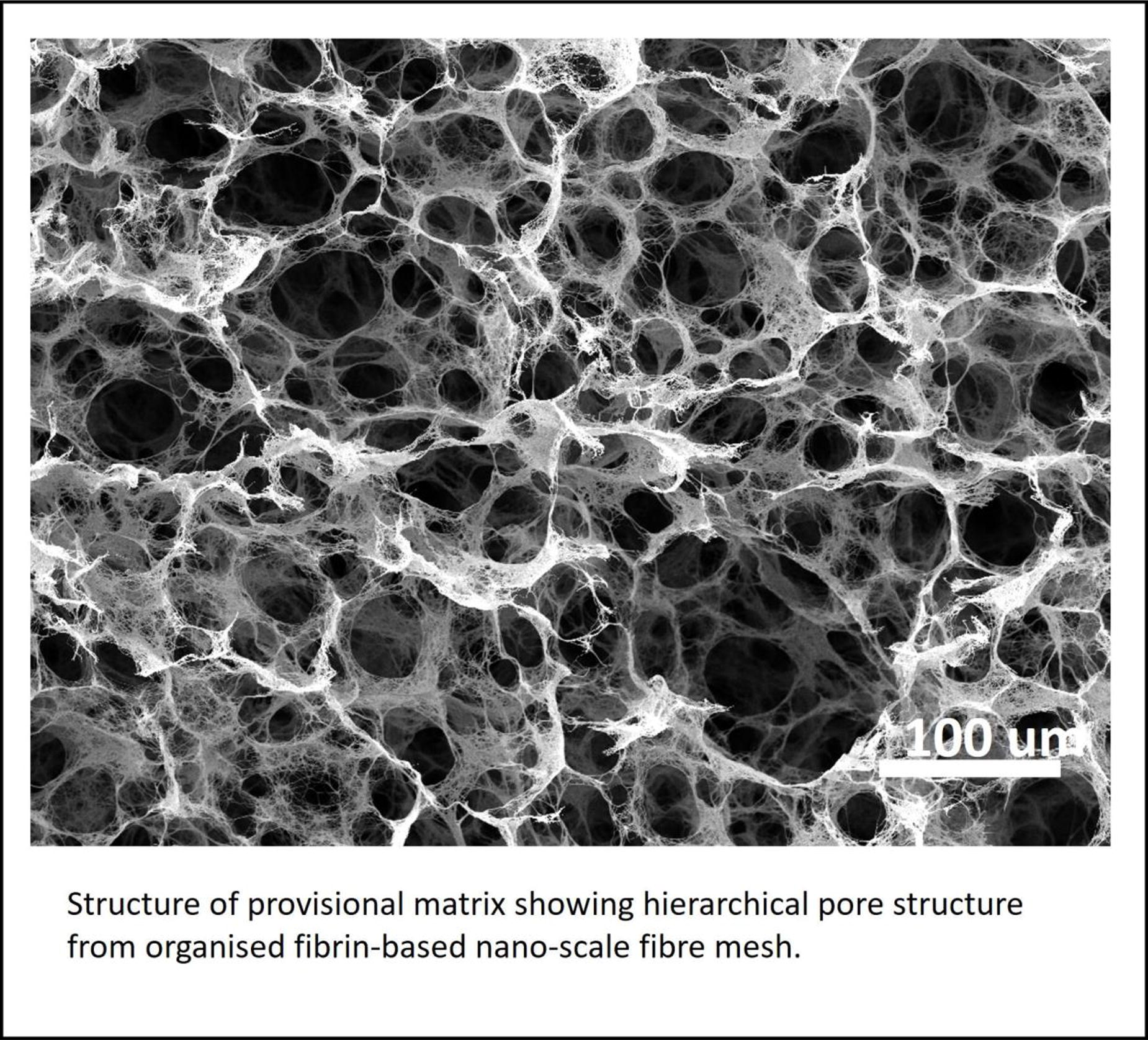
C345 - Three Dimensional Culture Conditioned Fibroblast Engineered Nanovesicle Accelerated The Wound Healing In Mouse
H. Kim
Kyungpook National University, Daegu, Korea, Republic of
*Purpose/Objectives: Three-dimensional (3D) cell culture offers a significant advantage over traditional two-dimensional (2D) culture systems by better recapitulating the physiological microenvironment of cells. In 3D culture, cells grow in a more native-like spatial arrangement, allowing for cell-cell interactions, cell-matrix interactions, and signaling gradients that closely mimic in vivo conditions. This enhanced mimicry of the in vivo environment provides valuable insights into cell behavior, tissue development, drug responses, and disease modeling, making 3D culture an invaluable tool in biomedical research and regenerative medicine. The limited availability, high expenses, and time-consuming harvesting process have constrained the therapeutic uses of extracellular vesicles (EVs). Consequently, engineered nanovesicles (NVs) have emerged as promising biomaterials for clinical applications in translational medicine.
*Methodology: We obtained NVs through the sequential extrusion of fibroblasts (FBs) using polycarbonate membrane filters and subsequent ultracentrifugation. Fibroblasts represent a potential source for NVs, enabling the exploration of their therapeutic effects on wound healing. In this study, we presented the isolation, characterization, and functional properties of nanovesicles derived from 2D and 3D cultured fibroblasts, denoted as 2D-FB-NVs and 3D-FB-NVs, respectively.
*Results: Notably, 3D-FB-NVs treatment exhibited superior wound-healing effects in vitro, promoting the proliferation, migration, and activation of fibroblast cells compared to 2D-FB-NVs. Additionally, 3D-FB-NVs treatment enhanced the proliferation, migration, and tube formation of endothelial cells when compared to 2D-FB-NVs. Furthermore, 3D-FB-NVs treatment expedited wound healing in a mouse skin wound model by augmenting collagen production, collagen maturation, and the formation of blood vessels in the injured skin, surpassing the effects of 2D-FB-NVs.
*Conclusion/Significance: These results unveil a novel role for NVs derived from 3D cultured fibroblasts and enhance our understanding of their potential in improving wound healing.
C348 - Topographically Patterned Gels For Scar-less Wound Healing
V. Ganesan, S. Sen
Indian Institute of Technology Bombay, Mumbai, India
*Purpose/Objectives: Wound healing naturally occurs after an injury but results in scars. Scar tissue lack the properties of the normal tissue leading to poor mechanical functions and aesthetics. We have fabricated hydrogels with topographic patterns that decrease scarring during wound healing. Our gels realign fibroblasts during migration causing changes in tension and ECM secretion.
*Methodology: We design various topographic patterns and simulated their effect on wound closure using a cellular Potts model in the Compucell3D software. The designs which aided in wound closure were then fabricated on a silicon wafer using soft lithography. From the silicon wafer we developed a PMDS mold which is used as a stamp to make the final patterned gels. Our gels consist of methacrylated gelatin that is crosslinked by UV irradiation. 3T3 fibroblasts and human dermal fibroblasts were used for cell culture experiments.
*Results: The patterned gels are able to govern the cell orientation. The spatial distribution of cell contractility was mapped in the region of the patterned gels and we saw the tension distribution was altered. The patterns also influence the ECM secretion.
*Conclusion/Significance: The topographic patterns aid in wound healing and modulate ECM secretion. These patterned hydrogels can be used for the application of scar-less wound healing.
C351 - 3d-printed Tool For Creating Standardized And High-throughput Burn Wounds In Ex Vivo Human Viable Skin Tissues
F. TABATABAEI1, M. Javid2
1
iFyber, ithaca, NY,
2
University of Wisconsin–Madison, Madison, WI
*Purpose/Objectives: The development of biomaterials and medical devices for burn wound treatment necessitates thorough investigation through in vitro/ex vivo models before transitioning to animal studies. Establishing a standardized and high-throughput burn wound model in ex vivo skin presents a considerable challenge. Our objective was to address this challenge by developing a practical and cost-effective 3D-printed burn wound tool capable of uniformly inducing burns in 12 skin samples simultaneously.
*Methodology: Utilizing Autodesk Inventor software, we designed a 3D model comprising a Plate-Base component (PBC) and a Rod-Base component (RBC). The design was exported as a Standard Triangulation Language (STL) file, processed through “Slicer” software to generate a G-code file tailored for 3D printing.
*Results: The Rod-Base component underwent iterative design modifications to optimize weight, airflow, and material consumption, resulting in a final design featuring a unique star shape for enhanced airflow. Simultaneously, the Plate-Base component design evolved to enable easy and secure plate placement, demonstrating compatibility with 12-well plates. The average production time for the model was 14.5 hours, with a production cost of approximately $20, covering printing material and steel rods.
*Conclusion/Significance: In conclusion, this study provides valuable insights into the required equipment and software, empowering researchers to efficiently produce their accurate and cost-effective 3D-printed tool for controlled and reproducible burn wound creation in ex vivo viable skin tissues.
C352 - Development Of Urinary Catheters Delivering Antibacterial Phages
F. Moghtader1, S. Genc Simsek2, N. Turkoglu2, E. Piskin3, E. Piskin3
1
Ti/PHAGE San.Tic. Ltd. Sti, Teknopark Istanbul, Istanbul, Turkey,
2
Faculty of Science and Arts, Department of Molecular Biology and Genetics, Yildiz Technical University, Istanbul, Turkey,
3
NanoBMT: Nanobiyomedtek Biyomedikal ve Biyoteknoloji San.Tic., Ltd. Sti.,, Istanbul/Mugla, Turkey
*Purpose/Objectives: Urinary catheters are simply inserted through the urethra into the bladder and left in place for a period of time (from minutes to days and even longer) to empty to it during the treatment of the hospitilized patients and then removed. The percentage of the hospitilized patients receiving urinary catheters during hospital stay has reached to 25% even more globally. Urinary tract infections due to urinary catheterization are the most common healthcare-associated problems - that may lead secondary nosocomial bloodstream infections - which means higher morbidity and mortality risks. All these strategies/materials will be briefly described/discussed in the min review by focusing on cold plasma applications and using bacteriophages as antibacterial agents to modify urinary catheters - which is our own interests in this field.
*Methodology: Different type of polymers, PVC, silicone, poliurethane, etc. are used to produce tubings in preparation of urinary catheters. These are hyrdophobic materials that pathogenic bacteria like to adhere, propagate and form biofilms which may highly complex 3D structures - could be describes as castles to protect them from the host immune system. Biofilms are dynamic - means that patogenic bacteria are detached and move other sites to developmmore castles. These are hard-tofight battlefields for the host. Bacterial invasions resulting infections is getting a huge health problem globally as a result of insreasing the number of antibiotic resistive pathogenic bacteria. We have to develop challenging strategies to fight with these deadly species - to prevent/reduce infections. Methods: Several strategies have been investigated. Preparation of passivated/antifouling surfaces is the easiest strategy in which surface hydrophilicities of catheter surfaces are increased which are much less prone to bacterial attachment and further biofilm formation. Coating of hydrophobic catheter surfaces with hydrophilic polymers (such as polyethylene glycol) and using several cold plasma techniques to create hydrophilic surfaces are leading strategies having already sucessful antifouling/hydrophilic urinary catheters commercially available. Using antibacterial bioactive surface coating/layers on the hyrophobic catheters is the other main strategy to prevent/reduce urinary catheter-associated infections. In the earlier periods, antibitic loaded/releaind catheters have been developed - seems that this is not and option now due to antibitic resistance. Other bactericidal agents, including nanotubes, graphene oxide, metal and metal oxide nanoparticles, antimicrobial peptides, bacteriophages have been studied but no commercial outcomes yet.
*Results: Under investigation.
*Conclusion/Significance: All these strategies/materials will be briefly described/discussed in the min review by focusing on cold plasma applications and using bacteriophages as antibacterial agents to modify urinary catheters - which is our own interests in this field.
C353 - Extracellular Vesicles Restore Glomerular Endothelial Lipid Metabolic Homeostasis In Alport Syndrome
H. Soloyan1, M. Thornton2, G. Clair3, J. Cuala2, S. Georgia2, Q. Zhang1, R. E. De Filippo1, S. Da Sacco1, L. Perin1, S. Sedrakyan1
1
Children's Hospital Los Angeles, Los Angeles, CA,
2
Keck School of Medicine, University of Southern California, Los Angeles, CA,
3
Pacific Northwest National Laboratory, Richland, WA
*Purpose/Objectives: Chronic kidney disease (CKD) affects more than 10% of the population worldwide, and our understanding of the mechanisms in many forms of CKD are poorly understood. Alport syndrome (AS), is a form of CKD caused by mutations in the collagen IVα3, α4, or α5 gene. Even though injury to glomerular endothelial cells (GEC) in AS is well established, the role of GEC in Alport progression has not been elucidated. Here, we describe the role of mitochondria and lipid metabolism in GEC injury in an animal model of AS, and the potential of using amniotic fluid stem cell (AFSC) derived extracellular vesicles, (EVs) as a rescue strategy to restore glomerular homeostasis.
*Methodology: The phasor approach to fluorescent lifetime imaging microscopy (FLIM) and flow cytometry was applied to evaluate the mitochondria and metabolic changes in GEC of AS vs WT mice. GEC isolated by FACS from Tek-tdT reporter AS and WT mice were compared by bulk RNA-seq, lipidomic and flow cytometric analysis. In vitro, silencing experiments on primary human GEC were performed to study the role of fatty acid synthase (FASN) in mitochondrial dysfunction and GEC damage. FASN-carrying AFSC-EVs and control nanoparticles were applied both in vitro and in vivo to restore lipid homeostasis in GEC.
*Results: FLIM studies showed strong correlation between the metabolic state of GEC and the age or severity of disease in AS mice. RNA-seq analysis revealed changes in the pathways associated with lipid metabolism and mitochondria function. Mitochondrial dysfunction was confirmed using flow cytometric analysis of the MitoTracker signal in tdTomato expressing GEC. Lipidome analysis revealed high abundance of triglycerides in AS GEC. We confirmed accumulation of lipid droplets in the glomeruli of AS mice and in FASN KO human primary GEC, in vitro. These results suggest potential mitochondrial dysfunction in GEC. AFSC derived EV treatment restored lipid homeostasis in GEC, both in vitro and in vivo.
*Conclusion/Significance: We report for the first-time mitochondrial dysfunction in Alport GEC, and the ability of AFSC-derived EVs to rescue this phenotype. Better understanding of the metabolic changes in AS GEC could lead to the development of targeted new therapies for other forms of CKD.
C355 - Effects Of Matrix-bound Nanovesicles On Biologic Aging
W. D’Angelo, H. Capella, N. Venkatasamy, G. Hussey, S. Badylak
University of Pittsburgh, Pittsburgh, PA
*Purpose/Objectives: Early mammalian development is characterized by a remarkable capacity for wound healing and regeneration. In contrast, old age is marked by a declining ability to repair injuries and maintain homeostasis. Underpinning these changes is a progressive loss of epigenetic regulatory marks throughout life, resulting in dysregulated cellular functions and age-related disease. We have shown that the extracellular matrix (ECM) of all tissues contains a type of extracellular vesicle which we have termed matrix-bound nanovesicles (MBV), with roles in tissue repair and immunomodulation and containing lipid, protein, and RNA cargo characteristic of the tissue of origin. More recent results suggest that MBV also encode information about the developmental age of their source tissue. Given the dynamic reciprocity between cells and ECM, in the current study we hypothesize that MBV from developmentally young tissues can restore the epigenetic landscape and cellular functioning of aged cells to a youthful state.
*Methodology: MBV were isolated from decellularized small intestine submucosa from neonate (one day old) and sow pigs (>12 months old) by enzymatic digestion and purified by ultracentrifugation and size exclusion chromatography. Six weekly injections of “young” or “aged” MBV were administered to young (6-8 week) or aged (>18 month) C57BL/6 mice and the effects on biological age were measured in peripheral blood cells by DNA methylation analyses, ATAC-sequencing, and RT-qPCR analysis of age-related marker expression.
*Results: Preliminary results show that systemically administered MBV are taken up by myeloid progenitor cells in the bone marrow, causing changes in chromatin distribution which are maintained following differentiation to macrophages, and resulting in significant changes in mRNA expression of multiple genes related to aging. Our ongoing analyses will use an “epigenetic clock” to measure the biologic age of MBV-treated animals and determine the effect of MBV donor tissue age.
*Conclusion/Significance: Our results suggest that MBV from young tissues can delay or reverse the epigenetic changes that underlie aging, highlighting the potential of ECM as a reservoir of anti-aging molecules that could be exploited for rejuvenation therapies to stave off age-related diseases such as cancer, cardiovascular disease, and neurodegeneration.
C356 - Dclk1+ Alveolar Tuft Cells From The Distal Rat Lung Form Multipotent Sox9+ Organoids
S. Mizoguchi1, T. Obata2, S. Edelstein1, H. Kim1, M. B. Raredon1
1
Yale University, New Haven, CT,
2
Nagasaki University Graduate School of Biomedical Sciences, Nagasaki, Japan
*Purpose/Objectives: Whole lung regeneration, based on the recellularization of decellularized scaffolds, requires the bioengineering of complex alveolar structures. However, many hurdles remain on the road to creating alveoli with complete blood-gas barrier capable of gas exchange. The choice of cells used for these efforts is particularly critical. An ideal epithelial population should be capable of differentiating into all of the various epithelial lineages that contribute to lung function. Two existing options include basal cells, which are stem cells in the proximal airways, and bronchioalveolar stem cells (BASCs), which are multipotent stem cells at the bronchioalveolar junction. Here, we present data showing that alveolar tuft cells, which are present throughout the alveoli of rats, co-express the endodermal pluripotency markers SOX9 and LGR5 and show clear, functional stem-cell character as well.
*Methodology: Single cell RNA sequencing (scRNAseq) was performed on the post-pneumonectomy lungs in rats to identify key players in alveolar epithelial regeneration and maintenance of lung homeostasis. To obtain alveolar tuft cell population, which were thought to be deeply involved in lung regeneration, we isolated DCLK1+ alveolar tuft cells from native rat lungs using magnetic selection, and cultured them within Matrigel organoid cultures. The cells were cultured under various conditions and evaluated histologically for their pluripotency and differentiation potential.
*Results: ScRNAseq of rat lung tissue shows that alveolar tuft cells expand following pneumonectomy and play an important role in epithelial regeneration. Isolated DCLK1+ alveolar tuft cells successfully formed organoids and this population maintains SOX9-positivity in vitro and shows the clear capability to differentiate into ciliated cells, goblet cells, and basal cells, as well as cells resembling alveolar Type I and Type II epithelial cells, indicating that they maintain stemness similar to BASCs in the peripheral lung.
*Conclusion/Significance: The establishment of a repeatable method for the isolation and expansion of millions of adult peripheral lung progenitor stem cells will have a significant impact on whole-organ tissue engineering. Our newly established SOX9+ organoid cultures have the potential to reproduce multiple alveolar constituent cells required to form a complex and well-regulated blood-gas barrier. The results of this research are expected to make a significant contribution to not only for lung bioengineering but also for basic science studies of lung biology and disease.
C357 - Development Of A Sustainable Multi-cell Organoid System: Exploring The Regulatory Factors Of Whole-lung Engineering
S. E. Edelstein, S. Mizoguchi, S. Brickman Raredon
Yale University, New Haven, CT
*Purpose/Objectives: The complexity of the human lung makes it challenging to engineer a functional gas exchange system. Successfully engineering new organs would offer a promising way to treat end-stage lung disease circumventing long donor lists and allograft rejection. To make headway in this field, we have developed an organoid model that facilitates the study of interactions between the resident cellular components of the lung—epithelial, endothelial, mesenchyme, and immune—and their dependence on exogenous cues.
*Methodology: First, we developed individual cultures of each resident cell type to confirm sustainability in extracellular matrix and epithelial progenitor expansion medium dosed with ROCK inhibitor, CHIR99021 (GSK-3 inhibitor), BIRB796 (pan-p38 MAPK inhibitor), and A8301 (Activin/NODAL/TGF-beta pathway inhibitor). We then studied whether or not each cell system formed organoids or not due to cell-cell interactions between the resident cellular components in the expansion medium. Cellular response was assessed by immunohistochemical staining, RT-PCR, and morphological characterization.
*Results: We developed a 3-dimensional, self-assembling model useful to probe the expansion and differentiation of pulmonary cell systems under the influence of exogenous factors. We have confirmed that epithelial progenitors are essential for the self-assembly of organoids and that growth medium plays a critical role in determining the cellular fate of epithelial progenitors, independent of their initial characterization. Additionally, our multicellular organoid system reveals evidence that increased cellular communication affects the stemness of epithelial progenitor cells in vitro.
*Conclusion/Significance: Together, these results demonstrate the power of multicellular organoid systems to inform regenerative lung biology and emphasize that growth medium and cell-to-cell crosstalk must both be carefully considered when engineering pulmonary tissues ex vivo.
C361
C362 - Effects Of Bariatric Surgery-related Weight Loss On The Basic Characteristics, Metabolism, And Immunomodulation Of Adipose Stromal Cells In A Follow-up Study
A. Adnan1, M. Juntunen1, T. Tyrväinen2, M. Kelloniemi2, L. Kummola1, R. Autio1, M. Patrikoski3, S. Miettinen1
1
Tampere University, Tampere, Finland,
2
Tampere University Hospital, Tampere, Finland,
3
Finnish Red Cross Blood Service, Helsinki, Finland
*Purpose/Objectives: Human adipose tissue derived stromal/stem cells (ASC) are attractive candidates for therapies requiring tissue regeneration and immunomodulation. Their therapeutic potential has been observed for many disorders. Since ASC are often obtained from overweight or donors with obesity, it is crucial to know how obesity affects ASC functions. The expansion of adipose tissue in obesity causes dysfunction of adipocytes and their mitochondria. These dysfunctional adipocytes secrete inflammatory adipokines regulating the functions of immune cells. Considering the importance of donor weight on ASC properties, this study aims to analyze the effect of bariatric surgery associated weight loss on ASC derived from the same donors before (obASC) and after the weight loss (wlASC).
*Methodology: ASC were isolated from adipose tissue samples obtained from donors undergoing bariatric surgery, before (obASC) and after (wlASC) the weight loss. Additionally, plasma inflammatory factors, adipose tissue samples, ASC’s oxygen consumption rate, cell surface marker profile, proliferation, differentiation capacity and inflammatory factors were investigated. To evaluate immunomodulatory capacity of obASC and wlASC, macrophages were prepolarized and pre-activated from peripheral blood mononuclear cells towards pro-inflammatory (M1), anti-inflammatory (M2), and regulatory macrophages (Mreg) and co-cultured with ASC. Pro- and anti-inflammatory surface marker levels (CD11C, HLA-DR, CD86, CD163 and CD206), gene expression (IFNY, TNF-α, CCL5, COX2, MRC1, TSG6, KLF4, PPARY, CD163, IDO-1, STAT6 and HLA-G) and cytokine secretion (MCP-1, IL-6, MDC, IL-12p70, MIP-1α, IL-1β, TNFα, TARC, IL-1RA, IL-4 and IL-10) were studied from the ASC co-cultures compared to macrophage monocultures.
*Results: Concomitantly with the weight loss, a significant decrease in plasma leptin and increase in adiponectin levels were detected as well as a significant decrease in adipocyte size. There was no crown like structures detected in the adipose tissue after the weight loss. Moreover, weight loss significantly improved the overall mitochondrial respiration of ASC. The inflammatory marker genes TNF-α, CCL5 and COX2 were significantly upregulated in ASC monocultures after weight loss, and only anti-inflammatory gene TSG6 was upregulated. Similarly, only proinflammatory cytokine IL-12p70 was significantly downregulated in wlASC monocultures after weight loss. However, the immunomodulative functions of ASC after weight loss and their capacity to drive macrophage polarization and cytokine secretion into anti-inflammatory direction were variably regulated in this study. Moreover, our cytokine secretion, cell surface protein expression and macrophage polarization data suggest that wlASC are as or even more pro-inflammatory as obASC.
*Conclusion/Significance: In conclusion, our results suggest that bariatric surgery-related weight loss improves donors’ metabolic health and impacts positively on ASC characteristics. However, ASC may not fully resume their immunosuppressive capacity after the weight-loss.
C365 - Defining Angiogenic And Myogenic Regenerative Responses Following Autologous Bone Marrow Cells Treatment In Chronic Limb Threatening Ischemia
L. Moldovan, L. Zhang, J. R. King, J. Stashevsky, A. R. Gutwein, R. I. Muraru, N. Moldovan, M. P. Murphy
IUPUI, Indianapolis, IN
*Purpose/Objectives: There has been significant interest in cell-based therapies for treating vascular disease. To date all mechanistic studies have been performed in murine models that present significant genomic discordances between mice and humans. In this first in man study we performed a multi-factorial analysis of muscle tissue in patients with critical limb threatening ischemia (CLTI) in response to the injection of concentrated autologous bone marrow aspirate (cBMA).
*Methodology: Patients requiring semi-elective below-knee amputation (BKA) due to CLTI received a cBMA (10cc) injection into the anterior tibialis muscle (ATM). ATM and soleus (as internal control) were harvested 3-21 days post-injection. Control tissue was collected from risk factor-matched CLTI patients undergoing amputation. The tissue was processed for either immunohistochemistry or RNA analysis. Immunostaining for the CD31 antigen was selected to measure the capillary density, reported as CD31-positive features normalized to fiber number. High multiplexed imaging and spatial analysis (PhenoCycler platform from Akoya Bioscinces) was used to assess the tissue response to injected cells: progenitor cell markers (PAX7, MYOD, PDGFRA), immune cells infiltrate (CD3, CD14, CD68, arginase), endothelial cells (CD31), as well as proliferation and apoptosis (KI67 and TUNEL, respectively). We also tested by real-time PCR the expression of genes encoding cytokines with pro-angiogenic activity, as well as some metabolic genes.
*Results: We found that immediately downstream of the injection site CD31 profiles increased in the ATM of cBMA-treated patients vs. controls (1.54 ± 0.15 vs. 0.83 ± 0.17, p < 0.05). Capillary density in cBMA-treated patients was also increased in the soleus. Fibers in advanced stages of degeneration were invaded by dense clusters of blood-derived mononuclear cells. Thus, in cBMA-treated patients we found (i) higher proportion of CD3+ cells (15.22% vs 7.78%); (ii) increased proportion of CD14+ cells (10.21% vs. 7.29%); (iii) decreased proportion of CD68+ cells (0.39% vs. 1.58%), concomitant with increased ARG+ cells (4.37% vs. 1.94%), which led to a reversal of the argiase:CD68 ratio (7.9 vs 0.3, respectively). The real-time PCR data showed significant (p<0.05) up-regulation of several regeneration-related genes in some or all segments tested.
*Conclusion/Significance: Besides confirming a pro-angiogenic and muscle repairing activity at the site of implantation, this study also highlights that the effect of cBMA is paracrine as there was a robust response seen in the soleus muscle, which resides in a separate fascial compartment. The injected cells speed up the local repairing of the ischemia-inflicted tissue damage, and also mobilize the whole body’s immuno-reparatory system.
C367 - Adipose-derived Cell And Cell-free Therapies For Contraction-suppressed Full-thickness Skin Wound Healing
S. Khan, S. Banu, M. Mamachan, A. Subash, K. Mathesh, O. Vinodhkumar, A. Pawde, S. Maiti, -.-. Amarpal
ICAR- Indian Veterinary Research Institute, Izatnagar, India
*Purpose/Objectives: To evaluate the therapeutic potential of adipose-derived stromal vascular fraction (Adipose-SVF), mesenchymal stem cells (Adipose-MSC), and conditioned media (Adipose-MSC-CM) for the repair of contraction-suppressed full-thickness wounds in a rabbit model.
*Methodology: In this study, twenty-four healthy adult New Zealand white rabbits were randomly assigned to four groups, each comprising six animals. The treatment protocol involved administering treatments at three doses, administrated one week apart via intradermal route. The groups included: control (No treatment), treatment 1 (Adipose-SVF), treatment 2 (Adipose-MSC), and treatment 3 (Adipose-MSC-CM). 8-point injection method was used to deliver the treatment to the wounds on days 0, 7, and 14. The evaluation of the therapeutic efficacy of various adipose-derived cell-based and cell-free treatments involved assessing parameters such as percentage wound healing, epithelialization duration, epidermal thickness, scar assessment, histopathological analysis, histochemical evaluation, immunohistochemistry specifically targeting collagen type I, and quantification of hydroxyproline levels. These assessments were compared with the outcomes observed in the control group to gauge the impact of the treatments on wound healing dynamics.
*Results: Analysis of collagen density through various staining methods, immunohistochemistry, and the hydroxyproline assay consistently demonstrated that the delivery of Adipose-MSCs and Adipose-MSC-CM significantly improved epithelialization, collagen production, and organization. This enhancement contributed notably to the augmentation of tissue strength and overall quality. While allogeneic Adipose-SVF exhibited some degree of efficacy in promoting wound healing within the contraction-suppressed full-thickness wound model in rabbits, its potential upon intradermal application was notably lower when compared to Adipose-MSC and Adipose-MSC-CM treatments. Immunohistochemistry specifically targeting collagen type I quantification revealed considerably higher densities in groups receiving Adipose-MSC and Adipose-MSC-CM therapies. This elevation pointed towards a more pronounced improvement in tissue remodelling and maturation.
*Conclusion/Significance: Adipose-MSCs possess low immunogenicity, making them suitable for allogeneic use without eliciting substantial immune responses. On the other hand, Adipose-MSC-CM, being devoid of cells, exhibits minimal antigenic potential, thereby reducing the likelihood of triggering immune reactions. This characteristic contributes significantly to its effectiveness in promoting wound healing. The study findings underscored the positive impact of topically applying both Adipose-MSCs and Adipose-MSC-CM. These treatments were observed to stimulate collagen production, enhance tissue organization, and facilitate epidermal regeneration, leading to notable improvements in overall healing outcomes. This study effectively highlighted the advantages of utilizing adipose cell-derived therapies and cell-free products in bolstering wound healing processes, indicating their promising potential for future translational research endeavours.
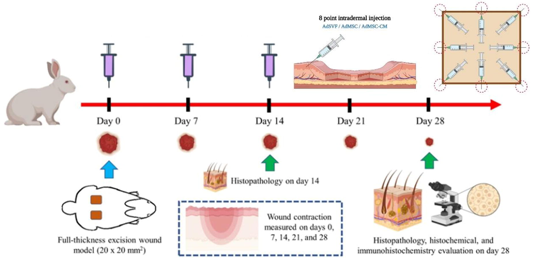
C368
C369
C370
C372 - Endothelial-differentiated Adipose-derived Mesenchymal Stem Cells In Re-endothelialization Of Decellularized Omentum Flaps
F. Biniazan, A. Stoian, C. Duru, S. Haykal
UHN, Toronto, ON, Canada
*Purpose/Objectives: Extensive soft tissue defects significantly impact both patient quality of life and functional capabilities, while appropriate repair strategies remain a significant challenge. Decellularization/Recellularization of tissues and organs emerges as a potential approach in the field of tissue engineering to address concerns related to donor site morbidity and the limitations of transplantation. Nonetheless, a primary impediment to accomplishing this goal lies in the angiogenesis and rendothelialization of acellular vasculature. Establishing an intact and functional vascular structure, which serves as a crucial conduit for delivering oxygen and nutrients, remains a significant challenge in the decellularization/re-endothelialization procedure. Endothelial cells (ECs) are important for neovascularization; however, their limited quantity poses challenges for sufficient therapeutic applications. The ability of adipose-derived mesenchymal stem cells (MSCs) to differentiate into other types of cells, such as endothelial cells, makes them attractive candidates for promoting the process of angiogenesis. This research aims to investigate the efficient differentiation of adipose-derived MSCs into ECs and explores the application of decellularization and re-endothelialization techniques on omentum flaps using endothelial-differentiated adipose-derived MSCs.
*Methodology: MSCs were isolated from porcine adipose tissue by collagenase digestion. Adipose-derived MSCs were cultured and used for the experiments at passage 3. Cells were characterized by flow cytometry using a panel of monoclonal antibodies. They were induced to differentiate into endothelial cells with endothelial-inducing medium and analyzed for expression of the endothelial-specific markers and functions. Otherwise, for flaps perfusion-decellularization, protocol with low concertation of detergent was used (based on 0.05% of SDS). Histological analysis, and quantification and DNA quality assessment was done for native and decellularized omentum flaps. A re-endothelialization of the preconditioned flaps with Endothelial-differentiated adipose-derived MSCs was achieved through perfusion-based seeding. Histological evaluation, Immonohistochemistry analysis, and viability of cells was tested after seeding.
*Results: Cultured MSCs at passage 3 were positive for the mesenchymal surface markers CD105, CD90, CD29, while negative for typical hematopoietic and endothelial markers (CD45 and CD31). Differentiation into ECs was induced by endothelial inducing medium. Results showed the differentiation of expanded adipose derived MSCs into cells with characteristics, and functional features of ECs. Otherwise, our techniques exhibited proof of decellularization of the omentum flaps, and cell attachment, accompanied by early indications of a regenerated neo-endothelium.
*Conclusion/Significance: The outcomes of this research provide a method to promote differentiation of MSCs into ECs in vitro and may offer valuable insights into the feasibility and efficacy of utilizing decellularized omentum flaps as scaffolds for endothelial-differentiated adipose-derived MSCs, contributing to advancements in tissue engineering and regenerative medicine.
C373 - Exploring The Impact Of Xeno-free Culture Conditions On Adipogenic Differentiation And Angiogenic Properties Of Adipose Tissue-derived Stem Cells
M. Brohlin1,2, A. Lauvrud2,3, M. Vittoria Giraudo2, R. Wiberg2,3, M. Wiberg2,3, P. J. Kingham2
1
Department of Clinical Microbiology, Umeå, Sweden,
2
Department of Medical and Translational Biology, Umea, Sweden,
3
Department of Diagnostics and Intervention, Umea, Sweden
*Purpose/Objectives: Adipose tissue-derived stem cells (ASCs) isolated from the stromal vascular fraction of fat tissue are a heterogeneous cell population that exhibit specific phenotypic features which are important for various types of regenerative medicine applications. Culturing ASCs in xenofree cell culture media formulations provide a safer, defined culture environment to meet regulatory requirements for clinical applications. Another option is to use a pool of human platelet lysates to avoid animal components contamination. In this study we investigated the effects on the angiogenic and adipogenic profile of the ASCs with focus on their potential applications in reconstructive surgery such as cell-assisted fat grafting. ASCs from lipoaspirates were cultured in platelet lysate (PLT) and xenofree medium and compared with traditional foetal bovine serum (FBS).
*Methodology: Human lipoaspirate samples (n=7) were digested to yield the stromal vascular fraction cells which were then expanded in Minimum Essential Medium-α (MEM-α) supplemented with either 10% (v/v) FBS or with 2% (v/v) human PLT (PLTGold®) or in PRIME-XV MSC expansion XSFM xeno-free, serum free medium (XV). Analysis of ASCs defined stem cells markers CD73, CD90 and CD105 together with the pericyte marker CD146 was performed by flow cytometry. Proliferation rates were monitored over multiple passages and adipogenic differentiation performed at early and expanded passage culture. Angiogenic and growth factor gene expression was analyzed by qRT-PCR and adiponectin protein levels were detected by ELISA. An in vitro angiogenesis assay was performed to measure biological activity of the secreted factors.
*Results: Cells in FBS and PLT showed similar growth rates whereas the cells cultured in XV-medium proliferated at a significantly faster rate. All cultures were >98% positive for CD73, CD90 and CD105, whereas CD146 expression was significantly higher in XV-cells. Adipogenic differentiation was most noticeable in cells which had been cultured in XV medium although cells grown in PLT showed impaired differentiation compared with cells from the FBS cultures. IGF1 gene expression was highest in cells cultured in PLT whilst cells grown in XV medium showed 10-fold lower expression compared with FBS cells. In contrast, HGF gene expression was 90-fold greater in cells cultured in XV medium compared with those cultured in FBS. Conditioned medium from XV cultured cells showed the most angiogenic activity, inducing the greatest endothelial cell network formation and maturation.
*Conclusion/Significance: Culture under different conditions alters the ASCs characteristics. Since cells cultured in XV medium showed the best adipogenic and angiogenic profile this might be a preferred medium formulation for preparing cells required for reconstructive surgical applications such as cell-assisted fat grafting.
C374
C375 - Enhanced Urokinase Plasminogen Activator Secretion In Xeno-free Cultures Of Human Adipose Stem Cells Through Forskolin Stimulation
M. Giraudo, G. Andersson, P. J. Kingham
Umeå University, Umeå, Sweden
*Purpose/Objectives: Adipose-derived stem cells (ASCs) hold significant potential for treating various clinical conditions. To enhance their regenerative properties, ASCs can be chemically stimulated in vitro. Different protocols have been studied. However, unsatisfactory results persist, partly due to the relatively costly long-term methods. Furthermore, current culturing techniques often rely on the use of xenogenic fetal bovine serum that can be immunogenic, limiting clinical applications. To facilitate clinical translation of ASCs-derived therapeutics, we investigated the effect of different stimulation protocols using a xeno-free medium (PRIME-XV MSC Expansion XSFM) on human ASCs.
*Methodology: The xeno-free medium was supplemented with stimulants (forskolin, basic fibroblast growth factor, platelet-derived growth factor-AA, neuregulin-1) in combination or alone for 72 hours. The protocols were studied using qRT-PCR and ELISA, and cellular signaling pathways were further investigated using a human phosphokinase array and Western blot. Ultimately, the functionality of the ASCs was evaluated by studying the impact of ASCs’ conditioned media (CM) on angiogenic properties of human umbilical vein endothelial cells (HUVECs).
*Results: Our findings revealed that forskolin (FSK) stimulated ASCs produced significantly higher levels of urokinase plasminogen activator (uPA), a serine protease involved in tissue remodeling processes. Both the phosphokinase array and Western blot confirmed the activation of the phosphorylated extracellular signal-regulated kinase 1/2 (ERK1/2) signaling pathway in the stimulated ASCs. Pharmacological inhibition then revealed that ERK1/2 mediated uPA protein and gene expression. Furthermore, the CM from stimulated ASCs elicited an enhanced angiogenic response in the HUVECs, characterized by the formation of a robust capillary-like network in culture.
*Conclusion/Significance: In conclusion, we demonstrate the potential of a short stimulation protocol for human ASCs in a xeno-free medium to successfully enhance the production of pro-angiogenic uPA if grown in the presence of FSK. Collectively, we demonstrate that our technique may enhance the regenerative properties of ASCs, potentially increasing their effectiveness in clinical treatments.