




Abstract
Biodegradable materials are widely used in the biomedical field because there is no postoperative surgery after implantation. Widely used synthetic biodegradable materials are polyesters, especially those used in tissue engineering. Advances in the tissue engineering field have brought much attention in terms of scaffold fabrication, such as with biodegradable polyester nanofibers. The rationale for using nanofibers for tissue engineering is that the nonwoven polymeric meshwork is a close representation of the nanoscale protein fiber meshwork in native extracellular matrix (ECM). Electrospinning technique is a promising way to fabricate controllable continuous nanofiber scaffold mimicking the ECM structure. Electrospun nanofibers provide high surface-to-volume ratio and high porosity as a promising scaffold for tissue engineering. Because the degradation behaviors of scaffolds significantly affect new tissue regeneration, the degradation of the material becomes one of the crucial factors when considering using polyester nanofibers as scaffolds in tissue engineering. In this review paper, we focus on the degradation studies of several bioresorbable polyester nanofibrous scaffolds used in tissue engineering. The degradable properties of nanofibers were compared with the corresponding degradable materials in macroscale. The factors that might affect the degradation behaviors were analyzed.
Introduction of Electrospinning and Its Application in Tissue Engineering
Tissue engineering and nanofiber
The nanotopographic environment is believed to be conducive to cell and tissue growth because the in vivo microenvironment where cells and tissue reside is a nanofeatured environment composed of a porous and nanofibrous extracellular matrix (ECM).2,3 It has also been suggested that the proper phenotypic cell expression may not be achieved within the cellular matrix if the scaffold's fiber diameter is equivalent to the size of the cell or of an order of magnitude greater than the cell size.4,5 In addition, the nanofibrous structure has a high surface-to-volume ratio (SVR), which may enhance cell attachment. Therefore, one strategy for scaffold fabrication is to construct an ECM-like nanofibrous structure.
Electrospun nanofibers as a tissue-engineered scaffold
Although tissue scaffolds can be manufactured using various methods, only some methods have the ability to produce nanofibrous scaffolds. Currently, nanofibrous structure can be generated using mainly three methods: self-assembly, 6 phase separation, 7 and electrospinning, 8 which are briefly described in Table 2. of these, electrospinning has become most the popular technique in recent years.9,10 This technology uses static electricity to draw fibers from a polymer solution and deposits the fibers on the surface, where the fibers cure to form a thin, uniform mesh. Electrospinning generates continuous, uniform, long fibers with diameters down to the nanoscale dimension. The advantages of the electrospinning technology make it suitable for small-quantity production for laboratory research use and mass production for industrial use. By using different setups, as shown in Figure 1, electrospinning can produce different nanofibrous structures with various two- or three-dimensional shapes, including aligned nanofibers,11,12 nanofibrous yarn,13,14 tubular structures, 15 and core-shell nanofibers. 16 The flexibility and versatility make electrospinning the most popular techniques for micro- and nanofibrous fabrication.
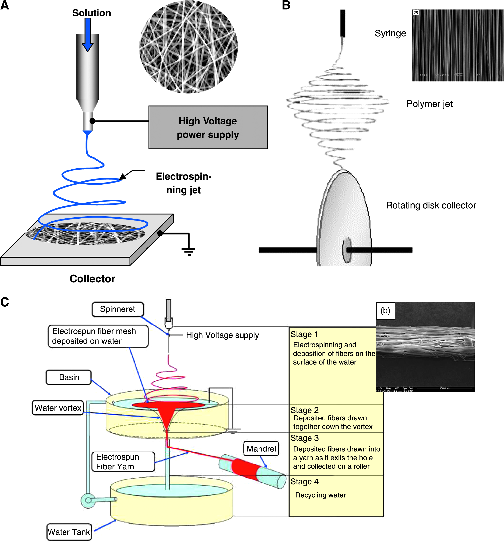
Schematic diagrams of different electrospinning set-ups with resultant structures on the upper right corner. (
Scope of this article
Although much effort has been made of the creation of novel nanofibrous scaffolds 1 and investigating cell–nanofiber interaction, 5 the biodegradability of nanofibrous scaffolds has been relatively understudied. This article briefly reviews popular degradable polymers used for electrospinning and their current applications in tissue engineering. Additionally, detailed reviews are given of the degradation studies on commonly used nanofibrous polyester scaffolds based on a limited number of studies. Finally, mechanisms and expectations of nanofiber degradation are discussed.
Biodegradable Polyester Nanofibers and Their Application in Tissue Engineering
Commonly used biodegradable polymers include synthetic and natural polymers. Degradable polyesters are one of the widely used synthetic materials to be electrospun as tissue-engineered scaffolds because they are biodegradable with metabolizable degradation products (e.g., lactic acid; LA), the degradation rate of polyester can be controlled by changing the constitute of the polymer, they are synthetic polymers and highly scalable in terms of production, some of the common members such as polylactic acid (PLA), polyglycolic acid (PGA), polycaprolactone (PCL), and their copolymers have been approved by the U.S. Food and Drug Administration (FDA) and used in medical application such as sutures and drug delivery systems, and their nanofibers can be conveniently and economically produced using electrospinning. 9 Commonly used biodegradable polyesters are PGA, PLA, PCL, poly(3-hydroxybutyrate-co-3-hydroxyvalerate) (PHBV), and their copolymers. Table 3 lists some commonly used polyester nanofibers for tissue engineering applications.
Polyglycolic acid
PGA is the simplest linear aliphatic polyester. It is a rigid thermoplastic material with high strength (up to 250 Mpa 17 ) and a metabolizable degradation product. Therefore, PGA has been used in various biomaterial applications. It was used to develop the first synthetic absorbable suture, marketed as Dexon in the 1960s by Davis and Geck, Inc. (Danbury, CT). Thanks to its high strength and degradability, PGA implants have been widely tested in bone fracture fixation in the forms of pins, screws, plates, and rods. 18 However, complications of PGA implants include displacement of fracture, fixation failure, and more importantly, inflammatory foreign body reaction. 18 It has been found that young patients with PGA bone implants had less risk of foreign body reaction. In particular, PGA pins have shown promising results in treating fractures in children. 19 Recently, PGA has been fabricated into a biodegradable conduit for nerve repair. 20 The clinical trail demonstrated the better outcome of PGA conduit than of a nerve graft. The product is now marketed as Neurotube (Synovis Micro Companies Alliance Inc., Birmingham, AL).
Although the degradation product glycolic acid (GA) is resorbable, at high concentrations, it can cause an increase in localized acid concentration and result in tissue damage.21–26 The ultimate fate of GA in vivo is considered to be the conversion to carbon dioxide and water, with removal from the body via the respiratory system. However, Hollinger 27 suggested that only the LA follows this pathway and that GA is converted into glyoxylate (by glycolate oxidase), which is then transferred into glycine after reacting with glycine transaminase.
Because of its high crystallinity, with reported values ranging from 35% to 50%,28–30 it is insoluble in general organic solvents except highly fluorinated solvents. Most PGA nanofibers were electrospun using hexafluoro-isopropanol as a solvent. However, because of its rapid degradation (<20 days 31 ), electrospun pure PGA has rarely been used as tissue scaffold. Boland et al. 32 claimed that electrospun PGA nanofibers, after pretreated with hydrochloride acid, showed an improved biocompatibility with rat cardiac fibroblasts in vitro and rat muscle tissue in vivo. It was also claimed that thinner PGA nanofiber (fiber diameter 220 nm) had better biocompatibility both in vitro and in vivo than thicker ones (fiber diameter 880 nm). However, the authors did not adequately address the degradation effect of hydrochloride acid on PGA nanofiber. Our group has cultured porcine smooth muscle cells on electrospun PGA nanofibers (d = 380nm). Results suggest that the PGA nanofiber can support cell growth only in the first 7 days, followed by rapid disintegration of nanofiber (unpublished data). One concern raised in the study was that the fast degradation of PGA nanofiber would not provide enough support for tissue regeneration.
Polylactic acid
PLA, with the addition of a methyl group in each unit, degrades more slowly than PGA, with greater hydrophobicity. 33 The lactide monomer exists in three different forms: two stereoisomers L- and D- lactide (L-LA and D-LA) and racemic D, L-lactide (DL-LA). The chirality of the LA units provides a method to adjust degradation rates and physical and mechanical properties. The three stereoisomers of PLA exhibit distinct properties. Poly(L-lactide) (PLLA) is a semicrystalline polymer, with a melting temperature between 173°C and 178°C and crystallinity varying from 37% to 72%.28,34,35 In contrast poly(D,L-lactide) (PDLLA) is an amorphous polymer, with no fixed melting point. PLA polymers are used in a broad variety of medical applications, including bioresorbable sutures, 36 dental implants, 37 bone screws and plates, 38 and controlled drug delivery. 39
Nanofibrous PLA has been used as a scaffold for nerve regeneration, 11 bone tissue engineering,40,41 vascular engineering, 42 and stem cell tissue engineering. 43 Yang et al. 11 designed an electrospun aligned PLLA nanofibrous scaffold to evaluate its efficacy in promoting neuron differentiation and guiding neurite outgrowth of C17.2 cells in vitro. A C17.2 cell is a primordial, multipotent, self-renewing cell that can be used as a neuron precursor and is involved in the normal development of the cerebellum embryonic neocortex and other structures upon implantation.11,44,45 The study revealed that C17.2 cells seeded on the scaffolds adhered to the scaffolds and started to differentiate on the fibrous scaffold 10 h after seeding and that, by 24 h, approximately 70% of cells exhibited a spindle-like shape with extended processes. The fiber alignment had a strong effect on the cell phenotype; neural cells on aligned fibers grew parallel to the fiber orientation, and the aligned nanofibers resulted in better neurite outgrowth than with random nanofibers or microfibrous scaffolds.
Poly(lactic-co-glycolic) acid
Poly(lactic-co-glycolic) acid (PLGA), the copolymer of PLA and PGA, is probably the most popular synthetic polymer in tissue engineering application because of its excellent biocompatibility and variable degradability.46,47 Several end-products are already on the biomedical market. Vicryl (Ethicon Inc., Somerville, NJ) and Polysorb (US Surgical, Mansfield, MA) sutures use PLGA with a GA:LA ratio of 90:10, and it has been reported that they are absorbed within 8 to 10 weeks, but Vicryl can maintain 50% of its original strength after 3 weeks, whereas Polysorb maintains 30% (information obtained from manufacturer's Website). PLGA with a LA:GA ratio of 7:3 has been chosen for biodegradable bone clips and staples (Lactomer, US Surgical, Mansfield, MA). PLGA microspheres have been widely used for drug delivery. Commercial products include Lupron Depot (TAP Pharmaceutical Products, Inc., Lake Forest, IL) and Prostap SR (Wyeth Pharmaceuticals, Madison, NJ) for cancer chemotherapy, Risperdal Consta (Janssen-Cilag Pty Ltd., Australia) for the treatment of schizophrenia, and Sandostatin LAR depot (Novartis, Switzerland) for severe watery diarrhea. Additionally, Zoladex (AstraZeneca, Wilmington, DE) is a subcutaneous PLGA rod implant for sustainable release of goserelin acetate, a luteinizing hormone-releasing hormone analogue to treat prostate cancer. The wide use of PLGA could be attributed to its flexibility of different copolymerizing ratios of PLA and PGA. Depending on the ratio of copolymerization, PLGA has different subtypes, each exhibiting different properties.
PLGA was one of the first electrospun biodegradable polymers for tissue engineering applications. The PLGA copolymer has an amorphous structure, because the constituent PGA and PLA molecules are unable to pack tightly to one another. Li et al. 48 have shown that nanofibrous PLGA scaffolds have an interconnected porous structure with more than 90% porosity and sound mechanical properties, close to that of skin. The authors found that electrospun PLGA possesses good properties for tissue engineered scaffolds. Electrospun PLGA has been successfully used as a scaffold for skin tissue engineering, 49 nerve regeneration, 50 vessel engineering,51,52 and bone regeneration.48,53 With a high SVR, electrospun PLGA nanofiber has been proposed for cell capture. Ma et al. 54 compared the ability of PLGA and blended PLGA–collagen to capture hematopoietic stem cells (HSCs) in vitro, using tissue culture polystyrene as a control. All three samples of nanofibers exhibited more than 5-fold increase of HSC capture. Blending collagen with PLGA nanofiber additionally increased the cell capture percentage from 7.5% to 23% in 30 min, possibly reaching 67% if the PLGA–collagen blended nanofiber is precoated with endothelial leukocyte adhesion molecule-1 (E-selectin).
Poly(ɛ-caprolactone)
Another member of the polyester family is PCL, a semicrystalline, biodegradable polymer. Its crystallinity has been reported to be 45% to 67%.35,55,56 PCL has been used less frequently than other polyester family members such as PLA, PGA, and PLGA as a material for fabricating biomaterial scaffolds, mainly because of concern about its slower degradation kinetics, 57 but the slow degradation makes PCL an ideal polymer for long-term drug delivery. 57 Capronor (Research Triangle Institute, Research Triangle Park, NC), a 1-year contraceptive, represents such a system. 58 The drug release is thus diffusion controlled rather than erosion controlled.
The ease of fabrication and low cost make PCL an attractive polymer for the fabrication of electrospun nanofibers.59–65 It is also known that the electrospinning process re-organizes PCL polymer chains and that electrospun nanofibers have less crystallinity than unprocessed PCL. 66 Electrospun PCL has been used in skin tissue engineering, 61 bone regeneration,59,65,67,68 and heart tissue engineering. 64 Our group has successfully fabricated coaxial, 16 porous, 69 gelatin-blended, 70 and hydroxyapatite (HA)-blended 71 PCL electrospun nanofibers. Zhang et al. 16 used coaxial electrospinning (core, polyethylene glycol; shell, PCL) to successfully encapsulate bovine serum albumin (BSA) in the polyethylene glycol matrix (Fig. 1e). The release kinetics of fluoro-isothiocyanate–conjugated BSA showed a burst release of 60% BSA within 24 h, although gradual release of BSA was maintained from day 3 until the end of study period (35 days).
Poly(L-lactide-co-epsilon-caprolactone)
Poly(L-lactide-co-epsilon-caprolactone) (P(LLA-CL)) is the copolymer of PLLA and PCL. Although less popular than PLGA, electrospun P(LLA-CL) has shown distinctive properties in tissue engineering. Our group first demonstrated that electrospun P(LLA-CL) could be a good candidate for vascular engineering, with biocompatibility of human coronary artery smooth muscle cells (CASMCs) and human coronary artery endothelial cells.12,42,72 Dong et al. 73 cultured porcine CASMCs on electrospun P(LLA-CL) for up to 100 days, and multilayers of cells were tightly grown on the scaffold, whereas porcine CASMCs on electrospun PLGA failed to reach full confluence. He et al. 15 implanted electrospun P(LLA-CL) vascular grafts into the epigastric vein of a rabbit model. The grafts remained patent 7 weeks after the implantation. The above studies indicated that electrospun P(LLA-CL) has good potential in vascular engineering applications.
Poly(3-hydroxybutyrate-co-3-hydroxyvalerate)
PHBV belongs to the family of polyhydroxyalkanoates (PHAs), a polyester class that French microbiologist Maurice Lemoigne first isolated from Bacillus megaterium in 1925. A number of micro-organisms (like Alcaligenes eutrophus or Bacillus megaterium) produce the most common form, poly(3-hydroxybutyrate) (PHB), apparently in response to conditions of physiological stress. Native PHB is amorphous but easily crystallized after purification. To reduce the brittleness of PHB, it is often copolymerized with 3-hydroxyvalerate in various ratios, forming PHBV. PHBV is now widely used in packing materials, such as thin film or paper coating. 74 In the last decade, PHBV has also attracted attention for tissue engineering applications. 75
A number of studies have investigated tissue engineering application for electrospun PHBV. Lee et al. 76 reported that chondrocytes attached better and appeared to have greater spread morphology on the surface of the electrospun PHBV fabric than they did on flat PHBV cast films in the early culture stage. Two hours after cell seeding, 30.1% of chondrocytes were attached on the surfaces of the PHBV nanofibrous mat, whereas only 19.0% were attached on flat PHBV film. Chondrocytes tended to spread on the nanofibers and remain rounded on the cast film. Ito et al. 77 fabricated a composite of hydroxyapatite and PHBV nanofibers, by soaking the electrospun nanofibrous membrane in simulated body fluid, a solution with similar concentrations of Na+, K+, Mg2+, Ca2+, Cl−, HCO3−, HPO4 2−, and SO42− as human plasma. HA deposition greatly increased the hydrophilicity of the scaffold. COS-7, a cell line originated from the kidney of an Africa green monkey, was seeded on the scaffold; 1.5 h after cell seeding, the number of attached cells on the electrospun membrane were significantly greater that on the cast film (p < 0.01), although combination with HA did not significantly affect cell adhesion. Han et al. 78 investigated electrospun PHBV mats for wound dressing and compared them with PHBV:collagen (70:30, wt/wt) and PHBV:gelatin (70:30, wt/wt) nanofiber matrices. Dermal sheath cells, one of the two major dermal (dermal sheath and dermal papilla) cells in the hair follicle, were used to study cell attachment. Although PHBV mixed with collagen and gelatin resulted in better attachment of dermal sheath cells, the pure PHBV mat promoted faster wound closure in a wounded mouse model than PHBV:collagen and PHBV:gelatin. The authors suggested that the mechanical stability of the matrices seemed to be more important for early-stage wound dressings because PHBV:collagen and PHBV:gelatin matrices have poor mechanical strength. The stress at yield of PHBV, PHBV:collagen, and PHBV:gelatin matrices were 2.96, 5.23, and 2.20 MPa, respectively, but the strength of PHBV:collagen and PHBV:gelatin was much lower only 10 min after contact with water, whereas the strength of the PHBV matrices was not affected.
Degradation Behaviors of Polyester Nanofibers
One advantage of degradable polyesters in medical applications is that their degradation products are metabolizable in the human body, with no future complication after their complete absorption. 79 Although degradation of traditional macroscale (in the dimension of millimeters) degradable polyesters have been comprehensively studied, 79 degradation studies of electrospun degradable polymeric nanofibers are still in their infancy stage. It is reasonable that the degradation behavior (in vivo and in vitro) of nanoscale materials will be different from that of macroscale ones, because the SVR of nanofibers is much higher than that of macroscale polymers; the crystallinity, polymer chain configuration and orientation, and hydrophobicity could be different; and the biomimic structure of nanofibers may affect the activity of surrounding cells or tissue, which may in turn affect their degradation. In the following sections, degradation behaviors of electrospun degradable polyesters will be systematically reviewed and compared with the degradation behaviors of polyesters in macroscale.
Many in vitro factors could influence the biodegradation of polyesters, including pH, ionic strength of the medium, and enzymatic activity. A commonly used in vitro method for the assessment of polymer degradation is immersing the sample in phosphate buffered saline (PBS) or equivalent buffer solution at pH ∼7.4 at 37°C. In the following discussion about in vitro degradation, it is assumed that this type of solution was used unless stated otherwise. For in vivo degradation, a piece of sample will normally be placed subdermally in a small animal, such as a mouse or rat.
High SVR greatly increase the degradation rate of PGA nanofibers
PGA was among the fastest-degrading polyesters because of its hydrophilicity and non-side-chained molecular structure. Size, crystallinity. and degradation environment greatly affect the degradation rate of PGA. 18 Chu et al.29,80–82 systematically studied the in vitro degradation of Dexon PGA sutures and established a simple degradation mechanism via homogeneous erosion. The degradation process occurs in two stages. The first stage involves the diffusion of water molecules into the amorphous regions of the matrix and simple hydrolytic chain scission of the ester groups. In this stage, degradation predominates during the first 21 days, whereas crystallinity increases from 40% to a peak of 52%. The second stage of degradation involves largely the crystalline areas of the polymer, which become predominant when the majority of the amorphous regions have been eroded. After 49 days, the weight loss of the PGA suture was approximately 42%, with complete loss of mechanical properties. 29 A number of studies 18 have investigated the in vivo degradation of a PGA bone implant. The results have revealed that the complete degradation time in vivo can range from 4 to 9 months. Ginde and Gupta 30 examined the degradation of PGA fibers and pellets of comparable crystallinities. Degradation of pellets was faster than that of fibers, but the authors attributed this to the long-range order of PGA fibers produced by melt spinning, not to the size difference. Later studies indicated that the degradation rate of PGA increases when its dimension decreases.83,84
A few studies have investigated the degradation behavior of electrospun PGA nanofibers. You et al. 85 reported that PGA nanofibers showed a rapid degradation rate without an induction period, the first stage of polymer degradation in which the mass and morphology remain unchanged. After 20 days of degradation in vitro, only 40% of the weight of the PGA nanofiber mesh remained. PGA nanofibers started to break down after 1 day. The surface defect was believed to lead to fiber rupture after several days of degradation. The authors also compared the degradation study of PGA microfibers by Shum and Mak 86 and found that PGA nanofibers showed a much faster degradation rate than microfibers, whereas the crystallinity of PGA nanofibers did not increase during degradation like that of microfibers did. Park et al. 87 observed similar a degradation profile of electrospun PGA nanofibers. The weight loss of PGA nanofibers was greater than 90% after 45 days. After only 12 days, PGA nanofibers were broken down into short fiber fragments. In contrast to You's study, the crystallinity of PGA nanofibers was greater and peaked at day 5, followed by a sharp decrease.
Table 4 lists the differences in in vitro degradation rates of different sizes of PGA polymers. To sum up, the degradation rates of PGA are in the order of nanofibers > pellets >microfibers. Only a few days of exposure to an aqueous medium could lead to complete disintegration of PGA nanofiber, 88 whereas macroscale PGA, such as a PGA rod, remains intact after 56 days of degradation (Fig. 2). 30
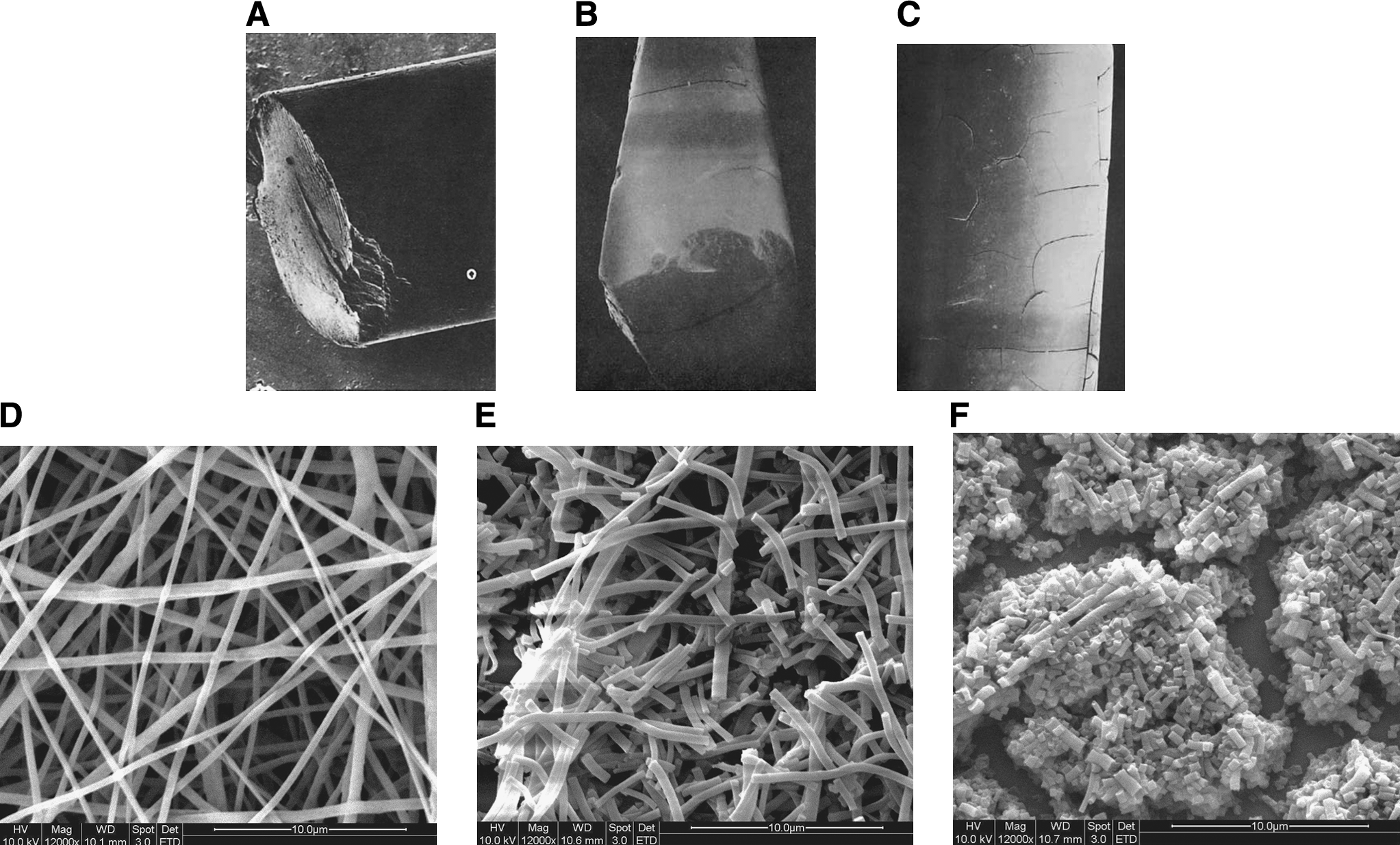
Morphology of polyglycolic acid (PGA) rod (a–c) (adapted from
30
with permission) and PGA nanofiber (d–f) after (
Calculations based on the assumption that pellet is cubic, films are infinitely large, and fibers are infinitely long.
Estimation made presumed that pellet was a 3-mm cube.
Composition of D-LA and L-LA greatly affect the degradation rate of PLA nanofibers
The degradation of macroscale PLA in aqueous medium generally occurs in two stages, similar to PGA. 34 The first stage starts with water diffusion into the amorphous regions, which are less organized, allowing water to penetrate more easily. The second stage starts when most of the amorphous regions are degraded. The hydrolytic attack then proceeds from the edge toward the center of the crystalline domains. This explains the much faster hydrolysis rate of the amorphous PDLLA than of semicrystalline PLLA.
The composition of the polymer chains (the content of L-LA and D-LA units) greatly influences the degradation rate of PLA nanofibers. 34 PDLLA is amorphous, whereas PLLA is semicrystalline. The half-life of PLA with different L-LA:D-LA ratios can vary from 10 weeks for PDLLA (50:50) to 110 weeks for PLLA. The higher the crystallinity is, the lower the degradation rate. During early degradation, the amorphous PDLLA showed greater crystallinity. That the shortened polymer chain was restructured and crystallized could explain this. In addition, the transition and melting temperature of PLA decreases with degradation time. Bulk amorphous PDLLA tends to degrade into a “hollow” structure, because of the autocatalytic effect together with ease of water penetration,34,89,90 See examples in Figure 3.
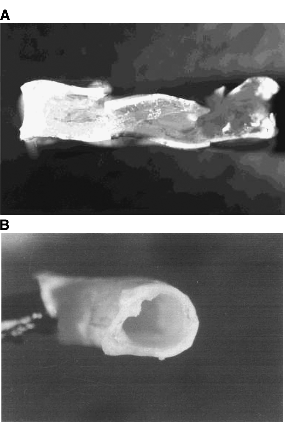
The size and shape of the polymers is another important factor affecting the degradation properties. Grizzi et al. 91 compared the degradation rate of different shapes of PDLLA: compression-molded plate, millimetric beads, microspheres, and cast films. Results showed that the degradation of plates and beads, finally leading to bulk disintegration, were faster than that of microspheres and films, during which only surface hydrolysis was observed. The autocatalytic degradation occurring in the large-scale materials can explain this. 92
Degradation studies on PLA nanofiber have mainly used amorphous PDLLA, because of the much lower degradation rate of PLLA. Cui et al. 93 compared the in vitro degradation of 5% paracetamol-loaded electrospun PDLLA fiber with average diameters of 212, 551, and 1310 nm with that of PDLLA cast film with a thickness of 100 μm. After incubation in the degradation medium, the fiber size of electrospun PDLLA increased, and fiber space decreased. This was explained by the shrinkage of fibers. Thicker fibers showed faster mass loss, a nanofibrous mat with an average fiber diameter of 212 nm lost 18% of mass in 9 weeks, whereas casting film and a microfibrous mat with average fiber diameter of 1.31 μm lost only 8%. However, reduction in molecular weight (50% in 9 weeks) was observed only in casting film, whereas no obvious change in molecular weight (Mw) was found on electrospun mats after 9 weeks of study time. The authors explained that casting film underwent bulk degradation with autocatalysis, resulting in reduction in Mw but minor mass loss, and electrospun mats were degraded by surface erosion, which led to mass loss but minor reduction in Mw. A recent study from the same group 94 confirmed similar degradation behavior of pure PDLLA electrospun fibers. In addition, the authors pointed out that high hydrophobicity of PDLLA electrospun mats could result in slower water penetration and thus a degradation profile of surface erosion.
Two studies85,95 have investigated the degradation of electrospun PLLA nanofibers with fiber diameters of 290 and 650 nm (Mw of 450 kD and 650 kD, respectively). No significant degradation was observed during the study periods (7 and 14 weeks, respectively). A more recent study found a 10% loss of mass of electrospun PLLA nanofiber (fiber diameter 368 nm), with Mw reduced from 238 kD to 200 kD in 8 weeks. 96
Table 5 lists the size-dependent degradation rate of PDLLA. As seen, the degradation rate of PDLLA was greater when the dimension of PDLLA is reduced from millimeters to micrometers, but the degradation rate decreased when the dimension was further reduced from micrometers to nanometers. As for PLLA, its degradation rate is so slow that there are only limited studies on its degradation. At present, no conclusive comparisons can be made of PLLA degradation with different sizes.
Calculations based on the assumption that pellet is cubic, films are infinitely large, and fibers are infinitely long.
Estimation made presumed that the rod is 3 mm in diameter and 3 mm in length.
Studies have been performed on enzymatic degradation of PLLA. It has been reported that 80% weight loss was accomplished within 65 h for a PLLA film when exposed to proteinase K with concentration of 200 μg/mL. 97 The same weight loss was achieved within 7 to 9 h for electrospun PLLA nanofiber mats when the concentration was only 2 μg/mL. The concentration of proteinase K was 99% lower, whereas the degradation rate was nearly 10 times as high. 98 The study revealed that a greater SVR of PDLLA nanofiber resulted in a faster enzymatic degradation rate.
Controllable degradation of PLGA nanofiber
The composition of the polymer chain of PLGA (the content of L-LA, D-LA, and GA units) greatly influences degradation behavior.34,99,100 GA-rich PLGA copolymers are more hydrophilic and degrade faster in aqueous media than PLA- and LA-rich PLGA copolymers.101,102 For blended PGA and PLA nanofibers, greater PGA content also induces faster degradation. 31 Mixing D-LA and L-LA copolymers reduces crystallinity, thus increasing the degradation rate.34,93,95 As a result, the half-life of PLGA with different compositions can vary from 3 weeks for P(DLLA-GA) (37.5:37.5:25) to 20 weeks for P(LLA-GA) (85:15). 34 However, commonly used of PLGA are Poly(DL-lactide-co-glycolide) [P(DL-LGA)], in which the LA component is amorphous. The PLGA mentioned in this article is P(DL-LGA) unless otherwise specified.
Shin et al. 101 compared the in vitro degradation of electrospun PLGA (50:50) and PLGA (75:25) (d = 550 nm) for a period of 8 weeks. There was little change in mass and pH for the 75:25 PLGA scaffolds until week 7, after which there was a decrease in mass (5%) and pH value (from 7.4 to 7.35) in week 8. Similarly, for the 50:50 PLGA scaffolds, mass and pH changed slightly during the first 4 weeks and decreased markedly thereafter (35% mass loss until 8 weeks and pH drop from 7.4 to 7.27). The Mw of all scaffolds decreased gradually, but the decrease in the Mw of the 50:50 PLGA scaffolds was more rapid than that of the 75:25 PLGA scaffolds. The molecular weights of the 75:25 and 50:50 PLGA scaffolds decreased by 36.8% and 88.0% over 8 weeks, respectively.
Size-dependent degradation of PLGA (75:25) and PLGA (50:50) is listed in Table 6. For both polymers, their degradation rates decreased with reduction in size. Autocatalysis greatly increases its degradation rate when the size of material is larger than millimeter scale.
Calculations based on the assumption that pellet is cubic, films are infinitely large, and fibers are infinitely long.
Estimation made presumed that the rod is 3 mm in diameter and 3 mm long.
Duan et al. 103 reported a degradation study of PLGA (80:20) electrospun fibers (d = 300 nm) over 10 weeks. The pH value of the degradation medium remained relatively constant (∼7.40) throughout the degradation period of 10 weeks. The Mw of PLGA, as determined using gel permeation chromatography, fell from 250 kD to 200 kD in 10 weeks. During the in vitro degradation period, the fibers shrunk a little and seemed to lose their initial surface smoothness, although no obvious morphological change in the PLGA scaffolds was observed for up to 10 weeks. The tensile strength and Young's modulus of electrospun PLGA membranes did not change significantly after 10 weeks of degradation. The electrospun PLGA showed a significant decrease in elongation at break (by 83%) after a 10-week degradation period with respect to its initial value. Electrospun PLGA membranes maintained their elongation at break during the first 2 weeks; thereafter elongation at break decreased sharply. The thermal-induced recrystallization, leading to a more-brittle structure, could explain this.
PLGA 10:90 is more crystalline than PLGA with a higher content of lactide because of the high ratio of GA. Zong et al. 104 divided the degradation of this semicrystalline polyester nanofiber into four stages (Fig. 4). In stage I (within the first day), because the glass transition temperature (Tg) is close to the incubation temperature (37°C), a rapid thermally induced crystallization process resulted in a two-phase lamellar structure. In stage II (day 2 to day 5) the polymer chains in the amorphous regions between the lamellar stacks begin to degrade because of ease of water penetration. This chain scission process increases the polymer chain mobility, which leads to further crystallization. The process is often termed “cleavage-induced crystallization.” Several studies have shown a similar phenomenon of greater crystallinity during degradation.31,85,104 In the first two stages, mass loss is trivial (10%), but Mw can decrease sharply. In stage III (day 6 to day 11), the degradation rate of the electrospun membrane increases because of autocatalysis. The sample fragmentizes because the amorphous regions degrade faster than the crystalline regions. In this stage, breach of nanofibers can be observed with large mass loss (40%). The degraded sample is more hydrophilic than the initial sample because of the exposed carboxylic groups from hydrolysis of ester bonds. Stage IV (day 12 onward) can be described as mass loss from the crystalline region. Considered the similar property of PLGA 10:90 and PGA, this model may also apply to similar semicrystalline PGA nanofibers.

A four-stage model of structure and morphology changes of electrospun poly(lacide-co-glycolide) (PLGA) (10:90) membranes during in vitro degradation. Stage I: thermally induced crystallization from amorphous PLGA (10:90) nanofibers and lamellar stacks are formed. Stage II: the mobility of polymer chains within large amorphous gaps increases after chain scission, cleavage-induced crystallization occurs, and thinner lamellae and lamellar stacks form. Stage III: mass loss rate is accelerated, large amorphous gaps disappear, and nanofibers start to break down. Stage IV: lamellar stacks start to collapse, and accelerated mass loss is observed. (adapted from 104 with permission).
A recent study by Pan et al. 105 investigated the degradation of electrospun PLGA (75:25) in a fibroblast–macrophage co-culture system. Results showed that fibroblasts and macrophages were able to accelerate the degradation of scaffold. Lysozyme, nonspecific esterase, gelatinase, hyaluronidase-1, and a-glucosidase were upregulated in the presence of the scaffold. The authors believed that these enzymes played important roles in the cell-mediated scaffold degradation. After comparing the co-culture system with a mouse subdermal model, the authors claimed that the co-culture system would be a useful in vitro tool for initial biomaterial evaluation.
Slow degradation of PCL nanofiber
Polycaprolactone is relatively stable against in vitro hydrolysis, but it was shown that microorganisms can degrade it. PCL has been shown to degrade in pure fungal cultures,106–109 in compost,107,110–112 in active sludge,107,111 by enzymes, 113 and in soil. 114 In the biodegradation of PCL, rapid mass loss was observed by surface erosion, with less reduction of molecular weight. In contrast, the in vitro hydrolysis of polyester is normally preceded with decrease in molecular weight and minor mass loss. Therefore, the microorganism plays a key role in PCL surface erosion.
Bolgen et al. 115 reported degradation of PCL nanofiber in vitro over 6 months and in vivo over 90 days. During the in vitro degradation period, samples with different diameters were characterized according to mechanical properties and Mws. Overall, thinner fibers degraded faster than thicker ones in mechanical strength, with PCL fiber with an average diameter of 196 nm losing 70% of their strength over 6 months, whereas that with a diameter of 689 nm lost only 35%. The reduction in Mw ranged from 7% to 15%, but there were no significant differences between fibers with different diameters. In the in vivo degradation study, electrospun PCL (d = 250 nm) meshes were subdermally implanted in the back skin of rats. At different time intervals, the samples were collected, and the Mws were analyzed. After 90 days of implantation, the Mw had fallen by 27%, compared with a 13% drop after 6 months of in vitro degradation, but it is likely that no mass change was observed throughout the study period.
Enzymatic and environmental degradation of PCL nanofibers has also been studied. PCL exhibited a fast degradation, with 47% weight loss in 8.5 h in the presence of lipase. 98 The crystallinity of PCL fibers increased during the degradation due to thermally induced recrystallization. Ohkawa et al.34,103,116 has done two biodegradation studies of electrospun PCL cultured with seven strains of fungi. As expected, PCL nanofibers with thinner diameters experienced faster degradation.
Enzymatic degradation of PHBV nanofiber
The in vitro degradation of PHBV under physiological conditions is slow. 117 Under accelerated conditions (high temperature and acidic or basic pH), the degradation proceeds through a Mw decrease, and when the Mw is sufficiently low, a weight loss is observable. Almost all of the mechanical strength is lost, and the remaining polymer breaks down into small fragments.117,118 The rate of chemical hydrolysis decreases with increasing crystallinity.119,120 There is no general consensus as to how the copolymer composition affects the hydrolysis rate.118,120 It has been suggested that it is the crystallinity, rather than the composition, that affects the hydrolysis rate. 120 On the other hand, PHBV of the same level of crystallinity, but of different composition, showed decreasing hydrolysis rates with increasing hydroxyvalerate content. 118
Degradation of PHBV nanofiber was studied in enzyme solution or a solid waste environment. Ito et al. 77 conducted an enzymatic degradation study on PHBV nanofiber by polyhydroxybutyrate (PHB) depolymerase. The results showed that enzymatic degradation of PHBV nanofibers was faster than that of PHBV film. The samples were incubated in PBS containing PHB depolymerase ranging from 100 ng/mL to 100 μg/mL for 16 days at 37°C. Results showed that, at 100 μg/mL, nanofibrous membrane was completely disintegrated at the end of study, whereas the majority of the cast film sample remained intact. It was also found that a HA-coated nanofiber degraded faster than a non-coated one. At 1 μg/mL PHB depolymerase, a HA-coated nanofiber completely disintegrated, whereas the non-coated one stayed intact. The greater hydrophilicity of HA particles could explain this. Another study by Choi et al. 121 examined the degradation of electrospun PHBV nanofiber in simulated solid waste. The simulated solid waste mixtures were composed of 39.8% food waste, 20.7% shredded computer and newspaper, 5.3% saw dust, 7.3% glass beads, 7.7% plastics, 4.5% rubbers, and 14.7% leaves. A reactor containing the waste was kept at pH 7.5 and 55°C, with 60% moisture content. The samples were exposed to the waste for 100 days. The mass loss of PHBV nanofiber was not observable for the first 12 days, but after that, the mass loss rate increased. Electrospun PHBV nanofiber lost 10% of its weight after 3 months of degradation, whereas PHBV film lost only 3%.
Nanofiber morphology during degradation
The nanofiber morphology changes significantly during degradation. Although degradation rate varies with polymer type, the morphological change of polymer nanofibers generally could be divided into two types: fiber melting (Fig. 5, unpublished data) and fiber breaking (Fig. 2d-f). The melt-like morphology is often observed on amorphous nanofibers with a low Tg, such as PLGA nanofibers or their blends.85,104,122 It is believed that fiber melting happens when the Mw of the polymer is reduced to the extent that its Tg is near or below the degradation temperature, normally 37°C, in which case the polymer chain in the amorphous region becomes mobilized. Therefore, the fibers tend to “melt” together to reduce the surface tension.122,123 On the other hand, polymer nanofibers with high crystallinity or high Tg, such as PGA,85,88 PLLA, 96 PCL, 98 and their copolymers, 124 tend to have broken fibers during degradation. This is because the polymer chains in the crystalline region are rigid and immobilized. Therefore, fiber breaks when there is a “weak” point along the fiber during degradation, and the broken ends are more susceptible to hydrolytic attack because of the higher exposure to degradation medium.

Scanning electron microscope images of PLGA nanofibers degraded in phosphate buffered saline at 37°C for (
Degradation Mechanisms of Nanofibers: Faster or Slower
It is intuition that the polymeric nanofibers degrade faster than the respective polymer in macroscale, simply because of the high surface area of nanofibers, but this is not always true. To address this issue, we need first to analyze the underlying mechanism of polyester hydrolysis.
Polyester hydrolysis and erosion
Erosion is defined as the physical disintegration of a polymer matrix as a result of degradation.125,126 Upon incubation, water penetrates into the polymer matrix, advancing toward the center of the device and inducing scission of polymer chain. Once a sufficiently low Mw is reached, the degradation products formed will diffuse to and dissolve in the degradation medium and be transported away from the polymer matrix.
Depending on the erosion mechanism, the polymer is classified as bulk eroding or surfacing eroding.125,126 If water penetration proceeds faster than the erosion of the matrix, degradation will occur throughout the matrix, and material will be lost from the entire polymer volume. This behavior is termed bulk erosion, but it is sometimes referred to as homogeneous erosion because mass loss proceeds at a more or less uniform rate throughout the matrix. 127 The size of structure within the bulk changes considerably. After erosion to a critical degree, the device eventually collapses. 127 A special case of bulk erosion is heterogeneous degradation. After a period of degradation, soluble oligomeric compounds are generated in the matrix. The soluble oligomers on the surface of the matrix are easily diffused into the degradation medium, whereas the oligomers inside the matrix are not. These acidic oligomers will thus accumulate inside the matrix, inducing faster degradation in the interior matrix by autocatalysis. In extreme cases, hollow structures will be formed when the internal materials are completely degraded, as in the example shown in Figure 3. Bulk erosion tends to happen on amorphous and hydrophilic polymers.
On the other hand, if water penetration is slower than the erosion process, mass loss is confined to the surface layers of the device, 126 termed “surface erosion.” The size of the device gradually decreases, but the bulk phase remains undegraded. 125 For ideal surface erosion, the erosion rate is directly proportional to the external surface area. Surface-eroding devices are hence often preferred over bulk-eroding materials for ease of predictability, although surface erosion is difficult to achieve. Most polymers are not sufficiently hydrophobic to prevent water from penetrating into the matrix while allowing degradation of the interior of the materials to occur faster than erosion of the surface layer. 126 However, hydrophobic polymer with high crystallinity is more likely to undergo surface erosion.
Nanofiber degradation: Surface erosion
According to the hypothesis stated above, for polyester that mainly experiences surface erosion, the corresponding nanofiber may degrade faster than the macroscale polymers simply because of the larger surface area (Fig. 6a), although for polyesters that mainly undergo bulk erosion, the situation becomes a bit complicated (Fig. 6b). Assuming that autocatalysis is induced during the degradation of macroscale polyesters such as PDLLA and PLGA, when SVR increases, the autocatalysis is less likely to happen because the soluble hydrolytic products from the nanofibers can be easily diffused into the medium. Therefore, at first, the degradation rate decreases with increase of SVR, but a further increase in SVR will lead to more contact area with the degradation medium, resulting in more hydrolysis attacks on the ester bond and faster diffusion of the degradation product. This will in turn increase the degradation rate. Therefore, there will be a certain SVR at which the degradation rate is lowest (Fig. 6b).
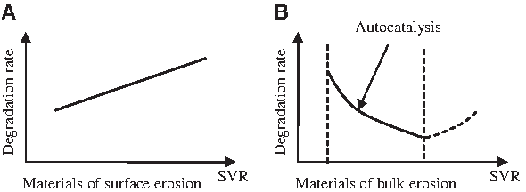
Schematic diagram of polyester degradation rate versus surface-to-volume ratio (SVR) in aqueous medium. (
PGA is a hydrophilic polymer with high crystallinity, which inhibits water from penetrating. Therefore, with larger surface area, PGA nanofiber should degrade faster than PGA in macroscale. Table 4 shows that PGA tends to degrade faster with higher SVR.
In contrast, PDLLA is an amorphous polymer with low crystallinity. Water molecules can easily penetrate into amorphous parts of the polymer matrix. As shown in Table 5, studies have revealed that the degradation rate of PDLLA decreases when the dimension decreases from millimeters to micrometers but increases when the dimension further decreases to nanofiber. PLGA, especially the commonly used P(DLLA-co-GA), is amorphous. 6 shows that PLGA (50:50) and PLGA (75:25) nanofiber both degrade more slowly when the dimension scale is lower.
PLLA and PCL are semicrystalline polyesters with high crystallinity and hydrophobicity. In this case, the penetration of water molecules into the polymer matrix is difficult. Nanofibers made from these polymers degrade faster than the respective polymers in macroscale. 115
To put it simply, polyester nanofibers degrade by surface erosion because there is no “bulk material.” If there is no autocatalysis in the matrix of nanofiber, the smaller the fiber diameter is, the faster it will degrade.
Nondimensional properties that affect degradation of nanofibers
Hydrophobicity
Electrospinning create more-hydrophobic surfaces than films made of the same polymer.93,128 An electrospun PDLLA nanofibrous mesh with an average diameter of 551 nm showed a water contact angle of 140.1°, whereas the PDLLA cast film showed only 69.58°. 93 It is believed that hydrophobic materials tend to degrade more slowly34,93 because they can reduce the water penetration as described.
Chain orientation
Chain orientation along the fiber axis renders the fiber material less susceptible to water penetration and more resistant to hydrolytic attack.30,129 This is because the lateral surface of the crystalline regions on the outer surface of the fiber has been shown to exhibit remarkable resistance to hydrolytic degradation. 130 The molecular chain orientation inside the nanofibers is reported to be along the fiber axis 104 (Fig. 4). This could reduce the water attack on the surface of nanofibers, especially those with high crystallinity.
Shrinkage
Amorphous PLGA or PDLLA electrospun nanofibers shrink during the degradation process because of the thermally induced relaxation of stretched amorphous chains. 104 The Tg of PDLLA and PLGA is close to 37°C, and the polymer chains inside the nanofiber are highly oriented but amorphous. As a result, the relaxation of polymer chains caused great shrinkage of the electrospun mesh. PLGA (75:25) and PDLLA shrinkage after 1 day of 37°C PBS incubation are 84% and 82%, respectively. 104 Figure 7 shows electrospun PLGA (75:25) nanofibers before and after shrinkage, where the size could be reduced by approximately 80% (unpublished data). During shrinkage, fiber diameter increases, and porosity decreases. Therefore, the effective surface area will reduce because of shrinkage. The degradation rate could be lower because of smaller surface area.
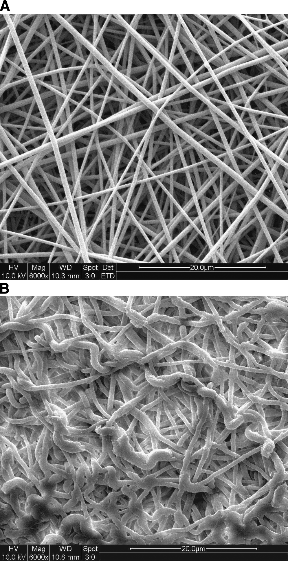
PLGA (75:25) electrospun nanofiber. (
External Factors of Nanofiber Degradation
Fabrication and treatment
Fiber diameter will affect the degradation rate of nanofibers, as previously mentioned. Electrospinning parameters can greatly affect the diameter of the resultant nanofibers. The concentration of polymer solution, the feed rate, and the electric field play are some contributing factors in determining the fiber diameter.9,131 Generally, higher concentrations and feed rates of polymer solutions result in larger diameters of nanofibers. Zong et al. 132 reported that annealing increased the mechanical strength of electrospun PLGA (90:10) membranes during degradation. The PLGA membranes were drawn to desired extension ratios at a constant rate of 4 mm/min at room temperature. The stretched membranes were subsequently heated to four different temperatures (60°C, 70°C, 80°C, and 90°C) at a rate of 5°C/min under constant strain for annealing. For the treated PLGA membranes, reasonable tensile strength retention time was prolonged to more than 8 days, whereas untreated ones completely lost their mechanical strength within 2 days of degradation. Greater crystallinity after annealing explained this.
Ultraviolet sterilization
Ultraviolet (UV) with wavelength of 254 nm is a common method of sterilizing tissue-engineered scaffolds. This common germicide, UV irradiation, can dramatically degrade the nanofiber compared. 124 After only 30 min of UV irradiation of PLGA (75:25) nanofiber on the bench in a biological cabinet, the Mw was reduced by 46%, with 23% reduction in tensile strength. The authors explained that the high surface area and high-energy UV scattering between the nanofibers can “etch” the “thin” nanofiber by breaking the ester bond. This degradation would not be observable on polymer in macroscale because the etch depth is only several hundred nanometers.
Enzymatic degradation
Several polyesters were susceptible to certain enzymes, for example, PLA to proteinase K, PCL to lipase, and PHBV to PHB depolymerase. Because the enzyme cannot penetrate into the polymer matrix, nanofiber polymers, because of their high surface area, will be more sensitive than macroscale polymers to enzymatic degradation. 98
Future Prospects
Although tissue engineering has become a popular topic, and more and more efforts have been made to explore new scaffolds, “nano” and “biodegradable” are always the “sexy” words to claim superiority of a new scaffold. However, although the emerging new type of scaffold or polymers have been claimed to be degradable by incorporating a hydrolytical bond (e.g., ester bond, amide bond), less studied are the actual degradation behaviors, either in vitro or in vivo. This review meant to summarize the degradation behaviors of popular nanofibrous scaffolds made mostly by using degradable polymer—polyesters, only to find that the literatures are limited, in both quantity and depth. Other than giving a “yes” or “no” answer to the question of degradability, an urgent request to is to answer “how” and “why.”
Most of the degradation studis on polymeric nanofibers have revealed only weight loss and morphological change. Mechanical strength is one of critical factors for tissue engineering application; the nanofiber scaffold must not degrade too rapidly before tissue growth takes place. However, only a few studies have been conducted to investigate mechanical loss during nanofiber degradation. Detailed characterization of nanofiber mechanical strength during degradation is desired.
Although the advantages of using biodegradable nanofibers are immense, much work needs to be done for these materials to be widely accepted in a wide array of tissue engineering applications. Considerations include how the nanofiber finally disappears, whether the degradation of the nanofibers affects the cellular growth when the nanofibers are used as scaffolds for in vitro tissue engineering, and lastly, how the degradation of nanofibers induces foreign body reaction in vivo. These need to be addressed.
The degradation mechanism of polyesters in macroscale has been extensively studied, but the degradation mechanism of polyester nanofibers has not. If there were models to estimate the degradation behavior of polyester nanofiber based on material, fiber diameter, fabrication method, and degradation environment, they could help tissue engineers choose suitable type of nanofibers for different applications.
Footnotes
Acknowledgments
This study was supported by the National University of Singapore, and NMRC/1151/2008, Singapore.
Disclosure Statement
No competing financial interests exist for any of the authors.