




Abstract
Tissue-engineered ligaments have received growing interest as a promising alternative for ligament reconstruction when traditional transplants are unavailable or fail. Mechanical stimulation was recently identified as a critical component in engineering load-bearing tissues. It is well established that living tissue responds to altered loads through endogenous changes in cellular behavior, tissue organization, and bulk mechanical properties. Without the appropriate biomechanical cues, new tissue formation lacks the necessary collagenous organization and alignment for sufficient load-bearing capacity. Therefore, tissue engineers utilize mechanical conditioning to guide tissue remodeling and improve the performance of ligament grafts. This review provides a comparative analysis of the response of ligament and tendon fibroblasts to mechanical loading in current bioreactor studies. The differential effect of mechanical stimulation on cellular processes such as protease production, matrix protein synthesis, and cell proliferation is examined in the context of tissue engineering design.
Introduction
The traditional tissue engineering paradigm consists of a biodegradable scaffold seeded with isolated cells and bioactive factors.4–6,10 As the scaffold degrades, neotissue forms until the injured tissue is completely replaced by healthy tissue and functionality is restored.4,5 The full mechanical load is initially supported by the biomaterial scaffold. During remodeling, the loss of scaffold strength because of biodegradation is offset by increased load bearing of the neotissue. Two variables must be controlled to maintain mechanical integrity: (1) neotissue formation at a rate complementary to scaffold degradation4,7,11 and (2) graded load transfer to the neotissue to guide organization.6,7,12 Healthy ligaments are composed of a highly oriented network of collagen fibrils and fibroblasts.7,13 It is this hierarchical structure that provides these tissues with the necessary tensile properties to withstand constant loading from locomotion. To achieve this level of organization in tissue-engineered ligaments, researchers have utilized mechanical loading of fibroblasts to induce cellular alignment and orientation of the extracellular matrix (ECM). Mechanical loading also enhances cell proliferation, increases ECM synthesis, and promotes differentiation of cells toward specific fibrous connective tissue lineages.11,14–18 For example, Altman et al. 15 reported enhanced differentiation of mesenchymal stem cells (MSCs) to ligament fibroblast phenotypes in response to mechanical stimulation. These studies have established that mechanical stimulation is central to the successful development of tissue-engineered ligaments.4–7,19
Tissue engineers utilize bioreactors to mimic native loading regimes in vitro and generate functional tissue.20–26 Initial bioreactor designs applied uniaxial strains to tethered constructs of tendon fibroblasts to induce differentiation. 23 More recent bioreactor studies have applied multidimensional strains, such as axial tension/compression and torsion, which more closely approach physiological conditions.4,5,11,22 Although improved outcomes have been demonstrated with the addition of bioreactor conditioning, current tissue constructs do not fully replicate the intricate ECM remodeling process of native ligament tissue. Therefore, researchers have focused on elucidating the effect of mechanical loading on ECM synthesis, degradation, and organization as a means to predict optimal loading conditions. Mechanical conditions necessary to guide the remodeling process may then be incorporated into bioreactor design to generate improved tissue grafts.
This review provides a comparative analysis of the response of ligament and tendon fibroblasts to mechanical loading in current bioreactor studies. Fibroblasts from different connective tissue lineages respond to a mechanical stimulus differently based on their functional environment. Therefore, a comparison of ACL, medial collateral ligament (MCL), and patellar tendon anatomy is necessary to provide insight into their respective cellular behavior. The effect of mechanical stimulation on processes such as cell proliferation, matrix protein synthesis, and protease production is examined in the context of tissue engineering design for each fibrous connective tissue phenotype.
Structure of Fibrous Connective Tissues
The functional characteristics of fibrous connective tissues are directly related to the composition and organization of the ECM (Table 1). Therefore, discussion of the impact of mechanical conditioning on tissue constructs begins with a descriptive analysis of the role of individual components of the ECM and the key fibroblastic mediators of ECM remodeling. The ECM of fibrous connective tissue is predominantly composed of collagen, elastin, proteoglycans (PGs), and glycoproteins. The most abundant protein in fibrous connective tissue is collagen—specifically, collagen types I and III. Collagen type I forms tough, nonelastic crosslinked fibers that contribute to the tensile strength of ligaments and tendons.27,28 In contrast, collagen type III forms loosely organized, thin fibrils that provide elasticity.27,28 Elastin, a highly crosslinked array of tropoelastin proteins, also contributes to the elastic behavior of ligaments. 29 The collagen and elastin makeup of fibrous connective tissue dictates its bulk mechanical properties and thus plays a critical role in establishing tissue function.
Adapted from Bramono et al. 37
ECM, extracellular matrix; MMP, matrix metalloproteinase; TIMP, tissue inhibitor of metalloproteinases; TGF, transforming growth factor; ACL, anterior cruciate ligament.
The primary role of PGs and glycoproteins is to guide and maintain the organization of fibrous connective tissue. In particular, biglycan and decorin are small, leucine-rich PGs that are associated with collagen fibrillogenesis and organization.30–32 Typically, biglycan is associated with thick collagen fibrillogenesis, whereas decorin binding results in the formation of thinner collagen fibrils. 33 Glycoproteins, such as fibronectin and tenascins, assist neotissue formation by providing physical links among cells and the ECM. Fibronectin binds to cell surface integrins to enable cell signaling and adhesion and other ECM proteins to control organization of the ECM network.27,34 Tenascin influences cell adhesion and migration by interacting with fibronectin and other ECM molecules. 27 The interaction between tenascin and fibronectin also upregulates the synthesis of enzymes in fibroblasts, which suggests that the composition of these proteins plays a role in tissue remodeling. 34 Overall, each of these ECM components plays a specific role in the maintenance of the structural integrity of connective tissue.
In response to injury, fibroblasts upregulate ECM protein synthesis and proliferation to generate scar tissue. In later stages of healing, fibroblasts produce enzymes that degrade this randomly organized ECM and synthesize oriented ECM proteins with improved mechanical properties similar to healthy tissue. Common enzymes associated with the degradation of the ECM in ligaments and tendons include matrix metalloproteinase-1 (MMP-1), MMP-2, and MMP-9.35–37 MMP-2, a gelatinase, accounts for the most efficient degradation of collagen. 35 Because of the high percentage of collagen in fibrous connective tissues, MMP-2 is often targeted as the key mediator of tendon and ligament degradation. To control enzymatic activity and thus tissue remodeling, tissue inhibitor of metalloproteinase-1 interacts with MMPs to prevent excessive degradation of the ECM network.
In addition to enzymatic activity, fibroblasts regulate ECM protein synthesis and organization by secreting growth factors that alter the structural composition of fibrous connective tissue. For example, proteins in the transforming growth factor-β (TGF-β) superfamily induce cell proliferation and modulate the synthesis of collagen, fibronectin, biglycan, and decorin.6,38–41 TGF-β1 upregulates the expression of collagen and biglycan while having a minimal to negative effect on the expression of decorin.39,42–46 Thus, the production of growth factors can alter mechanical properties by modulating the synthesis and organization of ECM proteins. 47
Overall, the bulk mechanical properties of fibrous connective tissue are dictated by the compositional makeup and organization of the ECM. Although general similarities exist among ligaments and tendons, the individual composition and organization of their ECM differ based on their distinct function and anatomy. Tendon grafts, particularly from the patellar or hamstring tendon, remain the gold standard for ACL repair.4,6,7,9 Therefore, a structural comparison of fibrous connective tissues not only provides insight into the distinct nature of the ACL with respect to other connective tissues, but also helps identify the key mechanisms to restore functionality in ACL reconstruction.
The ACL is predominantly composed of thick, close-packed collagen bundles that are oriented parallel to its longitudinal axis in a helical formation.4,5,7,48 Fibroblasts align between these collagen bundles and elongate in the direction of loading. 48 Synthesis of collagen makes up roughly 80% of all protein synthesis in the ACL, with a ratio of collagen type I to collagen type III at approximately 88% to 12%, respectively.28,48 In contrast, Yoshida and Fujii 41 showed that the percentage of collagen in MCL tissue is actually much greater than that of the ACL, at approximately 95% and 83%, respectively. Further, Amiel et al. reported that the ratio of collagen type I to collagen type III is higher in the MCL at approximately 91% and 9%, respectively.28,48 As a result, MCL tissue demonstrates superior tensile properties. Woo et al. 49 reported that the elastic modulus of the MCL (1120 ±153 MPa) is more than twice that of the ACL (516 ± 64 MPa) in rabbits.
Tendons typically exhibit higher collagen content than ligaments, at approximately 87% of total protein synthesis. 48 In addition, tendons demonstrate a much higher percentage of collagen type I than ligaments with minimal expression of collagen type III. The ratio of collagen type I to collagen type III in tendons is roughly 95% to 5%, respectively.28,48 Because of this high percentage of collagen type I, tendons possess sufficient mechanical strength to restore ACL function; however, their limited production of collagen type III and elastin does not fully mimic ACL tissue.
Mechanical Stimulation of Ligament and Tendon Fibroblasts
As neotissue forms in tissue-engineered constructs, mechanical loading modulates ECM synthesis and remodeling through mechanotransduction. Mechanotransduction refers to cellular mechanisms that convert mechanical stimuli into biochemical signals responsible for cell proliferation, differentiation, and ECM synthesis. In typical biochemical cell signaling cascades, binding of ECM proteins, growth factors, or cytokines to cell surface receptors transmits the biochemical signals to the interior of the cell. 50 This binding triggers intracellular messengers to phosphorylate proteins linked to specific gene expression.27,51,52 During mechanotransduction, the attachment of integrins to ECM proteins creates a physical link between the ECM and the interior of the cell. 53 Mechanical signaling pathways translate physical loading of the ECM into cell signaling cascades that alter gene expression. For example, Miyaki et al. 54 reported that mechanical stretch stimulates the extracellular signal-regulated kinase (ERK) signaling pathway that governs the expression of type I collagen and decorin in ACL-derived cells. Mechanical stimulation has been reported to induce cellular proliferation and differentiation, cellular alignment, and ECM synthesis and remodeling.46,54–56
Cellular proliferation
Current research has established that mechanical stimulation increases fibroblast proliferation and that this effect is dependent on the type, magnitude, and duration of loading. Lin et al. 6 reported that mechanical stimuli in the form of fluid mixing increased the number of cells in ACL and MCL cultures by almost three times that of nonstimulated controls. A similar effect was observed after cyclic loading ACL fibroblasts. 57 Further, Park et al. demonstrated that increasing the strain magnitude (4% to 8%) resulted in elevated levels of cell proliferation. 57 Studies of tendon fibroblasts also link mechanical stimuli and cell proliferation.46,58,59 Yang et al. 46 reported that stretch-induced proliferation of tendon fibroblasts was dependent on strain magnitude, and Zeichen et al. 58 demonstrated that the duration of stretch is also relevant when generating a desired cell response.
Cellular morphology and alignment
In addition to proliferation, mechanical stimulation plays a significant role in the differentiation of ligament and tendon-derived cells into fibroblasts. In particular, cyclic stretch causes ACL-derived cells to adopt an elongated, spindle-like morphology consistent with fibrous connective tissue phenotypes.54,57,60–62 Mechanical loading is also necessary for fibroblasts to maintain this unique shape. Hannafin et al. 62 subjected tendon fibroblasts to cyclic loading and stress deprivation to determine the effect of stress on cell morphology. After 2 weeks in culture, unloaded tendon fibroblasts started to lose their elongated, spindle-like shape, whereas fibroblasts subjected to 0.5% strain at one cycle/min for 2 h/day and 5 days/week remained elongated, with their long axes aligned parallel to collagen bundles. Yamamoto et al. 63 stress-shielded rabbit patellar tendons in vivo and found that the number of round-shaped fibroblasts increased after 2 to 6 weeks, indicating a shift from the spindle-like morphology because of stress deprivation.
Mechanical loading also induces cellular alignment via restructuring of the actin cytoskeleton. In general, mechanical stretch of two-dimensional monolayer cell cultures causes fibroblasts to reorganize their cytoskeleton to align perpendicular to the direction of stretch.17,57,61,64–66 Conversely, fibroblasts stretched on a three-dimensional scaffold or a flexible substrate align parallel to the direction of stretch, similar to native ligament behavior.54,55,67 The orientation of fibroblasts with respect to mechanical loading is of particular interest because of its influence on de novo tissue formation.60,68–71 First, fibroblasts oriented in the longitudinal direction demonstrate greater protein synthesis than cells aligned perpendicular to the direction of stretch. 60 ACL fibroblasts aligned parallel to the direction of stretch also generate an oriented collagen matrix. 69 Yamamoto et al. 63 confirmed this correlation between cellular and collagen alignment in vivo. Along with increasing the number of disorganized, round-shaped fibroblasts, stress shielding of rabbit patellar tendons decreased the amount of longitudinally aligned collagen fibers. These effects have clear relevance to the mechanical properties of the resulting tissue and will be discussed in greater detail in subsequent sections.
ECM synthesis and remodeling
ECM remodeling is guided by the homeostatic tendency of soft tissues to adapt in response to mechanical stimuli. 72 There are several factors that dictate ECM synthesis in response to mechanical loading, including the direction, magnitude, and frequency of stretch.57,60,67 Optimally, ECM reorganization generates functional tissue with enhanced mechanical properties to withstand loading. Although the response of fibroblasts to mechanical stimuli varies among tissue lineages, physical loading typically increases collagen synthesis as necessary for the given tissue.45,54,57,60,61,67,73,74
Increased collagen synthesis is of particular importance in the ACL because of its role in establishing the tensile properties of ligament tissue. Although ACL fibroblasts increase production of collagen type I in response to mechanical stimulation, there is generally little to no effect on type III collagen expression.61,73,74 Toyoda et al. 61 seeded ACL-derived cells onto a flexible culture well and applied cyclic tensile load for 24 h at 10 cycles/min. This loading regime resulted in an increase in type I collagen synthesis by ∼14% with no significant effect on type III collagen synthesis. Contrary to this report, Kim et al. 45 found that cyclic loading of ACL fibroblasts at 10 cycles/min increased both collagen type I and collagen type III mRNA expression. A more extensive study by Hsieh et al. 73 examined the effect of strain magnitude (5% and 7.5%) and duration (0.5 to 24 h) on the expression of type I and type III collagen. At both strain magnitudes, collagen type I expression increased at almost all time points; however, the increases seen at 7.5% strain were smaller than those found at 5% strain. No effect of loading on type III collagen expression was observed at 7.5%, and only a small increase was seen at 16 and 24 h at 5% loading. These studies indicate that the effect of mechanical loading on ACL fibroblasts is dependent on strain magnitude, strain rate, and duration of stretch. Further, type I collagen synthesis in ACL fibroblasts is much more responsive to mechanical loading than type III collagen synthesis. In contrast, these effects are reversed in MCL fibroblasts. Hsieh et al. 73 reported that cyclic loading of MCL fibroblasts resulted in an increase in type III collagen expression but did not affect type I collagen expression. This study also found that higher strain magnitudes provoke a time-dependent increase in type III collagen expression.
Similar to ACL tissue, cyclic uniaxial stretch also increases collagen type I synthesis in patellar tendon fibroblasts. Yang et al. 46 reported that 4% and 8% stretch at 0.5 Hz increases mRNA expression of collagen type I by 10% and 22%, respectively (Fig. 1). These results suggest that stretch-induced alterations in collagen synthesis by tendon fibroblasts are dependent on the magnitude of loading. Further, mechanical stimulation of tendon fibroblasts does not significantly affect the expression of type III collagen. 46 These results, along with studies of ligament fibroblasts, suggest that stretch-induced differentiation of fibroblasts varies among individual types of fibrous connective tissue, specifically the ACL, MCL, and patellar tendon.
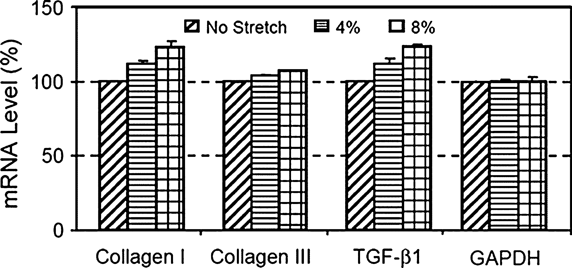
Effect of cyclic mechanical stretching on the mRNA expression of collagen type I, collagen type III, and transforming growth factor-β1 (TGF-β1) in human tendon fibroblasts. Cells were cyclically stretched at 0.5 Hz at a magnitude of 4% or 8% for 4 h, followed by 4 h of incubation in the stretch-conditioning medium. Quantitative measurements of reverse transcriptase (RT)-polymerase chain reaction results of tendon fibroblasts from three different subjects. Stretching-induced expression levels of collagen type I, collagen type III, and TGF-β1 in stretched tendon fibroblasts are expressed as a percentage of the mRNA level of nonstretched cells. Glyceraldehyde-3-phosphate dehydrogenase (GAPDH) was utilized as a housekeeping gene to normalize results. Reprinted with permission from Yang et al. 46 © 2004 by Elsevier Ltd.
In addition to collagen synthesis, elevated growth factor expression illustrates the effect of mechanical loading on cell differentiation and ECM formation.44–46,75 As stated previously, growth factors, such as TGF-β1, influence collagen, and PG synthesis in fibroblasts. Kim et al. 45 reported that ACL fibroblasts subjected to cyclic stretch demonstrate an increase in the level of TGF-β1 to approximately 149%, and that the stretch-induced expression of collagen mRNA was inhibited in the presence of an anti-TGF-β1 antibody. 45 This suggests that increased collagen production in response to mechanical loading is mediated via an autocrine mechanism of TGF-β1 released from ligament fibroblasts.
A correlation between growth factor expression and collagen synthesis in response to mechanical stimulation has also been reported for tendon fibroblasts.46,75 Skutek et al. 75 reported that cyclic biaxial stretch of tendon fibroblasts for 15 and 60 min elevates the expression of TGF-β1 by a factor of 1.26 and 1.40, respectively. Yang et al. 46 showed that 4% and 8% mechanical stretch elevates mRNA expression of TGF-β1 in patellar tendon fibroblasts by 11.5% and 24.6%, respectively. These results indicate that elevated growth factor expression is dependent on both the magnitude and duration of stretch. Similar to ACL tissue, Yang et al. 46 also found that stretch-induced increases in collagen synthesis were inhibited by the addition of an anti-TGF-β1 antibody. This confirms an active role for TGF-β1 in the ECM remodeling process of fibrous connective tissue as well as the mechanotransduction of fibroblasts.
The effect of mechanical stretch on the synthesis of PGs was also investigated. Studies of ACL fibroblasts have found that mRNA expression of PGs is typically downregulated in response to mechanical stimulation.54,74 Lee et al. 74 reported that ACL fibroblasts subjected to cyclic tensile stress of 5% strain at 0.5 Hz for 24 h demonstrate decreased expression of biglycan mRNA by approximately 30%. Miyaki et al. 54 observed that ACL-derived cells subjected to 10% stretch at 10 cycles/min demonstrate a reduction in decorin expression. Given the role of decorin and biglycan in the organization of fibrous connective tissue, further investigation is necessary to understand the effect of this downregulation on fibril diameter and packing.
Enzyme activity is elevated by ligament fibroblasts in response to cyclic stretch to offset increased collagen production and thus maintains balanced ECM remodeling. Upregulation of enzyme activity is also necessary to generate functional neotissue because it degrades randomly oriented scar tissue, which is then replaced with oriented ECM proteins. Zhou et al. 76 showed that ACL and MCL fibroblasts both demonstrate increased MMP-2 activity in response to stretch, with ACL fibroblasts generating significantly higher MMP levels than MCL fibroblasts. This study also found that ACL fibroblasts subjected to higher strain magnitudes (6%, 10%, 12%, and 14% stretch) further increase MMP-2 activity (35%, 105%, 423%, and 670%, respectively). Tendon fibroblasts also demonstrate increased enzyme activity in response to mechanical stretch.77–79 Yang et al. 77 reported that cyclic stretch of 8% elongation at 0.5 Hz increases MMP-1 expression in patellar tendon fibroblasts, whereas 4% stretch actually decreases MMP expression. This study, combined with the findings of Zhou et al., 76 suggests that stretch-induced enzyme production is also dependent on the magnitude of stretch.
Stress deprivation was also reported to induce a biochemical effect on ECM remodeling in fibrous connective tissue. Specifically, stress deprivation resulted in increased collagen turnover as measured by heightened collagen synthesis and degradation; however, degradation of existing collagen exceeded the formation of new collagen fibrils, which resulted in an overall decrease in collagen mass.80–82 Such mass loss coupled with changes in cellular morphology and collagen alignment in de novo tissue causes deterioration of the mechanical properties of immobilized ligaments and tendons.63,81–84 These results further establish the importance of mechanical loading on the development of functional tissue-engineered ligaments.
Mesenchymal Stem Cells
Although mechanical stimulation of ligament and tendon fibroblasts provides tissue engineers with a means to guide tissue organization during ligament reconstruction, fibroblasts are limited in their ability to generate a fully functional tissue-engineered ligament for ACL reconstruction. MSCs have emerged as an alternative cell source for tissue engineering because they can differentiate to multiple connective tissue cell types. 85 Additionally, isolation of MSCs from bone marrow provides tissue engineers with an unlimited supply of autologous cells that exhibit excellent regenerative properties, including superior proliferation. Current research has demonstrated that MSCs can differentiate toward the ligament lineage in response to mechanical loading.14–18 With appropriate mechanical stimulation, MSCs elongate, orient parallel to the direction of stretch, and form aligned collagen fibers characteristic of ligament cells.14–17 Noth et al. 14 seeded MSCs in type I collagen hydrogels and utilized uniaxial cyclic loading to induce ligament-like differentiation (Fig. 2). To better mimic physiological conditions, Altman et al. 15 applied translational (10%) and rotational strain (25%) to collagen gels seeded with bone marrow–derived cells and found that these constructs form helically organized collagen type I fiber bundles, similar to native ACL tissue.

Histochemical analysis of stretched anterior cruciate ligament (ACL) constructs (stretched) after 14 days compared with nonstretched controls and a human ACL. (
Mechanical stretch was also reported to increase collagen type I, collagen type III, elastin, and tenascin-C expression in MSCs.14–18 Altman et al. 15 reported that bone marrow–derived cells subjected to tensile and torsional loading demonstrate mRNA levels for collagen types I and III and tenascin-C that approach those quantified in native ligaments. Noth et al. 14 also reported an increase in the expression of fibronectin in mechanically stimulated stem cell constructs, although Juncosa-Melvin et al. 18 found no significant elevation of fibronectin or decorin expression after stimulating MSCs seeded within type I collagen sponges. In general, the effects of mechanical stimulation on the differentiation of MSCs to the ligament lineage are consistent with the effects in ligament and tendon fibroblast cultures.
Overall, mechanical stimulation has been a relatively successful mechanism to differentiate stem cell constructs toward the ligament lineage; however, before clinical implementation, further research is needed to develop functional ligament tissue using MSCs. 86 Although MSCs demonstrate proper morphology, alignment, and ECM production to mimic ligament fibroblasts in response to mechanical stimulation, the mechanical strength of the generated tissue still does not match healthy ACL tissue.14,86 Such differences in mechanical properties result from inconsistencies between the ECM synthesis of differentiated MSCs and the native ECM remodeling process. Nevertheless, Juncosa-Melvin et al. 86 found that mechanical conditioning of type I collagen sponges for patellar tendon repair improved cellular alignment within the construct and increased the linear stiffness by a factor of 2.5, as compared with nonstimulated controls. Given the advances in ligament and tendon fibroblast studies, it is expected that mechanical stimulation of MSCs can be optimized to induce appropriate ligament remodeling to restore ACL function. These loading conditions can then be incorporated into the design of bioreactors to generate improved tissue-engineered grafts for ACL reconstruction.
Summary
Mechanical stimulation is a critical component in the engineering of load-bearing tissues. Without the appropriate biomechanical cues, new tissue formation lacks the necessary ECM organization for sufficient load-bearing capacity. Tissue engineers utilize mechanical conditioning to guide tissue remodeling and improve the performance of ligament grafts. Current research has shown that cyclic mechanical stretch directs fibroblast alignment and guides cell morphology to an elongated, spindle-like shape. In addition, mechanical stimulation also increases proliferation, collagen synthesis, and growth factor expression. These effects were found to depend on the magnitude, frequency, and duration of stretch. Although stretch-induced alterations in fibroblast behavior should translate into improved tensile properties, fibroblasts are still limited in their ability to develop a functional tissue-engineered ligament. MSCs have received growing interest in ligament tissue engineering because of their ability to differentiate toward the ligament lineage in response to mechanical stimulation. Further investigation in tuning mechanical strength of tissue-engineered grafts that utilize MSCs is needed to match healthy ligaments. Overall, the results highlighted here indicate that bioreactors will play a critical role in the engineering of improved ligament grafts for ACL reconstruction. This comparative analysis is meant to serve as a resource to advance bioreactor design by probing the mechanisms of stretch-induced differentiation.
Footnotes
Disclosure Statement
No competing financial interests exist.