




Abstract
Background:
Osteoporosis is a frequent human metabolic bone disorder. Prospectively, global ageing of populations will lead to a major increase of subjects being diagnosed with osteoporosis and in need for dental rehabilitation. However, as local osteoporosis of the jaws affects bone quantity and quality of edentulous regions, osseointegration of dental implants might be hampered. Consequently, calcium phosphate ceramic-coated implants have been suggested to compensate for low bone quantity/density and for impaired bone healing in osteoporosis. Nonetheless, up to now no meta-analytical assessment of the relevant preclinical literature to quantify such a possible positive effect has been undertaken.
Materials and Methods:
PubMed search, limited to animal models, to identify a possible positive effect of calcium phosphate-coated implants on bone regeneration, was carried out. Further, the reference lists of related review articles and publications selected for inclusion in this review were systematically screened. The primary outcome variables were bone-to-implant contact percentage as assessed histomorphometrically and mechanical stability testing.
Results:
The electronic search in the database of the National Library of Medicine resulted in the identification of 2704 titles. These titles were initially screened by the two independent reviewers for possible inclusion, resulting in further consideration of 51 publications. Screening the abstracts led to 22 full-text articles. From these articles, 16 reports were excluded. Finally, six of these original research reports could be selected for evaluation. Additionally, eight publications were identified by manual search. Thus, a total of 14 articles were included for analysis.
Conclusions:
It was concluded that (1) in osteoporotic animal models calcium phosphate ceramic-coated implants are associated with improved bone-to-implant healing as compared to noncoated implants. Moreover, (2) essentially due to quality characteristics of the analyzed original research articles a negative impact of osteoporosis on bone-to-implant healing could not be confirmed. Besides, (3) the established positive bone-to-implant healing effect of calcium phosphate ceramic coatings does not differ between osteoporotic and nonosteoporotic, healthy animal models.
Introduction
In principle, for more challenging situations such as low bone density, improved implant stability, and accelerated bone healing have been shown for certain surface modifications of dental titanium implants.11–14 Surface modifications of dental titanium implants are in general accomplished by roughening or by altering the chemical composition. Various methods have been developed to create roughened surfaces, for example, titanium plasma spraying, grit-blasting, acid etching, and anodization. Coating of dental titanium implants with calcium phosphate (CaP) ceramic is the most frequently used method for changing the chemical surface composition. 15 It is well known that following implantation, the release of CaP into the peri-implant region increases the saturation of body fluids and results in the precipitation of a biological apatite onto the surface of the implant.16,17 This layer of biological apatite might contain endogenous proteins and serve as a matrix for osteogenic cell attachment and growth. 18
Cellular interactions with the apatite layer and its contained proteins onto an implant surface are mediated by integrins. These specific cell membrane molecules transduce signals from the surface proteins matrix to the cell and vice versa. Then, the signaling pathways through integrins can regulate bone forming cell activity to a certain extent.16,17 This desired bone-stimulating behavior of CaP coatings at implant surface is of prime importance when compared to dental implants without CaP coatings. 19 In addition, as the biological fixation of titanium implants to bone tissue is faster with a CaP coating than without,19,20 it seems rational to assume that the bone regeneration process around the implant is enhanced by the formation of the aforementioned biological apatite layer. Thus, it is not astonishing that especially for osteoporotic bone CaP ceramic-coated implants have been suggested to compensate for low bone quantity/density and for delayed and/or impaired bone regeneration.21–25 However, up to now no meta-analytical assessment of the relevant literature to quantify such a possible positive effect has been undertaken.
Hence, the aims of this study were (1) to systematically review the literature on the subject of bone healing and CaP ceramic-coated implants in osteoporotic animal models and (2) to investigate bone-to-implant contact (BIC) data and data retrieved from mechanical stability tests by means of a meta-analytical approach.
It was hypothesized that in osteoporotic animal models (1) CaP ceramic-coated implants are associated with improved bone healing as compared to noncoated implants, that (2) CaP ceramic-coated implants overcome a possible negative impact of osteoporosis on bone healing, and (3) that the effect of CaP ceramic coatings is greater in osteoporotic animal models as compared to nonosteoporotic, healthy animal models.
Materials and Methods
Outcome variables
The primary outcome variables were BIC percentage as assessed by histological analysis and biomechanical stability testing.
Inclusion/exclusion criteria
In general, only animal studies presenting BIC percentage or data regarding mechanical testing (push-out test or torque-out test) for CaP-coated implants versus noncoated, control implants in osteoporotic animals versus healthy, control animals were included.
The following detailed inclusion criteria were used:
1. Research article presenting in vivo animal data; 2. Data should be acquired in osteoporotic animals (test) and in healthy, nonosteoporotic animals (control); 3. Data should be presented for calcium phosphate ceramic-coated implants (test) and noncoated (control) implants; 4. Shape or configuration of used implants, that is, threaded (screw-shaped) or nonthreaded (cylinder-shaped), should be indicated; 5. Implantation site should be clearly stated; 6. Healing period should be clearly mentioned; 7. The osteoporotic animal model used should be described conspicuously, for example:(i) Basic characteristics of the animal model should be mentioned (e.g.: species and gender);(ii) Initiation method of osteoporosis should be stated;(iii) Time point of induction (i.e., before −, parallel to −, or after implant installation) should be mentioned;(iv) Method to confirm osteoporotic status of the implantation site should be stated; 8. At least four osteoporotic and control animals should have been included; 9. BIC percentage and/or data retrieved from biomechanical testing (push-out test or torque-out test) have to be presented. Further, torque-out testing data should be presented together with histomorphometrical data.
Studies that did not meet all above mentioned inclusion criteria were excluded.
Search strategy
An electronic search in the database of the National Library of Medicine (www.ncbi.nlm.nih.gov) up to November 7, 2011, was carried out. Only publications in English were considered and the search was narrowed to animals only. The following search strategy was applied: (((((“dental implantation” [MeSH Terms] OR “dental implants” [MeSH Terms]) OR “orthopedics” [MeSH Terms]) OR “implants, experimental” [MeSH Terms]) OR “blade implantation” [MeSH Terms]) OR “implants” [All Fields]) AND ((((surface[All Fields] AND modification[All Fields]) OR “surface coating” [All Fields]) OR “surface treatment”[All Fields]) OR (“surface properties”[MeSH Terms] OR “coated materials, biocompatible” [MeSH Terms])). Further, the reference lists of related review articles and publications selected for inclusion in this review were systematically screened.
Study selection
Two independent reviewers (Hamdan Alghamdi [H.A.] and Rüdiger Junker [R.J.]) initially screened the publication titles and abstracts as identified by the electronic and manual search for possible inclusion. Full texts of all articles that were considered eligible for inclusion by one or both of the reviewers were obtained for further assessment against the stated inclusion criteria (Fig. 1). Both reviewers used a data extraction form to extract the data independently. Any disagreements between the reviewers regarding inclusion of a certain publication or data extraction were resolved by discussion.

Articles selection process.
Quantitative data synthesis
As the central aim of the present study was to quantify a possible positive effect of CaP ceramic coatings on the subject of implant osseointegration in osteoporotic bone, a meta-analysis for the effect size of CaP ceramic coating with regard to (1) BIC percentage [%BIC], (2) mechanical push-out testing [N], and (3) mechanical torque-out testing [Ncm] in osteoporotic animal models (test) and in healthy, nonosteoporotic models (control) was performed. For (1) BIC percentage [%BIC], (2) mechanical push-out testing [N], and (3) mechanical torque-out testing [Ncm], Student's t-test was applied to calculate the difference of the weighted means of CaP ceramic-coated implants (test) and noncoated implants (control) within and between osteoporotic animal models (test) and healthy, nonosteoporotic models (control). Next to differences of the weighted means (MD), 95% Confidence Intervals (CI), and statistical significance difference (p<0.05) were obtained. In addition, for meta-analysis, inverse variance weighting was used for pooling. Analyses were performed with the statistical software package R, version 2.10.1 (www.R-project.org). 26 In case of heterogeneity, the DerSimonian-Laird estimate was used. 27
Results
Study selection
The electronic search in the database of the National Library of Medicine resulted in the identification of 2704 titles. As already mentioned, these titles were initially screened by the two independent reviewers for possible inclusion, resulting in further consideration of 51 publications. Screening the abstracts led to 22 full-text articles, which are detailed in Table 1. From these articles, 16 reports were excluded for reasons mentioned in Table 1. Finally, six of these original research reports could be selected for evaluation. Additionally, eight publications were identified by manual search. Thus, at last a total of 14 articles were included for analysis (Fig. 1). Regarding data extraction and interpretation, any disagreement between the reviewers was resolved by discussion. Included studies are summarized in Table 2.
x indicates selected article.
BIC, bone-to-implant contact; CaP, calcium phosphate.
Excluded from meta-analysis; the presented data were not transferrable to the measuring unit for push-out testing [N].
OVX, bilateral ovariectomy; NX, unilateral limb neurectomy; BMD, bone mineral density.
Study characteristics
In 13 out of the included 14 research articles, female animals were submitted to bilateral ovariectomy-induced, estrogen-deficiency osteoporosis (OVX), whereas in one study 28 unilateral limb sciatic neurectomy (sciatic NX) was used. Osteoporosis was diagnosed in 11 out of the 14 reports by BMD measurements, in one study by radiographic analysis of bone morphology (SOFTEX-C-SM), 28 and in two publications25,29 by histomorphometrical analysis of iliac bone biopsies. In all 14 reports CaP ceramic-coated implants (test) and noncoated implants (control) were used in osteoporotic animal models (test) and in nonosteoporotic, healthy animal models (control). In principle, from four articles data of more than one experiment could be retrieved. This was possible because in one publication, 25 different anatomical sites of implantation (i.e., femur or tibia within a sheep-model) were analyzed separately, and because in the three otherarticles,23,28,30 different healing periods after implantation were independently studied. As a result, 20 discrete trials were identified (Table 2).
CaP ceramic coating characteristics
Coating characteristics as provided in the included research articles are presented in Table 3.
Highly detailed information with regard to chemical and physical composition is generally not presented. Only one publication reports a more detailed chemical composition, 30 whereas the other 13 articles roughly describe the composition as a hydroxyapatite.
CaP-coated implants versus noncoated implants in osteoporotic animal models
First, the hypothesis that in osteoporotic animal models (test) CaP ceramic-coated implants (test) are associated with improved bone healing as compared to noncoated implants (control) was tested. In osteoporotic animal models, the difference of the weighted means of BIC percentage between CaP ceramic-coated implants and noncoated implants was 19.2%. The 95% CI of the difference ranged between 13.8% and 24.6%. This difference was statistically significant (p<0.05). The difference of the weighted means for mechanical push-out testing between CaP ceramic-coated implants and noncoated implants was 105.4 N. The 95% CI of the difference ranged between 53.5 and 157.4 N. Also this difference was statistically significant (p<0.05). Moreover, the difference of the weighted means for mechanical torque-out testing between CaP ceramic-coated implants and noncoated implants was 115.9 Ncm. The 95% CI of the difference ranged between 31.6 and 200.2 Ncm. However, this difference did not reach statistical significance (p>0.05; Table 4).
SD, standard deviation; CI, confidence intervals; MD, difference of the weighted means.
Noncoated implants in osteoporotic animal models versus nonosteoporotic, healthy animal models and CaP ceramic-coated implants in osteoporotic animal models versus noncoated implants in nonosteoporotic, healthy animal models
Second, the hypothesis that CaP ceramic-coated implants (test) overcome a possible negative impact of osteoporosis (test) on bone healing was investigated. Comparing noncoated implants in nonosteoporotic, healthy animal models versus osteoporotic animal models revealed that the difference of the weighted means of BIC percentage was 8.2%. The 95% CI of the difference ranged between −4.1% and 20.5%. This difference was not statistically significant (p>0.05). The difference of the weighted means of mechanical push-out testing was 22.4 N. The 95% CI of the difference ranged between −41.2 and 86.0 N. Also, this difference was not statistically significant (p>0.05). Further, the difference of the weighted means of mechanical torque-out testing was 15.0 Ncm. The 95% CI of the difference ranged between −45.1 and 75.1 Ncm. Again, this difference failed to reach statistical significance (p>0.05; Table 5, Fig. 2A–C). Further, comparing CaP ceramic-coated implants in osteoporotic animal models versus noncoated implants in nonosteoporotic, healthy animal models uncovered that the difference of the weighted means of BIC percentage was 11.3%. The 95% CI of the difference ranged between −0.4% and 23.0%. This difference was not statistically significant (p>0.05). The difference of the weighted means of mechanical push-out testing was 84.1 N. The 95% CI of the difference ranged between 39.8 and 128.4 N. This difference was statistically significant (p<0.05). Besides, the difference of the weighted means of mechanical torque-out testing was 101.6 Ncm. The 95% CI of the difference ranged between −13.4 and 216.6 Ncm. Again, this difference failed to reach statistical significance (p>0.05; Table 6, Fig. 3A–C).

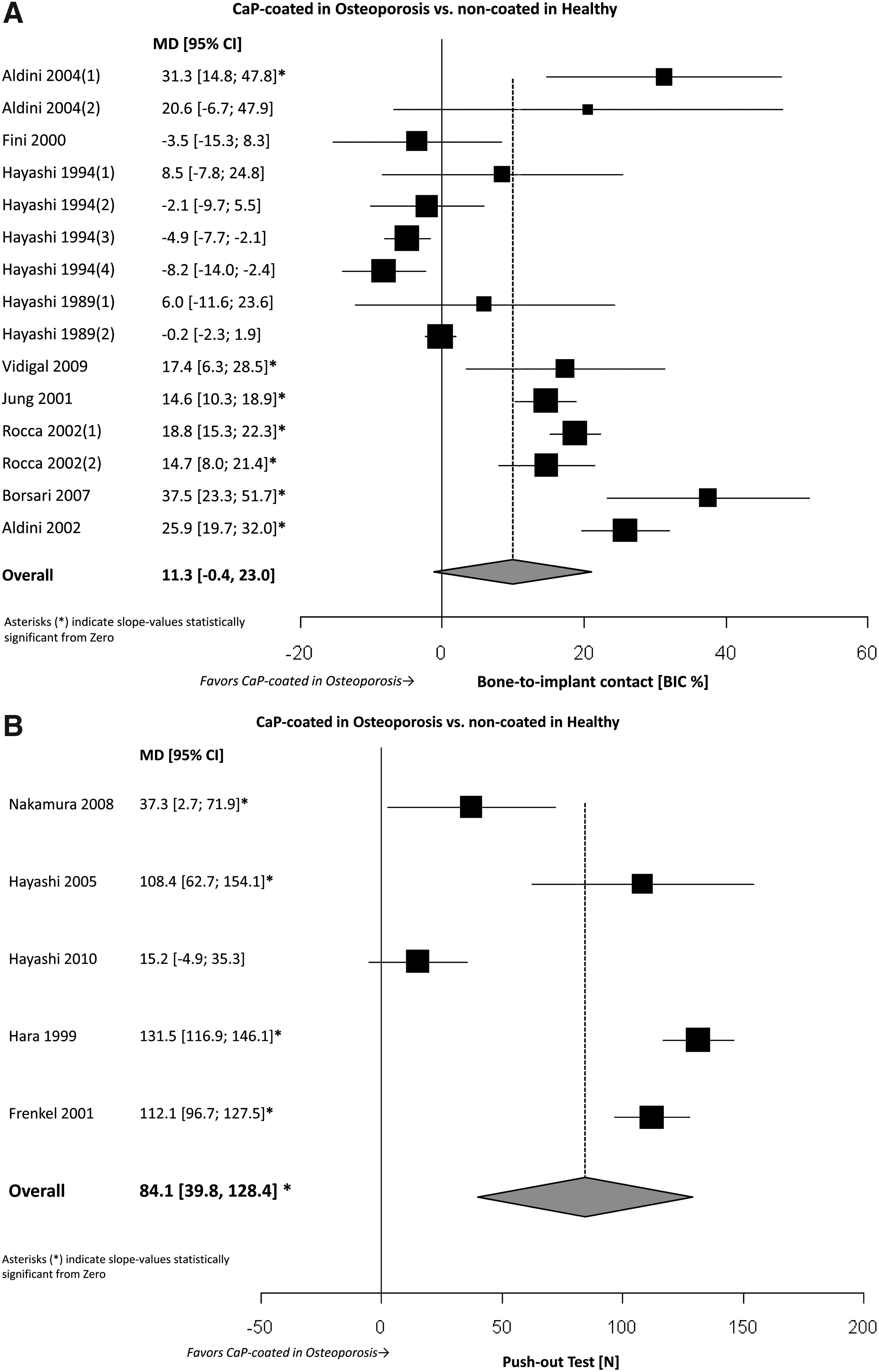
Effects of CaP coatings on bone healing in osteoporotic animal models and in nonosteoporotic, healthy animal models
Third, the hypothesis that the effect of CaP ceramic coatings (test) is greater in osteoporotic animal models (test) as compared to nonosteoporotic, healthy animal models (control) was examined. As already mentioned above, in osteoporotic animal models the difference of the weighted means of BIC percentage between CaP ceramic-coated implants and noncoated implants was 19.2%. The 95% CI of the difference ranged between 13.8% and 24.6%. This difference was statistically significant (p<0.05). The difference of the weighted means for mechanical push-out testing between CaP ceramic-coated implants and noncoated implants was 105.4 N. The 95% CI of the difference ranged between 53.5 and 157.4 N. Also, this difference was statistically significant (p<0.05). Moreover, the difference of the weighted means for mechanical torque-out testing between CaP ceramic-coated implants and noncoated implants was 115.9 Ncm. The 95% CI of the difference ranged between 31.6 and 200.2 Ncm. However, this difference did not reach statistical significance (p>0.05). In nonosteoporotic, healthy animal models the difference of the weighted means of BIC percentage between CaP ceramic-coated implants and noncoated implants was 13.7%. The 95% CI of the difference ranged between 6.8% and 20.6%. This difference was statistically significant (p<0.05). The difference of the weighted means for mechanical push-out testing between CaP ceramic-coated implants and noncoated implants was 147.7 N. The 95% CI of the difference ranged between 91.1 and 204.3 N. Again, this difference was statistically significant (p<0.05). Moreover, the difference of the weighted means for mechanical torque-out testing between CaP ceramic-coated implants and noncoated implants was 132.6 Ncm. The 95% CI of the difference ranged between 41.7 and 223.6 Ncm. Again, this difference reached statistical significance (p<0.05). Heterogeneity testing of the differences of the weighted means of BIC percentage, push-out testing, and torque-out testing between CaP ceramic-coated implants and noncoated implants in osteoporotic animal models and nonosteoporotic, healthy animal models revealed that the included experiments presenting BIC percentage data and the experiments presenting push-out testing data were heterogeneous (Q-value: 48.5, p<0.05; Q-value: 9.7, p<0.05; respectively), whereas the experiments presenting torque-out testing data were not heterogeneous (Q-value: 4.7, p>0.05). The applied statistical effect models, the random effect model for BIC percentage, and the push-out testing, and the fixed effect model for torque-out testing did not indicate statistically significant overall differences regarding the investigated outcome.
Discussion
The aims of the present report were (1) to systematically review the literature on the subject of bone healing around CaP ceramic-coated implants in osteoporotic animal models and (2) to investigate BIC data and data retrieved from mechanical stability tests by means of a meta-analytical approach. It was hypothesized that in osteoporotic animal models (1) CaP ceramic-coated implants are associated with improved bone healing as compared to noncoated implants, that (2) CaP ceramic-coated implants overcome a possible negative impact of osteoporosis on bone healing, and (3) that the effect of CaP ceramic coatings is greater in osteoporotic animal models as compared to nonosteoporotic, healthy animal models. As in osteoporotic animal models statistically significant differences between CaP ceramic-coated- and noncoated implants with regard to BIC percentage and mechanical push-out testing were found in favor of CaP-coated implants, we tend to accept the proposed H1-hypothesis. On the other hand, it should be kept in mind that for mechanical torque-out testing the alternative H0-hypothesis of no difference could not be rejected. Further, the proposed H1-hypothesis that CaP ceramic-coated implants overcome a possible negative impact of osteoporosis on bone healing could not be validated. Nonetheless, for mechanical push-out testing with regard to CaP ceramic-coated implants in osteoporotic animal models versus noncoated implants in nonosteoporotic animal models, the proposed H1-hypothesis seems to be substantiated. Additionally, statistically significant effect size differences for BIC percentage, mechanical push-out testing, and mechanical torque-out testing were not observed for CaP ceramic-coated implants versus noncoated implants in osteoporotic animal models and nonosteoporotic animal models. Therefore, the proposed H1-hypothesis could not be confirmed.
As already indicated above, the retrieved findings have to be interpreted with caution. 31 In principle, sample-size and thereby statistical power of the reviewed preclinical in vivo experiments tended to be low. For example, the compared eight weeks BIC data within the group of osteoporotic animals of Aldini et al. 30 have, with an assumed α-error of 0.05, a statistical power as low as 0.092. For that reason, a meta-analytical approach was intended to increase the power of statistical analysis by pooling the results of all retrieved available trials. Preferably, all research should be done in a similar manner when their results are being combined in a meta-analysis. As shown in Table 2, this is clearly not the case for the presently included articles. The publications that were eligible for inclusion in the present study display experimental variability for the utilized animal model, the method applied for induction of osteoporosis, the used technique to identify osteoporosis, the number of enrolled implants and/or animals, the anatomical site of implantation, the healing time after implant placement, and the type of implant and coating used. Although surgical technique might also have a critical effect on implant fixation,18,29 it was difficult to consider the surgical variables in this systematic review. This was because all included studies did not estimate all the possible effects of surgical technique on the implant outcomes. Not surprisingly, statistical heterogeneity of the evaluated experiments regarding BIC percentage and push-out testing was found and might be explained in part by experimental variability.32,33 However, subgroup analysis was unable to clarify the origin of the dissimilarity in terms of treatment effects. Consequently, for the outcome variables BIC percentage and push-out testing random effects meta-analysis was selected. 34 The applicability and problematic aspects of random effects meta-analysis are thoroughly discussed elsewhere.35,36 Further, as heterogeneity was not found for torque-out testing, fixed effects meta-analysis was chosen for that outcome variable. 34
Next to the meta-analytical method, to be confident that the combined outcome estimate points toward a meaningful picture of the effect of CaP ceramic coatings for the potential use in humans, it is important to critically appraise the utilized osteoporotic animal models.
Obviously, the used bilateral ovariectomy-induced, estrogen- deficient osteoporosis rat model,22–24,30,37–39 sheep model,21,25,29 and dog model 40 are respected,41–43 well documented, and in general, accepted osteoporotic disease models to simulate human osteoporosis for mucoskeletal research.41,44–46 In contrast, the employed rat unilateral limb sciatic neurectomy-induced osteoporosis model 28 and the bilateral ovariectomy-induced, estrogen-deficient osteoporosis rabbit model47,48 are evidently suitable osteoporosis models, but less accepted to mimic the human situation.46,49–51
For the careful interpretation of the current findings it should be mentioned that 11 of the 14 meta-analyzed research articles are published by just two different groups [group 1:21,25,29,30,39, group 2:22–24,28,37,38]. Apparently, only Frenkel et al., 40 Jung et al., 48 and Vidigal et al. 47 seem to be unrelated.
Unfortunately, highly detailed information with regard to chemical and physical composition is generally not presented. For example, only one publication reports a more detailed chemical composition, 30 whereas the other 13 articles roughly describe the composition as a hydroxyapatite. However, chemical and physical surface properties as ionic composition, hydrophilicity, and roughness are parameters that play a major role in implant-tissue interaction. 15 In other words, different CaP implant surface coatings possess different and unique chemical and physical surface properties, and might potentially lead to differences in the bone-to-implant reaction. 52 That finally implies that the statistical heterogeneity of the evaluated experiments regarding BIC percentage and push-out testing might be explained in part by differences of the chemical and physical composition of the applied CaP coatings. Accordingly, more detailed information regarding chemical and physical surface properties as ionic composition, hydrophilicity, and roughness might have enabled a subgroup analysis to clarify the origin of the dissimilarity as regards treatment effects.
Conclusion
Within the limits of the current meta-analysis, we conclude that (1) in osteoporotic animal models CaP ceramic-coated implants are associated with improved bone-to-implant healing as compared to noncoated implants. Further, (2) essentially due to quality characteristics of the analyzed original research articles, a negative impact of osteoporosis on bone-to-implant healing could not be confirmed. In addition, (3) the established positive bone-to-implant healing effect of CaP ceramic coatings does not differ between osteoporotic and nonosteoporotic, healthy animal models.
Footnotes
Disclosure Statement
No competing financial interests exist.