




Abstract
New advances in tissue engineering are being made through the application of different types of electrical stimuli to influence cell proliferation and differentiation. Developments made in the last decade have allowed us to improve the structure and functionality of tissue-engineered products through the use of growth factors, hormones, drugs, physical stimuli, bioreactor use, and two-dimensional (2-D) and three-dimensional (3-D) artificial extracellular matrices (with various material properties and topography). Another potential type of stimulus is electricity, which is important in the physiology and development of the majority of all human tissues. Despite its great potential, its role in tissue regeneration and its ability to influence cell migration, orientation, proliferation, and differentiation has rarely been considered in tissue engineering. This review highlights the importance of endogenous electrical stimulation, gathering the current knowledge on its natural occurrence and role in vivo, discussing the novel methods of delivering this stimulus and examining its cellular and tissue level effects, while evaluating how the technique could benefit the tissue engineering discipline in the future.
Introduction
In the following review, we discuss the various aspects of electrical stimulation, giving examples of endogenous electricity, the different types of stimuli, the various novel methods of delivering these and their effects on both the cellular and tissue level, to demonstrate the potential that this technique has for the discipline of tissue engineering.
In Vivo Electricity
The main sources of in vivo electricity are the cells. Through the constant pumping of ion channels, they establish a voltage gradient across their membrane, the membrane potential.19,20 When cells couple together into a continuous layer, they create a resistive barrier, paralleling the cellular membrane on a bigger scale.14,20,21 Polarized Na+, K+, and Cl− ion transport on the two sides of this layer establishes a tissue-level electric gradient across the cellular interface of typically 15–60 mV, an example of which is the trans-epithelial potential (TEP) found in skin, lens, and cornea.14,20–24 The TEP mechanism in vertebrate lens is particularly interesting, as it creates a complex pattern of current loops with magnitudes around 20–40 mA/cm2. These currents flow inward at the anterior and posterior poles of the lens, and outward at the equator of the lens, controlling the migration and differentiation of epithelial cells.7,22 Another important function of TEP can be observed in injuries of the epithelium. A wound short circuits the potential difference between the two sides of the epithelium and, therefore, gives rise to an EF (0.04–0.2 V/mm)8,9,14,16,20,24–26 that, in turn, controls the orientation and frequency of cell division 24 and induces directional migration toward the injury.8,16,21 This has been shown to override any other influence on the cell's functioning, such as chemical gradients and population pressure.14,21 In regeneration and embryonic development, intra- and extracellular EFs play a pivotal role in regulating cellular behavior 14 and the development of spatial patterns, such as left–right organ asymmetry.12,14,27–29 Disruption of these fields in embryos has been reported to result in serious defects, for example, the absence of the cranium, a malformed head and the loss of eyes. 14 In studies conducted on partially amputated Xenopus tails, electrical currents were observed to occur as a result of the injury. Disrupting these electrical currents reduced regeneration, while artificially reversing its polarity completely blocked the healing process. 29
The significance of bioelectricity is emphasized by its role in one of the rare examples of human regeneration. Illingworth and Barker 30 reported that the amputated fingertips of children could be fully regenerated as long as the stump is kept clean and hydrated and observed that electrical currents of 30 A/cm2 were present in wounds. In their article, they theorized that moisture in the wound ensures a continuous electric conductance path, enabling the wound's electrical fields to exert their regenerating effect. 30 Streaming potentials, streaming currents, and the piezoelectricity of collagen molecules all contribute to the generation of bioelectricity in bone.11,31–38 These electrical phenomena are suggested to be transduced by osteocytes and to play a similar role in remodeling and healing as TEPs.13,37–39 Bioelectricity is also suggested to be the ideal mechanism to deliver signals to chondrocyte cells in cartilage, as they usually exist in relative isolation. 40 Other examples of in vivo electricity include the rapid action potentials of nerves and muscles; long-lasting DC voltages around damaged nerves, known as injury potentials 14 and the trans-endothelial extracellular potential gradients in arteries and veins.25,41
The use of electrical stimulation is well-established in today's medicine. A wide range of electrical therapeutic systems have been developed to treat specific ailments and/or tissue types. These devices include implantable and external bone growth stimulators,42,43 chronic cutanous wound-healing systems, 44 functional electrical stimulation devices, to restore muscles in paralyzed patients, 45 and stimulators for pain relief.46,47
Transcutaneous nerve stimulation (TENS) units are mainly used to deliver electroanalgesia and have been proven to be greatly effective in the treatment of a wide range of pains.46,47 In a clinical study examining the effectiveness of these devices, it was shown that TENS treatment reduced pain by more than a half in 47% of the patients. 48 Additionally, these devices have also been shown to be useful in the treatment of chronic ulcers. 44 In a meta-analysis, where the effect of multiple systems (including TENS units) was assayed upon chronic wound healing, it was found that electrical stimulation produced an enhanced healing rate of 22% per week compared to the 9% of untreated samples. Upon the comparison of the healing effect of the devices involved in this study, no significant difference was observed. 44 In another investigation, examining the effectiveness of electrical therapy in the treatment on pressure ulcers, the wound surface decreased approximately 70% with electrical treatment, while only 36% without. 49 Electrical stimulation systems also have a proven record promoting bone growth, For example, an extensive study, including 175 patients, showed that electrical stimulation was able to induce solid bone union in 83.7% of the cases. 42
Due to the wide variety of bioelectrical presence and its significant influence on in vivo tissue, researchers are now beginning to explore the potential of using this stimulus in vitro.
The Methods of In Vitro Electrical Stimulation
Types of electrical stimulation
The most basic method of electric stimulation is an applied DC voltage, simply generated by batteries. 50 More complicated stimuli can be in the form of monophasic (DC) or biphasic (AC)13,32 sinusoidal,11,39,51,52 saw tooth,36,53,54 or square wave28,55 signals, injected in pulses,36,53,55 pulse bursts,11,56 or continuously.5,10,13 These signals can be generated by stimulator chips, 32 signal generators,28,54,57 or dedicated therapeutic systems,15,58 such as the Phyback, 59 TENS,46,47 or the EBI bone-healing systems.11,56,57
Methods of delivering the stimulus
Direct coupling
With direct coupling (Fig. 1A), the electrode is in direct contact with (e.g., inserted into) the cell culture or implanted into the patient or laboratory animal.31,32,37 Although this is the simplest way of delivering electrical stimulation, there are quite a few disadvantages to the approach. The problem of toxicity arises, due to the insufficient biocompatibility of the electrodes, 60 changes in pH, 31 reduced levels of molecular oxygen,60,61 and the generation of dangerous Faradic byproducts, for example, reactive oxygen species in the culture medium.13,31,60,61 The facts that the stimuli will be biased, due to the formation of a capacitive bilayer31,32,61 and that the effects of the stimulation was observed to depend heavily on whether it was measured near the anode or the cathode electrode, 61 further hinder the use of this method.
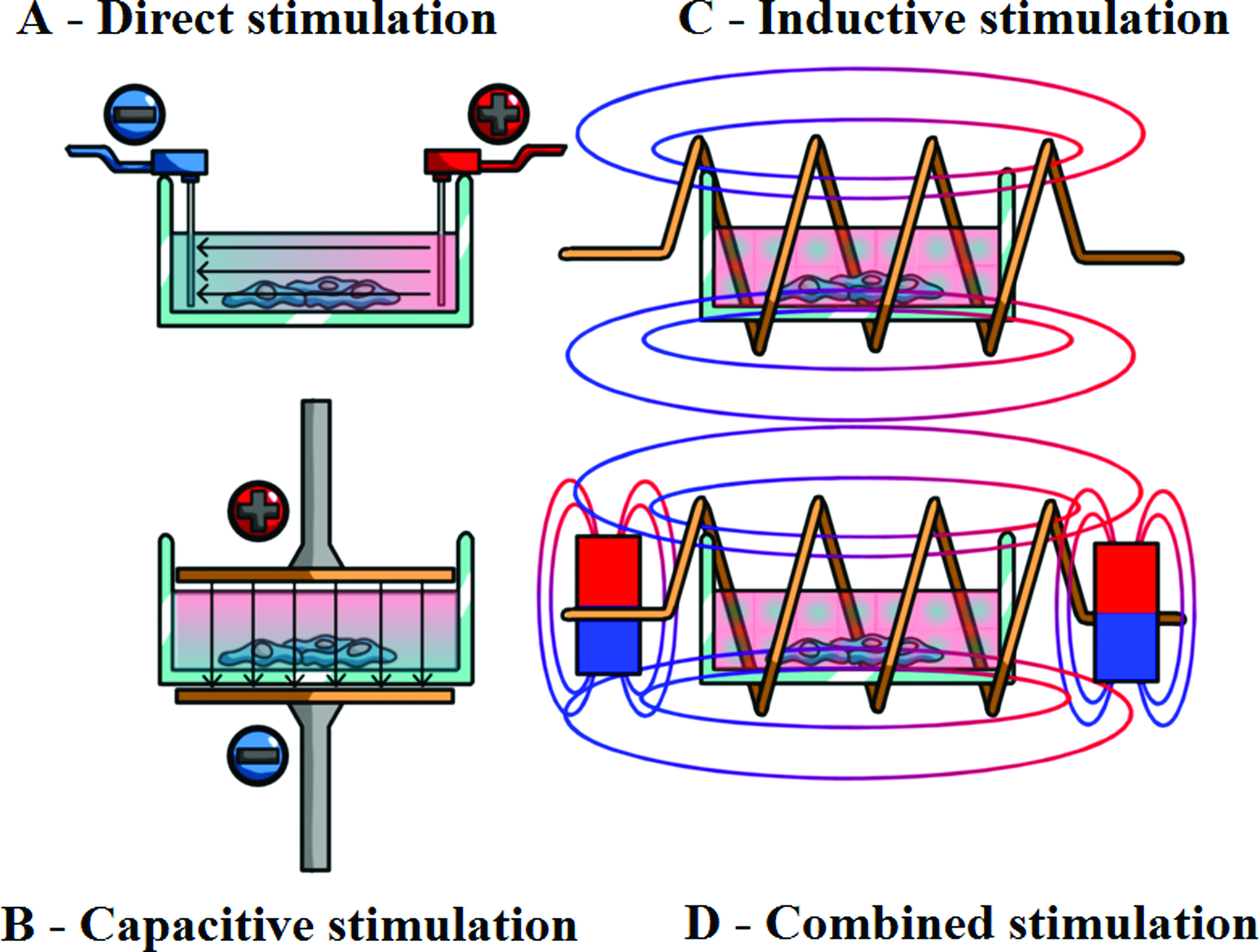
The four main techniques of delivering electrical stimulation: The direct
Indirect coupling
To avoid the difficulties inherent in direct coupling, an indirect, noninvasive approach is used in many therapeutic devices and in vitro experimental setups. The three main techniques used are capacitive, inductive, and combined coupling methods (Fig. 1B–D), where the latter is a combination of a static magnetic and an alternating current generated by a transient electromagnetic field.11,13,32 A comparison of the effects of the different techniques can be seen in Table 1.
Direct stimulation was mainly used to influence the morphology and orientation of cells. Capacitive stimulation seems to be more effective in increasing proliferation, while cells treated with inductive stimuli display greater ecm deposition.
ESC, embryonic stem cell; MSC, mesenchymal stem cell; ROS, reactive oxygen species; ECM, extracellular matrix; TGF transforming growth factor; ALP, alkaline phosphatase; VEGF, vascular endothelial growth factor.
Capacitive coupling: In capacitive coupling (CC), a homogenous electromagnetic field is created between two parallel layers of metal (e.g., stainless steel, gold) or carbon (i.e., a plate capacitor) that are placed above and below, the culture medium with a small (0.5–2 mm) gap between them.3,13,19,31,32,36,37,40,62–65 CC has the advantage of avoiding the issues with direct coupling and that of generating a homogenous EF, thus equal amount of stimulation for every cell regardless of the position in the culture vessel. However, its usefulness can be limited by the high voltage (e.g., 100 V) required to be generated between the electrodes. 66
Inductive coupling: Inductive coupling utilizes controlled electromagnetic fields generated by coils placed around the cell culture. This has the advantage of generating small-magnitude currents and potentials in the proximity of the targeted cells, rather than delivering the stimulation through electrodes.25,31,37,51,67,68 In many instances, the coils are used in pairs, placed in the Helmholtz configuration,39,53,57 meaning that distance between the coils is equal to their radius. 39 This generates a near homogenous magnetic field with uniform electromagnetic field properties across the cell culture. A subtype of this modality is known as pulsed electromagnetic field stimulation or pulsed electromagnetic field stimulation (PEMF), where the stimulus is delivered in pulses (rather than being static or continuously harmonic) to mimic, for example, the natural strain-generated potentials observed in bone.39,69
Scaffolds
In recent years, electrically conductive polymer scaffolds have been developed as a new means to deliver the electrical stimulus with the most popular materials being polypyrrole (PPy), used either alone5,18 or in combination with other materials, such as the biodegradable poly-L-lactide (PLA) in a 5:95 ratio. 4 In the future, these multifunctional scaffolds could act as bioactive substrates for cell attachment, while providing a way to better regulate cellular activities through evenly distributed, well-controlled electrical stimuli both on the surface and within the biomaterial scaffold. 4
The range of parameter variables in electrical stimulation
The level and nature of the voltage applied by the stimulation equipment is, arguably, the most important parameter influencing the cell cultures. The outcome, especially with regard to cellular migration, orientation,16,27,41,70 and gene expression,54,71 has been shown to depend upon the amplitude of the voltage applied, with strong indications that a relatively higher setting is favorable in nearly all circumstances9,16,27,41,54,70–73–see Table 2. However, effects, such as enhanced proliferation or increased extracellular matrices (ECM) deposition, were witnessed with stimulation currents (or current density) at both relatively high and low settings (Table 3). As such, the value of current does not seem to be important when considering the effectiveness of the stimuli. Indeed, it acts a limiting constrain, since any current above a certain threshold (e.g., 20 μA in case of treating nonunions 42 ) will result in cell death.10,13,31,32,42,73
It is also important to note that field strength was not necessarily the only parameter different between the stimuli.
The effect that the stimulation has seems not to depend on current density, but if the electrical current is raised above a certain threshold, for example, 5 A/m2 in the case of osteablasts, 61 necrosis will start to occur.
The maximum pulse width is limited by current density: the higher the current of the stimulation is, the shorter the duration needs to be to avoid necrosis (i.e., there is a limit on the electrical energy that can be absorbed by the cell safely).13,73 Nonetheless, none of the temporal durations, from 0.025 to 380 ms, that have already been applied in cell culture experiments, have proved to be ineffective or damaging.10,13,16,34,39,55,57,62,67,74 A wide range of frequencies (7.5-60000 Hz) have been applied in previous experiments, though most of these were below 100 Hz. This is a logical choice as frequencies in this low range are the ones that are assumed to occur naturally in vivo during normal use and are, therefore, the most likely to have a beneficial effect on the cells.10,11,13,31,32,34–36,51,63,75 Frequencies between 10 and 30 Hz, 51 and those less than 100 Hz, 39 have been reported as the most effective for stimulating new bone formation, while 7.5 Hz has been suggested as the best frequency to promote the proliferation of primary rat osteoblasts 75 and osteoclast differentiation of bone marrow cells of mice. 54
The Effects of Electrical Stimulation at the Cellular Level–Galvano-Transduction
Several theories exist on how EFs affect cells directly. What is known is that the electrical fields seem to be sensed through the same pathways observed to be involved in mechanotransduction and chemotaxis.9,21,26,27,76 A key component in this process, and one that has been observed to be greatly affected by electrical stimuli, is the intracellular calcium level.
Intracellular calcium
Electric signals are believed to be transduced, at least partially, through the calcium/calmodulin pathway. 32 This happens somewhat differently with the various methods of stimulation (Fig. 2). Direct and capacitive coupled stimuli exert their effect mainly on the cellular membrane, as these cannot overcome its high electrical resistance,20,25 raising the intracellular Ca 2 +concentration and prostaglandin E2 levels by activating the voltage-gated calcium channels in the cell membrane. In addition, inductively and combined coupled electromagnetic fields are theorized to generate potentials and currents in the cytoplasm, releasing intracellular calcium from reservoirs, such as the endoplasmic reticulum.25,75 The elevated calcium level in both cases activates the cytoskeletal calmodulin, resulting in, for example, enhanced proliferation, increased vascular endothelial growth factor (VEGF), and transforming growth factor (TGF)-β1 expression.5,11,13,25,32,40,63,75 This hypothesis is supported by the fact that blocking of the calcium channels by verapamil and nifedipine,13,63 the intracellular stores by TMB-8 75 and the calmodulin by W-7,63,75 actively impaired or completely blocked the stimulation effect.

An illustration of the effect of the different stimulation techniques: Though the signals delivered through the different methods will be transduced along the same intracellular pathway, they exert their effect on different parts of the cells and will result in an alternative cellular response. Color images available online at www.liebertpub.com/teb
Growth factors and receptors under electrical stimulation
The significance of growth factor receptors in electrical stimulation transduction has also been highlighted in many instances, particularly those of the epidermal growth factor (EGF), fibroblast growth factor (FGF), TGF-β1, and VEGF.20,23,41,71
This is supported by the observation that removing these growth factors from the culture medium disrupted the EF-induced migration of cells. 71 In another study, it was found that, as a result of DC electrical stimulation, EGF receptors on the cell membrane moved to the cathode electrode facing side of the cell. 21
The mechanisms behind galvanotaxis
How electrical stimulation can induce migration (i.e., galvanotaxis) and orientation is a difficult question. Asymmetric assembly and disassembly of the actin filaments and polarized redistribution of membrane receptors and integrins have been suggested to play a crucial role in this process.6,9,16,21,25,76,77 On the other hand, intracellular calcium's importance in this instance is still debated. Although intermittent discreet Ca2+ events 9 and waves, originating from the anodal and heading toward the cathodal side of the cells, 76 have been witnessed during galvanotaxis, researchers were unable to link these to changes in cytoskeletal tension (known to occur in a pattern corresponding to the direction of the applied fields) before movement, 78 or any other part of the migration process. Furthermore, inhibiting the calcium channels by nifedipine, gadolinium, nickel, or strontium did not alter the migratory rates, although the latter seemed to influence the directionality of the movement. Similar orientation disrupting effects were witnessed with T–type calcium inhibitors. These findings, together with reports that blocking PI3K, a member of an important motility pathway, inhibits the EF-driven migration to a greater extent than orientation, suggests that these two are regulated by a different mechanism.9,23
Cathode-wise polarization of the Golgi apparatus has been proposed as the key factor in defining the direction of the motion, as the presence of this organelle at the anterior side of the nucleus is a known prerequisite of the forward movement of cells. This polarization seems to be an overriding cue during the electrical field-driven migration and disrupting it through chemical means was shown to significantly reduce directional motility of cells. 21
Intracellular signaling pathways
There are several intracellular pathways, including the calcium/cadmium pathway mentioned previously, that are believed responsible for coupling the direct effects of electrical fields into cellular responses. The polarized activation of PI3K/Pten and its target Akt and rho, (already known to be important in chemotactic reactions),9,21,26,79 protein kinase C,20,23,80 and the mitogen-activated protein kinases p38 and Erk10,13,26,76,77,80 are believed to be involved in this process and responsible for the observed changes in the expression of proliferative and differentiative genes. This is supported by the strong evidence presented by Kim et al., that blocking p38, ERK, PI3K, or calcium signaling pathways inhibited biphasic electric current-induced proliferation and VEGF and BMP-2 expression in mesenchymal stem cells.10,13
The Effects of Electrical Stimulation at the Tissue Level
Galvanotaxis
Galvanotaxis or electrotaxis is the phenomenon of electric gradient-guided migration, where cells move, depending on the particular type, either toward the cathode or the anode of the stimulating electrodes.21,23,81 Control over the direction and rate of cell movement, orientation, and division through this mechanism has the potential of stimulating or inhibiting tissue construction/reconstruction, immune responses, blood vessel, and neuron growth and could be a powerful new tool for tissue engineering.3,6,16,26,71
In vitro studies have already shown the ability of EFs to influence the behavior of a variety of cell types. Corneal and lens epithelial cells, umbilical vein and aortic endothelial cells, neural crest cells, fibroblasts, osteoblasts, keratinocytes, lymphocytes, mASCs, and mesenchymal stem cells from various species have all been reported to exhibit cathodal migration and perpendicular orientation in a dose-dependant manner.6–8,16,22,26,27,41,70,78,82,83 Exceptions to this were the responses of rat primary osteoclasts 83 and human retinal pigment cells 72 who seemed to favor the anode migration.
A recent investigation, conducted by Au et al., 3 compared the efficiency of topography and electricity in influencing the alignment and elongation of cardiac myocytes and fibroblasts. Their results show that, although cells respond with a greater alignment to topography than to electrical stimulation, the best results can be achieved with the synergistic application of the two techniques. 3
Enhanced wound healing
The usefulness of electrical stimulation in skin tissue engineering and wound treatment is demonstrated by its ability to induce the re-epithelialization of cutaneous and corneal wounds through promoting migration and proliferation of fibroblasts, keratinocytes and epithelial cells, enhancing angiogenesis, improving blood circulation, and blocking edema formation.15,22,24,49,59,84,85 Furthermore, stimulation of cutaneous fibroblasts through conductive PPy and PLA scaffolds yielded a 10-fold increase in the expression of the genes IL-6 and IL-8, two cytokines known to play an important role in wound repair and promoting the growth of new blood vessels. 4
Improved nerve regeneration
There are also multiple indications that electrical stimulation can promote peripheral and central nervous system regeneration.50,74 Already used in numerous clinical applications, such as cohlear implants or the treatment of spinal cord injury and disuse atrophy, novel studies report the ability of electrical stimulation to increase nerve fiber and blood vessel density in sciatic nerve models, 50 double the amount of new cells after spinal cord injury, 74 or to significantly increase neurite length when applied through conductive PPy films. 18
Benefits for bone
The positive effects of electricity in healing bone fractures has been noted as early as 1812,11,86 but interest in such treatments increased considerably when Fukada and Yasuda demonstrated the piezoelectric properties of dry bone during the 1950s and 60s, providing a basis for the use of electrical stimulation as an osteoinductive tool.11,31–34,36 Such methods have been successful in treating osteoporosis, osteoarthrosis, normal, and nonunion fractures and promoting the integration of implanted biomaterials.11,13,61,68,87
In vitro studies on osteoblasts and mesenchymal stem cells show a certain disparity between the effects of the various coupling methods. Capacitive stimulation seems to enhance proliferation, alkaline phosphatase activity, and ECM deposition, while delaying differentiation. Expression of osteopontin, BMP-4, IGF-2, and other genes appear unchanged, while mRNA levels of BMP-2 and TGF-β1 have risen in a few instances. VEGF seems to be an exception, where upregulation has been observed, but only in connection to biphasic stimulation.10,13,31,32,34,36,63 The inductive PEMF method shows a tendency for inhibiting cell growth, while enhancing mineralization and expression of genes, such as BMP-2, BMP-4, TGF-β1, ALP, and prostaglandin E.35,53,54,57 A comparison of capacitive, inductive, and combined coupling, by Brighton et al., 11 showed that only the capacitive delivery of the stimulus results in a continuous increase of DNA levels. 11 However, both methods improved healing during treatment of nonunions and osteoarthritis in animal models as well as clinical trials.39,65,68,88–90 Both methods also seem to promote chondrogenesis, through enhancing proteoglycan and collagen secretion and the mRNA expression of TGF-β1 and aggrecan in chondrocytes.56,91
Effects on the cardiovascular system
Electrical stimulation has also been suggested as a promising tool for solving various cardiovascular-related problems.59,92 The reported ability of electricity to induce VEGF expression both in endothelial and muscle cells (observed in vitro as well as in vivo), together with its galvanotactic property could be crucial in better controlling angiogenesis, therefore improving tissue perfusion, promoting endothelialization of artificial vascular implants and preventing limb loss.41,52,59,92 Furthermore, bioelectricity can greatly benefit cardiac tissue engineering by helping to overcome the challenge of insufficient alignment and differentiation in engineered myocardium. 17 This is because greater proliferation and differentiation, together with better coupling between cells and improved contractile and conductive properties, have all been attributed to the positive influence of electrical stimulation.5,17,19,28
Future Possibilities
Electrical stimulation has the potential of alleviating some of the problems that currently prevail in tissue engineering, cancer treatment, regenerative medicine, and other biomedical scientific fields. This physical stimulatory method could take advantage of the galvanotactic property of cells, directing, concentrating, and isolating them.6,9,93 Furthermore, electrical stimulation could help in growing aligned and orientated tissue, opening up new possibilities in 3D tissue and organ engineering, and allowing greater complexity in the design of the tissue-engineered constructs. 9 Problems, such as the inappropriate colonization of lens epithelial cells after lens or cornea transplants or when engineering these organs, could be avoided by replacing the natural EFs with artificial ones.7,8
Electric stimulation has the potential to provide better control over the in vitro and in vivo proliferation and differentiation of cells and the properties of the resultant tissue, both spatially and temporally. 4 Stem cell fate could be defined more precisely, directing them to specific lineages, yielding a more homogenous cell culture, and making their therapeutic application more plausible.12,25,77 Cell proliferation, differentiation, 6 and ECM deposition could be enhanced or withheld in greater geometrical complexity than with any other stimuli (i.e., chemical, mechanical) giving tissue-engineered products that are more similar to their natural counterparts in architecture and function. The electrical stimulation technique also has the potential to give investigators the ability to regulate their construct not just in culture, but also after implantation, therefore allowing faster and better integration.
Electrical stimulation offers a cheap, simple, and flexible way to deliver growth factors by inducing the cells themselves to produce these materials through natural pathways. Another advantage of electrical stimulation is that, unlike treatments that involve incorporated growth factors and gene therapy, its effects disappear with the discontinuation of the treatment. This has obvious benefits for clinical usage allowing the clinician better control of the treatment and acting as a safety net to mitigate post-treatment complications. Electrical stimulation can be used synergistically with other techniques, reducing the required levels of expensive growth factors4,77 and/or other stimuli and, therefore, the cost of the whole process. Furthermore, tissue-engineered products could be created with a greater speed, bringing this scientific discipline one step closer to the ultimate goal of therapeutic application.21,77,93 It is also possible to create programmable, multiple electrode bioreactor systems that generate complex EFs customized to the needs of the particular treatment case. With increased research and further understanding, electrical stimuli have significant potential for the field of practical tissue engineering.
Footnotes
Acknowledgments
We wish to acknowledge the following sources for funding: EPSRC DTA Studentship (The University of Manchester), EPSRC Bridging The Gap 3ME Funding (University of Keele).
Disclosure Statement
There is no known actual or potential conflict of interest for any of the authors.