




Abstract
In the field of regenerative medicine, various types of biodegradable and nonbiodegradable scaffolds have been developed for urinary tract tissue-engineering applications. Naturally derived or synthetic materials have been tested to determine their properties and their effectiveness. However, the majority of the current literature focuses on the reconstruction of the urethra, urinary diversion, and urinary bladder, while limited data have been published regarding the use of biomaterials in ureteral reconstruction. Tissue engineering might offer alternative and less invasive therapeutic options for long ureteral defects compared with the current surgical reconstructive techniques and their potential complications. In this article, we aimed to review the literature regarding ureteral tissue engineering through a Medline search and describe new potential options for future clinical applications. We concluded that the available literature is inconclusive since the superiority of a specific scaffold has not been demonstrated and the latest developments of regenerative medicine have not been evaluated in ureteral tissue engineering yet.
Introduction
The site and the length of the affected ureter are of great importance for the surgical repair. 17 The classical surgical techniques for long ureteral defects (Boari flap, Psoas hitch, transureteroureterostomy, reimplantation, Blandy cystoplasty, and ileal interposition) are not always applicable and they also carry their own risks for complications such as recurrent strictures, urinary leakage, metabolic complications, and donor tissue harvesting problems. Since traditional surgical procedures for ureteral repair have their own limitations and complication rates, new therapeutic approaches are needed in ureteral surgery. Tissue engineering may contribute to ureteral reconstruction by developing new suitable tubular biomaterials that could serve as a ureter, and thus preserve the normal renal function.
In this article, we review the literature regarding ureteral tissue-engineering applications. A Medline search was performed for articles published between 1983 and February 2013 regarding ureteral tissue-engineering applications. Combined MeSH terms were ureter, biomaterials, tissue scaffolds, tissue engineering, regenerative medicine, and growth factors. Articles that examine the etiology and assessment of ureteral lesions were also included in the article. Finally, we propose new potential options for future clinical applications.
Ureteral Tissue Engineering Overview
The underlying supposition of tissue engineering is that the employment of the natural biology of the system will allow for greater success in developing therapeutic strategies aimed at the replacement, repair, maintenance, and/or enhancement of the tissue function. 17 The biomaterials involved must maintain the physiological anatomy and functionality of the original tissue and have the proper mechanical and structural properties. They should also provide a microenvironment capable of supporting certain cell types. Under these conditions, cells will be able to differentiate and regenerate tissues according to their tissue of origin. Alternatively, smooth muscle cells (SMCs) and urothelial cells (UCs) may be seeded onto scaffolds to enhance tissue regeneration. In time, cells produce new extracellular molecules, which gradually replace the initial structured scaffold leading to a healing and regenerative process. Autologous cells are preferred due to biocompatibility, and thus, avoidance of tissue rejection.
In the field of regenerative medicine, ureteral tissue engineering remains an underreported topic so far. This could be due to either the smaller percentage of ureteral lesions or cross-over knowledge from the urinary bladder and urethral tissue-engineering studies. In Table 1, we present the ureteral tissue-engineering studies with regard to the type of construct used, animal model, scaffold length, regeneration outcome, and tubular or onlay application of the biomaterial.
AM, acellular matrix; DSB, decellularized small bowel; T, tubular; T*, tubularized; O, onlay; UC, urothelial cells; SMC, smooth muscle cells; F, fibrosis; BM, bowel mucosa; SIS, small intestinal submucosa.
One of the first reported attempts to replace a ureteral segment with biomaterials was performed by Tachibana et al. They concluded that approximately 5-cm-long tubular collagen sponges in the canine ureter could promote the regeneration of UC layers with coeval SMCs regeneration present only at the junctional area between the primary ureter and the graft. No severe hydronephrosis was observed in six dogs where a ureteral stent was used, while in the remaining two dogs where no stent was used, severe strictures of the anastomotic sites were observed. 18 Another study by Dahms et al. examined the use of acellular collagen tubular scaffolds in a rat model. SMCs and nerve fibers were noticed at 10 and 12 weeks, respectively, while at 3 months, SMCs had assumed a regular configuration in a lower density compared to the normal contralateral ureter. The SMCs were arranged in parallel rows in the longitudinal direction with a decrease from the end to the central part of the scaffold. The ureteral segment that was replaced ranged from 0.3 to 0.8 cm. An examination of the specimens showed various degrees of hydronephrosis and this might be attributed to the migration of the stent to the distal ureter in all animals. 19
Baltaci et al. tested a 5- to 8-cm Gore-Tex ureteral graft in a canine model, but advanced hydronephrosis, atrophy of the renal parenchyma with calcium deposits, and no cellular growth was noted in all five animals. Although the lumen of the proximal and distal ureter was not obstructed, there was severe fibrosis and strictures at the proximal and distal anastomotic sites. The ureteral mucosa proximal to the Gore-Tex tubular graft revealed squamous metaplasia. 20 Gore-Tex was also evaluated in a canine model by Sabanegh et al. They used a 10-cm tubular graft and reported the absence of hydronephrosis in five of the total eight animals at 6 months or 1 year. Histology revealed a marked acute and chronic inflammatory reaction surrounding the graft, but the luminal diameter remained unaffected. Also, minimal cellular migration was noticed through the scaffold to the lumen. 21
Osman et al. tested a 3-cm tubular acellular matrix in a canine model. The constructs were prepared from heterologous canine ureters after cell lysis. Hydronephrosis, shrinkage of the graft and strictures with narrowing of the graft lumen were observed. 22 Small intestinal submucosa (SIS) has also been used for ureteral reconstruction. Liatsikos et al. replaced two-thirds of the diameter of a 7-cm ureteral segment in a pig model with SIS. They demonstrated epithelial regeneration supported by a prominent submucosal neovascularization. SMCs did not exhibit the normal organization found in the original ureter. 23 Smith et al. replaced half the diameter of a 2-cm-long ureteral segment in a pig model with SIS as an onlay patch laparoscopically. After 9 weeks, a primarily transitional epithelium was observed at the SIS graft with focal intestinal metaplasia. The submucosa and ureteral musculature appeared histologically normal. 24 Shalhav et al. also laparoscopically replaced a 1.5–2.8-cm ureteral segment with either the acellular matrix (prepared from mini pigs or domestic pigs) or tubularized SIS. They reported regeneration of urothelium, but also bone metaplasia with dense fibrosis and obstruction of the neoureter in all animals. 25 These findings were confirmed by Duchene et al. in a pig study, where a 2-cm ureteral segment was replaced laparoscopically by SIS. In contrast to tubularized SIS, where all animals demonstrated hydro-ureteronephrosis or renal atrophia, partial replacement of the ureteral wall with an SIS patch as onlay led to re-epitheliazation and normal appearance of the kidney. 26 Sofer et al. tested a 2-cm SIS graft tubularized over a 10F ureteral stent in a pig model. The histological evaluation demonstrated regeneration of both the urothelial and smooth muscle layers over the graft. However, this regeneration was associated with an intense fibrotic and inflammatory process resulting in complete ureteral obstruction and secondary hydronephrosis at 12 weeks postoperatively. In addition, mucous metaplasia of the epithelium, metaplastic bone and dystrophic calcification of sloughed luminal fragments and mucosal ulceration were observed. 27 El-Assmy et al. replaced a 4-cm-long ureteral segment with a tubularized one layer SIS graft in mongrel dogs. Regeneration of urothelial and smooth muscular layers was noticed with associated intense fibrosis and inflammation resulting in hydro-ureteronephrosis. 28 El-Hakim et al. published three sets of experiments. In the first set, they compared a 5-cm-long nonseeded versus seeded with autologous urothelial and SMC tubularized SIS in pigs. In the second set, they compared a 3-cm decellularized porcine ureteral segment seeded with autologous bladder cells versus nonseeded decellularized porcine ureteral segment in beagles. In the third set, they examined a 4-cm-long de-epithelialized small bowel segment seeded with autologous cells, which was retubularized transversely (Monti) in one mongrel dog. Successful outcomes were reported only in the last set, but bowel mucosa regeneration was noticed in the histology. 29
Ureteral regeneration has also been evaluated without in situ implantation of the scaffolds. Zhang et al. implanted an 8 Fr Silastic tube in the peritoneal cavity of female beagles. Within 3 weeks after implantation, the tubes had been completely encapsulated by a tubular tissue capsule. Histological analysis showed transversely arranged myofibroblasts embedded in homogenous collagen bundles and an outer layer of mesothelial cells. The tissue was everted and was used to replace a 3-cm ureteral segment. At 12 weeks, the urothelial lining, smooth muscle bundles, and surrounding fibrous adventitia became similar to the normal ureteral wall. 30 Matsunuma et al. demonstrated successful seeding of the canine decellularized ureteral matrix with the stratified urothelium and bone marrow-derived mononuclear cells using the subcutaneous tissue of nude mice or the omentum of rats as a natural bioreactor. 31 Baumert et al. reported successful urothelial regeneration upon a multilayer smooth muscle connective tissue by placing an SIS patch seeded with autologous cells shaped around a silicone drain in the omentum of female pigs. 32 Shi et al. evaluated the differentiation potential of human adipose-derived stem cells (hADSCs) into urothelial lineage after seeding in a hybrid polylactic acid collagen scaffold. These scaffolds were implanted subcutaneously in athymic mice for a period of 2 weeks. They reported differentiation of the hADSCs into UCs, which were maintained after the in vivo implantation. 33 ADSCs were also reported to differentiate into SMCs after proper induction. Zhao et al. used the decellularized Vessel Exracellular Matrix (VECM) from abdominal rabbit aortas. The VECM was seeded with inducted stem cells and replaced with an approximately 3-cm-long ureteral segment. At 16 weeks after implantation, the stratified epithelium and organized muscle bundles were observed that were similar to the native tissue. 34 Fu et al. constructed an electrospun composite poly(L-lactic acid)-collagen and examined the outcomes of seeded with UCs versus nonseeded scaffolds after subcutaneous implantation in nude mice. They also tested cell distribution after seeding with regard to the centrifugal or static seeding method. They concluded that this type of scaffold seeded with the centrifugal technique could be used as a biomatrix for UC growth. 35 Xu et al. prepared a spiral poly(L-lactid acid) scaffold and implanted it subcutaneously in Wistar rats. The scaffolds were harvested after 1, 2, and 3 weeks, decellularized, and finally seeded with autologous UCs. The entrapped cells grew well and UCs lined up in a continuous layer at all time points. Besides the cytocompatibility, neovascularization was also noticed. 36 Nevertheless, the functionality of these matrices remains to be evaluated.
Clinical Implication and Future Perspectives
The primary goal of the engineered ureter is to maintain the safe transportation of urine from the kidney to the bladder. The ureter is an active, contractile tissue that generates peristaltic waves and its role is critical in preserving the normal renal function and avoiding the development of hydronephrosis. In the porcine midureter, the propagation velocity of these peristaltic waves is 2.1±1.0 cm/s with a length of pressure peak 5.9±1.3 cm. 37 The native ureter is composed of two smooth muscle layers, an inner longitudinal and an outer circular, and therefore, the regenerated tissue should exhibit the same anatomic and functional properties as much as possible.
There is a clear difference in the outcome of published studies depending whether a partial or a complete ureteral segment is replaced. This can be justified by the fact that ureteral regeneration warrants cell migration from the original ureter onto and into the scaffold. Regeneration of the entire length and circumference of the scaffolds is challenging since it requires cell migration over a longer distance than in onlay techniques. When increasing the ureteral defect or the implanted scaffold, the cell growth and regeneration of acellular scaffolds decreases. Since current surgical techniques can be used to repair short ureteral defects, ureteral tissue engineering should contribute to the reconstruction of longer lesions.
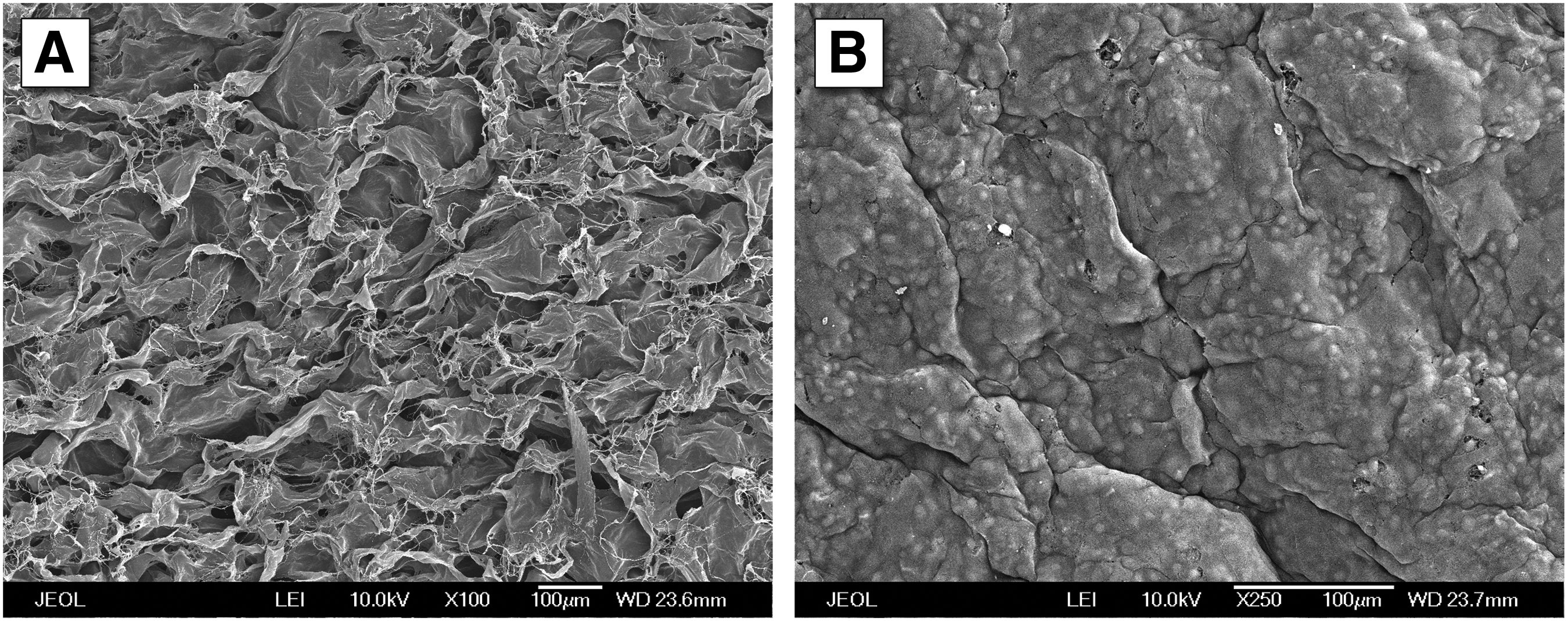
This issue can be potentially managed by seeding scaffolds with cells or loading them with growth factors. In urethral tissue engineering, cell seeding is critical for the avoidance of stricture formation when collagen scaffolds are used.38,39 When acellular tubular scaffolds were evaluated in urethral reconstruction, normal tissue regeneration was only noticed for 0.5-cm-long defects. 40 In a recent study regarding urinary diversion, Geutjes et al. reported that UC seeding may not provide any advantage to the development of urothelium. 41 On the other hand, the cytotoxicity of urine and also its negative influence in tissue regeneration has been demonstrated. 42 To protect the cells during ingrowth and tissue remodeling after in situ implantation, seeding UCs in the luminal surface might be beneficial 43 (Fig. 1). In studies where scaffolds are preimplanted into the peritoneal cavity, the toxic effect of urine is absent and further data regarding regeneration after in situ implantation would be interesting.
The construction of cell-seeded scaffolds involves cell harvesting, culture, and seeding onto the scaffold. Ideally, the scaffolds should also be tested in a bioreactor for their mechanical properties before implantation to ensure adequate mechanical strength, and thus avoid intra- and postoperative complications. With tensile and flow studies, it is possible to study the cell response to the forces that are normally applied to the ureter. This is a procedure that consumes time, work hours, and also increases the costs. Cell distribution after seeding is another issue that has to be determined before implantation. Sun et al. examined cell ingrowth in collagen and hybrid scaffolds of different collagen concentrations [ranging from 0.3% to 0.8% (w/v)]. They concluded that a hybrid scaffold prepared from 0.4% collagen strengthened with knitting achieved the best cellular distribution. 44 Autologous cells are preferred since their use ensures a minimal inflammatory response and biocompatibility. 45 To obtain these cells, in most cases, an additional surgery is necessary and this can cause additional morbidity. The technique used for seeding cells should provide equal distribution of living cells. Static, dynamic, and spinning seeding methods are most often used currently.46,47 The time needed to perform the aforementioned procedures may be a limitation in their use. Consideration should be given to determine whether the advantages of a cellular construct outweigh these disadvantages, the increased cost, and possibly decreased clinical applicability.
Growth factors can be an alternative to attract cells inside the ureteral scaffolds. They are known to have a stimulatory effect on various cellular processes, including cell influx, angiogenesis, and proliferation, thus improving the regenerative capacity of the scaffold. 48 Growth factors can be incorporated in biomaterials through a variety of methods, including entrapment within gel matrices, hydrophobic scaffolds, or microparticles, through affinity binding sites and covalent binding to matrices. 49 In ureteral tissue engineering, different growth factors are needed to stimulate different cell populations. The vascular endothelial growth factor (VEGF) and fibroblast growth factor-2 (FGF-2) are involved in angiogenesis and blood vessel maturation. 50 The epidermal growth factor (EGF) is known to play a key role in urothelial regeneration. 51 These growth factors are heparin-binding growth factors. Heparin can be incorporated in many different biomaterials to create controlled release systems. 52 In a study regarding urethral reconstruction using tubular collagen scaffolds loaded with growth factors (VEGF, FGF-2, and EGF) in a rabbit model, Nuininga et al. demonstrated narrowing of the lumen due to urothelium ingrowth. This may be prevented by loading the EGF only in the inner (luminal) side of the scaffold. 48 The insulin-like growth factor-1 (IGF-1) has been explored in an array of tissues, including skeletal and cardiac muscle, nerve, cartilage and bone. Lorentz et al. reported an engineered IGF-1 that improved SMC regeneration. 53 The main disadvantage of IGF-1 is that it does not bind to heparin. The main advantage of scaffolds loaded with growth factors is that they can be used off the shelf in urgent cases.
Stem cells (SCs) appear to be a promising area of research in urological regenerative medicine. Their use in tissue regeneration has been tested in bladder augmentation and detrusor regeneration in animal studies and in the treatment of stress urinary incontinence in clinical trials.54,55 Studies regarding ureteral regeneration by using SCs are still lacking. 56 Autologous urine-derived SCs exhibit a high expansion rate and capability of differentiating into both urothelial and SMCs. 57 Supplementation of growth factors in vivo promoted the survival of urine-derived SCs and their differentiation into muscle cells. Enhancement of nerve regeneration and native cell attraction were also noticed. 58 Issues that need further research are the control of SC proliferation rate, differentiation into the desirable line, and also their behavior in the long term. 59
Collagen currently seems to be the biomaterial of choice in the construction of small diameter tubular scaffolds. Type-I collagen is the most abundant type of collagen in organs and provides strength and structural integrity to tissues. Allogenic collagen, like bovine, exhibits excellent biocompatibility and low immunogenicity in humans.60–62 Highly purified type-I collagen is commercially available and it is technically feasible to prepare up to 10-cm-long tubular scaffolds. The major disadvantage of collagen scaffolds is their poor physical strength. 63 Chemical crosslinking can enhance their mechanical properties. 64 By experience of our institution, suturability of these scaffolds in vitro in a porcine ureteral model is satisfactory and patency is achieved without complications. Another option may be the use of high-degradable polymer-collagen scaffolds to increase the physical strength and ease of application. Further research regarding the development of small diameter hybrid scaffolds is required.
The animal model for preclinical studies is also a factor that may affect the outcome of the experiment. The natural algorithm for animal studies—which dictates the use of small animals before proceeding into large animal studies—cannot always be followed strictly as in the case of ureteral tissue engineering. Crossover knowledge from urethral tissue-engineering studies can provide data for the regeneration of scaffolds up to 2 cm long. 65 In ureteral reconstruction, a lesion of this length is clinically insignificant since it can be repaired by the available surgical techniques. In longer defects, and to mimic the clinical situation and extrapolate the preclinical data as much as possible, the animal model should have an abdominal and ureteral anatomy analogue to that of a human. This offers the advantages of testing the feasibility of such a procedure, the applicability of the scaffold, and the outcome of the regenerative process. The pig model seems to be the best alternative.66,67 Nevertheless, from the ethical perspective, it is important to perform extensive in vitro- and ex vivo-related experiments before animal testing, for example, pressure–flow experiments to characterize the mechanical properties and suturability of the construct.
The ideal scaffold should have a high regenerative capacity, ease of construction, and direct availability in urgent cases. Developing tubular scaffolds for ureteral reconstruction will be of great importance for both the patient and the surgeon. The simplified surgical technique may lead to a less invasive surgery, lower complication rates, and a reduction in health care costs. From this point of view, the evaluation of tubular collagen scaffolds loaded with growth factors seems to be promising. Finally, as in every tissue-engineering application, considerations should be made to bring these new options to applicable techniques in the clinical situation. However, the exact requirements and methods for conducting clinical trials should be defined. 68
Conclusions
Current literature regarding ureteral tissue engineering is lacking evidence as for the determination of a suitable biomaterial. Furthermore, progresses in regenerative medicine, like cell-seeded scaffolds, scaffolds loaded with growth factors, or the use of SCs have not been efficiently evaluated in ureteral reconstruction yet. Further, preclinical research is required to develop a suitable scaffold and improve the tissue-engineering applications for this domain of urological regenerative medicine.
Footnotes
Disclosure Statement
No competing financial interests exist.